Embed Size (px)
Citation preview
OpenStax-CNX module: m56830 1
Derived copy of Bis2A 16.2 Errors in
Meiosis*
Erin Easlon
Based on Bis2A 16.2 Errors in Meiosis� by
OpenStax
Mitch Singer
This work is produced by OpenStax-CNX and licensed under the
Creative Commons Attribution License 4.0�
Abstract
By the end of this section, you will be able to:
• Explain how nondisjunction leads to disorders in chromosome number• Describe how errors in chromosome structure occur through inversions and translocations
Inherited disorders can arise when chromosomes behave abnormally during meiosis. Chromosome disor-ders can be divided into two categories: abnormalities in chromosome number and chromosome structuralrearrangements. Because even small segments of chromosomes can span many genes, chromosomal disordersare characteristically dramatic and often fatal.
1 Disorders in Chromosome Number
The isolation and microscopic observation of chromosomes forms the basis of cytogenetics and is the primarymethod by which clinicians detect chromosomal abnormalities in humans. A karyotype is the number andappearance of chromosomes, including their length, banding pattern, and centromere position. To obtaina view of an individual's karyotype, cytologists photograph the chromosomes and then cut and paste eachchromosome into a chart, or karyogram (Figure 1).
*Version 1.1: Jul 17, 2015 2:24 pm -0500�http://cnx.org/content/m56091/1.2/�http://creativecommons.org/licenses/by/4.0/
http://cnx.org/content/m56830/1.1/
OpenStax-CNX module: m56830 2
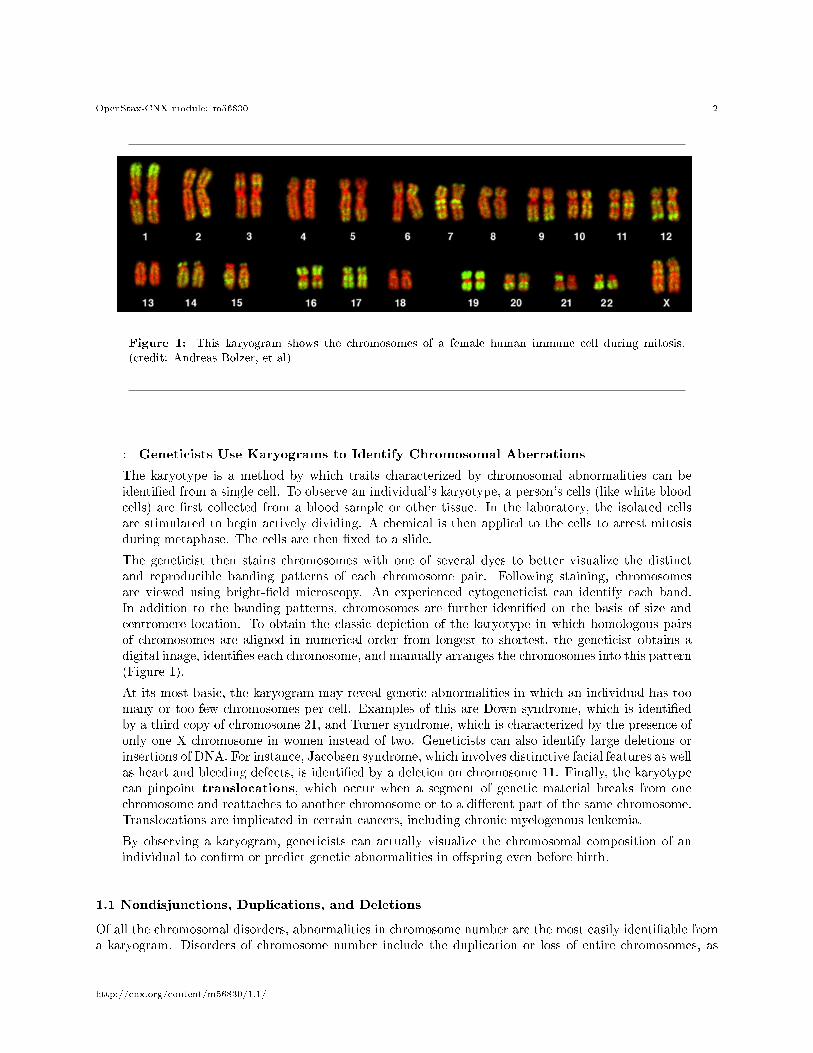
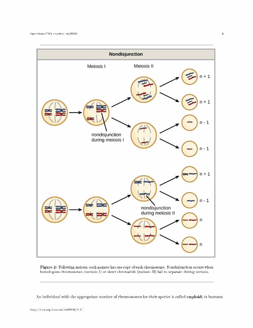
Figure 1: This karyogram shows the chromosomes of a female human immune cell during mitosis.(credit: Andreas Bolzer, et al)
: Geneticists Use Karyograms to Identify Chromosomal Aberrations
The karyotype is a method by which traits characterized by chromosomal abnormalities can beidenti�ed from a single cell. To observe an individual's karyotype, a person's cells (like white bloodcells) are �rst collected from a blood sample or other tissue. In the laboratory, the isolated cellsare stimulated to begin actively dividing. A chemical is then applied to the cells to arrest mitosisduring metaphase. The cells are then �xed to a slide.
The geneticist then stains chromosomes with one of several dyes to better visualize the distinctand reproducible banding patterns of each chromosome pair. Following staining, chromosomesare viewed using bright-�eld microscopy. An experienced cytogeneticist can identify each band.In addition to the banding patterns, chromosomes are further identi�ed on the basis of size andcentromere location. To obtain the classic depiction of the karyotype in which homologous pairsof chromosomes are aligned in numerical order from longest to shortest, the geneticist obtains adigital image, identi�es each chromosome, and manually arranges the chromosomes into this pattern(Figure 1).
At its most basic, the karyogram may reveal genetic abnormalities in which an individual has toomany or too few chromosomes per cell. Examples of this are Down syndrome, which is identi�edby a third copy of chromosome 21, and Turner syndrome, which is characterized by the presence ofonly one X chromosome in women instead of two. Geneticists can also identify large deletions orinsertions of DNA. For instance, Jacobsen syndrome, which involves distinctive facial features as wellas heart and bleeding defects, is identi�ed by a deletion on chromosome 11. Finally, the karyotypecan pinpoint translocations, which occur when a segment of genetic material breaks from onechromosome and reattaches to another chromosome or to a di�erent part of the same chromosome.Translocations are implicated in certain cancers, including chronic myelogenous leukemia.
By observing a karyogram, geneticists can actually visualize the chromosomal composition of anindividual to con�rm or predict genetic abnormalities in o�spring even before birth.
1.1 Nondisjunctions, Duplications, and Deletions
Of all the chromosomal disorders, abnormalities in chromosome number are the most easily identi�able froma karyogram. Disorders of chromosome number include the duplication or loss of entire chromosomes, as
http://cnx.org/content/m56830/1.1/
OpenStax-CNX module: m56830 3
well as changes in the number of complete sets of chromosomes. They are caused by nondisjunction, whichoccurs when pairs of homologous chromosomes or sister chromatids fail to separate during meiosis. The riskof nondisjunction increases with the age of the parents.
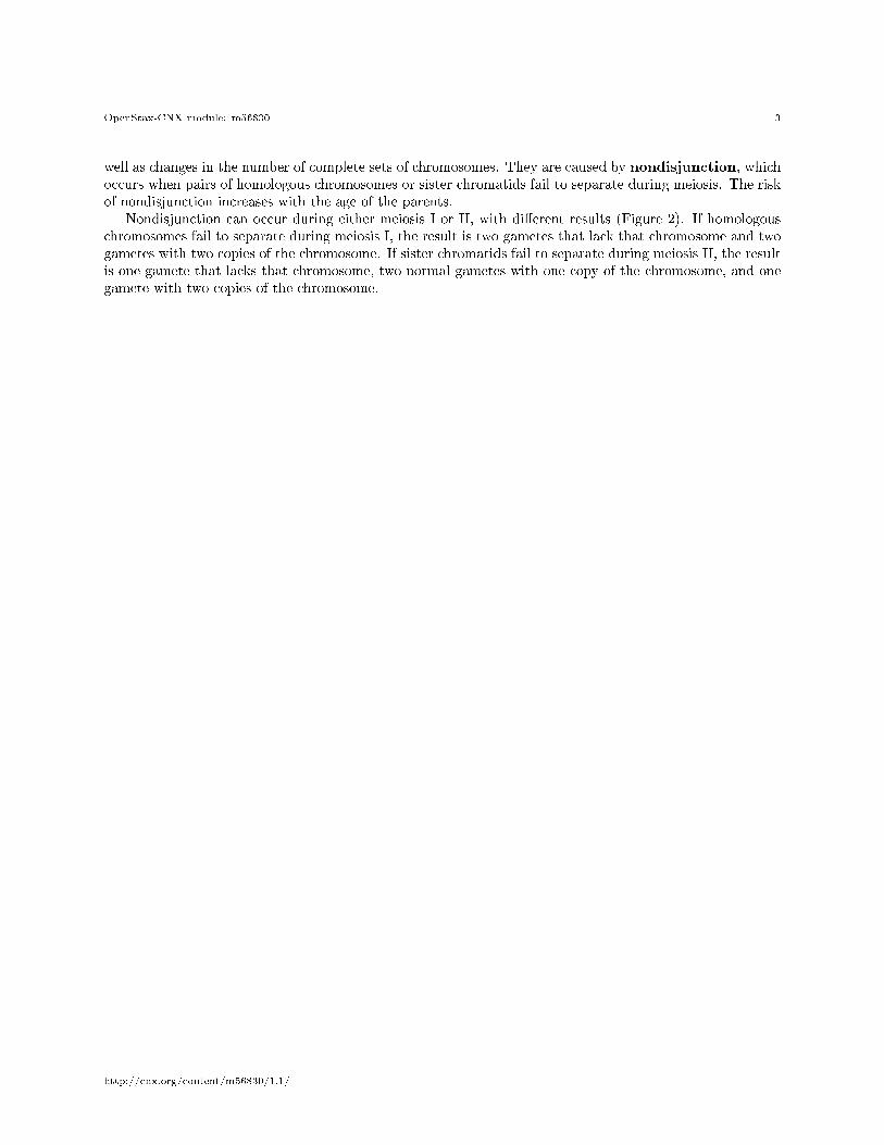
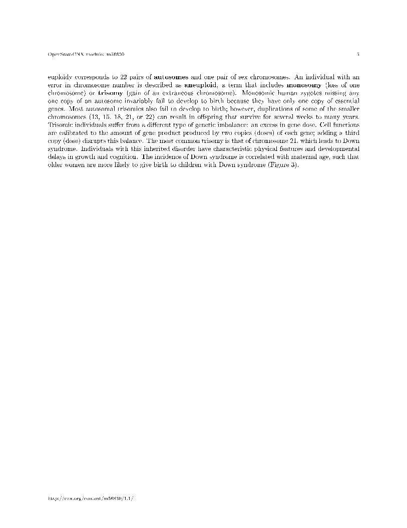
Nondisjunction can occur during either meiosis I or II, with di�erent results (Figure 2). If homologouschromosomes fail to separate during meiosis I, the result is two gametes that lack that chromosome and twogametes with two copies of the chromosome. If sister chromatids fail to separate during meiosis II, the resultis one gamete that lacks that chromosome, two normal gametes with one copy of the chromosome, and onegamete with two copies of the chromosome.
http://cnx.org/content/m56830/1.1/
OpenStax-CNX module: m56830 4
Figure 2: Following meiosis, each gamete has one copy of each chromosome. Nondisjunction occurs whenhomologous chromosomes (meiosis I) or sister chromatids (meiosis II) fail to separate during meiosis.
An individual with the appropriate number of chromosomes for their species is called euploid; in humans,
http://cnx.org/content/m56830/1.1/
OpenStax-CNX module: m56830 5
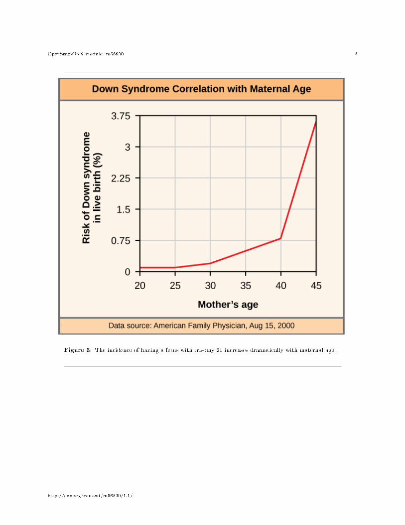
euploidy corresponds to 22 pairs of autosomes and one pair of sex chromosomes. An individual with anerror in chromosome number is described as aneuploid, a term that includes monosomy (loss of onechromosome) or trisomy (gain of an extraneous chromosome). Monosomic human zygotes missing anyone copy of an autosome invariably fail to develop to birth because they have only one copy of essentialgenes. Most autosomal trisomies also fail to develop to birth; however, duplications of some of the smallerchromosomes (13, 15, 18, 21, or 22) can result in o�spring that survive for several weeks to many years.Trisomic individuals su�er from a di�erent type of genetic imbalance: an excess in gene dose. Cell functionsare calibrated to the amount of gene product produced by two copies (doses) of each gene; adding a thirdcopy (dose) disrupts this balance. The most common trisomy is that of chromosome 21, which leads to Downsyndrome. Individuals with this inherited disorder have characteristic physical features and developmentaldelays in growth and cognition. The incidence of Down syndrome is correlated with maternal age, such thatolder women are more likely to give birth to children with Down syndrome (Figure 3).
http://cnx.org/content/m56830/1.1/
OpenStax-CNX module: m56830 6
Figure 3: The incidence of having a fetus with trisomy 21 increases dramatically with maternal age.
http://cnx.org/content/m56830/1.1/
OpenStax-CNX module: m56830 7
:
Visualize the addition of a chromosome that leads to Down syndrome in this video simulation1 .
Humans display dramatic deleterious e�ects with autosomal trisomies and monosomies. Therefore, it mayseem counterintuitive that human females and males can function normally, despite carrying di�erent num-bers of the X chromosome. In part, this occurs because of a process called X inactivation. Early indevelopment, when female mammalian embryos consist of just a few thousand cells, one X chromosome ineach cell inactivates by condensing into a structure called a Barr body. The genes on the inactive X chromo-some are not expressed. The particular X chromosome (maternally or paternally derived) that is inactivatedin each cell is random, but once the inactivation occurs, all cells descended from that cell will have the sameinactive X chromosome. By this process, females compensate for their double genetic dose of X chromosome.
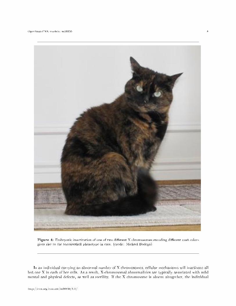
In so-called �tortoiseshell� cats, X inactivation is observed as coat-color variegation (Figure 4). Femalesheterozygous for an X-linked coat color gene will express one of two di�erent coat colors over di�erent regionsof their body, corresponding to whichever X chromosome is inactivated in the embryonic cell progenitor ofthat region. When you see a tortoiseshell cat, you will know that it has to be a female.
1http://openstaxcollege.org/l/down_syndrome2
http://cnx.org/content/m56830/1.1/
OpenStax-CNX module: m56830 8
Figure 4: Embryonic inactivation of one of two di�erent X chromosomes encoding di�erent coat colorsgives rise to the tortoiseshell phenotype in cats. (credit: Michael Bodega)
In an individual carrying an abnormal number of X chromosomes, cellular mechanisms will inactivate allbut one X in each of her cells. As a result, X-chromosomal abnormalities are typically associated with mildmental and physical defects, as well as sterility. If the X chromosome is absent altogether, the individual
http://cnx.org/content/m56830/1.1/
OpenStax-CNX module: m56830 9
will not develop.Several errors in sex chromosome number have been characterized. Individuals with three X chromosomes,
called triplo-X, appear female but express developmental delays and reduced fertility. The XXY chromosomecomplement, corresponding to one type of Klinefelter syndrome, corresponds to male individuals with smalltestes, enlarged breasts, and reduced body hair. The extra X chromosome undergoes inactivation to com-pensate for the excess genetic dosage. Turner syndrome, characterized as an X0 chromosome complement(i.e., only a single sex chromosome), corresponds to a female individual with short stature, webbed skin inthe neck region, hearing and cardiac impairments, and sterility.
An individual with more than the correct number of chromosome sets (two for diploid species) is calledpolyploid. For instance, fertilization of an abnormal diploid egg with a normal haploid sperm would yielda triploid zygote. Polyploid animals are extremely rare, with only a few examples among the �atworms,crustaceans, amphibians, �sh, and lizards. Triploid animals are sterile because meiosis cannot proceednormally with an odd number of chromosome sets. In contrast, polyploidy is very common in the plantkingdom, and polyploid plants tend to be larger and more robust than euploids of their species.
2 Chromosome Structural Rearrangements
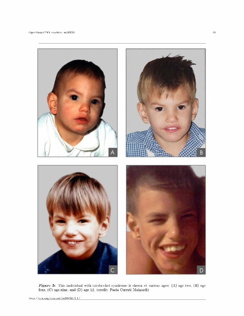
Cytologists have characterized numerous structural rearrangements in chromosomes, including partial du-plications, deletions, inversions, and translocations. Duplications and deletions often produce o�spring thatsurvive but exhibit physical and mental abnormalities. Cri-du-chat (from the French for �cry of the cat�)is a syndrome associated with nervous system abnormalities and identi�able physical features that resultsfrom a deletion of most of the small arm of chromosome 5 (Figure 5). Infants with this genotype emit acharacteristic high-pitched cry upon which the disorder's name is based.
http://cnx.org/content/m56830/1.1/
OpenStax-CNX module: m56830 10
Figure 5: This individual with cri-du-chat syndrome is shown at various ages: (A) age two, (B) agefour, (C) age nine, and (D) age 12. (credit: Paola Cerruti Mainardi)
http://cnx.org/content/m56830/1.1/
OpenStax-CNX module: m56830 11
Chromosome inversions and translocations can be identi�ed by observing cells during meiosis becausehomologous chromosomes with a rearrangement in one of the pair must contort to maintain appropriate genealignment and pair e�ectively during prophase I.
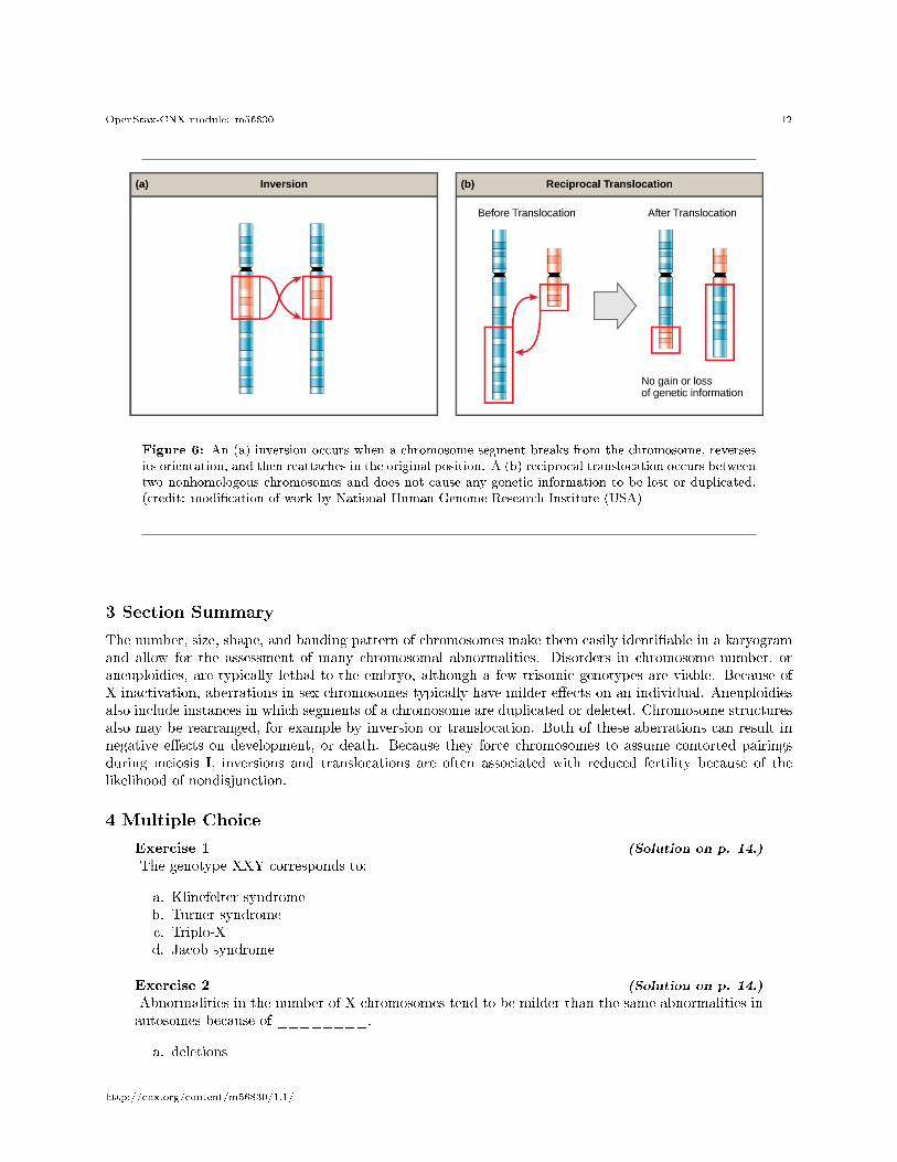
A chromosome inversion is the detachment, 180 ◦ rotation, and reinsertion of part of a chromosome(Figure 6). Unless they disrupt a gene sequence, inversions only change the orientation of genes and arelikely to have more mild e�ects than aneuploid errors.
: The Chromosome 18 Inversion
Not all structural rearrangements of chromosomes produce nonviable, impaired, or infertile indi-viduals. In rare instances, such a change can result in the evolution of a new species. In fact, aninversion in chromosome 18 appears to have contributed to the evolution of humans. This inversionis not present in our closest genetic relatives, the chimpanzees.
The chromosome 18 inversion is believed to have occurred in early humans following their divergencefrom a common ancestor with chimpanzees approximately �ve million years ago. Researchers havesuggested that a long stretch of DNA was duplicated on chromosome 18 of an ancestor to humans,but that during the duplication it was inverted (inserted into the chromosome in reverse orientation.
A comparison of human and chimpanzee genes in the region of this inversion indicates that twogenes�ROCK1 and USP14�are farther apart on human chromosome 18 than they are on thecorresponding chimpanzee chromosome. This suggests that one of the inversion breakpoints oc-curred between these two genes. Interestingly, humans and chimpanzees express USP14 at distinctlevels in speci�c cell types, including cortical cells and �broblasts. Perhaps the chromosome 18inversion in an ancestral human repositioned speci�c genes and reset their expression levels in auseful way. Because both ROCK1 and USP14 code for enzymes, a change in their expression couldalter cellular function. It is not known how this inversion contributed to hominid evolution, but itappears to be a signi�cant factor in the divergence of humans from other primates.
2
A translocation occurs when a segment of a chromosome dissociates and reattaches to a di�erent, nonho-mologous chromosome. Translocations can be benign or have devastating e�ects, depending on how thepositions of genes are altered with respect to regulatory sequences. Notably, speci�c translocations havebeen associated with several cancers and with schizophrenia. Reciprocal translocations result from the ex-change of chromosome segments between two nonhomologous chromosomes such that there is no gain or lossof genetic information (Figure 6).
http://cnx.org/content/m56830/1.1/
OpenStax-CNX module: m56830 12
Figure 6: An (a) inversion occurs when a chromosome segment breaks from the chromosome, reversesits orientation, and then reattaches in the original position. A (b) reciprocal translocation occurs betweentwo nonhomologous chromosomes and does not cause any genetic information to be lost or duplicated.(credit: modi�cation of work by National Human Genome Research Institute (USA)
3 Section Summary
The number, size, shape, and banding pattern of chromosomes make them easily identi�able in a karyogramand allow for the assessment of many chromosomal abnormalities. Disorders in chromosome number, oraneuploidies, are typically lethal to the embryo, although a few trisomic genotypes are viable. Because ofX inactivation, aberrations in sex chromosomes typically have milder e�ects on an individual. Aneuploidiesalso include instances in which segments of a chromosome are duplicated or deleted. Chromosome structuresalso may be rearranged, for example by inversion or translocation. Both of these aberrations can result innegative e�ects on development, or death. Because they force chromosomes to assume contorted pairingsduring meiosis I, inversions and translocations are often associated with reduced fertility because of thelikelihood of nondisjunction.
4 Multiple Choice
Exercise 1 (Solution on p. 14.)
The genotype XXY corresponds to:
a. Klinefelter syndromeb. Turner syndromec. Triplo-Xd. Jacob syndrome
Exercise 2 (Solution on p. 14.)
Abnormalities in the number of X chromosomes tend to be milder than the same abnormalities inautosomes because of ________.
a. deletions
http://cnx.org/content/m56830/1.1/
OpenStax-CNX module: m56830 13
b. nonhomologous recombinationc. synapsisd. X inactivation
Exercise 3 (Solution on p. 14.)
Aneuploidies are deleterious for the individual because of what phenomenon?
a. nondisjunctionb. gene dosagec. meiotic errorsd. X inactivation
5 Free Response
Exercise 4 (Solution on p. 14.)
Individuals with trisomy 21 are more likely to survive to adulthood than individuals with trisomy18. Based on what you know about aneuploidies from this module, what can you hypothesize aboutchromosomes 21 and 18?
http://cnx.org/content/m56830/1.1/
OpenStax-CNX module: m56830 14
Solutions to Exercises in this Module
to Exercise (p. 12)Ato Exercise (p. 12)Dto Exercise (p. 13)Bto Exercise (p. 13)The problems caused by trisomies arise because the genes on the chromosome that is present in three copiesproduce more product than genes on chromosomes with only two copies. The cell does not have a way toadjust the amount of product, and the lack of balance causes problems in development and the maintenanceof the individual. Each chromosome is di�erent, and the di�erences in survivability could have to do withthe numbers of genes on the two chromosomes. Chromosome 21 may be a smaller chromosome, so thereare fewer unbalanced gene products. It is also possible that chromosome 21 carries genes whose productsare less sensitive to di�erences in dosage than chromosome 18. The genes may be less involved in criticalpathways, or the di�erences in dosage may make less of a di�erence to those pathways.
Glossary
De�nition 6: aneuploidan individual with an error in chromosome number; includes deletions and duplications of chromo-some segments
De�nition 6: autosomeany of the non-sex chromosomes
De�nition 6: chromosome inversionthe detachment, 180 ◦ rotation, and reinsertion of a chromosome arm
De�nition 6: euploidan individual with the appropriate number of chromosomes for their species
De�nition 6: karyogramthe photographic image of a karyotype
De�nition 6: karyotypethe number and appearance of an individuals chromosomes, including the size, banding patterns,and centromere position
De�nition 6: monosomyan otherwise diploid genotype in which one chromosome is missing
De�nition 6: nondisjunctionthe failure of synapsed homologs to completely separate and migrate to separate poles during the�rst cell division of meiosis
De�nition 6: polyploidan individual with an incorrect number of chromosome sets
De�nition 6: translocationthe process by which one segment of a chromosome dissociates and reattaches to a di�erent, non-homologous chromosome
De�nition 6: trisomyan otherwise diploid genotype in which one entire chromosome is duplicated
http://cnx.org/content/m56830/1.1/
OpenStax-CNX module: m56830 15
De�nition 6: X inactivationthe condensation of X chromosomes into Barr bodies during embryonic development in females tocompensate for the double genetic dose
http://cnx.org/content/m56830/1.1/