Embed Size (px)
Citation preview

Title
DEVELOPMENT OF A PHYSIOLOGICALLY BASEDPHARMACOKINETIC (PBPK) MODEL FORENDOSULFAN AND ITS APPLICATION IN HEALTHRISK ASSESSMENTS( Dissertation_全文 )
Author(s) Melissa Chan Pui Ling
Citation Kyoto University (京都大学)
Issue Date 2005-09-26
URL https://doi.org/10.14989/doctor.k11874
Right
Type Thesis or Dissertation
Textversion author
Kyoto University
DEVELOPMENT OF A PHYSIOLOGICALLY BASEDPHARMACOKINETIC(PBPK)MOD肌FOR ENDOSULFANAND rTS APPLICATION lrg HEALTH RISK ASSESSMENTS
エンドサルファン生理学的薬動力学モデルの開発 と健康リスク評価への適用
!・i
r
Melissa Chan Pui Ling
Depa血nent of Globa1 EnVironment Enginee血1g
Kyoto University
June 2005

1)EV肌OPMENT OF A P正lYSIOLOGICA肌Y BASEI)
PHARMACOKINETIC(PBPK)MOI)EL FOR ENI)OSULFANANI)ITS APPLICATION IN HEALTH RISK ASSESSMENTS
エンドサルファン生理学的薬動力学モデルの開発 と健康リスク評価への適用
Melissa Chan Pui Ling
De麺ment of Global Environment Enginee亘ng
Kyoto University
Dissertation Submitted for the Degree of
Doctor of Engineering
Kyoto University
June 2005
、
ACKNOWLEI)GEMENTS
This study was financially supported by the Grant-in 一 Aid fbr Scientific Research
(B),No.13555150. I would like to express my deepest gratitude to the fbllowing
people for al1 their unfailing guidance, support, patience and constant encouragement
throughout my study.
1
2
3
4.
510丙-
8
9
10.
11.
Professor Shinsuke Morisawa, Department of Urban and Environmental
Engineering(Supervisor and Chief examiner)
Professor Iwao Uchiyama, Department of Environmental Engineering
(Examiner)
Professor Sadahiko Itoh, Department of Environmental Engineering
(Examiner)
Associate Professor Minoru Yoneda, Department of Urban and Environmental
Engineering
Dr. Aki Nakayama, Department of Urban and Environmental Engineering
Dr. Yuko Kawamoto, Department of Urban and Environmental Engineering
Dr. Miki Sugimoto, Department of Animal Science, Graduate School of
Agriculture
Satoshi㎞anishi and Hanako Nishizawa, Department of Animal Science,
Graduate School of Agriculture
Yoko Kitanaka, Ken Yasukouchi, Tadanao Miura, and Masahiro Tozaki,
Department of Urban and Environmental Engineering
Yoshito Matsui, Department of Environmental Engineering
Friends of the Chair of the Environmental Risk Analysis Laboratory

CONTENTS
AcknowledgementsList of figures
List of tables
i
viii
xii
Abstract 1
CHAPTER 1
1.0
1.1
Research background
Obj ectives of the research
10
12
1.2 Pesticides:An overview 13
1.3
1.4
1.5
Pesticides in Malaysia
1.3.1 Pesticide usage
1.3.2 Endosulfan in the Malaysian environment
Endosulfan in Japan
1・4・1 Sales of endosulfan in Japan
1.4.2Endosulfan in the Japanese environment and population
Endosulfa11-Ashort review1.5.1 1n廿oduction
1.5.2Summary o拙e characteristics of endosulfan
1.5.2.1 Chemical identity
1・5・2・2 Physical and chemical properties
1.5.2.3 Exposure guidelines
1・5・3 Possible routes of exposure
1.5.4 Exposure of endosulfan to humans
1.5.5E甑ts on the environment
1.5.6 Environmental fate of endosulfan
fJ7111
00(∠(∠
21
REFERENCES 29
CHAPTER 2 TOXICITY TO MAMMALS
2.0 Introduction 35
2.1 Toxicity to mammals(rats)
2.1.1 General toxic effects of endosulfan
2.1.2 Neurological effects
2.1.3 Reproductive effects
2.1.4 Endocrine effects
5只∨(U1334.4.
REFERENCES 43
●-

CHAPTER 3 TOXICOKINETICS OF 14C-ENI)OSULFAN FOLLOWING SINGLE ANI)REPEATE1) ORAL ADMINISTRATION IN THE MALE SPRAGUE-DAWLEY RATS
3.O Pharmacokinetics:An overview
3.1 Background introduction
23「」角」
Objective of the study
Materials and methods
3.3.1
3.3.2
3.3.3
3.3.4
3.3.5
3.3.6
3.3.7
3.3.8
3.3.9
Chemicals
Animals and genera 1 conditions
Preparation of endosulfan dosage
Rationale fbr dose selection
Experimental design and administration of i 4C-Endosulfan
N㏄ropsyMeasurement of radioactivity
Toxicokinetic parameters
Calculation of the Laplace Transfbmls
3.4 Results
3.4.1 General condition of animals
3.4.2 Disposition of i4C-Endosulfan(5 mg/kg):Excretion routes
3.4.3 14C-Endosulfan residues in tissues and contents of the GI tract
3.4.4 i4C ._ Endosulfan toxicokinetics in blood
3.5 Discussion
3.6 Conclusions
REFERENCES
0ノー惚」45く∨
53T4T4T4T5T6T7T7
ヨ
CHAPTER 4 1)EV肌OPMENT OF A PHYSIOLOGICA肌Y BASED PHARMACOKlNETIC MOD肌FOR ENDOSULFAN IN THE MALE SPRAGUE- DAWLEY RATS
4.O Physiologically based phamlacoldnetic(PBPK)model:An overview
4.1 Background introduction
4.2 0bj ective of the study
4.3 Materials and methods
4.3.1 PBPK model development
4.3.2 Parameterization
4.3.2.1 Physiological parameters
580880ノ
17’0/0ノ
iii
4.3.3
4.3.4
4.3.2.2
4.3.2.3
4.3.2.4
4.3.2.5
Physicochemical parameters
Biochemical parameters
Model calibration
Model verification and model simulations ofendosulfan disposition in other studies
Sensitivity analyses
Extrapolation from rat model to human model
4.3.4.1
4.3.4.2
4.3.4.3
4.3.4.4
4.3.4.5
Physiological parameters
Physicochemical parameters
Biochemical parameters
Model simulation
Model verification and model simulations ofendosulfan disposition in other studies
97101
101
101
102
102
103
105
105
107
4.4 Results
4.4.1
4.4.2
4.4.3
4.4.4
Parameterization and calibration
Model verification and model simulations of endosulfandisposition in other studies
Sensitivity analyses
4.4.3.1 Endosulfan
4.4.3.2 Endosulfan metabolites
Extrapolation from rat model to human model
4.4.4.1 Model verification and model simulations of endosulfan disposition in other studies
80ノ(UO11
0(U内∠〔∠11一-
123
4.5 Discussion 127
4.6 Conclusions 133
REFERENCES 134
CHAPTER 5 DEVELOPMENT OF AN I V VITRO BLOOI)-
BRAIN…MODEL TO STUDY THEPERMEABILITY EFFECTS OFENDOSULFAN ON THE TIGHT JUNCTIONS
5.0 Introduction
5.0.1 Blood-brain barrier(BBB)
5.0.2 Endpoints for acute toxic effects
5.0.3 Transendothelial electrical resistance(TEER)
145
146
146
5.1 Obj ective of the study 148
5.2 Materials and methods
5.2.1 Chemicals
5.2.2 Materials
5.2.3 Preparation of chemicals
5.2.4 Rationale fbr dose selection
444411111
iv
5.3
5.2.5
5.2.6
5.2.7
5.2.8
5.2.9
5.2.10
Results
5.3.1
5.3.2
5.3.3
5.3.4
Cell culture fbr transendothelial electrical resistance(TEER)
study
Measurements of TEER5.2.6.1 Calculation of the resistance of the cell monolayer
Cytotoxicity study of PBMECs after treatment withα一
endosulfan,β一endosulfan and endosulfan sulfate
Transendothelial transport(imer-to-outer)study with 14C-
Endosulfan
5.2.8.1 Data analysis
PBMECs reversible transport(outer-to-imer)study with14 C-EndosulfanStatistical analysis
Measurements of transendothelial electrical resistance(TEER)
Cytotoxicity analysis of endosulfan-treated PBMECs
Transendothelial transport(inner-to-outer)study with 14C-
Endosulfan
毘懸蒜蓋ble仕ansp°「t(°ute「-t°-inne「)study w’th
150
152
153
153
154
510く∨fJ11.且
156
156
160161
164
5.4 Discussion 164
5.5 Conclusions 169
REFERENCES 170
CHAPTER 6 A COMPARATIVE STUDY ON THENEUROTOXIC EFFECTS OF ENI)OSULFANON GLIAL AND NEURONAL CELL
C肌㎜smOM RAT蜘H皿州
6.0 lntroduction
6.0.1 Principles of the nervous system-Potential sites of neurotoxic
attack
6.0.2 Mode of action of endosulfan on the CNS
176
178
6.1 Objective of the study 179
6.2 Materials and methods
6.2.1
6.2.2
6.2.3
6.2.4
6.2.5
6.2.6
ChemicalsMaterials
Preparation of chemicals
Rationale fbr dose selection
Cell cultures
Cytotoxicity studies of PC12, C6, NT2 and CCF-STTG l cells
after treatment withα一endosulfan,β一endosulfan andendosulfan sulfate
179
180
180
180181
181
V

6.2.7 Statistical analysis 182
6.3 Results
6.3.1
6.3.2
6.3.3
6.3.4
6.3.5
Cytotoxicity study of rat C691ial cells
Cytotoxicity study of rat PC 12 neuronal cells
Cytotoxicity study of human CCF-STTG I glial cells
Cytotoxicity study of human NT2 neuronal cells
Lethal concentration to cause 50%cell death(LC50)values fbr
glial and neuronal cell cultures from rat and human
182
183
183
184
188
6.4 Discussion 190
6.5 Conclusions 191
REFERENCES 192
CHAPTER 7 ASSESSMENT OF THE HEALTH RISKSFO肌OWING EXPOUSRE TO ENDOSULFAN
7.0 An overview 197
7.1 Neurotoxicity 200
7.2 Reproductive toxicity 200
7.3 Objective of the study 201
7.4
7.5
Bioassay data and PBPK model simulations
●
Results and discussion
7.5.1 PBPK model simulations
7.5.2 Neurotoxic risk
7.5.2.1 PBPK model predictions
7.5.3 Reproductive risk
7.5.3.l PBPK model predictiolls
201
202202207209210
7.6 Conclusions 210
REFERENCES 212
CHAPTER 8
8.0 Conclusions 216
8.1 Recommendations for fUture study 221
vi

LIST OF FIGURES
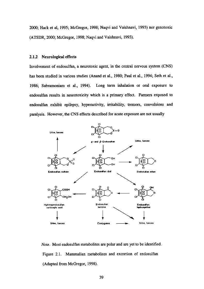
Figure 1.1 Mammalian metabolism and excretion of endosulfan 39
Figure 3.1
Figure 3.2
]Figure 3.3
Data 一一 based pharmacokinetic modeling
Two-compartment model system
Cum. ulative percentage of urinary and fecal elimination
of 14C_Endosulfan
50
59
67
Figure 3.4 Plot of the log concentration-time course of 14C-
Endosulfan in blood of male rats following single oral
administration
71
Figure 4.1
figure 4.2
Idealized approach of PBPK modeling
The schematic diagram of the PBPK model for
endosulfan in male rats
85
92
Figure 4.3 Comparison of PBPK model prediction and
experimental cumulative percentage of urinary and
fecal elimination of total endosulfan after single oral
administration
111
Figure 4.4 Comparison of PBPK model prediction and
experimental concentrations of tota1 endosulfan in
liver,1ddneys, brain, testes and blood fbllowing single
oral administration
112
Figure 4.5
Figure 4.6
Figure 4.7
Comparison of PBPK model prediction and
experimental concentrations of total endosulfan in
liver, kidneys, brain, testes and blood following three-
time repeated oral administration
Observed and simulated concentrations of endosulfan
metabolites in liver and endosulfan in kddneys after
multiple oral administration of 5 and 10 mg/kg body
weight per day of technical grade endosulfan in male
Sprague-Dawley rats
Observed and simulated concentrations of endosulfan
in liver,】Cidneys and brain after multiple oral
administration of 2.5 mg/kg body weight per day of
endosulfan in male rats for 60 days
113
114
115
viii

Figure 4.8
Figure 4.9
Figure 4.10
Observed and simulated concentrations of endosulfan
in liver, kidneys and brain after multiple oral
administration of 7.5 mg/kg body weight per day of
endosulfan in male rats for 60 days
Observed and simulated concentrations of endosulfan
in liver,1ddneys, brain and testes after multiple oral
administration of 1 1 mg/kg body weight per day of
endosulfan in male rats for 60 days
Observed and simulated concentrations of endosulfan
in liver, kidneys, and blood serum after multiple oral
administration of 5 mg/kg body weight per day of
endosulfan in male Wistar rats for 60 days
116
117
118
Figure 4.11
Figure 4.12
Observed and simulated concentrations of endosulfan
in liver, kidneys, and blood serum after multiple oral
administration of l.5 mg/kg body weight per day of
endosulfan in female Wistar rats for 60 days
Observed and simulated concentrations of endosulfan
in maternal serum of pregnant women from Chiba,
Japan fbllowing three-time ingested dose of
endosulfan
119
124
Figure 4.13 Observed and simulated concentrations of endosulfan
in maternal serum of pregnant women from Kyushu,
Japan following three-time ingested dose of
endosulfan
125
Figure 4.14
]Figure 5.1
Figure 5.2
Observed and simulated concentrations of endosulfan
in blood of Malaysian school children fbllowing three
- time ingested dose of endosulfan
Essential features of the blood-brain barrier(BBB)
Schematic representation of the in vitro mode1 of the
blood-brain barrier
126
147
151
Figure 5.3
]Figure 5.4
Measurement is taken by using the Millipore Millicell⑧
一ERSTime-dependent transendothelial electrical resistance
(TEER)values of PBMECs fbrα一endosulfan
152
157
]Figure 5.5 Time-dependent transendothelial electrical resistance
(TEER)values of PBMECs for fi 一 endosulfan
158
Figure 5.6 Time-dependent transendothelial electrical resistance
(TEER)values of PBMECs for endosulfan sulfate
159
ix
Figure 5.7 Concentration-response curves of cytotoxicity
induced by exposure of PBMECs to a-endosulfan,β
一endosulfan and endosulfan sulfate
161
]Figure 5.8
Figure 5.9
Figure 5.10
Figure 5.11
Figure 6.1
Transport profiles of】4C-Endosulfan for O.Ol pM,0.1
μM,1 pM and 10 pM from inner-outer compartment
Average permeability of the filter grown endothelial
cell monolayer to i4C-Endosulfan(Pe)(0.01μM,0.1
μM,1μM and 10 pM)across PBMEC monolayers
from inner 一 outer compartment
Transport profiles of i 4c-Endosulfan for O.Ol pM,0.1
μM,1 pM and 10 pM from outer-inner compartment
Average pemeability of the mter grown endo血elial
cell monolayer to i4C 一 Endosulfan(Pe)(0.Ol pM,0.1
μM,1μM and 10 pM)across PBMEC monolayers
from outer-inner compartment
Concentration-response curves of cytotoxicity
induced by exposure of C6 cells toα一 endosulfan,β一
endosulfan and endosulfan sulfate
162
163
165
166
185
Figure 6◆2 Concentration-response curves of cytotoxicity
induced by exposure of PC12 cells toα一endosulfan,β
一endosulfan and endosulfan sulfate
186
Figure 6.3
Figure 6.4
Concentration-response curves of cytotoxicity
induced by exposure of CCF-STTG l cells toα一
endosulfan,β一endosulfan and endosulfan sulfate
Concentration-response curves of cytotoxicity
illduced by exposure of NT2 cells toα一endosulfan,β
一endosulfan and endosulfan sulfate
187
188
Figure 7.1
Figure 7.2
Figure 7.3
Elements of risk assessment and risk management
Predicted concentrations of endosulfan in rat brain
during and post-exposure to 12.5 mg/kg and testes
during and post 一 exposure to 6 mg!kg for 5 days,
estimated by the rat PBPK model
Concentration-response curves of cytotoxicity
induced by exposure of rat C6 glial cells toα一
endosulfan,β一endosulfan and endosulfan sulfate
199
202
203
X
Figure 7.4
Figure 7.5
Figure 7.6
Figure 7.7
Figure 8.1
Concentration-response curves of cytotoxicity
induced by exposure of rat PC 12 glial cells toα一
endosulfan,β一endosulfan and endosulfan sulfate
Concentration-response curves of cytotoxicity
induced by exposure of rat CCF-STTG l glial cells to
α一endosulfan,β一endosulfan and endosulfan sulfate
Concentration-response curves of cytotoxicity
induced by exposure of rat C6 glial cells toα一
endosulfan,β一endosulfan and endosulfan sulfate
Percenlage of viability of detached cells versus log
concentrations of endosulfan a丘er 24 h and 48 h
exposure periods
Proposed risk assessnient paradigm for fUture study
204
205
206
209
222
xi
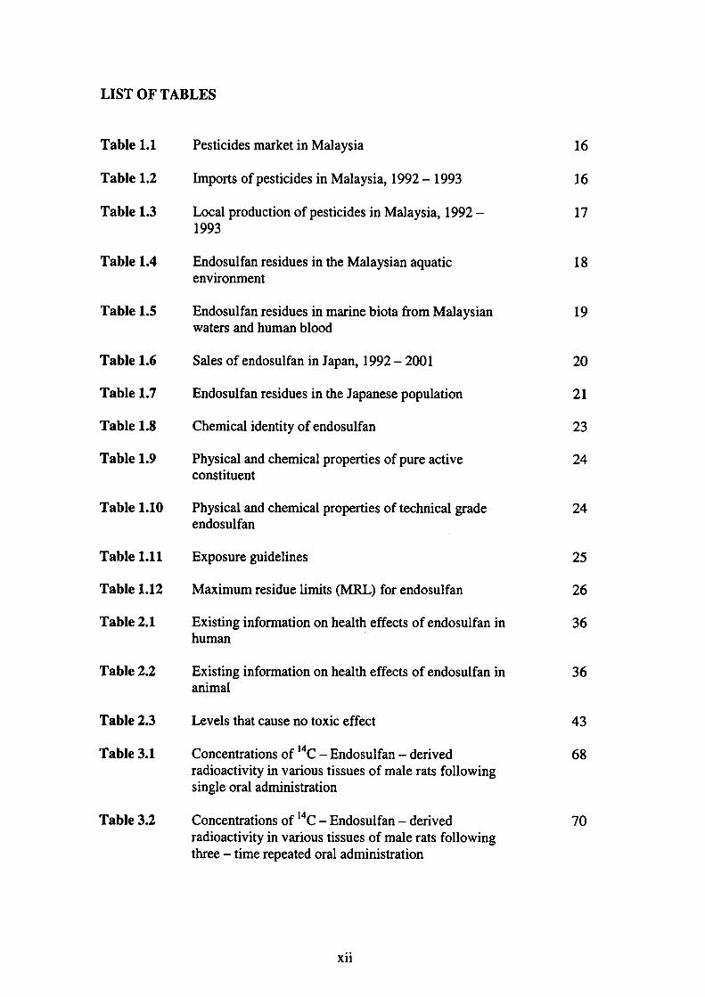
LIST OF TABLES
Table 1.1
Table 1.2
Table 1.3
Table 1.4
Table l.5
Table 1.6
Table 1.7
Table 1.8
Table 1.9
Table 1.10
Table 1.11
Table 1.12
Table 2.1
Table 2.2
Table 2.3
Table 3.1
Table 3.2
Pesticides market in Malaysia
㎞po丘s of pesticides in Malaysia,1992-1993
Local production of pesticides in Malaysia,1992-
1993
Endosulfan residues in the Malaysian aquatic
envrronment
Endosulfan residues in marine biota ffom Malaysian
waters and human blood
Sales of endosulfan in Japan,1992-2001
Endosulfan residues in the Japanese population
Chemical identity of endosulfan
Physical and chemical properdes of pure active
constltuent
Physical and chemical properties of technical grade
endosulfan
Exposure guidelines
Maximum residue limits(MRL)fbr endosulfan
Existing infbmlation on hea1中effects of endosulfan in
human
Exisdng infbmation on heal血e脆cts of endosulfan in
animal
Levels that cause no toxic effect
Concen仕ations of 14C-Endosulfan-derived
radioactivity in various tissues of male rats fbllowing
single oral ad】tninistration
Collcentrations of 14C-Endosulfan-derived
radioactivity in various tissues of male rats fbllowing
three-time repeated oral administration
xii
1010『11
1
1
18
19
01342222
24
56∠U
36
つ」846
70

Table 3.3
Table 4.1
Table 4.2
Table 4.3
Table 4.4
Table 4.5
Table 4.6
Table 4.7
Table 4.8
Table 4.9
Table 4.10
Table 5.1
Table 5.2
Table 5.3
Table 5.4
Table 5.5
Toxicokinetic parameters of i4C-Endosulfan -
derived radioactivity in blood of male rats following
single oral administration
Physiologic parameters fbr the male rats
The tissue:blood partition coefficient values fbr
endosulfan in male rats
The tissue:blood partition coefficient values fbr
endosulfan metabolites in male rats
Biochemical parameters of the PBPK model for
endosulfan and endosulfan metabolites in male rats
Experimental data reported on endosulfan disposition
in rats
Physiologic parameters for humans
Biochemical parameters of the PBPK model for
endosulfan and endosulfan metabolites in humans
Sensitivity analyses fbr the concentration of endosulfan
Sensitivity analyses for the concentration of endosulfan
metabolites
Concentrations of endosulfan(α一andβ一endosulfan)
and predicted range of concentrations in biological
materials伽m human
Time-dependent transendothelial electrical resistance
(TEER)values of PBMECs for a 一 endosulfan
Time-dependent transendothelial electrical resistance
(TEER)values of PBMECs for fi 一・ endosulfan ・
Time-dependent transendothelial electrical resistance
(TEER)values of PBMECs for endosulfan sulfate
Percentage of viability of PBMECs after ct-
endosulfan,13-endosulfan and endosulfan sulfate
treatments
Effect of inner concentrations of 14C-Endosulfan on
the average permeability(Pe)ofthe filter grown
endothelial cell monolayer
xiii
72
94
95
95
96
104
106
107
121
122
127
157
158
159
160
163

Table 5.6 Effect of outer concentrations of i4C 一 Endosulfan on
the average permeability(Pe)of the filter grown
endothelial cell monolayer
166
Table 6.1 Percentage of viability of C6 cells after exposure toα一
endosulfan,β一endosulfan and endosulfan sulfate
184
Table 6.2 Concentration 一 response curves of cytotoxicity
induced by exposure of PC 12 cells toα一endosulfan,β
一endosulfan and endosulfan sulfate
185
Table 6.3 Concentration-response curves of cytotoxicity
induced by exposure of CCF 一 ST”rG l cells toα一
endosulfan,β一endosulfan and endosulfan sulfate
186
Table 6.4 Concentration 一 response curves of cytotoxicity
induced by exposure of NT2 cells toα 一 endosulfan,β
一endosulfan and endosulfan sulfate
187
Table 6.5
Table 7.1
LC50 for the cytotoxic effects ofα一endosulfan,β一
endosulfan and endosulfan sulfate in rat glial and
neuronal, human glial and neuronal cell lines
Percentage of cell viability estimated from the
concentration-response curves of cytotoxicity after
exposure of endosulfan to glial and neuronal cell
cultures伽m rat and human
189
208
Table 7.2 Effect of endosulfan on the viability of detached cells
in rat Sertoli-gem cell cocultures
209
xiv
ABSTRACT
Generally, all humans are in contact from conception to death, illtentionally or
unintentionally, with a multitude of chemicals which are both natural and man-
made, and are present in the general env丘onment, the home, the workplace, in
ambient a口, fbod and drinking water. The use of chemicals in practically every aspect
of life has grown very rapidly over the last few decades in order to meet the social and
economic goals o価e world community. However, many of血ese che血cals can
pose substantial health risks due to the丘toxicity to living organisms. It is therefbre
important that their risks are recognized so that they call be eliminated or otherwise
controlled and adverse effects prevented.
Endosulfan(6,7,8,9,10,10-hexachloro-1,5,5a,6,9,9a-hexahydro-6,9-
methano-2,4,3-benzodioxathiepin-3-oxide), an organochlorine(OC)ins㏄ticide
of the cyclodiene group, has widespread use in aghculture and forestry to control a
wide variety of insect pests and on no11-fbod crops such as cotton and tobacco. It is
also used as wood preservative. Endosulfan is used in l ldia, Turkey, Malaysia,
Mexico and many other developing countries despite its low persistent characteristic
compared to other OC pesticides.
Endosulfan is a mixture of two stereoisomers:α一andβ一endosulfan in the ratio of
70:30.The metabolites of endosulfan include endosulfan sulfate, diol, hydroxyl-
ether, ether, and lactone but most of its metabolites are polar substances which have
not yet been identified. Residues of endosulfan are found in low levels in the
environment such as sediment, soil, water and air;in㎞mal tissues as well as in
humans. The accumulation of endosulfan has also been reported in various fbod
1
crops such as vegetables and fish, thus raising a concern that this pesticide may cause
health problems to humans.
Studies on the acute and subacute toxicity in rats, mice, rabbits and other species, its
regional disUibution in the brain and neurotoxic effects in rats have been documented.
Repeated administration of endosulfan increased the liver weight and produced
biochemical effects in the liver. Reduction in spermatid count in testis alld speml
count in cauda epididymis as well as decrease in the weights of testis, epididymis and
seminal vesicle were recorded when rats were given repeated admjnistration of
endosulfan. Adverse reproductive effects of endosulfan include testicular impairment
jηvivo, daily speml production along with increased speml abnomalities and altered
activities of testicular marker enzymes in both mature and immature rats as well as
reduction in senlm testosterone levels. Data based on these studies indicate that the
liver, kidneys, brain and testes are the main target organs. Although endosul負m has
been studied fbr the past decades, a kinetic profile fbr rodents is still missing.
hlvolvement of endosulfan, a neurotoxic agent, in the central nervous system(CNS)
has been studied in various studies. Astimulation by long term exposure of
endosulfan at a dose of 3 mg/kg body weight fbr 15 and 30 days respectively
increased fbot shock-induced fightillg behavior in male rats.,., lnduction of
hyperactivity, tremors and convulsions was observed in male rats given 40 mg/kg of
endosulfan intraperitoneally.
Data conceming human exposures are scarce and limited to a small number of cases,
with only sporadic data on the tissue concentrations of endosulfan and its isomers.
2
Toxicokinetic data in humans are lacking. Cases of acute intoxication or suicidal
attempts caused by ingestion of endosulfan have been well documented, all of which
developed life-threatening symptoms, resulting ill fatalities. Signs of poisoning
included vomiting, restlessness, i1Titability, convulsions, pulmonary edema and
cyanosis. The frrst case of a patient who developed chronic brain syndrome following
poisoning by endosulfan was reported elsewhere.
Chapter 1 compares the extent of contamination of endosulfan between the Malaysian
and Japanese environment. This chapter also describes the physical and chemical
properties of endosulfan fbllowed by the possible routes of exposure, human
exposures, effects on也e environment and its environmental fate. Chapter 2 describes
the general toxic effects of endosuぬn in mammals(rats)including the neurologica1,
reproductive and endocrine effects.
In Chapter 3, the toxicokinetics of 14C -Endosulfan fbllowing single oral
administration of 5 mg/kg body weight was investigated in male Sprague-Dawley
rats. Three rats were sacrificed at 30 min,1,2,4and 8 h after dosing. Radioactivity
・f14C-E・d・・ulf・n・w・・d・tect・d i・・ll・tissu・・at each tim・p・i・t. Uri…and・fece・
were collected in a separate experiment for up to 96 h. The total radioactivity
recovered in the excreta for four days was 106.8±26.2%of the total administered
dose with fecal elimination as the major elimination route(94.4±21.4%). The
cumulative excretion in the urine fbr four days was 12.4±4.8%. The relative
amounts of radioactivity found 8 h after administration was the highest in
gastrointestinal(GI)tract(20.28±16.35 mg-eq./L)and the lowest in muscle(0.18
±0.06mg-eq./L). Toxicokinetic parameters obtained from 14C-Endosulfan一
3

derived radioactivity in blood were the distribution half-life(T ,h x)=31min;and the
te血nal elimination half-life(τ%y)=193 h. The blood concelltration reached its
maximum(C”蹴)of O.36±0.08 mg-eq/Lat 2 h after oral dose. Endosulfan was
rapidly absorbed through into the GI tract in rats with an absorption rate constant(ka)
of 3.07 h-1.
To date, no physiologically based pharmacokinetic(PBPK)model was located fbr
endosulfim in animal species and humans. The estimation by a mathematical model is
essential since infbrmation on humans can scarcely be obtained experimentally. hl
Chapter 4, the PBPK model was constmcted based on the phamlacokinetic data of the
experiment fbllowing single oral administratioll of 14C-Endosulfan to male Sprague
-Dawley rats. The model was parameterized by using refbrence physiological
p訂ameter values and p飢tition coe伍cients that were detemined in也e expedment
and optimized by manual a(ljustment until the best visual fit of the simulations with
the experimental data was observed. The model was verified by simulating the
disposition of 14C-Endosulfan iηvjvo a血er single and multiple oral dosages and
comparing simulated with experimental results. The model was血rther verified by
using experimental data retrieved from the literature.
The present PBPK model could reasonably predict target tissue dosimetries in rats.
Simulation with three-time repeated administration of 14C-Endosulfan and
experimental data retrieved廿om the literature by the constnlcted model fitted fairly
well with the experimental results;thus suggesting that the newly 一 developed PBPK
model was developed and verified. Sensitivity analyses were used to determine those
input parameters with the greatest influence on endosulfan tissue concentrations.
4
With regards to the impact of the partition coefficients(PCs), the PCs for all the
tissues had similar impact on the endosulfan and endosulfan metabolites
concentrations across all time points. The impact of maximum metabolic rate(Vrnax)
and Michaelis-Menten constant(KM)on endosulfan tissue concentration was most
evident at 8 h and 24 h. Other parameters had little impact in the blood and tissues
concentrations of endosulfan and endosulfan metabolites across all time points, except
for the fecal elimination(KEG z))and biliary excretion rate(KbD)rates on endosulfan
metabolites concentrations, where the impact was most evident at 24 h.
The PBPK model for endosulfan in rats was extrapolated to humans, without
a(ljusting the previously established mode1 parameters to test the ability of the model
to predict the pharmacokinetic behavior of endosulfan in humans. The ability of the
model was tested by predicting the daily intake of endosulfan per individual and
comparing the residue levels of endosulfan in selected tissues fbr both the parent
isomers(endosulfan)and its metabolites(endosulfan metabolites). From the PBPK
model simulations, the estimated dietary illtake for the pregnant women from Chiba
and Kyushu, Japan were O.76 x lO’5 mg/kg/day and 9.09 x 10’5 mg/kg/day
respectively, whereas the estimated dietary intake for the Malaysian school children
was 1.06 x I O’s mg/kg/day. Generally, reasonable agreement was observed
between model predictions and experimental data, indicating that the parameters used
in the model were quite well predicted and the model was/could be partially
validated since data conceming human exposures are scarce and difficult to obtain
experimentally.
5
hl Chapter 5, the effects ofα一endosulfa11,β一endosulfan and endosul]fan sulfate on
the tight junctions of the blood-brain barrier(BBB)was examined by investigating
the transendothelial electrical resistance(TEER)and pemeability effects across
cultured monolayers of porcine brain microvascular endothelial cells(PBMECs).
Following exposure to a series of concentrations of endosulfan(0.01μM to 10μM),
the BBB pemeability, measured as TEER, decreased significantly in a dose-and
time-dependent manner when monolayers were treated withα一endosulfan,β一
endosulfan and endosulfan sulfate at concentrations of O.01,0.1,1 and 10 pM. TEER
declined significantly and reached the bottom level as concentrations and exposure
periods increased. Cytotoxicity tests indicated that the concentrations of 10 pM and
below did not cause cell death fbr all compounds. The integrity of the brain
endothelium was fUnher investigated by measuring the transendothelial permeability
to 14C-Endosulfan. It was observed that the transport of endosulfan through the
BBB was reversible, in which endosulfan transported from the blood-brain
compartment and from the brain-blood compartment, thus suggestillg that residues
of endosulfan has little potential to accumulate in the brain, although other
possibilities could not be ruled out. The ratio between the outer 一 to-inner and the
imer-to-outer compartments for the transport study of i4C-Endosulfan in the
concentration range of O.01-10 pM was approximately 1.2-2.1.
Taldllg illto account the present concem regarding the environmental impact of
endosulfan on public health, Chapter 6 studied the in vitro neurotoxic effects ofα一
endosulfan,β一endosulfan and endosulfan sulfate by comparing the ability ofα一
endosulfan,β一endosulfan and endosulfan sulfate to cause cell death in glial and
neuronal cell cultures from rat and human by using WST 一 8 assay. The present study
6
shows thatα一endosulfan,β一endosulfan and endosulfan sulfate cause cytotoxicity in
neurollal and glial cell cultures f『om rat and human in a concentration-dependent
manner.α一endosulfan was less cytotoxic in rat neuronal PC12 ceU line than in
human glial CCF-STrG l and human neuronal NT2 cell lines.β一endosulfan
showed a higher potency in human NT2 cell line than in other cultures. Endosulfan
sulfate was more cytotoxic in human CCF 一 STTG I cell line than in human NT2, rat
glial C6 and rat PC 12 cell lines.α一endosulfan produces a manifest selective
neurotoxicity with lethal concentration to cause 50%cell death(LC50)ranging from
11.2μM to 48.0μM. In contrast, selective neurotoxicity was not so manifested in
glial and neuronal cell cultures after exposure to endosulfan sulfate, as LC50 values
were in the range of 10.4-21.6 pM. Human glial cells were the most sensitive cell
type to the cytotoxic effects ofα一endosulfan followed by human neuronal, rat glial
and rat neuronal cells. Human neuronal cells were the most sensitive cell type to the
cytotoxic effects of 6-endosulfan followed by rat neuronal, rat glial and human glial
cells. Human glial cells were the most sensitive cell type to the cytotoxic effects of
endosulfan sulfate followed by human neuronal, rat neurollal and rat glial cells.
Chapter 7 was aimed to present a quantitative risk assessment of endosulfan, which
utilizes principles of PBPK modeling as well as in virro experiments of cytotoxicity.
The Ileurotoxic, and reproductive risks were estimated since the brain and testes were
among the target organs fbr toxicity. The neurotoxic risk was estimated by using the
cytotoxic data on the percentage of viability cells of glial and neuronal cell cultures
from rat and human after exposure to endosulfan as previously reported in Chapter 6.
At the no effect level of 12.5 mg/kg fbr neurotoxicity in rat, the brain concentration
was calculated by the PBPK model to be 1.56 mg/L. In humans, the same
7

concentration would be achieved fbllowing exposure to 12.7 ppb(0.0312μM)
endosulfan. Exposure of O.0312 pM endosulfan to glial and neuronal cell cultures
from human did not cause cell death and the estimated percentage of cell viability for
all cell lines were above 90%.
The reproductive risk was estimated by using the cytotoxic data on the percentage of
viability of detached cells in rat Sertoli- gelm cell cocultures after exposure to
endosulfan(丘om the literature). At the No-Observed-Adverse-Effect-Level
(NOAEL)of 6 mg/kg fbr reproductive toxicity in rat, the testes concentration was
calculated by the PBPK model to be 1.07 mg/L In humans, the same concentration
would be achieved fbllowing exposure to 6.14 ppb(1.51μM)endosulfan. It was
observed that the estimated percentages of viability of detached cells exposure to 1.51
pM endosulfan fbr 24 h and 48 h were 71.6%and 64.0%respectively. This may
suggest也at the shedding of germ cells in vi’ro may be a possible reason for low
sperm production observed in加ivo studies on mature and immature rats and may
lead to testicular dysfUnction.
Based on these results, the estimated exposure level for neurotoxicity in humans was
1.6-fbld higher and exceeded the acceptable daily intake(ADI)of O.008 mg/kg
(equivalent to 8 ppb). In contrast, the estimated exposure level fbr reproductive
toxicity in humans was 6.14 ppb, which was below the ADI.
The estimated exposure levels fbr the pregnant women from Chiba and Kyushu as
well as the Malaysian school children(previously mentioned in Chapter 4)were
below the llo effect level of 12.7 ppb for neurotoxicity and 6.14 ppb for reproductive
8

toxicity in human, thus suggesting that these people were unlikely to develop any
serious health problems.
9
CHAPTER l
1.O Research background
Large numbers and large quantities of man 一 made chemicals have been released into
the environment since World War ll and chemical industry began to boom in the
1950s. Many of man-made chemicals can disturb development of the endocrine
system and of the organs that respond to endocrine signals in organisms indirectly and
/or early postnatal life;effects of exposure du血g development are permanent and
irreversible. An endocrine disrupter has been defined as an exogenous agent that
interferes with the s ynthesis, secretion, transpo1t, binding, action, or elimination of
natural hormones in the body of an intact organism or its progeny that are responsible
for the maintenance of homeostasis, reproduction, development, and/or behavior.
Among the chemicals which are known or suspected to exert endocrine-disrupting
effects are organochlorine pesticides such as endosulfan, DDT and lindane;phthalate
plasticizers such as benzylbutylphthalate (BBP)and di-n」)utylphthalate (DBP);
industrial chemical such as bisphenol A (BPA) and styrenes. Target organs
potentially affected include male and female reproductive systems, the central
nervous system, the thyroid, and the immune system.
Since the late l950s to the present day, many dramatic declines in wildlife
populations, caused by reproductive failure and problems with the development of
young, have been associated with exposure to environmental pollutants. Yet it was
not until l 991, that scientists realized that a common thread could link these problems
in wildlife. Many of the observed effects were synonymous with what would be
expected from disruption of the body’s hormones.
10
In 1962, Rachel Carson warned of the dangers of man-made chemicals to the
environment and to humans in her book, Silent Spring, concluding,”Our/inte is
connected with the animals”. In 1993, after reviewing both human and wildlife data
on endocrine-disnlpters, scientists similarly proposed that wildlife could be acdng like
”sentinels”, or m廿rors to health effects which could also occur in humans. Presently,
it is only hypo由esis rather than fact that endocrine-disnlpting che㎡cals are affecting
human health, but evidence is mounting. Research suggests that the chemicals may
be implicated in the rise of several deleterious reproductive and neurological disorders
in humans over the past few decades, including reduced sperm counts and increased
breast cancer. They may also be associated with reduced intellectual capacity and
behavioral problems such as memory deficits and low IQ. Like the effects in wildlife,
many effects in humans appear to be on the next generatio11, although adults may be
also be affected.
There is already evidence which suggests that some endocrine-disnlptillg chemicals
have reached levels in the environment where they could cause adverse effects’on
development in humans and wildlife. Effects on development are often not gross, but
instead represent diminished potential-aloss of health and competency, such as
reduced fertility, reduced intellectual capacity and weakened immune systems. These
sorts of effects may not obviously threaten the existence of an individual but,
considered at a population leve1, they could change the whole character of human
society or destabilise wildlife populations. There is now great concem among many
scientists that endocrine-disrupting chemicals could pose a long-term threat to
world biodiversity and to human society.
11
1.1 0bjectives of the research
Generally, all humans are in contact丘om conception to death, intentionally or
unintentiollally, with a multitude of chemicals which are both natural and man-
made, and are present in the general environmellt, the home, the workplace, in
ambient air, fbod and drinking water. The use of chemicals in practically every aspect
of life has grown very rapidly over the last few decades in order to meet the social and
economic goals of the world community. However, many of these che輌s can
pose substantial health risks due to the丘toxicity to living organisms. It is therefbre
important that their risks are recognized so that they can be eliminated or otherwise
con仕oUed and adverse effects prevented.
Endosulfan, which is known as an organochlorine(OC)pesticide is still widely used
across the globe despite its low persistent compared to other OCs. It is currently one
of the major pesticides pemitted and used extensively in the Malaysian agriculture
s㏄tor but its usage is severely restricted in Japan. Despite its low persistent and
lipophilic characteristics, residue levels of endosulfan continued to be detected in a
wide range of biota and abiota environment. Brain, testes, liver and kidneys are the
main target organs for toxicity after exposure to endosulfan. Hence, there is a
research need to develop a risk assessment paradigm.
Briefly, the obj ectives of the current research are as follow:
(a) To develop a physiologically based phamlacokinetic(PBPK)model fbr
endosulfan in male rats that could reasonably predict tissues dosimetries after
single and repeated oral administration of i4C 一 Endosulfan.
12
(b) To extrapolate the newly-developed PBPK model伽m rat to human in order
to test the ability of the model to predict the pharmacokinetic behavior of
endosulfan in humans.
(c) To evaluate the transendothelial electrical resistance(TEER)and permeability
effects of・endosulfan on the tight junctions of the blood-brain banier(BBB)
within porcine brain microvascular endothelial cells(PBMECs).
(d) To study the in vitro neurotoxic effects of a-endosulfan,β一endosulfan and
endosulfan sulfate by comparing the ability of these compounds to cause cell
death in glial and neuronal cell cultures from rat and human by using the WST
-8assay.
(e) To use the newly-developed PBPK model coupled with in vitro assays
experiments as mentioned in(c)and(d)to predict/estimate the neurological
and reproductive risks.
1.2 Pesticides:An overview
The United States Environmental Protection Agency(U.S. EPA)defined“pesticides”
as any substance or mixture of substances intended fbr preventing, destroying,
repelling or mitigating any pest. Pesticides may also be described as any physica1,
chemical or biological agent that will kill an皿desirable plant or animal pest. The
term“垂?唐狽奄モ奄р?his a generic name for a variety of agents that are usually more
specifically classified on the basis of the pattern of use and organism killed. The
13

maj or agricultural classes
(Ecobichon,1995).
encompass insecticides, herbicides and fUngicides
The widespread use and disposal of pesticides by farmers, institutions and the gelleral
public provide many possible sources gf pesticides in the environment. Pesticides
may possess different fates and behavior when they are released into the environment,
namely they may be degraded by the action of sunlight, water or other chemicals or
microorganisms(i.e. bacteria). Some pesticides may be resistant to degradation and
remained unchanged in the environment for a certain period of time.(EXTOXNET,
1993).
From the mid-1940s to the mid-1960s, organochlorine(OC)pesticides were used
extensively in all aspects of agriculture and fbres廿y, in building and s血ctural
protectio11, and in human situations to control a wide variety of insect pests
(Ecobichon,1995). Their use was discontinued in many count亘es in subsequent years
fbllowing the廿inclinatioll to bio-accumulate in the lipid component of the biological
species and the廿resistance to degradation(Pandit et aL,2001).111 humans, the OC
pesticides affect the nervous system but are not cholinesterase inhibitors(Pinkston et
al). However, OC pesticides are still used in large quantities in some of the th故d-
world, developing nations fbr the control of agricultural pests because of the註low
cost and versatility in industry, agriculture and public health.
Endosulfan is still used in chemical formulations although it is no longer produced in
the United States(ATSDR,2000). Endosulfan is unrestricted and widely used in
Turkey(Oktay et a1.,2003), Mexico(Castillo et aL,2002), Malaysia(Chan et al.,
14
2004)and Brazil(Dalsellter et aL,1999).]㎞]lndia endosulfan is used against a variety
of agricultural pests with about 81,000 meUic tons of endosulfan being manufactured
during the l999-2000(Saiyed et al.,2003)and the annual consumption of
endosulfan is about 4,200 metric tons(Sinha et aL,2001). Endosulfan is severely
restricted in countries such as Japan, Korea and Taiwan(EJF,2002).
1.3 Pesticides in Malaysia
1.3.1Pesticide usage
Agriculture is an important sector in the Malaysian economy. h11997, it accounted
for 13.2%of Gross National Product(GNP),14.8%of total export eamings and
employed 21.3%of total work fbrce. Hence, pesticide industry is an essential
contribution to agriculture. In 1996, the pesticide market was valued at RM 301.O
million(end-user leve1). Most pesticides used are herbicides(75%)fbllowed by
insecticides(16%)and fUngicides(5%)(Table 1.1)(MACA,1997)and they are
mainly applied in the plantatioll industry, vegetable growing, rice cultivation and
public health control(Department of Statistics,1994).
The first few organochlorine(OC)insecticides to appear in the market were DDT,
lindane and the cyclodienes(aldrin, dieldrin, endrin). They were well received by
famers because they were cheap, effective, persistent and had a wide spectrum of
activity that could give total kill.
15
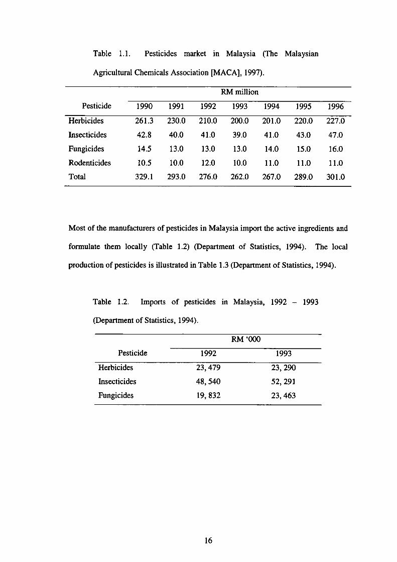
Table 1.1. Pesticides market in Malaysia(The Malaysian
Agricultural Chemicals Association【MACA】,1997).
RM million
Pesticide 1990 1991 1992 1993 1994 1995 1996
Herbicides
insecticides
Fungicides
Rodellticides
Total
261.3
42.8
14.5
10.5
329.1
230.0
40.0
13.0
10.0
293.0
210.0
41.0
13.0
12.0
276.0
200.0
39.O
l3.0
10.0
262.0
201.0
41.0
14.O
ll.0
267.0
220.0
43.0
15.0
11.0
289.0
227.0
47.0
16.0
11.0
301.0
Most of the manufacturers of pesticides in Malaysia import the active ingredients and
formulate them locally(Table 1.2)(Department of Statistics,1994). The local
production of pesticides i s illustrated in Table 1.3(Department of Statistics,1994).
Table 1.2. Imports of pesticides in Malaysia,1992-1993
(Department of Statistics,1994).
RM‘000
Pesticide 1992 1993
Herbicides
Insecticides
Fungicides
23,479
48,540
19,832
23,290
52,291
23,463
16

Table 1.3. Local production of pesticides in Malaysia,1992-1993
(Department of Statistics,1994).
RM‘000
Pesticide
Herbicides
Insecticides
Fungicides
1992
203,526
69,347
4,202
1993
216,173
57,698
3,623
In the rice growing region of West Malaysia, especially in Province Wellesley and in
Krian District of Perak, endosulfan was generally favored by many famers in both
localities.52%of the famlers reported that endosulfan was e脆ctive against many
species of paddy pests.32 90 of the farmers claimed that the chemical was effective
against leaf-rollers,4%fbund it to be effective against paddy field crabs and 20%
noted that it acted as a repellant to rats. The famlers in Krian District also favored
endosulfan fbr similar reasons.52%of the farmers noted that endosulfan was cheap
and fast acting on most insect pests(Yunus and Lim,1971), thus suggesting that
endosulfan is widespread in Malaysia. Endosulfan is currently pemitted in Malaysia.
1.3.2 Endosulfan in the Malaysian environment
Due predominantly to its persistent characteristic, residues of endosulfan continue to
in water, sediment and biota including fbodstuff such as agricultural products and
seafbod. Tables 1.4 and 1.5 show the extent of contamination of endosulfan in both
biotic and abiotic components in the Malaysian environment.
17

oo
一一
いひ9寸ひ巳
寸.
O.
一〔
bo?T自
No◎ひ一否oo9
壱田
盲o∈壱心o力
百o嘱日o=O
包ぷむヒ房
ロoるoo山
18

19
05盲喝§●.嚢
O.
n-唱ロ
毛晶言吟o言田
ひひ9
いOd
Oひ巳
一d処黶クoぶO
口oるoo山
.
1.4 Endosulfan in Japan
1・4・1 Sales of endosulfan in Japan
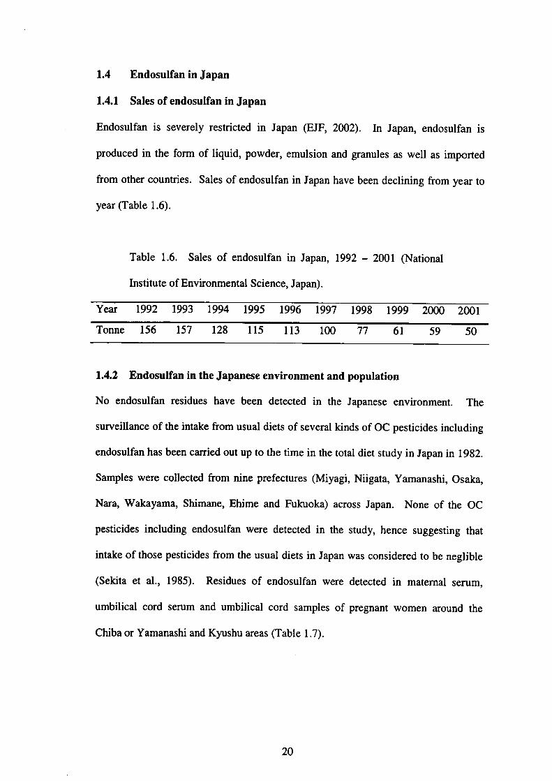
Endosulfan is severely restricted in Japan(EJF,2002). In Japan, endosulfan is
produced in the fbrm of liquid, powder, emulsion and granules as well as imported
from other countries. Sales of endosulfan in Japan have been declining from year to
year(Table 1.6).
Table 1.6. Sales of endosulfan in Japan,1992-2001(National
hlstitute of Environmental Science, Japan).
Yea} 1992 1993 1994 1995 1996 1997 1998 1999 2000 2001
To皿e 156 157 128 115 113 100 77 61 59 50
1・4・2Endosulfan in the Japanese environment and population
No endosulfan residues have b㏄n det㏄ted in the Japanese environmellt. The
surveillance of the intake from usual diets of several kinds of OC pesticides including
endosulfan has been carried out up to the time in the total diet study in Japan in l 982.
Samples were collected from nine prefectures(Miyagi, Niigata, Yamanashi, Osaka,
Nara, Wakayama, Shimane, Ehime and Fu㎞oka)across Japan. None of the OC
pesticides including endosulfan were detected in the study, hence suggesting that
intake of those pesticides from the usual diets in Japan was considered to be neglible
(Sekita et al.,1985). Residues of endosulfan were detected in matemal serum,
umbilical cord serum and umbilical cord samples of pregllant women around the
Chiba or Yamanashi and Kyushu areas(Table 1.7).
20
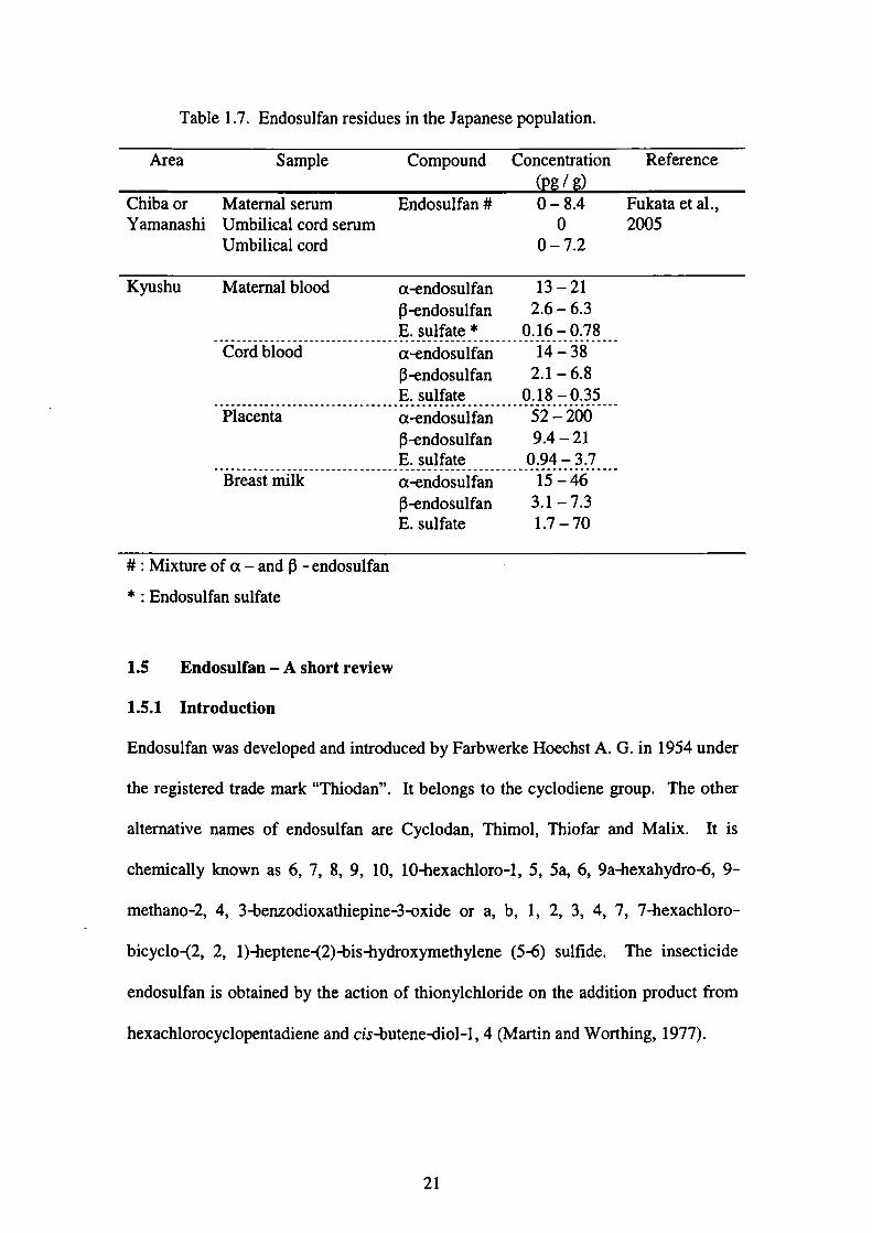
Table 1.7. Endosulfan residues in the Japallese population.
Area Sample Compound Concelltration ( /)
Reference
Chiba or
YamanashiMaternal serum
Umbilical cord serum
Umbilical cord
Endosulfan# 0-8.4
0
0-7.2
Fukata et al.,
2005
Kyushu Matemal blood α一endosulfan 13-21 β一endosulfan 2・6-6・3
E.sulfate* 0.16-0.78
Cord blood αendosulfan 14-38
β一endosulfan 2・1-6・8
E.sulfate O.18-0.35
Placellta α℃ndosulfan 52-200
β一endosulfan 9・4-21
E.sulfate O.94-3.7
Breast milk α一endosulfan 15-46
β一endosulfan 3・1-7・3
E.sulfate 1.7-70
#:Mixture of a-and li -endosulfan
*:Endosulfan sulfate
1.5 Endosulfan-A short review
1.5.1 1ntroduction
Endosulfan was developed and introduced by Farbwerke Hoechst A. G. in 1954 under
the registered trade mark“Thiodan”. It belongs to the cyclodiene group. The other
altemative names of endosulfan are Cyclodan, Thimo1, Thiofar and Malix. It is
chemically known as 6,7,8,9,10,10-hexachloro-1,5,5a,6,9a-hexahydro-6,9-
methano-2,4,3benzodioxathiepine-3-oxide or a, b,1,2,3,4,7,7-hexachloro-
bicyclo-(2,2,1)-heptene-(2)-bis-hydroxymethylelle(5-6)sulfide. The insecticide
endosulfan is obtained by the action of thionylchloride on the addition product from
hexachlorocyclopentadiene and cis-butene-diol-1,4(Martin and Worthing,1977).
21
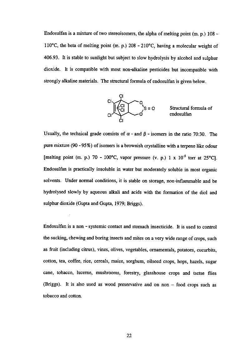
Endosulfan is a mixture of two stereoisomers, the alpha of meltillg point(m. p.)108-
110°C,the beta of melting point(m. p.)208-210°C, having a molecular weight of
406.93.It is stable to sunlight but sublect to slow hydrolysis by alcohol and sulphur
dioxide. It is compatible with most non-alkaline pesticides but incompatible with
strongly alkaline materials. The structural formula of endosulfan is given below.
Cl
Cl
Ct
l:l
Cl
0 、
S=0 〆
0Stnlctural fbrmula of
endosulfan
Usually, the technical grade consists ofα一alldβ一isomers in the ratio 70:30. The
pure mixture(90-95%)of isomers is a brownish crystalline with a terpene like odour
【melting point(m. p.)70-100°C, vapor pressure(v. p.)lxIO’5 torr at 25°C].
Endosulfan is practically insoluble in water but moderately soluble in most organic
solvents. Under normal conditions, it is stable on storage, non鴫nflammable and be
hydrolysed slowly by aqueous alkali and acids with the fbmlation of the diol and
sulphur dioxide(Gupta and Gupta,1979;Briggs).
Endosulfan is a non 一 systemic contact and stomach insecticide. It is used to control
the sucking, chewing and boring insects and mites on a very wide range of crops, such
as ftUit(including citrus), vines, olives, vegetables, omamentals, potatoes, cucurbits,
cotton, tea, coffee, rice, cereals, maize, sorghum, oilseed crops, hops, hazels, sugar
cane, tobacco, luceme, mushrooms, fbrestry, glasshouse crops and tsetse flies
(Briggs). It is also used as wood preservative and on non-fbod crops such as
tobacco and cotton.
22
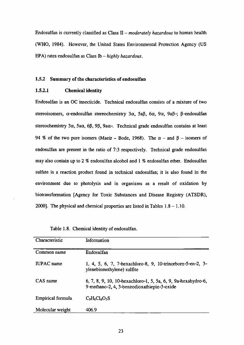
Endosulfan is currently classified as Class II-moderately hazardous to human health
(WHO,1984). However, the United States Environmental Protection Agency(US
EPA)rates endosulfan as Class ib 一 hi8hl:ソhazardous.
1.5.2 Summary of the characteristics of endosuぬn
1.5.2.l Chemical identity
Endosulfan is an OC insecticide. Technical endosulfan consists of a mixture of two
stereoisomers,α一endosul飴n stereochemistry 3α,5aβ,6α,9α,9aβ一;β一endosulfan
stereochemistry 3α,5aα,6β,9β,9aα一. Technical grade endosul飴n contains at least
94%of the two pure isomers(Mae丘一Bode,1968). Theα一andβ一isomers of
endosulfan are present in the ratio of 7:3 respectively. Technical grade endosulfan
may also contain up to 2%endosulfan alcohol and 1%endosulfan ether. Endosulfan
sulfate is a reaction product fbund in technical endosulfan;it is also f()und in the
environment due to photolysis and in organisms as a result of oxidation by
bio血ansformation【Agency for Toxic Substances and Disease Regis廿y(ATSDR),
2000].The physical and chemical properties are listed in Tables 1.8-1.10.
Table 1.8. Chemical identity of endosulfan.
Characteristic Infbrmation
Common name
IUPAC name
CAS name
Empirical fbrmula
Molecular wei ht
Endosulfan
1,4,5,6,7,7-hexachloro-8,9, 10-trinorborn-5-en-2,3-
ylenebismethylene)sulfite
6,7,8,9,10,10-hexachloro-1,5,5a,6,9,9a-hexahydro-6,
9-methano-2,4,3-benzodioxathiepin-3-oxide
CgH6Cl603S
406.9
23

L5.2.2 Physical and chemical properties
Table 1.9. Physical and chemical properties of pure active constituent.
Characteristic information
Color
Odour
Physical state
Melting Point
Vapor pressure
Specific gravity
Solubility in water
Solubility in organic
solvents(100g
solvent at 200C)
Colorless crystalline solid
Odourless
Pure aisomertrystalline solid;pureβ鴫somer-crystalline solid
α一:109.20C;β一:213.30C
α一:1.9;fi 一・:0.09 mPa at 25°C.α一:0.96;β一:0.04 mPa at 20°C
1.745at 200C
α一:0.33;β一:0.32mg/L at 22°C
Ethyl acetate, Dichloromethane, Toluene(200 g!L), Ethallol(65
g/L),Hexane(24 g/L)
Table 1.10. Physical and chemical propenies of technical grade endosulfan
Characteristic lnformation
Color
Odour
Physical state
Melting Point
Vapor pressure
Specific gravity
Solubility in water
DiffUsion coefficient
ln water
Solubility in organic
solvents(1009
solvellt at 20°C
Brown
Terpene odour
Crystalline flakes
70-100°C
1 x 10’5 mm Hg at 25°C;1.7 mPa
1.745at 20°C
60-150pg/L
4.55x106 cm2/sec at 37°C
Chloroform(50 g);Xylene(45 g);Benzene(37 g);Acetone
(33 g);Carbon tetrachloride(29 g);Kerosene(20 g);
Methanol(11g);Ethanol(5 g)
24
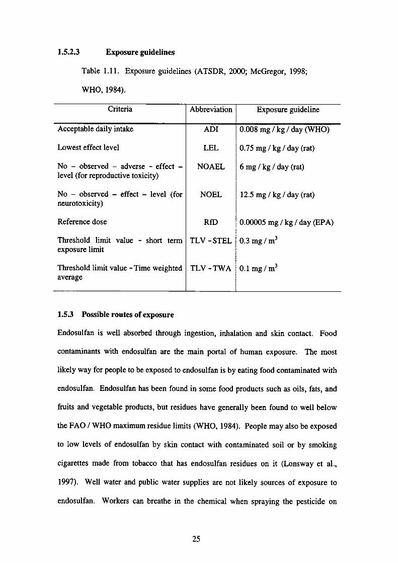
1・5・2・3 Exposure guidelines
Table 1.11. Exposure guidelines(ATSDR,2000;McGregor,1998;
WHO,1984).
Criteria Abbreviation Exposure guideline
Acceptable daily intake ADI 0.008mg/kg/day(WHO)
Lowest effect level LEL 0.75mg/kg!day(rat)
No-observed-adverse-effect一 NOAEL 6mg/kg/day(rat)level(fbr reproductive toxicity)
No-observed-effect-1evel(fbr NOEL 12.5mg/kg/day(rat)
neurotoxicity)
Reference dose RfD 0.00005mg/kg/day(EPA)
Threshold lilnit value - short term TLV-STEL 0.3mg/m3
exposure limit
Threshold limit value-Time weighted TLV-TWA 0.1mg/m3
average
1.5.3Possible routes of exposure
Endosulfan is well absorbed through ingestion, inhalation and skin contact. Food
contaminants with endosulfan are the main portal of human exposure. The most
likely way for people to be exposed to endosulfan is by eating food contaminated with
endosulfan. Endosulfan has been found in some food products such as oils, fats, and
血its alld vegetable products, but residues have generally been fbund to well below
the FAO/WHO maximum residue limits(WHO,1984). People may also be exposed
to low levels of endosulfan by skin contact with contaminated soil or by smoking
cigarettes made from tobacco that has endosulfan residues on it(Lonsway et al.,
1997).Well water and public water supplies are not likely sources of exposure to
endosulfan. Workers can breathe in the chemical when spraying the pesticide on
25

crops. Exposure can occur by breathing the dust or getting the pesticide on the skin if
people do not follow all the safety and handling procedures. Accidental spills and
releases to the environment at hazardous waste disposal sites are also possible sources
of exposure to endosulfan for occupational workers. The most likely exposure to
endosulfan fbr people living near hazardous waste sites is through contact with soils
contalnlng lt.
Populations that are usually susceptible to endosulfan include the unbom and
neonates, the elderly and people with liver, kidney, immunological, haematological or
neurological disease.
Table 1.12.
Handbook)
Maximum residue limits(MRL)for endosulfan(The Agrochemical
Compound Maximum residue limit(MRL)
Endosulfan Root vegetables O.2 ppm;other廿uit and vegetables
lppm;maize O.2 ppm;other cereals O.1 ppm;all
feedstuffs O.l ppm(except maize O.2 ppm, oilseeds O.5
ppm, complete feedstuffs for fish O.005 ppm)
1.5.4Exposure of endosulfan to humans
In general, characterization of the dose of endosulfan in poisoning cases has been
poor. Several cases of accidental and suicidal poisoning have been reported. The
lowest reported dose that resulted in death in humans was 35 mg!kg body weight,
and deaths have also been reported after ingestions of approximately 295 and 467
mg!kg, with death occurring within l hour of administration in some cases. Intensive
medical treatment within l hour of endosulfan administration was reportedly
26
successfUI at doses of 100 and 1000 mg/kg with clinical signs in these patients
consistent with those seen in laboratory animals, dominated by tonoclonic spasms. In
acase where the dose was 1000 mg/kg, neurological symptoms requiring anti-
epileptic therapy, which resulted from anoxia during treatment, were still required one
year after endosulfan exposure(NRA,1998). Signs of poisoning included vomiting,
restlessness,口㎡tability, collvulsions, pulmonary edema and cyanosis【Agency fbr
Toxic Substances and Disease Registry(ATSDR),2000;World Health Organization
(WHO),1984】.
Asmall number of case reports on the tissue concentrations of endosulfan and its
metabolites were reported in the literature(Chan et al.,2004;Cooper et a1.,2001;
Kumar et a1.,2000;Saiyed et al.,2003;Sarcinelli et aL,2003;Cerrillo et al.,2005).
This pesticide is also implicated in several cases of accidental(Demeter et al.,1977;
Oktay et al.,2003), non-accidental(Boereboom et al.,1998)and suicidal deaths
(Blanco-Coronado et aL,1992;Eyer et al.,2004;Lo et aL,1995)as well as
occupational human poisoning(Aleksandrowicz,1979;Brandt et al.,2001).
1.5.5 EffectS on the environment
Endosulfan is not readily bioaccumulated and it is not very persistent in biological
tissues as compared to other OC pesticides. It is hazardous as an acute poison fbr
some aquatic species, particularly fish, even at application rates recommended for
wetland areas and can cause massive mortalities. In fish, it caused marked changes in
sodium(Na)and potassium(K)concentrations, decrease in blood Ca2+and
magnesium(Mg)1evels and inhibits Na, K and Mg-dependent ATPase(in brain)
(Naqvi and Vaishnavi,1993). The high toxicity to fish means that fish kills can result
27
from discharges to waterways. It is moderately toxic to honey bees. It is moderately
to highly toxic for birds in a laboratory setting, but no poisonings have been repolted
under field conditions. Endosulfan contamination is not widespread in the aquatic
environment as it is easily degraded with a half 一 life of 4 days which could be
increased at low pH or under anaerobic conditions(Naqvi and Vaishnavi,1993).
1.5.6 Environmental fate of endosuifan
Endosulfan has low water solubility. It is s仕ongly absorbed onto soil麺cles and
hence immobile in the soil column. Transport of this insecticide is most likely in the
form of surface r皿off. Large amounts of endosulfan can be found in surface water
near areas of application(Farms Chemical Handbook,1992).
The breakdown of endosulfan in water is more rapid under neutral conditions with the
half life of five weekS that at more acidic condition with the half -life of five months.
Under strongly alkaline conditions, the halfコife of the insecticide is one day. The two
isomers have different degradation times in soil. The half 一 life for the a 一 isomer is
35days and 150 days for theβ 一 isomer under neutral conditions. The half-live on
plants is three to seven days fbr most㎞its and vegetables(Martin and Worthing,
1977).
Due to its widespread application, the accumulation of endosulfan has been reported
in the environment(Gam6n et al.,2003;Bhattacharya et al.,2003;Louie and Sin,
2003;Tan and Vij ayaletchumy,1994).
28
REFERENCES
Aleksandrowicz, D. R.(1979). Endosulfan poisoning and chronic brain syndrome.
ノlrchives of Toxicolo8y,43:65-68.
ノBhattacharya, B., Sarkar, S. K., Mukherjee, N.(2003). Organochlorine pesticide
residues in sediments of a tropical mangrove estuary, India:㎞plications fbr
monitoring. Environment lnterantional,29:587-592.
Blanco-Coronado, J. L, Repetto, M., Ginestal, R. J., Vicente,」. R., Yelamos, F.,
Lardelli, A.(1992). Acute intoxication by endosulfan. Ctinicat 7b」xicolo8y,30:575-
583.
Boereboom, F. T. J., van Dijk, A., van Zoonen, P., Meulenbelt, J.(1998).
Nollaccidental endosulfan intoxication:Acase report with toxicokinetic calculations
and tissue concentrations. Clinical Toxicology,36:345-352.
Brandt, V. A., Moon, S., Ehlers, J., Methner, M. M., Struttma皿, T.(2001). Exposure
to endosulfan in farmers:Two case studies. American Journal oflndustrial Medicine,
39:643-649.
Briggs, S. A. Basic guide to pesticides:Their characteristics and hazards. Rachel
Carson Council;15.
Cerrillo,1., Granada, A., L6pez-Espinosa, M. J., Olmos, B., Jim6nez, M., Caho, A.,
01ea, N.,01ea-Serrano, M. F.(2005). Endosulfan and its metabolites in fertile
29
women, placenta, cord blood, and human milk.
239.
Environmental Research,98:233一
Chan, M. P. L, Mustafa, A. M., Hussein, R., Hj Hussain, S., Zulkifli, S. N., Abdullah,
A.R.(2004). Pe’唐狽奄モ奄р?@residues in blood of schoolchildren from selected schools in
Peninsular Malaysia. Environmental Health Focus,2(1):18-24.
Cooper, S.P., Burau, K., Sw㏄ney, A., Robison, T., Smith, M. A., Symanski, E, Colt,
J.S., Laseter, J., Zahm, S. H.(2001). Prenatal exposure to pesticides:Afeasibility
study among migrant and seasonal famlworkers. American J加rnαZ()f lndustrial
Medicine,40:578-585.
Demeter, J., Heyndrickx, A., Timperman, J., Lefevere, M. and De Beer,」.(1977).
Toxicological analysis in a case of endosulfan suicide. Bulletin of Environmental
Contamination and Toxicolo8y,18:110-114.
Department of Statistics, Malaysia,1994.
Ecobichon, DJ., Toxic effects of pesticides.(1995). In:Amdur, M.0., Dou11,」.,
Klaassen, C. D.(Eds.), Toxico’08y:T7te●asic science q〆」poご∫onぷ,4「h ed., New York:
McGraw-Hill, Inc.
EIF(2002). End()f the roadプbr endosulfan:aco”プbr action a8ainぷ’ada’18e「ous
pesticide. Environmental Justice Foundation Ltd, London, UK, pp.1-4.
30
EXTOXNET(Extension Toxicology Network), Oregon State University,1993.
Eyer, F, Felgenhauer, N., Jetzinger, E., Pfab, R., Zilker, T. R.(2004). Acute
endosulfan poisoning with cerebral edema and cardiac failure. Journal of Toxicology
and Clinical To」xicology,42(6):927-932.
Farm Chemicals Handbook, Meister Publishing Company, Willoughby, Ohio,1992.
Fukata, H., Omori, M., Osada, H., Todaka, E., Mori, C.(2005). Necessity to measure
PCBs and organochlorine pesticide concentrations in human umbilical cords for fetal
exposure assessment. Environmental Health Perspectives,113:297-303.
Gam6n, M., Sfiez, E, Gil,」., Boluda, R.(2003). D口ect and ind丘ect exogenous
contamination by pesticides of rice-farming soils in a Mediterranean wetland.
ノlrchives of Environ〃lenta’Coη∫α〃linationαη4τb」xicolo8y,44:141-151.
Gupta, P. K.(1979). Phamlacology, toxicology and degradation of endosulfan. A
review. Toxicolo8y,13:115-130.
Kumar, R., Pant, N., Srivastava, S. P.(2000). Chlorinated pesticides and heavy
metals in human semen. lnternational Journal of Andrology,23:145 一 149.
Lo, R. S. K., Chan,」. C. N., Cockram, C. S., Lai, F. M. M.(1995). Acute tubular
necrosis fbllowing endosulphan insecticide poisoning. Clinical Toxicolo8y,33:67-
69.
31
Lonsway, J. A., Byers, M. E., Dowla, H. A., Panemangalore, M., Antonious, G. F.
(1997). Demal and respiratory exposure of mixers/sprayers to acephate,
methamidophos, and endosulfan during tobacco production. Bulletin of
Environmental Contamination and Toxicology,59:179-186.
Louie, P. K. K., Sin, D. W.(2003). A preliminary investigation of persistent organic
pollutants in ambient air in Hong Kong. Chemosphere,52:1397-1403.
MACA.(1997). Annual report 1996/97 and directories. The Malaysian Agricultural
Chemicals Association, Petaling Jaya.
Maier-Bode, H.(1968). Properties, effect, residues and analytics of the insecticide
endosulfan. Residue Reviews,22:1-44.
Martin, H., Worthing, C. R.(Eds.)(1977). Pesticide manua~ほasic information on
功εchemica’∫useばα∫act加εcompound∫(ゾρεぷτ輌cides,5’カed., British Crop Protection
Council.
McGregor, D・B・(1998). Endosulfan(JMPR evaluations 1998 Part II Toxicolog ical)
-Fir∫’4rψ. Lyon, France:international Agency for Research on Cancer.
Naqvi, S. M., Vaishnavi, C.(1993). Mini review:Bioaccumulative potential and
toxicity of endosulfan insecticide to non-target animals. Coハnparative Biochemistry
and Physioto8y,105C(3):347-361.
32

Oktay, C., Goksu, E., Bozdemir, N., and Soyuncu, S.(2003). Unintentional toxicity
due to endosulfan:acase report of two patients and characteristics of endosulfan
toxicity.レ「eterinary and. Human Toxicolo8y,45(6):318-320.
Pandit, G. G., Mohan Rao, A. M., Jha, S. K., Krisnamoorthy, T. M., Kale, S. P.,
Raghu, K., Murthy, N. B. K.(2001). Monitoring of organochlorine pesticide residues
in the lndian marine environment. ChemoSphere,44:301-305.
Pinkston, K., Criswell, J., Cupenls, G., Schnelle, M. A. Information on insecticides
/br 8reenhouse growerぷ. OSU Extension Facts F-6712, IPM in the Gr㏄nhouse
Series, Oklahoma Cooperative Extension Service, Division of Agricultural Sciences
and Natural Resources.
The agrochemical handbook,3「d ed., Cambridge:Royal Society of Chemistry,1991.
Saiyed, H., Dewan, A., Bhatnagar, V., Shenoy, U., Shenoy, R., Raj mohan, H., Patel,
K.,Kashyap, R, Kulkarni, P., Raj an, B., Lakkad, B.(2003). Effect of endosulfan on
male reproductive development. Environmental Health Perspectives,111(16):1958
-1962.
Sarcinelli, P. N., Pereira, A. C. S., Mesquita, S. A., Oliveira 一 Silva, J.」., Meyer, A.,
Menezes, M. A. C., Alves, S. R., Mattos, R. C.0. C., Moreira,」. S. and Wolff, M.
(2003).Dietary and reproductive determinants of plasma organochlorine levels in
pregnant women in Rio de Janeiro. Environmental Research,91:143-150.
33
Sekita, H., Sasaki, K., Kawamura, Y., Takeda, M., Uchiyama, M.(1985). Studies on
the analysis of pesticide residues in fbods(XLII). Surveillance of the daily intake of
endosulfan, chlorobenzilate, captan and others丘om total diet in 1982. Eisei Shikenjo
Hokoku,103:1129-133(ln Japanese).
Tan, G..H, Vijayaletchumy, K.(1994). Organochlorine pesticide residues levels in
Peninsular Malaysian rivers. Bu〃etin of Environmental Contai励ation and
To」xicology,53:351-356.
WHO(1984). Endosulfan. International Programme on Chemical Safeり}.
Environmental Health Criteria 40. Geneva, Switzerland:World Health Organization,
pp.1-62.
Yunus, A. Lim, G. S.(1971). A problem in the use of insecticides in paddy fields in
West Malaysia-Acase study. Malaysian Agricultural Journal,48:167-178.
34
CHAPTER 2
TOXICITY TO MAMMALS
2.O Introduction
Chlorinated hydrocarbon insecticides which are ubiquitous in the ellvironment in
nature have become an integral part in the tissues of animals. Recognition of the
incorporation of the parent compound and/or its metabolites in lower organisms, in
tissues of fishes, b口ds, wild animals and humans has caused serious concem(Martin,
1964).Endosulfan due to its insecticidal properties has an extensive use in aghculture
sector as a potellt pesticide in colltrol pests. Due to its wide application, it may enter
human or animal systems ei也er d丘ectly or as an env廿onmental pollutant. Generally,
mammals are not as sensitive to endosulfan as aquatic organisms. The degree of
toxicity depends upon the species and sex of animals, the route of exposure and
vehicle used(McGregor,1998). Brain, testes,1iver alld kidneys are the main target
organs fbr toxicity after exposure to endosulfan(ATSDR,2000).
2.1 Toxicity to mammals(rats)
2.1.1 General toxic effectS of endosuifan
Tables 2.1 and 2.2 show the existing information on health effects of endosulfan in
human and animal. The LD500f endosulfan(isomeric mixture)for rats varies
markedly depending upon the route of administration, species, dosillg vehicle and the
sex of the animal. The LD50 ranges from 47 一 89 mg/kg for male rats and 8-49 mg/
kg for female rats(Gupta,1976;Gupta and Gupta,1977). The inhalation lethal dose
(LC50)in male rats was 350 mg/m3 when exposed for 4 hours. The dermal LD50
35

Table 2.1.
human.
Existing information on health effects of endosulfan in
Health effect Route of ex osure
㎞alation Oral De㎜alDeath *
S stelnic(Acute) * * *
S stelnic(Intermediate) * *
Sstemic(Chro㎡c)
Immunolo ic * *
Neurolo ic * * *
Re roductive
Develo mental *
Genotoxic *
Cancer *
Table 2.2.
animal.
Existing infbmlation on health effects of endosulfan in
Health effect Route of ex osure
Inhalation Oral DermalDeath * * *
S.stelnic(Acute) * * *
S stelnic(Intermediate) * * *
Sstemic(Chronic) *
Immunolo ic * * *
Neurolo ic * * *
Re roductive * * *
Develo mental * *
Genotoxic *
Cancer *
Note.
*:Existing studies
varied伽m 74-681 mg/kg depending on the vehicle and sex used. When rats were
fed on diets containing O%,3.5%,9%,26%or 81%protein as casein, the LD50 was
5.1,24.0,57.0,102.O and 98.O mg/kg, respectively, but the prominent signs and
36

symptoms of intoxication remained unchanged(Gupta and Gupta,1977). The
prominent signs of intoxication include hypersensitivity, respiratory distress, diarrhea,
tremors, hunching and convulsions fbllowed by death. Rats fed two years on a diet
containing 30 ppm suffered no il1 effect(Martin and Worthing,1977).
The isomers of endosulfan show acute oral toxicity profiles similar to that of technical
grade endosulfan. The acute toxicity of formulations containing endosulfan was
dependent upon the concentration of the active ingredient in t le end-used products,
and was similar to those seen fbllowing administration of the active ingredient. The
toxicity of the metabolites varies depending upon vehicle and species used. Generally,
the toxicities of the metabolites were similar to the parent compound, except for
endosulfan diol which has low acute oral toxicity in the mouse[National Registration
Authority(NRA),1998].
More than 90%of an oral dose of endusulfan was absorbed in rats, with maximum
plasma concentrations occurring after 3-8hin males and about 18 h in females.
Elimination occurs mainly in the feces and to a lesser extent in the urine, more than
85%being excreted within 120 h. The highest tissue concentrations were in the
lddneys. The metabolites of endosulfan include endosulfan sulfate, diol, hydroxy-
ether, ether, and lactone but most of its metabolites are polar substances which have
not yet been idelltified. Endosulfan would not be expected to accumulate significantly
in human tissues.
Metabolism studies in rats, after an intraperitoneal injection of 20 mg/kg of technicaI
endosulfan in an oil solution revealed the presence of endosulfan diol and an
37
unknown compound in the urine as a water soluble conjugation metabolite. It was
also reported that endosulfan was not excreted in rat urine as endosulfan diol, but as
endosulfan一α一hydroxy ether. In addition, transient amounts of endosulfan and
endosulfan sulfate was also detected in the body fat and liver of mice after the
administration of【14q endosulfan where endosulfan metabolites were detected in
feces(Gupta and Gupta,1979). There is no accumulation in milk, fat or muscle and it
is excreted as conjugates of the diol and other highly polar compounds depending on
the species(Martin and Worthing,1977). Dorough et al.(1978)indicated that the
major portion of residues in the excreta and/or tissues consisted of unidentified polar
metabolites that could not be extracted from the substrate, whereas the non-polar
metabolites, including sulfate, diol, ct 一 hydroxyether, lactone and ether derivatives of
endosulfan, represented only minor amounts. The available evidence indicates that
endosulfan can be metabolized in animals to other lipophilic compounds, which can
rapidly enter tissues, and to more hydrophilic compounds that can be excreted.
The distribution pattern of endosulfan was estimated in the plasma and brain of the
rats when they were fed daily doses of endosulfan(5 or 10mg/kg)fbr 15 days. The
animals were sacrificed 24 hours later. The concentration ofαisomer was in the
order of cerebrum(3.76μg/g)>remaining parts of the brain(2.66μg/g)>
cerebellum(2.04μg/g). The concentration of theβisomer was O.06 pg/gin the
cerebrum and O.02μg/gin the cerebellum, where as no 6弓somer was detected in the
“remaining part”of the brain. The plasma collcentrations ofα一andβ 一 isomers were
2.26and O.46 pg/grespectively and endosulfan sulfate was the only metabolite
detected in the plasma(Gupta,1978). Endosulfan is neither carcinogenic(ATSDR,
38
2000;Hack et a1,1995;McGregor,1998;Naqvi and Vaishnavi,
(ATSDR,2000;McGregor,1998;Naqvi and Vaishnavi,1993).
1993)nor genotoxic
2.1.2 Neurological effectS
lnvolvement of endosulfan, a neurotoxic agent, in the central nervous system(CNS)
has been studied in various studies(Anand et al.,1980;Paul et al.,1994;Seth et al.,
1986;Subramoniam et a1.,1994). Long term inhalation or oral exposure to
endosulfan results in neurotoxicity which is a primary effect. Famers exposed to
endosulfan exhibit epilepsy, hyperactivity, i1’ritability, tremors, convulsions and
paralysis. However, the CNS e脆cts described fbr acute exposure are not usually
CI O
…一・・ Cl llS93S=°
↑/β㌣㎞/
Cl
::@C:)・ζ:
l
Endosulfan sulfite
::違:三H
Cl
HydroxyendosUlfin
carboxylic●cid
↓
Urine,‘aeces
ClCl OH
l目
Cl OH
l
Endosulfan d回
CC Cl
lEl
Cl
0
Endosulfan
lecton●
↓
Coniug●tes
O r鴫一一一一一一
Cl Cl
1呂 ・CI@ l
Endo5ul‘●n●ヒh◆r
Cl OHCt
lき1 ・Cl@ CI
EndosuFan hydroxyeth●r
↓
Uri∩e. Saeces
Note. Most endosulfan metabolites are polar and are yet to be identified.
Figure 2.1. Mammalian metabolism and excretion of endosulfan
(Adapted from McGregor,1998).
39

fbund with chronic oral exposure in experimental animals. A stimulation by long
term exposure of endosulfan at a dose of 3 mg/kg body weight fbr 15 and 30 days
respectively illcreased fbot shock-induced fighting behavior in male rats(Agrawal et
a1.,1983). lnduction of hyperactivity, tremors and convulsions was observed in male
rats given 40 mg/kg of endosulfan intraperitoneally(Ansari et a1.,1987). Dikshith
and co-workers(1984)observed signs of CNS simulation fbr the fhst 3-4days
only in rats given oral gavage doses of endosulfan fbr 30 days.
Dermal exposure to endosulfan has not been reported to cause neurological effects in
humans. No effects on brain weight or histopathology were observed with dermal
application of endosulfan to male rats(62.5 mg/kg/day)and to female rats(32 mg/
kg/day)for 30 days(Dikshith et a1.,1988).
2.1.3 Reproductive effectS
Human studies demonstrating reproductive effects due to inhalation, oral or dermal
exposures are unavailable in the literature survey. Reproductive perfbrmance in
animals is not adversely affected even though adverse effects on the reproductive
organs have been observed. Endosulfan induced alteration on spermatogenesis in
youllg growing as well as adult rats have been reported(Sinha et al.,2001). The
impaimients include decreased sperm count, intratesticular spermatid number, spem
morphology, altered activities of testicular marker enzymes【Pandey et al.,1990;
National Cancer Institute(NCI),1978]as well as reduction in senlm testosterone
levels(Choudhary and Joshi,2003;Wilson and LeBlanc,1998). Dalsenter et a1.
(1999,2003)reported reproductive effects of endosulfan on the male offspring of rat
exposed during pregnancy and lactation at high dose. The daily sperm production
40
was pemlanently decreased at puberty and adulthood. In addition, the percentage of
semi ni ferous tubules showing complete spermatogenesis was significantly decreased
at puberty. This finding may explain the decrease in daily sperm production observed
in the endosulfan-exposed male rats. Other testicular impairment in vivo have been
reported elsewhere(Sinha et al.,1995;Sinha et al.,1997;Chitra et aL,1999;Dikshith
et al.,1984;Singh and Pandey,1989).
2.1.4 Endocrine effectS
An’ ?唐狽窒盾№?氏@is a substance that can induce estrus or a biological response associated
with estnls;011e such effect is proliferation of cells in the female genital tract. In
recent ye訂s, naturally occuning and man-made subs伽ces in由e environment血at
may be estrogenic have come under increasing scnltiny. Suspicion that endosulfan
may have estrogenic properties was stimulated by observation of the reduced speml
counts described above and of testicular atrophy in rats given endosulfan in the diet in
longイem studies. Perhaps the血st study of es仕ogenic e飽cts仇vivo was conducted
by Raizada et a1.(1991), who仕eated groups of eight ovadectomized Wistar rats,
weighing an average of lOO g, with endosulfan at 1.5 mg/kg bw per day by gavage,
estradiol dipropionate intraperitoneally(dose unspecified), or a combination of these
treatments fbr 30 days. Endosulfan did not change the weights of the utenls, cervix, or
vagina, whereas estradiol propionate produced large increases(Hiremath and Kaliwal,
2003).The increased weights seen with the combined treatment were similar to those
with estradiol propionate alone. Persistent depressed testicular testosterone was seen
in male rats after intemediate oral exposures to endosulfan.
41
Concem that endosulfan might be estrogenic persisted as a result of the findings of an
’E -screen’assay, which was developed to assess the estrogenjc effects of
environmental chemicals by observing their proliferative effect on a target cell. The
numbers of cells present after similar inocula of the human breast cancer ce111ine,
MCF-7, were compared in the absence of estrogens(negative control), in the
presence of estradiol-17β(positive control), and with a range of concentrations of
endosulfan. In this assay, endosulfan was estrogenic at concentrations of 10-25μM,
with a proliferative effect about 80%that of 17β一estradiol at 10μM;the relative
proliferative potency (i.e. the ratio of the doses of endosulfan and estradiol -
17β required to produce the maximum effect)was O.0001%.111 addition, endosulfan
competed with estradio1-17βfbr binding to the estrogen receptor and illcreased the
concentrations of progesterone receptor and pS2 in MCF-7cells, as would be
expected fbr a compound that mimics estrogens(Soto et al.,1994, Soto et al.,1995).
No studies were located regarding elldocrine disnlption in humans after exposure to
endosulfan・ Overall, in vitro evidence in favor of endosulfan estrogenicity indicates
relatively weak potency compared to estradiol-17β. Contradictory results were
reported in different studies, indicating that cautions should be used in interPreting the
COIIeCtiVe in VitrO reSUItS.
42

Table 2.3. Levels that cause no toxic effect(ATSDR,2000;McGregor,1998).
Secies No toxic effect level
Mouse:
Rat:
Rabbit:
Do9:
3.9ppm, equal to O.58 mg/1(g bw per day(females in a 78-week
study of toxicity)
15ppm, equal to O.6 mg/kg bw per day(two- year dietary study of
toxicity)75 ppm, equal to 6 mg/kg bw per day(reproductive toxicity)
0.66mg!kg bw per day(maternal toxicity in a study of developmental
toxicity)2 mg/kg bw per day (fetotoxicity in a study of
developmental toxicity)
0.7mg!kg bw per day(maternal toxicity in a study of developmental
toxicity)
10ppm, equivalent to O.57 mg/kg bw per day(one-year study of
toxicity)
REFERENCES
Agrawal, A. K., Anand, M., Zaidi, N, F., Seth, P. K.(1983). Involvement of
serotonergic receptors in endosulfan neurotoxicity. Biochemical Pharmacology,32:
3591-3593.
Anand, M., Khanna, R. N., Misra, D.(1980). Electrical activity
endosulfan toxicity. Indian Journal〈of Pharmacology,12(4):229-235.
of brain in
Ansari, R. A., Husain, K., Gupta, P. K.(1987). Endosulfan toxicity influence on
biogenic amines of rat brain. Journat of Environmental Biolo8y,8:229-236.
ATSDR(2000). Toxicolo8ical、ρr()Lfile/br endosulfan.
Toxic Substances and Disease Registry.
Atlanta, GA:Agency for
43

Choudhary, N., Joshi, S. C.(2003). Reproductive toxicity of endosulfan in male
albino rats・Butletin ofEnvironmental Contamination and Toxicolo8y,70:285-289.
Chitra, K. C., Latchoumycandane, C., Mathur, P. P.(1999). Chronic effect of
endosulfan on the testicular functions of rat. ノlsian Journa~of/1ηばrolo8y,1:203-
206.
Dalsenter, P. R., Dallegrave, E., Mello, J. R. B., Langeloh, A., Oliveira, R. T., Faqi, A.
S.(1999).Reproductive effects of endosulfan on male offspring of rats exposed
during Pregnancy and lactation・Human and Experimental To」xicology,18:583-589.
Dalsenter, P. R., de Aratijo, S. L, da Silva de Assis, H. C., Andrade, A. J. M.,
Dallegrave, E(2003). Pre and post natal exposure to endosulfan in Wistar rats.
Human and Experimental Toxicolo8y,22:171-175.
Dikshith, T. S. S., Raizada, R. B., Srivastava, M. K., Kaphalia, B. S.(1984). Response
of rats to repeated oral administration of endosulfan. Industrial Health,22:295-304.
Dikshith, T. S. S., Raizada, R. B., Kumar, S. N., Srivastava, M. K., Kausha1, R. A.
(1988).Effect of repeated demlal application of endosulfan to rats. レ「eterinary and
Human Toxicology,30:219-224.
Dorough, H. W., Huhtanen, K., Marshall, T. C., Bryant, H. E.(1978). Fate of
endosulfan in rats and toxicological considerations of apolar metabolites. Pesticide
Biochemistry and Physiolo8y,8:241-252.
44
Gupta, P. K.(1978). Distribution of endosulfan in plasma and brain after repeated
oral administration to rats. Toxicolo8y,9:371-377.
Gupta, P. K., Gupta, R. C.(1979). Pharmacology, toxicology and degradation of
endosulfan. A review.7「bxicolo8y,13:115-130.
Hack,R., Ebert, E., Leist, K. H.(1995). Chronic toxicity and carcinogenicity studies
with the insecticide endosulfan in rats and mice. Food and Chemical Toxicolo8y,33
(11):941-950.
Hiremath, M. B., Kaliwal, B. B.(2003). Evaluation of estrogenic activity and effect
of endosulfan on biochemical constituents in ovariectomized(OVX)Swiss albino
mice. Bulletin of Environmental Contamination and Toxicolo8y,71:458-464.
Martin, H.(1964). Insecticide and/Ungicide hanabookプbr crOp pro’ection,4「んed.,
Blackwell, Oxfbrd, pp.44.
Martin, H., Worthing, C. R.(Eds.)(1977). Pesticide manual: Basic information on
the chemicats used as active compounds ofpesticides,5th ed., British Crop Protection
Counci1.
McGregor, D. B.(1998). Endosulfan(JMPR evaluations 1998 Part II Toxicolo8ical)
-Fjr∫’4rψ. Lyon, France:lnternational Agency for Research on Cancer.
45
National Cancer Institute(1978)・・B輌oo∬ay of endosulfan/br」ρo∬訪1εcarcino8enicity,
7セchnica’Report, Series No.62, Publたation No.但正1)78-1312. U. S. Department
of Health, Education and Weぬre.
NRA(1998). Review of endosutfan,レ「olume 2, The National Registration Authority
Australia.
Pandey, N., G皿devia, F., Prem, A. S., Ray, P. K.(1990). Studies on the genotoxicity
of endosulfan, an organochlorine insecticide, in mammalial germ cells. Mutation
Research,242:1-7.
Paul, V., Balasubramaniam, E, Kazi, M.(1994). The neurobehavioral toxicity of
endosulfan in rats:a serotogenergic involvement in leaming impatment. European
Journal of’Pharmacolo8y,270:1-7.
Raizada, R. B., Srivastava, M. K., Dikshith, T. S. S.(1991). Lack of estrogenic
effects of endosulfan:An organochlorine insecticide in rat. 1Vとztional/lcademy(ゾ
Science Letters,14:103-107.
Seth, P. K., Saidi, N. F., Agrawa1, A. K., Anand, M.(1986). Neurotoxicity of
endosulfan in young and adult rats. Neurotoxicology,7(2):623-636.
Singh, S. K., Palldey, P. S(1989). Gonadol toxicity of short teml chronic endosulfan
exposure to male rats. Indian Journal{ofExperimental Biolo8y,27:341-346.
46
Sinha, N., Narayan, R., Shanker, R., Saxena, D. K.(1995). Endosulfan induced
biochemical changes in the testis of rats. Veterinary and Human Toxicology,37:547
-549.
Sinha, N., Narayan, R., Saxena, D. K.(1997). Effect of endosulfan on the testis of
growing rats. Bulletin of Environmental Contamination and Toxicology,58:79-86.
Soto, A. M., Chung, K. L., Sonnenschein, C.(1994). The pesticides endosulfan,
toxaphene, and dieldrin have estrogenic effects on humap estrogen-sensitive cells.
Environmental Health Perspectives,102:380-383.
Soto, A. M., Sonnenschein, C., Chung, K. L, Fernandez, M. F., Olea, N., Seπano, F.
0.(1995).The E-SCREEN assay as a tool to identify estrogens:an update on
es仕09enic environmental Pollutants・En吻nm伽l Hεα励Perspectives,103
(Supplement 7):113-122.
Subramoniam, A., Khama,
phosphoinositide levels in
Pharmacology,26:49-51.
V.K., Mohindra, J., Seth, P. K.(1994). Reduced
endosulfan treated rat brain. Indian Journal of
Wilson, V. S, LeBlanc, G. A.(1998). Endosulfan elevates testosterone
biotransfbmlation and clearance in CD - 1 mice. Toxicology and Applied
Pharmacolo8y,148:158-168.
47

WHO(1984). Endosulfan. International P「08「amme on Chemical Safety.
Environmental Heatth Criteria 40. Geneva, Switzerland:World Health Organization,
pp.1-62.
48
CHAPTER 3
TOXICOKINETICS OF i4C 一 ENI)OSULFAN FO肌OWING SINGLE ANI)
REPEATEI)ORAL ADMINISTRATION IN THE MALE SPRAGUE-
1)AWLEY RATS
3.O Pharmacokinetics: An overview
Pharmacokinetics define the‘‘science and study of the factors which detemine the
amount of dmgs at sites of biologic effect at various times after application of an
agellt to a biologic system”. Pharmacokinetic data therefbre include all data leading
to the‘effective dose’of any chemical at the site of effect(Hoang,1995).
Phamlacokinetic models have the potential to estimate time-course concentrations
of parent compounds and metabolites for different exposure conditions. Much of the
earlier work with phamacokinetic modeling in toxicology was based oll deterlr血ing
the time-course of chemical concentrations in various tissues after different doses
(Figure 3.1). These time-course curves were then analyzed with the so-called data
-based compartmental pharmacokinetic models to estimate the dose-dependence of
ldnetic parameter. The compartments in these models do not correspond directly to
the allatomy, physiology, or biochemistry of the animal itself(Gibaldi and Pe㎡er,
1982). Despite the relative simplicity of these descriptions, these data-based
compartmental pharmacokinetic approaches were instrumental in uncovering dose
dependencies of pharmacokinetics and examining the influence of dose-dependent
kinetics on dosimetry ill target tissues. However, this approach of phamlacokinetic
analysis was primarily of use fbr interpolation between doses or for limited
extrapolation in the test animal. lnterpolation is not adequate for most risk assessment
49
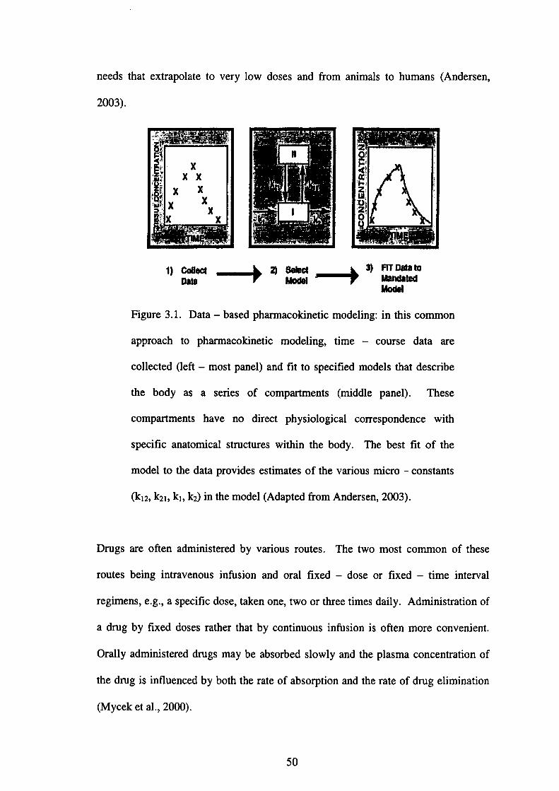
needs that extrapolate to very low doses and from animals to humans(Andersen,
2003).
1)罐→鋤誌→3)設ぽ 一
Figure 3.1. Data-based phamaco垣netic modeling:in血is common
approach to phamlacokinetic modeling, time-course data are
collected(left-most panel)and fit to specified models that describe
the body as a series of compartments(middle panel). These
compartments have no dir㏄t physiological correspondence with
specific anatomical structures within the body. The best fit of the
model to the data provides estimates of the various micro-constants
(k12, k21, k1, k2)in the model(Adapted伽m Andersen,2003).
Drugs are often ad㎡㎡stered by緬us routes. The two most common of these
routes being intravenous infUsion and oral fixed-dose or fixed-time interval
regimens, e.g., a specific dose, taken one, two or three times daily. Administration of
adnlg by fixed doses rather that by continuous in血sion is often more convenient.
Orally administered dnlgs may be absorbed slowly and the plasma concentration of
the drug is influenced by both the rate of absorption and the rate of dnlg elimination
(Mycek et aL,2㎜).
50
3.1 Background introduction
Orgallochlorine(OC)pesticides are ubiquitous in中e env丘onment and in organisms.
These chemicals, still in use in many countries have low volatility and slow rate of
biotransfbrmation, are highly lipid soluble, and resistant to degradation. Despite these
characteristics that make OC pesticides such effective pesticides also brought to their
demise in many countries due to its detrimelltal alld deleterious effects in the
environment, bioconcen仕ation and biomnagnification in various food chains
(Gonzalez-Farias et a1.,2002).
Endosulfan(6,7,8,9,10,10-hexachloro-1,5,5a,6,9,9a-hexahydro-6,9-
methano-2,4,3-benzodioxathiepin-3-oxide), an OC insecticide of the
cyclodiene group, has widespread use in agriculture and forestry to control a wide
variety of insect pests and on non-fbod crops such as cotton and tobacco. It is also
used as wood preservative【Agency fbr Toxic Substallces and Disease Registry
(ATSDR),2000】. Endosulfan is still used in chemical fbmulations although it is no
longer produced in the United States(ATSDR,2000). Endosulfan is unrestricted and
widely used in Turkey(Oktay et al.,2003), Mexico(Castillo et al.,2002)and Brazil
(Dalsenter et aL,1999).]㎞hldia endosulfan is used against a variety of agricultural
pests with about 81,000 metric tons of endosulfan being manufactured during the
1999-2000(Saiyed et aL,2003)and the annual consumption of endosulfan is about
4,200metric tons(Sinha et al.,2001). Endosulfan is severely restricted in countries
such as Japa11, Korea alld Taiwan(EIF,2002). Because of its widespread application,
the accumulation of endosulfan has been reported in various fbod crops(Arrebola et
aL,1999;Antonious et al.,1998;Novak and Ahmad,1989;Gunderson,1995),
environment(Gam6n et al.,2003;Bhattacharya et a1.,2003;Louie and Sin,2003;Tan
51

and Vijayaletchumy,1994)and animal tissues(Gupta,1978)as well as in humans
(Burke et al.,2003;Chan et aL,2004;Yo皿glai et a1.,2002).
Endosulfan is a mixture of two stereoisomers:α一andβ一endosulfan(Hayes and
Laws,1991)in the ratio of 70:30. Undiluted endosulfan is slowly and incompletely
absorbed in the、 digestive tract of warm-blooded animals but absorption is enhallced
in the presence of alcohols, oils and emulsifiers(Maeir-Bode,1968). When give加o
rats by various routes, endosulfan is rapidly metabolized and excreted in the urine and
feces as oxidation products such as endosulfan sulfate, diol, hydroxyether or ketone
resulting from the cleavage of the cyclic sulfite group. The ratio of the different
compounds in the excreta differs with routes of exposure and isomers(FAO/WHO,
1969).The LD500f endosulfan for rats, mice, dogs, guinea pigs and rabbits varies
markedly depending upon the route, strain, vehicle and the sex used(McGregor,
1998;WHO,1984;Gupta 1979).
Studies on the acute and subacute toxicity in rats, mice, rabbits and other species
(Gupta,1979;Gupta and Chandra,1977;Naqvi and Vaishnavi,1993;Singh and
Pandey,1989), its regional distribution in the brain(Gupta,1978)and neurotoxic
effects(Agrawal et al.,1983;Castillo et al.,2002;Dikshith et a1.,1984)in rats have
been documented. Repeated administration of endosulfan increased the liver weight
and produced biochemical effects in the liver(Tyagi et a1.,1984). Reduction ill
spermatid count in testis and speml count in cauda epididymis as well as decrease in
the weights of testis, epididymis and seminal vesicle were recorded when rats were
given repeated administration of endosulfan(Dalsenter et al.,2003;Sinha et al.,2001).
52
Data based on these studies illdicate that the liver ,
main target organs(ATSDR,2000).
kidneys, brain and testes are the
A pharmacokinetic profile for female rabbits(Gupta and Ehrnebo,1979)following
intravenous administration of racemic endosulfan showed pronounced di脆rences
betweel1α一andβ一isomers and these dissimilarities may partly explain reported
di脆rences in toxicity betweell the two isomers. However, it is uncertain that the
differences ih disposition are in the same direction in rats as in rabbits.
3・2 0bjective of the study
The present study was intended to establish the basic toxicokinetic parameters of
endosulfan in male Sprague-Dawley rats fbllowing oral administration of 14C-
Endosulfan at a dose of 5 mg/kg.
3.3 Materials and methods
3.3.1 Chemicals
Radiolabelled endosulfan(2,3-14C-Endosulfan)with a molar activity of 20.88 mCi
/㎜ole;specific activity of 1.895 MBq/mg and a radiochemical purity of>95%by
Radio TLC was obtained廿om the Institute of Isotopes Co., Ltd.(Budapest, Hungary).
All chemicals used in the present study were of reagent grade. Olive oi1,30%
hydrogen peroxide(H202),2-propanol and heparin sodium salt were supplied by
Nacalai Tesque,111c.(Kyoto, Japan). All solvents and chemicals used in the
experiment such as ethano1, diethyl ether, sodium chlgride and phosphate buffered
saline powder were purchased加m Wako Pure Chemical Industries, Ltd.(Osaka,
Japan). The tissue solubilizer(Soluene⑧一350), Hionic-FluorTM and Ultima Gold
53
used fbr liquid scintillating counting were obtained from Packard Bioscience B. V.
(Groningen, The Netherlands).
3.3.2Animals and general conditions
Thirty-seven male Sprague Dawley rats, weighing 92-110 g were bought from
Shimizu Laboratory Supplies(Kyoto, Japan)on post natal day(PND)21 and housed
in three per cage in plastic cages(20 x 25 x 47 cm)containing shredded wood chips
as bedding and roofed with stainless wire covers. Animals were fed pelleted rodent
chow supplied by MS Oriental Yeast(Tokyo, Japan)and waterα4励i’μη2, in a room
with a cycle of 12 hours light,12 hours dark, temperature of 23°C and a relative
humidity of 55%. The animals were allowed to acclimatize to their new surrounding
for one week prior the actual treatment started on PND 28. The use of animals in this
study was in accordance with the Guideline for Animal Experiments of Kyoto
university.
3.3.3Preparation of endosuぬn dosage
P・・chased i4C-E・d・sul飴・was diss・1・・蛆i・ethan・1・t由e c・ncen廿・ti…f10 mg/
mL A 5 mg 14C-Endosulfan/kg body weight dosing solution was prepared by
adding olive oil to the 10 mg/mL stock solution in order to obtain a final
concentration of l mg/mL 14C-Endosulfan.
3.3.4 Rationale for dose selection
The dose was selected based on the LD500f 7-121 mg/kg depending upon species,
sex, formulation tested, vehicle used and nutritional status of the animal established
by WHO(1984). At a dose of more than 7 mg/kg body weight;i.e.7.5 mg/kg
54

body weight administered fbr 60 days(Ansari et al.,1984)and lO mg/kg body
weight administered for 15 days(Gupta,1978;Gupta and Chandra,1977), mortality
was observed. ln order to maintain the number of animals in each group and to detect
minimal residue levels in various organs or tissues which were believed to be low,
administration dose was set at 5 mg/kg body weight.
傷
3.3.5 Experimental design and administration of 14C-Endosuぬn
(1) Single admmistration-Blood time course and distribution of 14C-
End・suぬn刷・wing single・ral administrati・n・f 5 mg 14C-End・suぬn
/ kg body weight
In the single dose experiment, eighteell animals were used. The animals were
arbitrarily divided into six groups(n=3);(i)Group I served as the control and was
given olive oil only;and(ii)Group n-VI were given 14C-Endosulfan once by oral
gavage at a dose of 5 mg/kg body weight and sacrificed at intervals of 30 min,1,2,4
and 8 h fbllowing administration. Each animal was weighed prior to treatment and
the dosage was a(加sted fbr body weight. Treatments were administered by oral
gavage, using an 18-gauge gavage needle(1 inch length, with a 2.25 mm ball)and a
2.5mL glass syringe. The compoulld was administered in O.5 mL per lOO g body
weight.
(2) Excretion analysis following single oral administration of s mg 14C-
Endosulfan / kg body weight
Three animals(n=3)were given 14C-Endosulfan once by oral gavage at a dose of 5
mg/kg body weight. Immediately following dosing, each rat was housed
55
individually in metabolic cages to collect fbr urille and feces. Urine and feces were
collected every 24 hour unti196 h.
(3) Repeated administration-Blood time course and distribution of 14C-
Endosulfan following three-time repeated oral administration of 5 mg
i4C 一 Endosuifan / kg body weight
hl the repeated dose experiment, thirteen animals were used. The animals were
randomly assignOd to five groups with three animals in each group(n=3)except for
Group I, where only one animal(n=1)was used.(i)Group I was given 14C-
Endosulfan once by oral gavage at a dose of s mg/kg body weight and sacrificed 2
hour following administration;(ii)Group ll was given 14C-Endosulfan pnce and
sacrificed 3 hour following administration;(iii)Group M was given i4C 一 Endosulfan
twice at an illterval of 3 hour per dose and sacrificed 2 hour fbllowing the last
administration;(iv)Group rV was given 14C-Endosulfan thrice at an interval of 3
hour per dose and sacrificed 2 hour following the last administration;and(v)Group V
was given 14C-Endosulfan thrice at an interval of 3 hour per dose and sacrificed 25
hour following the last administration.
3.3.6 Necropsy
Rats were lightly anesthetized with diethyl ether prior necropsy. At necropsy, tissue
samples such as liver, kidneys, fat, gastrointestinal(GD tract consists of large
intestine, small intestine, stomach and cecum, muscle, brain, heart, lung, spleen, testis
and thyroid gland were removed and blood was collected at the vena cava
immediately at each interval(30 min,1,2,4and 8 h)for radioactivity analysis for the
single dose experiment whereas blood was collected at the intervals of 2 h,3h,5h,8
56
hand 25 h fbr the three-time repeated dose experiment. The remaining blood
samples were collected in a 5 mL test tube and centrifUged at 3000 rpm for lO
minutes to obtain serum. The serum supernatant was aspirated out and stored in new
test tubes at-80°C until f血rther analysis. The contents of the GI tract were separated
from each of its tissues for the single dose experiment.
3.3.7 Measurement of radioactivity
㎞mediately upon necropsy tissue samples were minced, aliquots of blood, senlm,
feces and contents丘om the GI tract were dissolved in Soluene⑪一350. The
solubilized blood, feces and contents samples廿om the GI tract were mixed with
isopropyl alcohol(1/1v!v)and decolorized with 30%(v/v)H202 befbre mixing
with Hionic-FluorTM where as the solubilized tissues samples were mixed with
Hi・面・-H…M dire・tly・nd・・unt・d f…adi・acti・ity t・d。t。血。。血, t。t。l
concelltration with liquid scintillation co皿ter(LSC 6100, Aloka, Tokyo, Japan).
Aliquots of u血e were mixed with Ultima Gold directly and counted fbr radioactivity.
The quenching correction was made automatically by extemal standardization.
Radioactivity was represented as weight equivalent to Elldosulfan{ex. mg Elldosulfan
(ES)eq.l based on dle specific activity of 14C-Endosulfan.
3.3.8Toxicokinetic parameters
Toxicokinetic parameters were determined from the illdividual blood 14C -
Endosulfan-derived radioactivity concentration-time curve from the same dose(5
mg/kg)of orally administered 14C-Endosulfan. The area under the blood 14C-
Endosulfan-derived radioactivity concentration-time curve(AUC(o-8h))was
calculated by the linear trapezoidal method.
57
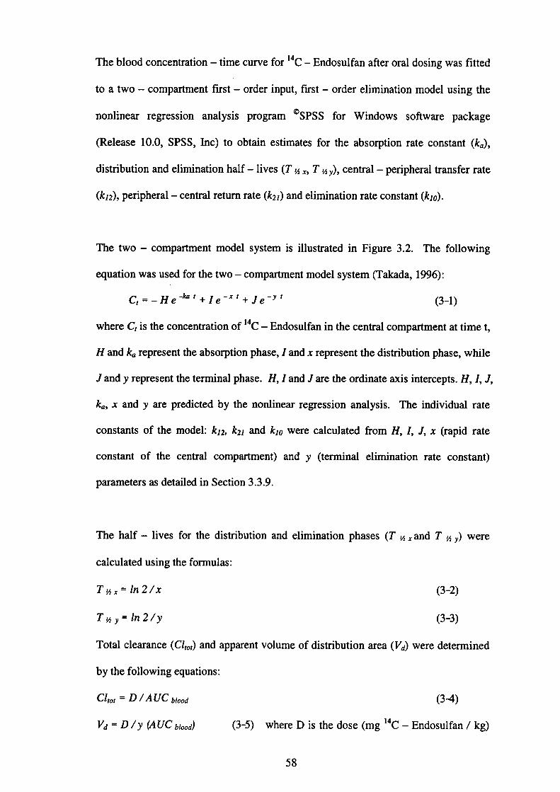
The blood concentration-time curve for i4C-Endosulfan after oral dosing was fitted
to a two -compartment first-order input, first-order elimination model using the
nonlinear regression analysis program◎SPSS fbr Windows software package
(Release l O.0, SPSS, Inc)to obtain estimates fbr the absorption rate constant(ka),
distribution and elinlination half-lives(T・h x, T’h y), central-peripheral transfer rate
(k12), peripheral-central return rate(k21)and elimination rate constant(kio).
The two-compartment model system is illustrated in Figure 3.2. The fbllowing
equation was used for the two 一 compartment model system(Takada,1996):
C,=-He -kOt+1e-xt+Je-yt (3-1)
where C}is the concentration of i 4C-Endosulfan in the central compartment at time t,
H and ka represent the absorption phase,1 and x represent the distribution phase, while
Jand y represent the ternlinal phase. H,1and J are the ordinate axis intercepts. H,1, J,
ka, x and y are predicted by the nonlinear regression analysis. The individual rate
constants of the model:k12, k21 and kio were calculated from H,1,」, x(rapid rate
constant of the central compartment)and y(terminal elimination rate constant)
parameters as detailed in Section 3.3.9.
The half-1ives fbr the distribution and elimination phases(T,h . and T,h y)were
calculated using the fbrmulas:
T%hr = ln 2/x (3-2)
T・hy=ln・2/y (3-3)
Total clearance(Clt。t)and apparent volume of distribution area(Vd)were determined
by the fbllowing equations:
Cltot=D/AUC b,ood (3-4)
Vd=D/y(A UC bi。。d) (3-5) where D is the dose(mg 14C-Endosul fan/kg)
58

DlIl▼
●X
Absorption’GUtCompartment
Pe巾heralCompartment V
Central
Compartment
Xt Vt
Xu
Ellmlna目onCompartment
D:Dose of the chemical(mg)
Xa:Amount of chemical absorbed in the gut(mg)
F:Oral bioavailability(%)
ka:Absorption rate constant(hゴ1)
k12:Central-peripheral transfer rate(hr-1)
k21:’Peripheral 一 central return rate(hゴ1)
kio:Elimination rate constant(hr-1)
X1:Amount of chemical in the central compartment(mg)
X2:Amount of chemical in the peripheral compartment(mg)
Xu:Amo皿t of chemical eliminated(mg)
Vl:Volume of distribution in the central compartment(L)
V2:Volume of distribution in the peripheral compartment(L)
C:Concentration of chemical in the blood(mg/L)
CLtot:Total clearance(mL/min)
Figure 3.2. Two-compartment model system.
59
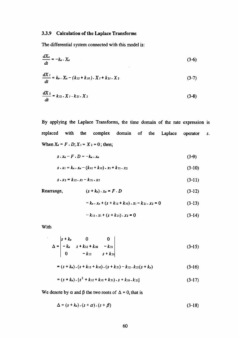
3.3.9 Calculation of the Laplace Transforms
The differential system connected with this model is:
t2[Xa=_輪.Xa
dt
dX i
=島・Xa-↓kn+kiの・Xi+k21・X2dt
aC( 2
= kn.Xl .. k21.X2
dt
(36)
(3-7)
(3-8)
By appl〕⑰g the Laplace Transfbrms, the time domain of the rate expression is
replaced with the complex domain of the Laplace operator ∫.
When X』=F・D;X1=X2=0;then;
3・x・-F・D=-ktr・Xa (3-9)
∫・x1=島・k-(kn+k1・)・Xl+k21・x2 (3-10)
s・x2=k12・x一k21・x2 (3-11)
Reaπallge, (s+輪).k=F.D (3-12)
-k,,・k+(s+k12+ki・)・x】-k21・x2・=O (3-13)
-k12.Xl+(s+k21).x2=0 (3-14)
with
∫+ん 0 0
△=-ka∫+k12+ki・一北21 (3-15) 0 -」ヒ12 ∫十」ヒ21
=(s+k,,).(ぷ+km+kiO).(s+k21)-k】2.k21(s+kロ) (3-16)
=(s+kρ).{s2+kn+kiO+k21).∫+k】o.k2i} (3-17)
We denote byαandβthe two roots of△=0, that is
△=(s+ki,)・(ぷ+α)・(s+β) (3-18)
60
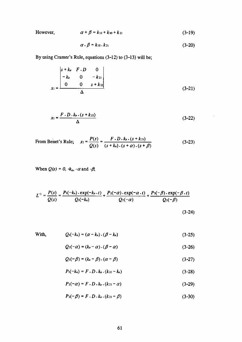
However, α+β=丸2+kio+k21 (3-19)
α・fi = ki・・k21 (3-20)
By using Cramer’s Rule, equations(3-12)to(3-13)will be;
∫+島 F.D O
-ktr O -k21
0 0 s+k21 Xl= △ (3-21)
…F’D’芸(5+k21) (3-22)
F,。mB。h。,・、R。1。;。1・P(s). F・D・k,,・(s+k21) (3-23) o(s) (∫十彪)・(∫十α)・(ぷ十fi)
When e(s)=0, rka,一αand -fi;
乙一已P(・).P1(-la)・・xp(-L・’).P・(一α)・exp(一α・t).P・(一β)・exp(一β・’)
(2(s) (21(-L) (~2(一α) 03(一β)
(3-24)
With, (1】(-k,,)=(α一島).(β一鳥) (3-25)
ρ2(一α)=(輪一α)・(β一α) (3-26)
Q3(一β)=(彪一β).(α一β) (3-27)
P1(-ka)=F.D.kct.(k 21一彪) (3-28)
P2(一α)=F.D.島.(k21一α) (3-29)
P3(一β)=F.D.輪.(k21一β) (3-30)
61
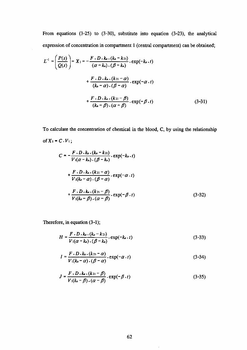
From equations(3-25)to(3-30), substitute into equation(3 -23), the analytical
expression of concentration in compartment 1(central compartment)can be obtained;
ピ慣〕・X・・一;蹴≡鴛)・ex⑭
・鴛ii器)・・xp(一α・t)
・篇ii;鵠)・exp(一β・t)(3-31)
To calculate the concentration of chemical in the blood, C, by using the relationship
ofX1=C・γ1;
F・D・k,,・(ka 一 k 2i)
C=一 ・exp(-1α・t) γ1(α一k,,)・(β一輪)
F・D・輪・(」ヒ21一α)十
Vl(k,,一α)・(β一α)
F・D・彪・(k21一β)十
・exp(一α・t)
・exp(一β・t)Vi(ka一β)・(α一β)
(3-32)
Therefore, in equation(3-1);
F・D・た6・(k,,-k21) H= ・exp(-k,,・t)
レ71(α一k,,)・(β一輪)
F・D・島・(k21一α)1= ・exp(一α・t) Vi(輪一α)・(β一α)
F・D・彪・(k21一β) ・exp(一β・t)J= γ1(la一β)・(α一β)
(3-33)
(3-34)
(3・・35)
62

By integrating equation(3-1);
H I J コ 輪αβ
F.D.瓦21 Vi・α・β
From equation(3-19);
F.D.k21 ∫ Cゴr= γ1.kio
By rearranging the form of equation(3-38);
Vl.kiO D Fr, CdI
By substituting equation(3-37);
γ1.kiO D
F HIJ の 彪αβ
?{/〔 k21 一 ka(α一輪)・(β一此o)〕
・÷Fl+i〔 k21一α(彪一α)・(β一α)〕
R÷当(k21一β輪一β)・(α一β)〕
F.Dand =ρ・(k,,一α)十R・(L一β)
γ1
63
r, Cd・・-H・r。 exp(一・a・t)dt…:・xp(一α・t)・」・〔exp(-fi・t)
.lf Cdt =
(3-36)
(3-37)
(3-38)
(3-39)
旬40
)13(
(3-42)
(3-43)
(3-44)
(3・45)
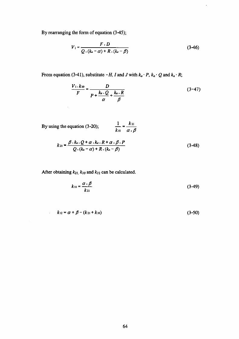
By rearranging the form of equation(3-45);
F.D Vi= 2・(k。 一α)+R・(し一β)
From equation(341), substitute-H,1 and J with k.・P, ka・e and k.・R;
Vi.kiO D
F p.ktr・ρ.ka・R
α β
. 1 k21By using the equation(3 -20); 一= kiO α・β
β・k,,・e+α・k,,・R+α・β・P k21 =
e・(k,,一α)+R・(ktz一β)
After obtaining k21, kio and k12 can be calculated.
α・β kiO =
k21
k12=α+β一(k21+kiO)
(3-46)
(3-47)
(348)
旬40
)03(
64
3.4 Results
3.4.1 General condition of animals
None of the rats showed any sign of toxicity through the period of observation
following single and three-time repeated oral administration of 5 mg/kg 14C-
Endosulfan.
3.4.2Disp・siti・n・f 14C-End・sulfan(5 mg/kg):・Excreti・n r・utes
Figure 3.3 show the cumulative percentage of urinary and fecal elimination after
single oral administration of 5 mg!kgハ4C-Endosulfan. Following single oral
administration of 5 mg/kg 14C-Endosulfan to male rats, the urinary excretion of the
radioactivity during 24 h was 9.6±5.0%of the total administered dose. The
cumulative excretion in the urine fbr fbur days was 12.4±4.8%. Fecal elimillation
・f14C-End・sulf・n-d・・i・ed・radi・acti・ity・rep・esent・d・the・m・j・・eliminati・n・・ute・i・
male rats. The fecal excretion of the radioactivity during four days was 94.4±21.4%.
The total radioactivity recovered in the excreta fbr fbur days was 106.8±26.2%.
Over four days, rats excreted more radioactivity in feces than in urine. Generally the
standard deviation(SD)for urinary elimination was below 10%for each time point,
which was considered excellent. The SD for fecal elimination at each time point was
above 20%;however it was consistent at each time point. The total elimination of
endosulfan recovered in the excreta, which exceeded 100%could be attributed to
experimental error.
3.4.3 14C-Endosulfan residues in tissues and contentS of the GI tract
Table 3.1 shows the residue levels of 14C-Endosulfan in various tissues of rats after
single oral administration of 5 mg/kg 14C-Endosulfan. The relative amo皿ts of
65
radioactivity found 8 h after administration were in the following sequence(mg ES eq.
/L):GI Tract(20.28)>liver(5.52)>fat(3.61)>thyroid gland(2.50)>kidneys
(1.83)>lung(1.31)>serum(0.75)>heart(0.42)>spleen(0.37)>blood(0.28)>
brain(0.27)>testes(0.25)>muscle(0.18).
The relative amounts of radioactivity found 8 h after administration for the contents of
each tissue from the GI tract were(mg ES eq./L):Cecum(25.84)>large intestine
(6.76)>small intestine(6.45)>stomach(3.57).
Table 3.2 shows the residue levels of 14C-Endosulfan in various tissues of rats after
three-time repeated ora l administration of 5 mg/kg 14C-Endosulfan. It was
observed that the radioactivity levels in all tissues on the 25th hour decreased after
administration was teminated.
3.4.4 14C-Endosm田fan toXicokinetics in blood
The log concentration-.time curve of i4C-Endosulfan-derived blood radioactivity
c・nce・廿ati・n・ve・8h・・al d・se at 5 mg 14C-E・d・su1飴・/kg is sh・w・i・Figure 3.4.
Toxicoldnetic parameters estimated伽m the blood-time course data fbr oral
ad㎡㎡stration are summarized in Table 3.3. Following single oral ad㎡㎡s仕ation of 5
mg 14C-E・d・・uぬ・/kg t・・at・,也e 14C-E・d・・uぬ・w…apidlダab…b・d t㎞・・gh
GI tract with absorption rate constant(ka)of 3.07 h’1 and peak blood concentration
(Cmex)of O.36±0.08 mg-eq./Lwith time to q”似(Tnrax)of 2 h. The terminal
elimination half life(τ%y)of the radioactivity in blood was 193 h.二
66
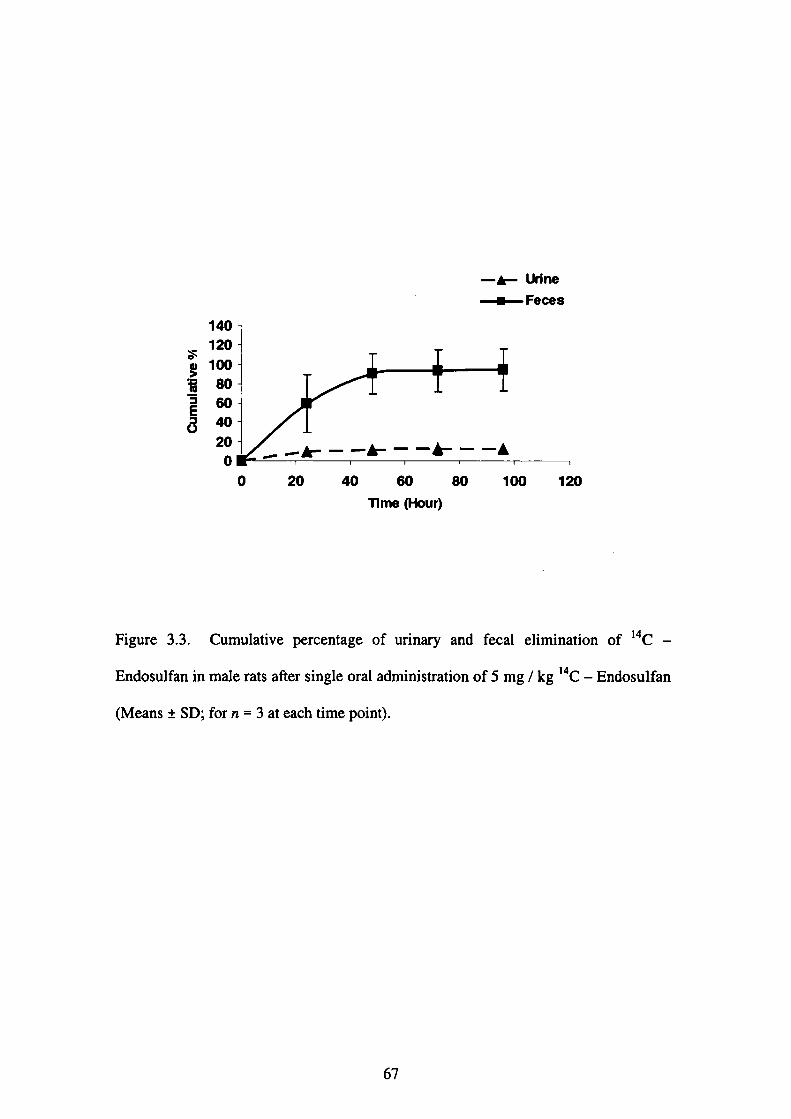
一▲-urine
十Feces140
120
100
80
60
40
20 0
一▲一一一▲一一一▲一一一▲
0 20 40 60 80 100 120
1ime (Hour)
Figure 3.3. Cumulative percentage of urinary and fecal elimination of 14C-
Endosulfan in male rats after single oral administration of 5 mg/kg 14C-Endosulfan
(Means±SD;for n = 3 at each time point).
67
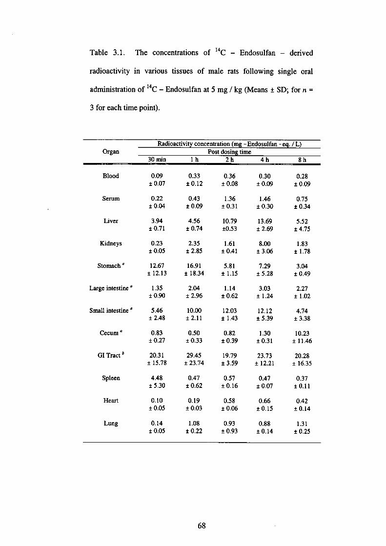
Table 3.1. The concentrations of 14C - Endosulfan - derived
radioactivity in various tissues of male rats fbllowillg single oral
administration of i4C-Endosulfan at 5 mg/kg(Means±SD;for n=
3fbr each time point).
Radioactivit concentration(m -Endosulfan 一一 .!L)
Organ Post dosin time
30 min lh 2h 4h 8h
Blood
Serum
Liver
Kidneys
Stomach a
Large intestine “
Small intestine a
Cecum a
GI Tract b
Spleen
Heart
Lung
㌶品鑑㌶認㌫蕊賜㌫場…迦鑑 3123α
㌶場鑑場㌫㎜剖鴉場温温㎜㎜
8603α
163301 ±
0/37’く∨001十一
1
血似1 ±
5118
・
・1
2461
◆
・0
1 ±
3304(∠11±
923潟0.
9975931±
67150
8065α
3399α0 十一
㌘翼盟器識欝罵品価還 顕賜嶽鑑温鑑㌫㍑鵠㎝㎜㎝⊇晶
68

Continue.....
OrganRadioactivity concentration(mg-Endosulfan-eq./L)
Post dosin time
30 min lh 2h 4h 8h Brain O.11 0.14
±0.05 ±0.01
Thyroid gland 6.50 0,30
±10.60 ±0.07
Muscle O.04 0.05
士0.02 ±0.01
Testes O.03 0.06
土0.01 ±0.02
Fat 2.17 1.43
±1.36 ±0.42
Contents of each tissue from GI tract
Stomach 6.95 25.71
±2.01 ±20.06
Large intestine 4.71 3.99
±2.61 ±5.37
Small intestine 3.85 3.03
±2.01 ±3.73
C㏄um O.02 0.03 ±0.02 ±0.04
鑑鍛品㍑鑑恕㍊盟㌫
37V5
Q8ヘ55幻781
豊鑑鑑漂
況漂卯託況㍗
74868153384胴蓋叉鑑鑑
∧Tote.
a:The radioactivities reported fbr the stomach, large intestine, small intestine and
cecum were counted without contents.
b:Sum of radioactivities in the stomach, large intestine, small intestine and cecum
without contents.
69

Table 3.2. The concentrations of 14C - Endosulfan - derived
radioactivity in various tissues of male rats fbllowillg three-time
repeated oral administration of i4C 一 Endosulfan at 5 mg!kg(Means
±SD;for n =3for each time point except for the frrst time point【2 h】
where n = 1).
Organ
2h 3h 5h 8h 25h
Blood
Serum
Liver
Kidney
Brain
Thyroid gland
Muscle
Testes
Fat
0.22
1.08
6.07
2.16
0.30
0.39
0.10
0.57
1.31
214
況ぷ㍗㌶㍑㎝託㎝口況ぷ 5071
.
・0
1 ±
50260
9710/401士
373フα
3602α1 土
377
・1
2602α0 十一
7371
■
・0
1 十一
86200 ・
・(∠
4 十一
490
18740
1889&21士
372⑨231±
882501 ±
1848σ1 ±
22040
6胴㎝
8ξ」8
フ0
塩蛋㍑霊㌫温撒塩鑑
70
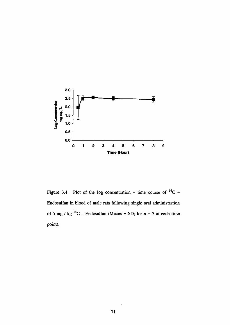
0505050a22tWαα 」、.90E
0 1 2 3 4 5 6
Ti me (Hour)
7 8 9
Figure 3.4. Plot of the log concentration-time course of 14C-
Endosulfan in blood of male rats fbllowing single oral administration
of 5 mg/kg i4C 一 Endosulfan(Means±SD;for n=3at each time
point).
71
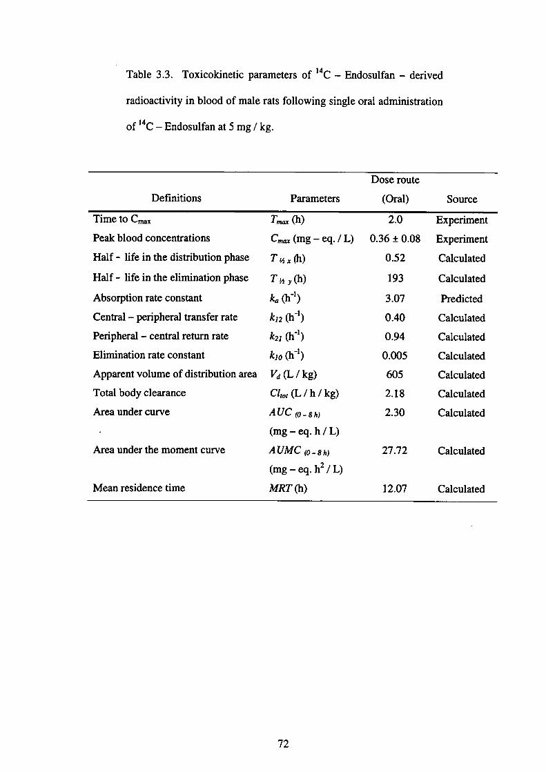
Table 3.3. Toxicokinetic parameters of 14C-Endosulfan-derived
radioactivity in blood of male rats following single oral administration
of 14C-Endosulfan at 5 mg/kg.
Definitions Parameters
Dose route
(Oral) Source
Time to Cmax
Peak blood collcelltratiolls
Half-life in the distribution phase
Half-life in the elimination phase
Absoηption rate constant
Central-peripheral transfer rate
Peripheral-central returll rate
Elimination rate constant
Apparellt volume of distribution area
Total body clearance
Aエea under curve
Area under the moment curve
Mean residence time
T,。ax(h)
Cmex(mg-eq./L)
τ撫(h)
T,hy(h)
kα(h-1)
k12(h-1)
k21(h-1)
晦o(h-1)
Vd(L/kg)
α,α(L/h/kg)
A UC (o_8力戊
(mg-eq. h/L)
AUMC (o_8ん戊
(mg-eq. h2/L)
MRT(h)
2.0
0.36±0.08
0.52
193
3.07
0.40
0.94
0.005
605
2.18
2.30
27.72
12.07
Experiment
Experiment
Calculated
Calculated
Predicted
Calculated
Calculated
Calculated
Calculated
Calculated
Calculated
Calculated
Calculated
72
3.5 Discussion
Following oral administration of 2 mg/kg of i4C ・一 Endosulfan to male Wistar rats,
90%of the oral dose was eliminated in the urille and feces within seven days;
elimination was essentially complete within the l-2days(McGregor,1998). Female
rats treated with a single dose(2 mg/kg)ofα一〇rβ一【14C】-Endosulfan eliminated
88%of theα一endosulfan and 87%of theβ一endosulfan within 5 days of oral
administration (Dorough et al., 1978). They also suggested that the biliary
metabolites of endosulfan would not enter the enterohepatic cycle and hence were
voided in the feces. ln metabolic studies using BALB/c mice fed with single dose of
O.3mg 14C-Endosulfan, approximately 65%of 14C-Endosulfan was recovered in
the excreta and tissues of mice 24 h later after ingesting endosulfan in their diet
(Deema et al.,1966). These observations were consistent with our results that most of
the radiocarbons were excreted in the urine(12.4±4.8%)and feces(94.4±21.4%)
after 96 h. Elimination was essentially complete after 48 h(101.6±25.1%). Our
data also suggested that endosulfan would not enter enterohepatic recirculation as
shown by the radioactivity concentrations in the contents of each tissue from the GI
tract(Table 3.1). The present study of disposition of endosulfan in rats showed that
the principal excretion route was the feces in rats.
The tissue concentrations of residues after 8 h as observed in the present study were
generally highest in the liver and kidneys and lower in other tissues such as brain,
testes and muscle;thus suggesting that these tissues do not appear to bioaccumulate.
High radioactivity which was fbund in the liver may indicate that liver is the site of
high metabolic activity(Khanna et al.,1979).
73

The distribution pattem ofα一andβ一 endosulfan in rats after administration of a
mixture varies depending on the tissues(Ansari et al.,1984;Nath et a1.,1978). The
regional differences in the quantity of fat content may also partly account for the
differences in the distribution of endosulfan in various organs(Ansari et al.,1984).
Ansari and co-workers(1984)indicated a differential ability to accumulate the two
isomers of endosulfan which may help to explain the difference in the toxic potential
ofα一andβ一isomers. Accumulation of endosulfan was greater in kidney than liver
as kidney is one of the target organs for toxicity(FAO/WHO,1999). A rapid
biodegradation in the liver may be one of the causes of low concentration in this
organ since it is the main site of detoxification, however the possibility of other
factors cannot be ruled out as suggested by Ansari and co-workers(1984).
The tissue concentration of residues in all tissues on the 25th hour decreased after the
administration was temlinated as observed in the present study. This suggests that
endosulfan does not appear to bioacのmulate and rats will recover once
administration is teminated. When technical endosulfan(mixture of a-andβ一
isomers)was fed orally to male rats at 5 mg/kg fbr l 5 days, maximum residues of
endosulfan, which occurred in the kidney and liver, were l.46μg/gand O.09 pg/g
respectively while only O.05μg/g was detected in the liver 15 days after the last
administration(Chan and Mustafa,2004). Similar distribution pattern was also
observed when technical endosulfan was administered orally to male rats at 10 mg/
kg fbr 15 days.2.26μg/g and O.09 pg/g of endosulfan residues were detected in the
kidney and liver respectively, whereas only O.06μg/g was detected in the liver 15
days after the last administration(Chan and Mustafa,2004). Deema and co-workers
(1966)observed that accumulation in liver(2883 cpm)was higher than lddney(1390
74
cpm)when BALB/c mice were fed with a single dose of【14q- Endosulfan after 24
h.Dorough and co-workers(1978)reported that tissue concentrations of residues
were generally highest in the kidneys(3 ppm)and liver(l ppm)and lower in other
tissues(<1ppm)when rats were fed endosulfan in the diet at a concentration of 5
ppm fbr l4 days. At the end of the l4-day recovery period, residues were confined
to the kidneys(0.9 ppm)and to the lesser extent, the liver(0.1ppm).
The log-concentration-time curve(Figure 3.4)for blood endosulfan after oral
administration demonstrates kinetics which can be described by a two-compartment
model. It consists of a central compartment and a peripheral compartment.
Elimination was allocated into the central compartment. All processes were assumed
to underline frst 一 order kinetics. The model delivers an excellent fit to the data with
・g・・dness・f・fi・(R・)value 6fo.999(Fig。,e 3.4).
The blood concentration reached its maximum(C”蹴)of O.36±0.08 mg-eq/Lat 2 h
(T,nax)a丘er oral dose(Table 3.1). Our observation was comparable when male Wistar
rats were administered 2 mg/kg 14C-Endosulfan orally, the blood concentration
reached its Cnzax of O.25±0.06 mg/Lat 7》顧of 3-8h(McGregor,1998). The
concentratlon-tlme curve ls approxlmated by two exponelltials:the first term with a
half-life(τ%x)of 31 min describes the decline after reaching the maximum
collcentration. Then, the slope flattens over time approaching a terminal phase with a
half-lifb(T%y)of 193 h. Both phases are governed by the distribution together with
the elimination process. However, McGregor(1998)reported that the half-life in
the distribution phase(T%ズ)for male Wistar rats was 8 h and the terminal half-1i允
(Tjhy)was 110h.
75

The present model)delds half-life of 31 min fbr the uptake into the central
compartment and an elimination rate constant of O.3 min from this compartment. The
obtained results also show that the absorption of endosulfan into the GI tract in rats is
rapid with a kαof 3.07 h-1. The absorption of endosulfan in the present study was
estimated to be 46%on a basis of a comparison of area under the curve(A U(ハafter
oral administration of 14C-Endosulfan as compared to 60-70%in male Wistar rats
(McGregor,1998). The rapid distribution of endosulfan to lipid tissues may be
responsible fbr its slow elimination half-life(Abdel-Rahman et al.,2002).
Intravenous administration of 2 mg/kg endosulfan(7:3 ratio ofα一andβ一isomers)
in rabbits produced much longer half-1ife of the terminal slope of theα一isomer
(235h)than fbrβ一endosul㎞(5.97 h)(Gupta and Ehrnebo,1979). Excretion of the
two isomers occurred primarily via the urine(29%)with much less excreted via the
允ces(2 %). It had b㏄n shown that marked diffbrences occurred in the
phamlacokinetics, metabolism and excretion betweenα一andβ一isomers in rabbits
(Gupta and Eimebo,1979). However, it was unclear whe也er the differences in
disposition were similar between rats and rabbits as中e level of radiocarbon was
insufficient to attempt identification of separate isomers in the present study.
The variations in the excretion and absorption pattems may be attributable to the
differences in exposure routes, species differences or to both. In a draft presented by
McGregor(1998), the percentage of elimination of endosulfan in the excreta,
absorption efficiency alld accumulation in various tissues differ with route of
exposure, sex, strain and species(McGregor,1998).
76

3.6 Conclusions
The present study indicates that 14C-Endosulfan was rapidly excreted in the feces
and urine after 96 h following single oral administration with fecal elimination as the
major elimination route in male rats. Elimination was essentially complete within a
few days. The present study also indicates that differences in sex, strain, route of
exposure may influence the pharmacokinetic parameters fbr endosulfan when
compared to other studies. Funher study is necessary to investigate the toxicokinetics
and biotransformation of 14C-Endosulfan in rats after oral and intravenous
administration. Amore reliable and sensitive analytical method would also be
lncorporated into our fUture experiments to enhance the separation and identification
of the parent compounds and metabolites present in different matrices.
Note.
Some sections in this chapter will be published and available in Environmental
Toxicology, Volume 20, Issue 5;October 2005.
REFERENCES
Abdel-Rahman, A. A., Blumenthal, G. M., Abou-Donia, S. A., Ali, F. A. F., Abdel
-Monem, A. E, Abou-Donia, A. B.(2002). Phamlacokinetic profile and placental
transfer of a single intravenous i可ection of[14q Chlorpyrifbs in pregnant rats.
ノlrchiveぷ〔ゾTb」κicolo8y,76:452-459.
Agrawal, A. K., Anand, M., Zaidi, N. F., Seth, P. K.(1983). i lvolvement of
serotonerglc receptors ln endosulfan neurotoxicity. Biochemicat Pharmacology,32:
3591-3593.
77

Andersen, M. E.(2003). Toxicokinetic modeling and its application in chemical risk
assessment. Toxicolo8y Letterぷ,138:9-27.
Ansari, R. A., Siddiqui, M. KJ., Gupta, P. K.(1984). Toxicity of endosulfan:
Distribution ofα一and β一isomers of racemic endosulfan fbllowillg oral
adnlinistration in rats. Toxicolo8y Lぴ’εr∫,21:29-33.
Antonious, G. F., Byers, M. E., Snyder,」. C.(1998). Residues and fate of endosulfan
on field-grown pepper and tomato. Pεぷticide Science,54:61-67.
Aπebola, F.」., Mar血ez Vidal,」. L, Fern4ndez-Guti6πez, A.(1999). Excretion of
endosulfan in urine of a pest control operator. Toxicology Letters,107:15-20.
ATSDR・2000・Toxicotogical proLfile/br endosutfan. U. S. Department of Health
and Human Services, Public Health Service, Agellcy fbr Toxic Substances and
Disease Registry, Atlanta, GA.
Bhattacharya, B., Sarkar, S. K., Mukhell ee, N.(2003). Organochlorine pesticide
residues in sediments of a tropical mangrove estuary, India:㎞plications fbr
monitoring. Environment lnternationa’,29:587-592.
Burke, E. R., Holden, AJ., Shaw,1. C.(2003). A method to determine residue levels
of persistent organochlorine pesticides in human mil】(from Illdonesian women.
Chemosphere,50:529-535.
’ 78
C・・til1・・C・G・・M・nt・・t・・M・,・D・f・u・, L, M・・tinez,・M. L, Jim6nez-C・pdevi11・, M.
E・(2002)・B・h・vi・・al・ffe・t・・f・xp・・ur・t・・nd・・ulf・n・and・m・thyl・par・thi・n in ad。lt
rats・Neurotoxicolo8y and Teratolo8y,24:797-804.
Chan・M・P・L・・M・・t・fa・A・M・, Hussei・, R., Hj・Hussai・, S., Z・1kifii,・S.・N., Abd。11、h,
A・R・(2004)・P・・ti・id・・e・idue・i・bl・・d・f・ch・・1・hild・en・fr・m・elect・d・ch・・1・i・
Peninsular Malaysia. Environmental Health Focus,2(1):18-24.
Chan, M. P. L, Mustafa, A. M.(2005). Analysis of endosulfan and its metabolites in
「at p1・・m・孤d・elect・d tissue・ampl・・by g・・一・祉・m・t・9・aphy-mass spec仕・m・t・,.
Eηvかon〃lenta’Toxicolo8y,20(1):45-52.
Dalsent…P・R・Dal1・9r・ve・E・M・11・」・R・B・, la・g・1・h, A., Olive丘・, R. T., F・qi, A.
S・(1999)・R・p・・d・・tive e輪t・・f・nd・・ulfa・・n m・1・・脆pri・g・・rat・exp・・ed
d岨・gP・eg・・n・y and l・・伍ti・n・肋m娠・4鋤・・im・nt・1撤・・1・8y,18・583-589.
Deema, P., Thompson, E, Ware, G. W.(1966). Metabolism, storage and excretion of
C14-E・d・・uぬ・i・・h・m・u・e. J・u・nal・・fE・・’・・Mi・E。t。m。1。8y,59(3),546-550.
Dik・hith・T・S・S・・R・izad・, R・B・, Sriva・tava, M. K., K・ph・1i・, B. S.(1984).
Response of rats to repeated oral administration of endosulfan.1ndustrial Health,22:
295-304.
79
Dorough, H. W., Huhtanen, K., Marshall, T. C., Bryant, H. E.(1978). Fate of
endosulfan in rats and toxicological considerations of apolar metabolites. Pesticide
Biochemistry and Physiolo8y,8:241-252.
EJF・(2002)・End of the roadプbr endosulfan:oco〃プbr action against a dan8erous
pesticide. Environmental Justice Foundation Ltd, Lρndon, UK, pp.1-4.
FAO/WHO(1969)FAO/PL:1968/M/9/1.1968 Evaluations ofso〃ze pesticide
residues in/bo4. Food and Agricultural Organization of the United Nations, World
Healt110rganization, Geneva.
FAO!WHO.(1999). Pesticide residues in/bo4-1998 evaluations. Part 11-
Toxicolo8ical. World Heald1 Organization, Geneva.
Gam6n, M., S4ez, E., Gi1,」., Boluda, R.(2003). D廿ect and ind口ect exogenous
contamination by pesticides of rice-farming soils in a Mediteπanean wetland.
Archives ofEnvironmental Conta〃証ηα’ion and Toxicology,44:141-151.
Gibaldi, M., Perrier, D.(1982). Phamlacokinetics,2nd ed., Marcel Dekker, hlc., New
York, NY.
Gonzalez-Farias, F., Cisneros, E. X., Fuentes, R. C., Diaz, G. G, Botello, A. V.
(2002).Pesticides disnibution in sediments of a tropical coastal lagoon a(ljacent to an
irrigation district in northwest Mexico. Environmental Technology,23(11):1247-
1256.
80
Gunderson, E. L(1995). FDA Total Diet Study, July 1986-April l 991, dietary
intakes of pesticides, selected elements, and other chemicals. Journal of 40A C
International,78:1353-1363.
Gupta, P. K.(1978). Distribution of endosulfan in plasma and brain after repeated
oral administration to rats. Toxicolo8y,9:371-377.
Gupta, P. K.(1979). Phamlacology, toxicology and degradation of endosulfan. A
review. T・xi・・1・8y,13:115-130.
Gupta, P. K., Chandra, C. V.(1977). Toxicity of endosulfan after repeated oral
administration to rats・Bulletin of Environmental Contamination and Toxicology,18:
378-384.
Gupta, P. K., Ehnebo, M.(1979). Pharmacokinetics of ct-andβ一isomers of
racemic endosulfan following intravenous administration in rabbits. Drug
Metabolism and Disposition,7(1):7-10.
Hayes, W. J., Laws, E. R.(1991). Handbook of pesticide toxicology. Academic
Press, San Diego.
Hayton, W. L.(1989). Pharmacokinetic parameters for interspecies scaling using
allometric techniques. Health Physics,57(Supplement 1):159-164.
81
Hoang, K. C. T.(1995). Physiologically based phamacokinetic models:
Mathematical fundamentals and simulation implementations. Toxicolo8y Letters,79:
99-106.
Kha㎝a, R. N., Misra,
endosulfan in cat brain.
22:72-79.
D.,Anand, M., Shamla, H. K.(1979). Distribution of
Bulletin of Environmental Contamination and Toxicolo8y,
Louie, P. K. K., Sin, D. W.(2003). A preliminary investigation of persistent organic
pollutants in ambient air in Hong Kong. Chemosphere,52:1397-1403.
Maier 一一 Bode, H.(1968). Properties, effect, residues and analytics of the insecticide
endosulfan. Residue Reviews,22:1-44.
McGregor, D. B.(1998). Endosulfan (JMPR evaluations 1998 Part II Toxicotogical♪
-First draLfi. lnternational Agency for Research on Cancer, Lyon, France.
Mycek, M. J., Harvey, R. A., Champe, P.(2000). Chapter 2:Pharmacokinetics and
dnlg r㏄eptors. h1:LipPincott ’s lllustrated Reviews:Pharmacolo8y, LipPincott
Williams&Wilkins, Philadelphia, pp.17-26.
Naqvi, S. M., Vaishnavi, C.(1993). Mini review:Bioaccumulative potential and
toxicity of endosulfall insecticide to non-target animals. Comρarative Biochemistiッ
and Phyぷiolo8y,105C(3):347-361.
82
Nath, G., Datta, K. K., Dikshith, T. S. S., Tandon, S. K., Pandya, K. P.(1978).
lnteraction of endosulfan and metapa in rats. Toxicolo8y,11:385 一 393.
Novak, B., Ahmad, N.(1989). Residues in fish exposed to sublethal doses of
endosulfan alld fish collected from cotton growing area. Journal of Environmentat
Science and Heatth, Part B B24:97-109.
Oktay, C., Goksu, E, Bozden血, N., Soyullcu, S.(2003). Unintentional toxicity due
to endosulfan:acase report of two patients and characteristics of endosulfan toxicity.
レ「eterinary and Hu〃tan Toxicolo8y,45(6):318-320.
Saiyed, H., Dewan, A., Bhatllagar, V., Shenoy, U., Shenoy, R., Rajmohan, H., Pate1,
K.,Kashyap, R., Kulkami, P., Raj an, B., Lakkad, B.(2003). Effect of endosulfan on
male reproductive development. Environmental Health PerSpectives,111(16):1958
-1962.
Singh, S. K., Pandey, R. S.(1989). Toxicity of endosulfan on kidney of male rats in
relation to dnlg metabolizing enzymes and microsomal lipid peroxidation. Indian
Journal()fExlワeri〃zental Biolo8y,27:725-728.
Sinha, N., Adhikari, N., Saxena, D. K.(2001). E脆ct of endosulfan during fetal
gonadal differentiation on spermatogenesis in rats. Environ〃iental Toxicolo8y and
Pharmacolo8y,10:29-32.
83

S輌tar, D. S.(2000). Chapter 23:Clinical pharmacokinetics and pharmacodynamics.
ln:Camlthers, S. G., Hoffman, B. B., Melmon, K. L., Nierenberg, D. W.(Eds.),
ルtelmons and Morrelli’s Clinical Pharmacolo8y,4’h ed., McGraw-Hil1, New York,
pp.1207-1221.
Takada, K.(1996). Chapter 2:Two-compartment model.㎞:Baぷ輌c and Clinical
Phar〃iacokinetics,4力ed., Jiro Publishing, Japan, pp.51-77侮Japaneぷの.
Tan, G..H, Vijayaletchumy, K.(1994). Organochlorine pesticide residues levels in
Peninsular Malaysian rivers. Bulletin
Toxicolo8y,53:351-356. 、
of Environmental Contamination and
Tyagi, S. R., Yogendra, S., Srivastava, P. K., Misra, U. K.(1984). hlduction of
hepatic mixed fUnction oxidase system by endosulfan in rats. Bulletin of
Environmental Contamination and Toxicolo8y,32:550-556.
WHO・(1984)・ Endoぷulfan. 1カ’εη励oηαI Pro8ramme on Chemical Safety.
Environmental Health Criteria 40. Geneva, Switzerland:World Health Organization,
pp.1-62.
Younglai, E V., Foster, W. G., Hughes, E. G., Trim, K., Jarrell,」. F.(2002). Levels
of environmental contaminants in human follicular fluid, serum, and seminal plasma
of couples undergoing in vitro fertilization. ノ1rchives (ゾ Environmental
Contamination and Toxi・colo8y,43:121-126.
84
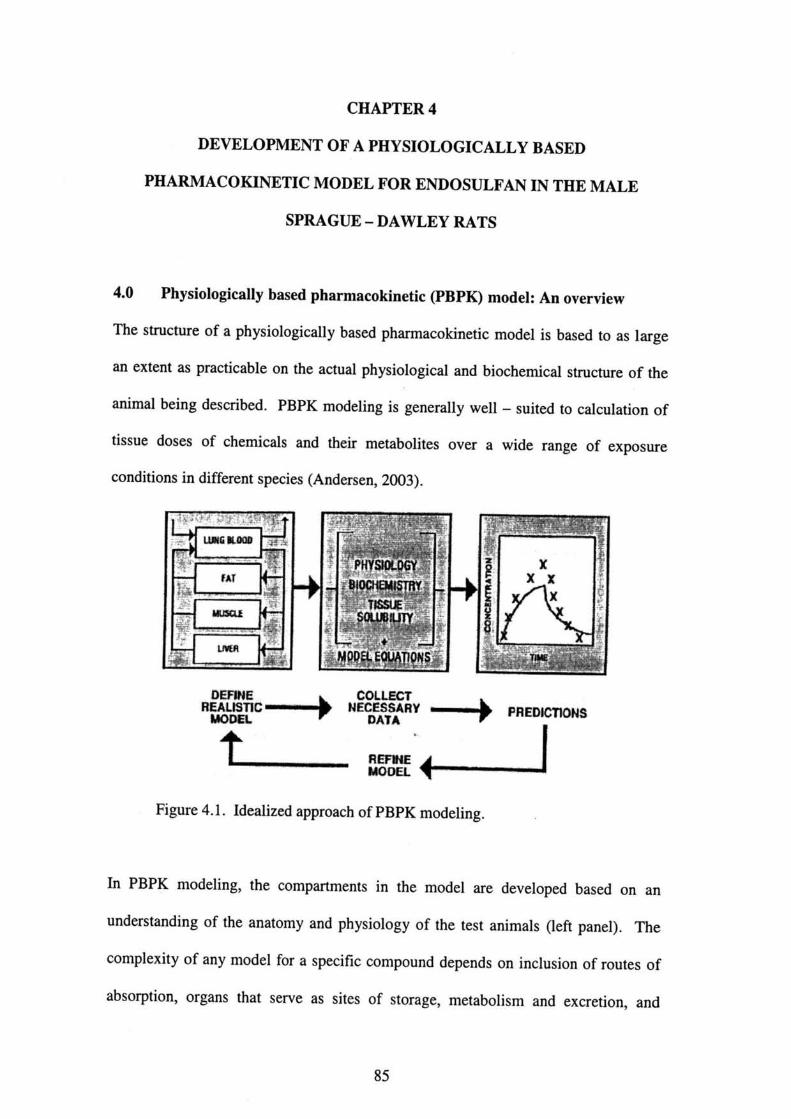
CHAPTER 4
DEV肌OPMENT OF A PHYSIOLOGICALLY BASEI)
PHARMACOKINETIC MOD肌FOR ENI)OSULFAN IN THE MALE
SPRAGUE-1)AVVLEY RATS
4.O Physiologically based pharmacokinetic(PBPK)modeL An overview
Th・・血・t・・e・f・phy・i・1・gically b・・ed・ph・・mac・垣n・ti・m・d・1 i・b・・ed t…1・・g・
an extent as practicable on the actual physiological and biochemical s血cture of the
animal being described. PBPK modeling is generally wel1-suited to calculation of
tiss・e d・・e・・f・h・mical・and th・丘m・t・b・lit…ve・awid・・ang・・f exp・・u・e
conditions in different species(Andersen,2003).
・罐C→Nε懸V→P-・NS
REF刑E
蝋》OEL
Figure 4.1. Idealized approach of PBPK modeling.
1・PBPK m・d・li・g・the c・mp・rtm・nt・i・th・m・d・1 ar・devel・P・d b・・ed。n、n
und・・st・ndi・g・f the an・t・my・nd phy・i・1・gy・f th・t・・t anim・1・(1・丘P…1). Th。
complexity of any model fbr a specific compo皿d depends on inclusion of routes of
・b・・中ti・n…gan・th・t・er・e ・・ ・it…f・t・rag・, m・t・b・1i・m・nd ex・・eti。n,、。d
85

tissues that are the target organs fbr chemical toxicity. The parameters fbr
metabolism, excretion, tissue partitioning, blood and air flow rates are introduced as
constants into the model and kinetic behavior in various compartments-blood,
tissues, excreta, etc. Middle panel is predicted by computer simulation. Predictions
made from the PBPK models can be tested by comparison with the observed time-
course results(right panel). When discrepancies are noted between experiment and
model predictions(as often happen), the model can be refined in a hypothesis
generation loop and tested against available data(Adapted from Andersen,2003).
In PBPK models(Figure 4.1), the biological system is envisaged as comprising of a
small number of physiologically relevant compartments and each compartment
corresponds to discrete tissues or groups of tissues with appropriate volumes, blood
flow rates, and pathways of metabolism of the test chemical.111 PBPK models,
pertinent biochemical and physical constants fbr metabolism and tissue solubility are
incorporated directly in the descriptions of each tissue compa血ellt. Routes of
administration are included in their proper relationship to the overall physiological
structure and exposure scenario differences are accounted for in the time sequence of
the dose input terms. Each compartment(i. e., tissue)in the model:is described by a
‘
mass-balance differential equation, and the set of equations is solved by numerical
integration to predict tissue time-course concentrations of the test ’chemical and its
metabolites(Andersen,2003).
86

The steps involved in developing a PBPK model are(Aarons et a1
●
●
●
●
●
.,1999):
Specification of the model based on the anatomical/physiological structure of
the species of interest and the determinants of disposition and
biotransforrnation of the substance concerned;
Development of a mathematical description of the biological processes
involved, including physiologica1, physicochemical and biochemical
constants;
Design of new experiments to test the robustness of the model description;
Collection of the necessary experimental data to conflrm or refUte the model
structure;
Adjustments to refine the model.
PBPK models offer a number of advantages over the compartmental models used
previously in classical phamacokinetic analyses. A physiologically based model can
be interpreted in biological tems, and lead to an’understanding of出e actual
pharmacokinetic processes goveming chemical disposition in the body. Lack of丘t of
・p頒i・ula・m・d・l may sugge・t・1t・m・ti・・h〕?・theses ab・ut phamac・垣neti。
processes involved in the distribution and metabolism of chemicals. They can be used
t・e・tim・t・d・・e-・ffe・t d・t・・v・・awid・・ang・・f exp・・u・e c・nditi・n・.踵・y訂・
validated, they can even be used to predict the outcomes of exposure conditions which
have n・t been・xp・亘mentally test・d・H・nce, PBPK m・del・can p・・vide a means。f
e緬polating丘om the high-dose situation commonly used in laboratory studies to
th・1・w-d・・e c・nditi…elev・・t t・envi・・nment・1・xp・・u・e. Simi1・・1y, it m・y
collceivably be used fbr extrapolating from acute to repeated exposure scenarios.
PBPK models may also offer a powerfUl tool fbr predicting target tissue dose from
87
one route of exposure to another and between species. A PBPK model established on
the basis of studies in non-human mammalian species can be applied to humans
after substitutillg the appiopriate allometric, biochemica1, and pharmacokinetic
parameters fbr humalls. Finally, by providing a means of estimating the dose of
reactive metabolites reaching target tissues, PBPK models may lead to more accurate
predictions of nsk也an can be obtained using environmental exposure levels,
particularly whell saturation effects lead to a non-lillear relationship between
environmental exposures and tissue doses(1品ung and Paustenbach,1995;
Moolgavkar et al.,1999).
4.1 Background introduction
Endosulfan(6,7,8,9,10,10-hexachloro-1,5,5a,6,9,9a-hexahydro-6,9-
methano-2,4,3-benzodioxathiepin-3-oxide), an organochlorine(OC)insecticide
of the cyclodiene group, has widespread use in agriculture and fbrestry to control a
wide variety of insect pests and on non-fbod crops such as cotton and tobacco. It is
also used as wood preservative【Agency fbr Toxic Substances and Disease Registry
(ATSDR),2000]. Endosulfan is used in Illdia(Saiyed et al.,2003), Turkey(Oktay et
al.,2003), Malaysia(Chan et al.,2004), Mexico(Castillo et a1.,2002;Gonz61ez-
Farias et al.,2004)and many other developing countries.
Residues of endosulfan are found in low levels in the environment such as sediment
(Gonz41ez-Farias et aL,2004;Bhattacharya et aL,2003), soil(Gam6n et al.,2003),
water(Tall and Vijayaletchumy,1994)and air(Louie and Sin,2003);in animal
tissues(Ansari et aL,1984;Chan and Mustafa,2005;Dikshith et al.,1984;Nath et al.,
1978;National Cancer Institute,1978)as well as in humans(Aleksandrowicz,1979;
88

Blanco 一一 Coronado et al.,1992;Boereboom et a1.,1998;Brandt et al.,2001;Chan et
al.,2004;Coutselinis et aL,1978;Demeter et al.,1977;Eyer et aL,2004;Lo et al.,
1995).The accumulation of endosulfan has also been reported in various fbod crops
such as vegetables(Antonious et aL,1998)and fish(Novak and Ahmad,1989).
Endosulfan is a mixture of two stereoisomers:α一andβ一endosulfan(Hayes and
Laws,1991)in the ratio of 70:30. When given to rats by various routes, endosulfan is
rapidly metabolized and excreted in the urine and feces as oxidation products such as
endosulfan sulfate, diol, hydroxyether or ketone resulting from the cleavage of the
cyclic sulfite group(McGregor,1998). The ratio of the different compounds in the
excreta differs with routes of exposure and isomers(FAO/WHO,1969).
Endosulfan induced alteration on spermatogenesis in young growing and adult rats.
The impairments included decreased sperm count, intratesticular spermatid number,
sperm morphology and altered activities of testicular marker enzymes(Chitra et al.,
1999;Choudhary and Joshi,2003;Sinha et aL,1995;Sinha et aL,1997;Sinha et al.,
2001)as wen as reduction in senlm testosterone levels(Choudhary alld Joshi,2003;
Wilson and LeBlanc,1998). ln addition, reduction in the weights of testes and sex
accessory orgalls was observed(Choudhary and Joshi,2003). In an in vitro
experiment, cytotoxicity in Sertoli 一 germ cell coculture was observed fbllowing
exposure to endosulfan, which might disturb the normal interaction between Sertoli
and gernl cells, thus leading to testicular dysfUnction(Sinha et al.,1999).
Involvement of endosulfan, a neurotoxic agent, in the central nervous system(CNS)
has been studied in various studies(Anand et al.,1980;Paul et al.,1994;Seth et al.,
89

1986;Subramoniam et al.,1994). A stimulation by long term exposure of endosulfan
at a dose of 3 mg/kg body weight fbr 15 and 30 days respectively increased fbot
shock-induced fighting behavior in male rats(Agrawal et al.,1983). Induction of
hyperactivity, tremors and convulsions was observed in male rats given 40 mg/kg of
endosulfan intraperitolleally(Ansari et aL,1987). Data based on these studies
indicate that the liver, kidneys, brain and testes are the main target organs(ATSDR,
2000).
According to ATSDR(2000)and the PubMed Database(2005),
validation of PBPK model predictions was located for endosulfan.
no calibration or
4.2 0bjective of the study
The present study was aimed to develop a physiologically based pharmacokinetic
(PBPK)model for endosulfan in male rats that could reasonably predict tissues
dosimetries a危er single oral administration of 14C-Endosulfan. The model was
verified and given a trial by simulating independent derived data on endosulfan
disposition after multiple oral administrations of 5 mg/kg body weight 14C-
Endosulfan. The model was fUrther verified by using experimental data retrieved
from t le literature.
The newly-developed model was extrapolated and scaled up to predict the behavior
of endosulfan in humans, and model predictions were compared with the data丘om
human volunteers in Japan and Malaysia.
90
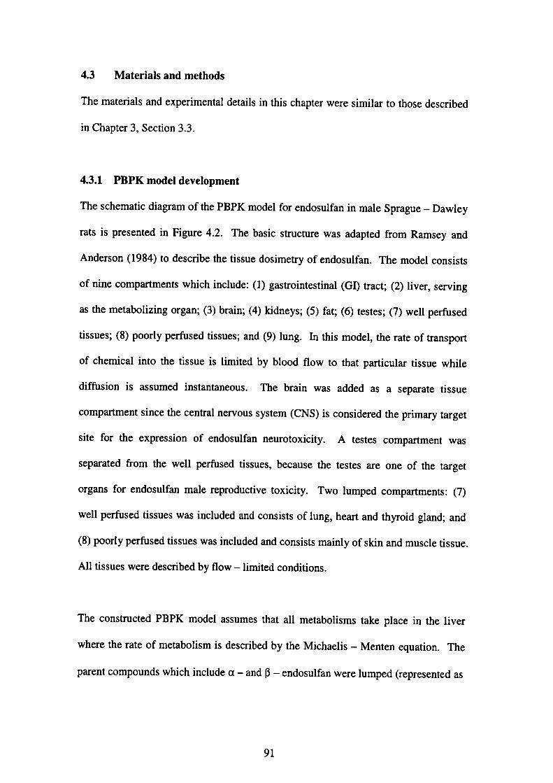
4.3 Materials and methods
The materials and experimental details in this chapter were similar to those described
in Chapter 3, Section 3.3.
4・3・1PBPK model development
The schematic diagram of the PBPK model for endosulfan in male Sprague-Dawley
rats is presented in Figure 4.2. The basic structure was adapted from Ramsey and
Anderson(1984)to describe the tissue dosimetry of endosulfan. The model consists
of nine compartments which include:(1)gastrointestinal(GD tract;(2)liver, serving
as the metabolizillg organ;(3)brain;(4)kidneys;(5)fat;(6)testes;(7)well perfUsed
tissues;(8)poorly per血sed tissues;and(9)1ung. In this model, the rate of transport
of chemical into the tissue is limited by blood flow to that particular tissue while
diffUsion is assumed instantaneous. The brain was added as a separate tissue
compartmellt since the central nervous system(CNS)is considered the primary target
site for the expression of endosulfan neurotoxicity. Atestes compartmellt was
separated from the well perfUsed tissues, because the testes are one of the target
organs fbr endosulfan male reproductive toxicity. Two lumped compartments:(7)
well perfUsed tissues was included and consists of lung, heart and thyroid gland;and
(8)poorly perfUsed tissues was included and consists mainly of skin and muscle tissue.
All tissues were described by flow-1imited conditions.
The constructed PBPK model assumes that all metabolisms take place in the liver
where the rate of metabolism is described by the Michaelis 一 Menten equation. The
parent compounds which include a-andβ 一 endosulfan were lumped(represented as
91
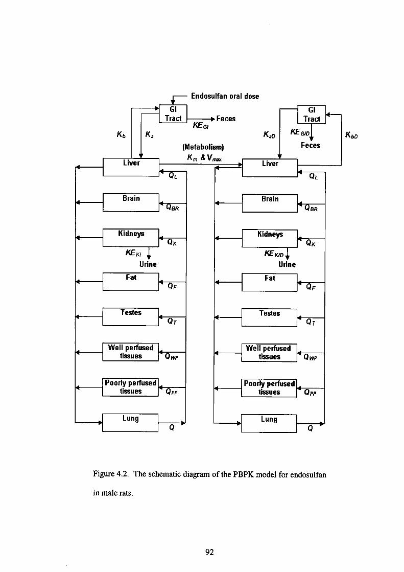
Endosolfan o「al dose
GI
sralコ
Gl
srad
κb κ∂
@ (Metabolism)
κEGJ
ネm&V鰍
Feces
@ κ∂o拍EG,o
@ Feces
Liver Liver
QL QL
Brain Brain
8R 朗
Kidneys Kidneys
Qκ Qκ
灼Eκ,
@ Urine
κεκノo
Urine
Fat Fat
F QF
Te舗es Te舗esQγ Qγ
Well pe㎡used
@ tissoes Q柳Well pe同used
@ tissues Q柳
Poorly per向sed
@ tissues QPPPoorlv pe而used
@ tissues QPP
Lung LungQ Q
KbD
Figure 4.2. The schematic diagram of the PBPK model for endosulfan
in male rats.
92

endosulfan)and the metabolites w ere represented as endosulfan metabolites in the
desc「ipti°n°f the m・d・1・i・・h・d・・cripti・n・f・h・m・d・1.・n、um町。,ally
administ・・ed・・d・・ulf・n・w・…k・・upbyth・1i。,,a。d m。、、b。lized.
陥4G・= c・・
dt PκI
r・ρ腔(G一鶏
弓P一ρ〃〔G一舞り
喋・ρ・{α笥
弓F・ρ・(Ca一曇〕
防芸γ一ρ・〔G一司
曙”・・イG一意り一弩Eτ一・-K・CuVu
陶4蜒ソ・D・蹴認α・K、CuVu.脚α
喋・Σρ’信一Ca
W
(mg/L);α’i・・h・b1・・d・fl・w・a・・t・a・iss・e(L/h);
concent「atlon(mg/L);pti is the tissue:blood ・・
ん’=G・η・i・・he end・・ulfa・・m・un・i・an。,g、。.
pa「amete「s ln the eq・・ti…a・e d・fi・ed i・T・b1,、4.1.4.4.
The m・d・1・quati…f…end・・ulf・n・ar・d。、crib。d、, f。ll。ws:
Q・・(c・ 一)-KE・・c・・v・・
…Kidneys
…Well pe血sed tissues
… Poorly per血sed tissues
...Brain
...Fat
...Testes
...】しiver
...GI Tract
〔り …Art。,i。1b1。。d
he「eηパs thev・1・m・・f・・jssu・(L);C,・ i・・heend・・uぬ。、。ncent,a,i。n.in、,iss。e
Ca is the arterial endosulfan
pa「tltlon coefficient of endosulfan;and
The abbrevjations of other
93
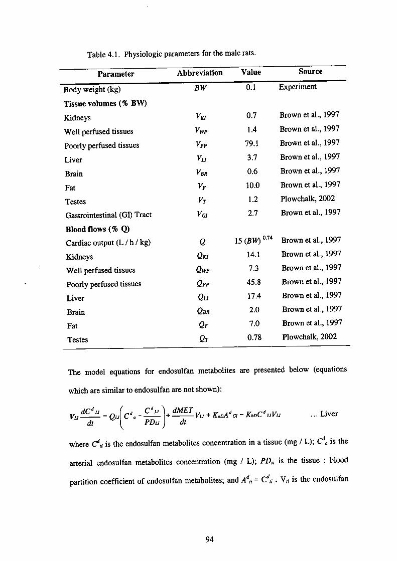
Table 4.1. Physiologic parameters for the male rats.
Parameter Abbreviation Value Source
Body weight(kg)
Tissue volumes(%BW)
Kidneys
Well pe血sed tissues
Poorly perfUsed tissues
Liver
Brain
Fat
Testes
Gastrointestinal(GI)Tract
Blood fiows(%Q)
Cardiac output(L!h/kg)
Kidneys
Well perfUsed tissues
Poorly perfUsed tissues
Liver
Brain
Fat
Testes
Bvv
喩聯㌦陥撫昨巧㌦
o伽伽伽鋤鋤色o,
0.1
0.7
1.4
79.1
3.7
0.6
10.0
1.2
2.7
15(B助゜・74
14.1
7.3
45.8
17.4
2.0
7.0
0.78
Experiment
Brown et al.,1997
Brown et a1.,1997
Brown et al.,1997
Brown et al.,1997
Brown et al.,1997
Brown et al.,1997
Plowchalk,2002
Brown et al.,1997
Brown et al.,1997
Brown et al.,1997
Brown et al.,1997
Brown et al.,1997
Brown et al.,1997
Brown et al.,1997
Brown et al.,1997
Plowchalk,2002
Th。 m・d・1・quati・n・f・・end・・uぬ・m・t・b・1it・・ar・p・e・ent・d b・1・w(・q・・ti・・s
which are simjlar to elldosulfan are not shown):
ぬ4eμ・eu〔C∂・一劃・ば㌘ぬ・ ’α一一…Live・
wh。,e Cd,、 i、血e e。d。、uぬ。 m…b・li…c・ncent・a・i・n i・・tissu・(mg!L);♂・i・・h・
arterial endosulfan metabolites concentration(mg/L);PDrf is the tissue:blood
P、rti・i・n…伍・i・・t・f・・d・・uぬ・m・t・b・lit・・;・nd・Ad’・・C∂…V・・i・・he end・・曲・
94
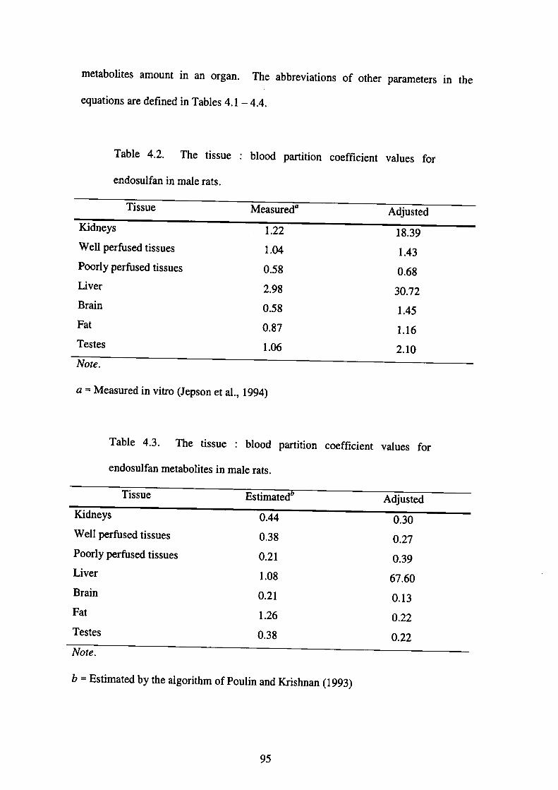
metab・lit・・am・皿t i・・n・・g・n・The abb・evi・ti・n・・f・th・r param,t。,、 i。 th。
equatlons are defined in Tables 4.1-4.4.
Table 4.2. The tissue :blood
endosulfan in male rats.
partition coefficient values for
TissueMeasuredα Adjusted
Kidneys
Well per血sed tissues
Poorly per負1sed tissues
Liver
Brain
Fat
Testes
1.22
1.04
0.58
2.98
0.58
0.87
1.06
18.39
1.43
0.68
30.72
1.45
1.16
2.10
Note.
a = Measured in vitro(Jepson et al.,1994)
Table 4.3. The tissue :blood partition coefficient values fbr
endosulfan metaboljtes in male rats.
Tissue Estimated Adjusted
Kidneys
Well perfUsed tissues
Poorly per血sed tissues
Liver
Brain
Fat
Testes
0.44
0.38
0.21
1.08
0.21
1.26
0.38
0.30
0.27
0.39
67.60
0.13
0.22
0.22
Note.
b=E・tim・t・d by the a19・・ithm・fP・・1i…d㎞・h・・n(1993)
95
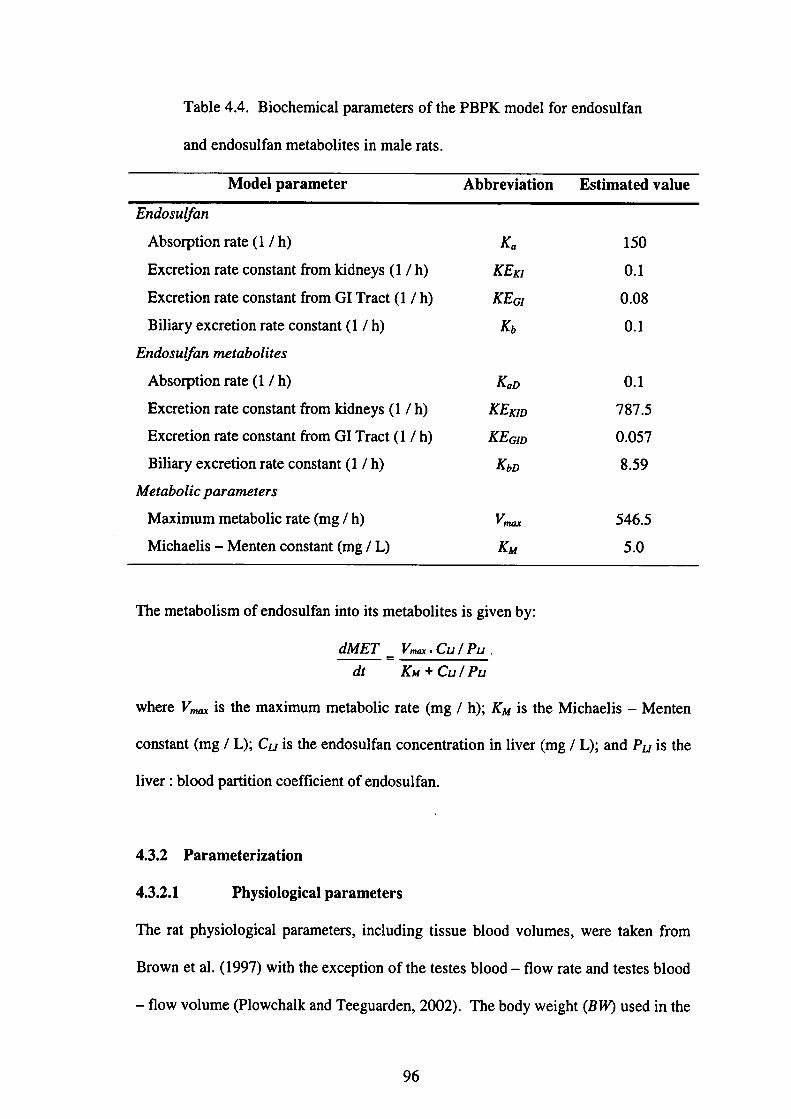
Table 4.4. Biochemical parameters of the PBPK model for endosulfan
and endosulfan metabolites in male rats.
Model parameter Abbreviation Estimated value
Endosulfan
AbsorPtion rate(1/h)
Excretion rate constant from kidneys(1/h)
Excretion rate constant from GI Tract(1!h)
Biliary excretion rate constant(1/h)
Endosulfan metabolites
Abso4)tion rate(1!h)
Excretion rate constant from kidneys(1/h)
Excretion rate constant from GI Tract(1/h)
Biliary excretion rate constant(1/h)
Metabolic parameters
Maximum metabolic rate(mg/h)
Michaelis-Menten constant(mg/L)
KEκI
KEGI
K。D
KEκID
KEGn)
KbD
㌦脳
150
0.1
0.08
0.1
0.1
7875
0.057
8.59
546.5
5.0
The metabolism of endosulfan into its metabolites is given by:
dMET Vmax.Cu/Pu,
dt KM+Cu!Pu
where Vmax is the maximum metabolic rate(mg/h);KM is the Michaelis-Menten
constant(mg/L);Cu is the endosulfan concentration in liver(mg/L);and Pu is the
liver:blood partition coefficient of endosulfan.
4.3.2 Parameterization
4.3.2.1 Physiologica1 parameters
The rat physiological parameters, including tissue blood volumes, were taken from
Brown et aL(1997)with the exception of the testes blood-flow rate and testes blood
-flow volume(Plowchalk and Teeguarden,2002). The body weight(B Ui) used in the
96
model was determined by our experiment.
4.1.
Parameter values are summarized in Table
4.3.2.2 Physicochemical parameters
1) Measured partition coefficients
l1 the partition coefficient(PC)determination experiment, three rats(n=3)were used.
PC values fbr 14C-Endosulfan were determined experimentally in male Sprague-
Dawley rats using the centrifUgation method for nonvolatile chemicals described by
Jepson et al.(1994). Several unexposed rats were lightly anesthetized with diethyl
ether and tissues such as liver, kidneys, fat, muscle, brain, heart, lung, spleen, testis,
thyroid gland and blood were collected. The blood and minced tissues were weighed
into 5 mL scintillation vials capped with Teflon/rubber septa to prevellt the
absolption of the chemica1. Vials were prepared usillg l pg/mL of i4C-Endosulfan
in PBS. Typically,0.25 g of blood or tissue and 5 mL of chemical in saline were
added to each vial. Vials were incubated and vortexed at a medium speed setting fbr
l8 h at 37°C. The’supernatant was cenUifuged for 10 min at 3000 rpm. The
resulting supernatant was filtered through a prewashed Centrifree⑧Micropartition
Device with YMT membrane(Millipore Corp., Bedfbrd, MA)by centrifUgation for 3
min at 3000 rpm.100 pL of the filtrate was removed from the filter cup and 5 mL of
Ultima Gold was mixed and counted fbr radioactivity. The tissue:saline partition
coefficients were calculated as in Jepson et a1.(1994)and a(ijusted until agreement
was reached between model-predicted and experimental data fbr the 5 mg/kg
single dose group. The physicochemical parameters are summarized in Table 4.2.
97
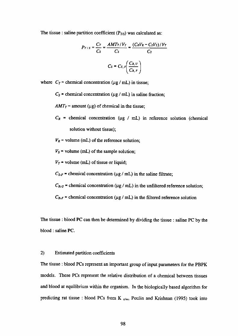
The tissue:saline partition coefficient(PTIs)was calculated as:
P〃、。皇.蜥靭防。(c・V・’c・Vs)IVT
Cs Cs C∫
α・G・剰
where CT = chemical concentration(μg/mL)ill tissue;
Cぷ=chemical concentration(μ9/mL)in saline飴ction;
AMT7・= amount(μg)of chemical in the tissue;
CR=chemical concentration(μg/mL)in reference solution(chemical
solution without tissue);
VR = volume(mL)of the reference solution;
Vs = volume(mL)of the sample solution;
VT = volume(mh)of tissue or liquid;
Cs,F = chemical concentration(pg/mL)in the saline filtrate;
CR, u = chemical concentration(pg/mL)in the unfiltered reference solution;
CR, F = chemical concentration(μg!mL)in the filtered reference solution
The tissue:blood PC can then be detemined by dividing the tissue:saline PC by the
blood:saline PC.
2) Estimated partition coefficients
The tissue:blood PCs represent an important group of input parameters for the PBPK
models. These PCs represent the relative distribution of a chemical between tissues
and blood at equilibrium within the organism. In the biologically based algorithm fbr
predicting rat tissue:blood PCs from K o/w, Poulin and Krishnan(1995)took into
98
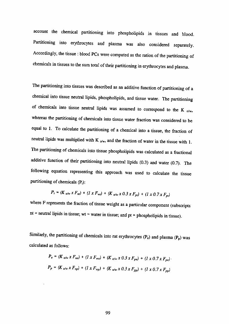
acc°unt the ch・mical p・niti・ni・g i・t・ph・・ph・lipid・i。 tissu,、 and bl。。d.
Pa「tjti°ni”g i・t・e・yth…yt・・a・d p1・・m・w・・al・・・…id・・ed・epar、・,ly.
Acc°「di”gly・ th・tissue・b1・・d PC・w・・e c・mp…d・・th・・ati・n・fth・par・i・i。ni。g。f
chemicals in tissue・…h・・umt・t・1・fth・ir・P・・titi・ni・g・i…ythr・・yt,, and p1、、m、.
The剛iti°㎡・g i・t・・issue・w・・d・・c・ib・d・・a・additi・・血・・ti・n・f麺i・i。㎡。g。fa
che血cal int・tissu・n・u・・al lipid・・ph・・ph・1ipid・,・nd・iss・e w・….Th・剛i・i。㎡。g
°f chemical・i・t・・issu・・e囎11ipid・w・・assum・d・…π・・p・nd・。 th, K 。f。,
whe「eas th・剛iti・㎡・g・f・h・㎡cal・i・t・tiss・・w・t・・丘acti。n w。、 c。。、id。,ed t。 be
equal t°1・T・・al・・1・t・th・卿iti・㎡・g・f・・h・mi・al i・t。 a tiss。e, th,丘acti。n。f
・・岨11ipid・w・・m・1・ipli・d w仙K・。f。,・nd晒ac・i。n。fw、t。, i。,h。,iss。e Wi,h l.
The卿iti・㎡・g・f・h・mica1・i・t・tissue ph・・ph・lipid・w。、 c、1、ul、t,d。、 a廿acti。n。l
additive血・・ti・n・f・h・丘声i・i・血・g i・t・ne眈・11ipid・(・.3)。。d w。・,,(・.7). The
飴11°wi・g・q・ati・n・ep・e・e・ti・g thi・apP・・ach w・・u、ed t。 ca1、ul。t, th。,i、sue
partltlomng of chemicals(pt):
P,=輪鵬川・坑∂・(K・・・・・・・…3・・Fp川1・α7・ち,戊
whe「e F「ep・e・e・t・・h・f「ac・i・n・ftissu・w・igh…ap・rti・ular・・mp・nent(、ub、c,ipts
nt = neut「al lipid・i・tissu・;w・・w・…i・・issue;and p・・ph・・ph・lipid、 i。 tissue).
Simila「ly・the畑iti・ni・g・f・h・mical・i・t・・at・・ythr・cyt・・(P,)・nd p1・・m、(P,)was
calculated as fbllows:
P・=(K・}/6・ x F・昭戊+(1x Fw・)+(κ吻xα3xち∂+↓l x(). 7 x Fp,)・
Pp=(K ・iS・ x F,rp戊+(1 x FM・P戊+(K ・iSv x()・3x FPP)+(1 x O. 7 x F,ρ)
99
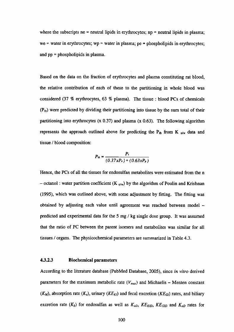
where the subscripts ne =neutral lipids in erythrocytes;np=neutral lipids in plasma;
we=water in erythrocytes;wp=water in plasma;pe = phospholipids in erythrocytes;
and pp = phospholipids in plasma.
Based on the data on the fraction of erythrocytes and plasma constituting rat blood,
the relative contribution of each of these to the partitioning in whole blood was
considered(37%er)血ocytes,63%plasma). The tissue:blood PCs of chemicals
(Ptb)were predicted by dividing the丘partitioning into tissue by the sum total of their
partitioning into erythrocytes(x O.37)and plasma(x O.63). The fbllowing algorithm
represents the approach outlined above fbr predicting the Ptb丘om K《ガw data and
tissue/blood composition:
Pr
Pゆ=
(0.37xP,)+↓0.63x1㌔)
Hence, the PCs of all the tissues fbr endosulfan metabolites were estimated伽m the n
_octanol:water partition coefficient(K(ガw)by the algorithm of Poulin and Krishnan
(1995),which was outlined above, with some a(加stment by fitting. The fitting was
obtained by a(輌sting each value until agreement was reached between model-
predicted and experimental data fbr the 5 mg/kg single dose group. It was assumed
that the ratio of PC between the parent isomers and metabolites was similar fbr all
tissues/organs. The physicochemical parameters are summarized in Table 4.3.
4.3.2.3 Biochemical parameters
According to the literature database(PubMed Database,2005), since in vitro derived
parameters fbr the maximum metabolic rate(Vntax)and Michaelis-Menten constant
(KM), absorption rate(Ka), urinary(KEKI)and fecal excretion(」KEα)rates, and biliary
excretion rate(Kb)fbr endosulfan as well as KaD,κEκノD,、KEGID and KaD rates fbr
100

end°sulfan m・t・b・lit・・w・・e u・・v・il・bl・,・h・・e p・・am・・…w・・e・P・imized、by_al
adjustm・nt皿tinh・b・・t・i・u・1・fit・f・h・・im・1・ti・n・wi・h・he exp,,im,nt、l d、,、 was
°bserv・d・Th・v・1ue・・fth・bi・・h・mical p・・am・t…ar,、umm、,ized・i。 T、b1,4.4.
4.3.2.4 Model calibration
The PBPK m・d・l w・・num・・ically・・1・・d・・i・g・h・E。1。, M。血。d. Th, m。d,l
slmulati°n was carri・d・ut・・i・g Mi・・…ft・Vi・u・I B・・i・6.3(Mi・・…ft・C。甲。,a,i。n).
The sim”1・ti・n・f・h・p鎚m・t・dzed m・d・l w・・c・m声・d wi・h・he exp。,im,。,、1 d。ta
ln@male Sp「agu・-D・wl・y・a・・a丘…i・g1…a1・d㎡㎡・tr・・i・n・f 5 mg/kg 14C一
End°sul㎞・・d・・c「ib・d p・evi…1y i・Ch・p…3(Sec・i・n 3.4.3, T、bl,3.1).
4・3・2・5 @M・d・1・・rificati・・and m・d・1・im・1・ti・n・・f・nd・,ulf。n・di,p。,iti。n
in other studies
The calib・at・d PBPK m・d・1 w・・apPli・d…h・・epea・・d d・・i・g・。ndi・i。n{,hr,e 一一
tlme「epeated admini・t「・・i…f5mg/kg・・p・evi…ly d・・crib・d i・・h・E。p,,im,n、。1
design・nd・d輌・廿・ti・n・f 14C-E・d…ぬ・(3)}. Th, m。d,1、im。1。ti。。 was
c°mpa「ed wiかhe exp・亘m・nt・1 d・t・・n time c・urse・。f t。t、1、。。ce。廿、ti。n、
f°ll°wi・g・㎞ee-・im・・epea・・d・・al・d㎡・i・t・a・i・n・f。。d。、曲。,。 m。1,,a,、 as
desc「ibed p・evi…1y i・Ch・p…3(Sec・i・n 3・4・3, T・bl・3.2). Th・m・d。r、 ability,。
P「edict也e end…1飴・di・p・・i・i・・i・・va・i・u・tiss・e・w・・血曲・・v・・i丘・d by。、i。g
expe「1mental data・e廿i・ved fr・m・h・li…a…e(A・・ari…1.,1984;Chan and M。、t、fa,
2°°5;Dikshith・t・・1984;N・・h…1・・1978)・nd・he exp・・imen・・l d・t。 ar,、pecifi,d in
Table 4・5・Since・he av・・ag・b・dy w・ight・w・・e unavail・bl・i。・h。、e、,。di。、,
simulati°n・w・・e d・ne by・・i・g th・・a・g・・fb・dy w・igh・・(㎡・im・m-m、xim。m)as
reported in the literature.
101
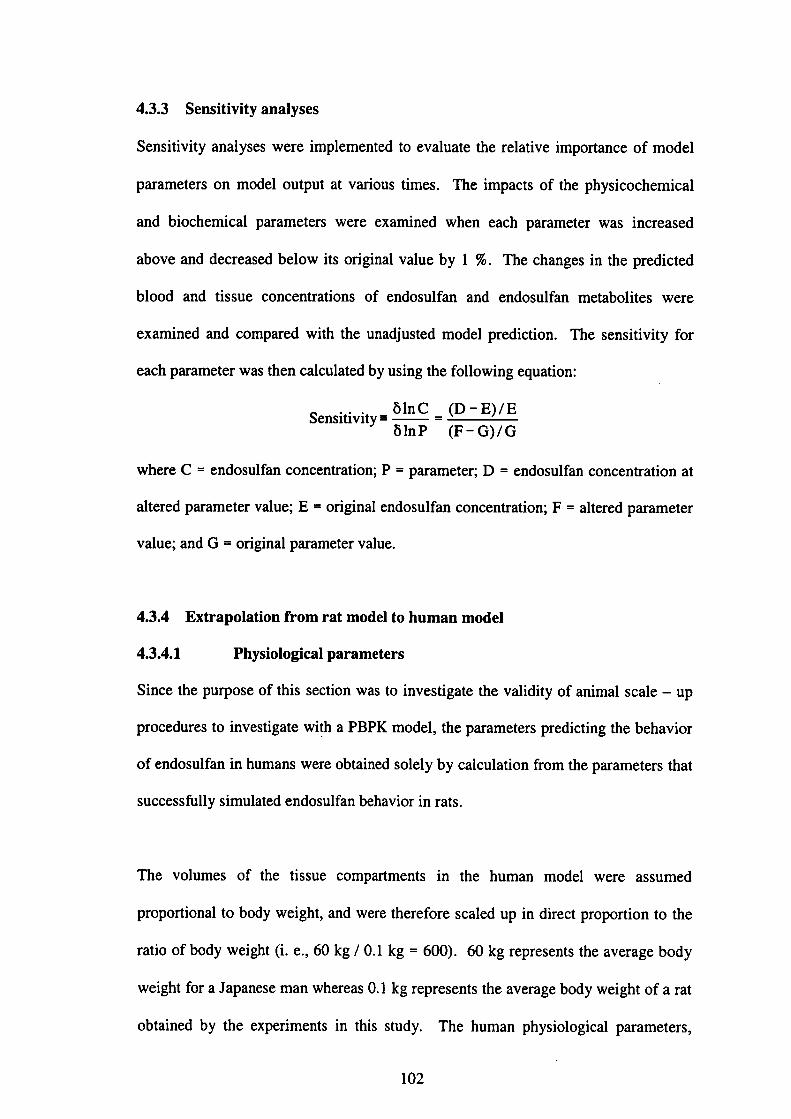
4.3.3 Sensitivity analyses
Sensitivity analyses were implemented to evaluate the relative importance of model
parameters on model output at various times. The impacts of the physicochemical
and biochemical parameters were examined when each parameter was increased
above and decreased below its original value by l%. The changes in the predicted
blood and tissue concentrations of endosulfan and endosulfan metabolites were
examined and compared with the una(ljusted model prediction. The sensitivity fbr
each parameter was then calculated by using the following equation:
S・n・i・i・i・y悉・i:≡ぎ}1:
where C=endosulfan concentration;P = parameter;D=endosulfan concentration at
altered parameter value;E = original endosulfan concentration;F=altered parameter
value;and G=original parameter value.
4.3.4 Extrapolation from rat model to human model
4.3.4.1 Physiological parameters
Since the purpose of this section was to investigate the validity of animal scale-up
procedures to investigate wi!h a PBPK model, the parameters predicting the behavior
of endosulfan in humans were obtained solely by calculation from the parameters that
successfUlly simulated endosulfan behavior in rats.
The volumes of the tissue compartments in the human model were assumed
proportional to body weight, and were therefbre scaled up in direct proportion to the
ratio of body weight(i. e.,60 kg/0.l kg=600).60 kg represents the average body
weight for a Japanese man whereas O.1 kg represents the average body weight of a rat
obtained by the experiments in this study. The human physiological parameters,
102
including tissue blood volumes, were taken丘om Brown et al.(1997)with the
exception of the testes blood-flow rate and testes blood-flow volume(Plowchalk
and Teeg・訂d…2002)・Th・b・dy w・ight(B助・・ed i・th・h・m・n m・d・l w・・60 kg,
which was an average weight of a Japallese man. Parameter values are summarized in
T・bl・4・6・Phy・i・1・gical p訂・m・t・・g・n・・ally・・n・id・・ed t・b・m・・e nea・ly
proportional to body surface area than to body weight(and hence to a fractional
power of the body weight ratio)is cardiac output. The O.74 power of the body weight
ratios was chosen to scale up these parameters to the human mode1, i. e.,6000’74=
113.7(Ramsey and Andersen,1984). BW of 49 kg was used fbr the data on the
M・1・y・i・n・ch・・1・hild・en(Ch・n・t・1・,2004)・BVV・f 70 kg, whi・h w・・an・verag・
B晒br a Western man, was used fbr other data re垣eved仕om the literature.
4.3.4.2 Physicochemica] parameters
PCs used fbr the rat model were unchanged fbr the human mode1. PCs are
summarized in Tables 4.2 and 4.3.
103

肩口肩ロ
㊨◎
§三否Σ題三否
一一Z§氏
ロo§亀
い.
い
(・。
?n℃ε)
言。。宣ロ宕Σ
マ
゜。
量国
吟
一一Zぢ日uエ
=
(江題O℃ε)言。畳ロ3Σ
ひ
ひーひひーひ
喜冨一〇Σ題“一〇
oon.nー②O.O
●on.nl㊨O.O
…o一§ぎ4
一皇8亘壱 (一〔一)百唱8肩‥=o
い.
mい.
m
(。・
ー題S占)
言o昔ヨロΣ
(・・
梠閧r」a)
冠。。宣ヨロΣ
N
104
冒Σ匂§自ぷO
〔(パ(
←ーマ卜ーマトーマ卜ーマ
喜も門喜召雲喜もの喜吾£
N『Oーヌ》.O岱.O18.〇怠.Ol文).O岱.O18.O
O◎oーロ3ら←O°oー§5←Oooーロ8タ←O◎◎ー§5←
22いい
(江題い二a)項8。一身ヨΣ芭。昔OヨΣ
一
旨一〇●日富」註2丙日一ミ
ロヨ昆●日ロ
唱oヒ8台O
(bo゚着。o匂●与苫
習頃図8菖2胡
、bo旦●8⇔
oZ

4・3・4・3 Bi・・h・mi・・l p・,。met,,、
Specific metab°1ic c・n・t・nt・ar・n・t・・u・11y・eg・・d・d・・v・・y、。、。。、i、t,ndy with
b°dyweight・alth・・ghb・・alm・t・b・1i・・a・・d…apP・ar・・飴11・w・h,加c,i。n、lp。wer
mlewithanexp・n・・t・f・・74・Th・m・xim・m・at・・f・he⑭、・i、,eacti。瓜was
assumed@apP「°ximately p・・P・rti・n・1・・b・・al m…b・1i・・at・(G・h,i。g,t、1.,1948;
RamseyandA・d…e・・1984)…dw・・也・・ef・・e・ca1・di。・h,、am,m、m,,a, cardiac
°utput’砺was a’s°scal・di・th・・am・m・m・・a・cardiac・u・p・・. Th。・ab、。叩,i。n,ate
(Ka・・u輌陶and・fecal・x・・e・i・n陶・at・・,and・bili・・y・x、,e,i。n,a,,(Kb)f。r
end°suぬn andtheab…p・i・n・a・・(Ka・・輌y(KEmo)・nd・fe・al exc,e、i。。(KE。ω)
「ates・andb’1助exc「et’・n・a・・㈲f・・e・d…1⑫・m・t・b・li・esrem。i。。dun。h、ng。d
f°「the human m・d・1・Th・v・1・…f・h・bi・・h・面cal・par。m,,,M。、u輌zed in
Table 4.7.
4.3.4.4 Model simulation
The model simulation was carried °ut uslng Mi・・…負Vi・u・1 Basi・6.3(Mi。,。、。丘
C卿゜「ati°n)・°nce・h・m・d・l p獅・t…w・・e・e・,・・1y・he c。。ce。tr。,i。n/d。、e。f
end°suぬn inth・d’・・w・・ch・ng・d…im・1・…he c・n・en廿、・i。。。f。。d。、。lfa。 i。,he
des’「ed tissues・Each・・’・mee・w・・ass・m・d・・・…um・mea1・伽ee,im,、 p,,d。y
Φ「eakfast・lun・h・nd dinn・・)・Each・・1…ee・w・・expec・・d・。 hav,由,i, m閉1、 at an
ave「age tlme・fO700・1300・・d 1900 d・ily.
105
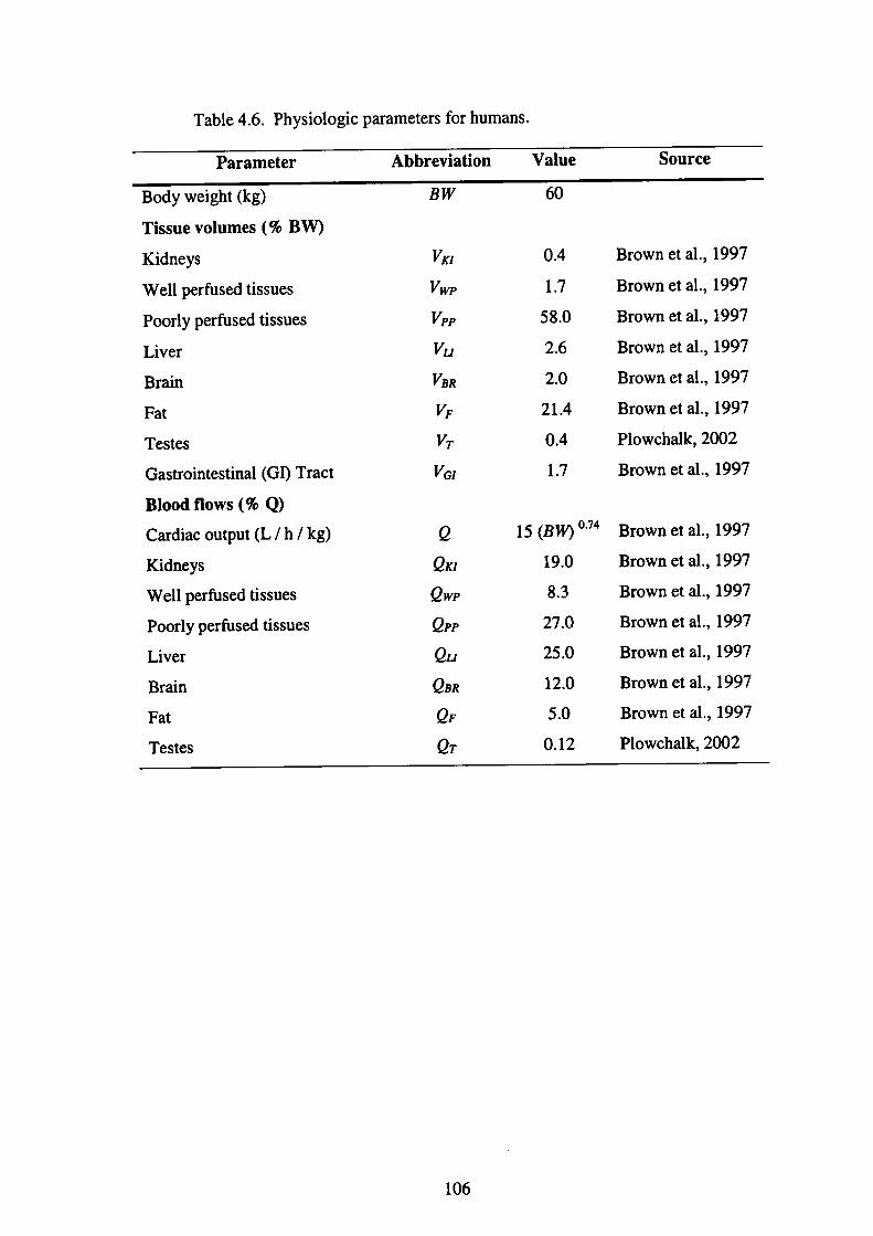
Table 4.6. Physiologic parameters for humans.
Parameter Abbreviation Value Source
Body weight(kg)
Tissue volumes(%BW)
Kidneys
Well pe血sed tissues
Poorly perfUsed tissues
Liver
Brain
Fat
Testes
Gastrointestinal(GI)Tract
Blood fiows(%Q)
Cardiac output(L/h/kg)
Kidneys
Well perfUsed tissues
Poorly perfUsed tissues
Liver
Brain
Fat
Testes
Bvv
喩晦撫晦撫昨巧㌧
e
eκI
eWP
OPP
eu
ρBR
OF
eア
60
0.4
1.7
58.0
2.6
2.0
21.4
0.4
1.7
15(BVV)°・74
19.0
8.3
27.0
25.0
12.0
5.0
0.12
Brown et al.,1997
Brown et al.,1997
Brown et al.,1997
Brown et al.,1997
Brown et al.,1997
Brown et al.,1997
Plowchalk,2002
Brown et al.,1997
Brown et al.,1997
Brown et al.,1997
Brown et al.,1997
Brown et al.,1997
Brown et al.,1997
Brown et al.,1997
Brown et al.,1997
Plowchal1(,2002
106
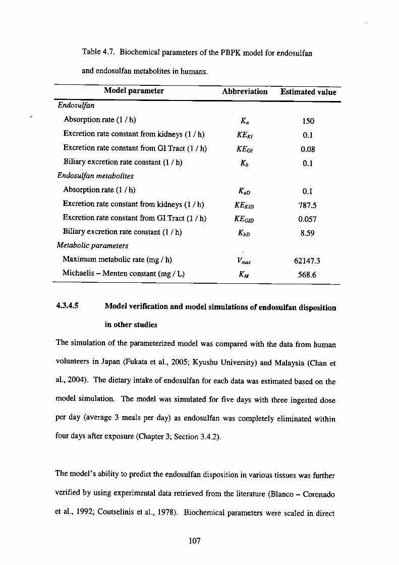
Table 4.7. Biochemical parameters of the PBPK model for endosulfan
and endosulfan metabolites in humans.
Model parameter Abbreviation Estimated value
Endosulfan
Absorption rate(1/h)
Excretion rate constant from kidneys(1/h)
Excretion rate constant from GI Tract(1/h)
Biliary excretion rate constant(1/h)
Endosulfan〃ietabolites
Absorption rate(1/h)
Excretion rate constant from kidneys(1/h)
Excretion rate constant from GI Tract(1/h)
Biliary excretion rate constant(1/h)
ルletabolic para〃zeters
Maximum metabolic rate(mg/h)
Michaelis-Menten constant(mg/L)
KEκI
KEGI
Kb
K。D
KEKID
KEGID
KbD
㌦砺
150
0.1
0.08
0.1
0.1
787.5
0.057
8.59
62147.3
568.6
4.3.4.5 Model verification and mode1 simulations of endosulfan disposition
in other studies
The simulation of the parameterized model was compared with the data from human
volunteers in Japan(Fukata et al.,2005;Kyushu University)and Malaysia(Chan et
al.,2004). The dietary intake of endosulfan for each data was estimated based on the
model simulation. The model was simulated for five days with three ingested dose
per day(average 3 meals per day)as endosulfan was completely eliminated within
f()ur days after exposure(Chapter 3;Section 3.4.2).
The model’s ability to predict the endosulfan disposition in various tissues was fUrther
verified by using experimental data retrieved from the literature(Blanco-Coronado
et al.,1992;Coutselinis et a1.,1978). Biochemical parameters were scaled in direct
107
proportion to the ratio of body weight as described in Section 4.3.4.1. For acute
intoxication or suicidal deaths, the model was simulated for 24 h with single ingested
dose of endosulfan. The ingested dose was predicted for data with unknown amo皿t
of ingested endosulfan.
4.4 Results
4.4.1 Parameterization and calibration
No overt toxicity was observed in any animal post-dosing. The data used for the
model simulations in this chapter were retrieved from Chapter 3(Section 3.4.3,
Tables 3.l and 3.2). The time course fbr cumulative amount of total endosulfan
excreted in urine and feces and model predictions are presented in Figure 4.3.
Cumulative amounts of total endosulfan excreted in urine and feces and model
predictions were also compared well with the experimental data. Following single
びoral administration of 5 mg/kg 14C-Endosulfan to male rats, the urinary excretion
of the radioactivity during 24 h was 9.6±21.4%where as the fecal elimination was
59.0±29.7%.The total radioactivity recovered in the excreta fbr four days was
106.8±26.2%(12.4±4.8%in urine and 94.4±21.4%in .feces). The results were
consistent with other studies that most of the radiocarbons were excreted in the urine
and feces after 96 h(Deema et al.,1966;Dorough et al.,1978;McGregor,1998).
Biochemical parameters that resulted in the best fit of the data are given in Table 4.4.
The parameterized model was compared with the experimental pharmacokinetic data
in male Sprague-Dawley rats after single oral administration of 5 mg/kg 14C-
Endosulfan. The resulting model simulations fbr blood and target tissues(liver,
kidneys, brain and testes)are shown in Figure 4.4. The model simulations correctly
108
captu「ed the shape°fthe exp・・im・…ld・…1ndica・i・g・h・t・th,m。d,l p,edi、,i。n、。f
t°talend°sulfan time c・u・・ep・・fil・・i・・11・iss・e・w・・ei・g,n,,al、g,eem,n,、with.,he
expe「1mental@data・G・・d・…i…n・y w…b・erv・d f。, t。,、1,nd。、ulfan
c°ncent「at1°ns i・1ive・・i・djca・i・g・h・t m・t、b。li、 kinetic
pa「amete「s were successfUlly
predicted.
442M°del v・・命ti・n・nd m・d・1・i輌・…f。nd。、uぬ。 di、p。、iti。n in
other studies
F°’1°w’ngm°delca1’b・ati・励ecalib・a・・dm・d・1w・・apPli・d…h,,epea,。dd。、i。g
expenmem(血ee-time「epea・・d・・a1・d輌・d・n・f 5 mg/kg). Gen,,ally,
「eas°nable ag「eement w…b・erv・db・・weenm・d・1P・edi・・i・。、and,xp。dm,。,、1d。,a
f°「aU@tissues・Th・・im…u・・ep・・m・・i・・arg…issu・, a。d m。d,1 P,edi、、i。。、 are
sh°wn@in Fig町・4・5・H・w・v・…im・1・・i・n・f・…1・。d。、uぬ。 di、p。,i,i。曲。m
栢dneysandlive・a負・・ml・ip1…al・d㎡・i・仕・・i…f5mg/kg,。m。1。,a、、re、。1,。d
mslight°ve「estim・t’・n・f・・ncen仕・・i・n・a・・11・im・p・i。・、 wh,,e as sim。1。,i。n。f
t°tal@end°sul伽di・p・・i・i・・廿・m・・・…re・・1・ed・i・・1igh・und。,e、、im。,i。n、,血,1、、t
tlme-P°int・butw・・g・…ally・…id・・ed・・beaccep・・b1。(日9。,e4.5).
Va「i°us expe「’ment・1 d・…etri・ved廿・m・h・li・era…e・n,。d。、。ぬ。 di、p。、i,i。n.in
v輌v°a負e「multiple°・al d・・ag・w・・e・im・1…dwi・h・u・・Φ・加g,h。 p,evi。u、1y
established@m°de1剛m・・ers・Sim・1…d…cent・a・i・n・。f。nd。、曲。 i。,h。 liver
and垣dneysa丘e「multipl…ald・・ag・・f5・・d 1・mg/kgb・dyw,igh,p。,d。晦a
pe「i°d°f 15 d・y・and・e…df・・an・・h・・15 d、y、 b,f。,e、ac,市
ce we「e generally in
ag「eement@with the expe「imen・・1・e・・1・・(Fl9・・e 46)(Chan・nd M噸2。。5).
An°the「study by A・・a・i…L(1984)・1・・i・dica・・d g。。d、g,eem。n, b。tween
109
exp。,im・…1・・d・im・1・t・d・・ncen廿・t1・…f・nd・・ul白・i・th・live・・b「ain and
垣d。,y、 aft・・m・ltipl…al d・・ag・・f 2・5・・7・5 mg!kg b・dy w・ight p・・day㊤「a
P・・i・d・f60 d・y・t・m・1・・at・(Fig・・e・4・7・nd 4・8)・
Sim。1。t。d。。。ce加、ti。n,・f・nd・・ul緬・th・垣d・・y・and live・aft・・mltipl…al
d。、ag。。f ll mg/kg P・・d・Y f・・ap・ri・d・f 30 d・y・t・m・1・・a・・(Figu「e 4・9)
、。mp訂。d w・ll wi也・xp・rim・nt・l d・t・(N・th・t・1・・1978)・H・weve・・血・・imulated
、。ncen仕、ti。n、 i。 th。 b・ai・and t・・t・・w・・e皿d・・e・tim・t・d・・mpar・d t・由e
exp,亘m・・tal・e・u1・・(Fig・・e 4.9)・The exp・・im・n・・1・e・ul…f・nd・・uぬ・levels in the
垣d。。y,, li…and bl・・d・f m・le an口ml・・a鱈・ft・・m・1・ipl…al d・・ag・・f 5 mg/
kg P,, d・Y f・・60 d・y・侮m・1・・at・)・nd 1・5 mg!kg P・・d・Y f・・6°d・y・(飴「female
,a,、)w。,e g・n・・ally・・mpar・d w・ll wi晒・・im・1・・i…(FigU・e・4・10孤d 4・11)
(Dikshith et aL,1984).
110

A:Urine 20
eX 15誓
警1・
∈
85 0
0 20 40 60 80
Tlm●, hour
100 120
蜘“ぬm8。6。ω釦・
B:Feces
0 20 40 60 80
Tlme, hour
100 120
Figu「e 4・3・C・mp・・i・・n・f PBPK m・d・1 P・edi、・i。n(1i。e、)、nd
expenmental cum・1・・i・・p・・cen・・g・(▲)・f・・i・ary(A)、。d fecal
elimjnati°n(B)・f・…1・nd・・ulfa・・丘er si・g1・・,al、dmini、t,a、i。n。f5
mg/kg ’4C-E・d・・u伽・m・1・・a・・(V・1…a,emean、±SD;f。,。=
3at each time poillt).
111

A
C
≒二
111
11:
121
1。
lii1
0.4白
0.3
咋
51015202530 nme, hour
5 10 寸5 20 25 30
1吊m●,けoor
B
2086420
e ”
l:::
§ 0.4
0.2
0
05to 152025 T㎞●,hoor
D
30
0 5 10 15 20 25
nm●, hour
30
‘
E
ii、
02
0.1
005 10 S5 20 25 30
’「lm●. hour
Figure 4.4. Comparison of PBPK model prediction(lines)and
experimelltal concentrations(▲)of total endosulfan in liver(A),
kidneys(B), brain(C), testes(D)and blood(E)following single oral
administration of 14C-Endosulfan to male rats(Values are means±
SD;for n =3 at each time point).
112
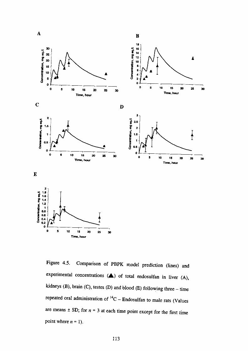
A
C
≒30 n
鋼
00
1,.,
闘 00
▲
1
▲
0
互
5 10 15 20 25 30
Wme, bOur
5 10 15 20 25 30
TIm●,㎞」r
1)
鷺
ljl:
0
0 5 10 15 20
「m●,hour
25
0 5 10 15 20
nme, hour
30
-25 30
E
il
0.8
0.6
0.4
0.2
00 5 10 15 20 25 30
nme, hour
Figu「e 4・5・C・mp・・i・・n・f PBPK m・d・l p・edi・ti・n(1ine、)、nd
expe「imental・・ncentr・・i・n・(▲)・f・…1・nd・・ulfa・i・live,(A),
kidney・(B)・b・ai・(C)・・・・…(D)・nd b1・・d(E)f・ll・wi。g,hree-,ime
「epeated°「al・d㎡・i・tr・ti…f 14C-E・d・・u脆・t・m・1・,at、(V、1。,、
a’e means±SD;f・・n・3・t・each・tim・p・i・t・xcept・f。, th, fi,、t tim。
point where n=1).
113
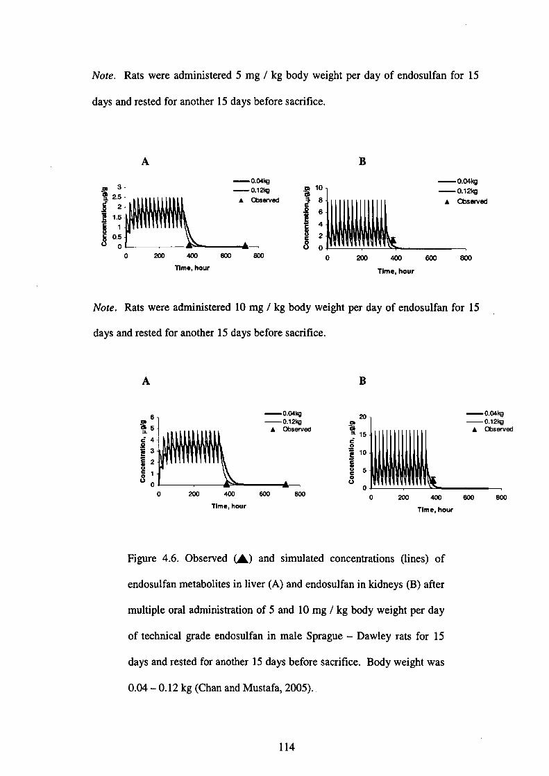
Note. Rats were administered 5 mg/kg body weight per day of endosulfan fbr 15
days and rested fbr another 15days befbre sacrifice.
3、
2.5
2
1.5
1
0.5
0
A 0.04㎏
O.12kg
▲ Obsαved
0 am 4。。 6。。 一二
Wm●, hour
0864201
B
O.04㎏
0 200 400 600
Tlme, hou「
800
Note. Rats were administered 10 mg/kg body weight per day of endosulfan for 15
days and rested fbr another 15 days befbre sacrifice.
A
6りB5三
亘4き3§2
§1 0
0.04㎏
OJ 2㎏
」 Observ
「一0
B
200 400 600
Time, hour
800
0.04㎏
0.12㎏
」 Observ
0 200 400 600
Tlme, hour
800
Figure 4.6. Observed(▲)and simulated concentrations(lines)of
endosulfan metabolites in liver(A)and endosulfan in kidneys(B)after
multiple oral administration of 5 and 10 mg/kg body weight per day
of technical grade endosulfan in male Sprague-Dawley rats fbr 15
days and rested for another 15 days before sacrifice. Body weight was
O.04-0.12kg(Chan and Mustafa,2005)..
114

A
0
、繊剖
B
500 1000
11m●,hou「
1500 2000
e41:
ll
田
鐵「
0 500 1000 1500
Tlm●. hour
2000
C
8:纈
▲ ()bseived
警。Pi:1
、So2 0.15き
8 0・t
玲 0 500 1000 1500
Time, hou「㎜
Fig・・e 4・7・Ob・e・v・d(▲)・・d・im・1・t・d・・ncentr・ti…(1ine・)。f
・nd・・曲・i・li…(A)・・kidn・y・(B)・nd b・ai・(C)・ft・・m・ltip1…al
administration of 2.5 mg/kg body weight per day of endosulfan in
m・1・・at・f・・60 d・y・・B・dy w・ight w・・0.06-0.08 kg(A・・ah・t、1.,
1984).
115

A
§ ’5
ξ1・
1・
5・ 0
8:鵬
▲ Observed
500 1000 1500
Wme, hour
2000
B
20864201噸1
0ρ6㎏
▲臨.d
500 1000 1500
Tim●, bOur
2㎜
C
0ρ鞠
▲曝剖言゜・8
ξρ6
種o.4毒・.・
§。0 500 1000 1500
Tlm●, hou「㎜
Figure 4.8. Observed(▲)alld simulated concentrations(lines)of
endosulfan in liver(A), kidneys(B)and brain(C)after multiple oral
administration of 7.5 mg/kg body weight per day of endosulfan in
male rats for 60 days. Body weight was O.06-0.08 kg(Ansari et aL,
1984).
116
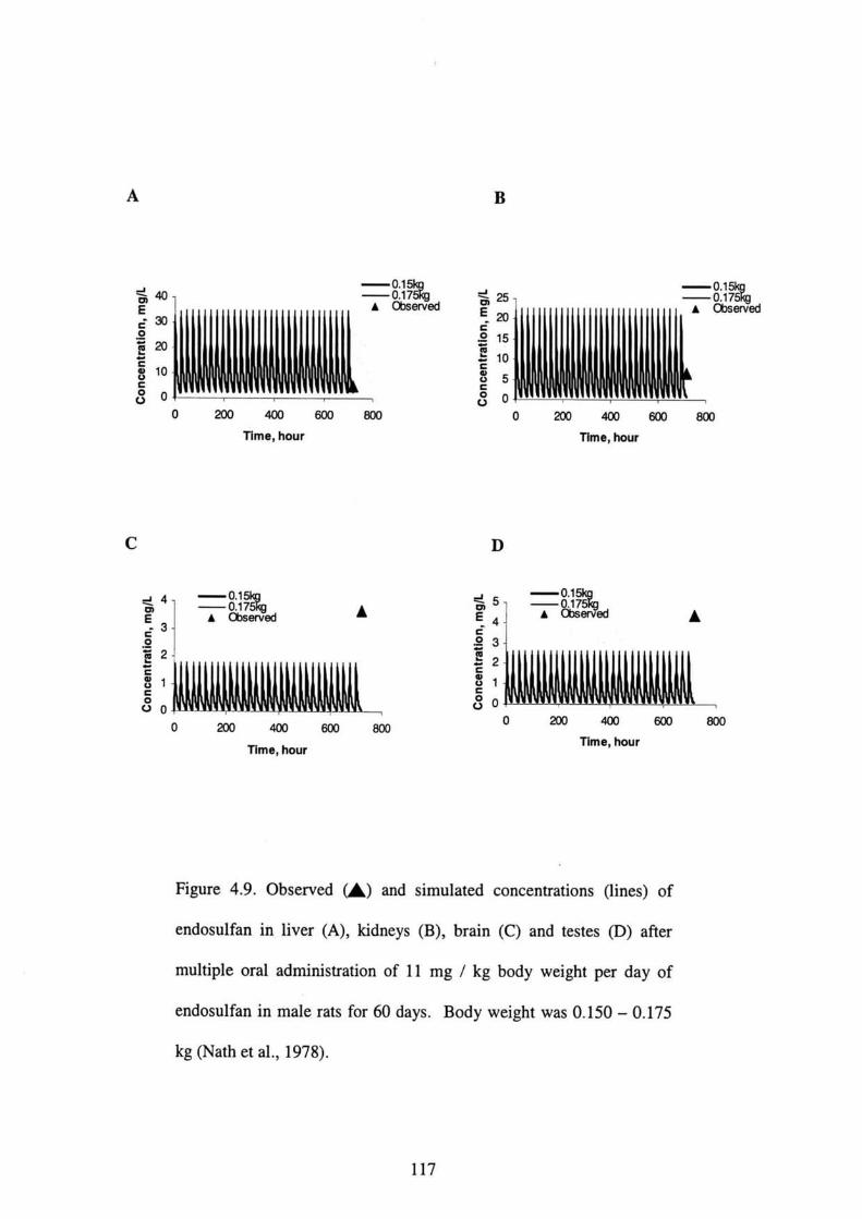
A B
▲
0 200 400 600
Tlme, hou「
800
8:隅
Observθd
お20 151050
一▲
0 200 400 600
Tlm●, hou「
議
800
C
8:跳▲ Observ6d
▲
0 200 400 600
Tlme, hou「
800
D
51[
▲繊、d▲
0 200 400 600
Tlme, hour
800
Figure 4.9. Observed(▲)and simulated concentrations(lines)of
endosulfan in liver(A), kidneys(B), brail1(C)and testes(D)after
multiple oral administration of l l mg/kg body weight per day of
endosulfan in male rats fbr 60 days. Body weight was O.150-0.175
kg(Nath et a1.,1978).
117
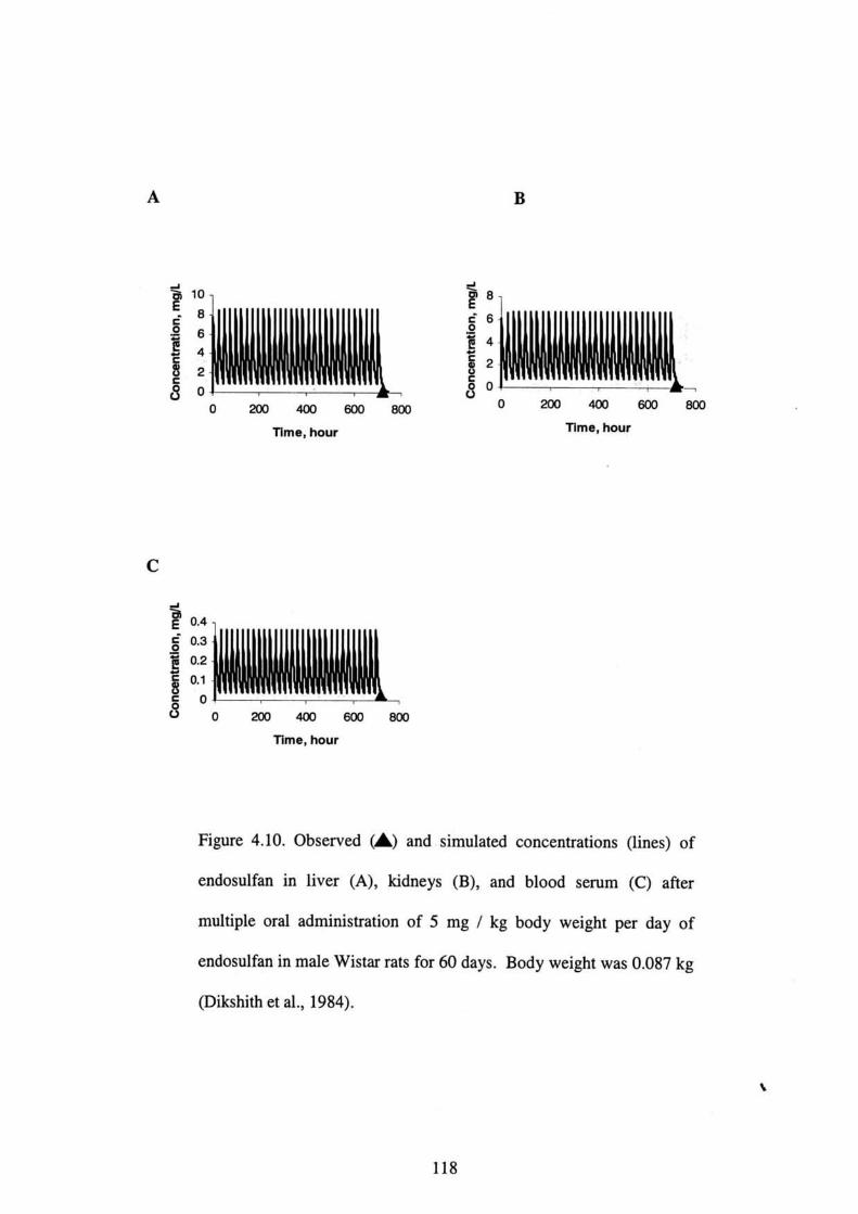
A B
086420ー
0 200 400 600
Tlme, hour
800
喜8
ξ6
ξ4§2ξ・
0 200 400 600
Tlme, hou「
「㎜
C
き。、
亘α3
盲o・2
§・1
5° 0 200 400 600 800
Time, hour
Figure 4.10. Observed(▲)and simulated concentrations(lines)of
endosulfan in liver(A), kidneys(B), and blood senlm(C)after
multiple oral administration of 5 mg/kg body weight per day of
endosulfan in male Wistar rats for 60 days. Body weight was O.087 kg
(Dikshith et a1.,1984).
s
118

A B
3
2
1
0
0 200 400 600
Time, hou「
800
525150
0 200 400 600
nme, hou「
800
C
醜iコ0.08
0.06
0.04
0.02
0
0 200 400 600 800
Time, hour
Fig・・e 4・11・Ob・e・ved(▲)・nd・im・1・t・d・・ncentr・ti・ns(line・)。f
endosulfan ill liver(A), kidneys(B), and blood serum(C)after
m・ltip1…a1・dmini・tr・ti・n・f 1.5 mg/kg b・dy w・ight p・・d・y・f
endosulfan in female Wistar rats for 60 days. Body weight was O.083
kg(Dikshith et al.,1984).
119
4.4.3 Sensitivity analyses
4.4.3.1 Endosulfan
The results of the sensitivity analyses at each time point(1 h,2h,4h,8hand 24 h)
that tissue endosulfan concentrations were calculated are shown in Table 4.8. With
regards to the impact of the PCs, the PCs for all the tissues had similar impact on the
endosulfan concentrations across all time points.
The sensitivity for maximum metabolic rate(Vmtvr)and urinary excretion rate(KEKI)
were consistently negative across tissue time, and extemal exposure concentration
where as the sensitivity for Michaelis-Menten constant(KM)was consistently
positive across tissue time, and externa 1 exposure concentration. The impact of Vntax
and KM on endosulfan tissue concentration was most evident at 8 h and 24 h. Other
parameters such as the absorption rate(Ka), biliary(Kb)and fecal excretion(KEGI)
rates for endosulfan had no impact in the blood and tissue concentrations of
endosulfan(Data not shown).
4.4.3.2 Endosulfan metabolites
The results of the sensitivity analyses at each time point(1 h,2h,4h,8h and 24 h)
that tissue endosulfan concentrations were calculated are shown in Table 4.9. With
regards to the impact of the PCs, the PCs for all the tissues had similar or little impact
on the endosulfan metabolites concentrations across all time points.
The sensitivity fbr maximum metabolic rate(Vmex)was consistently positive across
tissue timel and external exposure concentration except for the last time point at 24 h
where the sensitivity was negative. The sensitivity for Michaelis-Menten constant
120

(KM)was c°n・i・ten・ly neg・・ive ac・・ss tissue tim・,・nd ext・m、l exp。、。,e
c°ncen孜at1°n except f・・血・1・・t time at 24 h wh・・e th・・en・iti・ity f。, KM w。,e
P°sltlve・°the「par・m・・ers su・h…he ab・・叩ti…at・㈲・・d。,晦ex、,e,i。n
(κE…)・at・h・d litt1・impac・i・th・b1・・d・nd tissue・c。。centr。ti。n,。f。nd。、uぬn
metab°lites ac・・ss all・im・p・i・…Th・impact・f・fecal(KE。、D)and bili、,y ex、,e,i。n
(Ka・)・at…n・・d・・uぬ・m…b・1i…c・nce・廿・・i・n・w、, m。、、 evid,nt、t 24 h.
Tab1・4・8・S・n・iti・ity・n・1y・e・f・・the c・ncent・ati。n。f。。d。、uぬ。 i。
liver, kidneys, brain, testes and blood.
End。sulfan(mg/L) Time1h 2h 4h 8h
Lルerl」ver PC
Vmvc
KM
KE剛
Kidneys PC
Vm叙KMKEKl
Brain PC
Vm“
KM
KE閲
Testes PC
V晒KM
KE泊
Vm“KMKEi閲
24h
’)《●6ξ万”一一一“’’”◆“’”⊇’”一⇔’一’”一’一一一一一一一一一一一一一一一一一一一一一・・一一一・一一一一一一一一一一一一.一一..一一一.
一亘;ll万一一“一’”一一一一一一一一一’一”一一一一一一・一一・… 一・・一一一一・一一・一一一一一一・・一一.....一,一一一一一一,一一一’..一..一.
一一ウξiliξ゜一一”一一一“一一←一一“一’一“’一一一’”一・一・・一一一一一一一一一・・一一一一一一一一一・一一一一.一.一.一一.一.一..一一.一一...一..
1.38 鴫).29
0.27<ぺ).01
1.01
イ〕.05
0.05
<イ).01
1.45
・0.05
0.05
くく).01
1.04
イ).04
0.04
<<).01
0.99 イ).49
0.46
<㊨.01
1.01
℃.15
0、14
<イ).01
1.45
く).15
0.15
<弍).01
1.07
イ).14
0.13
<㊨,01
0.99 <D.72
0.68
<弍}.01
1.01
<).38
0.35
<イ).01
1.45
<[.38
0.36
<ぺ).01
1.07
く).37
0.35
<弍).01
一石」涜∂’≡一’”一゜t-一一一一”一一一’“’“一’’”一一一・・一一一一一一一一一・一・… 一一一一・一一一一一一一.一一一一一一一一.一一一一一一一..
く).05
0.05
<D.01
K).16
0.15
<弍).01
<}.38
0.36
くD,Ol
1.03
-1.20
1.14<イ).01
1.01
弍).84
0.80
く).01
1.45
弍D.84
0.80
<㊨.01
1.07
・{D.83
0.79
<弍).01
く).84
0.80
<℃.01
17P2
O4
O3
@01冷6603 45756703
1◇3℃
05ラ65臼
1立2㊨
欝砲
N°te・ Sen・iti・ity with・n・b・・1・t・・v・1…fO.50・, g,e、t。, i、 i。 b。1d.
an absolute value of less than O.01 was not shown.
Sensitivity with
121
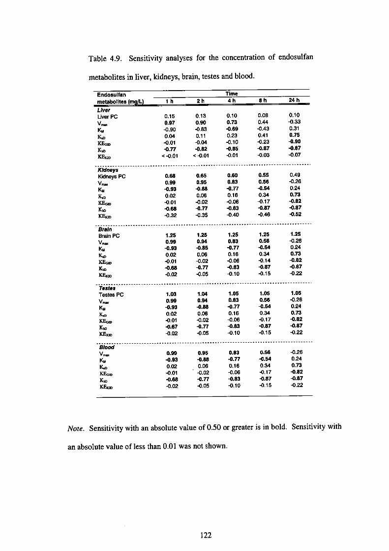
Table 4.9. Sensitivity allalyses for the concentration of endosulfan
metabolites in liver, kidneys, brain, testes and blood.
Endosulfan Tlme
-!:!1!111111ll.{11!llll t 2h 4h sh 24h
-o
P31万蜘”ロ
08S4イ41Ωぽ佃
10чP23㈹ぷ血
Oα㊨α℃㊨C
㎝認㎝㌫㊨㌫翻謂
㏄㍑浄監㌫
否■●●
49�ハ符鯉班Ω
O◇σα噸㊨0
…一5556胴鱗”廊調
……6・83召』・器 はぴゆロつゆゆ
……65篇。6劣卵
………6899鯛。2血豊
……c
野驚
‥…‥}19………………●●●一■●■■
25ィ餌符顕翻2
25T6ユ誕洞卯脂
25W3≠P6迫6顧沌
tα㊨O㊨㊨や25リぷ06皿刀価
…2599鯛促血鎚皿
…………陀…舳
Y監竃
■●
…‥ …………………………一}‥… …………‥●◆
05ユ別符鯉”92
t℃Oα㊨㊨◇05T6�鞄*|蒲
tO㊨0℃㊨㊨05W3≠P6迫6顧㈹
04X4≠O6皿刀迫6
03X9竄O2血翻皿
…㏄…欝鑑㌫
…讃腸認
…一■●●■
………………一◆
56ユ誕刀ガ怜
0㊨α℃㊨℃83≠P6迫6顧㈹
0㊨α℃㊨◇95冾O6皿刀迫6
α㊨α㊤㊨㊨
……‥99鯛02血臼舵
………≡隔監㌫
Note. Sensitivity with an absolute value of O.500r greater is in bold. Sensitivity with
an absolute value of less than O.01 was not shown.
122
4.4.4 Extrapolation from rat model to human model
4・4・4・1 M・d・1・・rificati・n・nd m・d・1・im・1・ti・n・・f・nd・・曲・di・p・・iti。。
in other studies
Th・f・ll・wi・g・ecti・n・f・thi・ch・pt・・p・e・ent・at・・t・fthe ability・f th。 PBPK m。d,l
fbr endosulfan in rats to predict the pharmacokinetic behavior of endosulfan in
h・m・…Th・par・m・t・rs f・・th・h・m・n PBPK m・d・1(T・b1・4.6)w。,e。bt、i。ed
solely by scale up of the parameters for the rat as described in Section 4.3.4.1. All
pa「amete…em・i・・d・・ch・ng・d・except・f・r th・Vmex・nd・KM, wh・・e th・・e p・・am・ters
were scaled up.
Figures 4.12 and 4.13 show the time course profiles in maternal semm and model
P「edi・ti・n・・f p・egnant w・m・吐・m J・p孤. The e・tim・t・d di・t・・y i・t、k。 w。、
simulated until reasonable agreement was observed between model simulations alld
experimental data. From the survey of thirty-two pregnant women who lived ill the
・iti・・(Chib…Y・m・n・・hi)ne訂T・ky・, J・p・n, i・2002・・d 2003(F・k、ta et。1.,
2005)・・nd・・uぬ・w・・d・tect・d i・th・m・t・m・1・emm・f也・・e・両㏄t,. Th。
c°ncen仕・ti・n w・・i・th・・ang・・f・d(・・n-d・tect・bl・)-8.4 P9/9. The e、tim。t。d
dietary intake fbr the su切ects was O.76 x 10“5 mg/kg/day.
F「°mth・d・t・・f・1ev・n p・egnant w・m・n li・i・9 ar・・nd th・Kyu・h・ ・・ea, J・p・n
・bt・i・・d f「・m Kyu・h・ Unive・・ity,・・d・・ulfa・(α一a・dβ一・・d・・ulfa。)with。
concentratlon range of 14.6-27.3 pg/g, where as endosulfan sulfate, with a
concentratlon range of nd-0.78 pg/gwere detected in the maternal serum. The
・・tim・t・d di・t・・y・i・t・k・f・・th・・ubject・w・・9.09・10’5 mg/kg/d・y.
123

Figure 4.14 shows the time course profiles in blood and model predictions ofpregnant
school children from Malaysia. The exposure of school children to pesticide residues
was investigated in a cross-sectional study involving 577 school children丘om 60
schools in Peninsular Malaysia. Only three su句ects had detectable residue levels of
endosulfan in the concentration range of nd-0.6 ng!g(Chan et aL,2004). The
estimated dietary intake fbr the su句ects was 1.06 x l Oづmg/kg/day.
Generally, reasonable agreement was observed between model predictions and
experimental data for blood. The time course profiles in blood and model predictions
are shown in Figures 4.12-4.14.
0.000009
m O.000008
豆o.㎜ピ 0.㎜量1:灘§・.・・…3
91:蒜 0
0 102030405060 708090100110120130 Tlm●, hour
Figure 4.12. Observed(▲)and.simulated collcentrations(lines)of
endosulfan in matemal serum of pregnant women f㌃om Chiba, Japan
fbllowing three-time ingested dose of endosulfan. Each volunteer
was assumed to consume meals three times per day(breakfast,1unch
and dinner). Each volunteer was expected to have their meals at an
average time of 0700,1300 and 1900 daily(Fukata et aL,2005). The
estimated dietary intake was O.76 x 10’5 mg/kg/day.
124
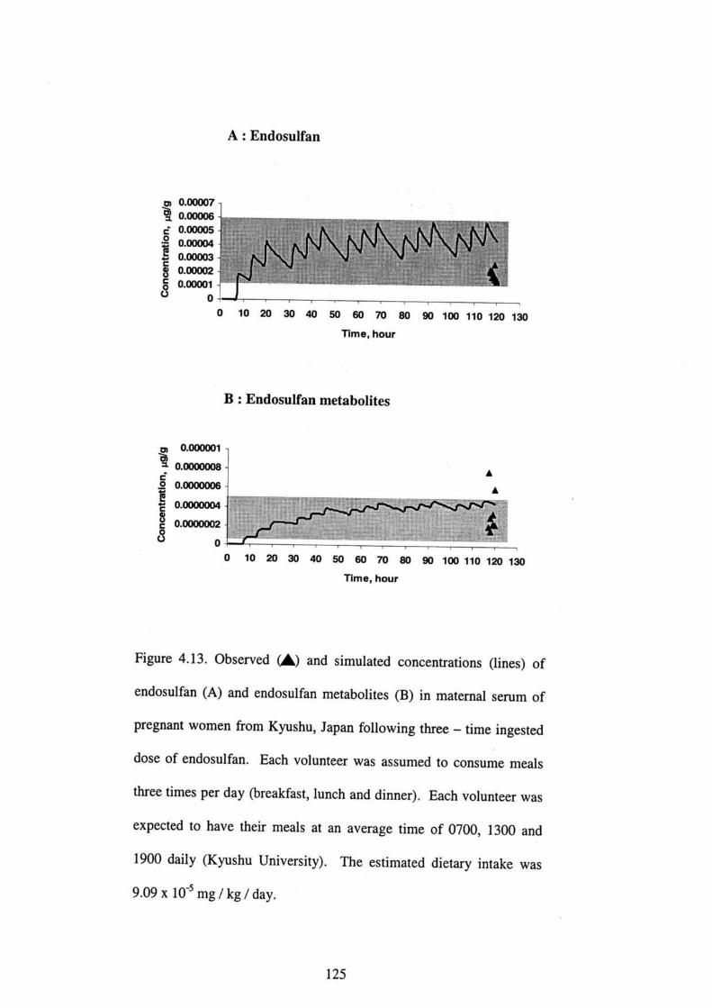
A:Endosulfan
00.00007盲o.oooo6
ピ0.00005
量1:畿
=
80・oooo2
ξα㎜1
0 10 20 30 40 50 60 70 80 90 100 110 120 130
nme, hour
B : Endosulfan metabolites
0.OOOOOt
O.0000008
0.0000006
0.㎜0.0000002
0
0 10 20 30 40
▲▲
50 60 70 80 90 100 110 120 130
Tlme, hour
Fig・・e 4・13・Ob・e・v・d(▲)・nd・im・1・t・d・・ncen廿・ti・n・(line・)。f
endosulfan(A)and endosulfan metabolites(B)in matemal senlm of
pregllant women from Kyushu, Japan fbllowing three-time ingested
dose of endosulfan. Each volullteer was assumed to consume meals
three times per day(breakfast, lunch and dinner). Each volunteer was
expected to have their meals at an average time of O700,1300 alld
1900d・ily(Kyu・h・U・i・…ity). The e・tim・t・d di・t・・y・i・t・k。 w。、
9.09x10’5 mg/kg/day.
125
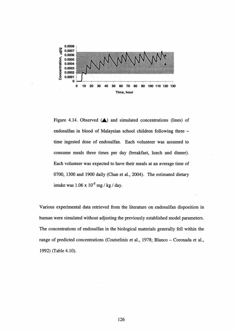
0.0008り 0.00079 .
ぎo.ooo5
宣o・ooo4乞 0.0003
80.oo促c80・OOOI O
0 10 20 30 40 50 6070 80 90100110120130 Tlm●, hour
Figure 4.14. Observed(▲)and simulated concentratiolls(lines)of
endosulfan in blood of Malaysian school children fbllowing thr㏄一
time ingested dose of endosulfan. Each vol皿teer was assumed to
consume meals three times per day(breakfast, lunch and di皿er).
Each volunteer was expected to have their meals at an average time of
O700,1300 and 1900 daily(Chan et al.,2004). The estimated dietary
intake was 1.06 x 10“5 mg/kg/day.
Various experimental data retrieved from the literature on endosulfan disposition in
human were simulated without a(ljusting the previously established model parameters.
The concentrations of endosulfan in the biological materials generally fell within the
range of predicted concentrations(Coutselinis et al.,1978;Blanco-Coronada et al.,
1992)(Table 4.10).
126

Tab1・4・10・C・ncent・ati・n・・f・・d・・ulf・n(α一・nd・S- end・・ulfa。)
and predicted range of concentrations in biological materials from
human.
Specimen
Ingested dose
Blood leve1-
peak
Brain
Liver
Kidney
Coutselinis et al.,1978
Case series(n=3)
Un㎞own(Predictedエ0.2 mg)
0.63mg!La
(0.12-0.03nig!L)
0.028pg!g
(0.18-0.05μg/g)
1・00μ9!9
(4.33-1.00μg/g)
2.OO P9/9
(2.24-0.61μg!g)
B】anco・一 Coronado et a1.,
1992
Case series(n=6)
Unknown
(Predicted=1.5)
0.87mg!La
(0.25-0.92mg!L)
Note.
Values in parentheses are predicted range of concentrations from the simulation.
a:Averaged blood level-peak.
4.5 1)iscussion
PBPK m・d・1・ar…ed剛・n・1y i・t・xi・・1・gy・nd亘・k assessm・nt・f・・e綱p。1、・i。n
ac「°ss d°se・「・ut・・f・xp・・u・e…nd・peci・・(Simm・n・et・1.,2002). h・a・ly・d、y、,。
typical PBPK m・d・1・・n・i・t・・f live・, P…ly p・・血・ed tissue・, w・11 P・・血、ed tissue、,
and白t(K・i・h・・n・nd A・d…en,1994). ln・・ea・i・gly, d・t、i1,d m。d,1、 with m。,e
c°m脚ments a「・b・i・g d・v・1・P・d t・・血・nce th・d・・c・ipti・n・fth・ph・・mac。kineti。、
°f・pecific c・mp・und・・The ad・・nt・g…fPBPK m・d・1・・mpar,d with、1assical
ph・nnac・ki・・ti・m・d・1・・e・(1)th・・e m・d・1・can p・・vid。 th。 time c。u,、e。f
127
distribution of xenobiotic to any organ or tissue;(2)they allow estimation of the
effects of changing physiological parameters of tissue concentrations(as done by
sensitivity analyses in the present study);(3)they can predict the toxicokinetics of
chemicals across species by allometric scaling;(4)complex dosillg regimes are easily
accommodated and more important, the resultant PBPK model have the potential fbr
extrapolations from observed data to predicted situations;while one of the major
disadvantages is that physiological input parameters are often ill-defined fbr various
strains and species(Medinsky and Klaassen,1996).
Given that no calibration or validation of PBPK model predictions of concentrations
of endosulfan in various tissues exist(ATSDR,2000;PubMed Database,2005), the
present study is the frrst attempt to do so and compares rat PBPK model predictions of
endosulfan to measured concentrations following single and multiple oral
administrations. It should be emphasized that the parent isomers were lumped
together in the current study as an initial step to developing the rat PBPK model.
Generally, results show reasonable concordance between model predictions and
measured values in all target tissues fbllowillg oral administration. Brain and testes
dosimetries of total endosulfan were reasonably predicted after oral exposures to i 4C-
Endosulfan with the a《ijustment of the brain:blood and testes:blood partition
coefficients.
For the present study, in vitro derived parameters for absorption and metabolism of
endosulfan were unavailable. Hence, these parameters were obtained by model
calibration to a single set of in vivo experimental data after rats were administered
single oral dose of 5 mg/kg 14C-Endosulfan, Subsequently, the validity of the
t
128
m°de’wastestedby・i伽1・・i・nwithth・three-tim・・epeat・dd。、eexperlm,nt、ld。ta
and°the「迦・d・…ne・d・・曲・di・p・・iti・n丘・m・h, li,,,a,。,e, whi、h。,e
’ndependent°fthe calib・a・i・n d・…A・・tt・mpt t・iden・i取、ep訂。,, i、。mers i。,he
cuπent study was imp・ssib1・due t・1・w・peci丘c acti・ity。f,adi。、a,b。。.
The simulation of endosulfa nc°ncent「ati°ns i・・h・・a・g…issue・aR・・m・1tipl。。,al
d°sagessh°ws由eab’1’・y・f由・m・d・1…xt・ap・1…丘・m・i・g1・。,ald。,ag。(Fig。,e
4旬t°multiple・・a’ d・・ag・・(Figti・e 4・5)・Th・・h・p・・f・h。㎞d。。y, and liver
c°ncen仕at1°n-time cu「ve・w・・e n・・w・llp・edi…d・丘・・m・1・ip1・。,al d。、ag。、. This
may be att「」b”tabl・t・・1・・m・・i・・p1・u・ib1・mech・m・ms su・h、、 di飽、i。n-1i㎡,、、i。n
°「ente「°hepatic「ec廿culat’・n・・b・・hi・・ccuπi・gi・・h・kidney・andlive,(K。y、e,。1.,
1999)・H・weve・,・nt…h・p・ti、,e、壮、u1、ti on was unlikely to occur fbr endosulfan
(D°「°ugh et a1・・1978;M・G・eg…1998)・D…ugh・nd・・-w。,kers(1978)
suggestedthattheb’1’a’y・m…b・1i・…f・・d…1塩・w・・1d・…n・…hee。t。,。h。p、、ic
cycle and we「e v・’d・d’・血・fece・・A…h・・p・ssib1・・ea・。。 m、y b, dueb也。、h。n
lntervals between administration and residu ewas not sufficiently excreted befbre the
next@ad血㎡s仕at’°n・H・nce・cau・i・n・h・uld be exe・ci・ed wh,。、pPlyi。g,h, m。d,h。
P「edictc°ncen仕ati°n・・f・・d・・uぬ・i・kid・・y・and livera丘・・m・hip1,。ral d。、ag,、.
Thi・al…ug9・・t・也・need・f・・血nherre,ea,ch.
Thesimulati°ns・fth・・e…di・・廿・m・h・1i…at・・eg・…ally・g,eewi,hexp,,imen,、1
data(Figu「es 4・6-4・11)・H・wev・…xp・・im・…1d・・、,ep。π。d。n end。、ul白n
disp°siti°n’n b・ai・・nd t・・・…fm・1・・at・(N・・h…L,1978)w,,e und。,e,,im、t,d
(Figu「e 4・9)・ReaΦ・tm・n・・f・h・p・ni・i・n…伍・ient・飼,、h,,e、pec,i。,,issues
「esulted@in an adequat・d・・c・ipti・n・f・・d…ぬ・di・p・・iti・n・ep・n,d i。,his s,。dy
129
(Data not shown). When evaluating these simulations, it has to be realized that these
studies sometimes differ from the calibration study in strain(Ansari et al.,1984;
Dikshith et al.,1984;Nath et al.,1978)or sex of the animals(Dikshith et aL,1984)
and also the volume and the type of vehicle used in which endosulfan was applied
(Chan and Mustafa,2005).
Sensitivity allalyses allow fbr a quantitative assessment of input parameters on the
model simulations of tissue concentrations(Simmons et al.,2002). The present
results illdicate that the influence of PBPK model input parameters on total
endosulfan tissue dosimetry varies, as expected, across parameter. Comparison of the
absolute magnitude of the sensitivity parameters may help guide decisions regarding
the usefUlness of data collection efforts to fUrther refine/define specific parameters
or may help to improve on the designs of fUture experiments.
It was observed that the estimated dietary intake fbr the pregnant women ffom
Kyushu area was l 2-fbld higher than the estimated dietary intake fbr the pregnant
women from Chiba or Yamanashi area with 9.09 x 10’5 mg/kg/day and O.76 x 10づ
mg/kg!day respectively. Residue levels of endosulfan from these women living in
the Kyushu area were higher[α一andβ一endosulfan(14.6-27.3 pg/g)and
endosulfan sulfate(nd-0.18 pg/g)]than those living in the urban areas in Chiba or
Yamanashi[endosuぬn(nd-8.4 pg/g)]. This could be attributed that perhaps some
parts of the Kyushu area, where the samples were collected were of agricultural areas
and fanning activities were carried out intensively in those areas. In add輌tion, these
su切ects may also be exposed to endosulfan through consumption of fbod
contaminated with endosulfan. Recent studies regarding predictors of organochlorine
130
levels indicat・th・t exp・…eam・・g・h・g・n・・al p・P・1・・i・n・cc…m・i・1y・hr。ugh,he
diet(Devoto et a1.,1998;Laden et a1.,1999).
E”d°sulfa・i・p・・㎡tt・d f・・agricul…a1・・e i・J・p…1・h・ugh it, u、ag。 i, re、Ui。t。d
(田F・2002)・R・・idue・・fend・・ul白・w・・e d・・ec・・d i・・he umbilical、。,d、emm廿。m
血enewb°m babi・・{α一・ndβ一・nd・・uぬ・(16・1-44.8 P9/9)and end。、u1飽。
suぬte(nd-°・78 P9/9)]・R・・idue・・f・nd…ぬ・w・・e al・・f・・nd i。 high lev,1、 in
theb「east milk f「・m也・p・egnant w・men[α一・ndβ一・・d・・ul白・(19.・-52.g P9/9)
and end°sulfa・・ulfa・・(・d-7…P9/9)]・i・dicati・g・h・tth・・e・ubjec・・w。,e exp。、ed
to high levels of endosulfan.
ln a surv・y…duc・・d by F・k・ta a・d・・-w・・k…(2005),,e、idue、。f。。d。、ulfan
we「e detect・d i・the umbilica1…d(・d-7.2 P9/9)b・・id・・m。・,m、1,emm. Th。、e
su「veys indicat・d th・t・nd・・ulfa・w・・tr・n・ferr・d fr・m m。th,, t。 f。t。、 a。d
b「eastfeedi”g w…h・high・…xp・・u・e・・u・ce a・i・w…h・m・i・m・・h。d f。, excre,i。n,
也us「aising a cau・e f・・c・n・em・ince m…m・1・・xic ch・血cal・d。,i。g,he c,i,ical
devel°pment ph・・e m・y gi・・ri・e・・fe・・1・・u・…xi・i・y(Sar・i・・11i…1.,2・・3;Til、。n,
1998).
Gene「ally・・ea・・n・b1・・9reem…w…b・erv・d b・・ween-m・d,l p,edi、,i。。、 a。d
expe「imental d・t・f・・ bl・・d・・n Fj9・・e・4・12・nd 4.14,・h・・b・erv,d,a。g,。f
expe「1mental data f°「th・p・eg・・nt w・m・n fr・m Chib・f・ll・wi・hi・・h・・im・1・t・d,ang。,
thus indicati・g・h・t th・・ewly-d・v・1・P・d m・d・1 h…he capaci・y・。,ep,。duce、he
°bse「ved・ang・・f・nd・・ulf・n・・ncentr・ti・n i・h・m….・n・Fig。,e 4.13,,h。。b、erv。d
「ange°f exp・・iment・l d・t・f・・th・p・eg・・nt w・m・n fr・m Kyu、h。 f,ll withi。 the
131

simulated range for endosulfan and endosulfan metabolites. By comparing the
experimental data with the simulation results for endosulfan, the experimental data fbr
endosulfan metabolites fell within the simulated range, thus indicating that the model
can be partially validated. This also demonstrates that the parameters used in the
current model were quite well predicted and the model can be applied to estimate the
concentrations of endosulfan in human tissues. The estimated dietary intakes fbr the
pregnant women from Chiba and Kyushu as well as school children from Malaysia
were below the acceptable daily intake(ADI), in which ADI for endosulfan in human
was O.008 mg!kg/day(WHO,1984). This suggests that the low estimated dietary
intakes fbr these groups of people will not cause serious health effects. However,
other possibilities cannot be Iuled out.
At present, the human PBPK model cannot strictly be validated since data conceming
human exposures are scarce and limited to a small number of cases, with only
sporadic data on the tissue concentrations of endosulfan and its isomers.
Toxicokinetic data in humans are lacking. All of the cases reported were of acute
intoX ication or suicidal attempts caused by ingestion of endosulfan. Another problem
fbr model validation may be due to the large differences among individual
concentration data. These differences in concentration may be partially explained by
differences in body weight, age, gastroilltestinal absorption and fbod consumption
habits.
132
4.6 Conclusions
In summary, it is concluded that the frrst and new PBPK model for endosulfan in male
rats has been developed in the present study and can predict tissue dosimetries
fbllowing single and multiple oral dosages of endosulfan. The model was verified
with experimental data retrieved from the literature. This model provides the
foundation for development of a more complex physiologi6ally based dynamic model
that would provide a basis for the design of fUrther experimental investigations. A
more reliable and sensitive analytical method would also be incorporated into our
fUture experiments to enhance the separation and identification of the parent
compo皿ds alld metabolites present in different matrices. Further research is also
necessary to expand its current model for endosulfan to include the two isomers of the
parent compounds and its major oxidation metabolites.
Generally, reasonable agreement was observed between model predictions and
experimental data for the extrapolated PBPK model from rat to human. This suggests
that the parameters used in the model were quite well predicted and the model has the
capacity to be applied to estimate the concentrations of endosulfan in human tissues.
However, more studies on human exposures to endosulfan are necessary in order to
validate the model for fUture health risk assessments..
Extensioll of the model with in vitro experimental data on the toxicodynamics of
endosulfan in the CNS would facilitate the interpretation of endosulfan’s
neurotoxlclty in vivo. Such studies are currently ca㎡ed out in our laboratory. The
extended model would also provide the basis fbr more accurate and reliable risk
assessments of the e脆cts of endosulfan on neurotoxicity alld male reproductive
133
toxicity since the CNS and testes are among the target organs fbr the respective toxic
effects. It is hoped that the newly developed model could be utilized in the human
health risk assessment in near fUture, particularly in the human reproductive and
neurotoxic risks.
REFERENCES
Aarons, L., Clewell, H.」., Conolly, R. B., Delic, J.1., Houston, J. B., Jarabek, A. M.,
Loizou, G., Mason, H.」., Nestorov,1., Rowland, M., Trans, C. L, Tucker, G. T.
(1999)・Physiolo8ically-based phar〃iacokinetic〃iode’in8:A、ρotential tool foアuse
in risk・asse∬〃lent・Report of a workShop organised by the Risk・A∬e∬〃zent and
Toxicology Steerin8 Committee. lnstitute for Environment and Health, Leicester, UK,
PP.3-29.
Agrawal, A. K., Anand, M., Zaidi, N, F., Seth, P. K.(1983). lnvolvement of
serotonergic receptors in endosulfan neurotoxicity. Biochemical Pharmacology,32,
3591-3593.
Aleksandrowicz, D. R.(1979). Endosulfan poisoning and chronic brain syndrome.
ノlrch加es()f Toxicolo8y,43,65-68.
Anand, M., Khanna, R. N., Misra, D.(1980). Electrical activity of brain in
endosulfan toxicity.」rndian Journal(ゾPhar〃laco’08y,12〈4),229-235.
Andersen, M. E.(2003). Toxicokinetic modeling and its application in chemical risk
assessment. Toxico~08y Lθ〃εr∫,138:9-27.
134
Ansari, R. A., Husain, K., Gupta, P. K.(1987). Endosulfan toxicity influence on
biogenic amines of rat brain. Journal ofEnvironmental Biology,8,229-236.
Ansari, R. A., Siddiqui, M. K. J., Gupta, P, K.(1984). Toxicity of endosulfan:
distribution of alpha-and beta-isomers of racemic endosulfan fbllowing oral
adlninistration in rats. Toxicology Letters,21,29-33.
Antonious, G. F., Byers, M. E., Snyder, J. C.(1998). Residues and fate of endosulfan
on field-grown pepper and tomato. Pesticide Science,54,61-67.
ATSDR(2000).伽・・1・8i・吻・φ1ゆ励卿・. Atl・n伍, GA・Ag・n・Y f。,
Toxic Substances and Disease Registry.
Bhattacharya, B., Sarkar, S. K., Mukheilee, N.(2003). Organochlorine pesticide
residues in sediments of a tropical mangrove estuary, ildia:㎞plications fbr
monitoring. Environment lnternational,29,587-592.
Blanco-Coronado, J. L, Repetto, M., Ginestal, R. J., Vicente, J. R., Yelamos, F.,
Lardelli, A・(1992). Acute intoxication by endosulfan. Clinicat To」πicology,30,575-
583.
Boereboom, F. T. J., van Dijk, A., van Zoonen, P., Meulenbelt, J.(1998).
Nonaccidental endosulfan intoxication:Acase report with toxicokilletic calculations
and tissue concentrations. Clinical Toxicolo8y,36,345-352.
135
Brandt, V. A., Moon, S., Ehlers,」., Methner, M. M., Struttmann, T.(2001). Exposure
to endosulfan in farmers:Two case studies.んηεricoπ」と)urnal〈)f lndustrial Medicine,
39,643-649.
Brown, R. P., Delp, M. D., Lindstedt, S. L., Rhomberg, L R., Beliles, R. P.(1997).
Physiological parameter values for physiologically based pharmacokinetic models.
Toxicoto8y and lndustrial Health,13,407-484.
Castillo, C. G., Montante, M., Dufour, L, Martinez, M. L, Jim6nez-Capdeville, M.
E.(2002).Behavioral effects of exposure to endosulfan and methyl parathion in adult
rats. Neurotoxicolo8y and Teratology,24,797-804.
Chan, M. P. L, Mustafa, A. M.(2005). Analysis of endosulfan’and its metabolites in
rat plasma and selected tissue samples by gas chromatograph-mass spectrometry.
Environmentat Toxicology,20(1),45-52.
Chan, M. P. L, Mustafa, A. M., Hussein, R., Hj Hussain, S., Zulkifli, S. N., Abdullah,
A.R.(2004). Pesticide residues in blood of schoolchildren from selected schools in
Peninsular Malaysia. Environmental Hea~th Focus,2(1),18-24.
Chitra, K.
endosulfan
206.
C.,Latchoumycandane, C., Mathur, P. P.(1999). Chronic effect of
on the testicular fUnctions of rat. ノlsian Journat〔ゾノlndrolo8y,1,203一
136

Choudhary, N., Joshi, S. C.(2003). Reproductive toxicity of endosulfan in male
albino rats.」Bulletin of Environmental Contamination and Toxicolo8y,70,285-289.
Coutselillis, A., Kelltarchou, P., Boukis, D.(1978). Concentration levels of
endosulfan in biological material(Report of three cases). Forensic Science,11:75.
Deema, P., Thompson, E., Ware, G. W.(1966). Metabolism, storage and excretion
of C 14 一 Endosulfan in the mouse. J()urnal of Economic Entomolo8y,59(3),546-
550.
Demeter, J, Heyndrickx, A., Timpemlan, J., Lefevere, M., De Beer, J.(1977).
Toxicological analysis in a case of endosulfan suicide. Bulletin of Environmental
Contamination and Toxicology,18,110-114.
Devoto, E. Kohlme廿, L, Heeschen, W.(1998). Some dietary predictors of plasma
organochlorine concentrations in an elderly Gemlan population. ∠4rchiveぷ q〆
Enviアonη昭ntal Health,53:147-155.
Dikshith, T. S. S., Raizada, R. B., Srivastava, M. K., Kaphalia, B. S.(1984). Response
of rats to repeated oral administration of endosulfan. Industrial Health,22,295-304.
Dorough, H. W., Huhtanen, K., Marshall, T. C., Bryant, H. E.(1978). Fate of
endosulfan in rats and toxicological considerations of apolar metabolites. Pesticide
Bioche〃listり)and Physiology,8,241-252.
137

EJF・(2002)・End of the road/br endoぷulfan:αcα〃ノbr action againstαdとzn8erous
pesticide. Environmental Justice Foundation Ltd, London, UK, pp.1-4.
Eyer, F., Felgenhauer, N., Jetzinger, E, Pfab, R., Zilker, T. R.(2004). Acute
endosulfan poisoning with cerebral edema and cardiac failure. Journat of Toxicolo8y
and Clinical Toxicolo8)),42(6),927-932.
FAO/WHO(1969). FA O/PL:1968/ルf/9/1.1968 Evaluationぷof so〃ie pesticide
residueぷin/bod. Geneva:Food and Agricultural Organization of the United Nations,
World Health Organization.
Fukata, H., Omori, M., Osada, H., Todaka, E, Mori, C.(2005). Necessity to measure
PCBs and organochlorine pesticide collcentratiOns in human umbilical cords for fetal
exposure assessment. Environmental Health Perspectives,113:297-303.
Gam6n, M., S4ez, E, Gil,」., Boluda, R.(2003). Direct and indirect exogenous
contamination by pesticides of rice-fatming soils in a Mediteπanean wetland.
ArchiveぷofEnvironmental Contamination and Toxicolo8y,44,141-151.
Gehring, P. J., Watanabe, P. G., Park, C. N.(1978). Resolution of dose-response
toxicity data fbr chemicals requiring metabolic activation:Example 一 vinyl chloride.
Toxicolo8y and Applied Pharmacolo8y,44:581-591.
Gonz61ez-Farias, F., Cisneros, E. X., Fuentes, R. C., Diaz, G. G., Botello, A. V.
(2002).Pesticides distribution in sediments of a tropical coastal lagoon a(ljacent to an
138
irrigation district in nortwest Mexico. Environmental Technolo8y,23(11),1247-
1256.
Hayes, W. J., Laws, E. R.(1991). Handbook of pesticide toxicology. San Diego:
Academic Press.
Jepson, G. W., Hoover, D. K., Black, R. K., McCafferty,」. D., Mahle, D. A., Gearhart,
J.M.(1994). A partition coefficient detemination method fbr nonvolatile chemicals
in biological tissues. Funcla〃iental and ApPlie∂Toxicotogy,22,519-524.
Keys, D. A., Wallace, D. G., Kepler, T. B., Conolly, R. B.(1999). Quantitative
evaluation of altemative mechanisms of blood and testes disposition of di(2-
ethylhexyl)phthalate and mono(2-ethylhexy1)phthalate in rats. ]roxico’08ical
Sciences,49,172-185.
Krishnan, K., Andersen, M. E.(1994). Physiologically based pharmacokinetic
modeling in toxicology. In:Hayes, A. W.(Ed.), Principlesαη4 MethodS仇
Toxicolo8y 3「d ed., PP.149-148. New York:Raven Press.
Laden, F., Neas, L. M., Spiegelman, D., Hanldnson, S. E., Willet, W. C.,丘eland, K.,
Wolff, M. S., Hunter, D.」.(1999). Predictors of plasma conce加ations of DDE and
PCBs in a group of US women. Eηvかonη1θη’al Health Perspectives,107:75-81.
139
Leung, H. W., Paustenbach, DJ.(1995). Physiologically based pharmacokinetic and
phamacodynamic modeling in health risk assessment and characterization of
hazardous substances. Toxicolo8y Letters,79:55-65.
Lo, R. S. K., Chan,」. C. N., Cockram, C. S., Lai, F. M. M.(1995). Acute tubular
nec…i・f・ll・wi・g end・・ulph・n i・・ecti・id・p・i・・㎡・g. Cli。i・。1伽。。1。8y,33,67-
69.
Louie, P. K. K., Sin, D. W.(2003). A preliminary investigation of persistent organic
pollutants in ambient air in Hong Kong. Chemosphere,52,1397-1403.
McGregor, D・B・(1998)・Endosulfan(7MPR evaluationぷ1998 Part II Toxico’08匡cα1)
rF輌rst drafi. Lyo11, France:hltemational Agency fbr Research on Cancer.
Medinsky, M. A., Klassen, C. D.(1996). Toxicokinetics. hl C. D. Klaassen(Ed.),
Casarett and Dou〃ぷToxicolo8y:T7ze Basic Science of Poisons, S「h ed., pp.187-198.
New York:McGraw-Hil1.
Moolgavkar, S., Krewski, D., Schwarz, M.(1999). Chapter 7:Mechanisms of
carcinogenesis and biologically based models fbr estimation and prediction of risk.
In:Moolgavkar, S., Krewski, D., Zeise, L, Cardis, E, Moller, H.(Eds.), euantitative
Estiruation and Prediciton of Human Cancer Riskぷ.1んRC Scientiific Publicationぷ1Vo.
131.International Agency fbr Research on Cancer, World Health Organization, Lyon,
France, pp.179-237.
140

Nath, G., Datta, K. K., Dikshith, T. S. S., Tandon, S. K., Pandya, K. P.(1978).
lnteraction of endosulfan and metepa in rats. Toxicolo8y,11,385-393.
National Cancer Institute(1978). Bioa∬ay of endosulfan/br」po∬ibte carcino8enicity,
Technical Report, Series No.62, Pu●lication No.(MH)78-1312. U. S. Department
of Health, Education and Wel白re.
Novak, B., A㎞ad, N.(1989). Residues in fish exposed to sublethal doses of
endosulfan and fish collected from cotton growing area. Journal of Environmental
Science and Health, Part B B24,97-109.
Oktay, C., Goksu, E., Bozdem丘, N., Soyuncu, S.(2003). Unintentional toxicity due
to endosulfan:acase report of two patients and characteristics of endosulfan toxicity.
レ「eterinary and Hu〃zan Toxicolo8ツ,45(6),318-320.
Paul, V., Balasubramaniam, E, Kazi, M.(1994). The neurobehavioral toxicity of
endosulfan in rats:a serotogenergic involvement in leaming impairment. European
Journal of Pharmacology,270,1-7.
Plowchalk, D. R., Teeguarden, J.(2002). Development of a physiologically based
pharmacokinetic model fbr estradiol in rats and humans:abiologically motivated
quantitative framework for evaluation responses to estradiol and other endocrine-
active compounds. Toxicolo8ical Sciences,69,60-78.
141

Poulin, P., Krishnan, K.(1995). An algorithm for predicting tissue:blood partition
coefficients fbr organic chemicals丘om n-octanol:water partition coefficient data.
Journal of Toxicolo8y and Environmental Health,46:117-129.
PubMed Database.(2005). National Center for Biotechnology lnformation(NCBI).
National Library of Medicine.
Available at htt:〃www.ncbi.nlm.nih. ov/entrez/uer.fc i?db=PubMed
Ramsey,」., Anderson, M. E(1984). A physiologically based description of the
inhalation pharmacokinetics of styrene in rats and humans. Toxicology and Applied
Pharmacolo8y,73,159-175.
Saiyed, H., Dewan, A., Bhatnagar, V., Shenoy, U., Shenoy, R., Rajmohan, H., Patel,
K.,Kashyap, R., Kulkami, P., Raj an, B., Lakkad, B.(2003). Effect of endosulfan on
male reproductive development. Environmental Health Perspectives,111(16),1958
-1962.
Sarcinelli P. N., Pereira A. C. S., Mesquita S. A.,01iveira-Silva J. J., Meyer A.,
Menezes M. A. C., Alves S. R., Mattos R. C.0. C., Moreira J. S., Wolff M.(2003).
Dietary and reproductive determinants of plasma organochlorine levels in pregnant
women in Rio de Janeiro. Environmental Research,91,143-150.
Seth, P. K., Saidi, N. F., Agrawal, A. K., Anand, M.(1986). Neurotoxicity of
endosulfan in young and adult rats. Neurotoxicolo8y,7(2),623-636.
142

Simmons,」. E, Boyes, W. K., Bushnell, P.」., Raymer,」. H., Limsa㎞n, T.,
McDonald, A., Sey, Y. M., Evans, M. V.(2002). Aphysiologically based
phamlacokinetic model fbr trichloroethylene in the male Long-Evans rat.
Toxicolo8ical Sciences,69,3-15.
Sinha N., Adhikari, N., Narayan, R., Saxena, D. K.(1999). Cytotoxic effect of
endosulfan on rat Sertoli-germ cell coculture. Reproductive Toxicolo8y,13,291-
294.
Sinha, N., Adhikari, N., Saxena, D. K.(2001). Effect of endosulfan during fetal
gonadal differentiation on spermatogenesis in rats. Environmental Toxicology and
Pharmacolo8y,10,29-32.
Sinha, N., Naraya11, R., Shanker, R, Saxena, D. K.(1995). Endosulfan induced
biochemical changes in the testis of rats. レ「eterinary and Hu〃zan Toxicolo8y,37,547
-549.
Sinha, N., Naraya11, R., Saxena, D. K.(1997). Effect of endosulfan on the testis of
growing rats・Bulletin ofEnvironmental Contamination and Toxicolo8y,58,79 一 86.
Subramoniam, A., Khama, V. K., Mohindra, J., Seth, P. K.(1994). Reduced
phosphoinositide levels in endosulfan treated rat brain. Indian Jと)urnal of
Pharinacolo8y,26,49-51.
143
Tan, G. H., Vijayaletchumy, K.(1994). Organochlorine pesticide residues levels in
Peninsular Malaysian rivers. Bulletin of Environmental Contamination and
Toxicology,53,351-356.
Tilso11, H. A.(1998). Developmental neurotoxicology of endocrine disnlptors and
pesticides:Identification of information gaps and research needs. Environmental
Health Perspectives,106:807-811.
Wilson, V. S, LeBlanc, G. A.
biotransformation and clearance in
Pharmacology,148,158-168.
(1998). Endosulfan elevates testosterone
CD - 1 mice. Toxicology and Applied
144
CHAPTER 5
DEV肌OPMENT OF AN IN VITRO BLOOD-BRAIN BARRIER MOD肌TO
STUI)Y THE PERMEABILITY EFFECTS OF ENDOSULFAN ON THE
TIGHT JUNCTIONS
5.O Introduction
As has been discussed in Chapter 2, endosulfan, a neurotoxic insecticide is involved
in the neurotoxicity of its central nervous system(CNS). The primary site of action of
endosulfan is believed to be the CNS. Involvement of CNS in the neurotoxicity of
endosulfan has been shown by several in vivo experiments(Anand et al.,1980;Paul et
a1.,1995;Seth et al.,1986;Subramoniam et al.,1994;Zaidi et al.,1985). However,
the precise mechanism of its toxicity and whether endosulfan causes a direct effect on
tight j皿ctions at the blood-brain・barrier(BBB)remained・unidentified.
5.0.l Blood-brain barrier(BBB)
BBB is fbrmed by complex tight jullctions of the brain capillary endothelial cells
(ECs)and expresses various transport systems. ECs are joilled by tight intercellular
junctions that provide a biological barrier to maintain the homeostasis of the brain
microenvironment. The distinct property of this barrier can be atnibuted, in卿, to
the presence of a continuous ring of tight junctions between neighboring cells. BBB
protects the brain from the blood milieu, and delivery of ions and solutes from blood
to CNS is limited by the selectivity of BBB.(Cucullo et aL,2004;Igarashi et al.,
1999;Jean Harry et al.,1997;Lauer et al.,2004;Lee et al.,2004;Lu et al.,2005;
Ohtsuki,2004;Rubin and Staddon,1999). Figure 5.1 shows the essential features of
the BBB.
145

From a toxicological viewpoint, three aspects of the BBB are of interest:(a)the BBB
regulates uptake and release of endogenous substances and also xenobiotics,(b)toxic
substances may interfere with the structural and fUnctional properties of the BBB, and
(c)certain parts of the CNS(e. g., areas in the hypothalamus and the choroids plexa),
have poorly developed BBB functions. The latter is also true fbr all parts of the
embryonic and juvenile brains[European Center fbr Validation of Altemative
Methods(ECVAM)].
5.0.2 Endpoints for acute toxic effects
For acute toxic effects, there are two endpoints for toxic insult to the BBB:(a)partial
or complete breakdown of the barrier fUnction(i. e., effects on the ability of the B B B
to exclude endogenous and exogenous substances)and (b)changes in the specific
transport capacity of the BBB. There is a need to measure the ability of the nomlal
BBB to transport toxicants into or out of the brain(ECVAM).
The literature varies considerably conceming the different BBB in vitro models set up
during the past decades, using primary endothelial cells and astrocytes or glial cells,
cultured under different experimental conditions and usillg di脆rent cell sources.
Several in vivo and in vitro models fbr estimation of brain permeation have been
proposed in the literature(Pardridge,1995). Most commonly, bovine, rat or porcine
endothelial cells are used for in vitro models.
5.0.3 Transendothelial electrica1 resistance(TEER)
The analysis of transendothelial electrical resistance(TEER)is a simple method to
quantify the fUnctionality of the tight junctions of the iil vitro BBB. TEER represents
146
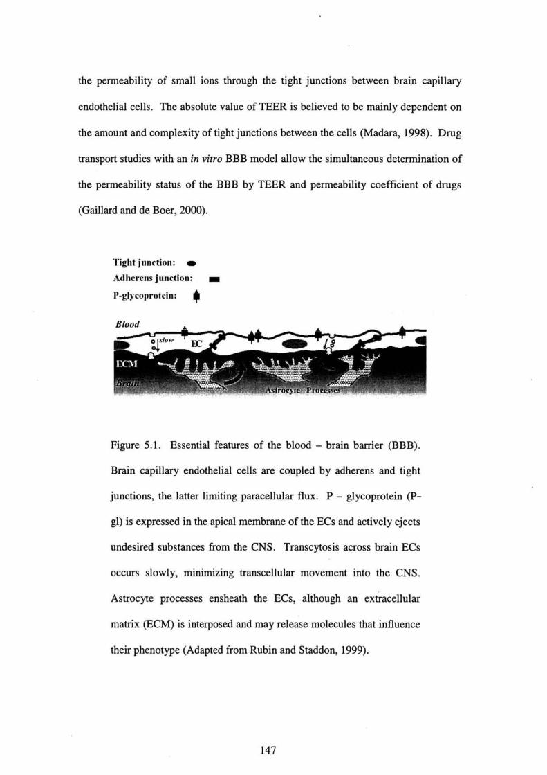
the permeability of small ions through the tight junctions between brain capillary
endothelial cells. The absolute value of TEER is believed to be mainly dependent on
the amount and complexity of tight junctions between the cells(Madara,1998). Dn19
transport studies with an in vitro BBB model allow the simultaneous determination of
the permeability status of the B B B by TEER and permeability coefficient of drugs
(Gaillard and de Boer,2000).
Tight junction: ●
Adherens jtanction: ■
P-gly・・pr・t・in・申
Btood
Figure 5.1. Esselltial features of the blood-brain barrier(BBB).
Brain capillary endothelial cells are coupled by adherens and tight
junctions, the latter limiting paracellular fiux. P-glycoprotein(P-
gl)is expressed in the apical membrane of the ECs and actively ejects
undesired substances f「om the CNS. Transcytosis across brain ECs
occurs slowly, minimizing transcellular movement into the CNS.
Astrocyte processes ensheath the ECs, although an extracellular
matrix(ECM)is interposed and may release molecules that influence
their phenotype(Adapted from Rubin and Staddon,1999).
147

5.1 0bjective of the study
The present study was aimed to evaluate the transendothelial electrical resistance
(TEER)and permeability effects of endosulfan on the tight junctions of the BBB
within porcine brain microvascular endothelial cells(PBMECs). The porcine BBB
model was used as the current in vitro BBB model because porcine brain physiology
is closely related to human(Tewes et aL,1997).
5.2 Materials and methods
5.2.1 Chemicals
Radiolabelled endosulfan(2,3-14C-Endosulfan)with a molar activity of 772.56
MBq;specific activity of 1.895 MBq/mg and a radiochemical purity of>95%by
Radio TLC vvas obtained from the取1stitute of Isgtopes Co. Ltd.(Budapest, Hungary).
All reference analytical standards ofα一endosulfan,β一endosulfan and endosulfan
sulfate(>99%purity), phosphate buffered saline(PBS)powder alld dimethyl
sulfoxide(DMSO)were purchased from Wako Pure Chemical lndustries, Ltd.(Osaka,
Japan). Trypsin-EDTA were purchased from Gibco(Grand Island, NY, USA).
Hionic-FluorTM used fbr liquid scintillating counting were obtained from Packard
Bioscience B. V.(Groningen, The Netherlands). CS 一 C Complete Medium Kit was
purchased伽m Cell Systems Corporation(Kirkland, WA, USA). Cell Viability Kit
(CCK-8)was obtained from D()jindo Laboratories(Kumamoto, Japan). All
chemicals used in the present study were of the highest grade available.
148
5.2.2 Materials
Millicell-PCF culture plate inserts(30 mm diameter;0.4 pm pore size)were
purchased伽m Millipore Corporation(Bedfbrd, MA, USA). Falcon MultiwellTM cell
culture plates were obtained from Becton-Dickinson(NJ, USA). Iwaki 96-well
micro plates, tissue culture flasks and scintillation vials were purchased from
WakenyakU Co. Ltd.(Kyoto, Japan).
5.2.3 Preparations of chemicals
α一endosulfan,β一endosulfan and endosulfan sulfate were dissolved in DMSO
individually to obtain concentrations of l nM,0.01μM,0.1μM,1pM,10μM,100
μM,lmM,10 mM. These standard solutions will be used fbr transendOthelial
electrical resistance(TEER)and cytotoxicity studies of porcine brain microvascular
endothelial cells(PBMECs). The final concentrations that will be used fbr these
studies were O.001 nM,0.01 nM,0.1 nM,1nM,0.Ol pM,0.1μM,1μM and 10 pM.
P・・ch・・ed 14C-E・d・・ulfa・w・・diss・1・・d i・・也・n・1・t the c・ncent・ati・n・f 10 mg/
mL. The 10 mg/mL stock solution of 14C-Endosulfan was fUrther diluted in
DMSO to obtain concentrations of 10μM,100μM,1mM and 10 mM. These
solutions will be used fbr transendothelial transport study of PBMECs. The伽al
concentrations that will be used were O.01μM,0.1μM,1μM and 10μM.
5.2.4 Rationale for dose selection
The concentrations of 14C-Endosulfan in the brain following single administration of
5mg/kg i4C 一 Endosulfan to male Sprague rats were O.11mg/L(equivalent to O.27
μM)after 30 min and O.27 mg/L(equivalent to O.66 pM)after 8 h as detailed and
149
shown in Chapter 3(Section 3.4.3, Table 3.1). The concentration of i4C-Endosulfan
in the brain following three-time repeated administration of 5 mg/kg 14C-
Endosulfan to male Sprague rats was in the range of O.74 pM to 3.88 pM as discussed
in Chapter 3(Section 3.4.3, Table 3.2). Since the concentration levels in the brain
were low in the experiments in Chapter 3 and in order to maintain maximum survival
of the cells, the dose range of O.001 nM to 10 pM was chosen for all the experiments
in Chapter 5.
5.2.5 Cell culture for transendothelial electrical resistance(TEER)study
Porcine brain microvascular endothelial cells(PBMECs)were obtained from
Dainippon Pharmaceutical Co. Ltd.(Osaka, Japan). PBMECs were grown to
confluence in 75 cm2 collagen-coated tissue culture flasks in CS-CComplete
Medium Kit(CSCCMK)and maintained in 95%air and 5%CO2 at 37°C. Upon
reaching confluence, PBMECs were briefly trypsinized to remove them from the
tissue culture flasks and plated at a density of 5 x 10⁵ cells/mL in 2 mL CSCCMK
cultUre medium on the upper side of 6 一 well PCF culture plate inserts and maintained
in 95% air and 5%CO2 at 379C.3 mL of the CSCCMK medium without PBMECs
were added to the lower compartments of the 6-well plates. Under these
・xp・rim・・t・1・・nditi・n・, PBMEC・・each・d・毛。nfluence three d、y、 a丘er seedi。g.
Figure 5.2 shows the schematic representation of the in vitro BBB model used in the
current study.
150

CI:Concentration of tracer chemical in the
inner chamber
V1:Volume of the inner chamber
Co:Concentration of tracer chemical in the
outer chamber
Vo:Volume the outer chamber
100 pm
Co, Vo
Cl, Vl
Filter insert
Cell culture well
Figure 5.2. Schematic representation of the in vitro model of the
blood-brain barrier(BBB).(A)Phase contrast micrograph of a
confluellt brain capillary endothelial cell monolayer. The inner
compartment represellts the blood compartment while the outer
compartment represents the brain compartment.(Modified from
Cecchelli et al,,1999).
151
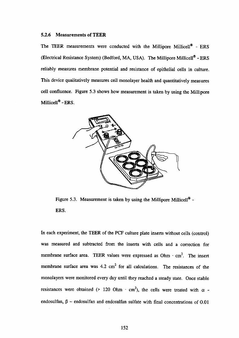
5.2.6Measurements of TEER
The TEER measurements were conducted with the Millipore Millicell⑪一ERS
(Electrical Resistance System)(Bedfbrd, MA, USA). The Millipore Millicell⑧ 一 ERS
reliably measures membrane potential and resistance of epithelial cells in culture.
This device qualitatively measures cell monolayer health and quantitatively measures
cell confluence. Figure 5.3 shows how measurement is taken by using the Millipore
Millicell⑧一ERS.
し
Figure 5.3. Measurement is taken by using the Millipore Millicell⑧一
ERS.
]㎞each experiment, the TEER of the PCF culture plate inserts without cells(control)
was measured and subtracted from the inserts with cells and a correction fbr
membrane surface area. TEER values were expressed as Ohm・cm2. The insert
membrane surface area was 4.2 cm2 fbr all calculations. The resistances of the
monolayers were monitored every day until they reached a steady state. Once stable
resistances were obtained(>1200hm・cm2), the cells were treated withα一
endosul fan,β一endosulfan and endosulfan sulfate with final concentrations of O.01
152

μM,0.1pM,1μM,10 pM respectively. TEER was measured just before adding the
respective endosulfan and at every hour fbr 6 hours. Measurements were taken in
quadnlplicate samples(n=4)from three independent experiments(Mean±SD).
5.2.6.1 Calculation of the resistance of the cell monolayer
(1)The resistance of the blank(Millicell-PCF insert without cells)was
measured and the blank resistance was denoted R biank = A Ohm
(2)The resistance of the sample-wel1(Millicell-PCF insert with cells)was
measured and the sample-well resistance was denoted R sample=BOhm
(3)The blank resistance was subtracted from the sample-well resistance
measurement. Suppose a typical sample reading was C Ohm. Then:
Rsample-Rblank=R monolayer
』BOhm_A・Ohm=COhm
(4)The resistance of the cell monolayer in this example was C Ohm. However, a
correction for the area covered by the cell monolayer was needed. Therefbre,
the resistance of the cell monolayer was multiplied by the area of the e脆ctive
membrane diameter on the Millicell-PCF culture plate insert. h this
・xp・riment, th・area・f・ffe・ti・・m・mb・an・di・m・t・・u・ed・w・・4.2・m2・
Th・n, R_1、yer・4.2・m2・COhm・4.2・m2-DOhm・・m2
5.2.7 Cytotoxicity study of PBMECs after treatment with ct 一 endosulfan,β一
endosu】lfan and endosulfan sulfate
Viability of PBMECs after treatment ofα一endosulfan,β一endosulfan and
endosulfan sulfate was assessed using a commercially available Cell Viability Kit
(CCK-8). The WST-8assay used here is more sensitive than the MTT[3-(4,5一
153
dim・thylthiaz・1-2-yl)-2・5-diph・nylt・t・az・1i・m b・・mid・]assay, whi・h i・wid・ly
used in the measuremellt of cell viability. It measures the activity of mitochondrial
d・hyd・・g・n・・e cat・1y・i・g the c・・versi・n・f【2-(2-m・th・xy-4一㎡仕・phenyl)-3
-(4一㎡t・・phenyl)-5-(2・4-di・ul飴ph・ny1)-2H-t・廿az・1i・m m・n・s・di・m・alt】
t・w・t・・一・・1・bl・fbmlaz紐(D句i・d・L・b・・at・d・・, K・m・m・t・」・p・n). B亘・ny,
PBMECs were plated onto the 96-well microplate in CSCCMK culture medium at a
d・n・ity・f 2・105 cell・/mL t・9・th・・withα一end・・uぬ・,β一。nd。、ul飴n and
endosulfan sulfate at different final concentrations of O.001 nM,0.01 nM,0.1 nM,1
nM,0.01μM,0.1μM,1μM,10μM respectively and incubated fbr 96 h. CCK-8
・・1・ti・n w・・add・d t・each w・11・f也・pl・te and i・・ub・t・d飴・an・th・・4hp亘・・t・
meas皿ements. The absorbance was measured at wavelength of 450 nm using Bio-
Rad Model 550 microplate reader(Bio-Rad Laboratories, Inc., CA, USA). Each
value was calculated f「om 6 to 8 culture wells(η=6-8)from three independent
expenmems. Viability was expressed as a percentage of control(Mean±SD).
5.2.8 Transendothelial transport(inner-to 一 皿ter)study with 14C -
EndOSUlft》n
T・an・p・rt・t・di・・with 14C-E・d・・ulfa・w・・e p・rf・rm・d。t・fi。、1、。nce。t,ati。。,。f
O.OlμM,0.1μM,1μM,10μM.3mL of the CSCCMK culture medium without
PBMECs were added to the outer compartments of the 6-well plates while 2 mL of
the CSCCMK culture medium containing PBMECs were added to the inner
・・mp・・tm・nt・・2μL・f 14C・-E・d・・ulfa・w・・e add・d t・the cult・・e p1・t, i。、ert、 at
various concentrations. Aliquots of 50 pL were collected from the outer
compartments at 20,30,45 mi11,1,2,3,4,5, and 6 h fbr radioactivity analysis. For
・adi・acti・ity・n・ly・i・・5mL・fHi・ni・-Fl…TM w・・add・d t・each 50μL・ample and
154
the radioactivity was measured by means of a liquid scintillation counter(LSC 6100,
Aloka, Tokyo, Japan).
5・2・8・1 D・taaゆ・i・
To obtain a concentration-dependent transport parameter, clearance principle was
used(Siflinger-Bimboim et al.,1987). The increment in cleared volumes between
successive sampling poillts was calculated by dividing the amount of solute transport
d・ri・g血・i・t・Ml by th・d・n・・ch・mb・・c・nce・t・ati・n・. The v・1・m・・f 14C-
Endosulfan cleared from the inner chamber to the outer chamber(のwas calculated
廿om:
Coxレ「o
V(μL)= (5-1) G
where Cl is the initial inner tracer concentration, Co is the outer tracer concentration
andレ’o is the volume of the outer chamber. During the 6 h experiment, the clearance
volume illcreased linearly with time. The clearance rate(dV/dt)is the slope of the
clearance volume over time obtained by linear regression analysis, and given inμL/
min(・r 10’3 cm3/minl.
Th・tr・n・end・th・li・l p・・meability・fth・filt・・g・・w・・nd・th・li・l cell m・n・1・y・・t・14C
-Endosulfan(Pe)was calculated from the clearance rate(dV/dt)and the surface area
(S),and given in pL/cm2/min(or 10-3 cm/min):
dV/dt (5-2) Pe=
∫
In this system, the movement of the tracer molecule occurs by di血sion since there
are no hydrostatic and oncotic gradients.
155
5.2.9 PBMECs reversible transport(outer-to-inner)study with 14C-
Endosu1飴n
To assess the reversible effect of endosulfan on outer-inner compartment, PBMECs
were grown on the culture plate inserts as previously described. 3μL of 14C-
Endosulfan were added to the outer compartmellts of the 6-well plates at various
concen仕ations. Aliquots of 50μL were collected丘om dle imer comp田tments at 20,
30,45min,1,2,3,4,5, and 6 h fbr radioactivity analysis. To obtain a concentration
-dependent transport parameter, clearance principle was used as described in Section
5.2.8.1.
5.2.10Statistical analysis
Results were expressed as Mean±SD of three independent experiments. Any
significant differences between test groups were assessed with one-or two-way
ANOVA followed by Tukey’s multiple comparison post test at significance level, P<
0.05(°SPSS劔Wi・d・ws s・ftwar・p・・k・g・, R・1…e10.0, SPSS, ln、).
5.3 Results
5.3.1MeasurementS of transendotheliar electrical resistance(TEER)
The effects ofα一endosulfan,β一endosulfan and endosulfan sulfate on TEER were
measured in PBMECs monolayers for 6 h(Tables 5.1-5.3, Figures 5.4-5.6). Blank
PCF inserts were used as an indicator of background effects on TEER and showed
・・n・i・t・n・y・t144±60hm・・m2. C・nt・・ls sh・w・d…ignificant・hang・i・TEER
over 6 h. TEER decreased significantly(P<0.05)and reached the bottom level after
6hof exposure toα一endosulfan,β一endosulfan and endosulfan sulfate. TEER
values were the lowest at 10μM as compared to other concentration levels fbr all
156

compounds. Generally, TEER declilled as concentrations and
illcreased fbrα一endosulfan,13-endosulfan and endosulfan sulfate.
exposure periods
Table 5.1. Time-dependent transendothelial electrical resistallce
(TEER)values of PBMECs fbrα一endosulfan.
for quadruplicate samples (n = 4) from
experiments(Mean±SD).
Data are measured
three independent
Time
(Hour)
Transendothelial electrical resistance(TEER)values(0㎞・cm2)
Contro1 0.OlμM 0.1μM 1pM 10 pM0123456
125±4
124±13
116±3
107±3
92±2
88±1
109±9
141±5
64±10
46±2
40±5
50±8
41±1
28±7
125±6
47±2
49±3
40±4
22±2
21±6
5±2
128±2
39±1
31±4
21±1
10±1
4±3
(う12±3
122±9
4±1
(う1±1
5±1
(ヨ11±6
(一)11±2
(う15±2
加欄㎜駒。劫 宅o.εエO ば山山←
一◆-Control
‡8:?1ぽ
‡}描
0 1 2 3 4 5
Time, hour
6 7
●
Figure 5.4. Time-dependent transendothelial electrical resistance
(TEER)values of PBMECs fbrα一endosulfan.
157
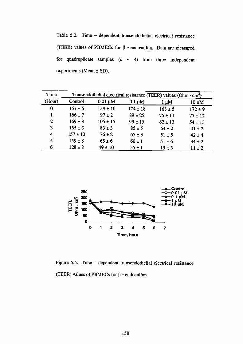
Table 5.2. Time-dependent transendothelial electrical resistance
(TEER)values of PBMECs fbrβ一endosulfan.
for quadruplicate samples (n = 4)from
experiments(Mean±SD).
Data areπieasured
three independent
Time
(Hour)
Transendothelial electrical resistance(TEER)values(Ohm・cm2)
Control 0.OlμM 0.1μM 1μM0123456
157±6
166±7
169±8
155±3
157±10
159±8
128±8
159±10
97±2
105±15
83±3
76±2
65±6
49±10
174±18
89±25
99±15
85±5
65±3
60±1
55±1
168±5
75±11
82±13
64±2
51±5
51±6
19±3
10μM
172±9
77±12
54±13
41±2
42±4
34±2
11±2
宅o.∈エ◎
蜘鋤欄働駒。一◆-Oontrol-・n-0.01pM
0 1 2 3 4 5
Time, hour
6 7
Figure 5.5. Time-dependent transendothelial electrical resistance
(TEER)values of PBMECs fbrβ一endosulfan.
158

Table 5.3. Time-dependent transendothelial electrical resistance
(TEER)values of PBMECs fbr endosulfan sulfate. Data are
measured fbr quadruplicate samples(n=4)from three independent
experiments(Mean±SD).
Time
(Hour)
Transendothelial electrical resistance(TEER)values(Ohm・cm2)
Controi0.01μM 0.1pM 1μM 10μM
210±7
213±7
183±4
155±10
154±8
148±3
172±16
210±1
175±6
144±2
135±3
111±1
108±6
102±3
214±3
146±13
133±3
124±7
94±2
101±7
94±4
225±3
147±6
126±2
99±2
85±4
77±3
74±2
233±1
120±3
100±3
64±2
47±2
39±3
39±2
宅o.∈エO
鋤加欄働駒。
一◆-Control
-O-0.01pM一白一〇.1pM
ま}撒
0 1 2 3 4 5
Time, hour
6 7
Figu「e 5・6・Time-d・p・nden噸n・end・th・li・1・lect・ical・e・i・t・nce
(TEER)values of PBMECs for endosulfan sulfate.
159

5.3.2 Cytotoxicity analysis of endosulfan-treated PBMECs
To test the possibility that changes in TEER resulted from the death of cells and the
subsequent formation of holes in the monolayer, the cell viability was measured using
CCK-8. The concentrations of O.001 nM,0.Ol nM,0.1 nM,1nM,0.01 pM,0.1μM,
1μM,and 10 pM for a-endosulfan,β一endosulfan and endosulfan sulfate did not
decrease cell viability as compared to the control(Table 5.4, Figure 5.7). This
indicates that the concentrations of 10 pM and below did not cause cell death.
Table 5.4. Percentage of viability of PBMECs after a-endosulfan,β
一endosulfan and endosulfan sulfate treatments. PBMECs were
grown fbr 96 h were exposed to several concentrations ofα一
endosulfan,β一endosulfan and endosulfan sulfate individually and
cell viability was measured by WST-8assay as detailed in Section
5.2.7.Cell viability is expressed as percentage of control. Data are
measured fbr six to eight samples(n=6-8)from three independent
experiments(Mean±SD).
Concentration
(pM)
Cell Viabilit(%of Control)
α一 Endosulfan β一Endosulfan Endosulfan sulfate
0.000001
0.00001
0.0001
0.001
0.01
0.1
1
10
93.9±7.2
91.8±1.2
92.4±3.5
97.9±6.3
96.0±7.3
92.3±3.9
89.9±4.4
89.4±3.1
97.3±7.0
95.2±4.0
87.7±4.4
106.2±5.0
98.7±7.2
98.7±2.6
97.9±7.O
l12.9±10.9
104.5±4.5
99.1±8.1
95.6±4.8
99.4±3.5
102.6±7.1
95.9±4.9
96.7±4.9
96.3±5.2
160

石担C8ちま
⑩⑳oo8060ω⑳
0
■一{コ■■a-Eidosultan
■■◎■-b一日ndosuHan
-■嚠鼈黷didosultan sultate
1 ErO6 1 EO5 1 ErO4 0.OO1 0.Ol O.1. 1 10
C◎ncentration,μM
Figure 5.7. Concentration-response curves of cytotoxicity induced
by exposure of PBMECs toα一endosulfan,β一endosulfan and
endosulfan sulfate.
5.3.3 Transendothelial transport(inner-to-outer)study with 14C -
Endosulfan
Figure 5.8 shows the plots of clearance per centimeter squared versus time fbr
different concentrations of 14C-Endosulfan. Figure 5.9 and Table 5.5 show the
average permeability ofthe filter grown endot lelial cell monolayer to endosulfan(Pe)
for different concentrations of i4C-Endosulfan. Pe, for 10 pM was high【0.47±0.09
(xIO“3)cm/min】as compared to 1 pM,0.1μM and O.01μM which remained almost
plateau. This indicates that no noticeable divergence of Pe value existed fbr the
concentrations of 1μM and below.
161
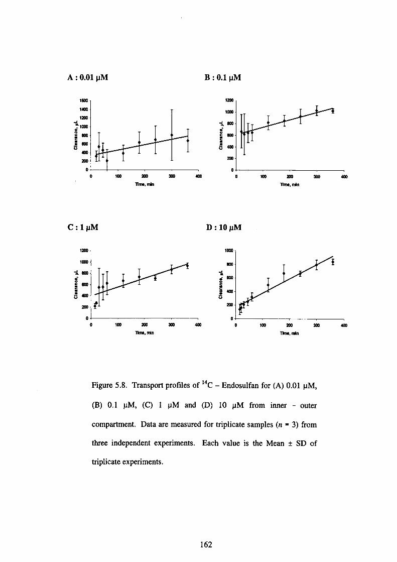
A:0.Ol pM
㎜蜘加働㎝砲伽加
コ
0
0 100 加τ1m●,耐n
伽
Mμ1 0◆●B
加1
㎜1
〈
㎜ ㎝ 蜘寸.85ヒ20
0 100 加丁1m●, mln
C:1μM
加1
励1
醐 緬 姻
寸.85」gδ
0
0 100 加nm●,耐o
姻
D:10 pM
㎝1
㎝ 姻
0 1co 加Wme, mln
伽
Figure 5.8. Transport profiles of i4c-Endosulfan for(A)0.01μM,
(B)0.1μM,(C)1μMand(D)10μM from inner-outer
compartment. Data are measured fbr triplicate samples(n=3)from
three independent experiments. Each value is the Mean±SD of
triplicate experiments.
162

Table 5.5. Effect of inner concentrations of 14C-Endosulfan on the
average permeability (Pe)of the filter growll endothelial cell
monolayer. Data are measured for triplicate samples(n=3)from
three independent experiments. Each value is the Mean±SD of
triplicate experiments.
14C-Endosulfan(μM) Average permeability, Pe(x 10-cm/min)
0.01
0.1
1
10
0.31±0.19
0.29±0.14
0.35±0.13
0.47±0.06
【ξ盲阜
㏄05㎝ΩΩ㎝o ロ0.01pM 但0.1 pM 目1pM 目10μM
Treatment
Figure 5.9. Average permeability of the filter grown endothelial cell
monolayer to 14C-Endosulfan(Pe)(0.Ol pM,0.1μM,1pM and 10
μM)across PBMEC monolayers from inner-outer compartment.
163

5.3.4 PBMECs reversible transport(outer-to-inner)study with 14C-
Endosulfan
Figure 5.10 shows the plots of clearance per centimeter squared versus time fbr
different concentrations of 14C-Endosulfan. Figure 5.11 and Table 5.6 show the
average Pe for different concentrations of i4C-Endosulfan. Pe for O.01 pM was low
【0.50±0.22(xIO’3)cm/min】as compared to O.1μM,1μM and lOμM. The ratio
between the outer-to-inner and i皿er-to 一 outer compartments for the transport
study of 14C-Endosulfan in the concentration range of O.01-10μM was
apProximately l.2-2.L
5◆4 Discussion
The homeostasis of血e microenvironment of central nervous system(CNS), which is
essential for normal fUnction, is maintained by blood-brain barrier(BBB). However,
the lack of reliable in vitro models is one of the major issues in BBB research to
examine in an easy and fast mamer cellular and molecular mechanisms of BBB
fUnction under normal and pathological conditions(Boveri et al.,2005;Cecchelli et
al.,1999). Transendothelial electrical resistance(TEER), an important indication of
the BBB integrity, is a use劔physiological marker and is well一㎞own that the
establishment of tight junction correlates with development of TEER(Gonzales-
Marisca et al.,1985).㎞areview by Madara(1998)on the regulation of the
movement of solutes across(epithelia1)tight junctions for inulin and mannitol, the
relationship between electrical resistance and transport of solutes is non-linear. He
explained that solute transport is esselltially dependent on the sum of transport across
all junctional pathways, whereas total electrical resistance is essentially dependent on
164
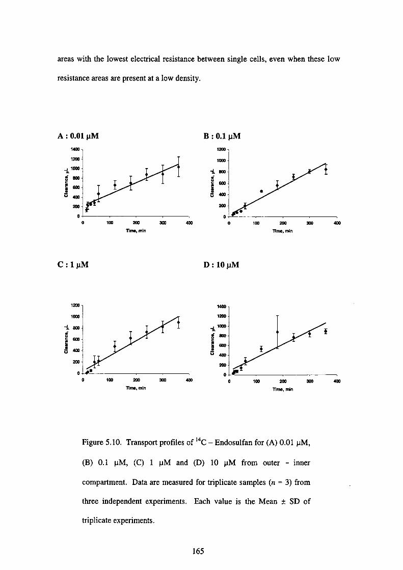
areas with the lowest electrical resistance between single cells,
resistance areas are present at a low density.
even when these low
Mμ10
0 A
働
伽加卿㎜鋤姻加0
0
C:1pM
加丁1m●・mln
鋤 伽
Mμ㎝㎜●●
㎜t
伽
0
0
B
D:10 pM
100 加
nme, mjn
鋤 400
加1
励 ㎜ 鋤 伽1
0
0 100 200
Wm●, mln
緬 400
伽加吻㎜㎝卿
0
0 100 200nme, m佃
伽
Figure 5.10. Transport profiles of i4c-Endosulfan for(A)0.01μM,
(B)0.1μM,(C)1μMand(D)10μM from outer-inner
compartment. Data are measured fbr triplicate samples(n=3)from
three independent experiments. Each value is the Mean±SD of
triplicate experiments.
165
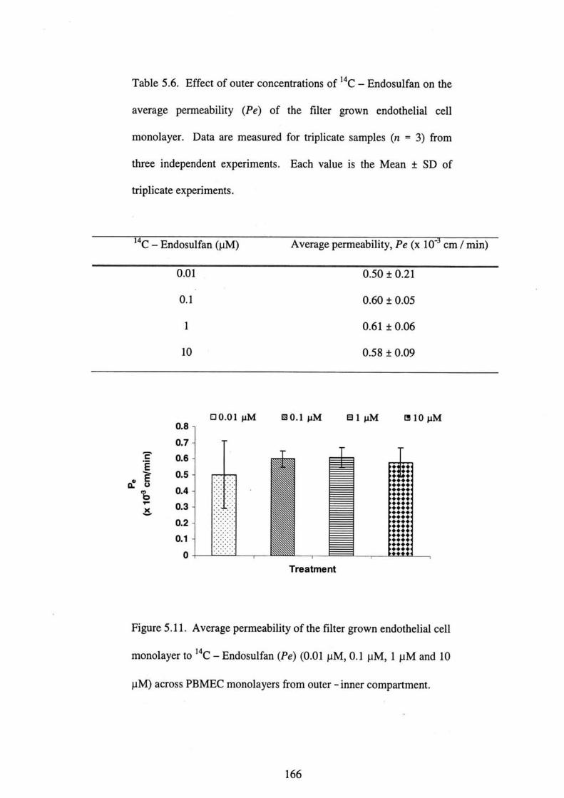
Table 5.6. Effect of outer concentrations of 14C-Endosulfan oll the
average permeability (Pe)of the filter grown endothelial cell
monolayer. Data are measured fbr triplicate samples(n=3)from
three independent experiments. Each value is the Mean±SD of
triplicate experiments.
14C-Endosulfan(μM) Average pemeability, Pe(x l O“cm/min)
0.01
0.1
1
10
0.50±0.21
0.60±0.05
0.61±0.06
0.58±0.09
⌒ε∈>5も三》
87654321000000000
ロ0.01pM 園0.1μM 日1pM 団10pM
T「eatment
Figure 5.1 1. Average permeability of the filter grown endothelial cell
monolayer to i4C-Endosulfan(Pe)(0.01μM,0.1μM,1μM and 10
μM)across PBMEC monolayers from outer-i皿er compartment.
166
Recent research has developed methods of cultivating the PBMECs or bovine brain
microvascular endothelial cells(BBMECs)with astrocyte co-cultures that results in
higher junctions(Boveri・et・al.,2005;Jeliazkova-Mecheva et aL,2003;Megard et al・,
2002).We decided to use the PBMECs without astrocyte co-culture to evaluate the
P・m・・bility・ffe・t・・f・・d・・ulfa・. Th・・e・i・tance・f l200hm・・m2 i・・u・BBB
model is adequate to show BBB fUnction as in other model using primary cultures of
BBMECs grown as monolayers on polycarbonate filters(Raub et al.,1992).
The present study focuses on how PBMECs respond to the treatment of endosulfan.
We studied the effects of a-endosulfan,β一endosulfan and endosulfan sulfate on the
TEER across cultured monolayers of PBMECs. It is also the first report
demonstrating the effects ofα一endosulfan,β一endosulfan and endosulfan sulfate in
PBMECs cultures. Concentrations of 10 pM and below did not affect cell viability as
demonstrated by the WST-8assay(Table 5.4). These data suggest that endosulfan
is not toXic to the PBMECs at the concentrations evaluated. The BBB pemleability,
measured as TEER, decreased significantly in a dose-and time-dependent manner
when monolayers were treated withα一endosulfan,β一endosulfan and endosulfan
sulfate(0.01,0.1, l and lOμM)as shown in Figures 5.4-5.6. TEER reached the
bottom level after 6 h of exposure to di脆rellt concentrations ofα一endosulfan,β一
endosulfan and endosulfan sulfate.11 Figure 5.4,10 pM a-endosulfan exerted the
most severe and instantaneous effect, with significant decrease in TEER as compared
to other concentration levels and compo皿ds. TEER dropped to 40hm・cm2 after l h
of the initiation of treatment and remained at the bottom level throughout the
exposure period. Generally, TEER declined as concentration levels and exposure
periods increased(Tables 5.1-5.3, Figures 5.4-5.6). This phenomenon
167
demonstrates that the alterations of the endothelial permeability are dependent on the
length and severity of endosulfan.
The role of the brain endothelial monolayer is to 1imit the nonspecific exchanges
between blood and brain compartments, behaving as a physical barrier not only to
ions, but also to hydrophilic molecules(Boveri et al.,2005;Rubin and Staddon,1999;
Ohtsuki,2004)・When BBB is disnlpted(e.9., by disruption of the tight junctions, or
by transendothelial channel or pore fbrmation, induced by dnlgs or diseases), changes
in the Pe of the drug are expected to be observed. Hence,1arge changes in Pεwill be
measured with a leaky in vitro BBB(i. e., when TEER values become low)(Gaillard
and de Boer,2000). We fUrther studied the integrity of the brain endothelium by
measuring the transendothelial permeability of the filter grown endothelial cell
monolayer to i4C 一 Endosulfan.
In the present study, we observed that the average Pe fbr the inner-to-outer
transport study after exposure to O.01,0.1, and l OμM i 4C 一 Endosulfan was lower
(rable 5.5)than the average Pe fbr the outer-to-inner transport study(Table 5.6),
thus suggesting that the brain-to-blood efflux transport rate is faster than the blood
-to-brain influx transport rate. This may be accounted for by active transport by P
-glycoprotein(P-gp)in the BBB(Megard et al.,2002;Pardridge,1995). The low
Pe from the inner-to-outer compartment may be attributed to the high lipophilic
nature of this compound(10g P octanol/wate,=3.55)[Agency fbr Toxic Substances and
Disease Registry(ATSDR),2000]. However, the reason for this phenomenon is not
clear, thus suggesting that fUrther research is necessary to elucidate this phenomenon.
168
It was unknown whether endosulfan crosses the cell monolayers via the transcellular
route(i. e., dif血sing through the cytoplasm of the cells, partitioning into and out of
the apical and basolateral cell membranes)or through the paracellular route by means
of the aqueous pores between cells. Since endosulf≧m is lipophilic, we speculated that
transcellular route is the most probable transport route as has been reported elsewhere
fbr lipophilic drugs(Megard et al.,2002;Yoo et al.,2003). Further studies are
necessary to idelltify transporters and tight junction proteins at the BBB as well as its
mechanism.
5.5 Conclusions
The BBB permeability, measured as TEER, decreased significantly in a dose-and
time-dependent manner when treated withα一endosulfan,β一endosulfan and
endosulfan sulfate illdividually. Cytotoxicity test revealed thatα一endosulfan,β一
endosul硲n and endosulfan sulfate did not cause cell death at O.001 nM,0.01 nM,0.1
nM, l nM,0.01μM,0.1μM,1μM, and 10μM. The average Pεfbr the imer-to-
・ut・・廿an・p・rt・st・dy・危・・exp・・u・e t・0.01,0.1,・nd 10 pM ’4C-E・d・・uぬ・w・・
lower than the average Pεfbr the outer-to-inner transport study. The ratio between
the outer-to-inner and也e imer-to-outer compartments fbr the transpo宜study
of 14C-Endosulfan in the concentration range of O.Ol-10μM was approximately
1.2-2.1.
To our knowledge, this is the frrst study that reports on the development of an in vitro
BBB model to study the ability of endosulfan to open tight junctions in PBMECs
cultures. It is hoped that the current model may proved usefU1 and as an initial step to
developing more complex models to understand the mechanism at the tight junctions
169
of the BBB. Additional research is necessary to improve the current model until
optimal conditions with respect to the medium composition, attachmellt factors,
seeding density and pore sizes of the Millicell filters are archived as well as the
possibility of co-culture models. It is hoped that the improved models will fUrther
lncrease our understanding on the pharmacology and toxicology effects of endosulfan
as well as its influences on the barrier under in vitro conditions.
REFERENCES
Anand, M., Khanna, R. N., Gopa1, K., Gupta, G. S.(1980). Effect of endosulfan on
bioel㏄trical activity of brain in rats・ レ「eterinar Y and Hu〃tan Toxicotogy,22:385-
387.
ATSDR.2000. Toxicoto8ical proLfile for endosulfan. U. S. Department of Health
and Human Services, Public Health Service, Agellcy fbr Toxic Substances and
Disease Registry, Atlanta, GA.
Boveri, M., Berezowski, V., Price, A., Slupek, S., Lenfant, A. M., Benaud, C.,
Hartung, T., Cecchelli, R., PrietO, P., Dehouck, M. P.(2005). Induction of blood-
brain properties in cultured brain capillary endothelial cells:Comparison between
primary glial cells and C6 cell line. Glia.
Castillo, C. G., Montante, M., Dufbur, L, Martinez, M. L, Jim6nez-Capdeville, M.
E(2002).Behavioral effects of exposure to endosulfan and methyl parathion in adult
rats・ハleurotoxicolo8y and Teratolo8y,24:797-804.
170
Cecchelli, R., Dehouck, B., Descamps, L, Fenart, L, Bu6e 一 Scherrer, V., Duhem, C.,
Lundquist, S., Rentfel, M., Torpier, G., Dehouck, M. P.(1999). In vitro model fbr
evaluating dnlg transport across the blood-brain barrier. Advanced Dru8 Delivery
Reviews,36:165-178.
Cucullo, L., Hallene, K., Dini, G., Dal Taso, R., Janigro, D. (2004).
Glycerophosphoinositol and dexamethasone improve transendothelial electrical
resistance in and in vitro study of the blood-brain barrier. Brain Research,997:147
-151.
ECVAM. Chapter 4:In vitro methods for organ 一 specific toxicity. The European
Center fbr Validation of Alternative Methods,111stitute fbr Health&Consumer
Protection, European Commission Joint Research Center, Italy, pp.61-88.
Gaillard, P.」., de Boer, A. G.(2000). Relationship between permeability status of the
blood-brain barrier and in vitro permeability coefficient of a drug. European
Journal ofPharmaceutical Sciences,12:95-102.
Gonzales-Marisca, L, Chavez de Ramirez, B., Cereij ido, M.(1985). Tight junction
fbmation in cultured epithelial cells(MDCK). Journal()f Membrane Biolo8y,86:
113-121.
Igarashi, Y., Utsumi, H., Chiba, H., Yamada-Sasamori, Y., Tobioka, H., Kamimura,
Y.,Furuuchi, K., Kokai, Y., Nakagawa, T., Mori, M., Sawada, N.(1999). Glial cell
line-d・・ived neu・・tr・phi・fa・t・・i・duce・b・r・i・・血・・ti・n・f end・th・li・1 cell・f・・mi・g
171
the blood-brain barrier. Bioche〃2ical and Biopノ鍵yぷical Rεぷearch Communication,
261:108-112.
Jean Harry, G., Billingsley, M., Bruinik, A., Campbell,1. L, Classen, W., Domlan, D.
C.,Galli, C., Ray, D., Smith, R. A., Tilson, H. A.(1997). In vitro techniques fbr the
assessment of neutotoxicity. Environmental Health PerSpectives,106(Supplement
1):131-158.
Jeliazkova-Mecheva, V. V., Bobilya, DJ.(2003). A porcine astrocyte/endothelial
cell co-culture model of the blood-brain barrier. Brain Research Protocols,12:91
-98.
LaueらR., Bauer, R., Linz, B., Pittner, F., Peschek, G. A., Ecker, G., Friedl, P., Noe, C.
R.(2004). Development of an in vitro blood-brain barrier model based on
immortalized porcine brain microvascular endothelial cells. Il Farntaco,59:133-
137.
Lee, H. S., Namkoong, K., Kim, D. H., Kim, K. J., Cheong, Y. H., Kim, S. S., Lee, W.
B.,Kim, K. Y.(2004). Hydrogen peroxide-induced alterations of tight junction
proteins in bovine brain microvascular elldothelial cells. Microvascular Research,68:
231-238.
Lu, W., Tan, Y. Z., Hu, K. L., Jiang, X. G.(2005). Cationic albumin conjugated
pegylated nanoparticle with its transcytosis ability and little toxicity against blood-
brain barrier. International Journal ofPharmaceutics,295:247-260.
172

Madara, J. L(1998). Regulation of the movement of solutes across tight junctions.
Annual Review ofPhysiology, 60:121-142.
Megard,1., Garrigues, A., Orlowski, S., Jorajuria, S., Clayette, P., Ezan, E,
Mabondzo, A.(2002). A co-culture-based model of human blood-brain barrier:
ApPlication to active transport of indinav廿and in vivo-in vitro colTelation. Brain
Rεぷearch,927:153-167.
Pardridge, W. M.(1995). Blood-brain barrier biology and methodology. Jb醐α’(ゾ
ハleuroviolo8y,5:556-569.
Pardridge, W. M.(1995). Transport of small molecules through blood-brain barrier:
Biology alld methodology. Advanced Dru8 Delivery Reviews,15:5-36.
Paul, V., Balasubramaniam, E, Jayakumar, A. R., Kazi, M.(1995). A sex-related
difference in the neurobehavioral and hepatic effects fbllowing chronic endosul」丘m
treatment in rats. European Journal of pharmacology,293:355-360.
Raub, T.」., Kuentzel, S. L, Sawada, G. A.(1992). Pemeability of bovine
microvessel endothelial cells in vitro:Barrier tightening by a factor released from
astroglioma cells. Ebeperimental Cell Research,199:330-340.
Rubin, L. L., Staddon, J. M.(1999). The cell biology of the blood-brain barrier.
Annual Review ofNeuroscience,22:11-28.
173
Ohtsuki, S.(2004). New aspects of the blood-brain barrier transporters:Its
physiological roles in the central nervous system. Biolo8y and Pharmacy BuUetin,27
(10):1489-1496.
Seth, P. K., Zaidi, N. F., Agrawa1, A. K., Anand, M.(1986). Neurotoxicity of
endosulfan in young and adult rats.∧leuroto」xicolo8y,7:627-635.
Siflinger-B㎞boim, A., Del Vecchio, P.」., Cooper,」. A., Blumenstock, F. A.,
Shepard, J. N., Malik, A. B.(1987). Molecular sieving characteristics of the cultured
endothelial monolayers・Journal of Cellular Physiolo8y,132:111-117.
Subramoniam, A., Khama, V. K., Mohindra, J., Deth, P. K.(1994). Reduced
phosphoinositide levels in endosulfan treated rat brain. Indian Journat of
Pharmacology,26:49-51.
Tewes・B・・F・ank・・H・・H・llwig・D・・Deck…S・・G・i・・ch・・D・, Tilli・g, T・, W・g・…,」・,
Galla, H. J.(1997). Preparation of endothelial cells in primary cultures obtained from
the brains of 6-month 一 old pigs・In:A. G. de Boer(Ed.), Dru8 transport across the
blood-brain barrier, Harwood Academic Publishers, Australia, pp.91-97.
Yoo, J. W., Kim, Y. S., Lee, S. H., Lee, M. K., Roh, H. J., Jhun, B. H., Lee, C. H.,
Kim, D. D.(2003). Serially passaged human nasal epithelial cell monolayer fbr in
vitro drug transport studies. Pharmaceutical Research,20(10):1690-1696.
174
Zaidi, N. F., Agrawal, A. K., Anand, M., Seth, P. K.(1985). Neonatal endosulfan
neurotoxicity:Behavioral and biochemical changes in rat pups. Neurobehavioral
Toxicolo8y and Teratology,7:439-442・
175

CHAPTER 6
A COMPARATIVE STUI)Y ON THE NEUROTOXIC EFFECTS OF
ENI)OSULFAN ON GLIAL AND NEURONAL C肌L CULTURES FROM
RAT ANI)HUMAN
6.O Introduction
As has b㏄n demonstrated in the in vitro experiments in Chapter 5, elldosulfan, a
chlolinated hydrocarbon insecticide, penetrated through the blood - brain barrier
(BBB). Residues of endosulfan have been detected in the brain of rats treated with
endosulfan(Ansari et al.,1984;Chan et a1.,2005;Dikshith et al.,1984;Gupta,1978;
Nath et al.,1978)and humans(Boereboom et a1.,1998;Coutselinis et al.,1978;Eyer
at al.,2004). Although toxicity is dependent on the sensitivity alld developmental
stage of target cells, concentration of test compound, and duration of exposure, it has
bee・p・・P・sed也・t孤y ch・mical th・t passe・th・BBB is neu・・t・xi・(Jean・Harry・et・1.,
1997;Walnum・t al・・1990)・He・ce,・・d・・uぬn i・classi五・d・・a・・u・・t・xic ag・nt
based oll the above mentioned hypothesis. This also raises more debate since the
actual mechanism remained皿㎞own although endosulfan has been extensively
studied fbr the past decades. Currently, the GABA-antagonism mechanism of
toxicity is the most widely accepted hypothesis【Agency fbr Toxic Substances and
Disease Registry(ATSDR),2000].
6・0・1Principles of the nervous system 一・ Potential sites of neurotoxic attack
The aut・n・mi・nerv・us sy・t・m・…di・・t・・th・・eg・1・ti・n・nd i・t・g・ati・n・f b・dy
fUnctions. The nervous system exerts its influence by the rapid transmission of
electrica1 impulses over nerve fibers that terminate at effector cells, where specific
176

effects are caused due to the release of a neuromediator substance(Mycek et al.,
2000).The nervous system comprises oftwo components:the central nervous system
(CNS), which is composed of the brain and spinal cord, and the peripheral nervous
system(PNS), which comprises the ganglia and the peripheral nerves lying outside of
the brain and spinal cord inclusive of the autonomic nervous system. Two general
types of cells are predominant in the nervous system:neurolls and neuroglial cells
(oligodelldrocites, astrocytes, microglia;and in the PNS, the Schwann cells)(Jean
Harry et al.,1990;Mycek et aL,2000). The principal neurotoxic action of chlorinated
hydrocarbon insecticide is on the aXons of nerve cells in the central nervous system
(CNS). They interfere with the normal flux of sodium and potassium ions across the
axon membrane and neural conduction and do not depress cholinesterase enzymes
(Loeta1.,1995).
Neurons are similar to all other cells of the body in general structure, but they have
additional anatomical features of axons and dendrites that allow conduction of nerve
impulses for communication with other neural cells and between neuronal populations.
Neurons are highly specialized cells and are responsible fbr the reception, integration,
transmission, and storage of infbrmation. Afferent neuronal pathways carry
infbmation illto the nervous system fbr processing;efferent pathways carry
commands to the periphery.㎞addition,血ere are interneurons that process local
infbmlation and transfer data within the nervous system. Within the CNS, neurons
are segregated into fUnctionally related nuclei that fbrm interconnecting bundles of
axonal fibers. Higher organizational levels consisting of several fUnctionally related
neurons are frequently called systems, e. g., motor, visual, associative, and
neuroendocrine systems. The CNS consists of a number of systems responsible fbr
177
the coordination of receiving and processing information from the environment,
maintaining the balance of all other organs systems, and responding to changes in the
enVlrOnment.
Glial cells compose a voluminous“support”system that is essential to the proper
operation of nervous tissue!the nervous system. Unlike neurons, glial cells have no
廿ue synaptic contacts;however, receptors fbr several neurotransmitters are present
and血nctional on various glial cells. Cell-cell contacts exist between the glial cells
and neurons that may regulate both neuronal and glial differentiation and are critical
fbr the glial-guided migration of neurons during development. Glial cells have a
dynamic nature and perfbrm a plethora of fUnctions in the nervous system. They
provide critical processes necessary to maintain normal fimctioning of the nervous
system(e. g., regulation of local pH and ionic balances, and tropic support fbr neurite
extension in the form of growt 1 factors and cell a(lhesion factors). They can also be
the target fbr a toxic response.
6.0.2 Mode of action of endosulfan on the CNS
Infbrmation indicates that the CNS is the major target of endosulfan-induced in
humans and animals following acute exposure by any routes(Aleksandrowicz,1979;
Blanco-Coronado et al.,1992;Boereboom et al.,1998;Boyd and Dobos,1969;
Boyd et al.,1970;Gupta and Chandra,1975;Shemesh et aL,1988;Terviez et a1.,
1974).The proposed modes of action of endosulfan include a global action on all
presynatic neurons in the CNS, inhibition of the calmodulin-dependent Ca2+-
ATPase activity(Srikanth et al.,1989), alteratiolls in the serotonergic system
(Agrawal et aL,1983)and inhibited GABA receptors(Abalis and Eldefrawi,1986;
178
●
Rosa et a1.,1996). Brain damage may also be due to the hypoxemia of seizures
(Shemesh et a1.,1988).
6.1 0bjective of the study
Taking into account the present concern regarding the environmental impact of
endosulfan on public health, the present study was aimed to study the in vitro
neurotoxic effects ofα一endosulfan,β一endosulfan and endosulfan sulfate by
comparing the ability ofα一endosulfan,β一endosulfan and endosulfan sulfate to
cause cell death in glial and neuronal cell cultures from rat and human. The
evaluation was based on the percentage of cell viability determined with WST 一一一8
assay・
6.2 Materials and methods
6.2.1 Chemicals
All reference analytical standards ofα一endosulfan,β一endosulfan and endosulfan
sulfate(>99%purity), phosphate buffered saline(PBS)powder and dimethyl
sulfbxide(DMSO)were supplied by Wako Pure Chemical Indus垣es Ltd.(Osaka,
Japan). Trypsin-EDTA, fetal bovine serum(FBS)and Ham’s l O medium were
purchased from Gibco BRL(Grand Island, NY, USA). RPMI 1640 medium and
Dulbecco’s modified Eagle’s medium(DMEM)were obtained from Nikken
Biomedical Laboratory(Kyoto, Japan). Horse serum was supplied by ICN
Bi・m・dical・ln・・(Ohi・・USA)・C・11 Vi・bility Kit.(CCK-8)w…bt・i・・d fr・m
D()jindo Laboratories(Kumamoto, Japan). All chemicals used in the present study
were of the highest grade available.
179
6.2.2 Materials
Falcon BIOCOATTM Poly D-Lysine tissue culture flasks were obtained from Becton
-Dickinson(NJ, USA). Iwaki 96-well micro plates and tissue culture flasks were
purchased from Wakenyaku Co. Ltd.(Kyoto, Japan).
6.2.3Preparations of chemicals
α一endosulfan,β一endosulfan and endosulfan sulfate were dissolved in DMSO
individually to obtain concentrations of 1 nM,0.01μM,0.1 pM,1μM,10 pM,100
μM,lmM,10 mM. These standard solutions will be used for the cytotoxicity studies
of rat(PC 12, C6)and human(NT2 and CCF-STTG 1)cells. ●
6.2.4 Rationale for dose selection
The concentrations of i4C-Endosulfan in the brain following single administration of
5mg/kg 14C-Endosulfan to male Sprague rats were O.11mg/L(equivalent to O.27
μM)after 30 min and O.27 mg/L(equivalent to O.66 pM)after 8 h as detailed and
shown in Chapter 3(Section 3.4.3, Table 3.1). The concentration of i4C 一 Endosulfan
in the brain following three-time repeated administration of 5 mg/kg 14C-
Endosulfan to male Sprague rats was in the range of O.74 pM to 3.88 pM as discussed
in Chapter 3(Section 3.4.3, Table 3.2). Since the concentration levels in the braill
were low in the experiments in Chapter 3 and in order to maintain maximum survival
of the cells, the dose range of O.001 nM to 10 pM was chosen for all the experiments
in Chapter 6.
180

6.2.5 Cell cultures
Rat PC12 cell line was purchased from American Type Culture Collection(ATCC)
while rat C6, human NT2 and human CCF 一 STTG I cell lines were purchased from
Dainippon Pharmaceutical Co. Ltd.(Osaka, Japan). PC 12 cells were grown to
confluence in Falcon BIOCOATiM Poly D-Lysine tissue culture flasks in RPMI
1640medium supplemented with 5%FBS and lO%horse senlm. C6 cells were
cultured in Ham’s 10 medium containing 2.5%FB S and 15%horse senlm. NT2
cells were grown in DMEM supplemented with 10%FBS while CCF-STTG I cells
were cultured in RPMI l640 medium containing 10%FBS. All cells were
maintained in 95%air and 5%CO2 at 37ΩC.
6.2.6 Cytotoxicity studies of PC12, C6, NT2 and CCF-STTGI cells after
treatment withα一endosulfan,β一endosuifan and endosuifan sulfate
Viability of all cells after treatment of a 一 endosulfan,β一endosulfan and endosulfan
sulfate was assessed using a commercially available Cell Viability Kit(CCK-8)as
described previously in Chapter 5, Section 5.2.7. Upon reaching confluence, all cells
were briefly trypsinized to remove them from the tissue culture flasks and plated onto
the 96-well microplates in respective mediums at a density of 2 x 10⁵ cells/mL
together withα一endosulfan,β一endosulfan and endosulfan sulfate at different
concentrations of O.001 nM,0.01 nM,0.1 nM,1nM,0.01μM,0.1μM,1pM,10μM
respectively and incubated fbr 96 h. CCK-8solution was added to each well of the
plate and incubated fbr another 4 h prior to measurements. The absorbance was
measured at wavelength of 450 nm using Bio 一 Rad Model 550 microplate reader(Bio
-Rad Laboratories, Inc., CA, USA). Viability was expressed as a percentage of
control(Mean±SD)ofthree independent experiments.
181

6.2.7 Statistical analysis
The study of each cell line consisted of three independent experiments with six to
eight samples(n=6-8)on each concelltration level. The means and standard errors
(Mean±SD)of independent tests were calculated on each concentration leve1.
Results were expressed as Mean±SD of three independent experiments. Any
significant differences between test groups were assessed with one-way ANOVA
fbllowed by Tukey’s multiple comparison post test at significance level, P<0.05.
The median toxic concentration(LC50)values were calculated by linear regression
analysis and evaluated by one-way ANOVA fbllowed by Tukey’s multiple
comparison test at significance level, P<0.05(◎SPSS for Windows software package,
Release 10.0, SPSS, hlc).
「
6.3 Results
6.3.1 Cytotoxicity study of rat C6 glial cells
Cell viability for the rat C6 glial cells as compared to control was above 80%fbr all
concentrations as shown in Table 6.1. Figure 6.l shows the concentration-response
curves of cytotoxicity for a 一 endosulfan,β一endosulfan and endosulfan sulfate. This
indicates that the endosulfan concentrations of 10μM and below did not cause cell
death. One-way ANOVA showed significant effect ofα一endosulfan,β一
endosulfan and endosulfan sulfate concentrations[F=17.663, P<0.05(α一
endosulfan);F=3.639, P<0.05(β一endosulfan);F=2.805, P<0.05(endosulfan
sulfate)].
182

6.3.2 Cytotoxicity study of rat PC12 neuronal cells
The viability of all cells was tested by WST-8assays. The concentrations of O.001
nM,0.01 nM,0.1 nM,1nM,0.01μM,0.1μM,1μM, and 10 pM for a -endosulfan,
β一endosulfan and endosulfan sulfate did not decrease cell viability as compared to
the control fbr rat PC 12 neuronal cells(Table 6.2). Figure 6.2 shows the
concen廿ation-response curves of cytotoxicity fbrα一endosulfan,β一endosulfan and
endosulfan sulfate. Cell viability was above 80%fbr all concentrations except that a
slightly below 80%of cell viability was observed fbr the 10μM concentration level
with 78.9%fbrβ一endosulfan and 71.9%for endosulfan sulfate respectively. One-
way ANOVA showed significant e脆ct ofβ一endosulfan and endosulfan sulfate
concentratiolls【F=5.985, P<0.05(β一endosul】fan);F=7.401, P<0.05(endosulfan
sulfate)】.
6.3.3 Cytotoxicity study of human CCF-STTGI glial ce皿s
Figure 6.3 shows the concentration-response curves of cytotoxicity forα一
endosulfan,β 一 endosulfan and endosulfan sulfate. Cell viability for the human CCF
-STTG l glial cells was above 80%for all concentrations except that after treatments
of O.1 pM,1pM, and 10 pM, cell viability decreased to 72.3%,68.2%and 51.6%
fbrα一endosulfan, and 75.6%,70.4%and 63.2%respectively fbr endosulfan
sulfate(Table 6.3). One-way ANOVA showed significant effect of a 一 endosulfan
and endosulfan sulfate concentrations【F=5.225, P<0.05(α一endosulfan);F=
7.412,P<0.05(endosulfan sulfate)】.
183

6.3.4 Cytotoxicity study of hunlan NT2 neuronal cells
Figure 6.4 shows the concentration-response curves of cytotoxicity fbrα一
endosulfan,β一endosulfan and endosulfan sulfate. Cell viability fbr the human NT2
neuronal cells as compared to control was above 80%fbr all concentrations except
that a丘er treatment with 10μM, cell viability was approximately 50%fbr 52.2%fbr
α一endosulfan,49.0%fbrβ一endosulfan and 46.5%fbr endosulfan sulfate
respectively(Table 6.4). One-way ANOVA showed significant effect ofα一
endosulfan,β一endosulfan and endosulfan sulf乞te concentrations[F=6.318, P<
0.001(α一endosulfan);F=5.787, P<0.05(β一endosulfan);F=5.980, P<0.05
(endosulfan sulfate)】.
Table 6.1. Percentage of viability of C6 cells after exposure toα一
endosulfan,β一endosulfan and endosulfan sulfate. Cell viability is
expressed as percentage of control. Data are Mean±SD from six-
eight samples(n=6-8)of three independent experiments. The
significance compared with the controls is expressed as*P<0.05.
Concentration
(μM)
Cell Viabilit (%of Contro1)
α一 Endosulfan β一Endosulfan Endosulfan sulfate
0.000001
0.00001
0.0001
0.001
0.Ol
O.1
1
10
88.7±1.5*
79.2±5.0*
76.6±2.0*
74.6±3.5*
75.9±1.9*
73.9±0.3*
73.1±0.8*
78.5±0.1*
102.4±10.6
92.1±8.6
87.1±9.0
87.2±7.7
84.7±5.3
85.3±2.2
79.1±6.1
84.9±5.7*
101.3±7.7
87.4±10.5
87.7±9.2
88.5±7.8
85.3±7.6
83.0±6.8
80.8±4.7
81.7±9.7
184
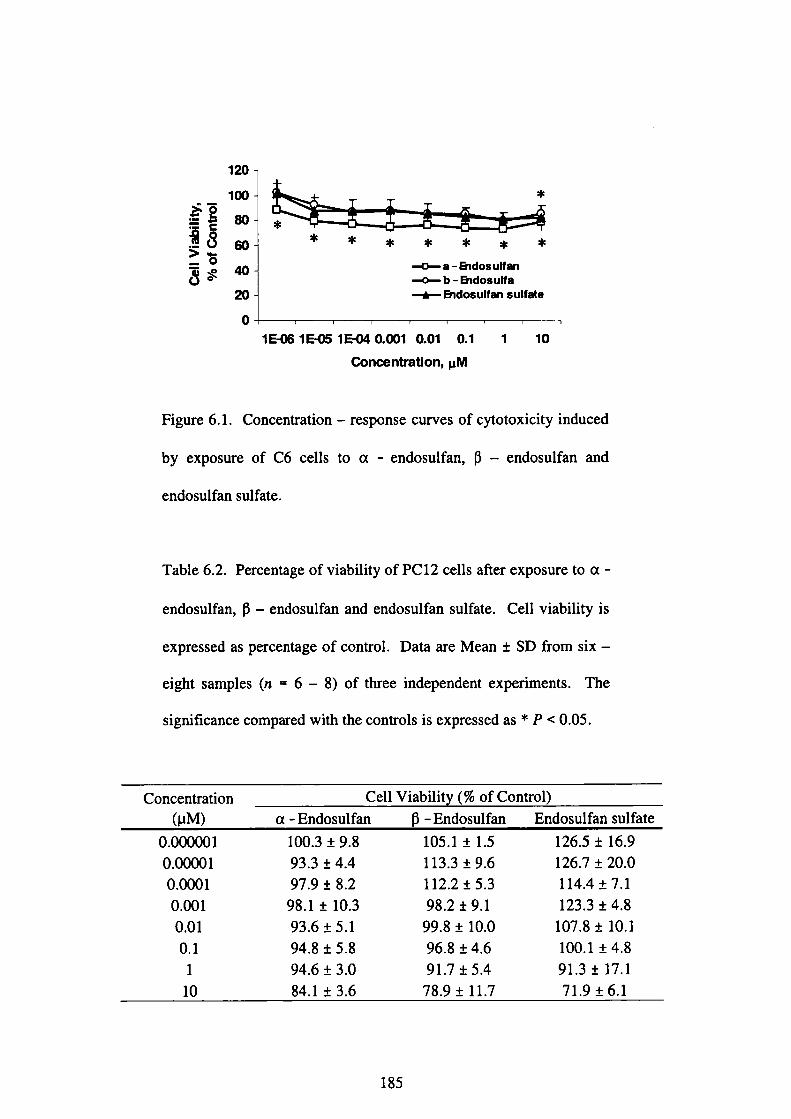
石曼」OOちま
“ぬ
o8060ω⑳o
*
*
* * * * * * *
■■口■-a-Ehdosulfan
{b-thdosulfa -■白一一Eidosulfan sulfate
1EOS l EO5 1 EO40.OO1 0.Ol O.1
Concent副on,μM
1 10
Figure 6.1. Concentration-response curves of cytotoxicity induced
by exposure of C6 cells toα一endosulfan,β一endosulfan and
endosulfan sulfate.
Table 6.2. Percentage of viability of PC12cells after exposure toα一
endosulfan,β一endosulfan and endosulfan sulfate. Cell viability is
expressed as percentage of control. Data are Mean±SD from six-
eight samples(n=6-8)of three independent experiments. The
significance compared with the controls is expressed as*P<0.05.
Concentration
(μM)
Cell Viabilit (%of Contro1)
α一 Endosulfan β一Endosulfan Endosulfan sulfate
0.000001
0.00001
0.0001
0.001
0.01
0.1
1
10
100.3±9.8
93.3±4.4
97.9±8.2
98.1±10.3
93.6±5.1
94.8±5.8
94.6±3.0
84.1±3.6
105.1±1.5
113.3±9.6
112.2±5.3
98.2±9.1
99.8±10.0
96.8±4.6
91.7±5.4
78.9±11.7
126.5±16.9
126.7±20.0
114.4±7.1
123.3±4.8
107.8±10.1
100.1±4.8
91.3±17.1
71.9±6.1
185
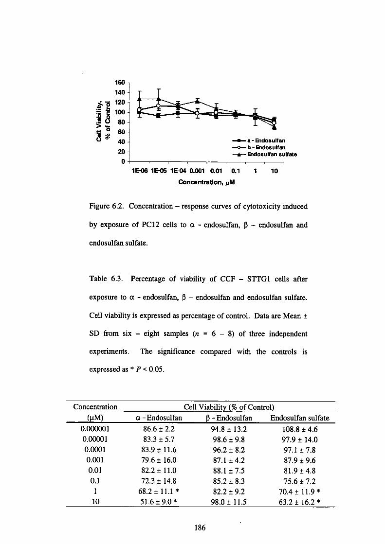
60ヨ⑳008060ω⑳0
■■■一一a一匡hdosulfan
■〈〉-b一旨idosultan
十thdosultan sultate
1E式圓51E弍5 1E-{図 0.001 0.01 0.1 1 10
Conce ntration, pM
Figure 6.2. Concentration-response curves of cytotoxicity induced
by exposure of PC 12 cells toα一endosulfan,β一endosulfan and
endosulfan sulfate.
Table 6.3. Percentage of viability of CCF-STTGI cells after
exposure toα一endosulfan,β一endosulfan and endosulfan sulfate.
Cell viability is expressed as percentage of control. Data are Mean±
SD from six-eight samples(n=6-8)of three independent
experimellts. The significance compared with the controls is
expressed as*P<0.05.
Concelltration
(μM)
Cell Viabilit (%of Control)
α一Endosulfan β一Endosulfan Endosulfan sulfate
0.000001
0.00001
0.0001
0.001
0.01
0.1
1
10
86.6±2.2
83.3±5.7
83.9±11.6
79.6±16.0
82.2±11.0
72.3±14.8
68.2±11.1*
51.6±9.0*
94.8±13.2
98.6±9.8
96.2±8.2
87.1±4.2
88.1±7.5
85.2±8.3
82.2±9.2
98.0±11.5
108.8±4.6
97.9±14.0
97.1±7.8
87.9±9.6
81.9±4.8
75.6±7.2
70.4±11.9*
63.2±16.2*
186
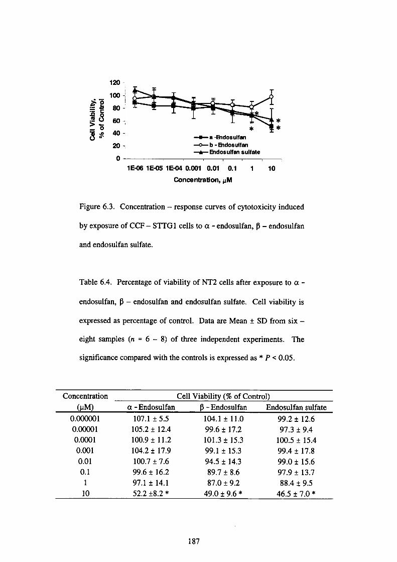
石担C◎Oちま
“㊥
ュ8060ω⑳o
■■●■■a“Ehdosulfan
-■◎■-b-Eidosulfan
十Endosuttan sultat●
1606 1ErO5 1 E砲 0.001 0.Ol O.1
CiOnoentration,μM
1 10
Figure 6.3. Concentration-response curves of cytotoxicity induced
by exposure of CCF-STTG l cells to a 一 endosulfan,β一endosulfan
and endosulfan sulfate.
Table 6.4. Percentage of viability of NT2 cells after exposure toα一
endosulfan,β一endosulfan and endosulfan sulfate. Cell viability is
expressed as percentage of control. Data are Mean±SD from six-
eight samples(n=6-8)of three independent experiments. The
significance compared with the controls is expressed as*P<0.05.
Concentration
(μM)
Cell Viabilit (%of Control)
α一Endosulfan β一Endosulfan Endosulfan sulfate
0.000001
0.00001
0.0001
0.001
0.01
0.1
1
10
107.1±5.5
105.2±12.4
100.9±11.2
104.2±17.9
100.7±7.6
99.6±16.2
97.1±14.1
52.2±8.2*
104.1±11.0
99.6±17.2
101.3±15.3
99.1±15.3
94.5±14.3
89.7±8.6
87.0±9.2
49.0±9.6*
99.2±12.6
97.3±9.4
100.5±15.4
99.4±17.8
99.0±15.6
97.9±13.7
88.4±9.5
46.5±7.0*
187
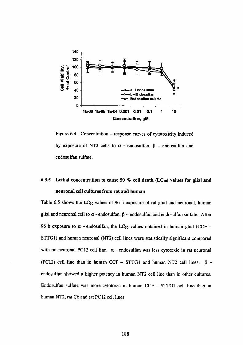
る担COOちボ
140
120
100
80
60
40
20
0
■■0■-a-H)dosultan
■■◎■-b●Endosulfan
一由声一Ehdosulfan su”就e
*
*
*
± 一一一一一一「一一一一『一「
1E≒{掲1E{田ilE{岡0.001 0.01 0.1 1 10
Concentration,μM
Figure 6.4. Concentration-response curves of cytotoxigitY induced
by exposure of NT2 cells toα一endosulfan,β一endosulfan and
endosulfan sulfate.
6.3.5 Lethal concentration to cause 50%cell death(LC50)values for glial and
neuronal cell cultures from rat and human
Table 6.5 shows the LC50 values of 96 h exposure of rat glial and neurona1, human
91ial and neuronal cell toα一endosulfan,13- endosulfan and endosulfan sulfate. A負er
96hexposure toα一endosulfan, the LC50 values obtained in human glial(CCF-
STTG 1)and human neuronal(NT2)cell lines were statistically significant compared
with rat neuronal PC 12 cell line.α一endosulfan was less cytotoxic in rat neurona1
(PCl2)cell line than in human CCF-STTGl alld human NT2 cell lines. β一
endosulfan showed a higher potency in human NT2 cell line than in other cultures.
Endosulfan sulfate was more cytotoxic in human CCF-STTGl cell line than in
human NT2, rat C6 and rat PC 12 cell lines.
188

Table 6.5. LC50 for the cytotoxic effects ofα一endosulfan,β一
endosulfan and endosulfan sulfate in rat glial and neuronal, human
glial and neuronal cell lines. Cell cultures were exposed to several
concentrations ofα一endosulfan,β一endosulfan and endosulfan
sulfate individually and cell viability was measured by WST-8
assay・・d・t・il・d i・S・・tig・6・2・6・LC・・val…ar・Mean±SD
calculated廿om the individual concentration 一一 response curves from
three independent experiments. The significance compared with the
controls is expressed as*P<0.05(Tukeプs multiple comparison
test).
Cell cultures LC50(μM)
α一 Endosulfan β一Endosulfan
Endosulfan
sulfate
Rat glial
Rat neuronal
Human glial
Humanneuronal
C6PCl2
CCF-STTGl
NT2
22.26±4.46
48.03±23.45
11.21±4.osa
13.45±2.40b
22.27±6.13
17.76±5.37
30.17±7.54
11.37±0.94c
21.63±4.68
12.97±2.61d
10.43±2.57e
12.64±1.69プ
aP<0.05 compared to rat neuronal PC 12 cell line
bP<0.05 compared to rat nellronal PC 12cell line
cP<O.05 compared to human glial CCF-STTG l cell line
dP<0.05 compared to rat glial C6 cell line
eP<0.05 compared to rat glial C6 cell line
fP<0.05 compared to rat glial C6 cell line
189

6.4 1)iscussion
In this study, the effects ofα一endosulfan,β一endosulfan and endosulfan sulfate
were compared and evaluated in PC 12(neuronal)and C6(glial)cells from rat and
NT2(neuronal)and CCF-STTG 1(glial)from human with WST-8 assay. The
present study shows thatα一endosulfan,β一 endosulfan and endosulfan sulfate cause
cytotoxlcity in neuronal and glial cell cultures from rat and human in a concentration
-dependent manner(Figures 6.1-6.4). It is also the frrst report demonstrating the
cytotoxic effects ofα一endosulfan,β一endosulfan and endosulfan sulfate in neuronal
and glial cultures from rat and human. The toxicity of these compounds varied in
different cell lines as shown in LC50 values(Table 6.5).α一endosulfan effects were
highly selective as shown by the wide range of LC50 values fbund in the different
cultures, ranging from l 1.2μM for human CCF-STTG I glial cells to 48.O pM for
rat PC 12 neuronal cells. ln contrast, selective neurotoxicity was not so manifested in
glial and neuronal cell cultures after exposure to endosulfan sulfate, as LC50 values
were in the range of 10.4-21.6μM.
Human CCF-STTG l glial cells were the most sensitive cell type toα一 endosulfan
toxlclty compared to human NT2 neuronal cells, rat C6 glial cells and rat PC 12
neu・・n・l cells・H・man NT2・・u・・n・l cells we・e th・m・st・en・itive cell typ・t・β一
end・・ulfa・・nd end・sulf・n・sulf・t・t・xi・ities c・mpar・d t・hum・n CCF-STrGl glial
cells, rat C691ial cells and rat PC 12neurollal cells.
It is㎞own that endosulfan concentration attained in rat brain after a single
・dmi・i・t・ati・n・f5mg/kg 14C-E・d・・ulfa・w・・0.11mg/L(・q・i・・1・nt t。0.27、pM)
after 30 min and O.27 mg/L(equivalent to O.66 pM)after 8 h as detailed and shown
190
in Chapter 3(Section 3.4.3, Table 3.1). The concentration of 14C- Endosulfan in the
braill fbllowing three-time repeated administration of 5 mg/kg I4C-Endosulfan to
male Sprague rats was in the range of O.74 pM to 3.88μM(Chapter 3, Section 3.4.3,
Table 3.2). Cerebral congestion and edema are often observed at necropsy in animals
that die fbllowing acute ingestion of endosulfan(ATSDR,2000;Boyd and Dobos,
1969;Boyd et a1.,1970;Terziev et al.,1974). lncreased brain weights were observed
in rats fbllowing consumption of 4.59 mg/kg/day of endosulfan fbr 13 weeks.
However, the significance of the increases in brain weight is皿㎞own, but it could
have been related to edema(ATSDR,2000).
In our in vitro study,α一endosulfan and endosulfan sulfate damaged human CCF-
STTG l glial cells and NT2 neuronal cells in a very similar potency(LCso=11.21 and
13.45μM respectively for a-endosulfan and 10.43 and 12.64 pM respectively fbr
endosulfan sulfate). Our results also indicate thatα一endosulfan and endosulfan
sulfate are very toxic to human glial and neuronal cells whereasβ一endosulfan is
toxic to human neurollal cells, thus suggesting that human cultures may be a useful
tool for studying and predicting the neurotoxic effects of these compounds. i 1 general,
the use of human cells may lead to an easier extrapolation from in vitro models to the
clinical situation and facilitate the accuracy of the human risk assessment.
6.5 Conclusions
Several end points have been proposed as simple and rapid methods to assess
neurotoxicity of chemicals in vitro. Cytotoxicity and viability end points provide
information on the intrinsic toxicity of chemicals. They have limited ability, if any,
to predict the neural-specific effects. However, these end points must be included to
191

determine the health status of the cells at the time of process evaluation and possibly
to differentiate specific effects from general cytotoxicity(Jean Harry et al.,1998).
hl summary, the results demonstrate thatα一endosulfan,β一endosulfan and
endosulfan sulfate cause cell death in glial and neurollal cell cultures from rat and
human brain.α一endosulfan produces a manifest selective neurotoxicity. Human
glial cells were the most sensitive cell type to the cytotoxic effects of a-endosulfan
followed by human neuronal, rat glial and rat neuronal cells. Human neuronal cells
were the most sensitive cell type to the cytotoxic effects ofβ一endosulfan fbllowed
by rat neuronal, rat glial and human glial cells. Human glial cells were the most
sensitive cell type to the cytotoxic effects of endoSulfan sulfate fbllowed by human
neuronal, rat neuronal and rat glial cells. This study also shows that human glial and
・・ur…1・ell・ar・m・・e・en・itive c・mpar・d t・・at gli・1・・d neu,。n。l cell、 and ’メB。1d b。
used as a good tool for studying the neurotoxic effects of these chemicals in the CNS,
and thus, predicting their impact on human health.
REFERENCES
Abalis, I M., Elde廿awi, M. E.(1986). Effects of insecticides on GABA-induced
chloride influx into rat brain microsacs. Journal(ゾToxicolo8y oη4 Environmentat
Hεo励,18:13-23.
Agrawa1, A. K., Anand, M., Zaidi, N. F., Seth, P. K.(1983). Involvement of
serotonergic receptors in endosulfan neurotoxicity. Biochemical Pharmacoto8y,32:
3591-3593.
192

Aleksandrowicz, D. R.(1979). Endosulfan poisoning and chronic brain syndrome.
ノlrchives of Toxicolo8y,43:65-68.
Ansari, R. A., Siddiqui, M. K. J., Gupta, P. K.(1984). Toxicity of endosulfan:
Distribution ofα一andβ一isomers of racemic endosulfan fbllowing oral
ad血nistration in rats. Toxicolo8y Letters,21:29-33.
ATSDR.2000. Toxicolo8ical proLfile/br endosulfan. U. S. Department of Health
and Human Services, Public Health Service, Agency fbr Toxic Substances and
Disease Registry, Atlanta, GA.
Blanco-Coronado, J. L, Repetto, M., Ginestal, R.」., Vicente,」. R., Yelamos, F.,
Lardelli, A.(1992). Acute intoxication by endosulfan. Clinical Toxicology,30:575-
583.
Boereboom, F. T.」., van Dijk, A., van Zoonen, P., Meulenbelt, J.(1998).
Nonaccidental endosulfan intoxication:Acase report with toxicokinetic calculations
and tissue concentrations. Clinical Toxicolo8y,36(4):345-352.
Boyd, E. M., Dobos,1.(1969). Protein deficiency and tolerated oral doses of
endosulfan. ノlrchiveぷ (ゾInternational Phar〃mcodyna〃lics and Therapeutics, 178:
152-165.
Boyd, E. M. Dobos,1., K両nen, CJ.(1970). Endosulfan toxicity and dietary protein.
ノlrchives()fEnvironmental Health,21:15-19.
193
Chan, M. P. L, Morisawa, S., Nakayama, A., Kawamoto, Y., Sugimoto, M., Yoneda,
M・(2005)・T・xi・・垣neti…f14C-E・d・・uぬ・f・ll・wi・g・i・gle a・d・epeat・d・・al
administration in male Sprague-Dawley rats. Environmental Toxicolo8y,γb1醐θ20,
Issue・5侮pre∬).
Coutselinis, A., Kentarchou, P., Boukis, D.(1978). Concentration levels of
endosulfan in biological material(report of three cases). Forensic Science,11:75.
Dikshith, T. S. S., Raizada, R. B., Srivastava, M. K., Kaphalia, B. S.(1984).
Response of rats to repeated oral administration of endosulfan. Industrial Health,22:
295-304.
Eyer, F., Felgenhauer, N., Jetzinger, E., Pfab, R., Zilker, T. R.(2004). Acute
endosulfan poisoning with cerebral edema and cardiac failure. Journal of Toxicology
and Clinical Toxicolo8y,42(6):927-932.
Gupta, P. K.(1978). Distribution of endosulfan in plasma alld brain after repeated
oral administration to rats. Toxicolo8y,9:371-377.
Gupta, P. K., Chandra, C. V.(1977). Toxicity of endosulfan after repeated oral
administration to rats・Bultetin of Environmental Contamination and Toxicolo8y,18:
378-384.
194
Jean Harry, G., Billingsley, M., Bruinik, A., Campbel1,1. L, Classen, W., Dorman, D.
C.,Galli, C., Ray, D., Smith, R. A., Tilson, H. A.(1997). In vitro techniques fbr the
assessment of neutotoxicity. Environmental Health Perspectives,106(Supplement
1):131-158.
Lo, R. S. K., Chan,」. C. N., Cockram, C. S., Lai, F. M. M.(1995). Acute tubular
necrosis fbllowing endosulfan insecticide poisoning・ 」侃r朋10f Toxicoto8y and
Clinical Toxicolo8y,33(1):67-69.
Mycek, M.」., Harvey, R. A., Champe, P.(2000). Chapter 3:The autonomic nervous
system. In:LipPincott’ぷ1〃ustrated Reviews: Pharmacology, LipPincott Williams&
Wilkins, Philadelphia, pp.27-34.
Nath, G., Datta, K. K., Dikshith, T. S. S., Tandon, S. K., Pandya, K. P.(1978).
Interaction of endosulfan and metapa in rats. Toxicolo8y,11:385-393.
Rosa, R. Rodriguez - Faπe, E., Sanfeliu, C. (1996). Cytotoxicity of
hexachlorocyclohexane isomers and cylodienes in primary cultures of cerebellar
granule cells. Journal()f Phar〃mcolo8y and EXperi〃zental 1「herapeutics,278(1):163
-169.
Shemesh, Y., Bourvine,
endosulfan intoxication.
268.
A.Gold, D., Bracha, P.(1988). Survival after acute
Journal of Toxicolo8y and Clinicat Toxicolo8y,26:265一
195
Srikanth, N. S., Seth, P. K., Desaiah, D.(1989). Inhibition of calmodulin activated
Ca2+-ATP・・e by end・・ulfa・i・・at b・ai・.」・u・nal・・f・T。。i,。1。8y、and、E。。ir。nm。n、。l
Health,28:473-481.
Terziev, G., Dimitrova, N., Rusev, F.(1974). Forensic medical and fbrensic chemical
study of acute lethal poisinnins with Thiodan.、Folia Medicine,16:325-329.
Walnum, E., Hansson, E., Harvey, A. L(1990).1ηvitro testing of neurotoxicity.
ノITLA,18:153-179.
196
CHAPTER 7
ASSESSMENT OF THE HEALTH RlSKS FO肌OWING EXPOUSRE TO
ENI)OSULFAN
7.O An overview
Risk assessment is an important tool in deciding on how to allocate resources to
・。・仕・lli・9・ri・k・.1・m・・t ca・e・, it i・b・・ed・n・haz・・d・d・t・d・・ived伽m anim・l
experiments and on exposure data f旨om an assessment of the likely or actual exposure
・fth・p・P・1・ti…fi・t・・est. Th・p・・cess・f assessi・g th・dsk ass・ciat・d wi舳・m・n
exposure to env辻onmental chemicals inevitably relies on a number of assumptions,
estimates and rationalizations. Some of the greatest challenges result加m the
necessity t・ex仕・p・1・t・・utsid・th・・ang・・f・・nditi…f…din exp・・im・nt・1・t・di…
For risk assessments based on animal data, the most obvious extrapolation that has to
b・p・由m・di・fr・m也・t・・ted・㎡m・1・peci・・t・h・m・n・(i・…the c・nve・・i・n・f血・
animal dose-response relationship to a predicted human dose-human relationship).
Furthermore, there is a need to extrapolate beyond the bounds of the animal data,
outside of the known dose-response region, to areas of likely human exposure both
in temls of level of exposure as well as other factors such as route(e. g., oral versus
inhalation)and duration of exposure. An additional difficulty often associated with
environmental health assessment is血at there may not be adequate toxicity
information available on some of the chemicals of concern(Aarons et aL,1999;
Clewell,1995).
Ak・y・・n・id・・ati・・f・・such・x血・p・1・ti・n i・h・w・nd・t wh・t・ate a ch・mical i・
absorbed, distributed, metabolized and elimillated (i. e., the pharmaco -or
197
toxicokinetics of the chemical)in different species. Various approaches have been
developed and employed depending upon the situation in question and the amount of
data available. The‘‘allometric”relationships may be used which assume that the
rates of absorption, distribution,, metabolism and elimination of a substance are a
fUnction of body size;as the body size varies betweens species, a(ljustments to allow
for this can be made.
Assessment of the risk fbllowing continuous exposure to very low levels of a
・h・mical i・也e env丘・nment i・alw・y・p・・b1・m・tic and・an n・m・lly be e、tim。t。d
丘・m1・bO・at・・y・㎡m・l d・t・w仙也e apPlicati…f・pP・・P・i・t・・af・ty・m・・gi・・t。
allow fbr the uncertainties involved. Frequendy, the animal data will have been
・bt・滅・t・・n・id・・ably high・・d・・e l・ve1・wi血di脆・ent・・ut・・fexp・・u・e.
PBPK m・d・1i・g・・uld p・・ve a・imp・rt・nt t・・l f・・imp・・vi・g血e acc・・acy・f h。man
heal也・i・k assessm・・t・f・・hanard・u・ch・血cal・i・血e env欽・㎜ent. h・P・・u、e。f
this technique can reduce uncertainties that currently exist in risk assessment
P…ed・・e・by p・・vidi・g m・・e・cienti丘cally・・edibl・・ext・ap・1・ti・n・a…ss speci・、 and
routes of exposures, and from high experimental doses to potential environmental
exp°su「es・C㎜・nt・pPlicati・n・・f PBPK m・d・1s ra・g・丘・m・el・tively
・廿・ightf・・w・・d・・e・f・曲e ex廿・p・1・ti・n・f・h・mical垣・・ti・・ac・・ss speci・・,,。ut,
and duration of exposure to much more demanding chemical risk assessment
・pPlicati・ns req・i「i・g・d・・c・ipti・n・f・・mp1・x ph・・mac・dy・・mi・ph・・。m。n、.
PBPK m・d・li・g h・1P・t・id・ntify・th・飴・t…th・t訂・m・・t imp・rt・nt i・d・t・・mi・i・g
the health risks associated with exposure to a chemical and provides a means fbr
198
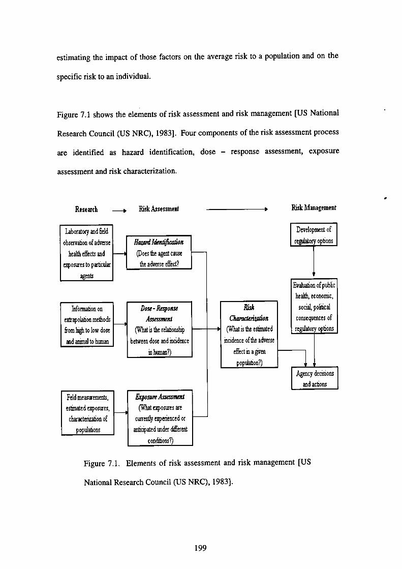
estimating the impact of those factors on the average risk to a population and on the
specific risk to an individual.
Figure 7.1 shows the elements of risk assessment and risk management【US National
Research Council(US NRC),1983]. Four components of the risk assessment process
are identified as hazard identification,
assessment and risk characterization.
Research
→Laboratory and丘eld
observadon of ad▽erse
heahh eflヒcts and
卿。sures to panicular
agerltS
Risk A$sessment
dose-response assessment, exposure
h偽π頑on。n
e蜘。囲o且me也。dS
fr。m⑩。1。w d。se
and animal to humaロ
∬㎞1i㎞輌わπ
①。es血e agent cause
the adverse e旺ed?
Do銘・吻㈱
加蹴‘(What is the relad。幽
betWeen dose and血cidence
娩h㎜?)
Feld measurem〔rnts,
eStimated exposures,
characterization of
popUlaions
鞠o㎜A㈱励刎(What⑳。sures・are
cuπen‖y expefieロced or
a血cipated under d舐re!lt
C。ndions?)
Risk Management
蹴
CimderiUto”
(What is出e es㎞ed
血cidence ofthe adverse
effect娩a卿
P。P曲ti。n?)
Development of
regUlatory。pti。ns
Evaluation ofput)亙c
he甑ec。n。㎡c,
s。d泣P。dical
C。nse卯ぴces。f
regUlat。ry。pti。ns
Agenry deeiSions
and achons
Figure 7.1. Elements of risk assessment and risk management[US
National Research Council(US NRC),1983].
199
’
7.1 Neurotoxicity
Involvement of endosulfan, a neurotoxic agent, in the central nervous system(CNS)
has been described previously in Chapter 2. The blood-brain barrier(BBB)
pemeability, measured as transendothelial electrical resistance(TEER), decreased
significantly in a dose-and time-dependent manner when treated withα一
endosulfan,β一endosulfan and endosulfan sulfate individually(Chapter 5, Section
5.3.1). Cytotoxicity studies(Chapter 6, Sections 6.3.1-6.3.4)indicated thatα一
endosulfan・β 一endosulfan and endosulfan sulfate cause cell dealth in glial and
neuronal cell cultures from rat and human brain. No effect was observed when male
rats were treated with 12.5 mg/kg bw per day(McGregor,1998).
7・2 Reproductive toxicity
Adverse reproductive effects of endosulfan include testicular impaiment in vivo
(Sinha et a1.,1995;Sinha et al.,1997;Chitra et al.,1999;Dalsenter et al.,1999;
Dalsenter et a1.,2003;Dikshith et al.,1984;Singh and Pandey,1989), daily sperm
production along with increased speml abnomlalities and altered activities of
testicular marker enzymes in both mature and immature rats【Pandey et a1.,1990;
National Cancer Institute(NCI),1978]as well as reduction in serum testosterone
levels(Choudhary and Joshi,2003;Wilson and LeBlanc,1998). hn addition,
vacuolation of Sertoli cells was observed histologically (Sinha et aL,1999).
Testicular cells, especially Sertoli-germ cells coculture have been used by many
researchers to investigate the mechanisms by which recognized toxicants exert their
effect(Reader and Foster,1990;Sundaran and Witorsch,1995). Metabolic
cooperation exists betweell Sertoli cells and geml cells and any disturbance in this
interaction may lead to testicular dysfUnction(Foster,1988). Cytotoxic changes
200
induced by endosulfan in mixed cultures of Sertoli and germ cells were seen after 24
hour and 48 hour of treatment(Sinha et al.,1999). The No-Observed 一 Adverse-
Effect 一 Level(NOAEL)for reproductive effects was 75 ppm, equal to 6 mg/kg bw
per day(McGregor,1998).
7.3 0bjective of the study
The purpose of this chapter is to present a quantitative risk assessment of endosulfan,
which utilizes principles of PBPK modeling as well as in vitro experiments of
cytotoxicity. The neurotoxic, and reproductive risks will be estimated since the brain
and testes are among the target organs for toxicity【Agency of Toxic Substances and
Disease Registry(ATSDR),2000】.
7.4 Bioassay data and PBPK model simulations
The brain compartment in the PBPK model, which was newly-developed for
endosulfan(Chapter 4)will be modeled to estimate the neurotoxic risk while the
testes compartment will be modeled to estimate the reproductive risk in humans.
For estimation of the neurotoxic risk, cytotoxic experiments on the glial and neuronal
cell cultures ffom rat and human, which have been described in Chapter 6 will be used.
For estimation of the reproductive risk, cytotoxicity experiment describing the
cytotoxic effects of endosulfan on rat Sertoli-gerln cell coculture(Sinha et al・・1999)
will be used.
201
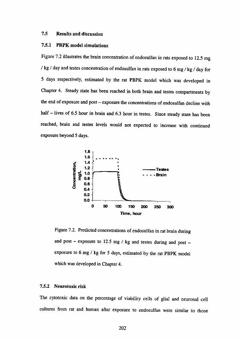
7.5 Results and discussion
7.5.1 PBPK model simulations
Fig・・e 7・2 i11・・t・at・・th・b・ai…ncen仕・ti・n・f・nd・・uぬ・i・・at・exp・・ed t・12.5 mg
/kg/d・y・nd t・・t・・c・ncen血・ti・n・f・nd・・uぬ・i・・at・exp・・ed t・6mg/kg/d・y允・
5d・y・re・pecti・・ly…tim・t・d by th・・at PBPK m・d・l whi・h w・・d・v・1・P・d i・
Ch・pt・・4・Steady・t・t・h・・been・each・d i・b・th b・ai・・nd t・・t・・c・mp頒ment・by
the end of exposure and post-exposure the concentrations of endosulfan decline with
half-lives of 6・5 hour in brain and 6・3 hour in testes. Since steady state has been
reached, brain and testes levels would not expected to increase with colltinued
exposure beyond 5 days.
1.8
1.6
ピ 1・4
1sI:1
8∈ 0.8
ξ1:l
l:1
0 50 100 150 200 250 300
Time, bOur
FigU・e 7・2・P・edi・t・d・・ncen仕・ti・n・・f・nd・・uぬ・i・・at b・ai・d・亘・g
alld post-exposure to 12.5 mg/kg and testes during alld post-
exposure to 6 mg/kg fbr 5 days, estimated by the rat PBPK model
which was developed in Chapter 4.
7.5.2 Neurotoxic risk
The cytotoxic data on the percentage of viability cells of glial and neuronal cell
cultures from rat and humall after exposure to endosulfan were similar to those
202
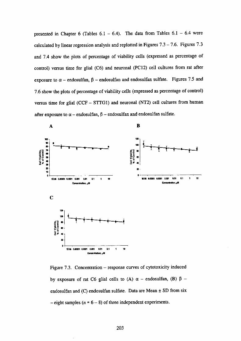
presented in Chapter 6(Tables 6.1-6.4). The data from Tables 6.1-6.4 were
calculated by linear regression analysis and replotted in Figures 7・3-7・6・Fi gures 7・3
and 7.4 show the plots of percentage of viability cells(expressed as percentage of
control)versus time fbr glial(C6)and neuronal(PC 12)cell cultures血om rat after
exposure toα一endosulfan,β一endosulfan and endosulfan sulfate・ Figures 7・5 and
7.6show the plots of percentage of viability cells(expressed as percelltage of contro1)
versus time fbr glial(CCF-STTG1)and neuronal(NT2)cell cultures from human
after exposure toα一endosulfan,β一endosulfan and endosulfan sulfate・
A B
oo
№潤
oηo
0
o
昏06息0㎜aaool oO団 0胴 0.1 1 10
Cooo●{μ■
506息㎜仏m1●001 00r O.1 1 10 Con6●血●叫口日
o
0
0
0
EO6 aoOO n O.oocn息001 0.or al 1 10
Cono●耐■tion P閉
igure 7.3. Concentration-response curves of cytotoxicity induced
y exposure of rat C6 glial cells to(A)α一endosulfan,(B)13-
ndosulfan and(C)endosulfan sulfate. Data are Mean±SD from six
eight samples(n=6-8)of three independent experlments.
03
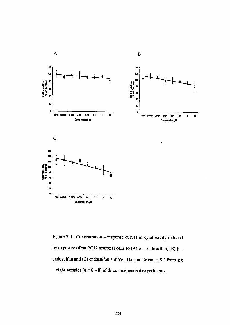
120
100
$る80≡治三ε旦⇔ 60こる
3ボ40
A
tEく060000 ” OOOC”息㎝ 001 01 1 10
0Dno●醐μ目
B
140、
1初
100云る≡き
三580ξ9=060δ’
40
tEO6 ao X”仙001α001 aOt al
OcacentreSeq P■
1
C●
ii]
戴
:
IEO60COCX” 乱㎜鋤胞or at l IO Cmo●r伽6叫抑鯖
Fjgure 7.4. Concentration-response curves of cytotoxicity induced
by exposure of rat PC 1 2 neuronal cells to(A)α一endosulfan,(B)β一
endosulfan and(C)endosulfan sulfate. Data are Mean±SD ffom six
-一@eight samples(n=6-8)of three independent experiments.
204

1初
100
云るooi≡3ち
三ε!り ぼ
:6
8*co
A
1EO6α㎜aOOOI aoel aor Ol 1 10 00nee,taation, pM
1初
100
云る80≡き三ε
旦060:る
3*幼
勿
B
IEOS O㎜0㎜0001α01 01 1
Cα一P日
01
C
1勾
100
sる 80
韮呈8●o:る
3ポ句
頒
11…06息0㎜息0001aonl息m al 1 10
map日
Figure 7.5. Concentration-response curves of cytotoxicity induced
by exposure of human CCF-STTGI glial cells to(A)α一
endosulfan,(B)β一endosulfan and(C)endosulfan sulfate. Data are
Mean±SD伽m six-eight samples(n=6-8)of three independent
expenments・
205
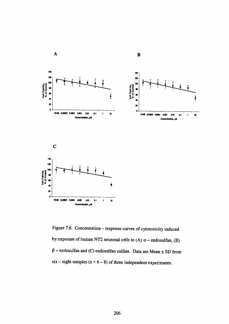
A
140
120
._100ξε= ‘ 80呈8三る●o
ε3 40
0
王
IEO6 aOOCO1一
140
1初
._100
ξ9三5●o旦o>一_0 603ポ 40
1
ao X” 0001 α胴 甑1 1 10
Cono●n悟6叫脚■
。L-____一___.___1EO6000CO1 OOOO1‘㎝ 息與 息1
00no●戚揃㎞L岬
t 10
C
140
120
._100
ξ9=: 6 00量8三も●oεま
co
O
1EO6駄0㎜ 0.OOOI aOO1 α0憎 al l
Cone●情m曽or鳥岬
10
Figure 7.6. Concentratio11-response curves of cytotoxicity induced
by exposure of human NT2 neuronal cells to(A)α一endosulfan,(B)
β一endosulfan and(C)endosulfan sulfate. Data are Mean±SD from
six-eight samples(n=6-8)of three independent experiments.
206

7.5.2.1 PBPK model predictions
In order to assess the lil(elihood that neurotoxicity would occur in humans fbllowing
exposure to endosulfan, the model(Chapter 4)was first used to predict the
concentration of endosulfan in the brain at the No-Observed-Effect-Level
(NOEL)of 12.5 mg/kg!day(equivalent to 30.72μM)for neurotoxicity in rats
(McGregor,1998). Exposure at this concentration gave a brain concentration of 1.56
mg!L. The no effect level for neurotoxicity in the rat equates, therefore, to a target
organ dose of l.56 mg!LTo produce the same concentration of endosulfan in the
humall brain, the model calculates that humans would have to be colltinuously
exposed to l.27 ppm endosulfan. An application of a safety margin of 100 to account
f()r cross - species (inter - species) and intra - species variability fbr non -
carcinogenic and reproductive effects resulted in an exposure of 12・7 PPb(equivalent
toO.0312μM).
Table 7.1 summarizes the estimated percentage of cell viability of glial and neuronal
cell cultures from rat and human after exposure to a-endosulfan,β一endosulfan and
endosulfan sulfate at a dose level of 12.5 mg/kg(rat)and 1.27 mg/kg(human)
respectively. It was observed that high exposure level of 12.5 mg/kg(30.72μM)
induced glial and neuronal cell death for cell cultures from rat and the estimated
percentage of cell viability was below 50%fbr all compounds except fbrα一
endosulfan in the rat neuronal PC 12 cell cultures. Endosulfan generally induced 50%
of glial and neuronal cell death fbr cell cultures from rat at LC500f as low as l 2.97
μM(Chapter 6, Section 6.3.5, Table 6.5). It seems that differences occur in the
response between in vivo and in vitro experiments as 12.5 mg/kg endosulfan was
established as a NOEL in rat(McGregor,1998). However, in vitro experiments can
207
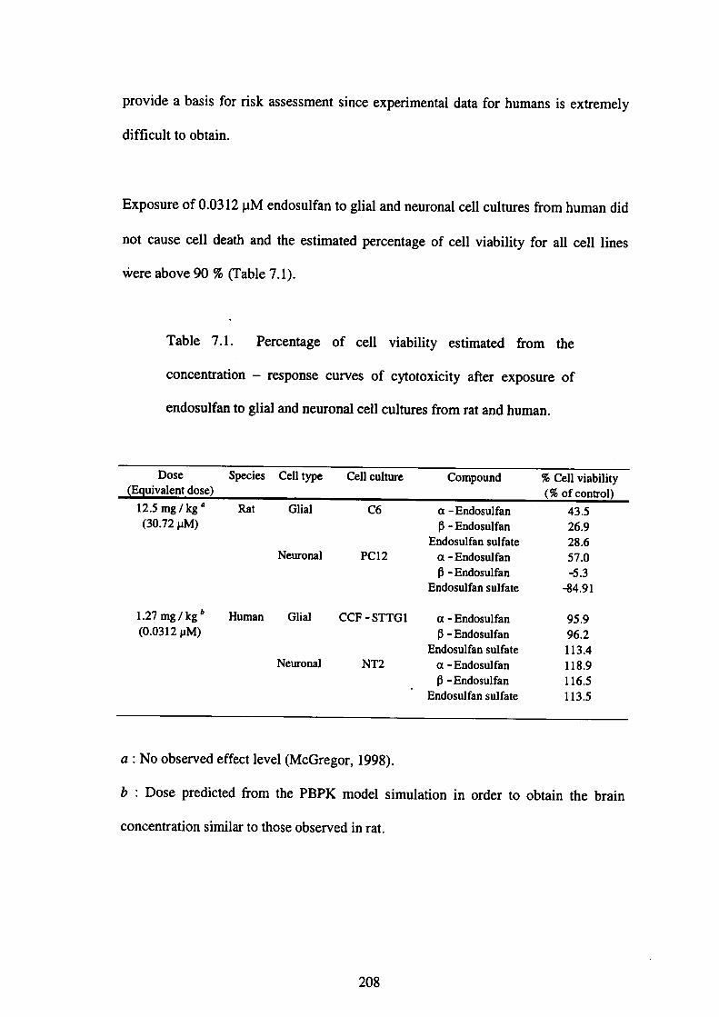
provide a basis fbr risk assessment since experimental data fbr humans is extremely
difficult to obtain.
Exposure of O.0312 pM endosulfan to glial and neuronal cell cultures from human did
not cause cell death and the estimated percentage of cell viability fbr all cell lines
vレere above 90%(Table 7.1).
、
Table 7.1. Percentage of cell viability estimated 伽m the
concentratlon-response curves of cytotoxicity after exposure of
endosulfan to glial and neuronal cell cultures from rat and human.
Dose Species Cell type Cell culture Compound %Cell viability
一91i}itll!ll!lillli
Rat12.5mg!kg a
(30.72μM)
1.27mg!kg b
(0.0312 pM)
副11G
泣㎝㎝N
6C
2 CP
Human G】ial CCF-STTG l
Neuronal NT2
α一Endosulfan
β一Endosulfan
Endosu】fan sulfate
α一Endosulfan
β一Endosul fan
Endosulfan su】fate
α一Endosulfan
fi -Endosulfan
Endosu】fan sulfate
α一 Endosul fan
β一Endosulfan
Endosulfan sUl fate
43526.9
28.6
57.0
-5.3
-84.91
95.9
96.2
113.4
118.9
116.5
113.5
a:No observed effect leve1(McGregor,1998).
b:Dose predicted from the PBPK model simulation in order to obtain the brain
concentration similar to those observed in rat.
208
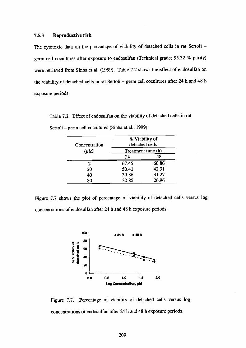
7.5.3 Reproductive risk
The cytotoxic data on the percentage of viability of detached cells in rat Sertoli-
germ cell cocultures after exposure to endosulfan(Technical grade;95.32%purity)
were retrieved from Sinha et al.(1999). Table 7.2 shows the effect of endosulfan on
the viability of detached cells in rat Sertoli-germ cell cocultures after 24 h and 48 h
exposure periods・
Table 7.2. Effect of endosulfan on the viability of detached cells in rat
Sertoli 一 germ cell cocultures(Sinha et al・,1999)・
Concentration
(μM)
%Viability of
detached cells
Treatment time(h)
24 48 0002 (∠4.8
67.45
50.41
39.86
30.85
60.86
42.31
31.27
26.96
Fi gUre 7.7 shows the plot of percentage of viability of detached cells versus log
concentrations of endosulfan after 24 h and 48 h exposure periods・
oo@80 60 40 ⑳
1
望W{噂ち≧云旦〉ま
0
0.0 0.5 1.0 1.5
Log Conoentration, pM
20
Figure 7.7. Percentage of viability of detached cells versus log
concentrations of endosulfan after 24 h and 48 h exposure periods・
209
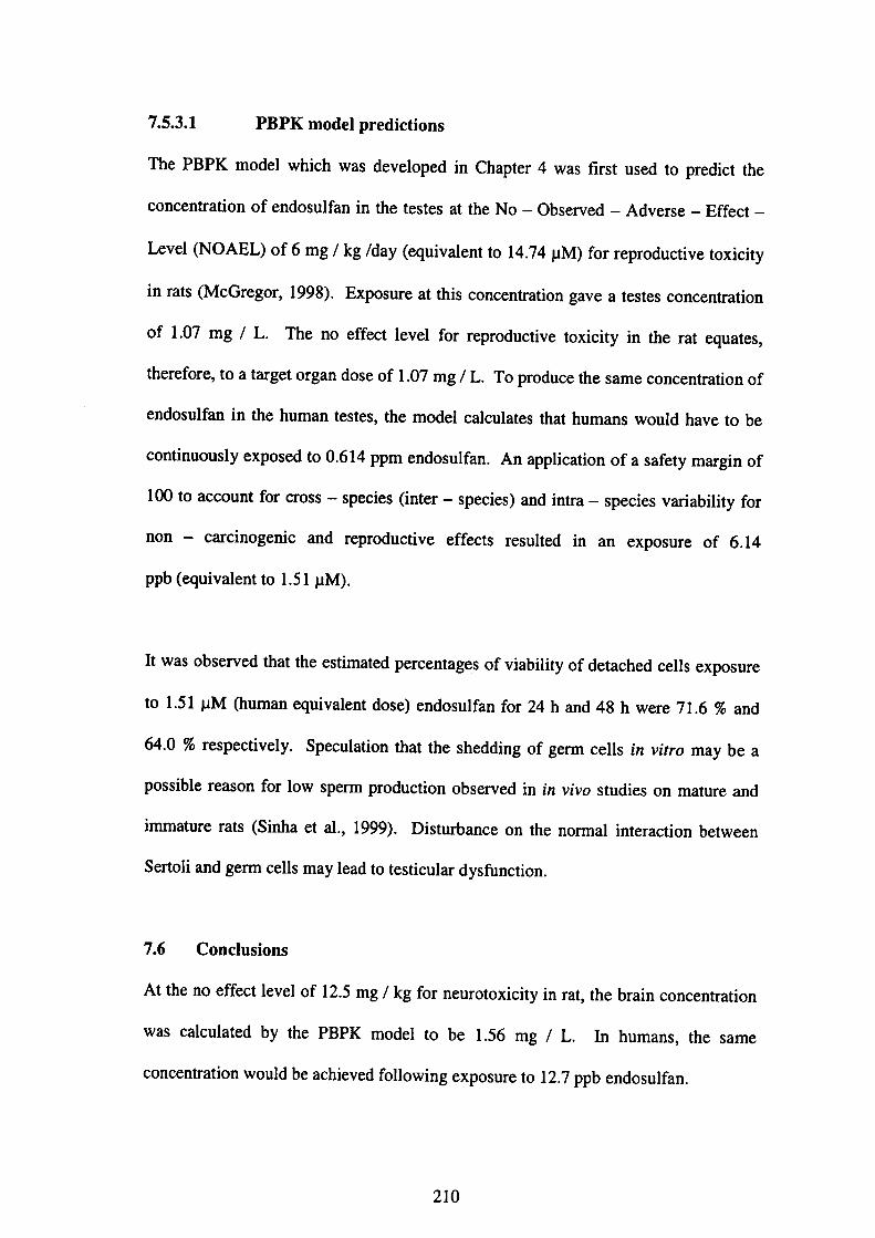
7.5.3.1 PBPK model predictions
The PBPK model which was developed in Chapter 4 was first used to predict the
concentration of endosulfan in the testes at the No-Observed-Adverse-Effect-
L・vel(NOAEL)・f 6 mg/kg!d・y(・q・ival・・t t・14.74μM)f…rep・・d・・ti・・t・xi・ity
in rats(McGregor,1998). Exposure at this concentration gave a testes concentration
・fl・07 mg/Lr「h・n・effect・1・v・l f…ep・・d・・ti・・t・xi・ity i・th・・at・quat・・,
therefore, to a target organ dose of 1.07 mg/L. To produce the same concentration of
endosulfan in the human testes, the model calculates that humans would have to be
・・nti…u・ly exp・・ed t・0・614 PPm・nd・・ulfa・. A・・pPlicati・n・f・・af・ty・m・・gi・・f
100t・acc・unt f・・er・ss-・peci・・(i・t・・一・peci・・)・・d i・tr・一・peci・・v・・i・bility・f・r
non - carcinogenic and reproductive effects resulted in an exposure of 6.14
ppb(equivalent to 1.51 pM).
It w…b・e・v・d也・t the e・tim・t・d p・・cent・g…f・i・bility・f d・tach・d cell・exp・・u・e
to 1.51μM(human equivalent dose)endosulfan fbr 24 h and 48 h were 71.6%and
64・0%・e・pecti・・ly・Specul・ti・曲・t th・・h・ddi・g・f g・m・ell・i・・vit・・m・y be a
possible reason fbr low spem production observed in加ivo studies on mature and
lmmature rats(Si血a et al.,1999). Disturbance on the nomal interaction between
Sertoli and germ cells may lead to testicular dysfUnction.
7.6 Conclusions
At the no effect level of 12.5 mg/kg for neurotoxicity in rat, the brain concentration
was calculated by the PBPK model to be 1.56 mg/L. In humans, the same
concentratlon would be achieved following exposure to 12.7 ppb endosulfan.
210
At the No-Observed-Adverse-Effect-Level(NOAEL)of 6 mg/kg fbr
reproductive toxicity in rat, the testes concentration was calculated by the PBPK
model to be 1.07 mg 1 L. In humans, the same concentration would be achieved
following exposure to 6.14 ppb endosulfan.
Based on these results, the estimated exposure level fbr neurotoxicity ill humans was
1.6-fbld higher alld exceeded the acceptable daily intake(ADD of O.008 mg/kg
(equivalent to 8 ppb)(WHO,1984). This could be attributed to interspecies
differences such as receptor binding as this factor was not introduced in the current
PBPK model. This parameter is very important in sensitivity and should be
introduced to the model in subsequent studies. ln contrast, the estimated exposure
level for reproductive toxicity in humans was 6.14 ppb, which was below the ADI.
As has been discussed in Chapter 4, S㏄tion 4.4.4.1, the pregnant women from Chiba
and Kyushu, Japan were estimated to be exposed to endosulfan through dietary intake
of O.76 x 10’5 mg/kg/day(equivalent to O.0076 ppb)and 9.09 x 10’5 mg/kg/day
(0.0909ppb)respectively whereas school children from Malaysia were estimated to
intake endosulfan for 1.06 x 10⁻⁵ mg/kg/day(0.0106 ppb). The estimated exposure
levels fbr tllese groups of people were below the no effect level of 12.7 ppb for
neurotoxicity and 6.14 ppb for reproductive toxicity in human, which were estimated
in Sections 7.5.2.l and 7.5.3.1 respectively, thus suggesting that these people were
unlikely. to develop any serious health problems. However, other possibilities cannot
be ruled out.
211

REFERENCES
Aarons, L, Clewell, H.」., Conolly, R. B., Delic,」.1., Houston, J. B., Jarabek, A. M.,
Loizou, G., Mason, H. J., Nestorov,1., Rowland, M., Trans, C. L, Tucker, G. T.
(1999)・Physiotogica〃y-based phaア〃Tacokinetic〃iodeting:、4 potentia〃oo1/br use
in riskα∬ε∬〃lellt・ R¢ρoπ々〆αworkshop organised by’he Risk∠4∬ε∬mentαπ4
Toxicolo8y Steerin8 Committee・lnstitute for Environment and Health, Leicester, UK,
PP.3-29.
ATSDR(2000). Toxicolo8ical proLtile for endosutfan. Atlanta, GA:Agency fbr
Toxic Substances and Disease Registry.
Chitra, K. C., Latchoumycandane, C., Mathur, P. P.(1999). Chronic effect of
endosulfan on the testicular fUnctions of rat. ノls輌an J〈)urnal qプノlndrolo8y,1:203-
206.
Choudhary, N., Joshi, S. C.(2003). Reproductive toxicity of endosulfan in male
albino rats・Bulletin ofEnvironmental Contamination and To」xicolo8y,70:285 一 289.
Clewell m, HJ.(1995). The application of physiologically based pharmacokinetic
modeling in human health risk assessment of hazardous substances. ]r()ズfco∫08y
乙επθrぷ,79:207-217.
Dalsenter, P. R., Dallegrave, E., Mello, J. R. B., Langeloh, A., Oliveira, R. T., Faqi, A.
S.(1999).Reproductive e脆cts of endosulfan on male offspring of rats exposed
during Pregnancy and lactation・Hu’nall and EXperimental Toxicology,18:583-589.
212

Dalsenter, P. R., de Aratijo, S. L, da Silva de Assis, H. C., Andrade, A. J. M.,
Dallegrave, E.(2003). Pre and post natal exposure to endosulfan in Wistar rats.
Human and Experimental Toxicology,22:171-175.
Dikshith, T. S. S., Raizada, R. B., Srivastava, M. K., Kaphalia, B. S.(1984). Response
of rats to repeated oral administration of endosulfan. Industrial Health,22,295-304・
Foster, P. M. D.(1988). Testicular organization and biochemical fUnction. In:Lamb,
∫C.,Foster, P. M D.,{Eぬ), Physiolo8y and Toxicolo8y of・Male Reproduction, PP.
7-31.USA:Academic Press.
McGregor, D. B.(1998). Endosutfan(JMPR evaluations 1998 Part II Toxicolo8ical)
-Fか∫’4rψ. Lyon, France:lnternational Agency for Research on Cancer.
National Cancer Institute(1978). Bioa∬ay of endosulfan/br po∬ibte carcino8enicity,
■
Technica’Report, Series No.62, Pubtication No.(MH)78-1312. U. S. Departmellt
of Health, Education and Welfare.
Pandey, N., Gundevia, F, Prem, A. S., Ray, P. K.(1990). Studies on the genotoxicity
of endosulfan, an organochlorine insecticide, in mammalial germ cells. Mutation
Research,242:1-7.
213

Reader, S. C. J., Foster, P. M. D.(1990). The in vitro effects of four isomers of
dinitrotoluene on rat Sertoli and Sertoli-germ cell cocultures:Germ cell detachment
and lactate and pynlvate production・ Toxicology and ApPlied Toxicolo8y,106:287-
294.
Sinha N., Adhikari, N., Narayan, R., Saxena, D. K.(1999). Cytotoxic effect of
endosulfan on rat Sertoli-germ cell coculture. Reproductive Toxicolo8y,13:291-
294.
Singh, S. K., Pandey, P. S(1989). Gonadol toxicity of short term chronic endosulfan
exposure to male rats・Indian Journal ofEixperimentat Biolo8y,27:341-346.
Sinha, N., Narayan, R., Shanker, R., Saxena, D. K.(1995). Endosulfan induced
biochemical changes in the testis of rats. レieterinai Y and Hu〃mn Toxicology,37:547
-549.
Sinha, N., Narayan, R., Saxena, D. K.(1997). Effect of endosulfan on the testis of
growing rats. Bulletin(~fEnvironmental Contamination and Toxicolo8y,58:79-86.
Sundaran, K., Witorscb, R.」.(1995). Toxic effects on testis. In:VVitorsch, R.∫,
(Edゆ・Reproductive Toxicology:Tar8et Or8αη Toxicolo8y, Series,2ηゴed., PP.99-
122.New York:Raven Press.
214
US・EPA.(1996). Guidelines for Reproductive Toxicity Risk A∬e∬ment, U.S.
Environmental Protection Agency, Washington, D. C. EPA/630/R-96/009
September l 996・
US EPA.(1997). Special report on environmental endocrine disrq〆ion:、4nψcf∫
asse∬ment an姻∫. Pr印αrε4カrτ乃ε蹴A∬θ∬ment Forum, U.S. Environmental
Protection Agency, Washington, D. C. EPA/630/R-96/012Febnlary 1997.
US NRC(1993). Risk a∬e∬ment in the Federal Government,ルtana8ing the Proce∬,
National Research Council, National Academy Press.
WHO/UNEP!m.1984. Endosulfan:international Programme on Chemical
Safety, Environmental Health Criteria 40. Geneva, World Health Organization.
Wilson, V. S, LeBlanc, G. A.(1998). Endosulfan elevates testosterone
biotransformation and clearance in CD-1mice. Toxicoto8y and Apptied
Pharmacolo8y,148:158-168.
215

CHAPTER 8
8.O Conclusions
With regards to the objectives of this research as mentioned in Chapter 1(Section l.1),
the findings are summarized as follow:
Chapter l
Chapter 1 compares the extent of contamination of endosulfan between the Malaysian
and Japallese environment. This chapter also describes the physical and chemical
properties of endosuぬn fbllowed by the possible routes of exposure, human
exposures, effects on the environment and its environmental fate.
Chapter 2
Chapter 2 describes the general toxic effects of endosulfan in mammals(rats)
including the neurologica1, reproductive and endocrine effects.
Chapter 3
The present study in Chapter 3 indicates that 14C-Endosulfan was rapidly excreted in
the feces and urine after 96 h following single oral administration of 5 mg/kg 14C-
Endosulfan with fecal elimination as the major elimination route in male rats.
Elimination was esselltially complete within a few days. The cumulative excretions
in the urine and feces fbr four days were 12.4%and 94.4%respectively of the total
administered dose. The total radioactivity recovered in the excreta fbr fbur days was
lO6.8%, thus suggesting that elimination was essentially complete within four days.
216
F・ll・wi・g・i・gle and th・ee-tim・・epeat・d・・al・dmi・i・t・ati・n・f 5 mg/kg 14C-
Endosulfan, it was observed that the radioactivity levels in all tissues decreased after
administration was terminated, rats will recover once administration is teminated.
The tissue concentrations of residues after 8 h as observed in the present study were
generally highest in the liver and kidneys and lower in other tissues such as brain,
testes and muscle.
The basic toxicokinetic parameters fbr 14C-Endosulfan was established fbllowing
・i・gl…創・dmi㎡・仕・ti・n・f 5 mg 14C-E・d・suぬ・/kg, withた。・f 3.07 h” and Cmax
of O.36±0.08 mg-eq./Lwith Tntax of 2 h. The terminal elimination half life,τ%
yof the radioactivity in blood was l 93 h.
Chapter 4
Given that no calibration or validation of PBPK model predictions of concentrations
of endosulfan in various tissues exist, the frrst and new PBPK model for endosulfan in
male rats has been developed in Chapter 4 and can predict tissue dosimetries
following single and multiple oral dosages of endosulfan. Generally, results show
reasonable concordance between model predictions and measured values in all target
tissues(liver, kidneys, brain, testes and blood)following oral administration. The
validity of the model was fUrther verified with experimental data retrieved from the
literature. The simulations of those studies from the literature generally agree with
experimental data. Sensitivity allalyses allow fbr a quantitative assessment of input
parameters on the model simulations of tissue concentrations. The present results
indicate that the influence of PBPK model input parameters on total endosulfan tissue
dosimetry varies, as expected, across parameter.
217
The PBPK model fbr endosulfan in rats was extrapolated to humans, without
adjusting the previously established model parameters to test the ability of the model
to predict the phamlacoldnetic behavior of endosulfan in humans. The ability of the
model was tested by predicting the daily intake of endosulfan per individual by
comparing the residue levels of endosulfan in selected tissues for both the parent
is°me・s(・nd・・ulfa・)and it・m・t・b・lit・・(・・d・・ulfa・m・t・b・lit・・)一. F・・m th・PBPK
model simulations, the estimated dietary intake fbr the pregnant women from Chiba
・nd・Kyu・h・・J・p・n w・・e O・76・10’5 mg/kg/d・y・nd 9.09・10’5 mg/kg/d・y
respectively, whereas the estimated dietary intake for the Malaysian school children
w・・1・06・10δ 高〟^kg/d・y・G・n・・ally,・ea・…ble ag・eemen・w…b・erv・d
betw㏄n model predictions and experimental data, indicating that the model was/
could be partially validated since data conceming human exposures are scarce and
difficult to obtain experimentally.
Chapter s
Chapter 5 f()cuses on the effects ofα一endosulfan,β一endosulfan and endosu1白n
sulfate on也e tight junctions of the blood-brain barrier(BBB)by investigating the
廿ansendothelial elec廿ical resistance(TEER)and permeability effe’cts across cultured
monolayers of porcine brain microvascular endothelial cells(PBMECs). Following
exposure to a series of concentrations of endosulfan(0.01μM t610μM), TEER
declined significantly and reached the bottom level as concentrations and exposure
periods increased. C)戊otoxicity tests indicated that the concentrations of 10μM and
below did not cause cell death fbr all compounds. The integrity of the brain
endothelium was fUrther investigated by measuring the transendothelial pemeability
・・14C-E・d・・uぬ・・1・w…b・erv・d・h・t th…an・p・rt・f end・・uぬ。・㎞。ugh・h。
218
BBB was reversible, in which endosulfan transported丘om the blood-brain
compartment and ffom the brain-blood compartment, thus suggesting that residues
●
of endosulfan has little potential to accumulate in the brain, although other
possibilities could not be nlled out. The ratio between the outer-to-inner and the
im・・-t・一・ut・・c・mp頒ments飼・th・t・ansp・宜st・dy・f 14C-E・d・suぬ・in th・
concentration range of O.Ol-10μM was approximately l.2-2.1.
Chapter 6
11Chapter 6, a study was carried with in vitro assays to assess the neurotoxic effects
ofα一endosulfan,β一endosulfan and endosulfan sulfate by comparing the ability of
the compounds to cause cell death in glial and neuronal cell cultures from rat and
human. The results demonstrate thatα一endosulfan,β一 endosulfan and endosulfan
sulfate cause cell dealth in glial and neuronal cell cultures from rat and human brain
in a concentration-dependent manner.α一endosulfan produces a manifest selective
neurotoxicity with LC50 ranging fξom 11.2μM to 48.0μM.111 contrast, selective
neurotoxicity was not so manifested in glial and neuronal cell cultures after exposure
to endosulfan sulfate, as LC50 values were in the range of 10.4-21.6 pM.
Human glial CCF-STTG l cells were the most sensitive cell type to the cytotoxic
effects ofα一endosulfan followed by human neuronal NT2, rat glial C6 and rat
neuronal PC 12 cells. Human neuronal cells were the most sensitive cell type to the
cytotoxic effects of S 一・ endosulfan followed by rat neuronal, rat glial and human glial
cells. Human glial cells were the most sensitive cell type to the cytotoxic effects of
endosulfan sulfate fbllowed by human neurona1, rat neuronal and rat glial cells. This
study also shows that human glial and neuronal cells are more sensitive compared to
N 219

rat glial and neuronal cells and could be used as a good tool fbr studying the
neurotoxic effects of these chemicals in the central nervous system(CNS), and thus,
predicting their impact on human health.
Chapter 7
1n Chapter 7, a quantitative risk assessment of endosulfan was presented by utilizing
the principles of PBPK modeling(as discussed previously in Chapter 4)as well as in
vitro experiments of cytotoxicity(as discussed previously in Chapter 5). The
neurotoxic and reproductive risks were estimated since the brain and testes were
among the target organs for toxicity.
At the no effect level of 12.5 mg/kg for neurotoxicity in rat, the brain concentration
was calculated by the PBPK model to be 1.56 mg/L. hl humans, the same
concentration would be achieved fbllowing exposure to 12.7 ppb(0.0312μM)
endosul fan. Exposure of O.0312μM endosulfan to glial and neuronal cell cultures
from human did not cause cell death and the estimated percentages of cell viability for
all cell lines were above 90%
At the No-Observed-Adverse-Effect-Leve1(NOAEL)of 6 mg/kg fbr
reproductive toxicity in rat, the testes concentration was calculated by the PBPK
model to be 1.07 mg/L In humans, the same concentratioll would be achieved
following exposure to 6.14 ppb(1.51 pM)endosulfan. The estimated percentages of
viability of detached cells in the Sertoli-germ cells cocultures exposed to 1.51μM
endosulfan fbr 24 h and 48 h were 71.6%and 64.0%respectively, indicating
220

disturbance on the normal interaction between Sertoli and germ cells may lead to
testicular dysfUnction. 、
Based on these results, the estimated exposure level for neurotoxicity in humans was
1.6-fbld higher and exceeded the acceptable daily intake(ADD of O.008 mg/kg
(equivalent to 8 ppb).111 contrast, the estimated exposure level fbr reproductive
toxicity in humans was 6.14 ppb, which was below the ADI.
8.l Recommendations fヒ)r仙ure study
It has been proposed that humans and wildlife have suffered adverse effects on health
as a result of environmental exposure to toxic chemicals. The o句ectives of this study
as mentioned in Chapter l(Section 1.2)were achieved. However, some issues and
research gaps need to be addressed fbr fUture investigation.
Amore reliable and sensitive analytical method would need to be incorporated into
the fUture experiments to enhance the separation and identification of the parent
compounds and its metabolites present in different matrices. Further research is also
necessary to expand the current model fbr endosulfan to illclude the two isomers of
the parent compo皿ds and its major oxidation metabolites.
The current in vitro blood-brain barrier(BBB)model to study the effects of
endosulfan on the BBB needs to be improved until optimum conditions are achieved
in order to increase our understanding on the pharmacology and toxicology effects of
endosulfan as well as its influences on the barrier under in vitro conditions.
s 221
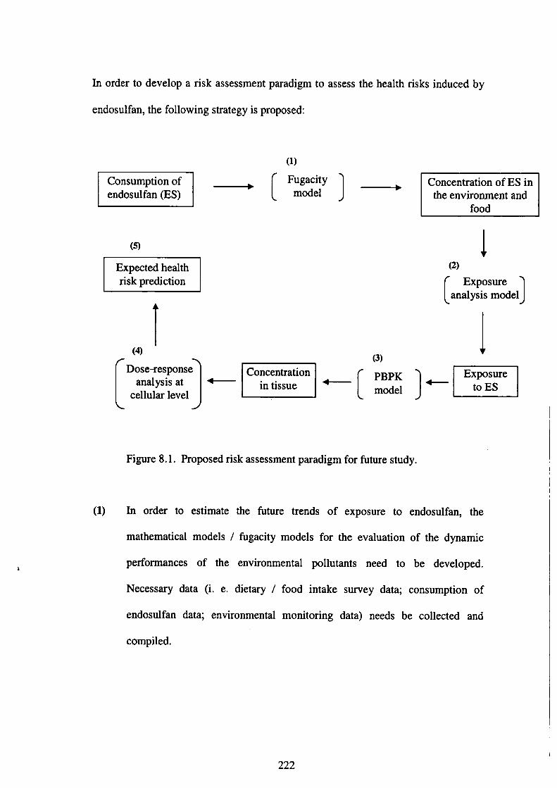
i
1110rder to develop a risk assessment paradigm to assess the health risks induced by
endosulfan, the fbllowing strategy is proposed:
(1)
fbod
⑤ ↓ Expected health (2)
「’sk P「edicti°n
(3)
蕊竃e-C°=°n一ご〕一[Exposur to ES]
(1)
Figure 8.1. Proposed risk assessment paradigm for fUture study.
㎞order to estimate the fUture trends of exposure to endosulfan, the
mathematical models/fUgacity models fbr the evaluation of the dynamic
pe㎡formances of the environmental pollutants need to be developed.
Necessary data(i. e. dietary/fbod intake survey data;consumption of
endosulfan data;environmental monitoring data)needs be collected and
compiled.
222

(2) To develop the exposure analysis models, exposure analysis of people to the
envirollmental pollutants through dietary and respiratory pathways need to be
conducted and the data of the concentration of endosulfan in the environment
and fbod intake survey data will be applied. An exposure model is an
empirical加mework, which allows estimation of exposure parameters from
available input data. Chemical release estimates, fate and transport modeling,
and exposure potentials based on life habits can be employed to estimate
exposure for wildlife and/or humans by way of air, fbod, water or in total.
(3) The PBPK model should be revised from time to time to accommodate for any
changes until the optimum conditions are achieved. More surveys on the
human exposures to endosulfan are necessary in order to validate the model.
(4) A dose response analysis fbr endosulfan at the cellular level needs to be
established in order to evaluate the magnitude of intemal exposure to the
target organs.111 addition, a reliable biomarker and appropriate endpoints
need to be identified before any risk assessment is attempted.
(5) By illtegrating and coupling all the necessary infbmlation, it is hoped that the
proposed strategy could be used to relate/evaluate possible adverse health
risk with the environmental pollutants with the hope that effective risk
reduction options/measures could be proposed subsequently.
223