Embed Size (px)
Citation preview
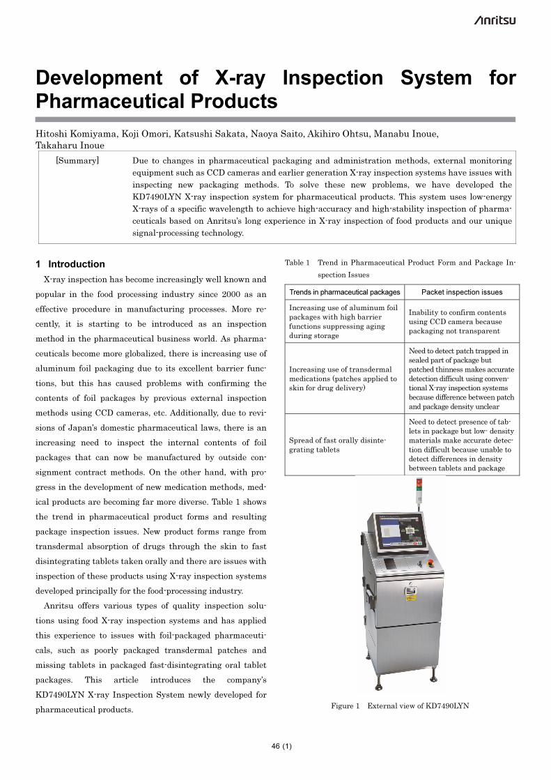
Development of X-ray Inspection System for Pharmaceutical Products
Hitoshi Komiyama, Koji Omori, Katsushi Sakata, Naoya Saito, Akihiro Ohtsu, Manabu Inoue,
Takaharu Inoue
[Summary] Due to changes in pharmaceutical packaging and administration methods, external monitoring
equipment such as CCD cameras and earlier generation X-ray inspection systems have issues with
inspecting new packaging methods. To solve these new problems, we have developed the
KD7490LYN X-ray inspection system for pharmaceutical products. This system uses low-energy
X-rays of a specific wavelength to achieve high-accuracy and high-stability inspection of pharma-
ceuticals based on Anritsu’s long experience in X-ray inspection of food products and our unique
signal-processing technology.
(1)
1 Introduction
X-ray inspection has become increasingly well known and
popular in the food processing industry since 2000 as an
effective procedure in manufacturing processes. More re-
cently, it is starting to be introduced as an inspection
method in the pharmaceutical business world. As pharma-
ceuticals become more globalized, there is increasing use of
aluminum foil packaging due to its excellent barrier func-
tions, but this has caused problems with confirming the
contents of foil packages by previous external inspection
methods using CCD cameras, etc. Additionally, due to revi-
sions of Japan’s domestic pharmaceutical laws, there is an
increasing need to inspect the internal contents of foil
packages that can now be manufactured by outside con-
signment contract methods. On the other hand, with pro-
gress in the development of new medication methods, med-
ical products are becoming far more diverse. Table 1 shows
the trend in pharmaceutical product forms and resulting
package inspection issues. New product forms range from
transdermal absorption of drugs through the skin to fast
disintegrating tablets taken orally and there are issues with
inspection of these products using X-ray inspection systems
developed principally for the food-processing industry.
Anritsu offers various types of quality inspection solu-
tions using food X-ray inspection systems and has applied
this experience to issues with foil-packaged pharmaceuti-
cals, such as poorly packaged transdermal patches and
missing tablets in packaged fast-disintegrating oral tablet
packages. This article introduces the company’s
KD7490LYN X-ray Inspection System newly developed for
pharmaceutical products.
Table 1 Trend in Pharmaceutical Product Form and Package In-
spection Issues
Trends in pharmaceutical packages Packet inspection issues
Increasing use of aluminum foil packages with high barrier functions suppressing aging during storage
Inability to confirm contents using CCD camera because packaging not transparent
Increasing use of transdermal medications (patches applied to skin for drug delivery)
Need to detect patch trapped in sealed part of package but patched thinness makes accurate detection difficult using conven-tional X-ray inspection systems because difference between patch and package density unclear
Spread of fast orally disinte-grating tablets
Need to detect presence of tab-lets in package but low- density materials make accurate detec-tion difficult because unable to detect differences in density between tablets and package
Figure 1 External view of KD7490LYN
46
Anritsu Technical Review No.23 September 2015 Development of X-ray Inspection System for Pharmaceutical Products
(2)
2 X-Ray Inspection System
X-ray inspections systems irradiate the inspected product
with X-rays and use the resulting captured X-ray transmis-
sion image to detect the presence of contaminants; they are
used on production lines for food products, etc., to automate
the inspection process for every product on the line. Due to
their ability to detect broken items or missing within
packaged products that cannot be seen by visual inspection,
they are used by many different types of production line.
Instead of using statistical sampling inspection on lines
producing large numbers of products with individually
packaged items purchased by consumers, it is now becoming
more common to install inspection equipment on the line to
inspect the quality of every product on the line. Not only
does this reduce the number of faulty products reaching the
market but it also provides data on the timing and trends in
production of faulty products, helping spot production line
problems early and increasing line efficiency.
2.1 Operation Principle
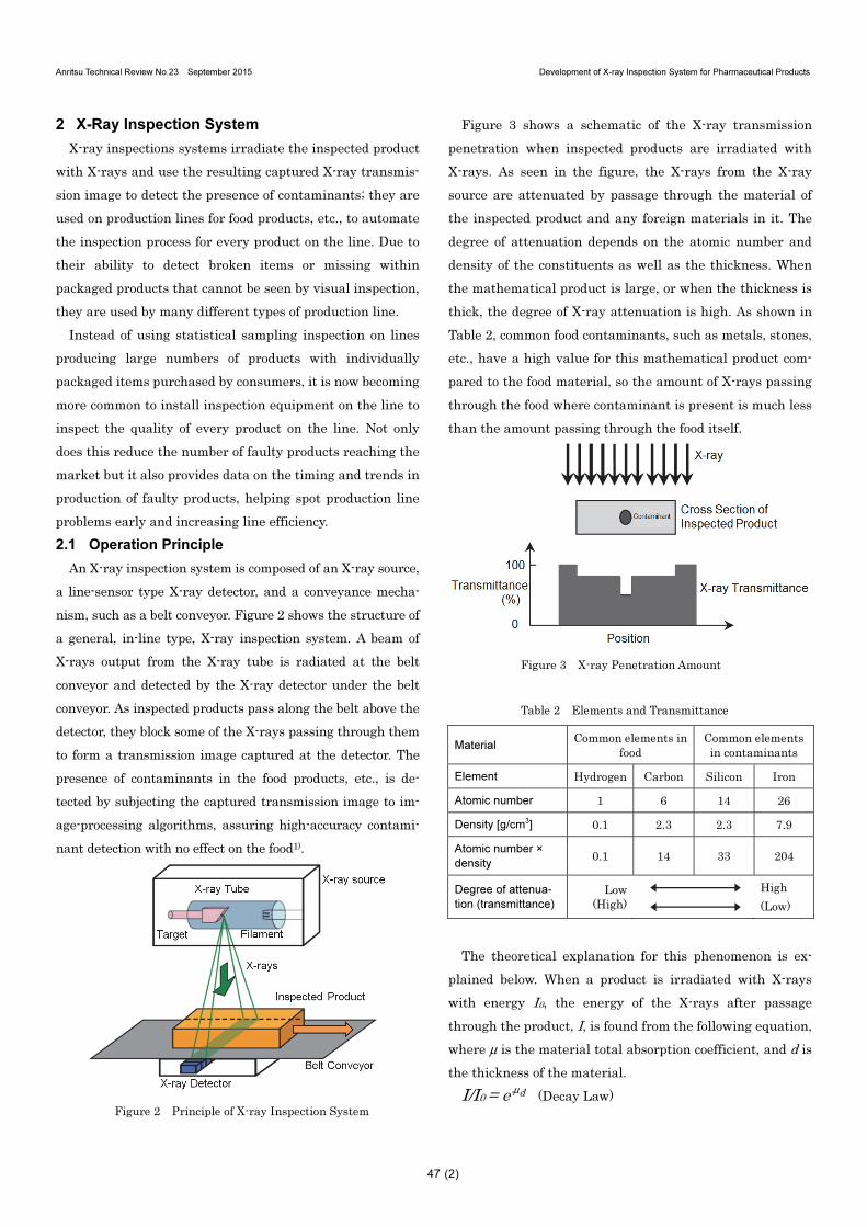
An X-ray inspection system is composed of an X-ray source,
a line-sensor type X-ray detector, and a conveyance mecha-
nism, such as a belt conveyor. Figure 2 shows the structure of
a general, in-line type, X-ray inspection system. A beam of
X-rays output from the X-ray tube is radiated at the belt
conveyor and detected by the X-ray detector under the belt
conveyor. As inspected products pass along the belt above the
detector, they block some of the X-rays passing through them
to form a transmission image captured at the detector. The
presence of contaminants in the food products, etc., is de-
tected by subjecting the captured transmission image to im-
age-processing algorithms, assuring high-accuracy contami-
nant detection with no effect on the food1).
Figure 2 Principle of X-ray Inspection System
Figure 3 shows a schematic of the X-ray transmission
penetration when inspected products are irradiated with
X-rays. As seen in the figure, the X-rays from the X-ray
source are attenuated by passage through the material of
the inspected product and any foreign materials in it. The
degree of attenuation depends on the atomic number and
density of the constituents as well as the thickness. When
the mathematical product is large, or when the thickness is
thick, the degree of X-ray attenuation is high. As shown in
Table 2, common food contaminants, such as metals, stones,
etc., have a high value for this mathematical product com-
pared to the food material, so the amount of X-rays passing
through the food where contaminant is present is much less
than the amount passing through the food itself.
Figure 3 X-ray Penetration Amount
Table 2 Elements and Transmittance
Material Common elements in
food Common elements in contaminants
Element Hydrogen Carbon Silicon Iron
Atomic number 1 6 14 26
Density [g/cm3] 0.1 2.3 2.3 7.9
Atomic number ×
density 0.1 14 33 204
Degree of attenua-
tion (transmittance)
Low (High)
High
(Low)
The theoretical explanation for this phenomenon is ex-
plained below. When a product is irradiated with X-rays
with energy I0, the energy of the X-rays after passage
through the product, I, is found from the following equation,
where μ is the material total absorption coefficient, and d is
the thickness of the material.
I/I0 = e-μd (Decay Law)
47
Anritsu Technical Review No.23 September 2015 Development of X-ray Inspection System for Pharmaceutical Products
(3)
Additionally, the total absorption coefficient μ is found
from the following equation, where λ is the X-ray wave-
length, ρ is the material density, Z is the material atomic
number, and C is a constant. From this, we can see that the
X-ray transmittance changes with the inspected product
and contaminant atomic number, thickness, and density.
μ=λ3ρCZ (cm−1)
Furthermore, shorter-wavelength X-rays (at higher X-ray
tube impressed voltage) have a lower absorption coefficient,
so the X-ray transmittance increases2),3),4).
3 Development Points and Implementation
In addition to resolving the issues with packaged products
listed in Table 1, the main point of this development was to
build a system that could be easily introduced into a pro-
duction line for pharmaceutical products to ensure strict
quality control.
3.1 Pharmaceutical Product Package Inspection
Issues
Transdermal medications with a diverse variety of forms
are becoming widespread. They are generally composed of a
thin patch type base film coated with medication covered by
another film called a liner to protect the medication and an
adhesive covering the medication surface all packaged in a
sealed rectangular form using an aluminum-laminate film,
etc. Sometimes, at the sealing stage, the patch can be
trapped between the sealing materials, causing it to be in-
completely packaged and requiring it to be discarded be-
cause the incomplete packaging is unable to maintain the
quality of the medication. Additionally, orally
fast-disintegrating tablet medications are becoming more
commonplace. These tablets are designed to dissolve in very
small amounts of water, so they are commonly packaged in
aluminum foil with high resistance to humidity. Conse-
quently, it is important to inspect these packages for miss-
ing tablets and failed packaging with high accuracy and
stability at the inspection stage after packaging.
Current medications, including these two examples, gen-
erally use packaging composed of polypropylene (PP), poly-
vinyl chloride (PVC), aluminum, etc., with thicknesses of 30
to 400 μm, requiring X-ray inspection technology able to
capture the transmission images of packaging materials
and tablets/patches with different densities.
3.2 Implementation Procedure
3.2.1 X-ray Management for Pharmaceutical Packages
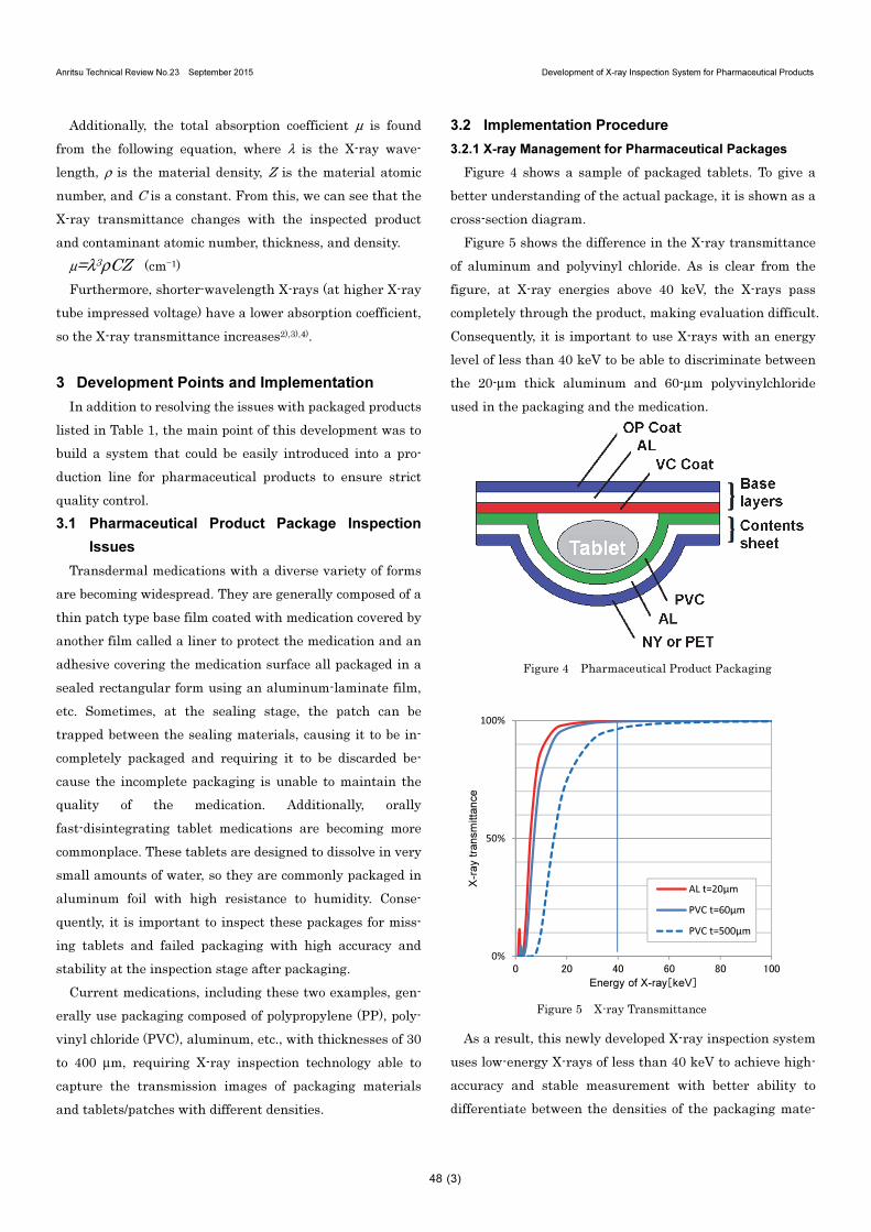
Figure 4 shows a sample of packaged tablets. To give a
better understanding of the actual package, it is shown as a
cross-section diagram.
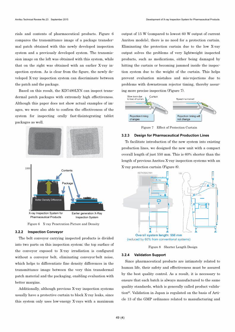
Figure 5 shows the difference in the X-ray transmittance
of aluminum and polyvinyl chloride. As is clear from the
figure, at X-ray energies above 40 keV, the X-rays pass
completely through the product, making evaluation difficult.
Consequently, it is important to use X-rays with an energy
level of less than 40 keV to be able to discriminate between
the 20-μm thick aluminum and 60-μm polyvinylchloride
used in the packaging and the medication.
Figure 4 Pharmaceutical Product Packaging
Figure 5 X-ray Transmittance
As a result, this newly developed X-ray inspection system
uses low-energy X-rays of less than 40 keV to achieve high-
accuracy and stable measurement with better ability to
differentiate between the densities of the packaging mate-
0%
50%
100%
0 20 40 60 80 100
X-r
ay t
ran
sm
itta
nce
Energy of X-ray[keV]
AL t=20μm
PVC t=60μm
PVC t=500μm
48
Anritsu Technical Review No.23 September 2015 Development of X-ray Inspection System for Pharmaceutical Products
(4)
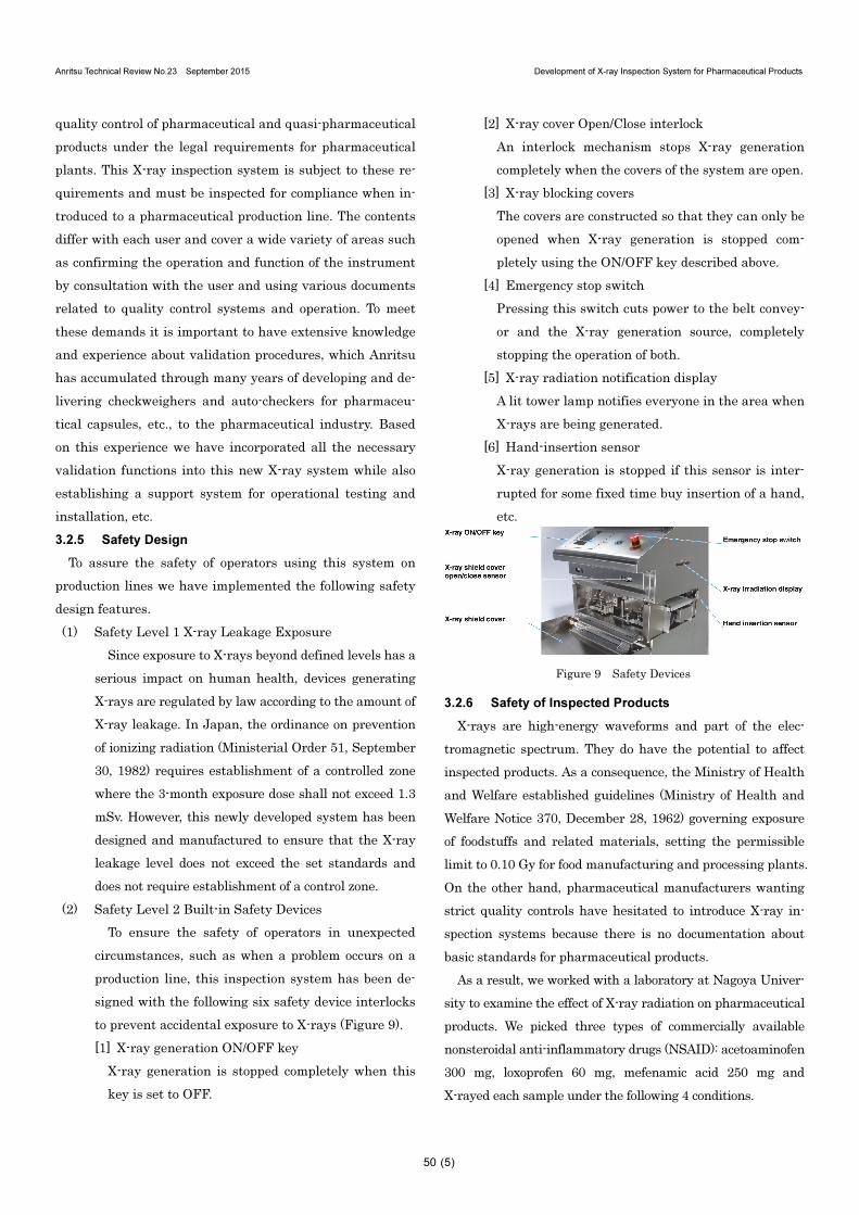
rials and contents of pharmaceutical products. Figure 6
compares the transmittance image of a package transder-
mal patch obtained with this newly developed inspection
system and a previously developed system. The transmis-
sion image on the left was obtained with this system, while
that on the right was obtained with an earlier X-ray in-
spection system. As is clear from the figure, the newly de-
veloped X-ray inspection system can discriminate between
the patch and the package.
Based on this result, the KD7490LYN can inspect trans-
dermal patch packages with extremely high effectiveness.
Although this paper does not show actual examples of im-
ages, we were also able to confirm the effectiveness of the
system for inspecting orally fast-disintegrating tablet
packages as well.
Figure 6 X-ray Penetration Picture and Density
3.2.2 Inspection Conveyor
The belt conveyor carrying inspected products is divided
into two parts on this inspection system; the top surface of
the conveyor exposed to X-ray irradiation is configured
without a conveyor belt, eliminating conveyor-belt noise,
which helps to differentiate fine density differences in the
transmittance image between the very thin transdermal
patch material and the packaging, enabling evaluation with
better margins.
Additionally, although previous X-ray inspection systems
usually have a protective curtain to block X-ray leaks, since
this system only uses low-energy X-rays with a maximum
output of 15 W (compared to lowest 60 W output of current
Anritsu models), there is no need for a protection curtain.
Eliminating the protection curtain due to the low X-ray
output solves the problems of very lightweight inspected
products, such as medications, either being damaged by
hitting the curtain or becoming jammed inside the inspec-
tion system due to the weight of the curtain. This helps
prevent evaluation mistakes and mis-rejections due to
problems with downstream rejector timing, thereby assur-
ing more precise inspection (Figure 7).
Figure 7 Effect of Protection Curtain
3.2.3 Design for Pharmaceutical Production Lines
To facilitate introduction of the new system into existing
production lines, we designed the new unit with a compact
overall length of just 550 mm. This is 60% shorter than the
length of previous Anritsu X-ray inspection systems with an
X-ray protection curtain (Figure 8).
Figure 8 Shorter Length Design
3.2.4 Validation Support
Since pharmaceutical products are intimately related to
human life, their safety and effectiveness must be assured
by the best quality control. As a result, it is necessary to
ensure that each batch is always manufactured to the same
quality standards, which is generally called product valida-
tion6). Validation in Japan is regulated on the basis of Arti-
cle 13 of the GMP ordinance related to manufacturing and
X-ray Inspection System for
Pharmaceutical Products
0
1000Contents
Package
Better Density Difference
Earlier generation X-Ray
Inspection System
49
Anritsu Technical Review No.23 September 2015 Development of X-ray Inspection System for Pharmaceutical Products
(5)
quality control of pharmaceutical and quasi-pharmaceutical
products under the legal requirements for pharmaceutical
plants. This X-ray inspection system is subject to these re-
quirements and must be inspected for compliance when in-
troduced to a pharmaceutical production line. The contents
differ with each user and cover a wide variety of areas such
as confirming the operation and function of the instrument
by consultation with the user and using various documents
related to quality control systems and operation. To meet
these demands it is important to have extensive knowledge
and experience about validation procedures, which Anritsu
has accumulated through many years of developing and de-
livering checkweighers and auto-checkers for pharmaceu-
tical capsules, etc., to the pharmaceutical industry. Based
on this experience we have incorporated all the necessary
validation functions into this new X-ray system while also
establishing a support system for operational testing and
installation, etc.
3.2.5 Safety Design
To assure the safety of operators using this system on
production lines we have implemented the following safety
design features.
(1) Safety Level 1 X-ray Leakage Exposure
Since exposure to X-rays beyond defined levels has a
serious impact on human health, devices generating
X-rays are regulated by law according to the amount of
X-ray leakage. In Japan, the ordinance on prevention
of ionizing radiation (Ministerial Order 51, September
30, 1982) requires establishment of a controlled zone
where the 3-month exposure dose shall not exceed 1.3
mSv. However, this newly developed system has been
designed and manufactured to ensure that the X-ray
leakage level does not exceed the set standards and
does not require establishment of a control zone.
(2) Safety Level 2 Built-in Safety Devices
To ensure the safety of operators in unexpected
circumstances, such as when a problem occurs on a
production line, this inspection system has been de-
signed with the following six safety device interlocks
to prevent accidental exposure to X-rays (Figure 9).
[1] X-ray generation ON/OFF key
X-ray generation is stopped completely when this
key is set to OFF.
[2] X-ray cover Open/Close interlock
An interlock mechanism stops X-ray generation
completely when the covers of the system are open.
[3] X-ray blocking covers
The covers are constructed so that they can only be
opened when X-ray generation is stopped com-
pletely using the ON/OFF key described above.
[4] Emergency stop switch
Pressing this switch cuts power to the belt convey-
or and the X-ray generation source, completely
stopping the operation of both.
[5] X-ray radiation notification display
A lit tower lamp notifies everyone in the area when
X-rays are being generated.
[6] Hand-insertion sensor
X-ray generation is stopped if this sensor is inter-
rupted for some fixed time buy insertion of a hand,
etc.
Figure 9 Safety Devices
3.2.6 Safety of Inspected Products
X-rays are high-energy waveforms and part of the elec-
tromagnetic spectrum. They do have the potential to affect
inspected products. As a consequence, the Ministry of Health
and Welfare established guidelines (Ministry of Health and
Welfare Notice 370, December 28, 1962) governing exposure
of foodstuffs and related materials, setting the permissible
limit to 0.10 Gy for food manufacturing and processing plants.
On the other hand, pharmaceutical manufacturers wanting
strict quality controls have hesitated to introduce X-ray in-
spection systems because there is no documentation about
basic standards for pharmaceutical products.
As a result, we worked with a laboratory at Nagoya Univer-
sity to examine the effect of X-ray radiation on pharmaceutical
products. We picked three types of commercially available
nonsteroidal anti-inflammatory drugs (NSAID): acetoaminofen
300 mg, loxoprofen 60 mg, mefenamic acid 250 mg and
X-rayed each sample under the following 4 conditions.
50
Anritsu Technical Review No.23 September 2015 Development of X-ray Inspection System for Pharmaceutical Products
(6)
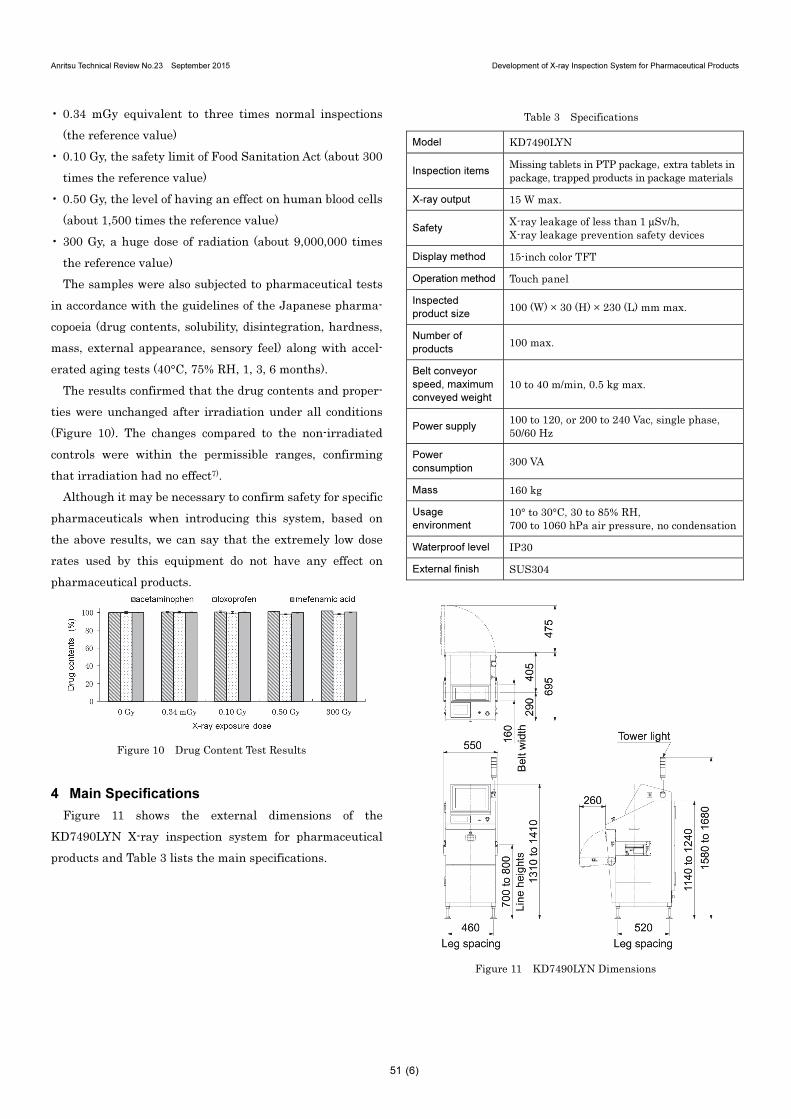
• 0.34 mGy equivalent to three times normal inspections
(the reference value)
• 0.10 Gy, the safety limit of Food Sanitation Act (about 300
times the reference value)
• 0.50 Gy, the level of having an effect on human blood cells
(about 1,500 times the reference value)
• 300 Gy, a huge dose of radiation (about 9,000,000 times
the reference value)
The samples were also subjected to pharmaceutical tests
in accordance with the guidelines of the Japanese pharma-
copoeia (drug contents, solubility, disintegration, hardness,
mass, external appearance, sensory feel) along with accel-
erated aging tests (40°C, 75% RH, 1, 3, 6 months).
The results confirmed that the drug contents and proper-
ties were unchanged after irradiation under all conditions
(Figure 10). The changes compared to the non-irradiated
controls were within the permissible ranges, confirming
that irradiation had no effect7).
Although it may be necessary to confirm safety for specific
pharmaceuticals when introducing this system, based on
the above results, we can say that the extremely low dose
rates used by this equipment do not have any effect on
pharmaceutical products.
Figure 10 Drug Content Test Results
4 Main Specifications
Figure 11 shows the external dimensions of the
KD7490LYN X-ray inspection system for pharmaceutical
products and Table 3 lists the main specifications.
Table 3 Specifications
Model KD7490LYN
Inspection items Missing tablets in PTP package,extra tablets in package, trapped products in package materials
X-ray output 15 W max.
Safety X-ray leakage of less than 1 μSv/h, X-ray leakage prevention safety devices
Display method 15-inch color TFT
Operation method Touch panel
Inspected
product size 100 (W) × 30 (H) × 230 (L) mm max.
Number of
products 100 max.
Belt conveyor
speed, maximum
conveyed weight
10 to 40 m/min, 0.5 kg max.
Power supply 100 to 120, or 200 to 240 Vac, single phase, 50/60 Hz
Power
consumption 300 VA
Mass 160 kg
Usage
environment
10° to 30°C, 30 to 85% RH, 700 to 1060 hPa air pressure, no condensation
Waterproof level IP30
External finish SUS304
Figure 11 KD7490LYN Dimensions
51
Anritsu Technical Review No.23 September 2015 Development of X-ray Inspection System for Pharmaceutical Products
(7)
5 Summary
We have developed a high-accuracy and stable X-ray in-
spection system for inspecting pharmaceutical products
using X-ray control and belt conveyor technologies solving
the problems of inspecting packaged pharmaceuticals that
are difficult to inspect using external visual techniques such
as CCD cameras and earlier-generation X-ray inspection
systems. Additionally, we clarified the effect of X-ray radia-
tion on pharmaceutical products to demonstrate the possi-
bilities of inspecting the diverse range of new pharmaceu-
tical packages to pharmaceutical manufacturers requiring
strict quality control systems.
Anritsu aims to become a leading partner in product
quality control assuring the safety and reliability of foods
and pharmaceuticals by offering new quality-control solu-
tions meeting its customers’ needs.
References
1) “Development of KD7203AW X-ray Inspection System”, AN-
RITSU TECHNICAL No.80 2002
2) “Development of Dual-Energy X-Ray Inspection System”, AN-
RITSU TECHNICAL No.87 2012
3) “Principle and practical use of Contaminant Inspection Sys-
tems for food contaminations”, ANRITSU TECHNICAL No.89
2014
4) H. Maekoshi sv.,
Tables and graphs of photon mass attenuation coefficients and
mass energy-absorption coefficients for photon energies, Jap-
anese society of radiological technology Measurement section
(1995.3)
5) T. Shimizu, T. Maebara, H. Mizuno, M. Minamino, H. Hitomi,
S. Nishida, H. Nishi, T. Sonoda, H. Fujii, S. Nakagawa,
T. Takaike, K. Murauchi, “【改訂版】医薬品包装・容器の材料要求
特性と 3極局方の品質基準・試験法” (2013.9) (in Japanese)
6) A. Nakao, S. Tahara, T. Fujioka, A. Yasumoto, M. Nasukawa,
“ハードからみた GMP” (2013.9) (in Japanese)
7) Murata, Miyazaki, Takahashi, Ohkubo, Uehara, Tagami, Ozeki,
“Quality Inspection of Pharmaceutical Products Using X-ray
Scanning”, Journal of Pharmaceutical Machinery and Engi-
neering Vol.23 No.4, pp. 34–41 (2014)
Authors
Hitoshi Komiyama
Anritsu Industrial Solutions Co., Ltd.
Development Division
2nd Development Dept.
Project Team
Koji Omori
Anritsu Industrial Solutions Co., Ltd.
Development Division
2nd Development Dept.
Project Team
Katsushi Sakata
Anritsu Industrial Solutions Co., Ltd.
Development Division
2nd Development Dept.
Project Team
Naoya Saito
Anritsu Industrial Solutions Co., Ltd.
Development Division
2nd Development Dept.
Project Team
Akihiro Ohtsu
Anritsu Industrial Solutions Co., Ltd.
Development Division
2nd Development Dept.
Project Team
Manabu Inoue
Anritsu Industrial Solutions Co., Ltd.
Development Division
2nd Development Dept.
Project Team
Takaharu Inoue
Anritsu Industrial Solutions USA Inc.
Product Design Manager for
Americas Region
Publicly available
52