Embed Size (px)
Citation preview
Developmental Changes in Spontaneous Beating Rhythm of Cardiac MyocytesCultured in Vitro by Molecular Diffusion Culture Method
AKIMASA TAKEUCHI,1 HAJIME OGAWA,2 HIROYUKI MORIGUCHI,2 JONG-KOOK LEE,3
MAKOTO NOSHIRO,1 KIYOSHI KOTANI,2 and YASUHIKO JIMBO21School of Allied Health Sciences, Kitasato University, Japan
2Graduate School of Frontier Sciences, University of Tokyo, Japan3Research Institute of Environmental Medicine, Nagoya University, Japan
SUMMARY
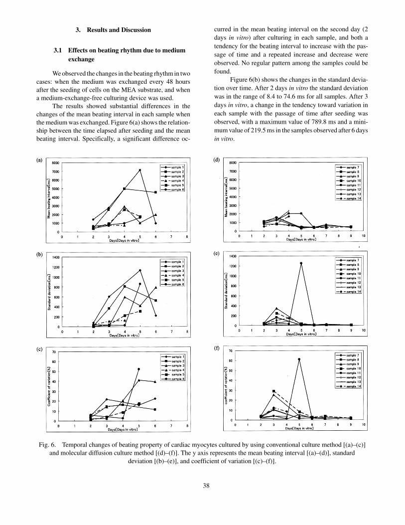
To understand the detailed mechanisms of arrhyth-mia and methods of its treatment, it is necessary to elucidatethe mechanisms of formation and breakdown of cardiacbeating at a microscopic level by using appropriate in vitromodels of cardiac beating. We developed a method formedium-exchange-free culture of rat cardiac myocytes, andcarried out long-term recording of the electrical activity ofcardiac myocytes from postnatal rats by means of a mi-croelectrode array (MEA) and our new culture method.Figures 6(a) to 6(f) show a series of properties of spontane-ous beating (mean beating interval, standard deviation, andcoefficient of variation) of cardiac myocytes cultured by aconventional culture method with alternate-day mediumexchange [(a) to (c)] and the newly developed “moleculardiffusion culture” method [(d) to (f)]. As shown in thesefigures, the difference in the beating property betweenindividual cultures that is found in conventional cultures[(a) to (c)] is dramatically reduced in the molecular diffu-sion cultures [(d) to (f)]. These results also indicate thatthere is a transient period when the mean beating interval,the standard deviation, and the coefficient of variation allincrease transiently before the formation of a stable rhyth-mic fast beat. © 2011 Wiley Periodicals, Inc. ElectronComm Jpn, 94(7): 35–42, 2011; Published online in WileyOnline Library (wileyonlinelibrary.com). DOI 10.1002/ecj.10244
Key words: spontaneous contraction; cultured car-diac myocytes; microelectrode array; molecular diffusionculture method.
1. Introduction
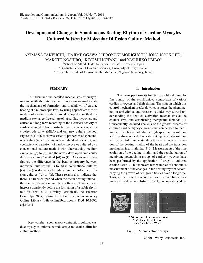
The heart performs its function as a blood pump byfine control of the synchronized contraction of variouscardiac myocytes and their timing. The state in which thiscontrol mechanism breaks down constitutes the phenome-non of arrhythmia, and research is under way toward un-derstanding the detailed activation mechanisms at thecellular level and establishing therapeutic methods [1].Consequently, detailed analysis of the growth process ofcultured cardiac myocyte groups that can be used to meas-ure cell membrane potential at high speed and resolutionand to perform optical observation at high spatial resolutionwill be helpful in understanding the mechanism of forma-tion of the beating rhythm of the heart and the transitionmechanism in arrhythmias [3–6]. Measurements of the timeevolution of the beating rhythm and the repolarization ofmembrane potentials in groups of cardiac myocytes havebeen performed by the application of drugs to culturedcardiac tissue [7], but there are few examples of continuousmeasurement of the changes in the beating rhythm accom-panying the growth of cell group tissues over a long time.Thus, in the present research we used cardiac tissue on amicroelectrode array substrate (Fig. 1), and investigated the
© 2011 Wiley Periodicals, Inc.
Electronics and Communications in Japan, Vol. 94, No. 7, 2011Translated from Denki Gakkai Ronbunshi, Vol. 128-C, No. 7, July 2008, pp. 1064–1069
Fig. 1. Microelectrode arrays.
35
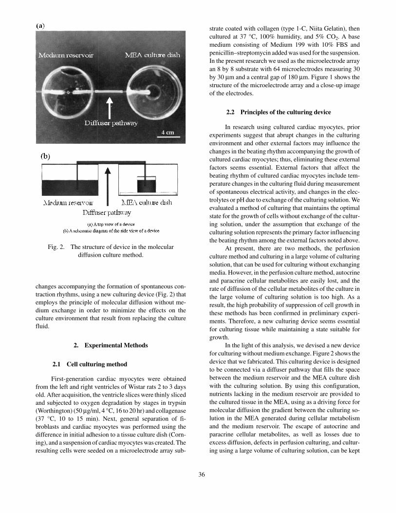
changes accompanying the formation of spontaneous con-traction rhythms, using a new culturing device (Fig. 2) thatemploys the principle of molecular diffusion without me-dium exchange in order to minimize the effects on theculture environment that result from replacing the culturefluid.
2. Experimental Methods
2.1 Cell culturing method
First-generation cardiac myocytes were obtainedfrom the left and right ventricles of Wistar rats 2 to 3 daysold. After acquisition, the ventricle slices were thinly slicedand subjected to oxygen degradation by stages in trypsin(Worthington) (50 µg/ml, 4 °C, 16 to 20 hr) and collagenase(37 °C, 10 to 15 min). Next, general separation of fi-broblasts and cardiac myocytes was performed using thedifference in initial adhesion to a tissue culture dish (Corn-ing), and a suspension of cardiac myocytes was created. Theresulting cells were seeded on a microelectrode array sub-
strate coated with collagen (type 1-C, Niita Gelatin), thencultured at 37 °C, 100% humidity, and 5% CO2. A basemedium consisting of Medium 199 with 10% FBS andpenicillin–streptomycin added was used for the suspension.In the present research we used as the microelectrode arrayan 8 by 8 substrate with 64 microelectrodes measuring 30by 30 µm and a central gap of 180 µm. Figure 1 shows thestructure of the microelectrode array and a close-up imageof the electrodes.
2.2 Principles of the culturing device
In research using cultured cardiac myocytes, priorexperiments suggest that abrupt changes in the culturingenvironment and other external factors may influence thechanges in the beating rhythm accompanying the growth ofcultured cardiac myocytes; thus, eliminating these externalfactors seems essential. External factors that affect thebeating rhythm of cultured cardiac myocytes include tem-perature changes in the culturing fluid during measurementof spontaneous electrical activity, and changes in the elec-trolytes or pH due to exchange of the culturing solution. Weevaluated a method of culturing that maintains the optimalstate for the growth of cells without exchange of the cultur-ing solution, under the assumption that exchange of theculturing solution represents the primary factor influencingthe beating rhythm among the external factors noted above.
At present, there are two methods, the perfusionculture method and culturing in a large volume of culturingsolution, that can be used for culturing without exchangingmedia. However, in the perfusion culture method, autocrineand paracrine cellular metabolites are easily lost, and therate of diffusion of the cellular metabolites of the culture inthe large volume of culturing solution is too high. As aresult, the high probability of suppression of cell growth inthese methods has been confirmed in preliminary experi-ments. Therefore, a new culturing device seems essentialfor culturing tissue while maintaining a state suitable forgrowth.
In the light of this analysis, we devised a new devicefor culturing without medium exchange. Figure 2 shows thedevice that we fabricated. This culturing device is designedto be connected via a diffuser pathway that fills the spacebetween the medium reservoir and the MEA culture dishwith the culturing solution. By using this configuration,nutrients lacking in the medium reservoir are provided tothe cultured tissue in the MEA, using as a driving force formolecular diffusion the gradient between the culturing so-lution in the MEA generated during cellular metabolismand the medium reservoir. The escape of autocrine andparacrine cellular metabolites, as well as losses due toexcess diffusion, defects in perfusion culturing, and cultur-ing using a large volume of culturing solution, can be kept
Fig. 2. The structure of device in the moleculardiffusion culture method.
36
to an absolute minimum. Furthermore, nitrogen com-pounds and other waste material generated during the proc-ess of cell growth can also be removed from the culture dishinto the medium reservoir by molecular diffusion.
2.3 Measurement methods
Cultured cardiac myocytes grown on a microelec-trode substrate adhere and extend themselves across thebottom of the medium in about 12 hours after the start ofculturing, and a signal representing a change in the extracel-lular potential for synchronous beating is detected after 24to 48 hours. Thus, in the present research we defined ourmeasurement sample as a group of cardiac myocytes 48hours or more after the settling that occurs 12 hours afterthe start of culturing. Visual observation was performedwith a system microscope (IX-71, Olympus) and extracel-lular potential recording was performed simultaneously atmultiple sites every 24 hours, beginning 48 hours after thecells were seeded. During the recording of the extracellularpotential, the recording time for the potential was set to 60seconds in order to minimize the changes in the pH andtemperature of the medium by minimizing the time duringwhich the sample was outside the incubator.
The signals obtained from the electrodes at 64 siteswere amplified 20-fold by a preamplifier (NF Corporation),then passed through a 10- to 300-Hz bandpass filter, ampli-fied to a total gain of 5000 to 10,000 by a postamplifier (NFCorporation), subjected to A/D conversion at a resolutionof 12 bits and a sampling frequency of 25 kHz, thenrecorded on a hard disk.
2.4 Method of calculating the mean beatinginterval
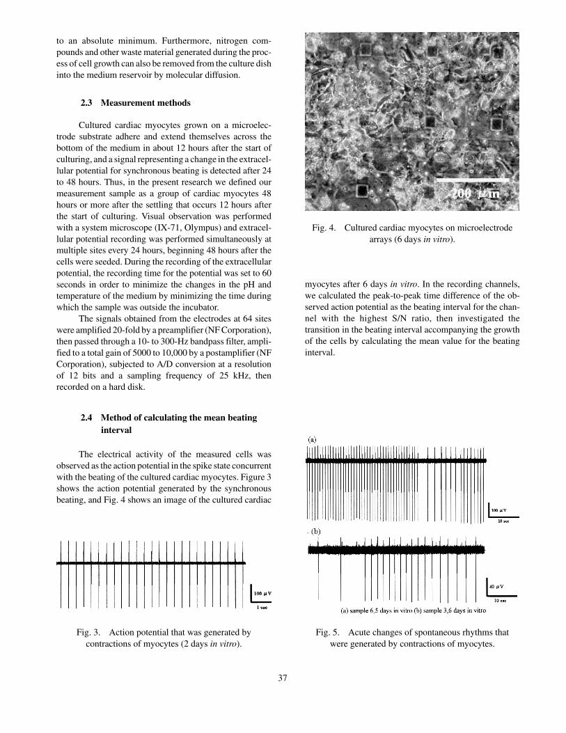
The electrical activity of the measured cells wasobserved as the action potential in the spike state concurrentwith the beating of the cultured cardiac myocytes. Figure 3shows the action potential generated by the synchronousbeating, and Fig. 4 shows an image of the cultured cardiac
myocytes after 6 days in vitro. In the recording channels,we calculated the peak-to-peak time difference of the ob-served action potential as the beating interval for the chan-nel with the highest S/N ratio, then investigated thetransition in the beating interval accompanying the growthof the cells by calculating the mean value for the beatinginterval.
Fig. 3. Action potential that was generated bycontractions of myocytes (2 days in vitro).
Fig. 4. Cultured cardiac myocytes on microelectrodearrays (6 days in vitro).
Fig. 5. Acute changes of spontaneous rhythms thatwere generated by contractions of myocytes.
37
3. Results and Discussion
3.1 Effects on beating rhythm due to mediumexchange
We observed the changes in the beating rhythm in twocases: when the medium was exchanged every 48 hoursafter the seeding of cells on the MEA substrate, and whena medium-exchange-free culturing device was used.
The results showed substantial differences in thechanges of the mean beating interval in each sample whenthe medium was exchanged. Figure 6(a) shows the relation-ship between the time elapsed after seeding and the meanbeating interval. Specifically, a significant difference oc-
curred in the mean beating interval on the second day (2days in vitro) after culturing in each sample, and both atendency for the beating interval to increase with the pas-sage of time and a repeated increase and decrease wereobserved. No regular pattern among the samples could befound.
Figure 6(b) shows the changes in the standard devia-tion over time. After 2 days in vitro the standard deviationwas in the range of 8.4 to 74.6 ms for all samples. After 3days in vitro, a change in the tendency toward variation ineach sample with the passage of time after seeding wasobserved, with a maximum value of 789.8 ms and a mini-mum value of 219.5 ms in the samples observed after 6 daysin vitro.
Fig. 6. Temporal changes of beating property of cardiac myocytes cultured by using conventional culture method [(a)–(c)]and molecular diffusion culture method [(d)–(f)]. The y axis represents the mean beating interval [(a)–(d)], standard
deviation [(b)–(e)], and coefficient of variation [(c)–(f)].
38
Based on the relationship between the mean beatinginterval and the standard deviation described above, weinvestigated the temporal relationship by calculating thecoefficient of variation. Figure 6(c) shows the temporalchanges in the coefficient of variation. In the results, a rangeof 2.2% to 6.2% was observed after 2 days in vitro, and anoverall increasing tendency with repeated up and downvariation was found after 3 days in vitro.
The temporal changes in the mean beating interval,the standard deviation, and the coefficient of variation whenthe medium was exchanged have been described. In addi-tion to these results, irregularities in the waveform of thespontaneous electrical activity were observed. Figure 5shows the waveform after 6 days in vitro for sample 3 andafter 5 days in vitro for sample 6 as examples of irregulari-ties in the observed waveforms. After 5 days in vitro forsample 6, the timing of the occurrence of spontaneouselectrical activity reached a maximum at 2460.5 ms and aminimum at 1120.2 ms in the measurement interval of 60seconds.
3.2 Growth of cells and changes in beatingrhythm
Our research showed no stable trend and charac-teristics of the temporal pattern of change in the meanbeating interval, its standard deviation, or the coefficient ofvariation for the samples when the medium was exchanged.Thus, in this section we describe the observed results forthe temporal changes in the beating rhythm of culturedcardiac myocytes after culturing under conditions thatminimize changes in the culturing environment by using amedium-exchange-free culturing device.
We were able to observe stable spontaneous beatingat up to 9 days in vitro by using the medium-exchange-freeculturing device. We were also able to find constant trendsin the temporal pattern of change in the mean beatinginterval and the standard deviation. Figures 6(d) and 6(e)show the temporal changes of the mean beating interval andits standard deviation. A tendency for the mean beatinginterval to increase after 2 to 3 days in vitro in the samplewas seen, and a tendency to decrease and converge to 400to 600 ms was found. The standard deviation reached amaximum value in each sample after 3 days in vitro, thentended to decrease to less than 50 ms. Furthermore, therewas no clear decrease in cardiac cells with an increasingnumber of days of culturing.
We calculated the coefficient of variation from themean beating intervals and standard deviations. Figure 6(f)shows the results. It will be seen that the coefficient ofvariation tended first to increase after 2 to 3 days in vitro,then to decrease to less than 5% after 6 days in vitro.
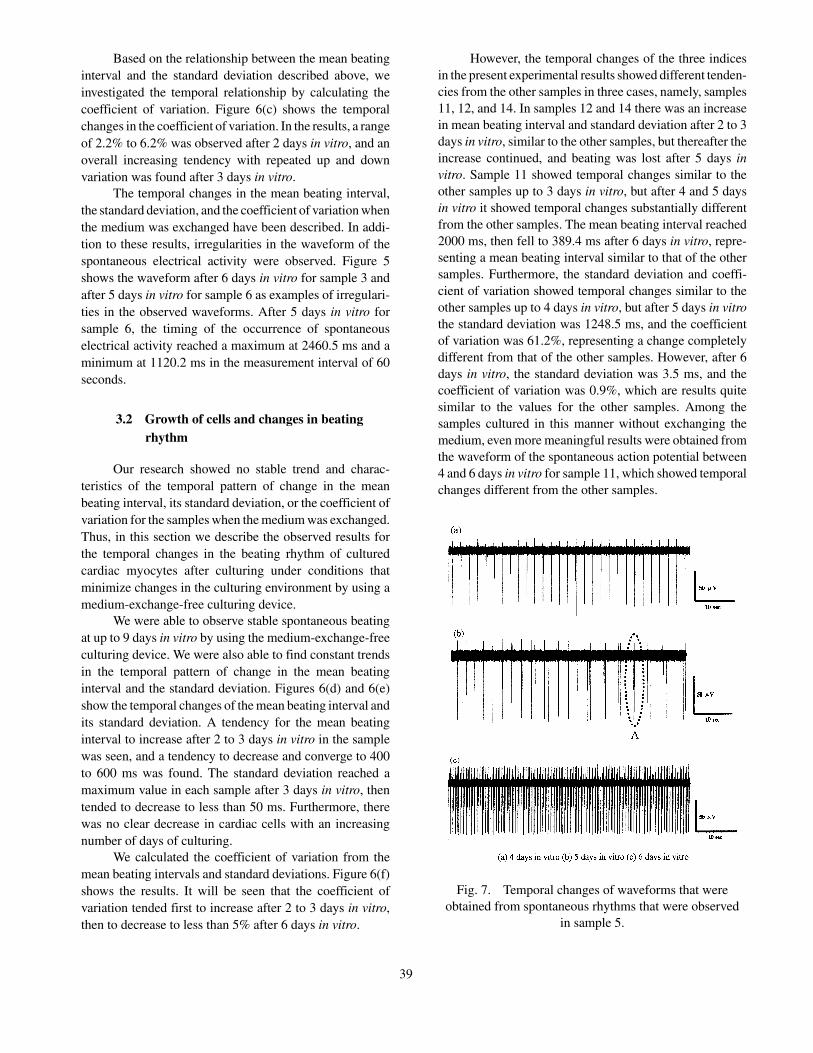
However, the temporal changes of the three indicesin the present experimental results showed different tenden-cies from the other samples in three cases, namely, samples11, 12, and 14. In samples 12 and 14 there was an increasein mean beating interval and standard deviation after 2 to 3days in vitro, similar to the other samples, but thereafter theincrease continued, and beating was lost after 5 days invitro. Sample 11 showed temporal changes similar to theother samples up to 3 days in vitro, but after 4 and 5 daysin vitro it showed temporal changes substantially differentfrom the other samples. The mean beating interval reached2000 ms, then fell to 389.4 ms after 6 days in vitro, repre-senting a mean beating interval similar to that of the othersamples. Furthermore, the standard deviation and coeffi-cient of variation showed temporal changes similar to theother samples up to 4 days in vitro, but after 5 days in vitrothe standard deviation was 1248.5 ms, and the coefficientof variation was 61.2%, representing a change completelydifferent from that of the other samples. However, after 6days in vitro, the standard deviation was 3.5 ms, and thecoefficient of variation was 0.9%, which are results quitesimilar to the values for the other samples. Among thesamples cultured in this manner without exchanging themedium, even more meaningful results were obtained fromthe waveform of the spontaneous action potential between4 and 6 days in vitro for sample 11, which showed temporalchanges different from the other samples.
Fig. 7. Temporal changes of waveforms that wereobtained from spontaneous rhythms that were observed
in sample 5.
39
Figure 7 shows the waveforms of the spontaneousaction potential at intervals of 24 hours from 4 days in vitroto 6 days in vitro for sample 11. At 4 days in vitro, a stablewaveform with a mean beating interval of 2030 ms, astandard deviation of 28.2 ms, and a coefficient of variationof 1.39% were recorded. However, after 5 days in vitro,irregularity in the beating rhythm occurred, clearly consti-tuting a factor in the increase in the standard deviation andthe coefficient of variation. The gap between the two actionpotentials shown at A in Fig. 7(b) was 428.8 ms, a resultvery close to the mean beating interval of 389.3 ms after 6days in vitro.
3.3 Discussion
In the present experiments, irregularities in the spon-taneous beating rhythm thought to result from the effectsof medium exchange were observed in the temporalchanges of the mean beating interval, the standard devia-tion, and the coefficient of variation. This suggests thatbased on the experimental results, the temporal changes inthe beating interval may be influenced by changes in thefluid composition of the medium due to medium exchange,because of the abrupt changes in the concentrations ofextracellular and intracellular electrolytes. This may beattributed to two factors: observation of small values foreach sample after 2 days in vitro during the temporalchanges of the three indices in Figs. 6(a) to 6(e), and thelack of a stable trend among the values for the samples after3 days in vitro when the medium was exchanged.
Furthermore, based on the experimental results [Figs.6(d) to 6(f)] obtained by using the culturing device newlyproposed here, a trend representing stable changes overtime of the three indices, the mean beating interval, thestandard deviation, and the coefficient of variation, wasseen. These results suggest that in the process of culturedcardiac tissue development, there are two polarized trends,one (samples 12 and 13) in which the mean beating intervaland the standard deviation of the beating rhythm mayundergo an initial increase, then a decrease, after which themean beating interval and standard deviation continue toincrease and beating stops, and one in which the meanbeating interval and standard deviation decrease and tran-sition to stable beating. Furthermore, Figs. 6(d) to 6(f) seemto show a different trend for samples 11, 12, and 14, whichdiffer from the other samples in the state of the gap connec-tion between cell groups and the distribution of fibroblasts,as a result of differences in the tendencies of the meanbeating interval, standard deviation, and coefficient of vari-ation. Differences in cell density may be the cause, and thedifferences in the trends when changing the cell densitiesmust be investigated in the future.
4. Conclusions and Future Topics
In the research reported here we used innovativeexperiments to perform long-term observations of thechanges in the beating interval accompanying the growthof cultured cardiac myocytes without medium exchange.The results clearly showed that during culturing with me-dium exchange every 48 hours, sharp changes in the beatingrhythm and other effects due to medium exchange werepossible.
Moreover, when performing observations in a cultur-ing device that used the principle of molecular diffusionwithout medium exchange, two clear categories werefound: one in which the mean interval of the beating rhythmfirst rose, then converged to 500 to 800 ms, and one in whichit continued to rise and beating was lost after a short periodof time. Among the samples in which the mean beatinginterval continued to increase, there was also a sample thatreturned to a stable rhythm after spontaneous electricalactivity.
In our experiments, we created an extracellular po-tential recording system outside the incubator. The changesin pH and temperature in the medium during measurementmust be minimized by using a measurement system thatallows recording of potentials inside the incubator. We alsoplan to further clarify the relationship between the changesin the beating rhythm after 2 to 4 days in vitro and thechanges in the rhythm that occur thereafter by performinglonger-term observations with more samples. In addition,we plan to clarify how the beating rhythm is affected by theapplication of electric shock or some other extracellularstimulus after 2 to 4 days in vitro.
REFERENCES
1. Rohr S, Kleber AG, Kucera JP. TCM 1999;9:173–179.
2. Klauke N, Smith GL, Cooper J. Biophys J2003;85:1766–1774.
3. Inoue N, Ohkusa T, Nao T, Lee JK, Matumoto T,Hisamatsu Y, Satoh T, Yano M, Yasui K, Kodama I,Matsuzaki M. J Am Coll Cardiol 2004;44:914–922.
4. Wrobel G, Yeung CK, Sommerhage F, Chan M, Of-fenhausser A, Ingebradt S. 5th Int Meeting on Sub-strate-Integrated Microelectrodes, p 115–116, 2006.
5. Efimov IR, Nikolski VP, Salama G. Circ Res2004;95:21–33.
6. Posenbaum D, Jalife J. Future Publishing Company;2001.
7. Stett A, Egert U, Guenther E et al. Anal Bioanal Chem2003;377:486–495.
40
AUTHORS (from left to right)
Akimasa Takeuchi (nonmember) received a bachelor’s degree from the Department of Medical Engineering of the Schoolof Allied Health Sciences, Kitasato University, in 2008, and entered the Graduate School of Frontier Sciences at the Universityof Tokyo. He is now engaged in the development of a next-generation pacemaker using medical engineering technology andtissue engineering technology. He is performing research on cardiac pulse rhythm control using autonomic nervous systemcultures.
Hajime Ogawa (nonmember) received a bachelor’s degree from the Department of Human Environmental Science ofKobe University in 2007. He is now enrolled in the Graduate School of Frontier Sciences of the University of Tokyo. He isengaged in research on the effects of the autonomic nervous system on the circulatory organs using biological signal processing.
Hiroyuki Moriguchi (nonmember) completed the program of study in the Graduate School of Humanities of the Universityof Tokyo in 2006 and became a researcher of the Japan Society for the Promotion of Science in 2006. He is performing technologydevelopment and biological experiments intended to advance understanding of the structure and functions of multicellular tissue.He holds a Ph.D. degree, and is a member of the Biophysical Society of Japan.
Jong-Kook Lee (nonmember) received a bachelor’s degree from the Department of Medicine at Nagoya University in1987. He completed his studies in the Graduate School of Medicine in 1998, receiving a D.Med.Sc. degree. Between 1995 and1998 he was a visiting researcher at UCLA. He became a lecturer on the circulatory system at the Research Institute forEnvironmental Medicine of Nagoya University in 1998, and an associate professor in 2004. He is now an associate professorof heart and circulation at the Research Institute of Environmental Medicine of Nagoya University. He is primarily engaged inresearch on cardiac electrophysiology. He is a member of the Japanese Circulation Society, the Japanese Society of Electro-cardiology, the Japanese Society for Regenerative Medicine, and the Japanese Pharmacological Society.
Makoto Noshiro (nonmember) received a bachelor’s degree from the Department of Electrical Engineering of theUniversity of Tokyo in 1969 and then joined Fujitsu Ltd. He moved to the Medical Materials Laboratory of Tokyo Medical andDental University in 1972, and became an associate professor in 1982. He was appointed a professor of clinical engineering inthe School of Allied Health Sciences of Kitasato University in 1995. In 1981–1982 he was a student at the BioengineeringResearch Facility of the University of Strathclyde. He is engaged in research on measurement and control of the respiratorysystem, bioinformatics analysis using impedance measurements, and bioinformatics related to the analysis of inclusive geneexpression. He holds a D.Eng. degree, and is a member of JSMBE, IEICE, and SICE.
Kiyoshi Kotani (nonmember) completed the doctoral program in precision engineering at the Graduate School ofEngineering of the University of Tokyo in 2003 and then became a research lecturer at the Graduate School of InformationScience. He is now a lecturer in the Graduate School of Frontier Sciences. He is engaged in research on nonlinear dynamics,statistical physics, biogenic signal processing, and human interfaces. He holds a D.Eng. degree, and is a member of JSMBE.
41
AUTHORS (continued)
Yasuhiko Jimbo (member) completed his studies in electrical engineering at the Graduate School of the University ofTokyo in 1988 and joined NTT Basic Research Laboratories. From 1992 to 1993 he was a visiting researcher at CNRS in France.In 2003 he became an associate professor of precision mechanical engineering at the Graduate School of Engineering of theUniversity of Tokyo. Since 2006 he has been a professor of human and environmental studies in the Graduate School of FrontierSciences. He is primarily engaged in research on neurological engineering. He is a member of IEICE, the Japan NeuroscienceSociety, JSMBE, and IEEE.
42