Embed Size (px)
Citation preview
Diagnosis of EGC and AGC
IM R3.송병근

위암의 증상
조기 위암 진행성 위암
무증상 80% 체중 감소 60%
속쓰림 10% 복통 50%
오심, 구토 8% 오심, 구토 30%
식욕 감퇴 8% 식욕 감퇴 30%
조기 포만감 5% 연하 곤란 25%
복통 2% 위장관 출혈 20%
위장관 출혈 < 2% 조기 포만감 20%
체중 감소 < 2% 속쓰림 20%
연하 곤란 < 1% 복부 팽만감 5%
무증상 < 5%

Histology

Histology
Kim WH, Park CK, et al. Korean J Pathol 2005;39:106-113

Histologic type of gastric cancer
Kim WH, Park CK, et al. Korean J Pathol 2005;39:106-113

Lauren classification
• Intestinal type
Better differentiated
Cohesive cells that form discrete glands
Resembles microscopically colorectal carcinoma
Predominant in high incidence areas
• Diffuse type
Less differentiated
Sheets of cells without gland formation
Occasional presence of signet ring cells and mucin
Similar prevalence throughout the world
Poor prognosis

Difficult to correlate
Vauhkonen. BPRCG 2006;20:651-674

Pathology report for ESD specimen
Kim WH, Park CK, et al. Korean J Pathol 2005;39:106-113

Depth of invasion
m1 -> Tis m2, m3 -> T1a sm -> T1b

Before endoscopic diagnosis …

TNM classification (UICC)
0 Tis N0 M0 III A T2 N2 M0
I A T1 N0 M0 T3 N1 M0
I B T1 N1 M0 T4 N0 M0
T2 N0 M0 III B T3 N2 M0
II T1 N2 M0 IV T4 N2 M0
T2 N1 M0 T1~3 N3 M0
T3 N0 M0 any T any N M1

병기별 생존율
Months
Cu
mu
lati
ve
Su
rviv
al
IV : 12.7%
IA : 96.1%
IB : 88.5%
II : 74.2%
IIIA : 53.8%
IIIB : 34.0%
외과 교과서 삼성서울 병원

Early gastric cancer (EGC)

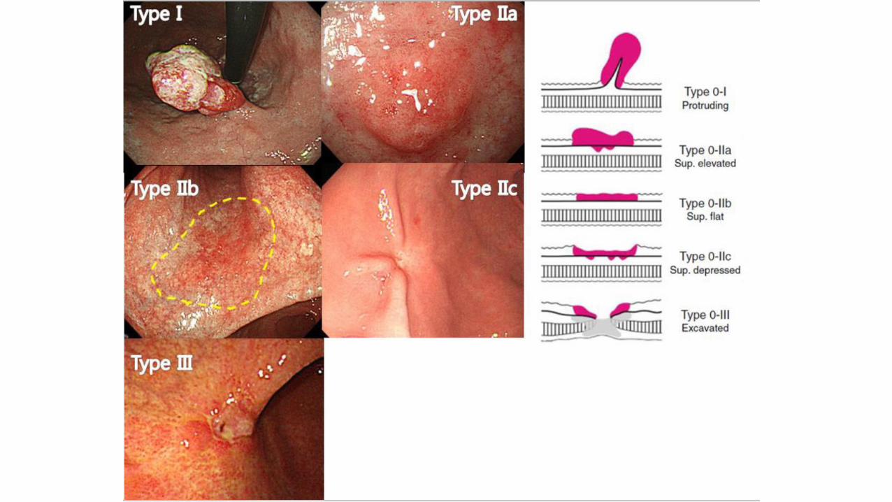
Classification of EGC
Protruded type
Superficial elevated type
Superficial depressed type
Flat type
Excavated type
Type 0 I
Type 0 IIa
Type 0 IIb
Type 0 IIc
Type 0 III

EGC type I과 type II의 경계는? - Update on the Paris classification
The cut-off limit is 2.5 mm in the columnar epithelium and
1.2 mm in the stratified epithelium of the esophagus.
Endoscopy 2005;37:570-578
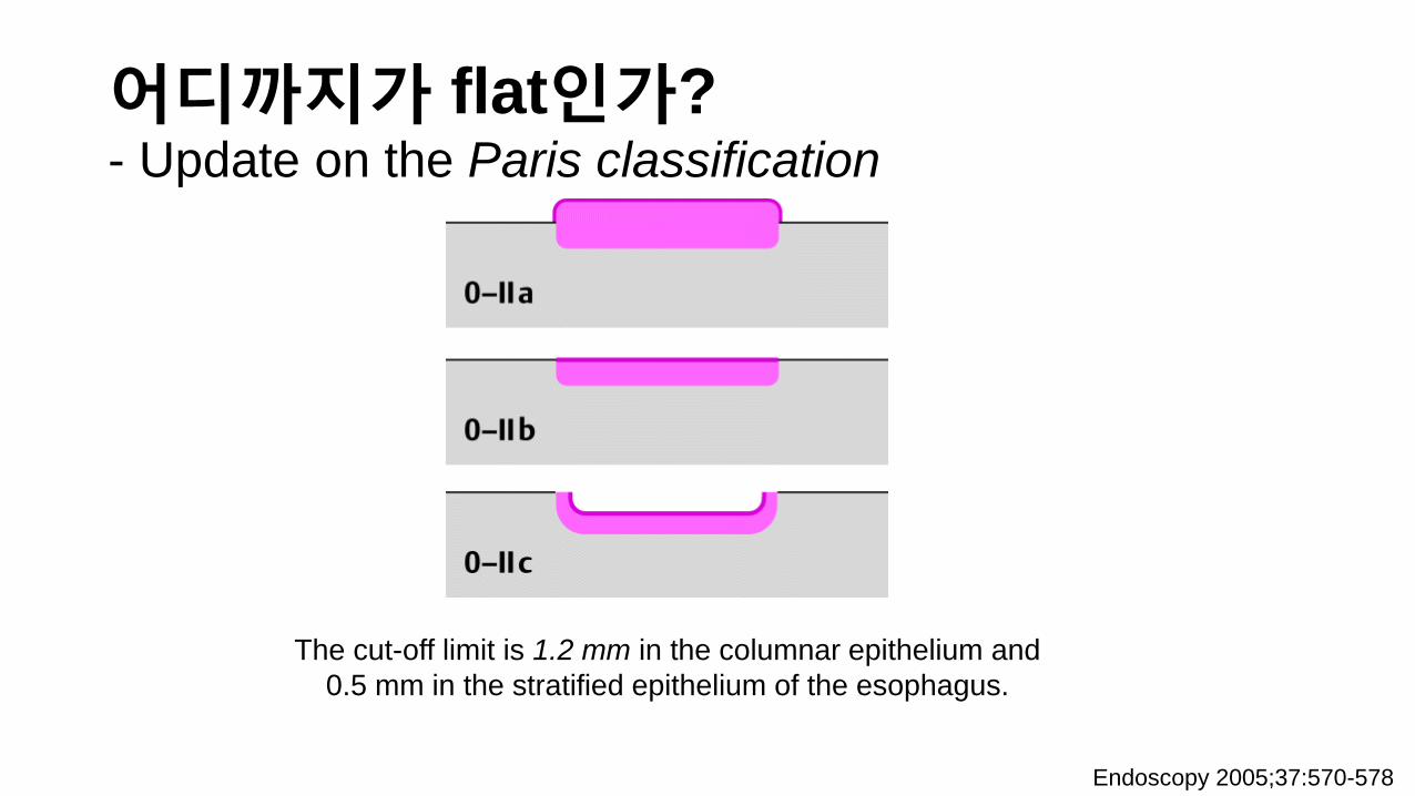
어디까지가 flat인가? - Update on the Paris classification
Endoscopy 2005;37:570-578
The cut-off limit is 1.2 mm in the columnar epithelium and
0.5 mm in the stratified epithelium of the esophagus.

Ulcer가 있으면 type III… - Update on the Paris classification
Endoscopy 2005;37:570-578
Endoscopic appearance of a superficial neoplastic lesion on the
surface of the digestive-tract mucosa: excavated type (0 - III).
An ulcer is seen.

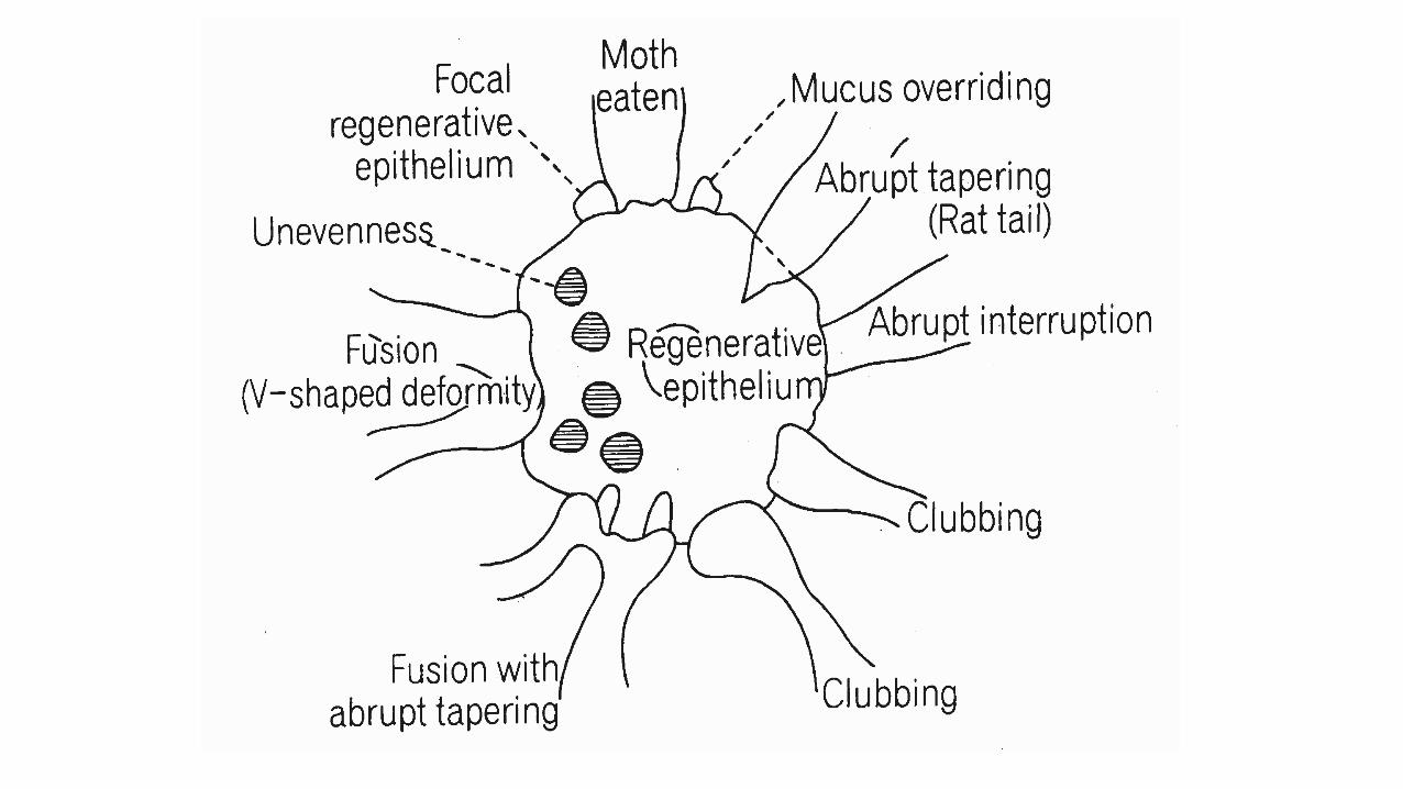
Depressed type에서는 fold 변화가 중요

함 요 형
1) 점막주름의 중단 2) 계단모양의 함요 3) 변색 4) 불규칙한 가늘어짐 5) 펜끝모양 가늘어짐 6) 점막주름주행의 함요내에 흔적이 남음
7) 점막주름 끝의 곤봉상 융기 결절모양의 융기 8) 점막주름사이에의 연결형성 9) 결절모양끝이 융합 10) 제방모양의 융기, 주위제방형성 11) 함요내 요철이 불규칙한 것이 현저함 섬형성, 결절성 융기, 두터운 백태부착, 출혈성 미란백태 등의 다재성
III 함요(Iic)내의 III의 축소소실을 보이는 것은 m이 많다.
B-II 불규칙한 깊은 분화구 형성, 주위제방의 경화, 결절형성
B-III 분화구 형성, 주변의 미란출혈, 요철부정
m
sm
pm~s
m에서는
흔하다
Depth of invasion

조기위암에서 보이는 fold abnormality
1. Interruption
2. Tapering
3. Clubbing
4. Fusion
5. Moth-eaten

전형적인 위암의 fold change (1)
Signet ring cell carcinoma, diffuse type, 1.9x1.2cm, SM3, 0/23

전형적인 위암의 fold change (2)

공기에 따른 변화

EGC type I
Tubular adenocarcinoma (W/D), 3.6x1.6 cm, in LP

EGC type IIa

EGC type IIb

EGC type IIc

EGC type III는 드물다 - BGU와 감별진단에 특히 주의
Adenocarcinoma (P/D), lamina propria, 0/39

Advanced gastric cancer (AGC)

http://www.cancer.go.kr/contentfile/cif/cifimg/0302_AGC_72dpi.jpg


AGC Borrmann type I

AGC Borrmann type II
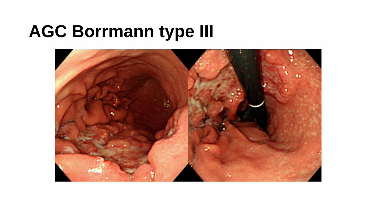
AGC Borrmann type III
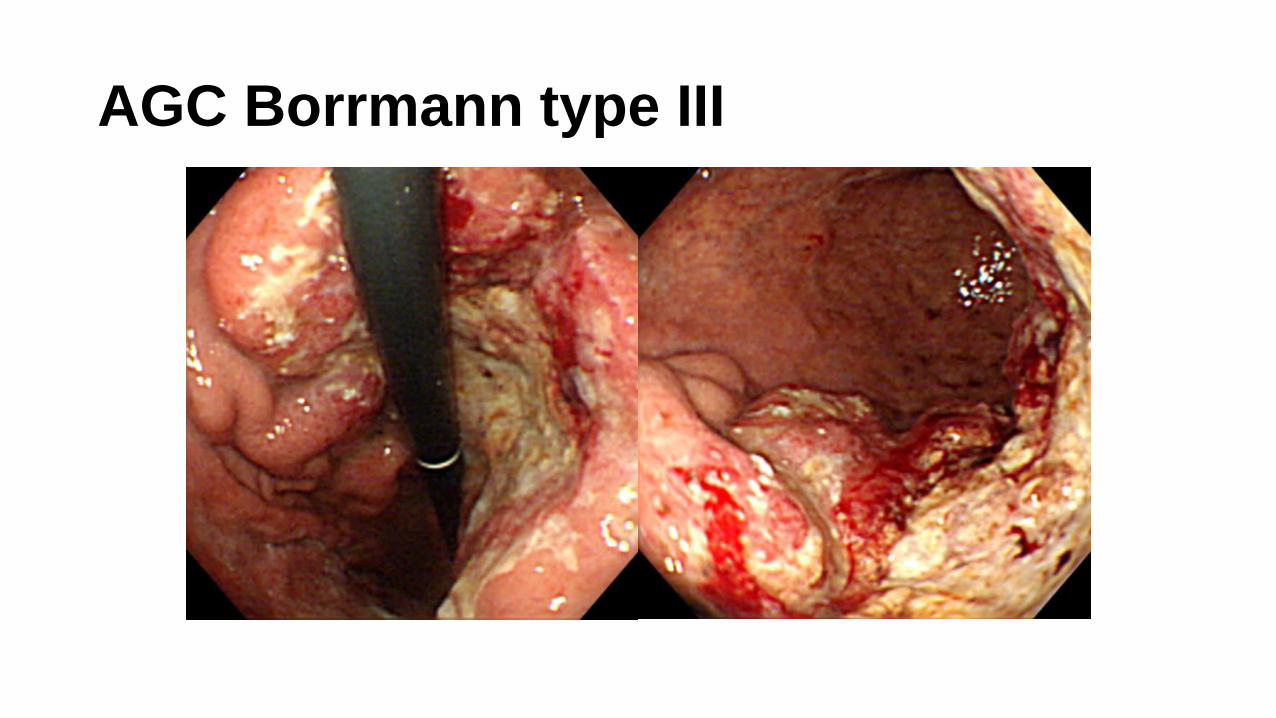
AGC Borrmann type III

AGC Borrmann IV - initial endoscopic examination

Take home message
- 무증상인 경우가 많다 -> 조기 발견을 위한 내시경 검사의 유용성
- 진단은 내시경 & 조직검사
- EGC : type I, IIa, IIb, IIc, III - EGC vs BGU : fold change, edge & margin, surface
- AGC : Borrmann type I, II, III, IV - Borrmann type IV의 중요성


![헬리코박터 파일로리 감염의 진단과 치료: 국내 및 …하는 방법(test-and-treat strategy)을 권장하고 있다[36-40]. 그 러나 우리나라는 위암의 유병률이](https://img.pdfslide.tips/doc/110x75/5e4dca98fc580216d239639e/ee-oeoeeoee-e-ee-eoe-ee-e-e-eetest-and-treat.jpg)