Embed Size (px)
Citation preview

Differentiation-Associated Localization of nPKCl1, a Ca++-Independent Protein Kinase C, in Normal Human Skin and Skin Diseases
Hiroko Koizumi,* Yohko Kohno,t Shin-ichi Osada,:j: Shigeo Ohno,:j: Akira Ohkawara,* and Toshio Kuroki§ • Department of Dermatology, Hokkaido University School of Medicine, Sapporo; tDepartment of Oral Pathology, Showa University School of Dentistry, Tokyo; :I:Department of Molecular Biology, Yokohama City University, School of Medicine Yokohama; and § Department of Cancer Cell Research, Instirute of Medical Science, University of Tokyo, Tokyo, Japan
The expression of nPKCI1, a Ca++-independent isoform of protein kinase C in normal human skin, and skin from patients with psoriasis, squamous cell carcinoma, basal cell epithelioma, nevus pigmentosus, and seborrheic keratosis, were examined by immunohistochemical staining using a polyclonal antibody raised against a synthetic peptide at a diverse region of the nPKCI1 molecule. In normal epidermis, the strongest staining was observed in the uppermost granular layer with no staining of the spinous or basal layers. The inner layer of the intra-epidermal eccrine duct was also strongly stained. Weak staining was observed in several layers of the outer root sheath of the follicular infundibulum. No staining was detected in the inner root sheath of the hair follicles, hair matrix, sebaceous gland, eccrine gland, intra-
Transduction of extracellular signals across the cell surface involves agonist-induced hydrolytic catalysis of phosphatidylinositol-4,S-diphosphate by phosphatidylinositol-specific phospholipase C. This reaction
. produces two intracellular second messengers, 1,2-diacyl glycerol (DAG) and inositol-1,4,S-triphosphate. The former, DAG, activates protein kinase C (PKC) whereas the latter releases calcium from intracellular stores. By implication, therefore, PKC is regarded as a key enzyme in transmembrane-signaling pathways in cells [1]. In epidermal keratinocytes, several hormones and peptides, such as bradykinin, platelet-activating factor, thrombin, and substance P, were reported to activate PKC [2-6].
PKC exists as a family of isoforms with closely related structures and enzymologic characteristics. Ten members of the PKC family have been identified to date. They are classified into three major groups: Ca++ -dependent conventional PKCs (cPKCa, -fJI, -fJII, and -Y), Ca++-independent novel PKCs (nPKC6, -E, -11, and -8), and DAG or phorbol ester-independent atypical PKC (aPKC( and -A) [7]. Conventional PKC isoforms contain three conserved domains, i.e., the C1 domain with two cysteine-rich zinc finger-like motives, the C2 domain determining Ca++-sensitivity, and the catalytic C3 domain. They are enzymatically characterized by the requirements of Ca++, phosphatidylserine, and DAG. The second group ofPKC isoforms, nPKCs, lacks the C2 domain and does not show a calcium
Manuscript received February 19, 1993; accepted for publication June 30, 1993.
Reprint requests to: Dr. Hiroko Koizumi, Department of Dermatology, Hokkaido University School of Medicine, Sapporo 060, Japan.
dermal eccrine duct, arrectores pilorum, melanocytes, Langerhans cells, fibroblasts, or blood vessels. In psoriatic skin, stained keratinocytes were distributed in the suprabasallayers with the most being observed in the uppermost layer and the least in layers closed to the basal layer. In squamous cell carcinoma, weak staining was observed in the keratotic cells around horny pearls. In the basal cell epithelioma and nevus pigmentosus, the cells were not stained, whereas in seborrheic keratosis, cells that stained were located in the granular layer. We conclude from the evidence presented above that nPKCI1 is expressed in close association with epidermal differentiation in normal skin and skin diseases. Key words: nPKCI1/ differentiation-associated localization/normal epidermis.] Invest Dermato1101:858-863, 1993
requirement [8-11]. The aPKC isoforms have only one cysteinerich repeat in the C1 domain and are independent of Ca++ or DAG. The presence of multiple isoforms and their tissue-specific distributions suggest that each performs a distinct role in the growth, differentiation, and functioning of cells [1,8,11-13].
We have cloned nPKCI1, a new family member of PKC, from a eDNA library of mouse skin [8] . A human version of nPKCI1, termed PKC-L, was reported by Bacher et al [14]. Northern blot analysis has shown that the mRNA of nPKCI1 is highly expressed in skin and lung but only slightly in brain [8]. This unique tissue distribution prompted us to examine the possibility that nPKCI1 is a major PKC isoform in most epithelial tissues. Indeed, we found that nPKCI1 is expressed predominantly in the epithelia of the skin, digestive, and respiratory tracts in association with differentiation of epithelial cells [13].
In this communication, we describe the differentiation-associated localization of nPKCI1 in normal human skin and that from various skin diseases such as psoriasis, squamous cell carcinoma, basal cell epithelioma, nevus pigmentosus, and seborrheic keratosis.
MATERIALS AND METHODS
Human Skin Samples Biopsy samples of normal skin were obtained from the lower extremities of two women (22 and 62 years old) and one man (32 years old), and the nape of a second man (54 years old). Biopsy samples of the following skin conditions were obtained also: psoriatic lesions from six patients of psoriasis vulgaris (two women, 38 and 75 years old, from the arms; four men, 46, 41, 53, and 71 years old, one sample from the chest, and three from the abdominal wall, respectively); squamous cell carcinoma (one woman, 91 years old, from the face; one male, 56 years old, from a dorsal site of the hand); basal cell epithelioma (one man, 65 years old, from the arm);
0022-202X/93/S06.00 Copyright © 1993 by The Society for Investigative Dermatology, Inc.
858

F
Figure 1. Immunohistochemical demonstration of nPKCI] in normal human skin. (A) Epidermis and dermis. Note that nPKC" is located exclusively in uppern10st granular layer. (B) High magnification of granular layer. C) Epidermis and dermis in which the staining is eliminated by the addition of peptide antigen. D) Upper portion of hair follicle in which several outer layers of the follicular infundibulum are stained. E) Epidermis with intraepidermal eccrine duct, which is also strongly stained. The dark staining of the basal layer is due to melanin pigment. F) Staining of cells in granular layer. Note that staining is strong in the cytoplasmic region devoid of nuclear space. G-I) Hair bulb (G) , sebaceous gland (H), and eccrine gland (l) are not stained by the antibody. Bars, 40 ~rn (A-E, G-I) and 20 J.lm (F).

860 KOlZUMI ET AL
nevus pigmentosus (one woman, 41 years old; one man, 65 years old; both samples from the face), and seborrhe~c keratosis (one. w.oman, 62 years .old, from the abdominal wall). The patients with psoriaSIS had not received treatment for the condition for at least 2 weeks before the biopsies were done, and none of the other patients had received any treatment. Biop~y materials were fixed with Bouin's solution, dehydrated, and embedded m paraffin.
Preparation of Antiserum Against nPKC" A polyclonal antibody was raised against a synthetic peptide with the sequ.ence Q~STK QKTNKPTYNEEFC, which corresponds to the N-termmal D1 diverse region (56- 73) of human and mouse nPKC" [13]. This sequence shares homology (61%, 11/18) with only one .of the PKC isofo!ms, n~m~ly nPKC€. The antibody was purified by chromatography and Its specificity was confirmed by immunoblotting; it reacted with an 82-kilodalton (Da) protein produced by COSl cells overexpressing nPKC", but not with the equivalent protein produced by cells expressing nPKCE.
Immunohistochemical Staining Paraffin-embedded tissues were cut into 4-J.lm sections and hydrated. The sections were pretreated with 3% hydrogen peroxide for 10 min at 40 C and washed with phosphate-buffered saline (PBS). They were then incubated with 10% normal horse serum at room temperature for 20 min, followed by overnight treatment with the antibody against nPKC'I (diluted 1: 5000 in PBS containing bovine serum albumin) . After washing with PBS, the sections were reacted with biotinated F[ab"] fragment of affinity-purified porcine anti-rabbit immunoglobulin (Dakopatts A/S, Denmark) for 30 min at room temperature. Then, the specimens were incubated with peroxide-conjugated streptavidin (Dakopatts A/S, Denmark) for 30 min at room temperature, washed with PBS, and reacted with 0.1 % diaminobenzidine. Some specimens were counterstained with Mayer's hematoxylin. Specimens were dehydrated and mounted. The specificity of the reaction was confirmed by elimination of the staining in the presence of an excess amount (1 J.lg/ml) of the peptide antigen.
RESULTS
Localization of nPKC'7 in Normal Skin In normal human skin, positive staining for nPKC'1 was observed in the keratinocytes comprising the granular layer of the interfoliicular epidermis (Fig lA, B). This staining was not observed in the basal or spinous layers. Melanocytes and dendritic Langerhans cells in the epidermis were not stained (data not shown).
In the hair follicle, weak staining was observed in several outer layers of the follicular infundibulum (Fig lD). Neither inner root sheath, hair matrix, nor papilla showed positive staining (Fig lG). The sebaceous glands and the arrectores pilorum, which consists of smooth muscle, did not stain (Fig 1H).
Strong positive staining was found in the cytoplasm of the inner cells of the intra-epidermal eccrine duct where granular cells are
THE JOURNAL OF INVESTIGATIVE DERMATOLOGY
present (Fig 1E), but the intra-dermal eccrine ducts and eccrine glands were negatively stained (Fig 11). Blood vessels, fibroblasts, and nerves in the dermis were devoid of staining. Adipose cells in the subcutaneous tissue were not stained (Fig 11).
Staining was strong in the cytoplasmic region devoid of nuclear space (Fig 1F).
In all the instances described above, and in those that follow below, staining was eliminated by the addition of peptide antigen (Fig le, 2C), thus demonstrating a specific reaction with nPKC17·
Localization of nPKC'7 in Psoriasis Psoriatic involved epidermis shows regular acanthosis and parakeratosis with a depleted granular layer. Six biopsy samples of involved psoriatic skin were examined for localization of nPKC17. In contrast to normal skin, staining was not limited to the uppermost two or three layers but was distributed in whole suprabasallayers, with the uppermost layer staining the strongest and the lower layer the weakest (Fig 2A). Weak staining was occasionally observed in the basal layer. As in normal skin, the immunoreaction was detected only in the cytoplasm (Fig 2B). Uninvolved skin adjacent to the psoriatic involved skin showed a similar pattern of nPKC17 expression as normal skin. Infiltrated cells in the upper dermis and epidermis did not stain.
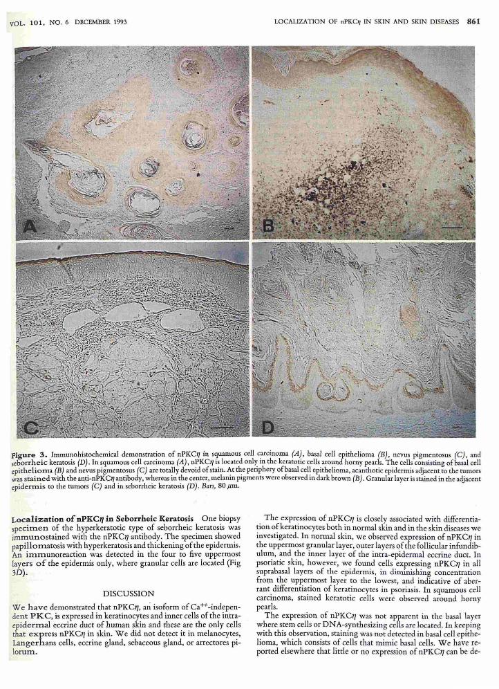
Localization of nPKC'7 in Squamous Cell Carcinoma Two biopsy specimens of well-differentiated squamous cell carcinoma of skin were immunostained with the nPKC17 antibody. Weak staining only was detected in the keratotic cells around horny pearls (Fig 3A). As with normal and psoriatic skin, the immunoreactive material was localized exclusively in the cytoplasm.
Absence of nPKC17 in Basal Cell Epithelioma Basal cell epithelioma consists of basophilic cells with a high nucleus: cytoplasm ratio that mimic the basal cells of the epidermis. We found that cells of basal cell epithelioma did not stain with the anti-nPKC17 antibody, most probably due to the absence of keratinization (Fig 3B). At the periphery of the tumor, acanthotic epidermis adjacent to the tumors was stained with the anti-nPKC17 antibody, whereas in the center, melanin pigments were observed in dark brown.
Absence of nPKC17 in Nevus Pigmentosus Two biopsy samples of intradermal nevus pigmentosus were examined for expression of nPKC17. Nevus cells were totally devoid of stain, from which we conclude that nPKC17 is not expressed in celis, including mel anocytes, that are derived from neural crest (Fig 3C).
c
Figure 2. Immunohistochemical demonstration of nPKC'I in psoriatic-involved epidermis. A,B) Psoriatic-involved epidermis by lower and higher magnifications. Note that stained keratinocytes are distributed in whole suprabasallayers with the uppermost layer staining the most strongly. C) Negative control in the presence of the antigen peptide. Bars, 40 J.lm.
VOL. 101, NO. 6 DECEMBER 1993 LOCALIZATION OF nPKClI IN SKIN AND SKIN DISEASES 861
Figure 3. Immunohistochemical demonstration of nPKe11 in s9uamous cell c.arcinoma (A~, basal cell epithelioma (B), nevus pigmentosus (e), and seborrheic keratosis (D) . In squamous cell carcinoma (A), nPKet/ls located only In the keratotic cells around horny pearls. The cells consisting of basal cell epithelioma (B) and nevus pigmentosus (e) are totally devoid of stain. ~t the periphery of basal cel~ epithelioma, acanthotic epidermis adjacent to the tumors was stained with the anti-nPKe11 antibody, whereas in the center, melamn pigments were observed In dark brown (B) . Granular layer is stained in the adjacent epidermis to the tumors (e) and in seborrheic keratosis (D) . Bars, 80 Jim.
Localization of nPKC" in Seborrheic Keratosis One biopsy specimen of the hyperkeratotic type of seborrheic keratosis was immunostained with the nPKCIi antibody. The specimen showed papillomatosis with hyperkeratosis and thickening of the epidermis. An immunoreaction was detected in the four to five uppermost layers of the epidermis only, where granular cells are located (Fig 3D).
DISCUSSION
We have demonstrated that nPKCl1, an isoform of Ca++ -independent PKC, is expressed in keratinocytes and inner cells of the intraepidermal eccrine duct of human skin and these are the only cells that express nPKCl1 in skin. We did not detect it in melanocytes, Langerhans cells, eccrine gland, sebaceous gland, or arrectores pilorum.
The expression of nPKCll is closely associated with differentiation of keratinocytes both in normal skin and in the skin diseases we investigated. In normal skin, we observed expression of nPKCIi in the uppermost granular layer, outer layers of the follicular infundibulum, and the inner layer of the intra-epidermal eccrine duct. In psoriatic skin, however, we found cells expressing nPKCIi in all suprabasal layers of the epidermis, in diminishing concentration from the uppermost layer to the lowest, and indicative of aberrant differentiation of keratinocytes in psoriasis. In squamous cell carcinoma, stained keratotic cells were observed around horny pearls.
The expression of nPKCl1 was not apparent in the basal layer where stem cells or DNA-synthesizing cells are located. In keeping with this observation, staining was not detected in basal cell epithelioma, which consists of cells that mimic basal cells. We have reported elsewhere that little or no expression of nPKCIi can be de-
862 KOIZUMI ET AL
tected at levels of mRNA and protein in the basal layer of interfollicular epidermis, tongue, esophagus, forestomach, or crypt of intestine of mice [13]. According to the existing data, we concluded that expression of nPKC1] is associated with differentiation of epithelial cells rather than their proliferation.
There are several reports of the localization of isoforms of the PKC family in skin [15-18]. Leibersperger et aJ [16] examined tissue distribution of nPKCo by immunologic methods and found that it was expressed in all epidermal layers and hair follicles as well as in papilloma and carcinoma. We have observed expression of cPKCa and nPKCo along with nPKCI] in the skin of mice by Northern blotting [13] . Recently Dlugosz et aJ [18] reported that keratinocytes in culture express mRNA encoding cPKCa, nPKCo, nPKCe, aPKC" and nPKCI], but not cPKCP or cPKCy. The expression of cPKCP in dendritic cells in mouse epidermis was reported by Koyama et aJ [19], which is consistent with the previous observation that immunologically competent cells express this isoform [2]. These PKC isoforms may play specific roles in the signal transduction of growth and differentiation of keratinocytes.
We demonstrated the presence of nPKC1] in the cytoplasm of keratinocytes devoid of nuclear spaces. However, Greif et aJ [20] reported the finding that PKC-L, the human version of nPKC1], was present exclusively in the nuclei of human cells derived from keratinocytes and their malignant counterparts. However, we have not been able to confirm their observation by the use of the same cell line they used and the antibody we used in the present study (unpublished data). The reason for this is not known.
Keratohyalin granules and cornified envelopes containing loricrin, filaggrin, involucrin, and cystatin-alpha (keratolinin) are formed in the granular layer [21 - 26]. The insoluble cornified envelope is formed by the cross-linking of involucrin, loricrin, and cystatin-alpha by trans glutaminase [27,28]. It is of particular interest that patterns of expression of involucrin and transglutaminase in the epidermis are similar to those of nPKCIj in normal and psoriatic skin. By immunohistochemical staining of normal skin, involucrin and trans glutaminase were detected in the granular layer and uppermost one or two layers of the spinous layer. In psoriatic epidermis, however, involucrin and trans glutaminase are also found in the deep spinous layer [29,30].
PKC could be involved in epidermal differentiation by activating certain genes and phosphorylating certain enzymes. Transglutaminase is known to be induced by 12-0-tetradecanoyl£horbol 13-acetate (TPA), which is a direct activator ofPKC [31,32J. Involucrin may also be induced by TPA because its promoter region contains a TPA-responsive element [33,34]. Chakravarty et aJ [35] reported that TPA treatment induced phosphorylation of transglutaminase ofkeratinocytes, suggesting that trans glutaminase is a substrate of PKC. Furthermore, cystatin-alpha is also a substrate for PKC and its phosphorylated form is contained in keratohyalin granules of granular cells [36]. Considering the localization and modification by phosphorylation of these keratinization-related molecules, nPKCI] is suggested to play a key role in epidermal differentiation, and is thereby involved in skin diseases that involve disorder of epidermal differentiation.
This work was supported i,l part by a Grant-in-Aid for Scielltific Research (Grant No. 02670469) (HK) and by a Grant-in-Aid for Special Project. Resea rch, Cancer-Bioscience (SOh, TK) from the Millistry of Education, Sciellce and Cu lture ofJapall.
REFERENCES
1. Nishizuka Y: The molecular heterogeneity of protein kinase C and its implications for cellular regulation. Nature 334:661-665,1988
2. Talwar HS. Fisher GJ, VoorheesJ] : Bradykinin induces phosphoinositides turn-
THE JOURNAL OF INVESTIGATIVE DERMATOLOGY
over, 1,2-diacylglyceride formation, and growth in cultured adult human keratinocytes. ] Invest Dermato195:705 - 710, 1990
3. T alwar HS, Fisher GJ, Harris VA, Voorhees 11: Agonist-induced hydrolysis of phosphoinositides and formation of 1,2-diacylglycerol in adult human keratinocytes.] I" vest Dermatol93:242 - 245, 1989
4. Rosenbach T , Greenlee WF: Inositolphosphate formation in the human squamous cell carcinoma line SCC-12F: studies with bradykinin, the calcium ionophore A 23187, and sodium fluoride.] I"vest DermatoI96:116-122, 1991
5. Fisher GJ, Talwar HS, Ryder NS, Voorhees 11: Differential activation of human skin cells by platelet activating factors: stimulation of phosphoinositide turnover and arachidonic acid mobilization in keratinocytes but not in fibroblasts. Biochem Biophys Res Comm"" 163:1344-1350, 1989
6. Koizumi H, Tanaka H, Fukaya T. Ohkawara A: Substance P induces intracellular calcium increase and translocation of protein kinase C in epidennis. Br] Dermatol 127:595-599, 1992
7. Nishizuka Y: Intracellular signaling by hydrolysis of phospholipid and activation of protein kinase C. Scie"ce 258:607-614, 1992
8. Osada S, Mizuno K, Takamori C, Akita Y. Suzuki K, Kuroki T, Ohno S: A phorbor ester receptor Iprotein kinase, nPKC" , a new member of protein kinase C family predommantly expressed m lung and skin.] BioI Ch,m 265:22434-22440, 1990
9. Ono Y, Fuji T, Ogita K, Kikkawa U, Igarashi K, Nishizuka Y: The structure, expression, and properties of additional members of the protein kinase C family.] Bioi Clrem 263:6927 - 6932, 1988
10. Ohno S, Akita Y, Konno Y, Imajoh S, Suzuki K: A novel phorbor ester receptorl protein kinase, nPKC, distantly related to the protein kinase C family. Cell 53:731-741,1988
11. Osada S, Mizuno K, Saido TC, Suzuki K, Kuroki T, Ohno S: A new member of the protein kinases C family, nPKCe, specifically expressed in skeletal muscle. Mol Cell Bioi 12:3930-3938,1992
12. Ohno S, Kawasaki H, Imajoh S, Suzuki K, Inagaki M, Yokokura H. Sakoh T, Hidaka H: Tissue specific expression of the distinct types of rabbit protein kinase C. Nature 325:161-166,1987
13. Osada S, Hashimoto Y, Nomura S, Kohno Y, Chida K, Tajima 0 , Kubo K, Akimoto K, Koizumi H, Kitamura Y, Suzuki K, Ohno S, Kuroki T: Predominant expression of nPKCII, a Ca2+ -independent isoform of protein kinase C in epithelial tissues. in association with epithelial differentiation. Cell Growtl. D!ffer4:167-175,1993
14. Bacher N , Zisman Y, Berent E, Livneh E: Isolation and characterization ofPKCL, a new member.of the protein kinase C-related gene family specially expressed In lung, skin, and heart. Mol Cell BioI 11:126-133, 1991
15. Koizumi H, Ohkawara A: Immunoblot demonstration of protein kinase C in pig epidermis.] Dermatol 17:24 - 27, 1990
16. Leibersperger H, Gschwendt M, Gernold M, Marks F: Immunological demonstration of a calcium unresponsive protein kinase C of the t5-type in different species and murine tissues. Predominance in epidermis. ] Bioi Chern 266:14778-14784, 1991
17. Wevers A, Wirnitzer U, Schaarschmidt H, Hagemann L, Mahrle G: Gene expression of protein kinase C subtypes in normal and psoriatic epidermis. Arch Dermatol Res 284:5-7, 1992
18. Dlugosz AA, Mischak H, Mushinski ]F, Yuspa SH: Transcripts encoding protein kinase C-a, -15, -€ , -C, and -1/ are expressed in basal and differentiating mouse keratinocytes in vitro and exhibit quantitative changes in neoplastic cells. Mol Carcinogellesis 5:286-292, 1992
19. Koyama Y, Hachiya T, Hagiwara M, Kobayashi M, Ohashi K, Hoshino T, Hidaka H, Marunouchi T : Expression of protein kinase C isozyme in epidermal Langerhans cells of the mouse.] Illvest Dermatol 94:677 -680, 1990
20. Greif H, Ben-Chaim J, Shimon T, Bevchor E, Elder H, Livneh E: The prorein kinase C-related PKC-L (II) gene product is localized in the cell nucleus. Mol Cell BioI 12:1304-1311 , 1992
21. Mehrel T , Hohl 0, Rothnagel J A, Longley MA, Bundman 0, Cheng C, Lichti U, Bisher ME, Steven AC, Streinert PM, Yuspa SH, Roop DR: Identification of a major keratinocyte cell envelope protein, Loricrin. Cell 61: 11 03 - 1112, 1990
22. Dale BA: Purification and characterization of a basic protein from the stratum corneum of mammarial epidermis. Biochim Biophys Acta 491:193-204, 1977
23. Simon M, Green H: Enzymatic cross-linking of involucrin and other proteins by keratinocyte particulates in vitro. Cell 40:677 - 683, 1985
24. Zettergren JG, Peterson LL, Wuepper KD: Keratolinin: the soluble substrate of epidermal transglutaminase from human and bovine tissues. Proc Noli Acad Sci USA 81 :238-242, 1984
25. Watt FM: Involucrin and other markers ofkeratinocyte terminal differentiation.] Illvest Dennatol 81 :100S- 103S, 1983
26. Peterson LL. Wuepper KD: Epidermal and hair follicle transglutaminase and crosslinking in skin. Mol Cell Biochem 58:99-11 1, 1984
27. Rice RH, Green H: Presence in human epidermal cells of a soluble protein precursorof the cross-linked envelope: activation of the cross-linking by calcium ions. Cell 18:681-694, 1979
28. Buxman MM, Lobitz CJ, Wuepper KD: Epidermal trans glutaminase: identification and purification of a soluble substrate with studies of in vitro cross-linking. ] BioI Chem 255:1200-1203, 1980
29. Parent 0, Bernard BA, Desbas C, Heenen M, Darmon MY: Spreading of psoriatic plaques: alteration of epidermal diHerentiation precedes capillary leakness and anomalies in vascular morphology. } I" vest Dermatol 95:333 - 340. 1990
30. Bernard BA, Reano A, Darmon YM, ThivoletJ: Precocious appearance of in volucrin and epidermal ttansglutaminase during differentiation of psoriatic skin. Br] DermatoII14:279-283, 1986

VOL. 101, NO. 6 DECEMBER 1993
~2.
~ 3.
J4.
Lichti U, Yuspa SH: Modulation of tissue and epidermal transglutaminascs in mouse epidermal cells after treatment with 12-0-tetradecanoylphorbol-13-acetate and/or retinoic acid in vivo and in culture. Callcer Res 48:74-81,1988
Dlugosz AA, Yuspa SH: Staurosporine induces protein kinase C agonist effects and m aturation of normal and neoplastic mOuse keratinocytes in vitro. Cattcer Res 51 :4677-4684, 1991
Eckert RL, Green H: Structure and evolution of the human involucrin gene. Cell 46:583-589, 1986
Takahashi H, Iizuka H: Analysis of the 5' -upstream promoter region of human
LOCALIZATION OF nPKC'l IN SKIN AND SKIN DISEASES 863
involucrin gene: activation by 12-0-tetradecanoylphorbol-13-acctate.] Invest Dennatoll00:10-15 . 1993
35. Chakravarty R. Rong X. Rice RH: Phorbol ester-stimulated phosphorylation of keratinocyte trans glutaminase in the membrane anchorage region. Biochem] 271 :25-30.1990
36. Takahashi M, Tezuka T. Towatan T. Katunuma N: Identification ofhematoxylin-stainable protein in epidermal keratohyalin granules as phosphorylated cystatin alpha by protein kinase C. FEBS LeI/287:178-180. 1991