Embed Size (px)
Citation preview
Disrupting the blood–brain barrier by focusedultrasound induces sterile inflammationZsofia I. Kovacsa,1, Saejeong Kima, Neekita Jikariaa, Farhan Qureshia, Blerta Miloa, Bobbi K. Lewisa, Michele Breslera,Scott R. Burksa, and Joseph A. Franka,b,1
aFrank Laboratory, Radiology and Imaging Sciences, Clinical Center, National Institutes of Health, Bethesda, MD 20892; and bNational Institute of BiomedicalImaging and Bioengineering, National Institutes of Health, Bethesda, MD 20892
Edited by Kevin J. Tracey, Feinstein Institute for Medical Research, Manhasset, NY, and accepted by Editorial Board Member Carl F. Nathan, November 23,2016 (received for review September 5, 2016)
MRI-guided pulsed focused ultrasound (pFUS) combined with sys-temic infusion of ultrasound contrast agent microbubbles (MB)causes localized blood–brain barrier (BBB) disruption that is currentlybeing advocated for increasing drug or gene delivery in neurologicaldiseases. The mechanical acoustic cavitation effects of opening theBBB by low-intensity pFUS+MB, as evidenced by contrast-enhancedMRI, resulted in an immediate damage-associated molecular pattern(DAMP) response including elevations in heat-shock protein 70, IL-1,IL-18, and TNFα indicative of a sterile inflammatory response (SIR) inthe parenchyma. Concurrent with DAMP presentation, significantelevations in proinflammatory, antiinflammatory, and trophic factorsalong with neurotrophic and neurogenesis factors were detected;these elevations lasted 24 h. Transcriptomic analysis of sonicatedbrain supported the proteomic findings and indicated that the SIRwas facilitated through the induction of the NFκB pathway. Histo-logical evaluation demonstrated increased albumin in the paren-chyma that cleared by 24 h along with TUNEL+ neurons, activatedastrocytes, microglia, and increased cell adhesion molecules in thevasculature. Infusion of fluorescent beads 3 d before pFUS+MBrevealed the infiltration of CD68+macrophages at 6 d postsonication,as is consistent with an innate immune response. pFUS+MB is beingconsidered as part of a noninvasive adjuvant treatment for malig-nancy or neurodegenerative diseases. These results demonstratethat pFUS+MB induces an SIR compatible with ischemia or mild trau-matic brain injury. Further investigation will be required before thisapproach can be widely implemented in clinical trials.
pulsed focused ultrasound | microbubbles | blood-brain barrier |sterile inflammation | magnetic resonance imaging
The temporal proteomic profile in response to blood–brainbarrier disruption (BBBD) consists of molecular features that
are common across noninfectious insults such as ischemia, trauma,or autoimmune diseases (1–7). The main purpose of the blood–brain barrier (BBB) is to maintain homeostasis, preventing thepassive crossing of cells and molecules that could induce in-flammation or damage to cells. The BBB consists of specializedendothelial cells connected through various tight junction proteins(TJP), astrocyte endplates, and a basement membrane. Thesecomponents form part of the neurovascular unit (NVU) that iscomprised of vessels, pericytes, microglia, astrocytes, and neuronsalong with the extracellular matrix (1, 3, 4, 8). BBBD secondary toischemia or trauma leads to increases in endothelial caveolae anddown-regulation of TJP, transcytosis of plasma proteins (i.e., al-bumin), and vasogenic edema (1, 6, 8–11). The presence of al-bumin in the parenchyma following BBBD can activate astrocytesand microglia and induce the production of cytokines, chemo-kines, and trophic factors (CCTFs) and cell adhesion molecules(CAMs) as observed with a sterile inflammatory response (SIR) toinjury (12–15). The release of CCTFs and intercellular adhesionmolecule (ICAM) following an insult can result from the transientrelease of damage-associated molecular patterns (DAMPs) thatalter the local metabolic and physiologic processes that occurbetween the vasculature and the rest of the NVU (8, 13–19).
Various methods have been developed for transient BBBD toenhance the delivery of chemotherapeutic agents, antibodies,genes, and nanoparticles to the parenchyma (20–27). Invasive andnoninvasive approaches have been used for drug and gene deliveryinto the parenchyma with or without altering BBB homeostasis.The BBB can be bypassed by direct injection or convection-enhanced delivery of drugs or viruses, but these means requiresurgical intervention, and the infiltration of agents into the pa-renchyma may be limited by diffusion (24–26). The injection ofhypertonic mannitol, i.v. or via the intracarotid artery, has beenused presumably to cause osmotic shrinkage and alter calcium fluxin endothelial cells, resulting in disruption of the TJP and openingthe BBB for drug delivery (24–26). The activation of bradykinin B2receptors by intraarterial infusion of bradykinin or its analog RMP-7also can result in calcium-medicated opening of the BBB and canenhance the delivery of agents (24). Paracellular and transcellularcrossing of the brain–endothelium barrier also can be accom-plished with directed infusion of vasogenic agents, trophic factorssuch as VEGF, or proinflammatory cytokines into the brain vas-culature, causing alteration in BBB function (27). MRI-guidedpulsed focused ultrasound (pFUS) combined with the infusion ofcontrast agent microbubbles (pFUS+MB) is a noninvasive tech-nique that can cause transient BBBD in targeted brain regions andfacilitate the delivery of large molecules into the parenchyma.Contrast-enhanced MR-guided pFUS+MB transiently opens theBBB in the targeted parenchyma without evidence of micro-hemorrhages (21, 28). It has been postulated that the BBBD fol-lowing pFUS+MB results from a combination of acoustic radiationforces [i.e., soft tissue displacement exerted by ultrasound (US)]
Significance
Pulsed focused ultrasound (pFUS) with systemic microbubble(MB) infusion is a noninvasive technique that opens the blood–brain barrier (BBB) and is currently advocated for increasing drugor gene delivery in neurological diseases. The opening of the BBBby pFUS+MB resulted in immediate damage-associated molecu-lar patterns that led to a sterile inflammation responsewithin theparenchyma that lasted 24 h. Currently, pFUS+MB exposure isunder consideration as an adjuvant in the treatment in malig-nancy or neurodegenerative disease. These results demonstratethat pFUS+MB induces a sterile inflammatory response com-patible with ischemia or mild traumatic brain injury. Furtherinvestigation will be required before translation to clinical trials.
Author contributions: Z.I.K., S.K., and J.A.F. designed research; Z.I.K., S.K., N.J., F.Q., B.M.,B.K.L., and M.B. performed research; Z.I.K., S.K., N.J., F.Q., B.M., B.K.L., M.B., S.R.B., andJ.A.F. analyzed data; and Z.I.K., S.K., S.R.B., and J.A.F. wrote the paper.
The authors declare no conflict of interest.
This article is a PNAS Direct Submission. K.J.T. is a Guest Editor invited by the EditorialBoard.1To whom correspondence may be addressed. Email: [email protected] or [email protected].
This article contains supporting information online at www.pnas.org/lookup/suppl/doi:10.1073/pnas.1614777114/-/DCSupplemental.
www.pnas.org/cgi/doi/10.1073/pnas.1614777114 PNAS | Published online December 19, 2016 | E75–E84
MED
ICALSC
IENCE
SPN
ASPL
US
Dow
nloa
ded
by g
uest
on
Mar
ch 1
9, 2
020
and acoustic cavitation forces inducing stable MB oscillations thatcan be accompanied by shear stress and microstreaming (i.e., ra-diating forces originating from MB) or inertial cavitation with un-stable oscillations and violent rapid collapse of MB at the endothelialsurface resulting in decreased tight junction integrity (29, 30).pFUS+MB has been reported to cause hemodynamic alterations inthe brain associated with temporary vasoconstriction, as seen withBBBD (31, 32). The molecular effects on the NVU associated withBBBD following pFUS+MB have received limited attention.Here, we report the temporal proteomic and transcriptomic
changes in the brain associated with BBBD following pFUS+MBthat were consistent with an SIR within the parenchyma (14, 18, 33).Increased numbers of TUNEL+ cells scattered through the targetedparenchyma, microglial and astrocyte activation, and increasedICAM up-regulation were observed within 1 h postsonication.Systemic prelabeling of macrophages with fluorescent beads beforepFUS+MB allowed the detection of these cells within the sonicatedparenchyma 6 d after treatment. These results indicate that BBBDby pFUS+MB induces a transient SIR as observed with trauma ordamage to NVU.
ResultsThe major findings of this study were as follows: (i) pFUS+MBinfusion resulted in acute BBBD observed by MRI and on histology;(ii) pFUS+MB rapidly (within 5 min) induced protein expression ofheat-shock protein 70 (HSP70) along with the proinflammatorycytokines TNFα, IL1α, IL1β, IL18, and IFNγ, lasting 12–24 h;(iii) molecular analyses of pFUS+MB–treated brains revealed in-creased CCTFs and transcriptomic changes associated with theNFκB pathway and sterile inflammation; (iv) histologic analyses ofpFUS+MB–treated brain showed increased numbers of TUNEL+
cells, up-regulation of ICAM, and activated astrocytes and microgliaup to/for 24 h postsonication injury; and (v) tropism of systemicCD68+ macrophages to targeted brain was found several dayspostsonication. For this study, the pFUS+MB parameters werechosen to cause BBBD and cerebral vasculature vasospasm withoutcausing parenchymal damage or microhemorrhages (31, 34). Wedemonstrate that pFUS+MB initiated a cascade of molecular profileand cellular changes consistent with the induction of a sterile in-flammation in the targeted brain.
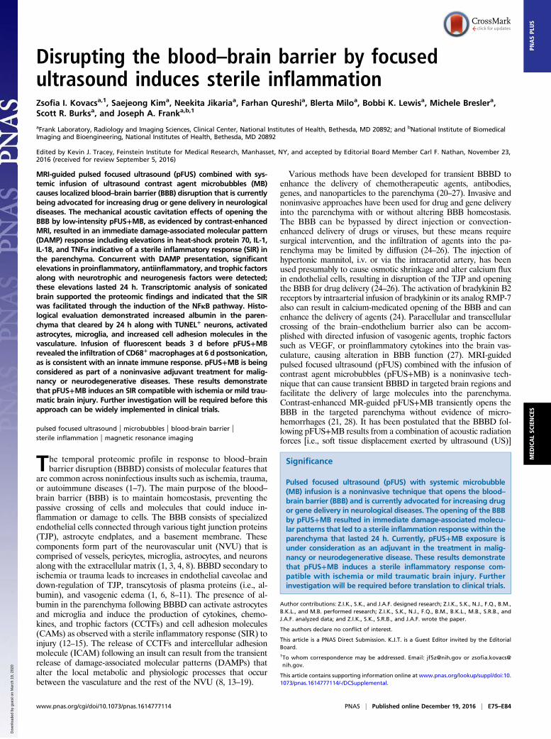
Assessment of BBBD.All animals underwent MRI before sonicationto ensure that rats had normal brain ventricular anatomy (35).Following pFUS+MB, gadolinium (Gd)-enhanced T1-weighted(T1w) images demonstrated contrast enhancement in the frontallobe demarcating the area that was harvested for molecular andhistological analysis at different time points (Fig. 1A). Immuno-fluorescence (IF) staining, coinciding with areas of BBBD onMRI, revealed the increased presence of albumin at 6 h post-sonication that returned to the levels in the contralateral cortex by24 h (Fig. 1B). H&E staining demonstrated no evidence ofmicrohemorrhages within the pFUS+MB–treated cortex (Fig. 1A).
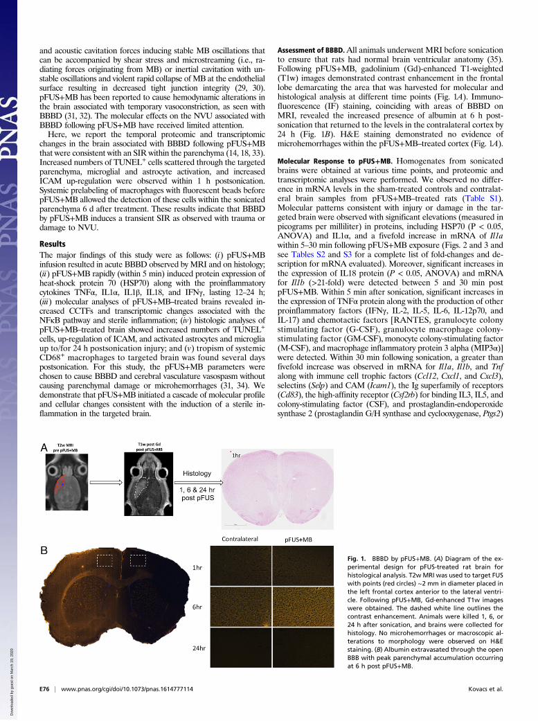
Molecular Response to pFUS+MB. Homogenates from sonicatedbrains were obtained at various time points, and proteomic andtranscriptomic analyses were performed. We observed no differ-ence in mRNA levels in the sham-treated controls and contralat-eral brain samples from pFUS+MB–treated rats (Table S1).Molecular patterns consistent with injury or damage in the tar-geted brain were observed with significant elevations (measured inpicograms per milliliter) in proteins, including HSP70 (P < 0.05,ANOVA) and IL1α, and a fivefold increase in mRNA of Il1awithin 5–30 min following pFUS+MB exposure (Figs. 2 and 3 andsee Tables S2 and S3 for a complete list of fold-changes and de-scription for mRNA evaluated). Moreover, significant increases inthe expression of IL18 protein (P < 0.05, ANOVA) and mRNAfor Il1b (>21-fold) were detected between 5 and 30 min postpFUS+MB. Within 5 min after sonication, significant increases inthe expression of TNFα protein along with the production of otherproinflammatory factors (IFNγ, IL-2, IL-5, IL-6, IL-12p70, andIL-17) and chemotactic factors [RANTES, granulocyte colonystimulating factor (G-CSF), granulocyte macrophage colony-stimulating factor (GM-CSF), monocyte colony-stimulating factor(M-CSF), and macrophage inflammatory protein 3 alpha (MIP3α)]were detected. Within 30 min following sonication, a greater thanfivefold increase was observed in mRNA for Il1a, Il1b, and Tnfalong with immune cell trophic factors (Ccl12, Cxcl1, and Cxcl3),selectins (Selp) and CAM (Icam1), the Ig superfamily of receptors(Cd83), the high-affinity receptor (Csf2rb) for binding IL3, IL5, andcolony-stimulating factor (CSF), and prostaglandin-endoperoxidesynthase 2 (prostaglandin G/H synthase and cyclooxygenase, Ptgs2)
Fig. 1. BBBD by pFUS+MB. (A) Diagram of the ex-perimental design for pFUS-treated rat brain forhistological analysis. T2w MRI was used to target FUSwith points (red circles) ∼2 mm in diameter placed inthe left frontal cortex anterior to the lateral ventri-cle. Following pFUS+MB, Gd-enhanced T1w imageswere obtained. The dashed white line outlines thecontrast enhancement. Animals were killed 1, 6, or24 h after sonication, and brains were collected forhistology. No microhemorrhages or macroscopic al-terations to morphology were observed on H&Estaining. (B) Albumin extravasated through the openBBB with peak parenchymal accumulation occurringat 6 h post pFUS+MB.
E76 | www.pnas.org/cgi/doi/10.1073/pnas.1614777114 Kovacs et al.
Dow
nloa
ded
by g
uest
on
Mar
ch 1
9, 2
020
(Fig. 3 B and C); these increases are consistent with activation ofthe NFκb pathway and sterile inflammation. Concurrent with theearly expression of inflammatory CCTFs, significant increases (P <0.05, ANOVA) in the expression of the proteins VEGF, erythro-poietin (EPO), basic fibroblast growth factor (bFGF), IL4, IL10,and IL13 were also detected in the sonicated parenchyma (Fig. 2 Band C). The increased expression in VEGF would be observedwith changes in vascular permeability to albumin and plasmaproteins resulting from BBBD. Between 0.5 and 12 h, persistentand significant expression of cell stress-reactive proteins, pro- andantiinflammatory CCTFs, was detected in the pFUS-treated brain(Fig. 2). The increase in ICAM1 started at 2 h postsonication andpersisted to 24 h (P < 0.05, ANOVA). Other factors, such asstromal cell-derived factor 1 alpha (SDF1α) and keratinocyte-de-rived chemokine (KC), were significantly elevated at a single timepoint from 2 to 6 h postsonication. Two hours postsonication, theprotein expression of proinflammatory factors (IL2, IL6, Il12p70,IL17, and IL18) and antiinflammatory factors (IL4, and IL13,
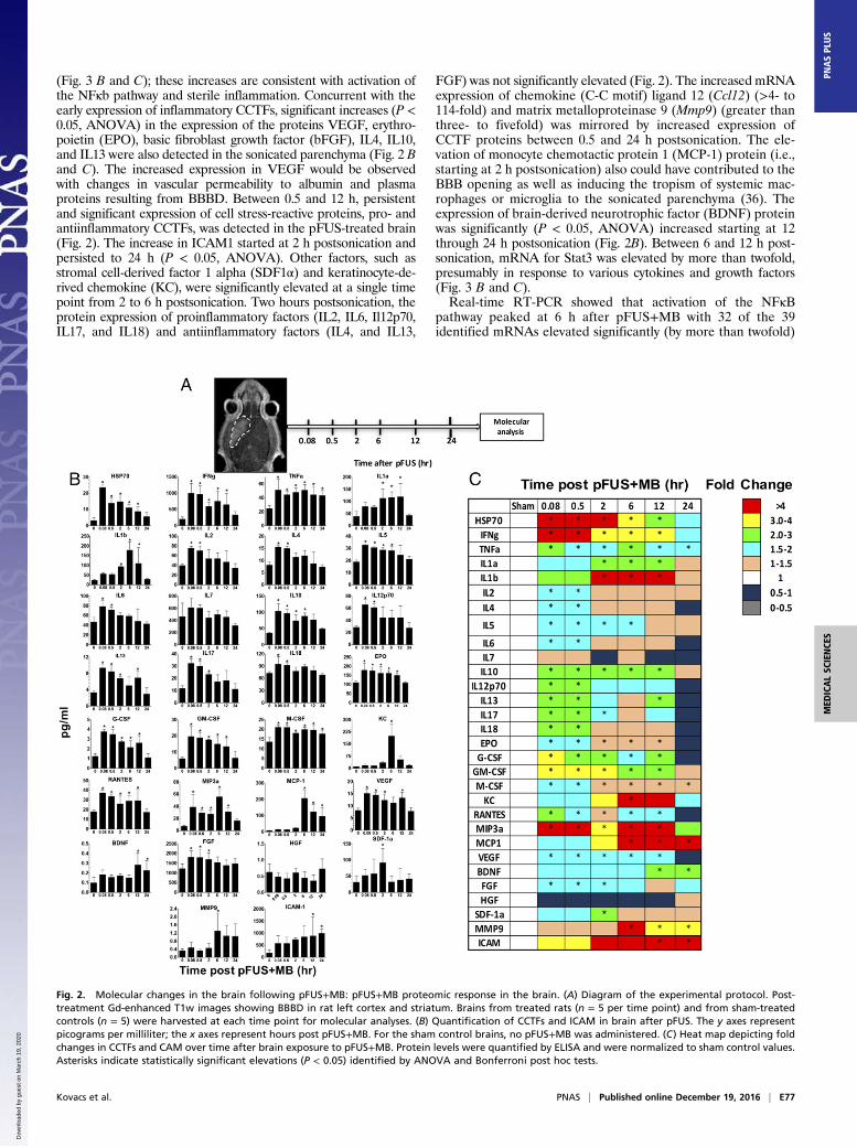
FGF) was not significantly elevated (Fig. 2). The increased mRNAexpression of chemokine (C-C motif) ligand 12 (Ccl12) (>4- to114-fold) and matrix metalloproteinase 9 (Mmp9) (greater thanthree- to fivefold) was mirrored by increased expression ofCCTF proteins between 0.5 and 24 h postsonication. The ele-vation of monocyte chemotactic protein 1 (MCP-1) protein (i.e.,starting at 2 h postsonication) also could have contributed to theBBB opening as well as inducing the tropism of systemic mac-rophages or microglia to the sonicated parenchyma (36). Theexpression of brain-derived neurotrophic factor (BDNF) proteinwas significantly (P < 0.05, ANOVA) increased starting at 12through 24 h postsonication (Fig. 2B). Between 6 and 12 h post-sonication, mRNA for Stat3 was elevated by more than twofold,presumably in response to various cytokines and growth factors(Fig. 3 B and C).Real-time RT-PCR showed that activation of the NFκB
pathway peaked at 6 h after pFUS+MB with 32 of the 39identified mRNAs elevated significantly (by more than twofold)
Fig. 2. Molecular changes in the brain following pFUS+MB: pFUS+MB proteomic response in the brain. (A) Diagram of the experimental protocol. Post-treatment Gd-enhanced T1w images showing BBBD in rat left cortex and striatum. Brains from treated rats (n = 5 per time point) and from sham-treatedcontrols (n = 5) were harvested at each time point for molecular analyses. (B) Quantification of CCTFs and ICAM in brain after pFUS. The y axes representpicograms per milliliter; the x axes represent hours post pFUS+MB. For the sham control brains, no pFUS+MB was administered. (C) Heat map depicting foldchanges in CCTFs and CAM over time after brain exposure to pFUS+MB. Protein levels were quantified by ELISA and were normalized to sham control values.Asterisks indicate statistically significant elevations (P < 0.05) identified by ANOVA and Bonferroni post hoc tests.
Kovacs et al. PNAS | Published online December 19, 2016 | E77
MED
ICALSC
IENCE
SPN
ASPL
US
Dow
nloa
ded
by g
uest
on
Mar
ch 1
9, 2
020
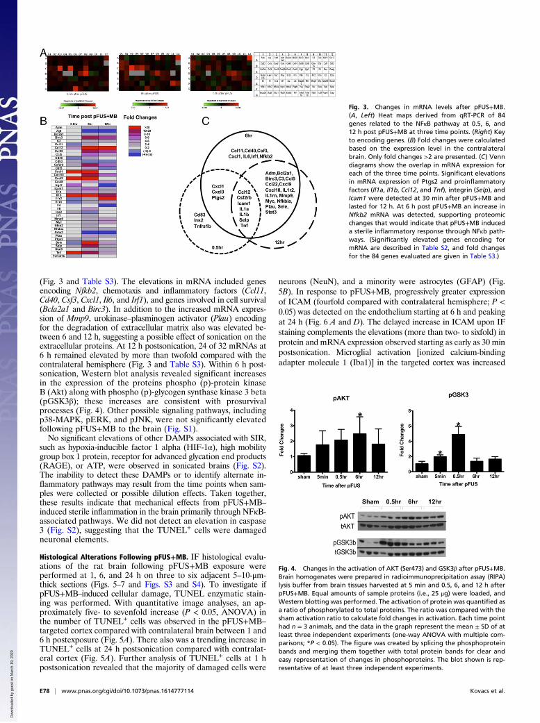
(Fig. 3 and Table S3). The elevations in mRNA included genesencoding Nfkb2, chemotaxis and inflammatory factors (Ccl11,Cd40, Csf3, Cxcl1, Il6, and Irf1), and genes involved in cell survival(Bcla2a1 and Birc3). In addition to the increased mRNA expres-sion of Mmp9, urokinase–plasminogen activator (Plau) encodingfor the degradation of extracellular matrix also was elevated be-tween 6 and 12 h, suggesting a possible effect of sonication on theextracellular proteins. At 12 h postsonication, 24 of 32 mRNAs at6 h remained elevated by more than twofold compared with thecontralateral hemisphere (Fig. 3 and Table S3). Within 6 h post-sonication, Western blot analysis revealed significant increasesin the expression of the proteins phospho (p)-protein kinaseB (Akt) along with phospho (p)-glycogen synthase kinase 3 beta(pGSK3β); these increases are consistent with prosurvivalprocesses (Fig. 4). Other possible signaling pathways, includingp38-MAPK, pERK, and pJNK, were not significantly elevatedfollowing pFUS+MB to the brain (Fig. S1).No significant elevations of other DAMPs associated with SIR,
such as hypoxia-inducible factor 1 alpha (HIF-1α), high mobilitygroup box 1 protein, receptor for advanced glycation end products(RAGE), or ATP, were observed in sonicated brains (Fig. S2).The inability to detect these DAMPs or to identify alternate in-flammatory pathways may result from the time points when sam-ples were collected or possible dilution effects. Taken together,these results indicate that mechanical effects from pFUS+MB–induced sterile inflammation in the brain primarily through NFκB-associated pathways. We did not detect an elevation in caspase3 (Fig. S2), suggesting that the TUNEL+ cells were damagedneuronal elements.
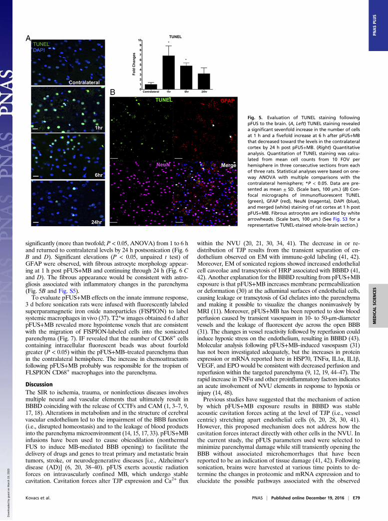
Histological Alterations Following pFUS+MB. IF histological evalu-ations of the rat brain following pFUS+MB exposure wereperformed at 1, 6, and 24 h on three to six adjacent 5–10-μm-thick sections (Figs. 5–7 and Figs. S3 and S4). To investigate ifpFUS+MB–induced cellular damage, TUNEL enzymatic stain-ing was performed. With quantitative image analyses, an ap-proximately five- to sevenfold increase (P < 0.05, ANOVA) inthe number of TUNEL+ cells was observed in the pFUS+MB–targeted cortex compared with contralateral brain between 1 and6 h postexposure (Fig. 5A). There also was a trending increase inTUNEL+ cells at 24 h postsonication compared with contralat-eral cortex (Fig. 5A). Further analysis of TUNEL+ cells at 1 hpostsonication revealed that the majority of damaged cells were
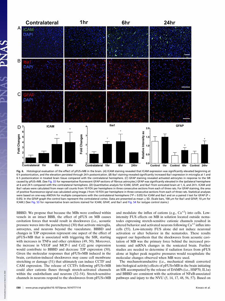
neurons (NeuN), and a minority were astrocytes (GFAP) (Fig.5B). In response to pFUS+MB, progressively greater expressionof ICAM (fourfold compared with contralateral hemisphere; P <0.05) was detected on the endothelium starting at 6 h and peakingat 24 h (Fig. 6 A and D). The delayed increase in ICAM upon IFstaining complements the elevations (more than two- to sixfold) inprotein and mRNA expression observed starting as early as 30 minpostsonication. Microglial activation [ionized calcium-bindingadapter molecule 1 (Iba1)] in the targeted cortex was increased
Fig. 3. Changes in mRNA levels after pFUS+MB.(A, Left) Heat maps derived from qRT-PCR of 84genes related to the NFκB pathway at 0.5, 6, and12 h post pFUS+MB at three time points. (Right) Keyto encoding genes. (B) Fold changes were calculatedbased on the expression level in the contralateralbrain. Only fold changes >2 are presented. (C) Venndiagrams show the overlap in mRNA expression foreach of the three time points. Significant elevationsin mRNA expression of Ptgs2 and proinflammatoryfactors (Il1a, Il1b, Ccl12, and Tnf), integrin (Selp), andIcam1 were detected at 30 min after pFUS+MB andlasted for 12 h. At 6 h post pFUS+MB an increase inNfkb2 mRNA was detected, supporting proteomicchanges that would indicate that pFUS+MB induceda sterile inflammatory response through NFκb path-ways. (Significantly elevated genes encoding formRNA are described in Table S2, and fold changesfor the 84 genes evaluated are given in Table S3.)
Fig. 4. Changes in the activation of AKT (Ser473) and GSK3β after pFUS+MB.Brain homogenates were prepared in radioimmunoprecipitation assay (RIPA)lysis buffer from brain tissues harvested at 5 min and 0.5, 6, and 12 h afterpFUS+MB. Equal amounts of sample proteins (i.e., 25 μg) were loaded, andWestern blotting was performed. The activation of protein was quantified asa ratio of phosphorylated to total proteins. The ratio was compared with thesham activation ratio to calculate fold changes in activation. Each time pointhad n = 3 animals, and the data in the graph represent the mean ± SD of atleast three independent experiments (one-way ANOVA with multiple com-parisons; *P < 0.05). The figure was created by splicing the phosphoproteinbands and merging them together with total protein bands for clear andeasy representation of changes in phosphoproteins. The blot shown is rep-resentative of at least three independent experiments.
E78 | www.pnas.org/cgi/doi/10.1073/pnas.1614777114 Kovacs et al.
Dow
nloa
ded
by g
uest
on
Mar
ch 1
9, 2
020
significantly (more than twofold; P < 0.05, ANOVA) from 1 to 6 hand returned to contralateral levels by 24 h postsonication (Fig. 6B and D). Significant elevations (P < 0.05, unpaired t test) ofGFAP were observed, with fibrous astrocyte morphology appear-ing at 1 h post pFUS+MB and continuing through 24 h (Fig. 6 Cand D). The fibrous appearance would be consistent with astro-gliosis associated with inflammatory changes in the parenchyma(Fig. 5B and Fig. S5).To evaluate pFUS+MB effects on the innate immune response,
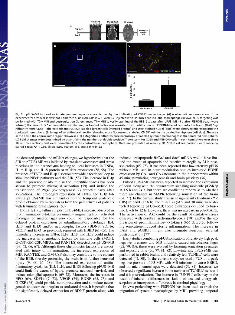
3 d before sonication rats were infused with fluorescently labeledsuperparamagnetic iron oxide nanoparticles (FlSPION) to labelsystemic macrophages in vivo (37). T2*w images obtained 6 d afterpFUS+MB revealed more hypointense voxels that are consistentwith the migration of FlSPION-labeled cells into the sonicatedparenchyma (Fig. 7). IF revealed that the number of CD68+ cellscontaining intracellular fluorescent beads was about fourfoldgreater (P < 0.05) within the pFUS+MB–treated parenchyma thanin the contralateral hemisphere. The increase in chemoattractantsfollowing pFUS+MB probably was responsible for the tropism ofFLSPION CD68+ macrophages into the parenchyma.
DiscussionThe SIR to ischemia, trauma, or noninfectious diseases involvesmultiple neural and vascular elements that ultimately result inBBBD coinciding with the release of CCTFs and CAM (1, 3–7, 9,17, 18). Alterations in metabolism and in the structure of cerebralvascular endothelium led to the impairment of the BBB function(i.e., disrupted homeostasis) and to the leakage of blood productsinto the parenchyma microenvironment (14, 15, 17, 33). pFUS+MBinfusions have been used to cause obicodilation (nonthermalFUS to induce MB-mediated BBB opening) to facilitate thedelivery of drugs and genes to treat primary and metastatic braintumors, stroke, or neurodegenerative diseases [i.e., Alzheimer’sdisease (AD)] (6, 20, 38–40). pFUS exerts acoustic radiationforces on intravascularly confined MB, which undergo stablecavitation. Cavitation forces alter TJP expression and Ca2+ flux
within the NVU (20, 21, 30, 34, 41). The decrease in or re-distribution of TJP results from the transient separation of en-dothelium observed on EM with immune-gold labeling (41, 42).Moreover, EM of sonicated regions showed increased endothelialcell caveolae and transcytosis of HRP associated with BBBD (41,42). Another explanation for the BBBD resulting from pFUS+MBexposure is that pFUS+MB increases membrane permeabilizationor deformation (30) at the adluminal surfaces of endothelial cells,causing leakage or transcytosis of Gd chelates into the parenchymaand making it possible to visualize the changes noninvasively byMRI (11). Moreover, pFUS+MB has been reported to slow bloodperfusion caused by transient vasospasm in 10- to 50-μm-diametervessels and the leakage of fluorescent dye across the open BBB(31). The changes in vessel reactivity followed by reperfusion couldinduce hypoxic stress on the endothelium, resulting in BBBD (43).Molecular analysis following pFUS+MB–induced vasospasm (31)has not been investigated adequately, but the increases in proteinexpression or mRNA reported here in HSP70, TNFα, IL1α, IL1β,VEGF, and EPO would be consistent with decreased perfusion andreperfusion within the targeted parenchyma (9, 12, 19, 44–47). Therapid increase in TNFα and other proinflammatory factors indicatesan acute involvement of NVU elements in response to hypoxia orinjury (14, 48).Previous studies have suggested that the mechanism of action
by which pFUS+MB exposure results in BBBD was stableacoustic cavitation forces acting at the level of TJP (i.e., vesselcentric) stretching apart endothelial cells (6, 20, 28, 30, 41).However, this proposed mechanism does not address how thecavitation forces interact directly with other cells in the NVU. Inthe current study, the pFUS parameters used were selected tominimize parenchymal damage while still transiently opening theBBB without associated microhemorrhages that have beenreported to be an indication of tissue damage (41, 42). Followingsonication, brains were harvested at various time points to de-termine the changes in proteomic and mRNA expression and toelucidate the possible pathways associated with the observed
Fig. 5. Evaluation of TUNEL staining followingpFUS to the brain. (A, Left) TUNEL staining revealeda significant sevenfold increase in the number of cellsat 1 h and a fivefold increase at 6 h after pFUS+MBthat decreased toward the levels in the contralateralcortex by 24 h post pFUS+MB. (Right) Quantitativeanalysis. Quantitation of TUNEL staining was calcu-lated from mean cell counts from 10 FOV perhemisphere in three consecutive sections from eachof three rats. Statistical analyses were based on one-way ANOVA with multiple comparisons with thecontralateral hemisphere; *P < 0.05. Data are pre-sented as mean ± SD. (Scale bars, 100 μm.) (B) Con-focal micrographs of immunofluorescent TUNEL(green), GFAP (red), NeuN (magenta), DAPI (blue),and merged (white) staining of rat cortex at 1 h postpFUS+MB. Fibrous astrocytes are indicated by whitearrowheads. (Scale bars, 100 μm.) (See Fig. S3 for arepresentative TUNEL-stained whole-brain section.)
Kovacs et al. PNAS | Published online December 19, 2016 | E79
MED
ICALSC
IENCE
SPN
ASPL
US
Dow
nloa
ded
by g
uest
on
Mar
ch 1
9, 2
020
BBBD. We propose that because the MBs were confined withinvessels in an intact BBB, the effect of pFUS on MB causescavitation forces that would result in shockwaves (i.e., acousticpressure waves into the parenchyma) (30) that activate microglia,astrocytes, and neurons beyond the vasculature. BBBD andchanges in TJP expression represent one aspect of the effect ofpFUS+MB that is associated with triggering the SIR, startingwith increases in TNFα and other cytokines (49, 50). Moreover,the increase in VEGF and MCP-1 and Ccl2 gene expressionwould contribute to BBBD and decrease TJP expression (50).Given the molecular responses that pFUS+MB induced in thebrain, cavitation-induced shockwaves may cause cell membranestretching or damage (51) that ultimately can induce CCTF andCAM expression. The release of CCTFs following pFUS+MBcould alter cationic fluxes through stretch-activated channelswithin the endothelium and neurons (52–54). Stretch-sensitivechannels in neurons respond to the shockwaves from pFUS+MB
and modulate the influx of cations (e.g., Ca+2) into cells. Low-intensity FUS effects on MB in solution located outside nema-todes expressing stretch-sensitive cationic channels resulted inaltered behavior and activated neurons following Ca+2 influx intocells (55). Low-intensity FUS alone did not induce neuronalactivation or alter behavior in the nematodes. These resultssupport our hypothesis that the shockwaves from acoustic cavi-tation of MB was the primary force behind the increased pro-teomic and mRNA changes in the sonicated brain. Furtherstudies are needed to determine if radiation forces from pFUSalone at higher peak negative pressures would recapitulate themolecular changes observed when MB were used.The mechanotransductive (i.e., mechanical stimuli converted
into biological activity) effects of pFUS+MB in the brain initiatingan SIR accompanied by the release of DAMPs (i.e., HSP70, IL1α)and BBBD are consistent with the activation of NFκB-associatedpathways and injury to the NVU (5, 14, 17, 48, 56, 57). Based on
Fig. 6. Histological evaluation of the effect of pFUS+MB in the brain. (A) ICAM staining revealed that ICAM expression was significantly elevated beginning at6 h postsonication, and the elevation persisted through 24 h postsonication. (B) Iba1 staining revealed significantly increased Iba1 expression in microglia at 1 and6 h postsonication in treated brain tissue compared with the contralateral hemisphere. (C) GFAP staining revealed activated astrocytes in response to the SIRcaused by pFUS+MB. (See Fig. S5 for representative fluorescent GFAP sections of fibrous astrocytes.) GFAP was significantly elevated in the ipsilateral hemisphereat 6 and 24 h compared with the contralateral hemisphere. (D) Quantitative analysis for ICAM, GFAP, and Iba1 from sonicated brain at 1, 6, and 24 h. ICAM andIba1 values were calculated from mean cell counts from 10 FOV per hemisphere in three consecutive sections from each of three rats. For GFAP staining, the areaof positive fluorescence signal was calculated using Image J from 10 FOV per hemisphere in three consecutive sections from each of three rats. Statistical analyseswere based on one-way ANOVA for multiple comparisons with the contralateral hemisphere (*P < 0.05) for ICAM and Iba1 and on a paired t test for GFAP (P <0.05). In the GFAP graph the control bars represent the contralateral cortex. Data are presented as mean ± SD. (Scale bars, 100 μm for Iba1 and GFAP; 10 μm forICAM.) (See Fig. S3 for representative brain sections stained for ICAM, GFAP, and Iba1 and Fig. S4 for isotype control stains.)
E80 | www.pnas.org/cgi/doi/10.1073/pnas.1614777114 Kovacs et al.
Dow
nloa
ded
by g
uest
on
Mar
ch 1
9, 2
020
the detected protein and mRNA changes, we hypothesize that theSIR to pFUS+MB was initiated by transient vasospasm and stressreactions in the parenchyma leading to local increases in TNFα,IL1α, IL1β, and IL18 protein or mRNA expression (56, 58). Thepresence of TNFα and IL1β also would provide a feedback loop tostimulate NFκB pathways and the SIR (58). The increase in IL1βand the presence of albumin in the interstitial spaces has beenshown to promote microglial activation (59) and induce thetranscription of Ptgs2 (cyclooxygenase 2) detected early aftersonications. The prolonged expression of TNFα and IL1β fol-lowing pFUS+MB has similarities to the temporal proteomicprofile obtained by microdialysis from the parenchyma of patientswith traumatic brain injuries (60).The early (i.e., within 2 h post pFUS+MB) increase observed in
proinflammatory cytokines presumably originating from activatedmicroglia or macrophages also could be responsible for thedelayed protein expression of antiinflammatory cytokines (IL4,IL10, and IL13) and/or neurotrophic factors (BDNF, SDF1α,VEGF, and EPO) as previously reported with BBBD (61–65). Theimmediate increase in TNFα, IL1α, IL1β, and IL18 could inducethe increases in chemotactic factors for immune cells (MCP1,G-CSF, GM-CSF, MIP3α, and RANTES) detected post pFUS+MB(33, 62, 66, 67). Although these chemotactic factors are associ-ated with injury or inflammation, the increased expression ofMIP, RANTES, and GM-CSF also may contribute to the closureof the BBB, thereby protecting the brain from further neuronalinjury (9, 48, 66, 68). The increased expression of antiin-flammatory cytokines (IL4, IL10, and IL13) following pFUS+MBcould limit the extent of injury, promote neuronal survival, andinduce microglial apoptosis (69–72). Moreover, the increases inEPO (69), SDF1α (7, 73), VEGF (74), BDNF (65, 75), andG-CSF (66) could provide neuroprotection and stimulate neuro-genesis and stem cell tropism to sonicated tissue. It is possible thatthe significantly delayed expression of BDNF protein along with
induced antiapoptotic Bcl2a1 and Birc3 mRNA would have lim-ited the extent of apoptosis and reactive microglia by 24 h post-sonication (65, 75). It has been reported that low-intensity pFUSwithout MB used in neuromodulation studies increased BDNFexpression by CA1 and CA3 neurons in the hippocampus within45 min, stimulating neurogenesis and brain plasticity (76).Pulsed FUS+MB has been reported to increase the expression
of pAkt along with the downstream signaling molecule pGSK3βat 1.5 h and 24 h, but there are conflicting reports as to whetherthere are changes in MAPK following sonication of the brain(34, 77). In the current study, transient significant elevations (P <0.05) in pAkt (at 6 h) and pGSK3β (at 5 and 30 min) were de-tected following pFUS+MB; these elevations declined to base-line levels by 12 h. However, there was no increase in p38-MAPK.The activation of Akt could be the result of oxidative stressobserved with cerebral ischemia/hypoxia (78) and/or the ex-pression of proinflammatory cytokines (65) detected follow-ing sonication-induced sterile inflammation. The increase inpAkt and pGSK3β might also promote neuronal survivalpostsonication (77).Early studies combining pFUS sonications of relatively high peak
negative pressures and MB infusions caused microhemorrhages(22, 79, 80); these were avoided by lowering sonication pressuresand exposure time (20, 77, 81, 82). Low-intensity pFUS+MB wasperformed in rabbit brains, and relatively few TUNEL+ cells weredetected (42, 80). In the current study, we used pFUS at a peaknegative pressure of 0.3 MPa with MB infusions to cause BBBD,and no microhemorrhages were detected (79, 81); however, weobserved a significant increase in the number of TUNEL+ cells at 1and 6 h postsonication. The increase in TUNEL+ cells may be theresult of inherent differences in skull thickness and energy ab-sorption or interspecies differences in cerebral physiology.In vivo prelabeling with FlSPION has been used to track the
migration of systemic macrophages by MRI, providing temporal
Fig. 7. pFUS+MB induced an innate immune response characterized by the infiltration of CD68+ macrophages. (A) A schematic representation of theexperimental protocol shows that 3 d before pFUS+MB, rats (n = 5) were i.v. injected with FlSPION beads to label macrophages in vivo. pFUS targeting wasperformed with T2w MRI and postsonication Gd-enhanced T1w MRI to verify opening of the BBB. Six days after pFUS+MB (9 d after FlSPION beads wereinfused) the area of T2* abnormalities (white oval) in treated cortex was consistent with infiltration of FlSPION-labeled cells into the brain. (B–D) Sig-nificantly more CD68+-labeled (red) and FLSPION-labeled (green) cells (merged orange) and DAPI-stained nuclei (blue) were observed migrating into thesonicated hemisphere. (B) Image of an entire brain section showing more fluorescently labeled CD 68+ cells in the treated hemisphere (left side). The areain the box is the approximate region shown in C. (C ) Magnified epifluorescence microscopy of labeled systemic macrophages in the sonicated hemisphere.(D) Fold changes were determined by quantifying the numbers of double-positive (fluorescent for CD68 and FlSPION) cells in each hemisphere over three10-μm-thick sections and were normalized to the contralateral hemisphere. Data are presented as mean ± SD. Statistical comparisons were made bypaired t test; *P < 0.05. (Scale bars, 100 μm in C and 2 mm in B.)
Kovacs et al. PNAS | Published online December 19, 2016 | E81
MED
ICALSC
IENCE
SPN
ASPL
US
Dow
nloa
ded
by g
uest
on
Mar
ch 1
9, 2
020
spatial localization of tagged cells to injured areas following trau-matic brain injury (37). We previously reported that in vivo la-beling with FlSPION provides the ability after pFUS to track viaMRI and histology the homing of these systemic macrophages topFUS-targeted sites within muscle (83). In our previous study,FlSPION beads were infused 3 d before sonication to providesufficient time for CD68+ macrophages to endocytose the particlesso labeled cells could be tracked by MRI and histology. Moreover,pFUS targeted to tissues also resulted in a shift toward antiin-flammatory (M2) rather than proinflammatory (M1) macrophagephenotypes in the absence of histological damage (84, 85). In thecurrent study, 6 d after pFUS+MB, hypointense voxels detected byT2*w MRI and FlSPION-labeled CD68+ macrophages observedvia IF staining were scattered throughout the targeted brain. Thepresence of labeled macrophages is consistent with the local in-creases in chemotactic factors inducing tropism of immune cellsfrom the circulation to the targeted parenchyma. Further in-vestigation is required to determine the relationship betweenpFUS+MB treatment and the initial homing of innate immunecells and macrophage phenotypes in the targeted parenchyma.It has been reported that exposure of the hippocampus to
pFUS+MB in AD models revealed microglial phagocytosing betaamyloid precursor protein (βAPP) and increased neurogenesisbased on bromo-deoxyuridine uptake or double-cortin staining ofproliferating cells (20, 38, 40, 86). Neurobehavioral testing of thesetreated mice demonstrated improvements in memory functionfollowing multiple pFUS+MB exposures. These reports specu-lated that sonication stimulated neurogenesis, presumably throughthe release of growth factors, and recommend further studies.Although the molecular mechanisms were not addressed in theseAD studies, we have clearly demonstrated that pFUS+MB in-duces increased expression of CCTFs that could contribute tomicroglial activation, which in turn could contribute to the clear-ance of βAPP plaques. Within 2 h after pFUS+MB we detected asignificant doubling of IL1β that would activate microglia andclear βAPP plaques from the hippocampus (87). Moreover, thepresence of inflammatory cytokines may contribute to the ex-pression of BDNF, EPO, and VEGF in our study and could ex-plain the enhanced neurogenesis observed in AD models (38, 40).Opening of the BBB was associated with the influx of serumproducts that represent damage signals to microglia, resulting inactivation (59). Persistent overexpression of IL1β has been shownto reduce amyloid plaques but to increase phosphorylated taupathology in an AD mouse model (87). Increased tau has beenassociated with an increase in GSK3β activity that was observedfollowing pFUS+MB in this study and others (77). Further studiesare necessary to investigate the long-term consequences of re-peated SIR events following multiple weekly exposures of thebrain to pFUS+MB and a role for pFUS+MB as a possibletreatment for neurodegenerative diseases.This study has a few limitations that need to be addressed. The
changes in proteomic and transcriptome expression observed fol-lowing pFUS+MB to the brain were determined only in femalerats. It is unknown if the molecular changes we observed are ap-plicable across strains, species, ages, or genders. Because pFUS+MBexerts a mechanical effect and has been shown to cause BBBD invarious experimental models, it is likely that the increased ex-pression of CCTFs resulting in a transient SIR would still occur.We also observed a heterogeneous response to pFUS+MB, in-cluding variations in contrast enhancement on MRI that was de-pendent on the distance from the top of the skull and potentialenergy dissipation of the FUS exposure. The variability in theextent and type of insult to the parenchyma may result from skullthickness or from the sensitivity of the BBB and neuronal elementsto the acoustic cavitation forces generated by the pFUS interactionwith MB. We also were unable to identify clearly which neuronalcellular elements primarily contributed to the SIR followingpFUS+MB. Furthermore, examination of pFUS+MB should be
performed in larger animal models mimicking sonication ex-posures to tumors to determine if SIR is induced within theparenchyma. Further research is needed to determine how themolecular and pathological response to multiple pFUS+MBwill impact the reparative or neurodegenerative processes inthe targeted brain.In summary, image-guided pFUS+MB is a clinically relevant,
noninvasive technique that can effectively cause transientopening of the BBB and has been used to enhance the retentionand permeability of drugs or gene therapy materials within tar-geted regions in the brain (6, 20, 30, 39). Previous studies haveindicated that the mechanical effects of pFUS+MB were con-fined primarily to the endothelium and vessel wall and wereassociated with cerebral vasculature vasospasm (31). Within5 min of pFUS+MB, we observed the transient BBBD and anincreased expression of DAMPs leading to an SIR throughNFκB pathways that lasted for ∼24 h. Moreover, there was evi-dence of early neuronal and astrocytic injury, based on TUNELstaining, and astrocyte and microglial activation along with in-creased expression of CAM. The SIR within the parenchymainfluenced the tropism of systemic macrophages to the targetedbrain. These results indicate that the mechanotransductive ef-fects of pFUS+MB in the brain require further investigation todetermine pFUS+MB’s potential clinical contribution to thetreatment of neurological diseases.
MethodsAnimals. The Animal Care and Use Committee at the National Institutes ofHealth approved all studies, and experiments were performed according tothe National Research Council’s Guide for the Care and Use of LaboratoryAnimals (88). Female 8- to 10-wk-old Sprague–Dawley rats purchased fromCharles River Laboratory were used in this study.
MRI-Guided pFUS+MB. Ratswere anesthetizedwith isofluorane [1–3.5% (vol/vol)]and 100% O2 through a nosecone for all experiments. MRI was performedusing a 3-T MR scanner (Achieva Philips Healthcare) using either a surface coil(RK-100; FUS Instruments) or a 3-cm diameter solenoid coil (Philips ResearchLaboratories). Targeting coordinates were registered (Fig. 1A) on axial T2wimages of the rat brain acquired before sonication with the following pa-rameters: turbo spin echo (TSE) with repetition time/echo time (TR/TE) =2,000/70 ms. pFUS was performed with the following parameters: 0.3 MPaacoustic pressure was applied in 10-ms bursts and a 1% duty cycle (120 s pernine focal points) using a single-element spherical FUS transducer (centerfrequency: 589.636 KHz; focal number: 0.8; active diameter: 7.5 cm) (FUS In-struments). The focal point of the FUS transducer was targeted based on anaxial T2w MRI from the anterior cortex to the lateral ventricle with non-overlapping 2-mm circles (Fig. 1). Sonications were accompanied by MB in-fusion of 100 μL Optison (GE Healthcare) through the tail vein at a rate of1 μL/s. After sonication, rats received 200 μL gadofosveset (Ablavar, an albumin-binding chelated Gd formulation; Lantheus Medical Imaging, Inc.) via thetail vein, and axial T1w contrast-enhanced images were obtained with thefollowing parameters: TSE (TR/TE 350/12 ms). High-resolution images follow-ing sonication to detect the presence of FlSPION-labeled macrophages wereobtained with the following sequences: T2w TSE (TR/TE 2,769/60 ms), T2*w(TR/TE 1,301/7.0 ms), number of echoes 5, ΔTE 7.0 ms, flip angle 30°, T1wGdTSE (TR/TE 600/20 ms).
Molecular Analysis. At 0 (sham, n = 5), 0.08, 0.5, 2, 6, 12, and 24 h aftersonication, rats (n = 5 per time point) were deeply anesthetized with iso-flurane before decapitation. The brains were dissected from the skull, andthe sonicated tissue and the corresponding sham tissue were isolated. Tissuewas snap-frozen in liquid nitrogen and kept at −80 °C until homogenizationin cell lysis buffer (9803S; Cell Signaling Technology) containing a proteaseinhibitor mixture (S8820-2TAB; Sigma-Aldrich). Samples were centrifuged at15,996 × g for 20 min at 4 °C, and the supernatant was used for analyses.Total protein was determined using a bicinchoninic acid (BCA) assay (23227;Thermo Scientific). Homogenates (2 mg/mL total protein) were analyzed byRat Bio-Plex Cytokine 24-Plex group I assay (171-K1001M; Bio-Rad Labora-tories, Inc.) or ELISAs for the following: stromal cell-derived factor 1 (SDF1)(RS0074; NeoBiolab), BDNF (ELR-BDNF; RayBiotech, Inc.), MMP9; FGF(MFB00), ICAM (RIC100), HSP70 (DYC1663-2), and hepatocyte growth factor(HGF) (MHG00), all from R&D Systems, Inc. All assays were performed
E82 | www.pnas.org/cgi/doi/10.1073/pnas.1614777114 Kovacs et al.
Dow
nloa
ded
by g
uest
on
Mar
ch 1
9, 2
020
according to the manufacturer’s protocols. Samples were run with a proteinconcentration of 2 mg/mL and were read on a spectrophotometric platereader (Spectra Max M5; Molecular Devices).
RNA Isolation and Quantitative Real-Time PCR. For the assessment sham andNFκΒ-treated brains were obtained at 0.5, 6, and 12 h after sonication (n = 3per time point) and were extracted into RNAlater (Ambion/Life Technolo-gies) and stored at −20 °C. Between 60 and 70 mg of tissue were homoge-nized in a Paris Cell Disruption Buffer with Omni Tip probes (OmniInternational), and RNA was isolated using the Paris RNA Isolation Kit(Ambion/Life Technologies) according to the manufacturer’s instructions.cDNA synthesis was performed using the RT2 First Strand Kit (Qiagen) fol-lowed by quantitative RT-PCR (qRT-PCR) using the CFX96 Touch Real-TimePCR Detection System (Bio-Rad Laboratories) with RT2 SYBR Green qPCRMaster Mix (Qiagen). cDNA samples from each time point were screenedwith RT2 Profiler PCR Array Rat NFκB Signaling Pathway (Qiagen). Data wereanalyzed using the SABiosciences PCR Array Data Analysis Web Portal (www.qiagen.com/shop/genes-and-pathways/data-analysis-center-overview-page/).
Western Blotting.Western blotting was performed in samples from sham (n =3) and treated brains obtained at 0.5, 6, and 12 h following sonication (n = 3per time point). Protein (25 μg) was electrophoresed by SDS/PAGE on 4–12%Novex Bis-Tris gels (Invitrogen, Thermo Fisher Scientific) under reducingconditions and was transferred to PVDF membranes. Membranes wereblocked in 5% BSA in TBS plus 0.05% Tween-20 (TBST) for 1 h and then werehybridized overnight at 4 °C in TBST containing 2% BSA with rabbit ormouse primary antibodies against following phospho- and total proteins:protein kinase B (Akt), glycogen synthase kinase 3B (GSK3b), ERK, JNK, andp38-MAPK. All antibodies were from Cell Signaling Technology and wereused in 1:1,000–1:2,000 dilutions. HIF1α, HMGB-1, and RAGE antibodies werefrom Thermo Fisher Scientific and were used in 1:1,000 dilutions. Secondaryantibodies were HRP-conjugated goat antibodies against mouse or rabbit(Jackson ImmunoResearch Laboratories) used in 1:10,000 dilutions with 1 hincubation at room temperature. Protein bands were detected using theAmersham ECL Prime Western Blotting Detection Reagent (GE Healthcare),and the relative protein amount was measured using ImageJ (National In-stitutes of Health).
Histological Staining. Rats (n = 3 per time point) were killed at 1, 6, and 24 hpost pFUS and were perfused with 4% (wt/vol) paraformaldehyde. Fixedbrains were embedded in paraffin or optimum cutting temperature (O.C.T.)tissue-mounting medium and were sliced into 5–10-μm sections. Paraffin (p)and frozen (f) tissue sections were stained with H&E and with IF for immu-nohistochemistry (IHC). Immunostaining of three sections from each animaldetected ICAM, GFAP, Iba1, and albumin. Primary antibodies used for IHCwere goat anti-ICAM, 1:20 (p) (AF583; R&D Systems Inc.), rabbit anti-GFAP,
1:1,500 (f) (Ab 33922; Abcam), goat anti-Iba1, 1:100 (p) 1:200 (f) (019-19741;Wako Chemicals), and sheep anti-albumin, 1:100 (f) (Ab53435; Abcam). Allthe secondary antibodies were from Abcam and were used in 1:200 dilutions[goat anti-rabbit antibody (Ab102293; Abcam) for GFAP and Iba1; donkeyanti-goat antibody (Ab150129; Abcam) for ICAM; and donkey anti-sheepantibody (Ab150178; Abcam) for albumin]. TUNEL+ cells were detected withan in situ cell-death detection kit (AP 11684809910; Roche Life Science)according to the manufacturer’s protocol. Isotype control antibody stains areshown in Fig. S4.
Macrophage Labeling. Three days before pFUS+MB, rats (n = 5) were injectedvia the tail vein with 8 mg/kg MicroTRACK biodegradable rhodamine en-capsulated magnetic polymers (CL-01Q02-B-50; BioPal, Inc.). MRI wasobtained at 3 T before and 6 d after pFUS+MB exposure. Rats were killed6 d after sonication. Frozen sections (10 μm thick) were IF-stained for amacrophage marker, CD68, 1:200 (f) (Ab31630; Abcam). Sections wereanalyzed for colocalized CD68 and nanoparticle-associated rhodamineusing Image J and were compared with the contralateral hemisphere.
Microscopy. Microscopy was performed with an Aperio ScanScope CS systemequipped with a 20× air objective (NA = 0.75) (Leica Microsystems). High-magnification (60×) images were taken with an Olympus BX61fluorescentmicroscope. A laser scanning confocal microscope (model 710; Carl Zeiss AG;www.zeiss.com/global/home.html) using Plan-Apochromat objectives (20×air, NA = 0.8) was used for confocal microscopy. Illumination was providedby argon-ion (Lasos, www.lasos.com), diode, and diode-pumped solid-statelasers (Roithner Lasertechnik, www.roithner-laser.com).
Histologic Analysis. Histological evaluation of the microscopy sections wasperformed at 20× magnification. Fold changes were based on cell countsthat were normalized from 10 fields of view (FOV) per hemisphere fromthree rats using Image J. For GFAP staining, the area of fluorescent signalwas quantified using Image J by setting thresholds from 10 FOV per hemi-sphere of three rats followed by normalization of the pixel area to thecontralateral hemisphere.
Statistical Analysis. All values are presented as mean ± SD. Statistical analysesand data presentation were performed with Prism (version 6, GraphPadSoftware, Inc.). Student’s unpaired t tests were used for pairwise compari-sons, and one-way ANOVA with Bonferroni post hoc tests was used formultiple comparisons. P values < 0.05 were considered significant.
ACKNOWLEDGMENTS. This research was funded by the Intramural ResearchPrograms of the Clinical Center and the National Institute of BiomedicalImaging and Bioengineering at the National Institutes of Health.
1. Schoknecht K, David Y, Heinemann U (2015) The blood-brain barrier-gatekeeper to
neuronal homeostasis: Clinical implications in the setting of stroke. Semin Cell Dev
Biol 38:35–42.2. Cardoso FL, et al. (2015) Systemic inflammation in early neonatal mice induces tran-
sient and lasting neurodegenerative effects. J Neuroinflammation 12:82.3. Daneman R, Prat A (2015) The blood-brain barrier. Cold Spring Harb Perspect Biol
7(1):a020412.4. Cernak I, O’Connor C, Vink R (2002) Inhibition of cyclooxygenase 2 by nimesulide
improves cognitive outcome more than motor outcome following diffuse traumatic
brain injury in rats. Exp Brain Res 147(2):193–199.5. Denes A, Thornton P, Rothwell NJ, Allan SM (2010) Inflammation and brain injury:
Acute cerebral ischaemia, peripheral and central inflammation. Brain Behav Immun
24(5):708–723.6. Leinenga G, Langton C, Nisbet R, Götz J (2016) Ultrasound treatment of neurological
diseases–current and emerging applications. Nat Rev Neurol 12(3):161–174.7. Yin W, et al. (2013) The migration of neural progenitor cell mediated by SDF-1 is NF-
κB/HIF-1α dependent upon hypoxia. CNS Neurosci Ther 19(3):145–153.8. Shlosberg D, Benifla M, Kaufer D, Friedman A (2010) Blood-brain barrier breakdown
as a therapeutic target in traumatic brain injury. Nat Rev Neurol 6(7):393–403.9. Cardoso FL, Brites D, Brito MA (2010) Looking at the blood-brain barrier: Molecular
anatomy and possible investigation approaches. Brain Res Brain Res Rev 64(2):
328–363.10. Eltzschig HK, Carmeliet P (2011) Hypoxia and inflammation. N Engl J Med 364(7):
656–665.11. Hawkins CP, et al. (1990) Duration and selectivity of blood-brain barrier breakdown in
chronic relapsing experimental allergic encephalomyelitis studied by gadolinium-
DTPA and protein markers. Brain 113(Pt 2):365–378.12. Arvin B, Neville LF, Barone FC, Feuerstein GZ (1996) The role of inflammation and
cytokines in brain injury. Neurosci Biobehav Rev 20(3):445–452.
13. Famakin BM (2014) The immune response to acute focal cerebral ischemia and as-sociated post-stroke immunodepression: A focused review. Aging Dis 5(5):307–326.
14. Gadani SP, Walsh JT, Lukens JR, Kipnis J (2015) Dealing with danger in the CNS: Theresponse of the immune system to injury. Neuron 87(1):47–62.
15. Shechter R, Schwartz M (2013) CNS sterile injury: Just another wound healing? TrendsMol Med 19(3):135–143.
16. Álvarez S, Muñoz-Fernández MA (2013) TNF-Αmay mediate inflammasome activationin the absence of bacterial infection in more than one way. PLoS One 8(8):e71477.
17. Amantea D, et al. (2015) Rational modulation of the innate immune system forneuroprotection in ischemic stroke. Front Neurosci 9:147.
18. Chen GY, Nuñez G (2010) Sterile inflammation: Sensing and reacting to damage. NatRev Immunol 10(12):826–837.
19. Tuttolomondo A, Pecoraro R, Pinto A (2014) Studies of selective TNF inhibitors in thetreatment of brain injury from stroke and trauma: A review of the evidence to date.Drug Des Devel Ther 8:2221–2238.
20. Burgess A, Hynynen K (2014) Drug delivery across the blood-brain barrier using fo-cused ultrasound. Expert Opin Drug Deliv 11(5):711–721.
21. Hynynen K, McDannold N, Vykhodtseva N, Jolesz FA (2001) Noninvasive MR imaging-guided focal opening of the blood-brain barrier in rabbits. Radiology 220(3):640–646.
22. McDannold N, Vykhodtseva N, Hynynen K (2008) Effects of acoustic parameters andultrasound contrast agent dose on focused-ultrasound induced blood-brain barrierdisruption. Ultrasound Med Biol 34(6):930–937.
23. Suzuki Y, Nagai N, Umemura K (2016) A review of the mechanisms of blood-brainbarrier permeability by tissue-type plasminogen activator treatment for cerebral is-chemia. Front Cell Neurosci 10:2.
24. Gabathuler R (2010) Approaches to transport therapeutic drugs across the blood-brain barrier to treat brain diseases. Neurobiol Dis 37(1):48–57.
25. Kroll RA, Neuwelt EA (1998) Outwitting the blood-brain barrier for therapeuticpurposes: Osmotic opening and other means. Neurosurgery 42(5):1083–1099, dis-cussion 1099–1100.
Kovacs et al. PNAS | Published online December 19, 2016 | E83
MED
ICALSC
IENCE
SPN
ASPL
US
Dow
nloa
ded
by g
uest
on
Mar
ch 1
9, 2
020
26. Neuwelt E, et al. (2008) Strategies to advance translational research into brain bar-riers. Lancet Neurol 7(1):84–96.
27. Stamatovic SM, Keep RF, Andjelkovic AV (2008) Brain endothelial cell-cell junctions:How to “open” the blood brain barrier. Curr Neuropharmacol 6(3):179–192.
28. Tung YS, et al. (2010) In vivo transcranial cavitation threshold detection duringultrasound-induced blood-brain barrier opening in mice. Phys Med Biol 55(20):6141–6155.
29. McDannold N, Zhang Y, Vykhodtseva N (2011) Blood-brain barrier disruption andvascular damage induced by ultrasound bursts combined with microbubbles can beinfluenced by choice of anesthesia protocol. Ultrasound Med Biol 37(8):1259–1270.
30. Goertz DE (2015) An overview of the influence of therapeutic ultrasound exposureson the vasculature: High intensity ultrasound and microbubble-mediated bioeffects.Int J Hyperthermia 31(2):134–144.
31. Raymond SB, Skoch J, Hynynen K, Bacskai BJ (2007) Multiphoton imaging of ultra-sound/Optison mediated cerebrovascular effects in vivo. J Cereb Blood Flow Metab27(2):393–403.
32. Nhan T, et al. (2013) Drug delivery to the brain by focused ultrasound induced blood-brain barrier disruption: Quantitative evaluation of enhanced permeability of cere-bral vasculature using two-photon microscopy. J Control Release 172(1):274–280.
33. Brough D, Denes A (2015) Interleukin-1α and brain inflammation. IUBMB Life 67(5):323–330.
34. Baseri B, et al. (2012) Activation of signaling pathways following localized delivery ofsystemically administered neurotrophic factors across the blood-brain barrier usingfocused ultrasound and microbubbles. Phys Med Biol 57(7):N65–N81.
35. Tu TW, et al. (2014) Imaging of spontaneous ventriculomegaly and vascular malfor-mations in Wistar rats: Implications for preclinical research. J Neuropathol Exp Neurol73(12):1152–1165.
36. Stamatovic SM, et al. (2005) Monocyte chemoattractant protein-1 regulation ofblood-brain barrier permeability. J Cereb Blood Flow Metab 25(5):593–606.
37. Foley LM, et al. (2009) Magnetic resonance imaging assessment of macrophage ac-cumulation in mouse brain after experimental traumatic brain injury. J Neurotrauma26(9):1509–1519.
38. Burgess A, et al. (2014) Alzheimer disease in a mouse model: MR imaging-guidedfocused ultrasound targeted to the hippocampus opens the blood-brain barrier andimproves pathologic abnormalities and behavior. Radiology 273(3):736–745.
39. Kovacs Z, et al. (2014) Prolonged survival upon ultrasound-enhanced doxorubicindelivery in two syngenic glioblastoma mouse models. J Control Release 187:74–82.
40. Leinenga G, Götz J (2015) Scanning ultrasound removes amyloid-β and restoresmemory in an Alzheimer’s disease mouse model. Sci Transl Med 7(278):278ra33.
41. Sheikov N, McDannold N, Sharma S, Hynynen K (2008) Effect of focused ultrasoundapplied with an ultrasound contrast agent on the tight junctional integrity of thebrain microvascular endothelium. Ultrasound Med Biol 34(7):1093–1104.
42. Hynynen K, McDannold N, Sheikov NA, Jolesz FA, Vykhodtseva N (2005) Local andreversible blood-brain barrier disruption by noninvasive focused ultrasound at fre-quencies suitable for trans-skull sonications. Neuroimage 24(1):12–20.
43. Eltzschig HK, Eckle T (2011) Ischemia and reperfusion–from mechanism to translation.Nat Med 17(11):1391–1401.
44. Huang T, et al. (2014) Hypoxia-inducible factor-1α upregulation in microglia followinghypoxia protects against ischemia-induced cerebral infarction. Neuroreport 25(14):1122–1128.
45. Lin Y, Wen L (2013) Inflammatory response following diffuse axonal injury. Int J MedSci 10(5):515–521.
46. Lu KT, Wang YW, Wo YY, Yang YL (2005) Extracellular signal-regulated kinase-mediated IL-1-induced cortical neuron damage during traumatic brain injury. NeurosciLett 386(1):40–45.
47. Taupin V, Toulmond S, Serrano A, Benavides J, Zavala F (1993) Increase in IL-6, IL-1and TNF levels in rat brain following traumatic lesion. Influence of pre- and post-traumatic treatment with Ro5 4864, a peripheral-type (p site) benzodiazepine ligand.J Neuroimmunol 42(2):177–185.
48. Amantea D, et al. (2014) Understanding the multifaceted role of inflammatory me-diators in ischemic stroke. Curr Med Chem 21(18):2098–2117.
49. Capaldo CT, Nusrat A (2009) Cytokine regulation of tight junctions. Biochim BiophysActa 1788(4):864–871.
50. Yao Y, Tsirka SE (2014) Monocyte chemoattractant protein-1 and the blood-brainbarrier. Cell Mol Life Sci 71(4):683–697.
51. Krasovitski B, Frenkel V, Shoham S, Kimmel E (2011) Intramembrane cavitation as aunifying mechanism for ultrasound-induced bioeffects. Proc Natl Acad Sci USA 108(8):3258–3263.
52. Spangenburg EE, McBride TA (2006) Inhibition of stretch-activated channels duringeccentric muscle contraction attenuates p70S6K activation. J Appl Physiol (1985)100(1):129–135.
53. Coste B, et al. (2012) Piezo proteins are pore-forming subunits of mechanically acti-vated channels. Nature 483(7388):176–181.
54. Li J, et al. (2014) Piezo1 integration of vascular architecture with physiological force.Nature 515(7526):279–282.
55. Ibsen S, Tong A, Schutt C, Esener S, Chalasani SH (2015) Sonogenetics is a non-invasiveapproach to activating neurons in Caenorhabditis elegans. Nat Commun 6:8264.
56. Mohamed IN, Ishrat T, Fagan SC, El-Remessy AB (2015) Role of inflammasome acti-vation in the pathophysiology of vascular diseases of the neurovascular unit. AntioxidRedox Signal 22(13):1188–1206.
57. Savage CD, Lopez-Castejon G, Denes A, Brough D (2012) NLRP3-inflammasome acti-vating DAMPs stimulate an inflammatory response in glia in the absence of primingwhich contributes to brain inflammation after injury. Front Immunol 3:288.
58. Singhal G, Jaehne EJ, Corrigan F, Toben C, Baune BT (2014) Inflammasomes in neuro-inflammation and changes in brain function: A focused review. Front Neurosci 8:315.
59. Ransohoff RM, Perry VH (2009) Microglial physiology: Unique stimuli, specializedresponses. Annu Rev Immunol 27:119–145.
60. Helmy A, Carpenter KL, Menon DK, Pickard JD, Hutchinson PJ (2011) The cytokineresponse to human traumatic brain injury: Temporal profiles and evidence for cere-bral parenchymal production. J Cereb Blood Flow Metab 31(2):658–670.
61. Burda JE, Bernstein AM, Sofroniew MV (2016) Astrocyte roles in traumatic brain in-jury. Exp Neurol 275(Pt 3):305–315.
62. Hedtjärn M, et al. (2002) Interleukin-18 involvement in hypoxic-ischemic brain injury.J Neurosci 22(14):5910–5919.
63. SofroniewMV (2015) Astrocyte barriers to neurotoxic inflammation. Nat Rev Neurosci16(5):249–263.
64. Takamiya M, Fujita S, Saigusa K, Aoki Y (2007) Simultaneous detections of 27 cyto-kines during cerebral wound healing by multiplexed bead-based immunoassay forwound age estimation. J Neurotrauma 24(12):1833–1844.
65. Zhou J, Ping FF, Lv WT, Feng JY, Shang J (2014) Interleukin-18 directly protects corticalneurons by activating PI3K/AKT/NF-κB/CREB pathways. Cytokine 69(1):29–38.
66. Kelso ML, Elliott BR, Haverland NA, Mosley RL, Gendelman HE (2015) Granulocyte-macrophage colony stimulating factor exerts protective and immunomodulatory ef-fects in cortical trauma. J Neuroimmunol 278:162–173.
67. Thompson WL, Van Eldik LJ (2009) Inflammatory cytokines stimulate the chemokinesCCL2/MCP-1 and CCL7/MCP-3 through NFkB and MAPK dependent pathways in ratastrocytes [corrected]. Brain Res 1287:47–57.
68. Tokami H, et al.; REBIOS Investigators (2013) RANTES has a potential to play a neu-roprotective role in an autocrine/paracrine manner after ischemic stroke. Brain Res1517:122–132.
69. Marti HH (2004) Erythropoietin and the hypoxic brain. J Exp Biol 207(Pt 18):3233–3242.
70. Shin WH, et al. (2004) Microglia expressing interleukin-13 undergo cell death andcontribute to neuronal survival in vivo. Glia 46(2):142–152.
71. Strle K, et al. (2001) Interleukin-10 in the brain. Crit Rev Immunol 21(5):427–449.72. Yang MS, et al. (2006) Interleukin-13 enhances cyclooxygenase-2 expression in acti-
vated rat brain microglia: Implications for death of activated microglia. J Immunol177(2):1323–1329.
73. Stumm RK, et al. (2002) A dual role for the SDF-1/CXCR4 chemokine receptor systemin adult brain: Isoform-selective regulation of SDF-1 expression modulates CXCR4-dependent neuronal plasticity and cerebral leukocyte recruitment after focal ische-mia. J Neurosci 22(14):5865–5878.
74. Kilic E, et al. (2006) The phosphatidylinositol-3 kinase/Akt pathway mediates VEGF’sneuroprotective activity and induces blood brain barrier permeability after focal ce-rebral ischemia. FASEB J 20(8):1185–1187.
75. Wang H, Ward N, Boswell M, Katz DM (2006) Secretion of brain-derived neurotrophicfactor from brain microvascular endothelial cells. Eur J Neurosci 23(6):1665–1670.
76. Tufail Y, et al. (2010) Transcranial pulsed ultrasound stimulates intact brain circuits.Neuron 66(5):681–694.
77. Jalali S, Huang Y, Dumont DJ, Hynynen K (2010) Focused ultrasound-mediated bbbdisruption is associated with an increase in activation of AKT: Experimental study inrats. BMC Neurol 10:114.
78. Song YS, et al. (2008) The role of Akt signaling in oxidative stress mediates NF-kappaBactivation in mild transient focal cerebral ischemia. J Cereb Blood Flow Metab 28(12):1917–1926.
79. Hynynen K, McDannold N, Martin H, Jolesz FA, Vykhodtseva N (2003) The thresholdfor brain damage in rabbits induced by bursts of ultrasound in the presence of anultrasound contrast agent (Optison). Ultrasound Med Biol 29(3):473–481.
80. McDannold N, Vykhodtseva N, Raymond S, Jolesz FA, Hynynen K (2005) MRI-guidedtargeted blood-brain barrier disruption with focused ultrasound: Histological findingsin rabbits. Ultrasound Med Biol 31(11):1527–1537.
81. Konofagou EE (2012) Optimization of the ultrasound-induced blood-brain barrieropening. Theranostics 2(12):1223–1237.
82. McDannold N, Arvanitis CD, Vykhodtseva N, Livingstone MS (2012) Temporary dis-ruption of the blood-brain barrier by use of ultrasound and microbubbles: Safety andefficacy evaluation in rhesus macaques. Cancer Res 72(14):3652–3663.
83. Burks SR, et al. (2011) Investigation of cellular and molecular responses to pulsedfocused ultrasound in a mouse model. PLoS One 6(9):e24730.
84. Burks SR, et al. (2015) Pulsed focused ultrasound pretreatment improves mesenchy-mal stromal cell efficacy in preventing and rescuing established acute kidney injury inmice. Stem Cells 33(4):1241–1253.
85. Burks SR, Ziadloo A, Kim SJ, Nguyen BA, Frank JA (2013) Noninvasive pulsed focusedultrasound allows spatiotemporal control of targeted homing for multiple stem celltypes in murine skeletal muscle and the magnitude of cell homing can be increasedthrough repeated applications. Stem Cells 31(11):2551–2560.
86. Scarcelli T, et al. (2014) Stimulation of hippocampal neurogenesis by transcranialfocused ultrasound and microbubbles in adult mice. Brain Stimulat 7(2):304–307.
87. Ghosh S, et al. (2013) Sustained interleukin-1β overexpression exacerbates tau pa-thology despite reduced amyloid burden in an Alzheimer’s mouse model. J Neurosci33(11):5053–5064.
88. National Research Council (2011) Guide for the Care and Use of Laboratory Animals(National Academy Press, Washington, DC), 1st Ed.
E84 | www.pnas.org/cgi/doi/10.1073/pnas.1614777114 Kovacs et al.
Dow
nloa
ded
by g
uest
on
Mar
ch 1
9, 2
020