Embed Size (px)
Citation preview
Dose-dependent role of claudin-1 in vivo inorchestrating features of atopic dermatitisReitaro Tokumasua, Kosuke Yamagaa,b, Yuji Yamazakia,c, Hiroyuki Murotab, Koya Suzukia, Atsushi Tamuraa,Kana Bandod, Yasuhide Furutad, Ichiro Katayamab, and Sachiko Tsukitaa,1
aLaboratory of Biological Science, Graduate School of Frontier Biosciences and Graduate School of Medicine, Osaka University, 2-2 Yamadaoka, Suita, Osaka565-0871, Japan; bDepartment of Dermatology, Course of integrated Medicine, Graduate School of Medicine, Osaka University, 2-2 Yamadaoka, Suita,Osaka 565-0871, Japan; cLewis–Sigler Institute for Integrative Genomics, Princeton University, Princeton, NJ 08544; and dLaboratory for Animal Resourcesand Genetic Engineering, RIKEN Center for Developmental Biology, 2-2-3 Minatojima Minami-machi, Chuou-ku, Kobe 650-0047, Japan
Edited by Mina Bissell, E. O. Lawrence Berkeley National Laboratory, Berkeley, CA, and approved May 17, 2016 (received for review December 23, 2015)
Atopic dermatitis (AD) is a chronic inflammatory skin disease inhumans. It was recently noted that the characteristics of epidermalbarrier functions critically influence the pathological features ofAD. Evidence suggests that claudin-1 (CLDN1), a major componentof tight junctions (TJs) in the epidermis, plays a key role in humanAD, but the mechanism underlying this role is poorly understood.One of the main challenges in studying CLDN1’s effects is thatCldn1 knock-out mice cannot survive beyond 1 d after birth, dueto lethal dehydration. Here, we established a series of mouse linesthat express Cldn1 at various levels and used these mice to studyCldn1’s effects in vivo. Notably, we discovered a dose-dependenteffect of Cldn1’s expression in orchestrating features of AD. In ourexperimental model, epithelial barrier functions and morphologi-cal changes in the skin varied exponentially with the decrease inCldn1 expression level. At low Cldn1 expression levels, miceexhibited morphological features of AD and an innate immuneresponse that included neutrophil and macrophage recruitmentto the skin. These phenotypes were especially apparent in the in-fant stages and lessened as the mice became adults, depending onthe expression level of Cldn1. Still, these adult mice with improvedphenotypes showed an enhanced hapten-induced contact hyper-sensitivity response compared with WT mice. Furthermore, werevealed a relationship between macrophage recruitment andCLDN1 levels in human AD patients. Our findings collectively sug-gest that CLDN1 regulates the pathogenesis, severity, and naturalcourse of human AD.
claudin-1 | tight junctions | atopic dermatitis
Claudins, a multigene family with at least 27 members in hu-man and mouse, are the main components of tight junctions
(TJs) (1), which create the paracellular barrier and, in somecases, the channel functions of epithelial cell sheets (2, 3). Theexpression patterns of claudins vary according to cell type andcontribute to a variety of paracellular barrier functions thatspecifically maintain the homeostasis of tissues and organs (4). Inthe skin, TJs are key contributors to the epidermal paracellularbarrier, and claudin-1 (CLDN1), a main component of TJs in theepidermis, is reported to be indispensable for this barrier func-tion; abnormalities in CLDN1 cause human skin diseases (5–9).However, because Cldn1 knock-out (KO) mice die within 1 d ofbirth due to dehydration (10, 11), it has been difficult to studyhow Cldn1 contributes to skin diseases.Recent evidence indicates that atopic dermatitis (AD), which
is a common chronic inflammatory skin disease (12, 13), is as-sociated with decreased CLDN1 expression levels in humans (8,14). AD appears in ∼20% of children and 3% of adults, and itssymptoms include itching and eczema, which decrease patients’quality of life (15). Around 70% of AD cases start in childrenunder 5 y of age, and the disease tends to show spontaneousremission with aging (16, 17). Although immunological imbal-ances were previously proposed to explain the features of AD,such as increased immunoglobulin E (IgE) levels and the acquired
immune response, recent studies showed that epidermal barrierfunctions critically influence the pathological features of AD (18,19). However, the cause and development of AD with respect toepidermal barrier functions have not been systematically studied.Here, we established a experimental model system consisting
of mouse lines in which the Cldn1 expression was systematicallyregulated to elicit different levels of expression. Although manygene functions have been uncovered using a gene KO strategy inmice to compare the all-vs.-none situations, an alternative ap-proach examines how variations in gene expression levels canchange the normal function of a gene, sometimes leading todisease. Using our systematic Cldn1 knock-down (KD) mousesystem, we revealed the in vitro and in vivo responses to dose-dependent expression levels of Cldn1, and the relationship be-tween Cldn1 expression and AD. The epithelial barrier functionsand morphological changes in the skin varied exponentially withthe decreased expression level of Cldn1. The Cldn1 KD micemimicked human AD in several respects: They showed mor-phological features typical of AD and similar innate immuneresponses, both of which improved with age. However, the Cldn1KD skin that had improved with age was still weakened againstthe penetration of hapten compared with WT. Our findingssuggest that the severity of these features is determined by theexpression level of CLDN1 in human AD. In support of thispossibility, we found that macrophage recruitment to the skinwas related to the protein expression level of CLDN1 in humanAD patients. Taking these findings together, we propose that adecreased expression of CLDN1 may be a critical risk factor forthe pathogenesis and progression of AD.
Significance
Claudin-1 (CLDN1), which is thought to be a key gene for humanskin disease, especially atopic dermatitis (AD), encodes thedominant claudin responsible for the paracellular barrier at tightjunctions in the epidermis. Although decreased CLDN1 expressionlevels are reported in AD patients, it has been difficult to studyhow CLDN1 contributes to AD development, mainly becauseCldn1 knock-out mice die within 1 d after birth from dehydration.In this report, we reproduced features of human AD in mice, bysystematically regulating the Cldn1 expression level. Our experi-mental approach contributes to the understanding of AD’s eti-ology and suggests a therapeutic target for this disorder.
Author contributions: R.T., Y.Y., H.M., and S.T. designed research; R.T., K.Y., Y.Y., and K.S.performed research; R.T., Y.Y., K.B., and Y.F. contributed new reagents/analytic tools; R.T.,K.Y., Y.Y., H.M., K.S., A.T., I.K., and S.T. analyzed data; and R.T., Y.Y., H.M., and S.T. wrotethe paper.
The authors declare no conflict of interest.
This article is a PNAS Direct Submission.1To whom correspondence should be addressed. Email: [email protected].
This article contains supporting information online at www.pnas.org/lookup/suppl/doi:10.1073/pnas.1525474113/-/DCSupplemental.
www.pnas.org/cgi/doi/10.1073/pnas.1525474113 PNAS | Published online June 24, 2016 | E4061–E4068
MED
ICALSC
IENCE
SPN
ASPL
US
Dow
nloa
ded
by g
uest
on
Janu
ary
29, 2
020
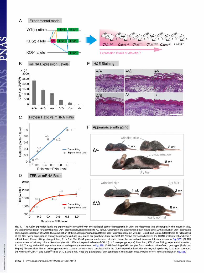
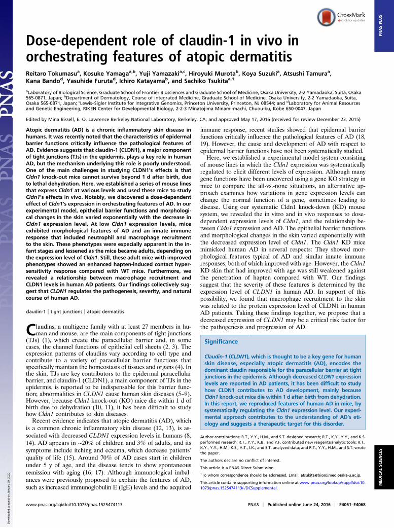
Fig. 1. The Cldn1 expression levels are exponentially associated with the epithelial barrier characteristics in vitro and determine skin phenotypes in the mouse in vivo.(A) Experimental design for analyzing how Cldn1 expression levels contribute toAD in vivo. Generation of a Cldn1 knock-downmouse serieswith six levels ofCldn1 expression(pink, higher expression of Cldn1). The combination of three alleles generated six different Cldn1 expression levels in vivo. Ex1, Exon1; Ex2, Exon2. (B) Real-time RT-PCR analysisof the Cldn1 gene expression in primary keratinocyte cultures (n = 5 mice per genotype). Error bar, SEM. (C) Positive correlation between the CLDN1 protein level and Cldn1mRNA level. Curve fitting, straight line, R2 > 9.4. The Cldn1 protein levels were calculated from the normalized immunoblot data shown in Fig. S2C. (D) TERmeasurement of primary cultured keratinocytes with different expression levels of Cldn1 (n = 5 mice per genotype). Error bars, SEM. Curve fitting, exponential equation,R2 > 9.5. The t1/2 and mRNA expression level of each genotype are shown in Fig. S3B. (E) H&E staining of skin samples from newborn mice of each genotype. (Scale bar:50 μm.) Abnormalities like an orthohyperkeratotic stratum corneum were correlated with the Cldn1 expression level. der, dermis; epi, epidermis; Sc, stratum corneum.(F) Pictures of Cldn1Δ/- and Cldn1Δ/Δ mice at 1, 2, and 8 wk. Note the pathological skin condition in the mutant mice. Pictures of WT mice are shown in Fig. S5B.
E4062 | www.pnas.org/cgi/doi/10.1073/pnas.1525474113 Tokumasu et al.
Dow
nloa
ded
by g
uest
on
Janu
ary
29, 2
020
ResultsEstablishment of an Experimental System of Mouse Lines withSystematically Regulated Cldn1 Expression Levels. To examine therelationship between Cldn1 and skin disease, we first sought toestablish an experimental model by generating mice harboringWT (+), knock-down [KD (Δ)], or knock-out [KO (−)] alleles invarious combinations (Fig. 1A). First, we generated Cldn1 mu-tant (conditional) KO mice (Fig. S1), using a strategy in whichinsertion of the neo gene down-regulates floxed gene expressionlevels (20, 21). We observed that the neo-flox allele led to sig-nificant reductions in the Cldn1 gene expression and proteinlevels (Fig. S2 A and B). By exploiting this phenomenon, weobtained mice with six different Cldn1 expression levels: Cldn1+/+,Cldn1+/Δ, Cldn1+/−, Cldn1Δ/Δ, Cldn1Δ/-, and Cldn1−/−. This seriesof mutant mouse lines served as a sophisticated experimental modelwith which to examine the role of Cldn1.To determine the expression levels of Cldn1 quantitatively, we
prepared primary keratinocyte cultures from newborn mice of eachgenotype. Real-time (RT) PCR confirmed that the Cldn1 mRNAlevels differed according to genotype (Fig. 1B). In addition, im-munoblots of Cldn1 and β-actin showed a similar pattern of Cldn1protein levels for each genotype (Fig. S2 C and D). The resultsrevealed a strong positive correlation between the Cldn1 mRNAand protein levels (Fig. 1C and Fig. S2 A, B, and D).
Exponential Correlations of the Expression Level of Cldn1 with ItsEpithelial Barrier Function and with the Phenotype in Vivo. To ex-amine the relationship between Cldn1 expression levels and thephenotypes of our mouse lines, we first analyzed the paracellular
barrier function of the skin epithelium. For this analysis, wemeasured the transepithelial electrical resistance (TER) andparacellular flux (FLUX) of 4-kDa dextran tracers in primarycultures of keratinocytes prepared from each mouse line. Al-though the Cldn1 mRNA level was proportional to its proteinlevel (Fig. 1C), the TER and FLUX results showed that thebarrier function in keratinocyte changed exponentially as theCldn1 mRNA expression level increased (Fig. 1D and Fig. S3A).The t1/2 values for the plateaus of the TER and FLUX datacorresponded approximately to the mRNA level in Cldn1Δ/-
keratinocytes, which was 10% of the level in WT (Fig. S3B).Furthermore, we observed a difference in permeability in vivo bytracer experiments using a biotinylation reagent (Fig. S3C). Inthe Cldn1Δ/-and Cldn1−/− newborn epidermis, the biotinylationreagent leaked through occludin-positive TJs. Notably, immuno-histochemical analyses revealed that the Cldn1−/− keratinocytes stillcontained TJs (Fig. S4A). In addition, the Claudin-3 and OccludinmRNA levels tended to be correlated with the level of Cldn1mRNA expression whereas other junctional components, includingClaudin-4, Tricellin, ZO-1, ZO-2, E-cadherin, and K5, did not (Fig.S4B). These findings indicated that the differences in the para-cellular barrier functions of keratinocytes were closely related tothe Cldn-1 expression levels in vivo.We next examined the Cldn1-dependent morphological changes
in the epidermis of newborn mice. Previous reports showed thatCldn1 affects the stratum granulosum (SG), from which the stra-tum corneum (SC) is generated, and that Cldn1−/− newborn micehave an abnormal SC (10, 11). In our experimental model, H&Estaining revealed that not only Cdln1−/− but also Cldn1Δ/Δ and
Fig. 2. The Cldn1 KD mice exhibit AD-like morphological features. (A) H&E staining at 2 and 8 wk. (Scale bar: 50 μm.) der, dermis; epi, epidermis. (B) Thicknessof the epidermis in Cldn1+/+, Cldn1Δ/Δ, and Cldn1Δ/- skin at 2 and 8 wk (n = 3). (C) Immunofluorescence micrographs of Claudin-1 (Cldn1, red); E-cadherin(Ecad, green). (Scale bar: 50 μm.) (D) Immunofluorescence micrographs of the differentiation marker keratin-5 (K5, green); integrin-b4 (Itgb4, red); and DAPI(blue). (Scale bar: 50 μm.) epi, epidermis; der, dermis.
Tokumasu et al. PNAS | Published online June 24, 2016 | E4063
MED
ICALSC
IENCE
SPN
ASPL
US
Dow
nloa
ded
by g
uest
on
Janu
ary
29, 2
020
Cldn1Δ/- newborn mice showed abnormal differentiation of theSC, the severity of which was correlated with the epithelial barrierfunction and Cldn1 expression level (Fig. 1E). These findings in-dicated that differences in the paracellular barrier functions andmorphology of the epidermis were closely related to the Cldn-1expression level in vivo.
Cldn1 Expression Level-Dependent Regulation of AD Phenotypes inMice at Different Ages. To understand CLDN1’s role in the age-related changes in human AD, we examined the phenotypes of themodel mice over time, from birth to adulthood. Although theCldn1−/−mice died within 1 d of birth, the other genotypes survivedto adulthood with varying mortality rates. The survival rate at 8 wkwas over 80% for Cldn1+/+, Cldn1+/Δ, Cldn1+/−, and Cldn1Δ/Δ; 4%for Cldn1Δ/-; and 0% for Cldn1−/− mice (Fig. S5A). These results
suggested that the low Cldn1 expression level in Cldn1Δ/- mice wasnear the threshold for lethality.Among the Cldn1 mutant mice, the Cldn1Δ/Δ and Cldn1Δ/- mice
showed age-dependent changes in their skin appearance (Fig. 1Fand Fig. S5 B–E). Cldn1Δ/Δ mice showed wrinkled skin at 1 wk,showed abnormal dry hair at 2 wk, and were nearly normal at 8 wk.The Cldn1Δ/- mice, which usually did not survive beyond weaning,showed more severe skin phenotypes with severe desquamation andwrinkled skin at 2 wk; this phenotype had improved but was stillapparent at 8 wk, and only the Cldn1Δ/- mice, of all of the mutantgenotypes, still exhibited a different severity of skin lesion from WTat 8 wk by dermatitis score (Fig. S6D) (22). Overall, these findingsrevealed that the decreased expression of Cldn1 caused severe skindefects in infancy that improved with age in the mouse, similar tothe natural history of infantile eczema in human AD patients (16).
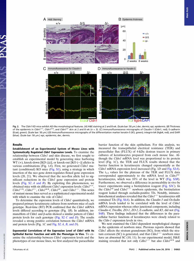
Fig. 3. AD-like phenotypes of Cldn1 KD mice include inflammation. (A) Immunofluorescence micrographs of neutrophil at 2 wk. Keratin-5 (K5, green); Gr-1(red). (Scale bar: 200 μm.) (B) Immunofluorescence micrographs of macrophages at 2 wk. Keratin-5 (K5, green); F4/80 (red). (Scale bar: 100 μm.) (C) Time-dependent changes in the mRNA expression levels of IL-1β, KC, and MCP-1 in the skin (n = 5 mice per genotype). Error bar, SEM. (D) Percentage of earthickness by hapten (DNFB)-induced contact hypersensitivity (CHS). Error bar, SEM. H&E staining of the ear skin after the hapten-induced CHS trial. (Scalebar: 100 μm.) (E) Real-time RT-PCR analysis of IL-12p40, IL-12p35, IFN-γ, IL-10, and IL-4 in the skin (n = 5 mice per genotype). Error bar, SEM. *P < 0.05.
E4064 | www.pnas.org/cgi/doi/10.1073/pnas.1525474113 Tokumasu et al.
Dow
nloa
ded
by g
uest
on
Janu
ary
29, 2
020
Cldn1 Expression Level-Dependent Morphological Features of theEpidermis. We next performed a detailed analysis of the skinmorphology of the Cldn1 mutant mice with aging. H&E-stainedpreparations from 2- and 8-wk-old mice revealed pathologicalcharacteristics of the Cldn1Δ/Δ and Cldn1Δ/- skin, including hy-perkeratosis, acanthosis, and increased numbers of hair follicles(Fig. 2 A and B). Immunofluorescence staining showed thatCldn1 localized only at the SG layer in Cldn1 KD epidermis, andthe TJs in 2-wk-old Cldn1Δ/- epidermis lacked CLDN1 (Fig. 2C).The expression domain of keratin-5 (K5), a basal epidermismarker, was restricted to the most basal layers in the Cldn1+/+
epidermis but expanded to the suprabasal layers in the Cldn1Δ/Δ
and Cldn1Δ/- epidermis (Fig. 2D). In addition, the 2-wk-oldCldn1Δ/- mice showed ectopic proliferation in the subapical basallayer, as indicated by Ki-67 staining (Fig. S6A). In contrast, themutant mice showed the same expression domain for keratin-10(K10), a marker for the stratum spinosum and SG layers, as theCldn1+/+ mice (Fig. S6B). Similar changes in the expressiondomains of the human orthologs of these molecules were foundin AD patients (see Fig. 4A) (Fig. S7 A–D and Dataset S1),consistent with a previous report (23). These findings suggestedthat a decrease in Cldn1 expression causes abnormal skin dif-ferentiation in the infant stages, especially in the proliferation
layers, and that the skin of Cldn1 KD mice mimics the changesseen in human AD tissue.
Correlation of Cldn1 Expression Level with Immunological Features ofthe Epidermis. In human AD patients, both macrophages andneutrophils are recruited to the skin to eliminate pathogens (24,25). In our experimental model, substantial numbers of Gr-1–positive neutrophils were detected in Cldn1Δ/Δ and Cldn1Δ/-
mouse skin samples at 2 wk, and very few were seen at 8 wk (Fig.3A and Fig. S6C). F4/80-positive macrophages were detected inthe Cldn1Δ/Δ mouse epidermis at 2 wk but not at 8 wk, and in theCldn1Δ/- skin at both 2 and 8 wk (Fig. 3B and Fig. S6D). Real-time RT-PCR showed similar levels of inflammation markersbetween Cldn1+/+ and the Cldn1 KD mouse series in newborns(Fig. 3C). By 1 wk, however, the skin of Cldn1Δ/- infants showedhigher expression of the proinflammatory marker IL-β, whichlater decreased in the adults, consistent with the visible neutro-phil infiltration. A similar pattern was observed for the expres-sion of the neutrophil chemoattractant KC in the Cldn1Δ/Δ andCldn1Δ/- skin, and of the macrophage chemoattractant MCP-1 inthe Cldn1Δ/- skin. These phenotypes were apparent in the infantstages and lessened as the mice became adults, depending on the
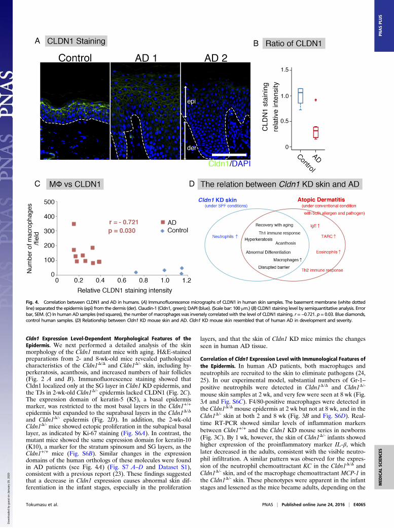
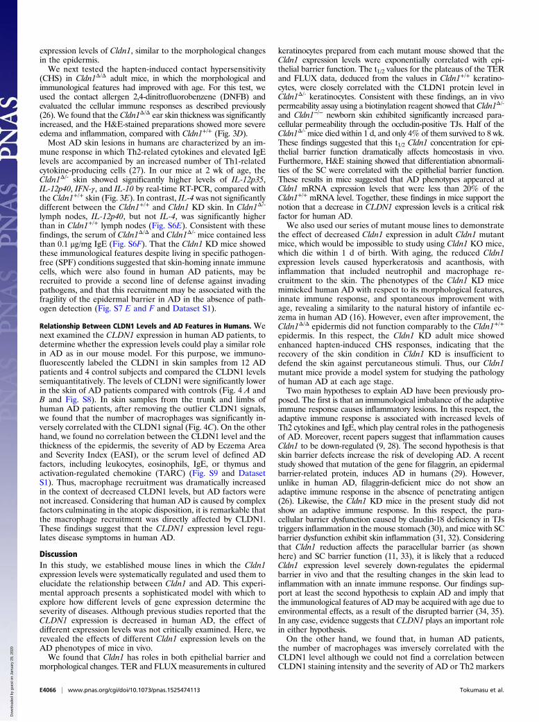
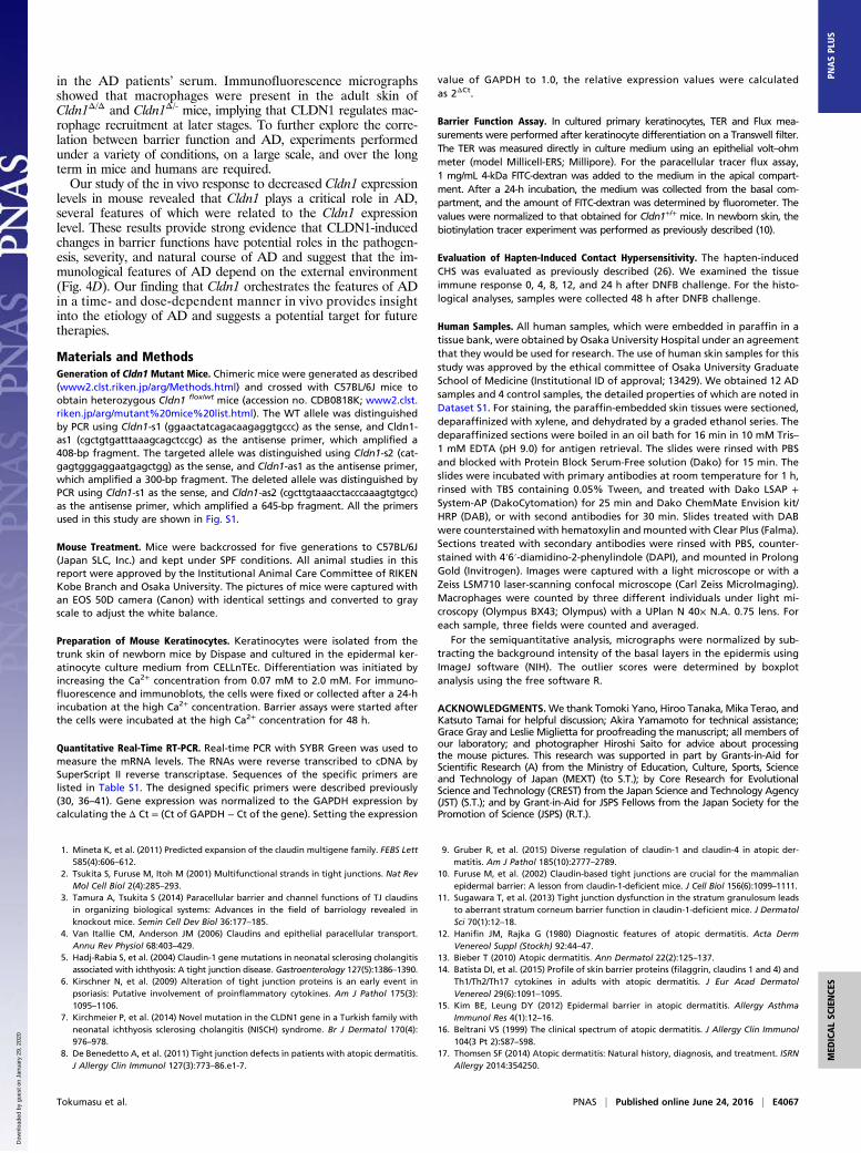
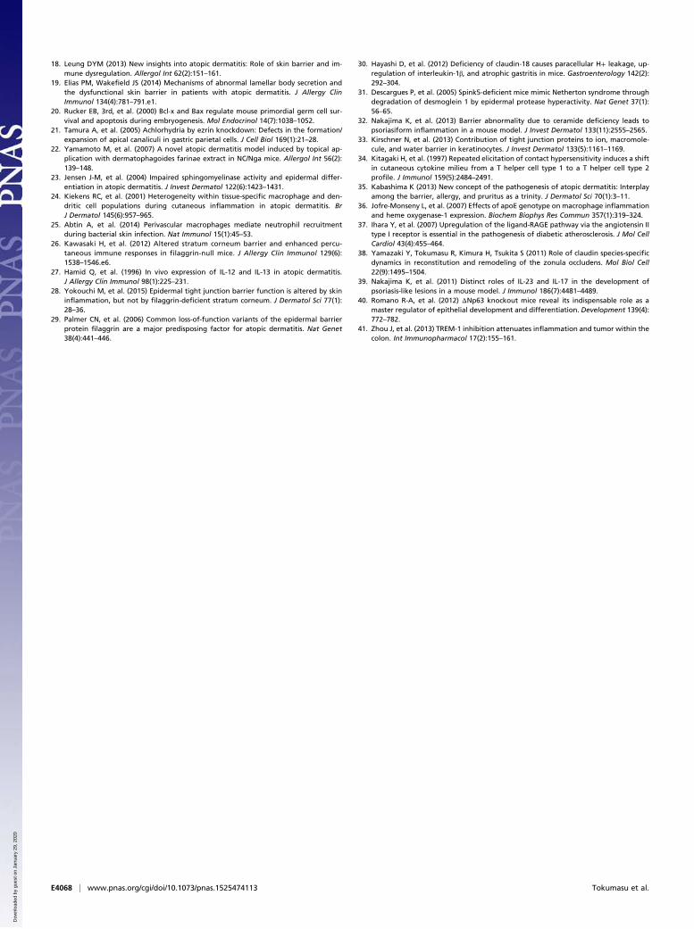
Fig. 4. Correlation between CLDN1 and AD in humans. (A) Immunofluorescence micrographs of CLDN1 in human skin samples. The basement membrane (white dottedline) separated the epidermis (epi) from the dermis (der). Claudin-1 (Cldn1, green); DAPI (blue). (Scale bar: 100 μm.) (B) CLDN1 staining level by semiquantitative analysis. Errorbar, SEM. (C) In humanAD samples (red squares), the number ofmacrophages was inversely correlatedwith the level of CLDN1 staining. r=−0.721. p= 0.03. Blue diamonds,control human samples. (D) Relationship between Cldn1 KD mouse skin and AD. Cldn1 KD mouse skin resembled that of human AD in development and severity.
Tokumasu et al. PNAS | Published online June 24, 2016 | E4065
MED
ICALSC
IENCE
SPN
ASPL
US
Dow
nloa
ded
by g
uest
on
Janu
ary
29, 2
020
expression levels of Cldn1, similar to the morphological changesin the epidermis.We next tested the hapten-induced contact hypersensitivity
(CHS) in Cldn1Δ/Δ adult mice, in which the morphological andimmunological features had improved with age. For this test, weused the contact allergen 2,4-dinitrofluorobenzene (DNFB) andevaluated the cellular immune responses as described previously(26). We found that the Cldn1Δ/Δ ear skin thickness was significantlyincreased, and the H&E-stained preparations showed more severeedema and inflammation, compared with Cldn1+/+ (Fig. 3D).Most AD skin lesions in humans are characterized by an im-
mune response in which Th2-related cytokines and elevated IgElevels are accompanied by an increased number of Th1-relatedcytokine-producing cells (27). In our mice at 2 wk of age, theCldn1Δ/- skin showed significantly higher levels of IL-12p35,IL-12p40, IFN-γ, and IL-10 by real-time RT-PCR, compared withthe Cldn1+/+ skin (Fig. 3E). In contrast, IL-4 was not significantlydifferent between the Cldn1+/+ and Cldn1 KD skin. In Cldn1Δ/-
lymph nodes, IL-12p40, but not IL-4, was significantly higherthan in Cldn1+/+ lymph nodes (Fig. S6E). Consistent with thesefindings, the serum of Cldn1Δ/Δ and Cldn1Δ/- mice contained lessthan 0.1 μg/mg IgE (Fig. S6F). That the Cldn1 KD mice showedthese immunological features despite living in specific pathogen-free (SPF) conditions suggested that skin-homing innate immunecells, which were also found in human AD patients, may berecruited to provide a second line of defense against invadingpathogens, and that this recruitment may be associated with thefragility of the epidermal barrier in AD in the absence of path-ogen detection (Fig. S7 E and F and Dataset S1).
Relationship Between CLDN1 Levels and AD Features in Humans. Wenext examined the CLDN1 expression in human AD patients, todetermine whether the expression levels could play a similar rolein AD as in our mouse model. For this purpose, we immuno-fluorescently labeled the CLDN1 in skin samples from 12 ADpatients and 4 control subjects and compared the CLDN1 levelssemiquantitatively. The levels of CLDN1 were significantly lowerin the skin of AD patients compared with controls (Fig. 4 A andB and Fig. S8). In skin samples from the trunk and limbs ofhuman AD patients, after removing the outlier CLDN1 signals,we found that the number of macrophages was significantly in-versely correlated with the CLDN1 signal (Fig. 4C). On the otherhand, we found no correlation between the CLDN1 level and thethickness of the epidermis, the severity of AD by Eczema Areaand Severity Index (EASI), or the serum level of defined ADfactors, including leukocytes, eosinophils, IgE, or thymus andactivation-regulated chemokine (TARC) (Fig. S9 and DatasetS1). Thus, macrophage recruitment was dramatically increasedin the context of decreased CLDN1 levels, but AD factors werenot increased. Considering that human AD is caused by complexfactors culminating in the atopic disposition, it is remarkable thatthe macrophage recruitment was directly affected by CLDN1.These findings suggest that the CLDN1 expression level regu-lates disease symptoms in human AD.
DiscussionIn this study, we established mouse lines in which the Cldn1expression levels were systematically regulated and used them toelucidate the relationship between Cldn1 and AD. This experi-mental approach presents a sophisticated model with which toexplore how different levels of gene expression determine theseverity of diseases. Although previous studies reported that theCLDN1 expression is decreased in human AD, the effect ofdifferent expression levels was not critically examined. Here, werevealed the effects of different Cldn1 expression levels on theAD phenotypes of mice in vivo.We found that Cldn1 has roles in both epithelial barrier and
morphological changes. TER and FLUXmeasurements in cultured
keratinocytes prepared from each mutant mouse showed that theCldn1 expression levels were exponentially correlated with epi-thelial barrier function. The t1/2 values for the plateaus of the TERand FLUX data, deduced from the values in Cldn1+/+ keratino-cytes, were closely correlated with the CLDN1 protein level inCldn1Δ/- keratinocytes. Consistent with these findings, an in vivopermeability assay using a biotinylation reagent showed that Cldn1Δ/-
and Cldn1−/− newborn skin exhibited significantly increased para-cellular permeability through the occludin-positive TJs. Half of theCldn1Δ/- mice died within 1 d, and only 4% of them survived to 8 wk.These findings suggested that this t1/2 Cldn1 concentration for epi-thelial barrier function dramatically affects homeostasis in vivo.Furthermore, H&E staining showed that differentiation abnormali-ties of the SC were correlated with the epithelial barrier function.These results in mice suggested that AD phenotypes appeared atCldn1 mRNA expression levels that were less than 20% of theCldn1+/+ mRNA level. Together, these findings in mice support thenotion that a decrease in CLDN1 expression levels is a critical riskfactor for human AD.We also used our series of mutant mouse lines to demonstrate
the effect of decreased Cldn1 expression in adult Cldn1 mutantmice, which would be impossible to study using Cldn1 KO mice,which die within 1 d of birth. With aging, the reduced Cldn1expression levels caused hyperkeratosis and acanthosis, withinflammation that included neutrophil and macrophage re-cruitment to the skin. The phenotypes of the Cldn1 KD micemimicked human AD with respect to its morphological features,innate immune response, and spontaneous improvement withage, revealing a similarity to the natural history of infantile ec-zema in human AD (16). However, even after improvement, theCldn1Δ/Δ epidermis did not function comparably to the Cldn1+/+
epidermis. In this respect, the Cldn1 KD adult mice showedenhanced hapten-induced CHS responses, indicating that therecovery of the skin condition in Cldn1 KD is insufficient todefend the skin against percutaneous stimuli. Thus, our Cldn1mutant mice provide a model system for studying the pathologyof human AD at each age stage.Two main hypotheses to explain AD have been previously pro-
posed. The first is that an immunological imbalance of the adaptiveimmune response causes inflammatory lesions. In this respect, theadaptive immune response is associated with increased levels ofTh2 cytokines and IgE, which play central roles in the pathogenesisof AD. Moreover, recent papers suggest that inflammation causesCldn1 to be down-regulated (9, 28). The second hypothesis is thatskin barrier defects increase the risk of developing AD. A recentstudy showed that mutation of the gene for filaggrin, an epidermalbarrier-related protein, induces AD in humans (29). However,unlike in human AD, filaggrin-deficient mice do not show anadaptive immune response in the absence of penetrating antigen(26). Likewise, the Cldn1 KD mice in the present study did notshow an adaptive immune response. In this respect, the para-cellular barrier dysfunction caused by claudin-18 deficiency in TJstriggers inflammation in the mouse stomach (30), and mice with SCbarrier dysfunction exhibit skin inflammation (31, 32). Consideringthat Cldn1 reduction affects the paracellular barrier (as shownhere) and SC barrier function (11, 33), it is likely that a reducedCldn1 expression level severely down-regulates the epidermalbarrier in vivo and that the resulting changes in the skin lead toinflammation with an innate immune response. Our findings sup-port at least the second hypothesis to explain AD and imply thatthe immunological features of ADmay be acquired with age due toenvironmental effects, as a result of the disrupted barrier (34, 35).In any case, evidence suggests that CLDN1 plays an important rolein either hypothesis.On the other hand, we found that, in human AD patients,
the number of macrophages was inversely correlated with theCLDN1 level although we could not find a correlation betweenCLDN1 staining intensity and the severity of AD or Th2 markers
E4066 | www.pnas.org/cgi/doi/10.1073/pnas.1525474113 Tokumasu et al.
Dow
nloa
ded
by g
uest
on
Janu
ary
29, 2
020
in the AD patients’ serum. Immunofluorescence micrographsshowed that macrophages were present in the adult skin ofCldn1Δ/Δ and Cldn1Δ/- mice, implying that CLDN1 regulates mac-rophage recruitment at later stages. To further explore the corre-lation between barrier function and AD, experiments performedunder a variety of conditions, on a large scale, and over the longterm in mice and humans are required.Our study of the in vivo response to decreased Cldn1 expression
levels in mouse revealed that Cldn1 plays a critical role in AD,several features of which were related to the Cldn1 expressionlevel. These results provide strong evidence that CLDN1-inducedchanges in barrier functions have potential roles in the pathogen-esis, severity, and natural course of AD and suggest that the im-munological features of AD depend on the external environment(Fig. 4D). Our finding that Cldn1 orchestrates the features of ADin a time- and dose-dependent manner in vivo provides insightinto the etiology of AD and suggests a potential target for futuretherapies.
Materials and MethodsGeneration of Cldn1 Mutant Mice. Chimeric mice were generated as described(www2.clst.riken.jp/arg/Methods.html) and crossed with C57BL/6J mice toobtain heterozygous Cldn1 flox/wt mice (accession no. CDB0818K; www2.clst.riken.jp/arg/mutant%20mice%20list.html). The WT allele was distinguishedby PCR using Cldn1-s1 (ggaactatcagacaagaggtgccc) as the sense, and Cldn1-as1 (cgctgtgatttaaagcagctccgc) as the antisense primer, which amplified a408-bp fragment. The targeted allele was distinguished using Cldn1-s2 (cat-gagtgggaggaatgagctgg) as the sense, and Cldn1-as1 as the antisense primer,which amplified a 300-bp fragment. The deleted allele was distinguished byPCR using Cldn1-s1 as the sense, and Cldn1-as2 (cgcttgtaaacctacccaaagtgtgcc)as the antisense primer, which amplified a 645-bp fragment. All the primersused in this study are shown in Fig. S1.
Mouse Treatment. Mice were backcrossed for five generations to C57BL/6J(Japan SLC, Inc.) and kept under SPF conditions. All animal studies in thisreport were approved by the Institutional Animal Care Committee of RIKENKobe Branch and Osaka University. The pictures of mice were captured withan EOS 50D camera (Canon) with identical settings and converted to grayscale to adjust the white balance.
Preparation of Mouse Keratinocytes. Keratinocytes were isolated from thetrunk skin of newborn mice by Dispase and cultured in the epidermal ker-atinocyte culture medium from CELLnTEc. Differentiation was initiated byincreasing the Ca2+ concentration from 0.07 mM to 2.0 mM. For immuno-fluorescence and immunoblots, the cells were fixed or collected after a 24-hincubation at the high Ca2+ concentration. Barrier assays were started afterthe cells were incubated at the high Ca2+ concentration for 48 h.
Quantitative Real-Time RT-PCR. Real-time PCR with SYBR Green was used tomeasure the mRNA levels. The RNAs were reverse transcribed to cDNA bySuperScript II reverse transcriptase. Sequences of the specific primers arelisted in Table S1. The designed specific primers were described previously(30, 36–41). Gene expression was normalized to the GAPDH expression bycalculating the Δ Ct = (Ct of GAPDH − Ct of the gene). Setting the expression
value of GAPDH to 1.0, the relative expression values were calculatedas 2ΔCt.
Barrier Function Assay. In cultured primary keratinocytes, TER and Flux mea-surements were performed after keratinocyte differentiation on a Transwell filter.The TER was measured directly in culture medium using an epithelial volt–ohmmeter (model Millicell-ERS; Millipore). For the paracellular tracer flux assay,1 mg/mL 4-kDa FITC-dextran was added to the medium in the apical compart-ment. After a 24-h incubation, the medium was collected from the basal com-partment, and the amount of FITC-dextran was determined by fluorometer. Thevalues were normalized to that obtained for Cldn1+/+ mice. In newborn skin, thebiotinylation tracer experiment was performed as previously described (10).
Evaluation of Hapten-Induced Contact Hypersensitivity. The hapten-inducedCHS was evaluated as previously described (26). We examined the tissueimmune response 0, 4, 8, 12, and 24 h after DNFB challenge. For the histo-logical analyses, samples were collected 48 h after DNFB challenge.
Human Samples. All human samples, which were embedded in paraffin in atissue bank, were obtained by Osaka University Hospital under an agreementthat they would be used for research. The use of human skin samples for thisstudy was approved by the ethical committee of Osaka University GraduateSchool of Medicine (Institutional ID of approval; 13429). We obtained 12 ADsamples and 4 control samples, the detailed properties of which are noted inDataset S1. For staining, the paraffin-embedded skin tissues were sectioned,deparaffinized with xylene, and dehydrated by a graded ethanol series. Thedeparaffinized sections were boiled in an oil bath for 16 min in 10 mM Tris–1 mM EDTA (pH 9.0) for antigen retrieval. The slides were rinsed with PBSand blocked with Protein Block Serum-Free solution (Dako) for 15 min. Theslides were incubated with primary antibodies at room temperature for 1 h,rinsed with TBS containing 0.05% Tween, and treated with Dako LSAP +System-AP (DakoCytomation) for 25 min and Dako ChemMate Envision kit/HRP (DAB), or with second antibodies for 30 min. Slides treated with DABwere counterstained with hematoxylin and mounted with Clear Plus (Falma).Sections treated with secondary antibodies were rinsed with PBS, counter-stained with 4′6′-diamidino-2-phenylindole (DAPI), and mounted in ProlongGold (Invitrogen). Images were captured with a light microscope or with aZeiss LSM710 laser-scanning confocal microscope (Carl Zeiss MicroImaging).Macrophages were counted by three different individuals under light mi-croscopy (Olympus BX43; Olympus) with a UPlan N 40× N.A. 0.75 lens. Foreach sample, three fields were counted and averaged.
For the semiquantitative analysis, micrographs were normalized by sub-tracting the background intensity of the basal layers in the epidermis usingImageJ software (NIH). The outlier scores were determined by boxplotanalysis using the free software R.
ACKNOWLEDGMENTS.We thank Tomoki Yano, Hiroo Tanaka, Mika Terao, andKatsuto Tamai for helpful discussion; Akira Yamamoto for technical assistance;Grace Gray and Leslie Miglietta for proofreading the manuscript; all members ofour laboratory; and photographer Hiroshi Saito for advice about processingthe mouse pictures. This research was supported in part by Grants-in-Aid forScientific Research (A) from the Ministry of Education, Culture, Sports, Scienceand Technology of Japan (MEXT) (to S.T.); by Core Research for EvolutionalScience and Technology (CREST) from the Japan Science and Technology Agency(JST) (S.T.); and by Grant-in-Aid for JSPS Fellows from the Japan Society for thePromotion of Science (JSPS) (R.T.).
1. Mineta K, et al. (2011) Predicted expansion of the claudin multigene family. FEBS Lett585(4):606–612.
2. Tsukita S, Furuse M, Itoh M (2001) Multifunctional strands in tight junctions. Nat RevMol Cell Biol 2(4):285–293.
3. Tamura A, Tsukita S (2014) Paracellular barrier and channel functions of TJ claudinsin organizing biological systems: Advances in the field of barriology revealed inknockout mice. Semin Cell Dev Biol 36:177–185.
4. Van Itallie CM, Anderson JM (2006) Claudins and epithelial paracellular transport.Annu Rev Physiol 68:403–429.
5. Hadj-Rabia S, et al. (2004) Claudin-1 gene mutations in neonatal sclerosing cholangitisassociated with ichthyosis: A tight junction disease. Gastroenterology 127(5):1386–1390.
6. Kirschner N, et al. (2009) Alteration of tight junction proteins is an early event inpsoriasis: Putative involvement of proinflammatory cytokines. Am J Pathol 175(3):1095–1106.
7. Kirchmeier P, et al. (2014) Novel mutation in the CLDN1 gene in a Turkish family withneonatal ichthyosis sclerosing cholangitis (NISCH) syndrome. Br J Dermatol 170(4):976–978.
8. De Benedetto A, et al. (2011) Tight junction defects in patients with atopic dermatitis.J Allergy Clin Immunol 127(3):773–86.e1-7.
9. Gruber R, et al. (2015) Diverse regulation of claudin-1 and claudin-4 in atopic der-matitis. Am J Pathol 185(10):2777–2789.
10. Furuse M, et al. (2002) Claudin-based tight junctions are crucial for the mammalianepidermal barrier: A lesson from claudin-1-deficient mice. J Cell Biol 156(6):1099–1111.
11. Sugawara T, et al. (2013) Tight junction dysfunction in the stratum granulosum leadsto aberrant stratum corneum barrier function in claudin-1-deficient mice. J DermatolSci 70(1):12–18.
12. Hanifin JM, Rajka G (1980) Diagnostic features of atopic dermatitis. Acta DermVenereol Suppl (Stockh) 92:44–47.
13. Bieber T (2010) Atopic dermatitis. Ann Dermatol 22(2):125–137.14. Batista DI, et al. (2015) Profile of skin barrier proteins (filaggrin, claudins 1 and 4) and
Th1/Th2/Th17 cytokines in adults with atopic dermatitis. J Eur Acad DermatolVenereol 29(6):1091–1095.
15. Kim BE, Leung DY (2012) Epidermal barrier in atopic dermatitis. Allergy AsthmaImmunol Res 4(1):12–16.
16. Beltrani VS (1999) The clinical spectrum of atopic dermatitis. J Allergy Clin Immunol104(3 Pt 2):S87–S98.
17. Thomsen SF (2014) Atopic dermatitis: Natural history, diagnosis, and treatment. ISRNAllergy 2014:354250.
Tokumasu et al. PNAS | Published online June 24, 2016 | E4067
MED
ICALSC
IENCE
SPN
ASPL
US
Dow
nloa
ded
by g
uest
on
Janu
ary
29, 2
020
18. Leung DYM (2013) New insights into atopic dermatitis: Role of skin barrier and im-mune dysregulation. Allergol Int 62(2):151–161.
19. Elias PM, Wakefield JS (2014) Mechanisms of abnormal lamellar body secretion andthe dysfunctional skin barrier in patients with atopic dermatitis. J Allergy ClinImmunol 134(4):781–791.e1.
20. Rucker EB, 3rd, et al. (2000) Bcl-x and Bax regulate mouse primordial germ cell sur-vival and apoptosis during embryogenesis. Mol Endocrinol 14(7):1038–1052.
21. Tamura A, et al. (2005) Achlorhydria by ezrin knockdown: Defects in the formation/expansion of apical canaliculi in gastric parietal cells. J Cell Biol 169(1):21–28.
22. Yamamoto M, et al. (2007) A novel atopic dermatitis model induced by topical ap-plication with dermatophagoides farinae extract in NC/Nga mice. Allergol Int 56(2):139–148.
23. Jensen J-M, et al. (2004) Impaired sphingomyelinase activity and epidermal differ-entiation in atopic dermatitis. J Invest Dermatol 122(6):1423–1431.
24. Kiekens RC, et al. (2001) Heterogeneity within tissue-specific macrophage and den-dritic cell populations during cutaneous inflammation in atopic dermatitis. BrJ Dermatol 145(6):957–965.
25. Abtin A, et al. (2014) Perivascular macrophages mediate neutrophil recruitmentduring bacterial skin infection. Nat Immunol 15(1):45–53.
26. Kawasaki H, et al. (2012) Altered stratum corneum barrier and enhanced percu-taneous immune responses in filaggrin-null mice. J Allergy Clin Immunol 129(6):1538–1546.e6.
27. Hamid Q, et al. (1996) In vivo expression of IL-12 and IL-13 in atopic dermatitis.J Allergy Clin Immunol 98(1):225–231.
28. Yokouchi M, et al. (2015) Epidermal tight junction barrier function is altered by skininflammation, but not by filaggrin-deficient stratum corneum. J Dermatol Sci 77(1):28–36.
29. Palmer CN, et al. (2006) Common loss-of-function variants of the epidermal barrierprotein filaggrin are a major predisposing factor for atopic dermatitis. Nat Genet38(4):441–446.
30. Hayashi D, et al. (2012) Deficiency of claudin-18 causes paracellular H+ leakage, up-regulation of interleukin-1β, and atrophic gastritis in mice. Gastroenterology 142(2):292–304.
31. Descargues P, et al. (2005) Spink5-deficient mice mimic Netherton syndrome throughdegradation of desmoglein 1 by epidermal protease hyperactivity. Nat Genet 37(1):56–65.
32. Nakajima K, et al. (2013) Barrier abnormality due to ceramide deficiency leads topsoriasiform inflammation in a mouse model. J Invest Dermatol 133(11):2555–2565.
33. Kirschner N, et al. (2013) Contribution of tight junction proteins to ion, macromole-cule, and water barrier in keratinocytes. J Invest Dermatol 133(5):1161–1169.
34. Kitagaki H, et al. (1997) Repeated elicitation of contact hypersensitivity induces a shiftin cutaneous cytokine milieu from a T helper cell type 1 to a T helper cell type 2profile. J Immunol 159(5):2484–2491.
35. Kabashima K (2013) New concept of the pathogenesis of atopic dermatitis: Interplayamong the barrier, allergy, and pruritus as a trinity. J Dermatol Sci 70(1):3–11.
36. Jofre-Monseny L, et al. (2007) Effects of apoE genotype on macrophage inflammationand heme oxygenase-1 expression. Biochem Biophys Res Commun 357(1):319–324.
37. Ihara Y, et al. (2007) Upregulation of the ligand-RAGE pathway via the angiotensin IItype I receptor is essential in the pathogenesis of diabetic atherosclerosis. J Mol CellCardiol 43(4):455–464.
38. Yamazaki Y, Tokumasu R, Kimura H, Tsukita S (2011) Role of claudin species-specificdynamics in reconstitution and remodeling of the zonula occludens. Mol Biol Cell22(9):1495–1504.
39. Nakajima K, et al. (2011) Distinct roles of IL-23 and IL-17 in the development ofpsoriasis-like lesions in a mouse model. J Immunol 186(7):4481–4489.
40. Romano R-A, et al. (2012) ΔNp63 knockout mice reveal its indispensable role as amaster regulator of epithelial development and differentiation. Development 139(4):772–782.
41. Zhou J, et al. (2013) TREM-1 inhibition attenuates inflammation and tumor within thecolon. Int Immunopharmacol 17(2):155–161.
E4068 | www.pnas.org/cgi/doi/10.1073/pnas.1525474113 Tokumasu et al.
Dow
nloa
ded
by g
uest
on
Janu
ary
29, 2
020


![- Fernando Claudin, Marx, Engels y La Revolucion de 1848[1]](https://img.pdfslide.tips/doc/110x75/55cf9d45550346d033ace9ff/-fernando-claudin-marx-engels-y-la-revolucion-de-18481.jpg)