Embed Size (px)
Citation preview
Linköping University Medical Dissertations No. 1036
Dynamic knee stability after
anterior cruciate ligament injury
Emphasis on rehabilitation
Sofi Tagesson
Division of Physiotherapy Department of Medical and Health Sciences
Linköping University, Sweden
Linköping 2008
© Sofi Tagesson, 2008 [email protected] http://www.imh.liu.se Electronic publication of the thesis: http://urn.kb.se/resolve?urn=urn:nbn:se:liu:diva‐10498 Published articles have been reprinted with the permission of the copyright holder. Paper I reprinted with permission of the Scandinavian Journal of Medicine & Science in Sports, Blackwell Publishing. Paper II reprinted with permission of the Journal of Strength and Conditioning Research, Allen Press Publishing Services. Paper III reprinted with permission of the American Journal of Sports Medicine, Sage Publications, Inc. Printed in Sweden by LiU‐Tryck, Linköping, Sweden, 2008 ISBN 978‐91‐85895‐05‐2 ISSN 0345‐0082

To my family


Contents
CONTENTS
ABSTRACT .................................................................................................................. 1
LIST OF PAPERS ........................................................................................................ 3
DESCRIPTION OF CONTRIBUTION................................................................... 4
ABBREVIATIONS...................................................................................................... 5
DEFINITIONS............................................................................................................. 6
INTRODUCTION....................................................................................................... 8
Anatomy and function of the knee joint........................................................... 8
Mechanical properties of ligaments............................................................... 8
Anterior cruciate ligament .............................................................................. 9
ACL injury............................................................................................................. 10
Incidence and injury mechanisms................................................................ 10
Consequences of the injury ........................................................................... 11
Static knee stability ............................................................................................. 12
Dynamic knee stability....................................................................................... 12
Proprioception and neuromuscular control ............................................... 13
Muscle function............................................................................................... 14
Treatment of ACL injury.................................................................................... 15
ACL reconstruction ........................................................................................ 15
Rehabilitation after ACL injury or reconstruction..................................... 16
Assessment of knee joint function................................................................... 21
Knee joint stability.......................................................................................... 21
Neuromuscular function ............................................................................... 22
Subjective knee function................................................................................ 23
Contents
Rationale for the thesis ....................................................................................... 23
AIMS OF THE THESIS............................................................................................ 24
General aim........................................................................................................... 24
Specific aims......................................................................................................... 24
MATERIALS AND METHODS............................................................................. 26
Design .................................................................................................................... 26
Overview of the studies................................................................................. 26
Subjects.................................................................................................................. 27
Study I and II................................................................................................... 28
Study III............................................................................................................ 28
Study IV ........................................................................................................... 29
Equipment............................................................................................................. 29
Electrogoniometer .......................................................................................... 29
Electromyography .......................................................................................... 31
Isokinetic device.............................................................................................. 32
Force measurements....................................................................................... 32
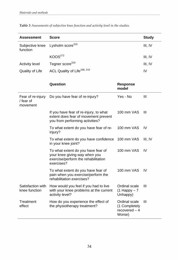
Questionnaires about subjective knee function and activity level.......... 33
Assessments .......................................................................................................... 35
Static tibial translation (study I, III, IV) ....................................................... 35
Dynamic tibial translation and electromyography (study I, III, IV) ....... 35
Muscle strength (study II, III, IV)................................................................. 38
Functional performance (study III).............................................................. 40
Knee joint circumference (study III, IV) ...................................................... 40
Passive range of motion (study III, IV)........................................................ 41
Data analysis......................................................................................................... 41
Tibial translation............................................................................................. 41
Electromyography .......................................................................................... 42
Muscle strength............................................................................................... 42
Statistical analyses............................................................................................... 42
Ethical considerations......................................................................................... 44
RESULTS .................................................................................................................... 45

Contents
Tibial translation and muscle activation after an exercise session in uninjured individuals (study I) ........................................................................ 45
Static tibial translation ................................................................................... 45
Dynamic tibial translation............................................................................. 45
Electromyography .......................................................................................... 46
The procedure for the establishment of one repetition maximum (study II) ............................................................................................................................. 47
Reliability of squat on one leg (study II) ........................................................ 47
Reliability of seated knee extension on one leg (study II).......................... 47
Effects of rehabilitation programs with quadriceps strengthening in closed or open kinetic chain in patients with ACL deficiency (study III)49
Static tibial translation ................................................................................... 49
Dynamic tibial translation............................................................................. 49
Electromyography .......................................................................................... 50
Muscle strength............................................................................................... 51
Functional performance................................................................................. 51
Swelling and passive range of motion ........................................................ 52
Follow‐up two to three years after the injury............................................. 52
Tibial translation and muscle activation during rehabilitation exercises five weeks after ACL reconstruction (study IV)............................................ 53
Static tibial translation ................................................................................... 53
Dynamic tibial translation............................................................................. 53
Electromyography .......................................................................................... 53
The ACL reconstructed leg compared to the uninjured leg (study IV) .... 55
Static tibial translation ................................................................................... 55
Dynamic tibial translation............................................................................. 55
Electromyography .......................................................................................... 55
The ACL reconstructed leg compared to the ACL deficient leg (study IV)56
Static tibial translation ................................................................................... 56
Dynamic tibial translation............................................................................. 56
Electromyography .......................................................................................... 56
Subjective knee function................................................................................ 57
DISCUSSION............................................................................................................ 58
Contents
Main findings....................................................................................................... 58
Dynamic knee stability during and after exercises in closed and open kinetic chain..................................................................................................... 59
Altered dynamic stability .............................................................................. 61
Muscle activation during and after exercises in closed and open kinetic chain.................................................................................................................. 64
Muscle function after rehabilitation............................................................. 66
Strength training and dosage of rehabilitation programs ........................ 68
Non‐surgical treatment or ACL reconstruction......................................... 69
Methodological considerations......................................................................... 70
Tibial translation............................................................................................. 70
Electromyography .......................................................................................... 72
Measurements of muscle strength and functional performance............. 74
Measurements on uninjured individuals and patients with ACL injury75
Selection of leg for assessment...................................................................... 76
Using the uninvolved leg as a reference ..................................................... 76
Design of the randomised clinical study (III) ............................................. 77
Compliance to the rehabilitation programs................................................ 77
Contribution to research..................................................................................... 78
Clinical implications........................................................................................... 78
Future research ..................................................................................................... 79
CONCLUSIONS ....................................................................................................... 80
SAMMANFATTNING ............................................................................................ 82
ACKNOWLEDGEMENTS...................................................................................... 84
REFERENCES ............................................................................................................ 86
Abstract
1
ABSTRACT
Anterior cruciate ligament injury leads to increased sagittal tibial translation, and perceptions of instability and low confidence in the knee joint are common. Many patients have remaining problems despite treatment and are forced to lower their activity level and prematurely end their career in sports. The effect of ACL reconstruction and/or rehabilitation on dynamic knee stability is not completely understood. The overall aim of this thesis was to study the dynamic knee stability during and after rehabilitation in individuals with ACL injury. More specific aims were 1) to elaborate an evaluation method for muscle strength, 2) to evaluate the effect of exercises in closed and open kinetic chain, and 3) to evaluate dynamic knee stability in patients with ACL deficiency or ACL reconstruction. Sagittal tibial translation and knee flexion angle were measured using the CA‐4000 computerised goniometer linkage. Muscle activation was registered with electromyography. The intra‐ and inter‐rater reliability of 1 repetition maximum (RM) of seated knee extension was clinically acceptable. The inter‐rater reliability of 1RM of squat was also acceptable, but the intra‐rater reliability was lower. The systematic procedure for the establishment of 1RM that was developed can be recommended for use in the clinic. One specific exercise session including cycling and a maximum number of knee extensions and heel raises did not influence static or dynamic sagittal tibial translation in uninjured individuals. A comprehensive rehabilitation program with isolated quadriceps training in OKC led to significantly greater isokinetic quadriceps strength compared to CKC rehabilitation in patients with ACL deficiency. Hamstring strength, static and dynamic translation, and functional outcome were similar between groups. Five weeks after ACL reconstruction, seated knee extension produced more anterior tibial translation compared to the straight leg raise and standing on one leg. All exercises produced less or equal amount of anterior tibial translation as the 90N Lachman test. Five weeks after the ACL reconstruction the static and dynamic tibial translation in the ACL reconstructed knee did not differ from the tibial translation on the uninjured leg. Patients in the early phase after ACL injury or ACL reconstruction used a joint stiffening strategy including a reduced peak
Abstract
2
knee extension angle during gait and increased hamstring activation during activity, which reduces the dynamic tibial translation. Patients with ACL deficiency that completed a four months rehabilitation program used a movement pattern that was more close to normal.
List of papers
3
LIST OF PAPERS
This thesis is based on the following papers, which are referred to in the text by their Roman numerals: I. Sofi Tagesson, Birgitta Öberg, Joanna Kvist. Passive and dynamic
translation in the knee is not influenced by knee exercises in healthy individuals. Scandinavian Journal of Medicine & Science in Sports 2005; 15 (3) 139‐147.
II. Sofi Tagesson, Joanna Kvist. Intra‐ and interrater reliability of the
establishment of one repetition maximum on squat and seated knee extension. Journal of Strength and Conditioning Research 2007; 21 (3) 801‐807.
III. Sofi Tagesson, Birgitta Öberg, Lars Good, Joanna Kvist. A
comprehensive rehabilitation program with quadriceps strengthening in closed versus open kinetic chain in patients with anterior cruciate ligament deficiency: a randomized clinical trial evaluating dynamic tibial translation and muscle function. American Journal of Sports Medicine 2008; 36 (2) 298‐307.
IV. Sofi Tagesson, Birgitta Öberg, Joanna Kvist. Tibial translation and
muscle activation during rehabilitation exercises 5 weeks after anterior cruciate ligament reconstruction. Submitted 2007.
Decription of contribution
4
DESCRIPTION OF CONTRIBUTION
Paper I Study design Joanna Kvist, Sofi Tagesson, Jan Gillquist Data collection Sofi Tagesson Data reduction Sofi Tagesson Data analysis Sofi Tagesson, Joanna Kvist, Birgitta Öberg Manuscript writing Sofi Tagesson Manuskript revision Joanna Kvist, Birgitta Öberg Paper II Study design Sofi Tagesson, Joanna Kvist Data collection Sofi Tagesson, Per Axelsson, Bettina Hillborg, Marie
Rydén Data reduction Sofi Tagesson Data analysis Sofi Tagesson Manuscript writing Sofi Tagesson Manuscript revision Joanna Kvist, Birgitta Öberg Paper III Study design Sofi Tagesson, Joanna Kvist, Birgitta Öberg, Lars Good Data collection Sofi Tagesson Data reduction Sofi Tagesson Data analysis Sofi Tagesson Manuscript writing Sofi Tagesson Manuscript revision Joanna Kvist, Birgitta Öberg, Lars Good Paper IV Study design Sofi Tagesson, Joanna Kvist, Birgitta Öberg Data collection Sofi Tagesson Data reduction Sofi Tagesson Data analysis Sofi Tagesson Manuscript writing Sofi Tagesson Manuscript revision Joanna Kvist, Birgitta Öberg
Abbreviations
5
ABBREVIATIONS
ACL Anterior cruciate ligament
CKC Closed kinetic chain
EMG Electromyography
ICC Intra class correlation coefficient
KOOS Knee injury and Osteoarthritis Outcome Score
M Mean
MIVC Maximal isometric voluntary contraction
OKC Open kinetic chain
RCT Randomised Controlled Trial
RM Repetition maximum
ROM Range of motion
SD Standard Deviation
VAS Visual Analogue Scale
Definitions
6
DEFINITIONS
ACL deficiency The state or condition of lacking a substance, quality, or characteristic essential for completeness;234 i.e., absence of the anterior cruciate ligament
ACL graft The biological substitute used for reconstruction of a ruptured anterior cruciate ligament
Closed kinetic chain exercise
Exercise that is modelled as a closed linkage where a movement in one joint simultaneously produces movements in other joints of the extremity243, 339
Compliance A patient’s collaboration with treatment directions
Concentric muscle action
When a muscle shortens while producing force155
Dynamic knee stability
The ability of the knee joint to remain stable during physical activity.342 Synonymous with functional stability
Dynamic tibial translation
Anterior tibial translation during activity
Eccentric muscle action
When a muscle lengthens while producing force155
Giving way The knee buckles or it feels as if it would not hold the patient’s weight47
Instability A condition of a joint characterized by an abnormal increased range of motion due to injury to the ligaments, capsule, menisci, cartilage or bone234
Laxity Slackness or lack of tension (a characteristic of a ligament) and looseness, referring to a normal or abnormal range of motion of a joint234
Definitions
7
Muscle strength The maximal force a muscle or muscle group can
generate at a specified velocity155
Neuromuscular control
The ability to produce controlled movement through coordinated muscle activity342
Open kinetic chain exercise
Exercises that isolate one link of the kinetic chain and where the distal segment is free to move243, 339
Reliability Consistency of measurements and the absence of random and systematic measurement error12, 313
Repetition maximum
The heaviest resistance that can be lifted for one complete repetition of an exercise76, 155
Static tibial translation
Total sagittal tibial translation during the Lachman test
Strain Change in the dimension of a material divided by its original size176
Tibial position Relative location of tibia with respect to femur
Torque Moment of force (term generally applied to rotation of shaft) (unit Nm)176
Translation A type of motion or displacement of a rigid body in which all lines attached to it remain parallel to their original orientation176, 234
Introduction
8
INTRODUCTION
Anatomy and function of the knee joint The knee joint has six degrees of freedom, three rotational and three translational.103 It is a complex joint that provides both stability, to allow weight bearing, and mobility. The knee joint obtains just modest stability from the bones due to incongruity of the tibia and femoral condyles. The menisci, on the other hand, provide stability through improved joint congruity because of their shape, orientation, and functional properties.295 In addition, the ligaments, capsule, and the muscolutendinous soft tissue that surrounds the knee interact with complex gliding and rolling movements in the joint to maintain functional stability.342 The knee ligaments guide the skeletal segments during motion and are the primary restraints to knee joint translations during passive loads.20 There is great range of movement (ROM) in flexion and extension in the sagittal plane, and a small amount of rotation in the transversal plane, especially when the knee is flexed and the foot is not fixed to a surface. Movements in the knee joint are a combination of rolling and gliding. Beginning with full extension in a weight bearing position, the femoral condyles only roll, and with increased flexion the sliding movement becomes increasingly more significant. At the end of flexion, the condyles only slide.103, 242 The tibial plateaus slopes approximately 9° posterior.103, 204, 229 During weight‐bearing, a vertical compression causes the tibia to translate anteriorly.67, 170, 295, 326 At activities such as running and cutting the knee is flexed, which means that the tibial plateau is almost parallel with the weight‐bearing surface.69
Mechanical properties of ligaments
Ligaments are viscoelastic structures and have a combination of elastic and viscous qualities. An elastic material returns to its original shape after it has
Introduction
9
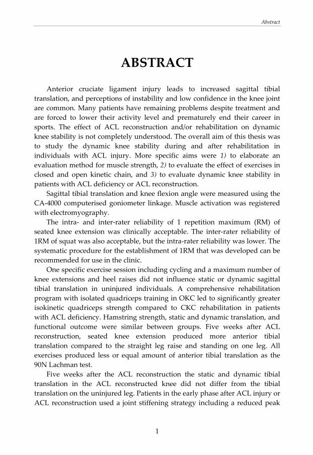
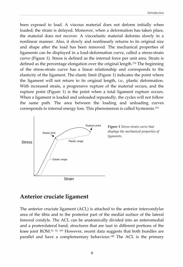
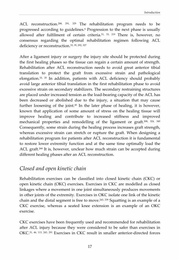
been exposed to load. A viscous material does not deform initially when loaded; the strain is delayed. Moreover, when a deformation has taken place, the material does not recover. A viscoelastic material deforms slowly in a nonlinear manner. Also, it slowly and nonlinearly returns to its original size and shape after the load has been removed. The mechanical properties of ligaments can be displayed in a load‐deformation curve, called a stress‐strain curve (Figure 1). Stress is defined as the internal force per unit area. Strain is defined as the percentage elongation over the original length.176 The beginning of the stress‐strain curve has a linear relationship and corresponds to the elasticity of the ligament. The elastic limit (Figure 1) indicates the point where the ligament will not return to its original length, i.e., plastic deformation. With increased strain, a progressive rupture of the material occurs, and the rupture point (Figure 1) is the point when a total ligament rupture occurs. When a ligament is loaded and unloaded repeatedly, the cycles will not follow the same path. The area between the loading and unloading curves corresponds to internal energy loss. This phenomenon is called hysteresis.176
Figure 1 Stress‐strain curve that displays the mechanical properties of ligaments.
Anterior cruciate ligament
The anterior cruciate ligament (ACL) is attached to the anterior intercondylar area of the tibia and to the posterior part of the medial surface of the lateral femoral condyle. The ACL can be anatomically divided into an anteromedial and a posterolateral band, structures that are taut in different portions of the knee joint ROM.53, 92, 103 However, recent data suggests that both bundles are parallel and have a complementary behaviour.148 The ACL is the primary
Stress
Strain
Elastic limit
Rupture point
Elastic range
Plastic range
Introduction
10
restraint to anterior tibial translation, and it provides an average of 86% of the total resisting force. Additionally, it is a secondary restraint to rotation, especially internal rotation of tibia.45 Accordingly, the strain in the ACL is increased when the tibia is exposed to anterior force. After 15 to 25% of elongation of the ACL, in relation to its original length, plastic deformation occurs.173 The average strain in the anteromedial bundle of the ACL during 90N Lachman test is 2.16 ± 1.1% and during 150N it is 3.4 ± 1.8%.82 The failure load of an ACL is reported to be about 2160 ± 157N in young individuals, and ultimate load and energy absorbed decrease significantly with age.347, 348 Another study states that load at failure is about 1818 ± 699N in males and about 1266 ± 527N in females.52 Moreover, the strain at failure was 30 ± 6% in males and 27 ± 8% in females.52 During daily activities and commonly prescribed rehabilitation exercises, the ACL is loaded only to about 20% of its failure capacity.31, 32, 124
ACL injury
Incidence and injury mechanisms
Anterior cruciate ligament injury is the most common total ligament rupture in the knee.34, 208 The injury is relatively uncommon in the general population; the yearly incidence is 0.3 to 0.81 per 1000 inhabitants.90, 227 The incidence is considerably greater among athletes.104 An estimated 80000 ACL injuries occur annually in the United States.104 In Sweden, about 6000 ACL injuries occur yearly, and approximately 3000 ACL reconstructions are performed.1 Soccer is the most common activity that results in injury,90 and ACL injuries represent 43% of all soccer related injuries.275 Also basketball, handball, football, alpine skiing and gymnastics are common activities that result in injury.41, 127, 205, 220, 221, 256 Rates of ACL injury in women’s soccer and basketball have been reported to be 0.32 and 0.28 injuries per 1000 athlete exposures respectively. Correspondingly, rates of ACL injury for men are 0.11 and 0.08 injuries per 1000 athlete exposures respectively.205 More males than females sustain an ACL injury due to the greater absolute number of male participants in sport activities.104 However, the risk of sustaining an ACL injury is reported to be two to eight times higher among
Introduction
11
female athletes compared to their male counterpart. 4, 8‐10, 104, 117, 139, 221, 275 The females also get injured at a younger age than men.275 A recent study found small gender differences in the overall risk of sustaining an ACL tear although gender differences in injury rates were found when specific sports were compared.218 Anterior cruciate ligament injuries are thought to occur due to unsuccessful postural adjustments and abnormal dynamic loading, i.e., inter‐segmental loads in the knee joint.105 The majority of the ACL injuries (about 70%) occur in a non‐contact situation.41, 104 Non‐contact injuries often occur with the knee close to extension during a sudden deceleration or landing motion. Contact injuries are frequently the result a contact blow to the lateral aspect of the leg or knee, a motion that causes a valgus collapse.41 The injury mechanism for ACL injuries in female team handball is reported to be a forceful valgus collapse with the knee close to full extension combined with external or internal tibial rotation.238 Associated injuries in the capsule, ligament or menisci are common, and most patients have complex injuries.90, 154, 239
Consequences of the injury
An ACL rupture leads to increased laxity in the knee.45, 63, 154, 184, 232, 284 In the ACL deficient knee, anterior tibial translation is limited by secondary restraining structures, such as the posterior joint capsule, the collateral ligaments, and the menisci.45, 201 The injury leads to loss of mechanoreceptor feedback and loss of reflex muscular contractions.19‐21, 25, 26, 144, 145, 162, 305 Furthermore, the ACL injury often results in perceptions of instability in the knee joint.47, 236 Giving way is common, and this is described as the knee buckles or a feeling as if the knee would not hold the patient’s weight.47 The patients frequently describe low confidence in their knee joint. Individuals are often forced to lower their activity level and prematurely end their career in sports.96, 163, 169, 236, 275, 303 An ACL injury predisposes the knee to subsequent injuries332 and the early onset of osteoarthritis.95, 185, 186, 223, 273, 346 About 50% of patients with ACL injury have osteoarthritis with associated pain and functional impairment 10 to 20 years after the injury. Considering that a majority of individuals with acute ACL injury are younger than 30 years and many are younger than 20 years,104, 303 ACL injuries are responsible for a large number of patients between 30 and
Introduction
12
50 years with early‐onset osteoarthritis.185 The joint instability280 and muscle weakness115 are thought to lead to progressive arthritic changes. Some patients use a stabilization strategy after the injury that stiffens the knee joint, and that may lead to changed loading on the joint surfaces and excessive joint contact forces. This could damage articular structures.279 Moreover, intra‐articular pathogenic processes are initiated at time of the injury, and this together with the changed loading pattern on the joint surfaces is thought to contribute to the osteoarthritis.185, 283 In addition, associated injuries, especially menisci injury, probably increase the risk of future osteoarthritis.5, 95, 147, 185 An ACL reconstruction does not seem to prevent osteoarthritis.60, 185
Static knee stability The knee anatomy includes a supportive system of osseous, contractile, and non‐contractile structures, which together contributes to stability in the joint.103 In a passive situation, for example during a static laxity test, the bones and other non‐contractile structures provide joint stability. An ACL injury leads to decreased static stability due to loss of an important stabilizer.45, 154, 232 The static tibial translation is frequently assessed when evaluating treatment after ACL injury.46, 206, 249, 287 However, static tibial translation does not correlate with functional outcome in patients with ACL deficiency71, 134, 246, 301 or ACL reconstruction121, 287 or with quadriceps or hamstring muscle strength in patients with ACL deficiency.246 Furthermore, the static tibial translation does not correlate with the dynamic tibial translation, i.e. translation during activity.166
Dynamic knee stability Dynamic knee stability is the ability of the knee joint to remain stable when it is exposed to the rapidly changing loads that occur during activity.342 Dynamic stability depends on integration of articular geometry, soft tissue restraints, and the loads applied to the joint through weight bearing and muscle activation.342 Generally, several ligaments work synergistically to provide joint stability.103 In addition, joint compressive forces, gained by weight bearing and muscle activity,188, 195, 196 provide additional stability.132 The neuromuscular system regulates joint motion.122, 137, 293, 342 When high loads are placed on the
Introduction
13
ligaments and other soft tissues, for example during some high speed sport activities, additional stabilizing forces are required to keep the strain in knee ligaments within safe ranges. Although muscle contraction can stabilize a mechanically unstable knee, these contractions can increase joint stiffness. Co‐contraction of agonist‐antagonist may further enhance joint stiffness by increasing the joint compression.179, 180, 188, 326, 342 Increased joint stability, and accordingly a probable reduction in ACL strain, can be the result of co‐contraction.18, 180, 260, 305 Co‐contraction regulates joint motion, and the antagonist can regulate the effect of gravity and velocity of a movement. When performing an unpractised exercise, increased co‐contraction is present. However, as a skill is acquired by practice, the activation of the antagonist muscle is reduced, and the efficiency of the movement is increased.305 In an ACL injured knee, the muscles play an even greater role in achieving dynamic stability. Some patients can maintain knee stability in dynamic situations despite a mechanically unstable knee.301 Dynamic tibial translation is a factor of importance for good function after ACL injury.165 It has been described that patients with ACL deficiency who do not function well after the injury, non‐copers, use a joint stiffening strategy with increased co‐contraction. In contrast, patients who function well after the injury, copers, use a movement pattern that is more close to normal.278‐280
Proprioception and neuromuscular control
The sensorimotor system represents complex neurosensory and neuromuscular processes.178 Proprioception is the afferent information arising from internal peripheral areas of the body that contribute to postural control, joint stability, and several conscious sensations.264 Proprioception involves acquisition of stimuli from mechanoreceptors primarily situated in the muscle, tendon, ligament, and capsule. The mechanoreceptors sensitive to this stimuli are Ruffini receptors, Pacinian corpuscles, Golgi tendon organ like endings, and free nerve endings. Sensory input is integrated into the central nervous system at the spinal cord and supraspinal levels and forms the basis of all motor output.20, 39, 144, 264, 265, 297, 305 The motor components controlling body motor control are the spinal cord, brain stem, cerebral cortex, cerebellum, and the basal ganglia.111, 264, 297 The proprioceptive information regarding posture and movements of a joint is fundamental for neuromuscular control.264, 297 Neuromuscular control is the ability to produce controlled movement through coordinated muscle activity.342 In an intact knee, the sensory innervations in
Introduction
14
the ligament transmit impulses through the nervous system to the muscles. When a ligament is overstretched, strong impulses are forwarded to muscles that are synergists to the ligament. A subsequent muscle contraction may protect the ligament from getting injured.20, 162 However, the cruciate ligament reflex has a latency that is too long to activate the muscles in time to prevent ligament rupture in sport accidents.162, 305 There are mechanoreceptors situated in the ACL.144, 145, 285, 286, 305 The ACL has a proprioceptive role, and loss of proprioceptive feedback in the ACL injured knee may be of importance in functional knee instability.19‐21, 25, 26, 144, 145, 162, 305 Reflex hamstring contraction as a response to shear force on tibia or stress of the ACL have been reported.25, 91, 304 ACL deficient knees have longer latency of the reflex hamstring contraction compared to uninjured knees, and this is suggested to be a factor contributing to functional instability in the injured knee joint.25, 26, 162, 305 Thus normal knee function depends on intact neuromuscular control. The results regarding if afferent input and the reflex muscle contraction are restored through an ACL reconstruction are somewhat contradictory, although deficits seems to persist to some degree.19, 144, 162, 259
Muscle function
Muscle function depends on neural, muscular, and biomechanical factors.75 Weakness of the thigh muscles, especially the quadriceps, is a common consequence after knee trauma. Joint pathology can cause inhibition of muscle activity.135, 136, 213, 312 Moreover, immobilization results in decreased muscle volume and function. Even after one or two weeks of immobilization the muscle is adversely affected.138, 146 Restoration of muscle function and strength is a central goal in ACL rehabilitation, since good muscle strength has been shown to be important after ACL injury or reconstruction. The rehabilitation needs to include all lower extremity muscles, although quadriceps weakness is one of the major challenges.268 Many patients have muscle weakness in spite of completed rehabilitation after ACL injury or reconstruction,16, 71, 74, 119, 149, 187, 241, 246, 267, 317, 341 and quadriceps weakness is common in patients who do not compensate well for the injury.71 Moreover, quadriceps atrophy or weakness have been noted to be correlated to poor knee function.71, 98, 143, 152, 216, 230, 300, 338, 343 It is unclear why quadriceps muscles weaken. Activation failure and lack of control of the quadriceps muscle have been reported,54, 135, 136, 187, 330, 331 and that
Introduction
15
can be caused by abnormal articular afferent information and reflex inhibition in the muscle.78, 135, 136, 157, 158, 213, 299, 312 Swelling has an inhibitory effect on the quadriceps muscle.78, 128, 153, 213, 325 It is also known that pain may affect the ability to activate the muscle.56, 213, 311, 333 In addition, instability can lead to a different movement pattern, which probably alters the muscle recruitment.279 A reduced external flexion moment has been found in patients with ACL deficiency.30, 278 This is interpreted as a quadriceps avoidance pattern and is suggested to be an adaptive strategy to decrease the anterior directed force on the tibia.30 However, the occurrence of this gait alteration is questioned.270 Furthermore, weakness in thigh muscles, particularly the quadriceps, persists with lack of structured rehabilitation,328 and it is questionable if the strength training previously performed has been sufficient.206, 277
Treatment of ACL injury The treatment choices for individuals with ACL injury are either surgery and subsequent rehabilitation or exclusively rehabilitation. The surgery involves a reconstruction of the ligament with a graft. Candidates for surgery are young active individuals, patients with persistent functional instability and giving way symptoms after the rehabilitation, and combined ligamentous and meniscal injuries.60, 68, 87, 274 Approximately one‐third of the patients with an ACL injury compensate enough to resume activity. One‐third can partly compensate but are forced to give up many activities. One‐third experience instability despite rehabilitation and are referred to surgical intervention.235 Irrespective of the possible reconstruction, the rehabilitation exerts great influence on the healing response,35 and structured rehabilitation significantly helps patients gain dynamic stability in the knee joint.35, 51, 70, 87
ACL reconstruction
The aim of an ACL reconstruction is to regain knee stability.70, 87, 350 There are several surgery techniques. There is no consensus regarding the most optimal graft for ACL reconstruction. Among the autologous tendon graft options, the bone‐patellar tendon‐bone graft and the hamstring graft are most frequently used.87, 350 Earlier, use of the bone‐patellar tendon‐bone graft was seen as the “gold standard” in ACL reconstruction. However, issues relating to donor site morbidity such as arthrofibrosis, anterior knee pain and pain during kneeling,
Introduction
16
and quadriceps weakness have resulted in the use of the hamstring graft, a technique that has become more common.86, 88, 350 Earlier meta‐analyses comparing patellar tendon and hamstring tendon graft concluded that patellar tendon grafts had a lower rate of graft failure88 and resulted in better static knee stability88, 357 and increased patient satisfaction88 compared to hamstring tendon graft. However, a recent meta‐analysis revealed that the knee stability and graft failure with the two graft alternatives are similar.38 In addition, use of the hamstring tendon graft resulted in a lower rate of anterior knee pain38, 88 and extension deficits38 compared to patellar tendon graft. There were no differences between the graft alternatives with respect to returning to previous sporting activities.88 An ACL reconstruction can reduce the static knee laxity to within normal or clinically satisfactory limits.34, 43, 244, 284, 318, 356 However, the procedure does not seem to restore normal tibiofemoral kinematics.27, 34, 43, 183, 235, 244, 318, 356 Another study reported that ACL reconstruction with hamstring graft restored the tibiofemoral contact pattern to that of the uninjured contralateral knee. However, the kinematics of both knees appears to change with time.284 Yet, an ACL reconstruction may decrease the risk of future meniscal or cartilage injury requiring surgery.68 However, the risk of osteoarthritis does not seem to be reduced.60, 185
Rehabilitation after ACL injury or reconstruction
The rehabilitation of patients with ACL deficiency aims to improve the dynamic stability despite the decreased mechanical stability. The main goal of the rehabilitation after ACL rupture or reconstruction is to restore knee function by enhanced neuromuscular control, which can be achieved by training of muscle strength, coordination, and proprioceptive ability.53, 269, 342 Neuromuscular training programs for patients with ACL reconstruction aim to improve muscle activation, increase dynamic joint stability, and relearn movement patterns and skills used during daily activities and sports activities.269 A neuromuscular training program is reported to be effective in improving the knee function.266 Many other rehabilitation programs also include exercises to improve the neuromuscular function. Current practices commonly employ accelerated rehabilitation, permitting immediate full weight bearing and restoration of full range of motion after
Introduction
17
ACL reconstruction.288, 291, 329 The rehabilitation program needs to be progressed according to guidelines.6 Progression to the next phase is usually allowed after fulfilment of certain criteria.51, 53, 219 There is, however, no consensus regarding the optimal rehabilitation regimen following ACL deficiency or reconstruction.33, 35, 292, 327 After a ligament injury or surgery the injury site should be protected during the first healing phases so the tissue can regain a certain amount of strength. Rehabilitation after ACL reconstruction needs to avoid great anterior tibial translation to protect the graft from excessive strain and pathological elongation.33, 35 In addition, patients with ACL deficiency should probably avoid large anterior tibial translation in the first rehabilitation phase to avoid excessive strain on secondary stabilizers. The secondary restraining structures are placed under increased tension as the load‐bearing capacity of the ACL has been decreased or abolished due to the injury, a situation that may cause further loosening of the joint.45 In the later phase of healing, it is however, known that application of some amount of stress on the healing tissue will improve healing and contribute to increased stiffness and improved mechanical properties and remodelling of the ligament or graft.308, 324, 349 Consequently, some strain during the healing process increases graft strength, whereas excessive strain can stretch or rupture the graft. When designing a rehabilitation program for patients after ACL reconstruction it is fundamental to restore lower extremity function and at the same time optimally load the ACL graft.268 It is, however, unclear how much strain can be accepted during different healing phases after an ACL reconstruction.
Closed and open kinetic chain
Rehabilitation exercises can be classified into closed kinetic chain (CKC) or open kinetic chain (OKC) exercises. Exercises in CKC are modelled as closed linkages where a movement in one joint simultaneously produces movements in other joints of the extremity. Exercises in OKC isolate one link of the kinetic chain and the distal segment is free to move.243, 339 Squatting is an example of a CKC exercise, whereas a seated knee extension is an example of an OKC exercise. CKC exercises have been frequently used and recommended for rehabilitation after ACL injury because they were considered to be safer than exercises in OKC.31, 46, 113, 243, 291 Exercises in CKC result in smaller anterior‐directed forces
Introduction
18
on the tibia relative to femur,31, 46, 77, 84, 171, 190, 337, 354 increased tibiofemoral compressive forces,46, 77, 84, 337 and increased hamstrings co‐contraction.31, 171 In addition, CKC exercises are similar to functional activities.77, 126 A CKC exercise involves motion in all joints in the extremity, which requires coordinated muscle activity in several muscles to control all segments in the extremity.66, 140 Moreover, there are fewer reported patellofemoral complications related to CKC exercises than to OKC exercises.46, 77, 126 However, there is evidence that CKC and OKC exercises are equally effective in rehabilitation for patellofemoral pain syndrome.112 Quadriceps contraction to near full extension leads to strain on the ACL. Knee extension exercises involve different strain values depending on knee flexion angle and the magnitude of muscle contraction. In contrast, exercises that involve isolated hamstring contraction do not strain the ACL at any knee position or magnitude of the muscle contraction.11, 32, 35, 36, 107, 197, 260, 337 Furthermore, exercises in CKC cause smaller anterior tibial translation than exercises in OKC in patients with ACL deficiency.171, 354 The smaller translation can be explained by higher joint compression forces that decrease anterior‐posterior translation during a CKC exercise.326, 337 However, application of a compressive load and muscle activation produces an anterior shift of the tibia326 that could strain the ACL, a result that supports the fact that weight bearing also increases ACL strain.84 In addition, it is unclear whether small differences in tibial translation would produce excessive increase in the strain on the ACL or secondary restraints.131 Moreover, recent research has proposed that the effect of CKC and OKC exercises do not differ in terms of graft healing, postoperative knee function, and patient satisfaction.83 There is still lack of evidence concerning what is optimal rehabilitation for ACL injured patients. Evidence regarding the strain on the ACL during various types of exercises31 is limited regarding the magnitude of loading that is detrimental to the graft following ACL reconstruction. A disadvantage with CKC exercises is that, due to their complex character, they may not isolate separate muscles sufficiently to achieve optimal increases in muscle strength. A weak quadriceps musculature will possibly not receive enough stimuli to regain maximal strength.140, 228 In contrast, OKC exercises isolate one muscle339 and demand considerably activity in the quadriceps musculature99 and are thereby essential for strength development.206 Still, there is no clear evidence that this type of exercise can be performed without risk.
Introduction
19
A few RCT studies have evaluated CKC and OKC exercises in ACL rehabilitation. Perry et al.250 reported no differences in laxity or knee function after rehabilitation in CKC or OKC in patients with ACL deficiency although data on muscle strength is not reported. Another study showed that patients with ACL reconstruction who trained with OKC exercises had greater anterior tibial translation compared to patients who trained with CKC exercises. However, there was a difference in translation only for the static knee stability (KT 1000) test with maximum applied force ‐ 1.6 mm in the CKC group vs. 3.3 mm in the OKC group. No information was obtained regarding muscle strength after the rehabilitation programs.46 In contrast, Mikkelsen et al.206 found that the addition of exercises in OKC after ACL reconstruction resulted in a significantly better improvement in quadriceps torque without reducing knee joint stability and led to a significantly higher number of athletes returning to their previous activity earlier and at the same level as before injury. Similarly, no differences in knee laxity in patients with ACL reconstruction who performed quadriceps exercises in CKC or OKC in the early period after surgery is reported.217 Moreover, there were no differences in functional improvement126 or knee pain214 between groups. Furthermore, CKC and OKC exercises have been compared in the middle period of rehabilitation after ACL reconstruction. There were no differences between groups regarding knee laxity or knee function.249 Heijne and Werner110 evaluated early versus late initiation of OKC exercises for quadriceps in patients with ACL reconstruction receiving either patellar tendon or hamstring tendon grafts. Early start of OKC exercises after ACL reconstruction with hamstring tendon graft resulted in increased knee laxity compared to late start and to both early and late start after ACL reconstruction with patellar tendon graft. On the contrary, in the patients receiving a patellar tendon graft the laxity did not differ between groups with early or late start of OKC exercises. This result is in line with other studies.37, 142, 289 Furthermore, early start of OKC exercises did not lead to increased quadriceps muscle torques in patients with hamstring tendon or with patellar tendon graft.110
Strength training
Strength training is an important component in rehabilitation as it is effective in increasing the strength of muscle, tendon, and ligament.76 Muscle strength is developed through neural adaptations and morphological changes in the muscle.50, 75, 133, 138, 251, 282, 309 This causes the hypertrophy of individual muscle fibres rather than development of new fibres.138, 146 During the early stages of
Introduction
20
strength training, muscle volume is not increased proportional to the strength gains. Particularly in the early period of training a great proportion of the muscle strength can be attributed to neuronal adaptation resulting in enhanced motor unit activation.75, 138, 146, 251, 282 Application of appropriate load is central in strength training.2, 49, 76, 251, 252, 262, 336 Once muscles adapt to a stimuli, additional load needs to be placed on these structures to achieve further strength gains.251, 252, 262 Overload leads to compensatory hypertrophy and an increase in muscle strength.76, 138, 146 One repetition maximum (1RM) is defined as the heaviest resistance that can be lifted for one complete repetition of an exercise.76, 155 Load in exercise programs are generally expressed as percent of 1RM. The dose‐response relationship of strength development has shown to be different for untrained individuals, recreationally trained individuals, and athletes. Untrained individuals achieve maximal strength gains at a training intensity of about 60% of 1RM, recreationally trained individuals exhibit maximal strength gains with a mean intensity of 80% of 1RM, and athletes need a training intensity of 85% of 1RM to achieve optimal results.251, 252, 262 Total training volume in weight training is given by sets x repetitions x load accomplished.76 Performing three261, 271 or four262 sets per muscle group has been shown to be superior compared to one set for maximal strength gains in the leg muscles. Training frequency is the number of exercise sessions per week. For maximized strength gains, two to three sessions per week has shown to be optimal.76 Effect size for frequency of training is reported to differ by training status. Untrained individuals elicited the greatest increases in strength with training of each muscle group three days per week, while trained individuals achieved optimal strength gains through training two days per week.262
Factors that may interfere with the rehabilitation
Musculoskeletal pain may affect the performance of exercises and alter the muscle activation pattern.122, 247, 311, 333 In addition, swelling in the knee joint may also contribute to a changed movement pattern. Since effusion has been shown to lead to quadriceps inhibition, it may interfere with quadriceps strengthening.78, 128, 153, 307 Therefore, most ACL rehabilitation programs incorporate early joint motion as it is beneficial for pain reduction and can
Introduction
21
minimize capsular contractions and normalize ROM. Goals in the early rehabilitation phase are full knee joint ROM, achieved muscular control, and reduced swelling.33, 35, 51, 193, 219, 266, 268, 269, 288, 289, 329 Furthermore, fear of pain affects the ability to move and the performance of the rehabilitation exercises. A patient can manage fear through confrontation or avoidance. Fear‐avoidance, which refers to avoidance of movements or activities based on fear, has been suggested to contribute to long term pain problems. Avoidance of movements and activities probably cause deterioration of the musculoskeletal system.177, 345 Pain related fear is associated with functional limitations in patients with osteoarthritis.116 Moreover, fear of re‐injury is common among patients with ACL reconstruction who do not succeed to return to their pre‐injury activity level.169 In addition, the psychological profile of the patients,96 and perceived self‐efficacy (the judgment of oneʹs potential ability to carry out a task) 322 may be of importance for the rehabilitation outcome.
Assessment of knee joint function
Knee joint stability
In the clinical situation knee laxity is evaluated using a Lachman test59, 184, 232 and pivot shift test.59, 232, 233 A clinical examination provides a subjective grading of the knee motion. There is, however, poor agreement between clinical laxity test on an acute knee injury and magnetic resonance imaging (MRI) verified ACL rupture.90 In addition, there are great inter‐observer variations in the estimations.231 In research, analysis of knee joint motion have been made using in vitro approaches with cadavers,132, 180, 195, 281, 350 and in vivo methods including strain measurements,31 goniometer chains,164, 334 optoelectronic motion analysis system,94 roentgen stereophotogrammetry,276 MRI,22 and fluoroscopy.192 Static tibial translation can be assessed for the anterior‐posterior displacement as a result of an anterior‐posterior force using an instrument.61, 196 Several devices exists: KT‐1000,62, 120, 306, 310 Stryker laxity tester,120, 306, 310 CA‐4000,164, 306, 334 Acufex Knee Signature System (KSS),263, 310 Instrumented Spatial Linkage,314 and Genucom knee analysis system.120, 306, 310 KSS is the same device as the CA‐4000 with a different brand name. Dynamic
Introduction
22
tibial translation can be measured using CA‐4000,164, 334 KSS,263, 354 and Instrumented Spatial Linkage.194
Neuromuscular function
Muscle strength is commonly evaluated with isokinetic dynamometry after ACL injury or reconstruction. Low angular velocities (30 or 60°/sec)64, 65, 98, 118, 143, 150, 151, 211 and higher angular velocities (≥ 120°/sec)64, 65, 98, 118, 143, 150, 151, 199, 211, 338 have been used. In addition, assessment of 1RM or prediction of 1RM from submaximal tests is used to evaluate strength and to determine appropriate load when prescribing training.248 Moreover, a strength test battery, which includes knee extension, knee flexion, and leg press muscle power tests, can significantly reveal deficits in leg muscle power in patients with ACL deficiency or ACL reconstruction.224 Electromyography (EMG) analysis can assess muscle activation with respect to magnitude and timing. The EMG signal consists of the electrical signal of the neuromuscular activation.23 The most established test to measure proprioception is the threshold to detection of passive motion. Another frequently used test is the reproduction of passive positioning, which uses active reproduction of a previously positioned knee joint angle.89 Postural control is a person’s ability to control the body’s position in space while maintaining stability and orientation342 and can be assessed using measurements of balance in stance.3 Jump tests are frequently used to assess knee function in patients with ACL deficiency or reconstruction. Several jump tests and jump test batteries exist; a selection is described here. Vertical jump, hop for distance, drop jump followed by double hop for distance, square hop, and side hop have high test‐retest reliability.108 A test battery including vertical jump, hop for distance and side hop is reported to have high ability to discriminate between the hop performance of the injured and uninjured leg in patients with ACL deficiency or ACL reconstruction.108 Noyes et al developed a series of one‐legged hop tests ‐ single, triple, cross‐over and timed hop tests ‐ to create a measure of alterations in lower limb function in patients with ACL deficiency.230 This
Introduction
23
series of hop test is reliable and valid for patients with an ACL reconstruction.258 These hop tests are included in a decision‐making scheme for classifying patients as rehabilitation versus surgical candidates based on their dynamic knee stability.81
Subjective knee function
The Lysholm score319 and the Knee Injury and Osteoarthritis Outcome Score (KOOS)272 are frequently used to evaluate subjective knee function in patients with ACL deficiency or reconstruction. The level of activity is regularly determined with the Tegner score.319 Knee related quality of life can be assessed using the ACL Quality of Life Questionnaire.168, 210 Moreover, pain related fear can be measured with the Tampa Scale for Kinesiophobia.169, 344 Self efficacy can be evaluated using the Knee Self‐Efficacy Scale, which is developed to evaluate prognostic and outcome expectations of perceived self‐efficacy in patients with ACL deficiency.321
Rationale for the thesis Anterior cruciate ligament injury is common among young active individuals, and many patients have remaining problems despite treatment. The rehabilitation aims to improve dynamic knee stability. The dynamic anterior tibial translation is of importance for the knee function. However, the effect of ACL reconstruction and/or rehabilitation on dynamic knee stability is not completely understood. Consequently, there is a need of more knowledge regarding the effect of different rehabilitation exercises and rehabilitation programs on knee joint motion and muscle activation in patients with ACL deficiency or ACL reconstruction. This information may imply that the rehabilitation can be adjusted to develop stability in the knee joint.
Aims
24
AIMS OF THE THESIS
General aim The overall aim of this thesis was to study the dynamic knee stability during and after rehabilitation in individuals with ACL injury.
Specific aims 1) To elaborate an evaluation method for muscle strength
• To develop a systematic procedure for the establishment of one repetition maximum (1RM) (study II).
• To investigate the intra‐ and inter‐rater reliability of 1RM of squat on one leg and seated knee extension on one leg (study II).
2) To evaluate the effect of exercises in closed and open kinetic chain
• To evaluate the effect of a specific exercise session on static and dynamic sagittal tibial translation in uninjured individuals (study I).
• To compare the effects of a comprehensive rehabilitation program supplemented with quadriceps strengthening in closed kinetic chain (CKC) with the same comprehensive rehabilitation program supplemented with quadriceps strengthening in open kinetic chain (OKC) in patients with acute ACL deficiency on static and dynamic sagittal tibial translation, muscle function, and subjective knee function (study III).
Aims
25
• To evaluate different rehabilitation exercises regarding dynamic anterior tibial translation and muscle activation five weeks after an ACL reconstruction (study IV).
3) To evaluate dynamic knee stability in patients with ACL deficiency or ACL reconstruction
• To compare anterior tibial translation and muscle activation during rehabilitation exercises in an ACL reconstructed knee with the same ACL injured knee before ACL reconstruction, and with the uninjured knee on the same patient (study IV).
Materials and methods
26
MATERIALS AND METHODS
Design The thesis consists of the following studies:
• a methodological study performed on uninjured individuals, aiming to develop an evaluation method for muscle strength (study II).
• experimental laboratory studies to evaluate the effect of repeated knee exercises in uninjured individuals (study I) and to identify effective and safe knee exercises in patients with ACL reconstruction (study IV).
• an experimental clinical study performed on patients with ACL deficiency, to evaluate specific rehabilitation programs in a randomised study design (study III).
Overview of the studies
The thesis is mainly based on data collection of knee motion including tibial translation, registration of muscle activation, muscle strength and functional performance, and subjective ratings of knee function and activity level.
Study I
Sagittal tibial translation and muscle activation were measured on uninjured individuals before, during, and after a specific exercise session with heavy load, including cycling and maximum number of knee extensions and heel raises.
Study II
A systematic procedure for the establishment of 1RM was developed. The intra‐ and inter‐rater reliability of 1RM of a squat on one leg and seated knee
Materials and methods
27
extension on one leg was investigated on uninjured individuals using a test retest design.
Study III
Patients were tested (median 43 days, range 20‐96) after an ACL injury. Patients were randomised to a rehabilitation program supplemented with quadriceps strengthening in CKC or OKC. Aside from these exercises, the two rehabilitation programs were identical (paper III Appendix 1). Patients were assessed after four months of rehabilitation. Sagittal tibial translation, muscle strength, jump performance, muscle activation, and functional outcome were evaluated.
Study IV
Sagittal tibial translation and muscle activation were registered during rehabilitation exercises on patients before (uninjured and the ACL deficient knee) and five weeks after an ACL reconstruction (ACL reconstructed knee).
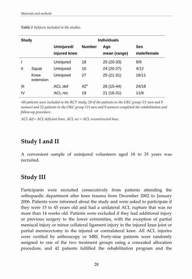
Subjects Uninjured individuals and patients with an ACL deficiency or ACL reconstruction participated in the thesis (Table 1).
Materials and methods
28
Table 1 Subjects included in the studies.
Study Individuals Uninjured/
injured knee Number Age
mean (range) Sex male/female
I Uninjured 18 25 (20-33) 9/9 II Squat Uninjured 16 24 (20-27) 4/12 Knee
extension Uninjured 27 25 (21-31) 16/11
III ACL def 42a 26 (15-44) 24/18 IV ACL rec 19 21 (16-31) 11/8
a49 patients were included in the RCT study; 20 of the patients in the CKC group (11 men and 9 women) and 22 patients in the OKC group (13 men and 9 women) completed the rehabilitation and follow‐up procedure.
ACL def = ACL deficient knee, ACL rec = ACL reconstructed knee.
Study I and II
A convenient sample of uninjured volunteers aged 18 to 35 years was recruited.
Study III
Participants were recruited consecutively from patients attending the orthopaedic department after knee trauma from December 2002 to January 2006. Patients were informed about the study and were asked to participate if they were 15 to 45 years old and had a unilateral ACL rupture that was no more than 14 weeks old. Patients were excluded if they had additional injury or previous surgery to the lower extremities, with the exception of partial meniscal injury or minor collateral ligament injury in the injured knee joint or partial meniscectomy in the injured or contralateral knee. All ACL injuries were verified by arthroscopy or MRI. Forty‐nine patients were randomly assigned to one of the two treatment groups using a concealed allocation procedure, and 42 patients fulfilled the rehabilitation program and the
Materials and methods
29
assessments. Numbers of isolated and combined ACL injures were not significantly different between groups.
Study IV
Participants were recruited consecutively from patients who were diagnosed with unilateral ACL rupture and were on the waiting list for ACL reconstruction with a quadruple hamstring tendon graft from October 2004 to May 2006. Patients were informed about the study and were asked to participate if they were 15 to 45 years old and had a unilateral ACL rupture that was no more than 4 years old. Exclusion criteria were other total ligament rupture, menisci suture in the injured knee joint, total ligament rupture, or reconstruction on the contralateral knee. All patients followed a carefully defined rehabilitation program (paper IV). Immediately after being operated on, the patients were allowed unrestricted range of motion exercises plus quadriceps sets and straight leg raise. Weight‐bearing when walking was allowed after one week, and single crutch weight bearing indoors after two weeks.
Equipment
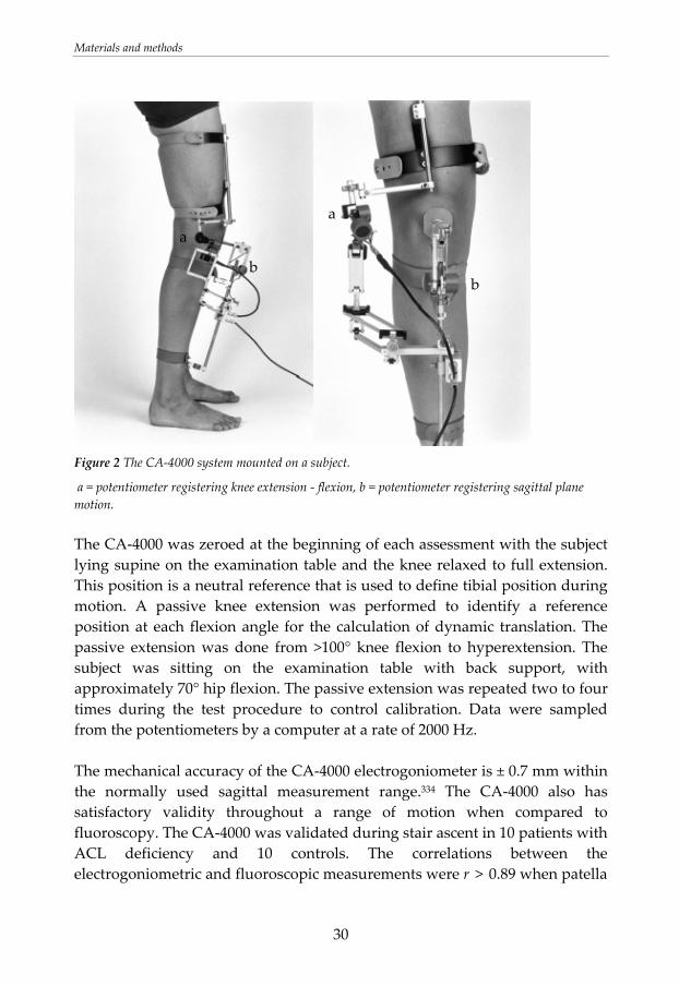
Electrogoniometer
A computerised goniometer linkage, CA‐4000 (OSI Inc., Hayward CA, USA), was used to measure the flexion angle and sagittal tibial translation (Figure 2). The system is composed of three parts: the femoral and tibial frames, and a rotation module. Three potentiometers in the rotation module measure the relative rotations between the femur and tibia. The potentiometer for sagittal motion mounted on the tibial frame registers the difference in position between a spring‐loaded patellar pad and the fixation point on the tibial tuberosity during knee motion. The sagittal plane direction is perpendicular to the tibial frame. The potentiometer registering knee extension flexion was aligned with an approximate knee flexion axis in the centre of the lateral femur epicondyle. The alignment was checked repeatedly during the examination.
Materials and methods
30
Figure 2 The CA‐4000 system mounted on a subject.
a = potentiometer registering knee extension ‐ flexion, b = potentiometer registering sagittal plane motion. The CA‐4000 was zeroed at the beginning of each assessment with the subject lying supine on the examination table and the knee relaxed to full extension. This position is a neutral reference that is used to define tibial position during motion. A passive knee extension was performed to identify a reference position at each flexion angle for the calculation of dynamic translation. The passive extension was done from >100° knee flexion to hyperextension. The subject was sitting on the examination table with back support, with approximately 70° hip flexion. The passive extension was repeated two to four times during the test procedure to control calibration. Data were sampled from the potentiometers by a computer at a rate of 2000 Hz. The mechanical accuracy of the CA‐4000 electrogoniometer is ± 0.7 mm within the normally used sagittal measurement range.334 The CA‐4000 also has satisfactory validity throughout a range of motion when compared to fluoroscopy. The CA‐4000 was validated during stair ascent in 10 patients with ACL deficiency and 10 controls. The correlations between the electrogoniometric and fluoroscopic measurements were r > 0.89 when patella
aa
b b
Materials and methods
31
was used as reference point and r > 0.94 when femur was used as reference point at the fluoroscopic analysis.335 The measurement system has satisfactory reproducibility: the mean variation between three consecutive dynamic measurements (gait) is 0.03 ± 0.5 mm. The mean variation throughout a range of motion (squat on two legs, 0° to 90° to 0°) on two different days was 0.73 ± 0.41 mm.170 The repeatability of three consecutive measurements of total sagittal tibial translation during the Lachman test was investigated from the tests performed in study IV. Three repetitions of the 90N Lachman tests using the CA‐4000 electrogoniometer and a force handle were carried out. The assessment was performed on the uninjured and the ACL deficient knee at assessment before ACL reconstruction and on the operated leg at assessment five weeks after ACL reconstruction. The mean variations between the three consecutive repetitions of the Lachman test were: for the uninjured leg 0.00 ± 0.06 mm, for the ACL deficient leg 0.00 ± 0.12 mm and for the ACL reconstructed leg 0.00 ± 0.09 mm.
Electromyography
Muscle activation was registered with EMG. The EMG was used to explain possible differences in dynamic translation. Furthermore, the EMG was used to assess the effectiveness of different exercises in reaching a high level of activation in the different muscles. Skin preparation and electrode placement were made according to recommendations from “Surface EMG for the Non‐Invasive Assessment of Muscles” (SENIAM).114 The EMG activity was registered by surface electrodes. The muscles registered are showed in Table 2. Table 2 Muscles assessed in the studies.
Muscle Study
m. vastus medialis I, III, IV m. vastus lateralis I, III, IV mm. hamstrings I, III, IV m. gastrocnemius I, III, IV m. gluteus maximus III m. soleus IV
Materials and methods
32
The muscles were located by palpation during a submaximal isometric contraction and the electrodes were placed at the most prominent place of the muscle. The skin at each electrode site was first prepared by shaving and cleaning with 70% alcohol to facilitate electrode adherence and conduction of EMG signals. On the skin above each muscle, two recording pre‐gelled silver‐chloride electrodes (Blue sensor, M‐00‐S, Medicotest, Denmark, diameter of active part 10 mm) were placed with 2 cm centre‐to‐centre distance, and one ground electrode with an amplifier was placed about 10 cm from the measuring area. The electrode placement was verified through functional testing while observing the recording on the computer screen. EMG signals were sampled at 2000Hz by the MESPEC 4000 EMG unit system (MEGA Electronics Ltd., Kuopio, Finland). Three repeated maximal isometric voluntary contractions (MIVC) were performed for knee extension, knee flexion, plantar flexion, and hip extension. Peak value during MIVC served as reference values for calculations of EMG activity. For knee extension and flexion, the patients sat with the knee positioned at 60° and 110° of knee flexion respectively and restrained by a strap. For plantar flexion, the patients stood on one leg in an upright position with light balance support and raised on toes as high as possible. The patients held a strap that was attached under the plate they were standing on to restrain their movement. The hip extension was performed with the patient lying prone at the examination table with the hip maximally extended against resistance and the knee in 90° flexion. The reliability of EMG is acceptable.257, 351
Isokinetic device
A Biodex machine (Biodex Medical Systems Inc., Ronkonkoma, NY) was used to record quadriceps and hamstring muscle torque. Isokinetic testing at 60°/sec is a reliable measurement method.255, 323
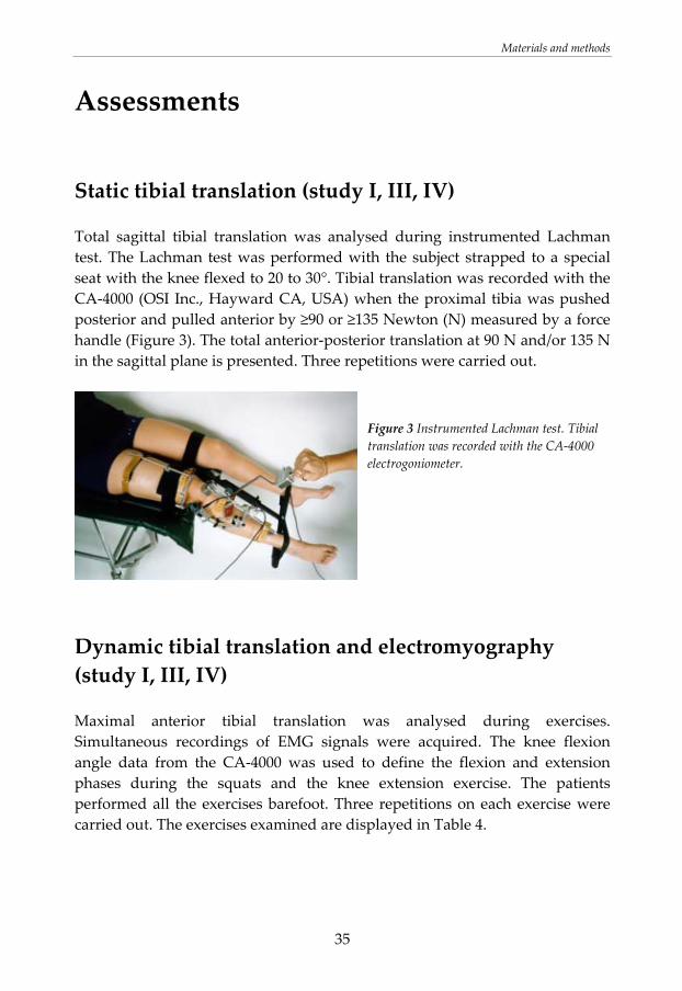
Force measurements
During the static laxity measurements, a force handle (Figure 3) was used to apply anterior and posterior force directed perpendicular to the proximal tibia.
Materials and methods
33
The force handle was calibrated before measurement and the signals were acquired simultaneously using the CA‐4000 electrogoniometer and the EMG. During gait and jump assessments, a Kistler force plate was used to identify phases during locomotion. Moreover, the force plate was used when assessing exercises performed on two legs in study IV. The tested leg was placed on the force plate. The data from the force plate was analysed after the assessment to control that the tested leg carried about 50% of the body weight.
Questionnaires about subjective knee function and activity level
The Lysholm score319 and the KOOS272 were used to evaluate subjective knee function in the patients with ACL deficiency or reconstruction. The Lysholm score consists of 8 different items on a 100‐point scale with 25 points each attributed to instability and pain. The score is meant to be used by an examiner. Lysholm score has satisfactory reliability and validity.191, 319 The KOOS is a self‐administered 42‐item questionnaire evaluating five dimensions; symptoms, pain, function in daily living, function in sport and recreation and knee related quality of life, using a 5‐point Likert scale response format. The reliability and validity are satisfactory.272 The level of activity was determined with the Tegner score,319 that grade activities regarding the demands put on the knee. The score meets basic criteria for outcome measures.319 Quality of life was evaluated using the ACL Quality of Life Questionnaire. It is a reliable and valid disease‐specific 32‐item quality of life questionnaire using a 100‐mm visual analogue scale (VAS) response format.168, 210 Scores and separate questions used in the studies to assess subjective knee function, activity level, ACL related quality of life, fear of reinjury and fear of movement, satisfaction with knee function, and treatment effect are displayed in Table 3.
Materials and methods
34
Table 3 Assessments of subjective knee function and activity level in the studies.
Assessment Score Study
Subjective knee function
Lysholm score319 III, IV
KOOS272 III, IV
Activity level Tegner score319 III, IV
Quality of Life ACL Quality of Life168, 210 IV
Question Response model
Fear of re-injury / fear of movement
Do you have fear of re-injury? Yes - No III
If you have fear of re-injury, to what extent does fear of movement prevent you from performing activities?
100 mm VAS III
To what extent do you have fear of re-injury?
100 mm VAS IV
To what extent do you have confidence in your knee joint?
100 mm VAS III, IV
To what extent do you have fear of your knee giving way when you exercise/perform the rehabilitation exercises?
100 mm VAS IV
To what extent do you have fear of pain when you exercise/perform the rehabilitation exercises?
100 mm VAS IV
Satisfaction with knee function
How would you feel if you had to live with your knee problems at the current activity level?
Ordinal scale (1 Happy – 7 Unhappy)
III
Treatment effect
How do you experience the effect of the physiotherapy treatment?
Ordinal scale (1 Completely recovered – 4 Worse)
III
Materials and methods
35
Assessments
Static tibial translation (study I, III, IV)
Total sagittal tibial translation was analysed during instrumented Lachman test. The Lachman test was performed with the subject strapped to a special seat with the knee flexed to 20 to 30°. Tibial translation was recorded with the CA‐4000 (OSI Inc., Hayward CA, USA) when the proximal tibia was pushed posterior and pulled anterior by ≥90 or ≥135 Newton (N) measured by a force handle (Figure 3). The total anterior‐posterior translation at 90 N and/or 135 N in the sagittal plane is presented. Three repetitions were carried out.
Figure 3 Instrumented Lachman test. Tibial translation was recorded with the CA‐4000 electrogoniometer.
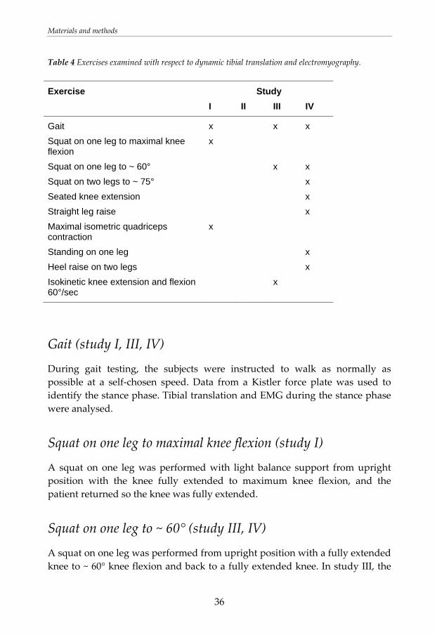
Dynamic tibial translation and electromyography (study I, III, IV)
Maximal anterior tibial translation was analysed during exercises. Simultaneous recordings of EMG signals were acquired. The knee flexion angle data from the CA‐4000 was used to define the flexion and extension phases during the squats and the knee extension exercise. The patients performed all the exercises barefoot. Three repetitions on each exercise were carried out. The exercises examined are displayed in Table 4.
Materials and methods
36
Table 4 Exercises examined with respect to dynamic tibial translation and electromyography.
Exercise Study I II III IV
Gait x x x Squat on one leg to maximal knee flexion
x
Squat on one leg to ~ 60° x x Squat on two legs to ~ 75° x Seated knee extension x Straight leg raise x Maximal isometric quadriceps contraction
x
Standing on one leg x Heel raise on two legs x Isokinetic knee extension and flexion 60°/sec
x
Gait (study I, III, IV)
During gait testing, the subjects were instructed to walk as normally as possible at a self‐chosen speed. Data from a Kistler force plate was used to identify the stance phase. Tibial translation and EMG during the stance phase were analysed.
Squat on one leg to maximal knee flexion (study I)
A squat on one leg was performed with light balance support from upright position with the knee fully extended to maximum knee flexion, and the patient returned so the knee was fully extended.
Squat on one leg to ~ 60° (study III, IV)
A squat on one leg was performed from upright position with a fully extended knee to ~ 60° knee flexion and back to a fully extended knee. In study III, the
Materials and methods
37
patients stood on the floor with light balance support and wore a weight west ‐ 10 kg for men and 8 kg for women. The exercise was performed at the uninjured leg at the assessment before the rehabilitation and bilaterally at the assessment after the rehabilitation. In study IV, the exercise was performed without additional weight. The patients were standing on a step board, without balance support, and flexed the knee until the contralateral foot reached the floor.
Squat on two legs to ~ 75° (study IV)
A squat on two legs was performed from an upright position with fully extended knees to 60 ‐ 80° knee flexion and back to fully extended knees. The tested leg was placed on a Kistler force plate to control that the tested leg carried about 50% of bodyweight.
Seated knee extension (study IV)
The patients sat on the examination table and flexed their knee to approximately 100° and then extended the knee to full extension and flexed it back to approximately 100° knee flexion. The pace was about 2 seconds during extension and 2 seconds during flexion phase.
Straight leg raise (quadriceps contraction) (study IV)
The patients were standing on the floor leaning against the examination table with approximately 40° of hip flexion and performed a quadriceps contraction; while keeping the contraction, they raised the straight leg to about 30 cm above the floor and lowered it back to the floor.
Maximal isometric quadriceps contraction (study I)
A maximal isometric quadriceps contraction was done with the subject seated with approximately 50° knee flexion.
Materials and methods
38
Standing on one leg (study IV)
The patients stood on the contralateral leg with the tested leg in approximately 0° knee extension, then they stood on the tested leg on a Kistler force plate (Kistler, Switzerland) and stood with a straight leg for 2 seconds. Tibial translation and EMG during the stance phase were analysed.
Heel raise on two legs (study IV)
The heel raise was performed on two legs with light balance support from standing on the floor with fully extended knees to maximum plantar flexion and back to the starting position. The tested leg was placed on a Kistler force plate to control that the tested leg carried about 50% of bodyweight.
Isokinetic knee extension and flexion 60°/sec (study III)
Evaluation of maximal anterior tibial translation during isokinetic 60°/sec knee extension and flexion was performed in study III for the uninjured leg at the test before the rehabilitation and bilaterally at the test after the rehabilitation.
Muscle strength (study II, III, IV)
Isokinetic assessment (study III, IV)
Torque of the quadriceps and hamstring muscles was recorded using a Biodex machine. Three repetitions of maximal isokinetic 60°/sec knee extension and flexion were performed. The patients were secured to the chair with body straps, and the resistance pad on the measuring arm was placed at the level of the ankle joint. Before recording, some submaximal familiarization repetitions were performed. The evaluation was performed in study III on the uninjured leg at the assessment before the rehabilitation and bilaterally at the assessment after the rehabilitation. The evaluation was performed in study IV bilaterally at the assessment before ACL reconstruction. Peak torque was analysed.
Materials and methods
39
Establishment of one repetition maximum (study II, III)
Procedure
Study II involved a development of a systematic procedure for the establishment of 1RM of squat on one leg and seated knee extension on one leg. During the procedure for the establishment of 1RM, the subjects estimated their leg strength compared to other individuals of equal age and gender using the following scale: much weaker, slightly weaker, normal, slightly stronger, and much stronger. The estimate formed the basis of the amount of load that was put on the machine before the first attempt to determine 1RM. After the first attempt to perform the movement, the subject rated perceived exertion in the leg during the exercise according to the Borg scale.42 This rating formed the basis of the amount of load that was added before the second attempt.
Squat on one leg
A barbell squat (0‐60° of knee flexion) was performed standing on one leg in a Smith machine (Life Fitness Strength; Life Fitness, Schiller Park, IL); the barbell slid in a safety rack. The subject was standing with the centre of gravity over the foot and the non‐supporting leg was flexed in the knee joint. The barbell was placed on the back. The subjects were instructed to keep their trunk in an upright position while flexing the knee of the leg on which they were standing. The ROM was measured using a standard plastic goniometer. In addition, the CA‐4000 electrogoniometer was used to verify ROM during the exercise. In study III, a test of 1RM for a squat on one leg was performed at the first and the last rehabilitation appointments at the physiotherapist. The test was performed on the uninjured leg at week 1 and on the uninjured leg and the injured leg at week 16 in both the CKC and the OKC group. These tests were used to compare strength and performance in a CKC exercise between the CKC and the OKC group. Moreover, to set the optimal load for the exercises, the test of 1RM for a squat on one leg was performed at weeks 1, 5, 9, and 13 for the CKC group. When application of the maximal load was considered unsuitable, a repetitions‐to‐failure method with submaximal load106 was used. The load was adjusted after the 1RM test.

Materials and methods
40
Seated knee extension on one leg
A knee extension was performed seated with 70° of hip flexion and back support in a leg extension machine (Life Fitness Strength; Life Fitness, Schiller Park, IL). The exercise was performed through extension of one knee from 100° to 0°. The ROM was measured by a standard plastic goniometer. To set the optimal load for the exercises in study III, test of 1RM on seated knee extension on one leg was performed at weeks 1, 5, 9, and 13 for the OKC group. When application of the maximal load was considered unsuitable, a repetitions‐to‐failure method with submaximal load106 was used. The load was adjusted after the 1RM test.
Functional performance (study III)
Unilateral vertical jump
At assessment after the rehabilitation in study III, patients were assessed for their ability to perform a unilateral vertical jump with their hands at their sides. Each patient stood on one leg on a Kistler force plate and performed one jump as high as possible and landing on the same foot. The time from take‐off to landing was determined. The reliability and validity for unilateral vertical jump is reported to be high.108, 130
Unilateral horizontal jump
At assessment after the rehabilitation in study III each patient performed a unilateral horizontal jump for distance; the patient’s hands were kept behind the back to prevent their use in generating momentum. Horizontal jump for distance has high reliability and validity.108, 258
Knee joint circumference (study III, IV)
Swelling, measured as knee joint circumference at mid‐patellae, was measured using a tape measure with the patient relaxed supine on the examination table.
Materials and methods
41
Knee joint circumference measured by a tape measure are reported as reliable measurements.226, 302, 320
Passive range of motion (study III, IV)
Passive range of motion (PROM) in knee extension and flexion was measured with a standard plastic goniometer with the patient relaxed supine on the examination table. Extension was measured with a block under the patient’s heel to allow for hyperextension. The arms of the goniometer were aligned with the greater trochanter and lateral malleolus and the axis of the goniometer was placed over the knee joint line just below the lateral femoral epicondyle. Goniometric measurements of the knee using a standard plastic goniometer have shown to be reliable and valid.55, 97, 200 Under controlled conditions, repeated measurements can be expected to fall within approximately 4°.200
Data analysis
Tibial translation
The position of the tibia is given in relation to the patella (femur) with the relative position at the passive extension as a neutral reference. Dynamic tibia translation was calculated by subtracting the tibial position values during the passive extension from the position values during motion.171 A position anterior to the neutral reference is referred to as positive and posterior as negative. The position of the passive extension curve has been shown to be constant with a mean difference of 0.09 ± 0.08 mm within three consecutive repetitions and 0.06 ± 0.09 mm between seven repetitions during a 1.5 hour test session with other movements in between.170 Calculation of tibial translation in the described manner eliminates a change in position resulting from rollback of the femur. For each measurement of static and dynamic tibial translation, the maximal translation was derived from each repetition, and the mean of the three repetitions was calculated.
Materials and methods
42
Electromyography
The mean and/or maximum EMG activation is presented. The peak EMG value during 100 msec was used for analysis. The peak value during exercise was normalised to the peak value at the MIVC test for each muscle respectively (relative value of one muscle = peak value during a movement / peak value during MIVC). In addition, in study III, the activation in each muscle during gait and squat was also calculated as a percentage of the total leg muscle activation in order to detect any differences in share of total leg muscle work. The following formula was used: relative value of one muscle / Σ relative values of all muscles. In this analysis, a mean of EMG activity in vastus medialis and vastus lateralis was used for the calculation of activity in quadriceps.
Muscle strength
Peak torque achieved during any of the three repetitions performed during isokinetic testing is reported. Muscle strength in the injured leg is expressed as percent of muscle strength in the uninjured leg.
Statistical analyses For detailed information regarding statistical methods refer to the separate papers. The statistical methods used are shown in Table 5. Statistics were calculated using SPSS (version 10.1–15.0; SPSS Inc., Chicago, IL). In all studies a significance level of P < 0.05 was used for all variables except the EMG data in study III where a significance level of P < 0.001 was used due to multiple analyses. Sample size calculations in the studies were executed based on the primary outcome variables. The calculations estimated that 17 individuals would be required to detect a 1.1 mm change in translation when comparing before and after exercise within one group (study I) or comparing legs within one group (study IV), as significant (α = 0.05, β = 0.20). For comparisons of exercises within one leg at one assessment session (study IV), a smaller sample had been sufficient. In study III, 19 patients were required in each group to detect a 1.5
Materials and methods
43
mm difference in translation and a 10% difference in quadriceps strength between groups as significant (α = 0.05, β = 0.20). Paired samples t‐test was used for comparisons of parametric variables within the same individual (study I, II, III). Independent samples t‐test was used to compare parametric variables between groups (study III). Two‐way analysis of variance (ANOVA) with Bonferroni adjustment for multiple comparisons was used for comparisons of tibial translation and muscle activation between the exercises, and two way ANOVA with Dunnett´s post hoc test was used for comparisons of tibial translation and muscle activation between the legs in study IV. In addition, due to skewed distribution of data in some EMG variables, the EMG data in study IV was also calculated using Friedman test for comparisons between exercises and between legs, and Wilcoxon Signed Ranks test for pair ways comparisons with Bonferroni adjustment for multiple comparisons. The Tegner score, Lysholm score, and KOOS were analysed with Mann‐Whitney U test (study III). The supplementary questions were analysed using the chi‐square test (nominal data) and the Mann‐Whitney U test (ordinal data) (study III). In study II, the two‐way random effect model (absolute agreement definition), single measure intra‐class correlation coefficient (ICC2.1), was used for analysis of relative reliability.296 Measurement error (SDdiff/√2) was used to describe absolute reliability.129 A Bland and Altman plot diagram was used to display the measurement error schematically.40
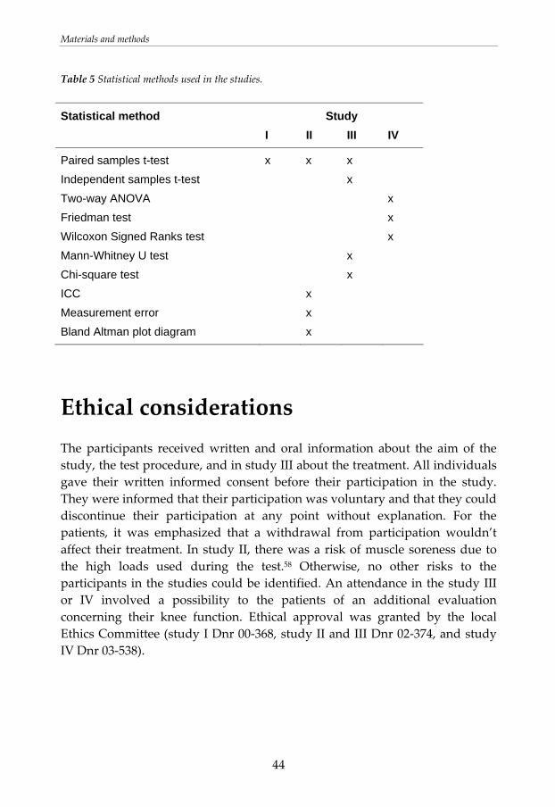
Materials and methods
44
Table 5 Statistical methods used in the studies.
Statistical method Study I II III IV
Paired samples t-test x x x Independent samples t-test x Two-way ANOVA x Friedman test x Wilcoxon Signed Ranks test x Mann-Whitney U test x Chi-square test x ICC x Measurement error x Bland Altman plot diagram x
Ethical considerations The participants received written and oral information about the aim of the study, the test procedure, and in study III about the treatment. All individuals gave their written informed consent before their participation in the study. They were informed that their participation was voluntary and that they could discontinue their participation at any point without explanation. For the patients, it was emphasized that a withdrawal from participation wouldn’t affect their treatment. In study II, there was a risk of muscle soreness due to the high loads used during the test.58 Otherwise, no other risks to the participants in the studies could be identified. An attendance in the study III or IV involved a possibility to the patients of an additional evaluation concerning their knee function. Ethical approval was granted by the local Ethics Committee (study I Dnr 00‐368, study II and III Dnr 02‐374, and study IV Dnr 03‐538).
Results
45
RESULTS
Additional results are presented in the separate papers.
Tibial translation and muscle activation after an exercise session in uninjured individuals (study I)
Static tibial translation
Maximal tibial translation during the Lachman test did not differ after the exercise session as compared to before (Table 6). A force‐displacement curve of the Lachman test showed no differences in tibial displacement between the test before and after the exercise session. Consequently, the anterior tibial translation was similarly restricted before and after the exercise. Figure 4 illustrates the tibial displacement during the Lachman test before and after the exercise session in one individual.
Dynamic tibial translation
The anterior tibial translation was equal during the first three repetitions and the last three repetitions of knee extensions during the exercise session (diff 0.5 ± 1.7 mm, P = 0.22). Furthermore, there were no differences in maximal dynamic anterior translation after compared to before the exercise session (Table 6).
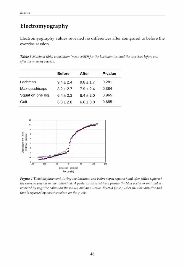
Results
46
Electromyography
Electromyography values revealed no differences after compared to before the exercise session. Table 6 Maximal tibial translation (mean ± SD) for the Lachman test and the exercises before and after the exercise session.
Before After P-value
Lachman 9.4 ± 2.4 9.8 ± 1.7 0.281
Max quadriceps 8.2 ± 2.7 7.9 ± 2.4 0.384
Squat on one leg 6.4 ± 2.3 6.4 ± 2.0 0.965
Gait 6.3 ± 2.8 6.6 ± 3.0 0.685
Force (N)posterior - anterior
Dis
plac
emen
t (m
m)
post
erio
r - a
nter
ior
-6
-4
-2
0
2
4
6
8
10
12
-180 -120 -60 0 60 120 180
Figure 4 Tibial displacement during the Lachman test before (open squares) and after (filled squares) the exercise session in one individual. A posterior directed force pushes the tibia posterior and that is reported by negative values on the y‐axis, and an anterior directed force pushes the tibia anterior and that is reported by positive values on the y‐axis.
Results
47
The procedure for the establishment of one repetition maximum (study II) The standardised test protocol with estimates of leg strength and ratings of perceived exertion resulted in only a few unsuccessful trials before detecting 1RM for a squat on one leg and seated knee extension on one leg. The information regarding the amount of load that was added based on the estimations of leg strength and the ratings of perceived exertion appeared to be useful when testing individuals 20 to 37 years of age.
Reliability of squat on one leg (study II) The intra‐rater reliability of 1RM for a squat on one leg, expressed as the ICC, was 0.64, and the inter‐rater reliability was 0.94. The measurement error for tests performed on different days was somewhat high, but it was acceptable for tests performed by different test leaders at the same occasion (Table 7).
Reliability of seated knee extension on one leg (study II) The intra‐rater reliability of 1RM for a seated knee extension on one leg, expressed as the ICC, was 0.90, and the inter‐rater reliability was 0.96. The measurement errors were acceptable (Table 7).
Results
48
Table 7 The number of unsuccessful trials before detecting 1RM (median (range)), the weight lifted at 1RM squat and knee extension (kg) (mean ± SD) obtained from the two test leaders (1 and 2) at the two test occasions (A and B), mean difference between the test leaders and test occasions (mean diff) (mean ± SD), measurement error, and ICC‐values.
Testa n Numbers of trials
1RM
Mean diff
Measure-ment error
ICC
Squat A 14 4 (1-8) 86.4 ± 19.9 Squat B 14 5 (1-8) 95.7 ± 25.8
-9.3 ± 18.6 13.1 0.64
Squat 1 16 5 (1-8) 97.5 ± 24.9 Squat 2 16 4.5 (0-8) 93.3 ± 22.6
4.2 ± 7.3 5.2 0.94
Knee extension A 23 2 (0-6) 58.8 ± 15.7 Knee extension B 23 2 (0-6) 59.5 ± 15.4
-0.6 ±7.2 5.1 0.90
Knee extension 1 26 2 (0-6) 56.6 ± 16.1
Knee extension 2 26 1.5 (0-5) 57.5 ± 17.1 -0.9 ± 4.5 3.2 0.96
a A = the first test occasion, B = the second test occasion, 1 = test leader 1, 2 = test leader 2. A and B represent values from the same test leader and were used for assessment of intra‐rater reliability. 1 and 2 represent values from the same test occasion and were used for assessment of inter‐rater reliability.
Results
49
Effects of rehabilitation programs with quadriceps strengthening in closed or open kinetic chain in patients with ACL deficiency (study III)
Static tibial translation
There were no differences between the CKC and the OKC group in maximal total translation of the tibia during the Lachman test for the uninjured leg, injured leg, or the ratio of injured: uninjured leg before and after rehabilitation. In both groups, there was no change in static translation of either leg following rehabilitation (P > 0.05).
Dynamic tibial translation
Gait
There were no group differences in maximal anterior translation of the tibia during gait for the uninjured or injured leg before or after rehabilitation. The anterior tibial translation in the injured leg increased after compared to before rehabilitation in both the CKC (P = 0.011) and the OKC (P = 0.044) group. After rehabilitation, there was greater anterior tibial translation in the injured leg compared to the uninjured leg in both the CKC group (P = 0.013) and the OKC group (P = 0.001).
Squat on one leg
There were no differences between groups in maximal tibial translation during the squat on one leg (P > 0.05).
Results
50
Isokinetic knee extension and flexion 60°/sec
Maximal anterior tibial translation during isokinetic knee extension or flexion of the uninjured leg before or after rehabilitation or of the injured leg after rehabilitation did not differ between groups. After rehabilitation, translation during the extension phase was greater for the injured leg than the uninjured leg in both the CKC group (P = 0.002) and the OKC group (P = 0.031).
Electromyography
Gait
There were no group differences in muscle activation before or after rehabilitation (P > 0.001). In the CKC group, the mean activation of hamstrings of the injured leg was significantly reduced after rehabilitation (P = 0.001). Furthermore, in both groups, there was a trend toward decreased mean activation of vastus lateralis in the injured leg after versus before rehabilitation (CKC group: P = 0.002; OKC group: P = 0.038). In the CKC group, there was also a trend toward reduced mean activation of vastus medialis (P = 0.018) and the gastrocnemius (P = 0.052) in the injured leg after versus before rehabilitation.
Squat on one leg
Both groups used a similar muscular stabilizing strategy when performing the squat on one leg.
Isokinetic knee extension and flexion 60°/sec
There were no significant differences in patterns of muscle activation between groups.
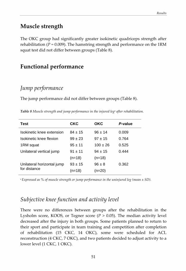
Results
51
Muscle strength
The OKC group had significantly greater isokinetic quadriceps strength after rehabilitation (P = 0.009). The hamstring strength and performance on the 1RM squat test did not differ between groups (Table 8).
Functional performance
Jump performance
The jump performance did not differ between groups (Table 8). Table 8 Muscle strength and jump performance in the injured lega after rehabilitation.
Test CKC OKC P-value
Isokinetic knee extension 84 ± 15 96 ± 14 0.009 Isokinetic knee flexion 99 ± 23 97 ± 15 0.764 1RM squat 95 ± 11 100 ± 26 0.525 Unilateral vertical jump 91 ± 11
(n=18) 94 ± 15 (n=18)
0.444
Unilateral horizontal jump for distance
93 ± 15 (n=18)
96 ± 8 (n=20)
0.362
a Expressed as % of muscle strength or jump performance in the uninjured leg (mean ± SD).
Subjective knee function and activity level
There were no differences between groups after the rehabilitation in the Lysholm score, KOOS, or Tegner score (P > 0.05). The median activity level decreased after the injury in both groups. Some patients planned to return to their sport and participate in team training and competition after completion of rehabilitation (15 CKC, 14 OKC), some were scheduled for ACL reconstruction (4 CKC, 7 OKC), and two patients decided to adjust activity to a lower level (1 CKC, 1 OKC).
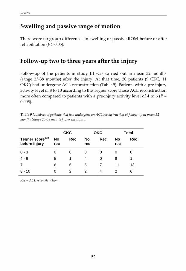
Results
52
Swelling and passive range of motion
There were no group differences in swelling or passive ROM before or after rehabilitation (P > 0.05).
Follow‐up two to three years after the injury
Follow‐up of the patients in study III was carried out in mean 32 months (range 23‐38 months) after the injury. At that time, 20 patients (9 CKC, 11 OKC) had undergone ACL reconstruction (Table 9). Patients with a pre‐injury activity level of 8 to 10 according to the Tegner score chose ACL reconstruction more often compared to patients with a pre‐injury activity level of 4 to 6 (P = 0.005). Table 9 Numbers of patients that had undergone an ACL reconstruction at follow‐up in mean 32 months (range 23‐38 months) after the injury.
CKC OKC Total Tegner score319 before injury
No rec
Rec No rec
Rec No rec
Rec
0 - 3 0 0 0 0 0 0 4 - 6 5 1 4 0 9 1 7 6 6 5 7 11 13 8 - 10 0 2 2 4 2 6
Rec = ACL reconstruction.
Results
53
Tibial translation and muscle activation during rehabilitation exercises five weeks after ACL reconstruction (study IV)
Static tibial translation
The total tibial translation at the 90N Lachman test was 6.1 ± 1.7 mm.
Dynamic tibial translation
Exercises performed with a straight leg; heel raise (P = 0.041), standing on one leg (P = 0.005), and straight leg raise (P = 0.002) produced less anterior translation than the maximal anterior translation during the 90N Lachman test. Exercises with greater ROM in the knee joint; seated knee extension (ROM (0)‐3‐94°), gait (ROM (0)‐6‐38°), squat on one leg (ROM (0)‐4‐61°), and squat on two legs (ROM (0)‐1‐77°) did not differ in maximal anterior translation from the amount of anterior translation produced during the Lachman test. There was greater anterior tibial translation during the seated knee extension compared to standing on one leg (P = 0.046) and straight leg raise (P = 0.018) (Table 10).
Electromyography
The EMG activation in vastus medialis was greater during seated knee extension and squat on one leg compared to heel raise and standing on one leg. Moreover, the medialis activation was greater during gait compared to standing on one leg. The EMG activation in vastus lateralis was greater during seated knee extension and squat on one leg compared to gait, heel raise and standing on one leg. The hamstring activation was larger during gait compared to all other exercises except squat on one leg. The EMG activation in gastrocnemius was larger during the heel raise than during all other exercises.
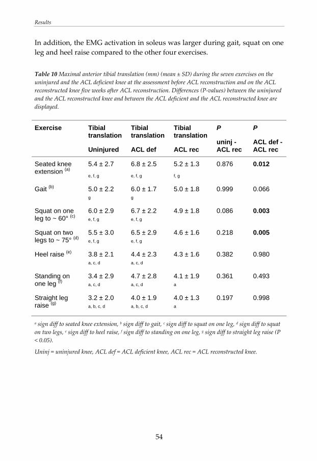
Results
54
In addition, the EMG activation in soleus was larger during gait, squat on one leg and heel raise compared to the other four exercises. Table 10 Maximal anterior tibial translation (mm) (mean ± SD) during the seven exercises on the uninjured and the ACL deficient knee at the assessment before ACL reconstruction and on the ACL reconstructed knee five weeks after ACL reconstruction. Differences (P‐values) between the uninjured and the ACL reconstructed knee and between the ACL deficient and the ACL reconstructed knee are displayed.
Exercise
Tibial translation
Uninjured
Tibial translation
ACL def
Tibial translation
ACL rec
P
uninj - ACL rec
P
ACL def - ACL rec
Seated knee extension (a)
5.4 ± 2.7
e, f, g
6.8 ± 2.5
e, f, g
5.2 ± 1.3
f, g
0.876 0.012
Gait (b)
5.0 ± 2.2
g
6.0 ± 1.7
g 5.0 ± 1.8 0.999 0.066
Squat on one leg to ~ 60° (c)
6.0 ± 2.9 e, f, g
6.7 ± 2.2
e, f, g 4.9 ± 1.8 0.086 0.003
Squat on two legs to ~ 75° (d)
5.5 ± 3.0
e, f, g 6.5 ± 2.9
e, f, g 4.6 ± 1.6 0.218 0.005
Heel raise (e) 3.8 ± 2.1 a, c, d
4.4 ± 2.3
a, c, d 4.3 ± 1.6 0.382 0.980
Standing on one leg (f)
3.4 ± 2.9 a, c, d
4.7 ± 2.8
a, c, d
4.1 ± 1.9 a
0.361 0.493
Straight leg raise (g)
3.2 ± 2.0
a, b, c, d 4.0 ± 1.9
a, b, c, d
4.0 ± 1.3 a
0.197 0.998
a sign diff to seated knee extension, b sign diff to gait, c sign diff to squat on one leg, d sign diff to squat on two legs, e sign diff to heel raise, f sign diff to standing on one leg, g sign diff to straight leg raise (P < 0.05).
Uninj = uninjured knee, ACL def = ACL deficient knee, ACL rec = ACL reconstructed knee.
Results
55
The ACL reconstructed leg compared to the uninjured leg (study IV)
Static tibial translation
There was no difference in total tibial translation at the 90N Lachman test between the ACL reconstructed leg and the uninjured leg (P = 0.118).
Dynamic tibial translation
The maximal anterior tibial translation during the exercises in the ACL reconstructed knee did not differ from the amount of tibial translation produced in the uninjured knee (Table 10).
Electromyography
The ACL reconstructed leg used more hamstring activation during seated knee extension, gait, squat on one leg, squat on two legs, heel raise, and standing on one leg compared to the uninjured leg. In addition, there was more gastrocnemius activation during seated knee extension, squat on one leg and straight leg raise, and more soleus activation during squat on one leg and squat on two legs. The lateralis activation was greater during gait and standing on one leg. In contrast, the medialis activation was lower during straight leg raise after the ACL reconstruction.
Results
56
The ACL reconstructed leg compared to the ACL deficient leg (study IV)
Static tibial translation
The ACL reconstruction reduced the total tibial translation at the 90N Lachman test from 7.2 ± 1.8 mm to 6.1 ± 1.7 mm (P = 0.013).
Dynamic tibial translation
After the ACL reconstruction, the anterior tibial translation during the seated knee extension, squat on one leg and squat on two legs was significantly reduced. During the other exercises there were no differences in tibial translation between the ACL reconstructed knee and the ACL deficient knee (Table 10).
Electromyography
After the ACL reconstruction, the operated leg used more hamstring activation in seated knee extension, gait, squat on one leg, squat on two legs, heel raise, and standing on one leg compared to the same leg before the ACL reconstruction. In addition, there was more gastrocnemius activation during seated knee extension, squat on one leg and straight leg raise. Moreover, the soleus activation was higher during squat on one leg and squat on two legs. On the contrary, there was less EMG activation in both vastus medialis and lateralis during straight leg raise after the ACL reconstruction.
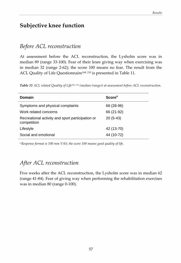
Results
57
Subjective knee function
Before ACL reconstruction
At assessment before the ACL reconstruction, the Lysholm score was in median 89 (range 33‐100). Fear of their knee giving way when exercising was in median 32 (range 2‐62); the score 100 means no fear. The result from the ACL Quality of Life Questionnaire168, 210 is presented in Table 11. Table 11 ACL related Quality of Life168, 210 (median (range)) at assessment before ACL reconstruction.
Domain Scorea
Symptoms and physical complaints 68 (28-96) Work related concerns 66 (21-92) Recreational activity and sport participation or competition
20 (5-43)
Lifestyle 42 (13-70) Social and emotional 44 (10-72)
a Response format is 100 mm VAS; the score 100 means good quality of life.
After ACL reconstruction
Five weeks after the ACL reconstruction, the Lysholm score was in median 62 (range 41‐84). Fear of giving way when performing the rehabilitation exercises was in median 80 (range 0‐100).
Discussion
58
DISCUSSION
Main findings The main findings in this thesis were that isolated quadriceps training in OKC resulted in greater quadriceps strength compared to quadriceps training in CKC in patients with ACL deficiency. Therefore, quadriceps strengthening in OKC should be included in rehabilitation programs for patients with ACL deficiency. The tibial translation did not differ between groups, indicating that OKC quadriceps training can be performed by patients with ACL deficiency without risk of further loosening of the knee joint. Persistent muscle strength deficits in patients with ACL deficiency and ACL reconstruction have previously been reported. A comprehensive rehabilitation program, with emphasis on continuous progression in load and degree of difficulty of the exercises, performed three days/week in four months, led to good muscle strength and knee function for most patients in the present study. Patients in the early phase after ACL injury or ACL reconstruction used an altered dynamic stabilization strategy including a reduced peak knee extension angle during gait and increased hamstring activation during activity. This stabilization strategy stiffens the knee joint and reduces the tibial translation. Patients that had completed a rehabilitation program used a movement pattern (including hamstring activation and knee flexion) that was more close to normal. The following sections include discussion concerning the effect of training in CKC and OKC on dynamic knee stability. Next, some ideas of altered dynamic stabilization in patients with ACL deficiency or ACL reconstruction are discussed. Finally, a discussion about muscle activation during and after rehabilitation and muscle function after rehabilitation follows.
Discussion
59
Dynamic knee stability during and after exercises in closed and open kinetic chain
Effects of an exercise session
An exercise session, including cycling and a maximum number of knee extensions and heel raises, did not influence the amount of tibial translation in individuals without knee injury. This means that the restraining mechanism of the ACL, and the stabilising effect of the leg muscles were not altered. Moreover, no differences in muscle activation that could explain the lack of difference in translation could be detected.
Rehabilitation exercises after ACL reconstruction
In the early rehabilitation phase after ACL reconstruction the graft needs to be protected against excessive strain. Some strain during the healing process increases graft strength although excessive strain can stretch or rupture the graft. 33, 35, 308, 324, 349 At the same time, it is important to introduce more demanding exercises to the patients to facilitate the regain of muscle function and neuromuscular control. CKC exercises are recommended in rehabilitation due to its similarities to daily and athletic movements concerning biomechanics and neuromuscular characteristics.77, 126 In a CKC exercise, balance and coordination are stimulated simultaneously as the muscle strengthens.66, 140 A possible disadvantage is that a weak quadriceps muscle may be supported by other muscles, a compensation that can take over a greater share of the work, interfering with an optimal strength development in the quadriceps muscle.206 In study IV, we evaluated the kinematics during rehabilitation exercises in patients five weeks after the ACL reconstruction, when these exercises normally are introduced to the patients. In addition, the patients were assessed bilateral before surgery to compare the ACL reconstructed knee with the uninjured and the ACL deficient knee on the same patient. The ACL reconstruction reduced the static tibial translation. The dynamic translation was also reduced during three exercises compared to the translation in the ACL deficient knee. This may be explained by the increased static stability together with changed muscle activation that stiffened the knee joint. Muscle
Discussion
60
contractions exert great influence on the stability in the knee joint. Addition of antagonistic hamstring contraction to an isolated quadriceps contraction reduces anterior tibial translation at flexion angles of 10 to 30° or more.180, 197, 237 After the ACL reconstruction, the amount of anterior tibial translation during an exercise was not determined by whether the exercise was performed in CKC or OKC. Instead, the level of quadriceps activation and the knee joint ROM during the exercise seemed to be of greater importance. Other authors have also suggested that the tibial translation is related to the quadriceps generated shear force.141 In the present study, exercises performed with a straight leg produced less translation compared to maximal translation at the 90N Lachman test, whereas exercises with greater knee joint ROM produced similar amount of anterior tibial translation as the Lachman test. Seated knee extension produced significantly more tibial translation compared to standing on one leg and straight leg raise. Both seated knee extension and straight leg raise are quadriceps exercises in OKC. In addition, both exercises involve knee flexion angles between 40 and 0°, angles that are considered to be the most harmful to the ACL graft.31 The activation of vastus medialis and lateralis was somewhat higher during the seated knee extension compared to the straight leg raise; however no significant differences could be detected. The flexion angle where the maximal tibial translation occurred was for seated knee extension 10 ± 8°, and for straight leg raise the flexion angle was 3 ± 5°. The difference was significant and might be of clinical importance; Li et al. have shown that quadriceps contraction causes an anterior tibial translation that increases from full knee extension to 30°.180 Standing on one leg required generally low muscle activation, and quadriceps activation was significantly smaller compared to the seated knee extension. Maximal tibial translation during standing on one leg occurred at 6 ± 7°, which was significant different from the seated knee extension. Since none of the exercises examined in study IV exceeded the amount of anterior tibial translation produced during the 90N Lachman test, all exercises can be recommended in rehabilitation in the early period after ACL reconstruction. The categorization of rehabilitation exercises can contribute to the development of safe and effective rehabilitation programs. However, knowledge regarding how much translation or ACL strain that can be accepted during different healing phases after an ACL reconstruction is lacking. Further research is needed to evaluate exercises used in the later phases in the rehabilitation after ACL reconstruction. Accelerated
Discussion
61
rehabilitation with early weight bearing is popular in rehabilitation after ACL reconstruction. An accelerated protocol can limit muscle weakness as a consequence of immobilization after surgery. This approach has led to that patients can return to activity and sports after a considerable shorter rehabilitation period.288, 291 There is, however, still little evidence regarding long‐term effects of a short intensive rehabilitation period and a fast return to sports.
Effects of rehabilitation programs with quadriceps strengthening in closed or open kinetic chain
The patients with ACL deficiency who completed the rehabilitation program supplemented with quadriceps exercise in CKC or OKC in study III did not differ in terms of static or dynamic tibial translation. The results are in line with other studies that also concluded that CKC and OKC training leads to similar knee laxity.206, 217, 249, 250 The addition of OKC quadriceps training has been found to lead to greater quadriceps strength without increased knee laxity compared to quadriceps exercises only in CKC in the rehabilitation after ACL reconstruction.206 Moreover, some results indicate that active knee extension training in the early142, 217 or middle period249 after an ACL reconstruction does not increase knee joint laxity. On the contrary, start of OKC quadriceps exercises four weeks after ACL reconstruction with a hamstring graft has been reported to result in increased knee laxity. Moreover, the early introduction of OKC quadriceps exercises did not influence the quadriceps strength compared to a late start.110 Hence both CKC and OKC exercises appear to be suitable for ACL rehabilitation;80, 83, 206, 217, 249, 277 however, the appropriate time for introducing OKC quadriceps training after ACL reconstruction is indecisive and needs further investigation.
Altered dynamic stability
A reduced peak knee extension angle during gait compared to the uninjured knee was seen in the patients with acute ACL injury before rehabilitation (study III), in patients with ACL deficiency scheduled for ACL reconstruction, and in these patients five weeks after ACL reconstruction (study IV). Moreover, patients in the early period following ACL injury or reconstruction used increased co‐contraction with more hamstring activation during activity.
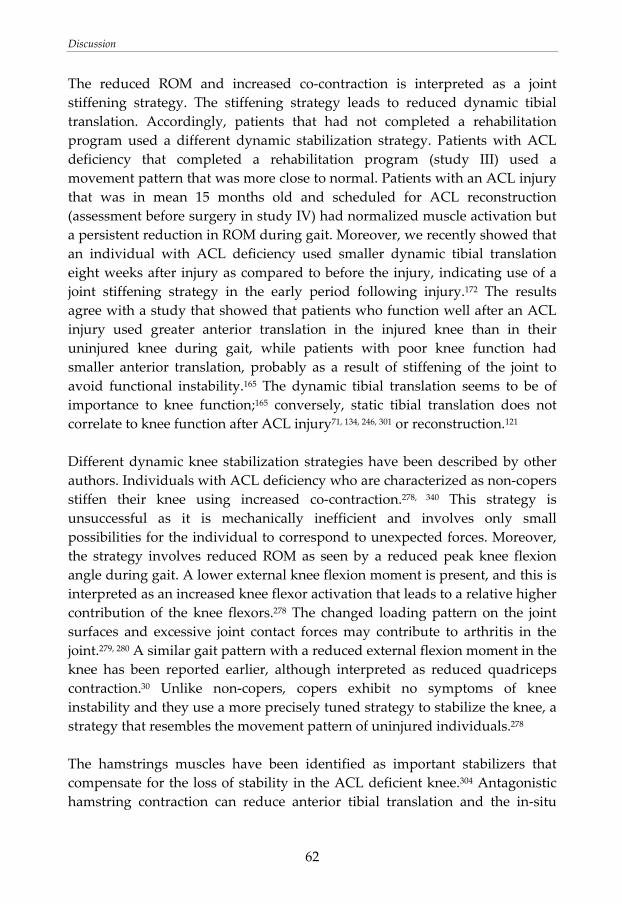
Discussion
62
The reduced ROM and increased co‐contraction is interpreted as a joint stiffening strategy. The stiffening strategy leads to reduced dynamic tibial translation. Accordingly, patients that had not completed a rehabilitation program used a different dynamic stabilization strategy. Patients with ACL deficiency that completed a rehabilitation program (study III) used a movement pattern that was more close to normal. Patients with an ACL injury that was in mean 15 months old and scheduled for ACL reconstruction (assessment before surgery in study IV) had normalized muscle activation but a persistent reduction in ROM during gait. Moreover, we recently showed that an individual with ACL deficiency used smaller dynamic tibial translation eight weeks after injury as compared to before the injury, indicating use of a joint stiffening strategy in the early period following injury.172 The results agree with a study that showed that patients who function well after an ACL injury used greater anterior translation in the injured knee than in their uninjured knee during gait, while patients with poor knee function had smaller anterior translation, probably as a result of stiffening of the joint to avoid functional instability.165 The dynamic tibial translation seems to be of importance to knee function;165 conversely, static tibial translation does not correlate to knee function after ACL injury71, 134, 246, 301 or reconstruction.121 Different dynamic knee stabilization strategies have been described by other authors. Individuals with ACL deficiency who are characterized as non‐copers stiffen their knee using increased co‐contraction.278, 340 This strategy is unsuccessful as it is mechanically inefficient and involves only small possibilities for the individual to correspond to unexpected forces. Moreover, the strategy involves reduced ROM as seen by a reduced peak knee flexion angle during gait. A lower external knee flexion moment is present, and this is interpreted as an increased knee flexor activation that leads to a relative higher contribution of the knee flexors.278 The changed loading pattern on the joint surfaces and excessive joint contact forces may contribute to arthritis in the joint.279, 280 A similar gait pattern with a reduced external flexion moment in the knee has been reported earlier, although interpreted as reduced quadriceps contraction.30 Unlike non‐copers, copers exhibit no symptoms of knee instability and they use a more precisely tuned strategy to stabilize the knee, a strategy that resembles the movement pattern of uninjured individuals.278 The hamstrings muscles have been identified as important stabilizers that compensate for the loss of stability in the ACL deficient knee.304 Antagonistic hamstring contraction can reduce anterior tibial translation and the in‐situ
Discussion
63
forces in the ACL.180, 197, 212, 237 Model simulations have shown that increased hamstring force can stabilize the ACL deficient knee during gait, reducing the strain on secondary restraints.182, 294 On the contrary, the efficiency of the hamstrings in restricting anterior tibial translation164, 170 and protecting the ACL298 have been questioned by others. The hamstring muscle can not counteract anterior tibial shear forces at knee flexion angles near full extension.180, 197, 237 Co‐activation of quadriceps and gastrocnemius muscles produce strain in the ACL.85, 237 Yet, co‐activation of quadriceps and gastrocnemius muscles is reported to be an important stabilizing mechanism of the knee joint in the sagittal plane.164, 171 During weight‐bearing the tibia is translated anteriorly,170, 326 and quadriceps and gastrocnemius contraction can stabilize the tibia in that position.170 The compressive force, produced by weight bearing and by muscle tension, reduces the total sagittal tibial translation.132, 195, 326 The quadriceps and gastrocnemius muscles also contribute to knee stability in the frontal plane.293 Swelling and limitations in ROM affect the movement pattern of the knee joint. A knee effusion can inhibit quadriceps contraction and may increase hamstring and soleus activity.78, 128, 153, 307, 325 Furthermore, knee effusion may increase the tibial translation.353 This was controlled for in the present studies by measurements of joint circumference and ROM at every assessment on all patients. As expected, patients had increased knee joint circumference and decreased ROM in the early period after injury or surgery. There needs to be an awareness of this when interpreting the results. Additionally, pain may alter the performance of a movement and inhibit or excite muscle activity.56, 122, 189, 247, 311, 333 Muscle pain is reported to cause alterations in muscle recruitment during gait with decreased EMG activity in the agonist muscle and increased EMG activity in the antagonist to the painful muscle. The increased EMG activity of the muscle antagonistic to the painful muscle is interpreted as a functional adaptation of muscle coordination in order to limit movements.101, 189, 311 Consequently, pain interferes with joint stability. Muscle inhibition and altered patterns of muscle recruitment can result in decreased joint stability.278, 311 Patients with joint disability regularly have changes in kinematics during movement.278, 279, 342 At assessment early after ACL reconstruction, it is possible that the patients are restrained by pain in the knee joint or at the harvest site on the hamstring tendon. Therefore, to control for any pain during the assessment, estimations of pain during the
Discussion
64
exercise were performed after each exercise in study IV. The pain estimations revealed that the great majority of the patients had no or minimal pain during the exercises. Fear of pain and fear of movement may lead to disordered muscle coordination during activity and inhibit muscle activation. Electromyography pattern with less modulation and more continuous activity, and hyperstability (reduced motion of a body segment) is also reported. Some patients may use guarded movements that lead to altered movement patterns.177, 345 However, every exercise performed in the laboratory in the present studies was voluntary. The patients could decline to perform, for example, the jump tests due to fear or perceptions of instability.
Muscle activation during and after exercises in closed and open kinetic chain
Effects of an exercise session
An exercise session including cycling and a maximum number of knee extensions and heel raises did not alter the maximum levels of activation in vastus medialis and lateralis, hamstrings, or gastrocnemius during a squat after compared to before the exercise session in uninjured individuals. The activation level of one muscle expressed as a percentage of the total activation in quadriceps, hamstrings, and gastrocnemius together was also similar. This can be interpreted as that the effectiveness of the squat exercise in challenging these leg muscles was not changed after an exercise session where individuals were loaded near fatigue.
Rehabilitation exercises after ACL reconstruction
In study IV, EMG was used to investigate each muscles contribution to the performance of an exercise. The maximum muscle activation during each exercise shows the effectiveness of the exercise to activate the muscle. Previously, the EMG data of different exercises in CKC and leg extension in OKC has been compared in healthy individuals.99 The previous study found that the average quadriceps activation was greatest in the knee extension
Discussion
65
exercise. Furthermore, during the knee extension, the hamstrings produced 25% as much activation as the quadriceps. In the CKC exercises, the percentage was greater than 64%.99 However, in the present study there was high quadriceps activation in both seated knee extension and squat on one leg exercises. The seated knee extension involved somewhat higher muscle activation in vastus medialis and lateralis compared to straight leg raise, although differences were not significant. There was less EMG activation in medialis during straight leg raise after the ACL reconstruction compared to the uninjured leg on the same patients, and less activation in both medialis and lateralis compared to the ACL deficient leg (the same patients before surgery). The results indicate that the ability to activate the quadriceps muscle during straight leg raise after the ACL reconstruction might be reduced. Another limitation with the straight leg raise is that it involves merely an isometric contraction, which is not as functional as a dynamic contraction. Moreover, vastus medialis and lateralis were also activated to a great extent during the squats. The activation levels were somewhat higher during squat on one leg as compared to squat on two legs; however, there were no significant differences. When comparing the two squats, the squat on one leg involved more EMG activation in gastrocnemius and soleus compared to squat on two legs. The squat on one leg can be considered as an effective exercise for strength development in vastus medialis and lateralis, hamstrings, and soleus as well as challenging the gastrocnemius to some extent. Gait challenged the medialis, hamstring, gastrocnemius, and soleus to a great extent, and significantly more compared to standing on one leg. Standing on one leg required generally low muscle activation and caused small tibial translation and is consequently suitable as an early exercise after the ACL reconstruction. The heel raise can be recommended as an effective exercise for strength development in gastrocnemius and soleus.
Effects of rehabilitation programs with quadriceps strengthening in closed or open kinetic chain
The rehabilitation program used in study III was a comprehensive program including exercises to improve neuromuscular function, and it was supplemented with quadriceps exercise in CKC or OKC. The CKC group performed quadriceps exercise in a complex movement where several muscles are activated simultaneously, and the OKC group performed quadriceps
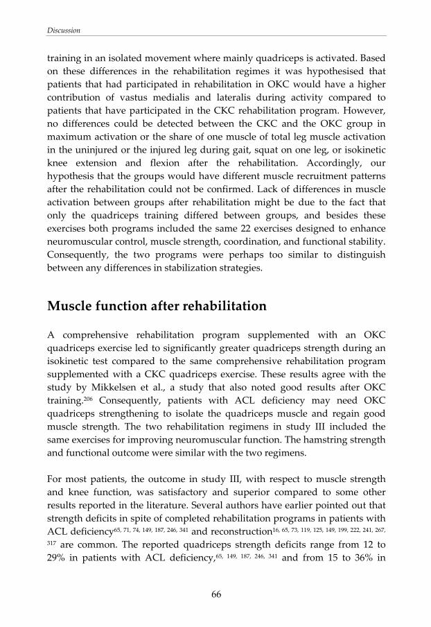
Discussion
66
training in an isolated movement where mainly quadriceps is activated. Based on these differences in the rehabilitation regimes it was hypothesised that patients that had participated in rehabilitation in OKC would have a higher contribution of vastus medialis and lateralis during activity compared to patients that have participated in the CKC rehabilitation program. However, no differences could be detected between the CKC and the OKC group in maximum activation or the share of one muscle of total leg muscle activation in the uninjured or the injured leg during gait, squat on one leg, or isokinetic knee extension and flexion after the rehabilitation. Accordingly, our hypothesis that the groups would have different muscle recruitment patterns after the rehabilitation could not be confirmed. Lack of differences in muscle activation between groups after rehabilitation might be due to the fact that only the quadriceps training differed between groups, and besides these exercises both programs included the same 22 exercises designed to enhance neuromuscular control, muscle strength, coordination, and functional stability. Consequently, the two programs were perhaps too similar to distinguish between any differences in stabilization strategies.
Muscle function after rehabilitation
A comprehensive rehabilitation program supplemented with an OKC quadriceps exercise led to significantly greater quadriceps strength during an isokinetic test compared to the same comprehensive rehabilitation program supplemented with a CKC quadriceps exercise. These results agree with the study by Mikkelsen et al., a study that also noted good results after OKC training.206 Consequently, patients with ACL deficiency may need OKC quadriceps strengthening to isolate the quadriceps muscle and regain good muscle strength. The two rehabilitation regimens in study III included the same exercises for improving neuromuscular function. The hamstring strength and functional outcome were similar with the two regimens. For most patients, the outcome in study III, with respect to muscle strength and knee function, was satisfactory and superior compared to some other results reported in the literature. Several authors have earlier pointed out that strength deficits in spite of completed rehabilitation programs in patients with ACL deficiency65, 71, 74, 149, 187, 246, 341 and reconstruction16, 65, 73, 119, 125, 149, 199, 222, 241, 267,
317 are common. The reported quadriceps strength deficits range from 12 to 29% in patients with ACL deficiency,65, 149, 187, 246, 341 and from 15 to 36% in
Discussion
67
patients with ACL reconstruction. 65, 73, 119, 125, 149, 199, 222, 241, 267, 317 It is recommended that patients should have less than 15% deficits in muscle strength and less than 10% deficits in one leg hop test to resume sport activities.163 In study III, the mean deficit in quadriceps muscle strength during isokinetic testing was 16% for the CKC group, slightly worse than the recommended level, and only 4% for the OKC group. The mean deficits in jump performance were 9% and 7% in the CKC group and 6% and 4% in the OKC group. In spite of the somewhat poor quadriceps strength in the CKC group, most patients in both groups met the criteria necessary to return to sports. It is known that quadriceps strength is correlated with knee function after ACL injury71, 152, 230, 343 and reconstruction.152, 216, 300 However, as seen in study III, muscle strength does not necessarily correlate to the functional outcome. The lack of differences in knee function between the CKC and the OKC group may be explained by the fact that the assessment after rehabilitation was performed immediately after completed rehabilitation in the present study. Because the majority of the patients had at that time not yet returned to sports, they had not exposed their knee to all situations they wanted to manage after the rehabilitation. Twenty‐nine patients planned to return to their sport after the final assessment, 11 patients were scheduled for ACL reconstruction, and only two patients had decided to adjust activity to a lower level. The return to sports would test the knee further and deficits in functional performance may not be obvious until several months later. It is possible that different results regarding knee function would have been obtained if the knee were assessed at a later time. Another conceivable explanation to why the muscle strength was not correlated with the knee function is that the measurements used to assess knee function were not sensitive enough to discriminate between patients with good and poor knee function at assessment immediately after completed rehabilitation. A test battery including vertical jump, hop for distance, and side hop have greater sensitivity compared to any of the hop tests individually.108 Likewise, low correlations between isokinetic strength measurements and functional performance in uninjured individuals have been reported102, 240 indicating that there is no linear relationship between these variables. Functional performance involves not only strength, but also other skills such as coordination and postural control.
Discussion
68
Strength training and dosage of rehabilitation programs
In study III, the load of the exercises was carefully controlled and a continuous progression in the rehabilitation program was emphasized. This approach seems to be successful in gaining muscle function after ACL injury. A need for more effective strength training programs for patients with ACL reconstruction has been identified. There is a dose‐response relationship between training stimulus and increases in muscular strength. Exercise programs involving low‐volume resistance training are probably not sufficient.2, 251, 252, 262, 336 Augustsson pointed out the importance of progression of load and exercises in strength training and recommended a training intensity of at least 8 to 12 RM.13 The design of the rehabilitation programs in study III ‐ load of 50 to 80% of 1RM, continuous progression, ten repetitions and three sets of each exercise, and exercise frequency of three days per week ‐ is supported in the literature.13, 203, 251, 252, 262, 336 The dosage of the rehabilitation programs in study III seems to be sufficient for most patients. The compliance, according to our criteria, was optimal in 14 patients in the CKC group and 15 patients in the OKC group. Consequently, because some patients had somewhat low adherence to the rehabilitation programs, it is difficult to interpret the response to the dosage of the programs. The results of the rehabilitation programs may be biased toward the failure of some patients to fully comply with the regimen. However, the compliance was similar in both groups. There was a great range in number of performed training sessions at the gym, but the median number did not differ between groups. Analysis of only the patients that met the criteria for optimal compliance showed an increased difference in quadriceps strength between the CKC and the OKC group. The quadriceps strength for the injured leg (expressed as a percentage of the strength of the uninjured leg) was 80 ± 15% for the CKC group and 99 ± 11% for the OKC group (P = 0.001). In the CKC group, numbers of completed exercise sessions did not correlate with the quadriceps strength. In the OKC group, there was a weak correlation (r = 0.49, P = 0.028). Consequently, there was not a straight association between compliance to the rehabilitation program and outcome for all patients. This result agrees with findings by others.290 Supplementary training performed beside the rehabilitation program, especially in the last rehabilitation phase, also probably stimulated development of muscle strength. Additionally, psychological aspects96 and perceived self efficacy322 may have affected the
Discussion
69
patient’s ability to follow guidelines and complete the rehabilitation. However, this was unfortunately not controlled for in the present study.
Use of one repetition maximum tests
It is well known that the dosage of the exercises is of great importance for the outcome in strength training and rehabilitation programs.2, 49, 76, 251, 252, 262, 336 It has also been pointed out that the training needs to be subscribed on individual basis and that a continuous progression is necessary for muscular strength development.251 To accomplish this in clinical practise, it is essential to have an accessible method for strength evaluation. Use of 1RM test was applicable to regularly control the current muscle strength and adjust the load of the exercises after the test in study III. Moreover, it is preferable to evaluate the training effect in a similar movement as the practised exercise as there is a specificity in the training effect.2, 14, 50, 160, 161, 215 Therefore, the 1RM test of squat on one leg (CKC) was used together with the isokinetic testing (OKC) in order to evaluate muscle strength in both CKC and OKC. The systematic procedure for the establishment of 1RM developed in the present study can be recommended for use in the clinic. However, high loads are put on the patient during the test, and this is not suitable for patients that have low tolerance towards loads such as patients at an early rehabilitation phase. Also, the CKC exercise involves high loads not only on the knee, but also on the spine and hip. In an early rehabilitation period, 1RM testing can be performed on the uninjured leg to get an initial strength value. Tests on the injured leg may be performed in the late rehabilitation period when involved structures can tolerate the heavy load. In the RCT study, an alternative method with submaximal load for assessing strength was used for some patients. The repetitions‐to‐failure method with submaximal load is frequently used in the clinic on these patients. The method is based on the relationship between the percentage of 1RM and the number of repetitions that can be performed.106
Non‐surgical treatment or ACL reconstruction
At follow‐up (study III) in mean 32 months (range 23‐38 months) after the injury 20 patients (9 CKC, 11 OKC) had undergone ACL reconstruction. There is insufficient evidence to determine whether non‐surgical treatment or ACL reconstruction is preferable.181 Non‐surgical treatment, including rehabilitation
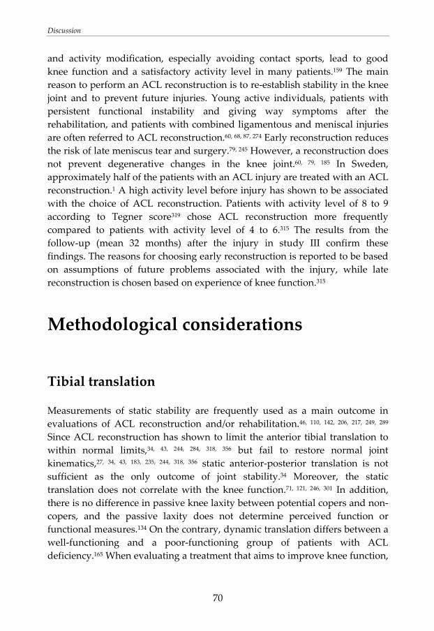
Discussion
70
and activity modification, especially avoiding contact sports, lead to good knee function and a satisfactory activity level in many patients.159 The main reason to perform an ACL reconstruction is to re‐establish stability in the knee joint and to prevent future injuries. Young active individuals, patients with persistent functional instability and giving way symptoms after the rehabilitation, and patients with combined ligamentous and meniscal injuries are often referred to ACL reconstruction.60, 68, 87, 274 Early reconstruction reduces the risk of late meniscus tear and surgery.79, 245 However, a reconstruction does not prevent degenerative changes in the knee joint.60, 79, 185 In Sweden, approximately half of the patients with an ACL injury are treated with an ACL reconstruction.1 A high activity level before injury has shown to be associated with the choice of ACL reconstruction. Patients with activity level of 8 to 9 according to Tegner score319 chose ACL reconstruction more frequently compared to patients with activity level of 4 to 6.315 The results from the follow‐up (mean 32 months) after the injury in study III confirm these findings. The reasons for choosing early reconstruction is reported to be based on assumptions of future problems associated with the injury, while late reconstruction is chosen based on experience of knee function.315
Methodological considerations
Tibial translation
Measurements of static stability are frequently used as a main outcome in evaluations of ACL reconstruction and/or rehabilitation.46, 110, 142, 206, 217, 249, 289 Since ACL reconstruction has shown to limit the anterior tibial translation to within normal limits,34, 43, 244, 284, 318, 356 but fail to restore normal joint kinematics,27, 34, 43, 183, 235, 244, 318, 356 static anterior‐posterior translation is not sufficient as the only outcome of joint stability.34 Moreover, the static translation does not correlate with the knee function.71, 121, 246, 301 In addition, there is no difference in passive knee laxity between potential copers and non‐copers, and the passive laxity does not determine perceived function or functional measures.134 On the contrary, dynamic translation differs between a well‐functioning and a poor‐functioning group of patients with ACL deficiency.165 When evaluating a treatment that aims to improve knee function,
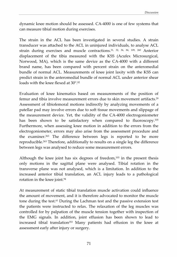
Discussion
71
dynamic knee motion should be assessed. CA‐4000 is one of few systems that can measure tibial motion during exercises. The strain in the ACL has been investigated in several studies. A strain transducer was attached to the ACL in uninjured individuals, to analyse ACL strain during exercises and muscle contractions.31, 32, 36, 84, 109, 260 Anterior displacement of the tibia measured with the KSS (Acufex Microsurgical, Norwood, MA), which is the same device as the CA‐4000 with a different brand name, has been compared with percent strain on the anteromedial bundle of normal ACL. Measurements of knee joint laxity with the KSS can predict strain in the anteromedial bundle of normal ACL under anterior shear loads with the knee flexed at 30°.82 Evaluation of knee kinematics based on measurements of the position of femur and tibia involve measurement errors due to skin movement artifacts.29 Assessment of tibiofemoral motions indirectly by analyzing movements of a patellar pad may involve errors due to soft tissue movements and slippage of the measurement device. Yet, the validity of the CA‐4000 electrogoniometer has been shown to be satisfactory when compared to fluoroscopy.335 Furthermore, when assessing knee motion in addition to the errors from the electrogoniometer, errors may also arise from the assessment procedure and the examiner.263 The difference between legs is reported to be more reproducible.263 Therefore, additionally to results on a single leg the difference between legs was analysed to reduce some measurement errors. Although the knee joint has six degrees of freedom,103 in the present thesis only motions in the sagittal plane were analysed. Tibial rotation in the transverse plane was not analysed, which is a limitation. In addition to the increased anterior tibial translation, an ACL injury leads to a pathological rotation in the knee joint.94 At measurement of static tibial translation muscle activation could influence the amount of movement, and it is therefore advocated to monitor the muscle tone during the test.61 During the Lachman test and the passive extension test the patients were instructed to relax. The relaxation of the leg muscles was controlled for by palpation of the muscle tension together with inspection of the EMG signals. In addition, joint effusion has been shown to lead to increased tibial translation353 Many patients had effusion in the knee at assessment early after injury or surgery.
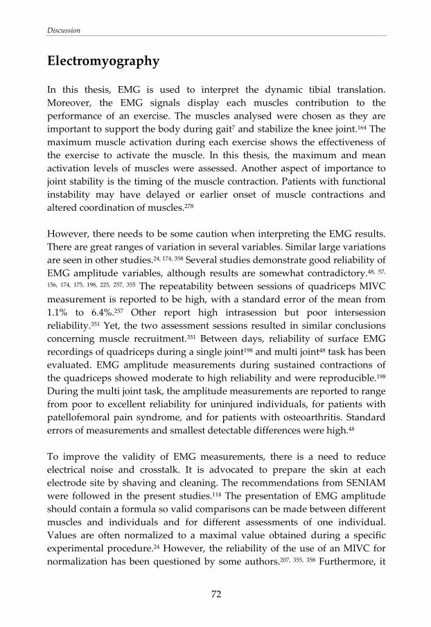
Discussion
72
Electromyography
In this thesis, EMG is used to interpret the dynamic tibial translation. Moreover, the EMG signals display each muscles contribution to the performance of an exercise. The muscles analysed were chosen as they are important to support the body during gait7 and stabilize the knee joint.164 The maximum muscle activation during each exercise shows the effectiveness of the exercise to activate the muscle. In this thesis, the maximum and mean activation levels of muscles were assessed. Another aspect of importance to joint stability is the timing of the muscle contraction. Patients with functional instability may have delayed or earlier onset of muscle contractions and altered coordination of muscles.278 However, there needs to be some caution when interpreting the EMG results. There are great ranges of variation in several variables. Similar large variations are seen in other studies.24, 174, 358 Several studies demonstrate good reliability of EMG amplitude variables, although results are somewhat contradictory.48, 57, 156, 174, 175, 198, 225, 257, 355 The repeatability between sessions of quadriceps MIVC measurement is reported to be high, with a standard error of the mean from 1.1% to 6.4%.257 Other report high intrasession but poor intersession reliability.351 Yet, the two assessment sessions resulted in similar conclusions concerning muscle recruitment.351 Between days, reliability of surface EMG recordings of quadriceps during a single joint198 and multi joint48 task has been evaluated. EMG amplitude measurements during sustained contractions of the quadriceps showed moderate to high reliability and were reproducible.198 During the multi joint task, the amplitude measurements are reported to range from poor to excellent reliability for uninjured individuals, for patients with patellofemoral pain syndrome, and for patients with osteoarthritis. Standard errors of measurements and smallest detectable differences were high.48 To improve the validity of EMG measurements, there is a need to reduce electrical noise and crosstalk. It is advocated to prepare the skin at each electrode site by shaving and cleaning. The recommendations from SENIAM were followed in the present studies.114 The presentation of EMG amplitude should contain a formula so valid comparisons can be made between different muscles and individuals and for different assessments of one individual. Values are often normalized to a maximal value obtained during a specific experimental procedure.24 However, the reliability of the use of an MIVC for normalization has been questioned by some authors.207, 355, 358 Furthermore, it
Discussion
73
should be remembered that the reference value that is used for calculation of the relative values determines the magnitude of the relative value. The performance during the MIVC test is crucial. Also, the magnitude of the EMG signal is affected by different positions ‐ other flexion angles in the joints. An MIVC test performed in another position would probably result in other values since maximal EMG is angle‐dependent.24, 352 Normalization of an EMG value collected during a dynamic movement with respect to an EMG value obtained during a single joint angle has been shown to result in large errors. At a given joint angle the portion of the muscle being registered is the same. On the contrary, in another joint angle the muscle moves in relation to the skin, and the portion of the muscle sampled thereby changes. Moreover, other conditions are changed, such as which muscles fibres that are sampled and the length/strength relationship of the muscle.207 Yet, other data show high intrasession reliability of percent MIVC values of lower extremity muscles.351 Some of the exercises analysed in the present studies are performed with other joint angles than the MIVC test. Furthermore, muscle activation depends on the type of muscular actions performed.17 In the present studies, the reference values are obtained during isometric contractions, which are used as references to the dynamic contractions. This may have contributed to the fact that some relative EMG values achieved were above 100%MIVC. However, all exercises are compared to the same reference value. In addition, when assessing patients five weeks after ACL reconstruction (study IV), it was evident that some patients had difficulty activating the hamstrings in a flexed knee position due to pain or discomfort on the harvest site on the hamstring tendon. The MIVC is reported to be significantly lower in a condition with muscle pain compared to a control condition.101, 189 In a previous study, despite a reduction in MIVC during pain, a submaximal voluntary contraction (80% of the MIVC before pain) was possible.101 The different response to maximal and submaximal contractions is suggested to be caused by changes in the neuronal drive. Decreased MIVC may be explained by an inhibitory influence on the motorneurons.101 This mechanism would lead to greater relative EMG values of painful muscles. Moreover, it is possible that the surgical procedure with graft harvesting on the hamstrings tendon may affect the EMG signal from the hamstrings muscle at assessment five weeks after the ACL reconstruction. The tendon was at that time at a healing phase. This may lead to an increase of the EMG signal. The amplitude of the EMG signal depends on geometrical properties of the motor unit and the muscle tissue.23 Increased activity in muscle spindle afferents have been
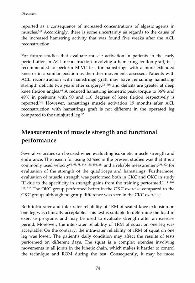
Discussion
74
reported as a consequence of increased concentrations of algesic agents in muscles.247 Accordingly, there is some uncertainty as regards to the cause of the increased hamstring activity that was found five weeks after the ACL reconstruction. For future studies that evaluate muscle activation in patients in the early period after an ACL reconstruction involving a hamstring tendon graft, it is recommended to perform MIVC test for hamstrings with a more extended knee or in a similar position as the other movements assessed. Patients with ACL reconstruction with hamstrings graft may have remaining hamstring strength deficits two years after surgery,72, 316 and deficits are greater at deep knee flexion angles.93 A reduced hamstring isometric peak torque to 86% and 49% in positions with 90 and 110 degrees of knee flexion respectively is reported.316 However, hamstrings muscle activation 19 months after ACL reconstruction with hamstrings graft is not different in the operated leg compared to the uninjured leg.93
Measurements of muscle strength and functional performance
Several velocities can be used when evaluating isokinetic muscle strength and endurance. The reason for using 60°/sec in the present studies was that it is a commonly used velocity64, 65, 98, 143, 150, 151, 211 and a reliable measurement255, 323 for evaluation of the strength of the quadriceps and hamstrings. Furthermore, evaluation of muscle strength was performed both in CKC and OKC in study III due to the specificity in strength gains from the training performed.2, 14, 160, 161, 215 The OKC group performed better in the OKC exercise compared to the CKC group, although no group difference was seen in the CKC exercise. Both intra‐rater and inter‐rater reliability of 1RM of seated knee extension on one leg was clinically acceptable. This test is suitable to determine the load in exercise programs and may be used to evaluate strength after an exercise period. Moreover, the inter‐rater reliability of 1RM of squat on one leg was acceptable. On the contrary, the intra‐rater reliability of 1RM of squat on one leg was lower. The patient’s daily condition may affect the results of tests performed on different days. The squat is a complex exercise involving movements in all joints in the kinetic chain, which makes it harder to control the technique and ROM during the test. Consequently, it may be more
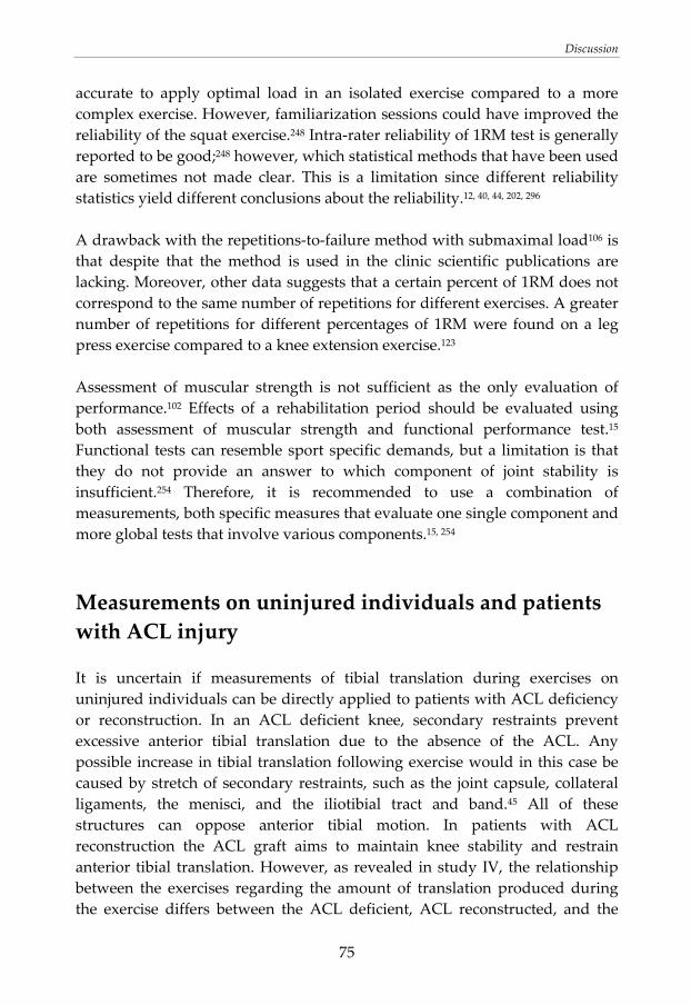
Discussion
75
accurate to apply optimal load in an isolated exercise compared to a more complex exercise. However, familiarization sessions could have improved the reliability of the squat exercise.248 Intra‐rater reliability of 1RM test is generally reported to be good;248 however, which statistical methods that have been used are sometimes not made clear. This is a limitation since different reliability statistics yield different conclusions about the reliability.12, 40, 44, 202, 296 A drawback with the repetitions‐to‐failure method with submaximal load106 is that despite that the method is used in the clinic scientific publications are lacking. Moreover, other data suggests that a certain percent of 1RM does not correspond to the same number of repetitions for different exercises. A greater number of repetitions for different percentages of 1RM were found on a leg press exercise compared to a knee extension exercise.123 Assessment of muscular strength is not sufficient as the only evaluation of performance.102 Effects of a rehabilitation period should be evaluated using both assessment of muscular strength and functional performance test.15 Functional tests can resemble sport specific demands, but a limitation is that they do not provide an answer to which component of joint stability is insufficient.254 Therefore, it is recommended to use a combination of measurements, both specific measures that evaluate one single component and more global tests that involve various components.15, 254
Measurements on uninjured individuals and patients with ACL injury
It is uncertain if measurements of tibial translation during exercises on uninjured individuals can be directly applied to patients with ACL deficiency or reconstruction. In an ACL deficient knee, secondary restraints prevent excessive anterior tibial translation due to the absence of the ACL. Any possible increase in tibial translation following exercise would in this case be caused by stretch of secondary restraints, such as the joint capsule, collateral ligaments, the menisci, and the iliotibial tract and band.45 All of these structures can oppose anterior tibial motion. In patients with ACL reconstruction the ACL graft aims to maintain knee stability and restrain anterior tibial translation. However, as revealed in study IV, the relationship between the exercises regarding the amount of translation produced during the exercise differs between the ACL deficient, ACL reconstructed, and the
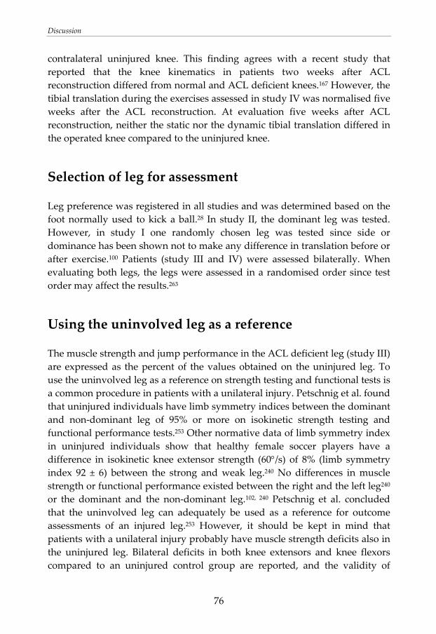
Discussion
76
contralateral uninjured knee. This finding agrees with a recent study that reported that the knee kinematics in patients two weeks after ACL reconstruction differed from normal and ACL deficient knees.167 However, the tibial translation during the exercises assessed in study IV was normalised five weeks after the ACL reconstruction. At evaluation five weeks after ACL reconstruction, neither the static nor the dynamic tibial translation differed in the operated knee compared to the uninjured knee.
Selection of leg for assessment
Leg preference was registered in all studies and was determined based on the foot normally used to kick a ball.28 In study II, the dominant leg was tested. However, in study I one randomly chosen leg was tested since side or dominance has been shown not to make any difference in translation before or after exercise.100 Patients (study III and IV) were assessed bilaterally. When evaluating both legs, the legs were assessed in a randomised order since test order may affect the results.263
Using the uninvolved leg as a reference
The muscle strength and jump performance in the ACL deficient leg (study III) are expressed as the percent of the values obtained on the uninjured leg. To use the uninvolved leg as a reference on strength testing and functional tests is a common procedure in patients with a unilateral injury. Petschnig et al. found that uninjured individuals have limb symmetry indices between the dominant and non‐dominant leg of 95% or more on isokinetic strength testing and functional performance tests.253 Other normative data of limb symmetry index in uninjured individuals show that healthy female soccer players have a difference in isokinetic knee extensor strength (60°/s) of 8% (limb symmetry index 92 ± 6) between the strong and weak leg.240 No differences in muscle strength or functional performance existed between the right and the left leg240 or the dominant and the non‐dominant leg.102, 240 Petschnig et al. concluded that the uninvolved leg can adequately be used as a reference for outcome assessments of an injured leg.253 However, it should be kept in mind that patients with a unilateral injury probably have muscle strength deficits also in the uninjured leg. Bilateral deficits in both knee extensors and knee flexors compared to an uninjured control group are reported, and the validity of
Discussion
77
using the contralateral leg as a rehabilitation endpoint may thereby be questioned.118
Design of the randomised clinical study (III)
The inclusion procedure in the RCT study may have failed to include all patients that met the inclusion criteria. A possible source of imperfection is that the routine with information about the study to the patient and information about the patient to the research investigator failed for some patients. However, the study sample can be considered to be representative to a population with ACL deficiency as regards to sex, age, and activity level.104 There has been a great demand for RCT studies evaluating the rehabilitation after ACL injury.35, 268, 327 Consolidated Standards of Reporting Trials (CONSORT) statement include a checklist and a flow diagram to improve reporting of a RCT.209 The present RCT study (study III) was designed, as far as possible, in accordance with these criteria. Completely blinding, including those administrating the interventions, may not be possible in a rehabilitation study. In the present study, the monitoring physiotherapists were naturally aware of group assignment. Moreover, the patients in the two groups performed the rehabilitation in the same gym, and it can not be eliminated that they saw the exercises performed of the patients in the other regimen. However, it was emphasized that no other exercises for the legs were allowed, and this was controlled for by the monitoring physiotherapist. The principal investigator that performed all outcome measurements in the laboratory (S.T.) was blinded to the group allocation for the great majority of the patients; it was not possible to secure a blinded test procedure for seven patients whose treatment she monitored, which is a limitation of the study. However, all data were coded during analysis and the analyses were performed several months after the assessments.
Compliance to the rehabilitation programs
Several earlier studies evaluating ACL rehabilitation did not report compliance to the rehabilitation programs, which is a limitation. It has been recommended that information regarding compliance should be included in RCT trials.268 Compliance to the rehabilitation regimen in study III was
Discussion
78
monitored with self‐reported exercises diaries. In addition, performed exercise sessions were entered in the patient journal if the patient at the same occasion was monitored by the physiotherapist. Some sessions were performed without supervision from the physiotherapist, and these sessions were registered at the reception desk at the physiotherapy clinic. Consequently, it is evident that the reported exercises sessions have been performed. In study IV compliance with the rehabilitation program was monitored with self‐reported exercises diaries. The diaries were used to make sure that no patient got treatment that differed from the customary rehabilitation program; no further analyses were performed. Since all patients went through the same surgical procedure and followed the same rehabilitation program, they can be considered a homogenous group.
Contribution to research The main contribution to research is the publication of the first RCT study on dynamic knee stability after rehabilitation in patients with ACL deficiency. There has been a debate regarding the optimal rehabilitation program for patients with ACL deficiency and reconstruction. In particular, there has been a controversy whether CKC or OKC exercises or a combination of both is preferable. This thesis demonstrates that both CKC and OKC exercises are suitable for ACL rehabilitation and that the quadriceps muscle needs to be challenged to regain strength. Persistent muscle strength deficits in patients with ACL deficiency or reconstruction have been reported. The present results support theories that structured rehabilitation with continuous progression in load is beneficial and essential. Furthermore, this thesis increases knowledge of dynamic knee stability after an exercise session and during rehabilitation exercises five weeks after ACL reconstruction.
Clinical implications Quadriceps strengthening in OKC should be included in rehabilitation programs for patients with ACL deficiency in order to achieve good quadriceps strength. The exercise does not reduce the knee joint stability in patients with ACL deficiency.
Discussion
79
Establishment of 1RM of a seated knee extension on one leg can be used to determine the load in exercise programs and may be used to evaluate strength after an exercise period. The reliability of 1RM of a squat on one leg between different days was lower, which may be due to the complexity of that exercise. The systematic procedure for the establishment of 1RM developed in the present study can be recommended for use in the clinic. However, this assessment is appropriate exclusively when maximal load is allowed. In the early rehabilitation period, 1RM testing can be performed on the uninjured leg to get an initial strength value. Tests on the injured leg may be performed in the late rehabilitation period when involved structures are able to tolerate the heavy load. All exercises tested in study IV can be recommended in rehabilitation in the early period after ACL reconstruction. All exercises produced less or equal amount of anterior tibial translation as the 90N Lachman test. Yet, it is unclear how much translation that is detrimental. In order to minimize strain on the ACL graft, the straight leg raise can be chosen for quadriceps training in the early phase after ACL reconstruction since that exercise produces less translation compared to the seated knee extension. However, it appeared that after the ACL reconstruction the patients had difficulty activating the quadriceps muscle during the straight leg raise. In that case, the seated knee extension is probably more effective in restoring muscle strength. To achieve great hamstring activation, gait and squat on one leg were superior among the exercises tested. To challenge the gastrocnemius and soleus, gait, squat on one leg and heel rise can be recommended.
Future research Further knowledge and understanding of the effect of different rehabilitation exercises and rehabilitation programs on patients with ACL deficiency or ACL reconstruction is essential in order to improve the outcome of rehabilitation. Future research ideas involve analysis of more challenging rehabilitation exercises in patients with ACL reconstruction. Exercises used at later phases in the rehabilitation need to be evaluated for their effect on dynamic knee stability. Subsequently, it would be interesting to investigate dynamic tibial translation and muscle function after different rehabilitation programs for patients with ACL reconstruction using a RCT design.
Conclusions
80
CONCLUSIONS
1) 1RM as an evaluation method for muscle strength • The intra‐ and inter‐rater reliability of 1RM of a seated knee
extension on one leg were clinically acceptable. The inter‐rater reliability of 1RM of a squat on one leg was clinically acceptable. On the contrary, the intra‐rater reliability, with assessment of 1RM of a squat on one leg performed on different days, was lower.
• The systematic procedure for the establishment of 1RM developed
in the present study can be recommended for use in a clinic or in research. The assessment is suitable exclusively when maximal load is allowed.
2) Exercises in closed and open kinetic chain
• One specific exercise session including cycling and a maximum number of knee extensions and heel raises did not influence static or dynamic sagittal tibial translation (the Lachman test, maximal isometric quadriceps contraction, squat on one leg and gait) in uninjured individuals.
• A comprehensive rehabilitation program with isolated
quadriceps training in OKC led to significantly greater isokinetic quadriceps strength compared to CKC rehabilitation in patients with ACL deficiency. Hamstring strength, static and dynamic translation, and functional outcome were similar between groups. Patients with ACL deficiency may need OKC quadriceps strengthening to regain good muscle strength.
• Comparing different rehabilitation exercises five weeks after ACL reconstruction:
Seated knee extension produced significantly more anterior tibial translation compared to the straight leg raise and standing on one leg. However, all exercises produced less or equal amount of anterior tibial
Conclusions
81
translation as the 90N Lachman test and should not involve excessive strain on the ACL graft.
Vastus medialis and lateralis activation was greater during the seated knee extension and squat on one leg compared to heel raise and standing on one leg. Hamstring activation was high during gait and squat on one leg. Gastrocnemius and soleus were challenged during gait, squat on one leg and heel rise.
3) Dynamic and static knee stability in patients with ACL deficiency or ACL reconstruction
• Five weeks after the ACL reconstruction the static and dynamic tibial translation in the ACL reconstructed knee did not differ from the tibial translation on the uninjured knee.
• Tibial translation during the seated knee extension, squat on one
leg and squat on two legs was significantly reduced after the ACL reconstruction as compared to before.
• Patients in the early phase after ACL injury or ACL reconstruction
used a joint stiffening strategy including a reduced peak knee extension angle during gait and increased hamstring activation during activity, which reduces the dynamic tibial translation. Patients with ACL deficiency that completed a four months rehabilitation program used a movement pattern that was more close to normal. The results suggest that the rehabilitation leads to a more coordinated movement pattern without excessive hamstring activation.
Summary in Swedish
82
SAMMANFATTNING
Främre korsbandsskador är vanligt förekommande bland unga idrottsaktiva individer. Skadan resulterar i en ökad framåtglidning (anterior translation) av tibia i förhållande till femur, och leder därmed till att rörelsemönstret i knäleden förändras. Många patienter besväras av instabilitet och upplever att de inte kan lita på knäleden. Kvarstående besvär trots behandling är vanligt och leder i många fall till att individer tvingas sänka sin aktivitetsnivå eller sluta med sin idrott. Skadan behandlas med enbart rehabilitering alternativt rekonstruktion av korsbandet med efterföljande rehabilitering. Det saknas kunskap om hur en främre korsbands‐rekonstruktion och/eller rehabilitering påverkar den dynamiska stabiliteten i knäleden. Det övergripande syftet med denna avhandling var att studera dynamisk stabilitet i knäleden under och efter rehabilitering hos individer med främre korsbandsskada. Mer specifika syften var 1) att utveckla en metod för utvärdering av muskelfunktion och 2) att utvärdera effekten av övningar i sluten och öppen kinetisk kedja samt 3) att utvärdera dynamisk stabilitet i knäleden hos patienter med främre korsbandsskada eller främre korsbandsrekonstruktion. Intra‐ och interbedömarreliabilitet av 1 repetitionsmaximum (RM) på sittande knäextension var acceptabel. Interbedömarreliabilitet av 1RM på stående knäflexion var också acceptabel, men intrabedömarreliabiliteten var lägre. Ett specifikt träningspass inkluderande cykling och ett maximalt antal knäextensioner och tåhävningar påverkade inte statisk eller dynamisk translation av tibia på individer utan knäskada. Ett sammansatt rehabiliteringsprogram med isolerad quadricepsträning i öppen kinetisk kedja resulterade i signifikant bättre quadricepsstyrka jämfört med rehabilitering i sluten kinetisk kedja för patienter med främre korsbandsskada. Hamstringsstyrka, statisk och dynamisk translation av tibia samt knäfunktion skiljde sig inte mellan grupperna. Fem veckor efter främre korsbands‐rekonstruktion producerade sittande knäextension mer anterior translation av tibia jämfört med rakt benlyft och stående på ett ben. Alla övningar producerade mindre eller lika mycket anterior translation av tibia som 90N Lachman test.
Summary in Swedish
83
Fem veckor efter främre korsbandsrekonstruktion skilde sig inte statisk och dynamisk translation av tibia i den korsbandsrekonstruerade knäleden från translationen i knäleden som inte var skadad. Patienter i en tidig fas efter främre korsbandsskada eller främre korsbandsrekonstruktion rörde knäleden stelare och aktiverade hamstrings i större utsträckning för att stabilisera leden. Detta rörelsemönster reducerar den dynamiska translationen av tibia. Patienter med främre korsbandsskada som genomgått ett fyra månaders rehabiliteringsprogram hade ett mer normalt rörelsemönster.
Acknowledgements
84
ACKNOWLEDGEMENTS
I wish to express my appreciation and gratitude to all of you who made this thesis possible, and in different ways have supported me during this work. Especially I would like to thank: Associate professor Joanna Kvist, my main tutor, for sharing all your knowledge in this research area with me. Thanks for introducing me in the laboratory and teaching me the methods, and for continuous inspiration, great support and excellent guiding throughout the work with my thesis. Professor Birgitta Öberg, my co‐tutor, for all support. Thank you for your wise and valuable comments, and sharing your analytic skills and great knowledge in research. Associate professor Lars Good, my co‐author in paper III, for cooperation in recruiting the patients and for constructive and valuable comments on the study design and the manuscript. My colleagues at the Division of Physiotherapy, for kind support. Thanks for fruitful discussions and valuable comments during the seminars throughout the years. Especially thanks to my PhD student colleagues Jenny Sjödahl, Charlotte Norgren, Emma Nilsing, Pia Ödman and Annelie Gutke for nice cooperation and friendship. Jenny also for helping me in the laboratory, and for discussions about research and all other things in life. Åsa Fahlstedt, Emma Winquist and Elin Wistrand for excellent administrative support. My physiotherapy colleagues Anna Enblom, Jenny Nordqvist and Emma Abou‐Zeid, for friendship and for showing interest in my work. Jan Gillquist for valuable comments on the study design in study I. Karola Messner for making it possible for me to be admitted as a doctorate student. Anna Fahlgren for support and help at the Division of Sport Medicine when I begun this work, and for continuous constructive criticism. My physiotherapy colleagues at Feelgood and Aktiva Rehab for stimulating discussions and cooperation. Special gratitude to Sören Ivarsson, Johan Carlsson,
Acknowledgements
85
Johan Holmberg and Magnus Wretman for monitoring the patient rehabilitation in the RCT study. Catrine for administrative support in the RCT study, and Malin, Anna, Rose‐Marie, Emma, Lisa and all others at the reception desk for help with the inclusion of the patients. Stenove Ringborg, Torbjörn Lindblom and Peter Erlandsson, my former and present bosses at Feelgood, for showing interest in my work, and letting my work fulltime as PhD student this last year to finish my thesis. The physical therapists Per Axelsson, Bettina Hillborg, and Marie Rydén for help with data collection in study II. Olle Eriksson and Henrik Magnusson for statistical advice. All participants in the studies. My parents, Helen and Jörgen for always believing in me and giving me endless support. My sisters Lisa and Åsa for friendship. All friends and relatives for being there. Linus, for love and support and for always encouraging me. The Centre for Orthopaedics at the University Hospital in Linköping for co‐operation. This project was supported by the Faculty of Health Sciences at Linköpings Universitet and by grants from the Swedish National Centre for Research in Sports and the County Council of Östergötland.
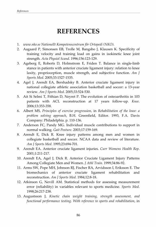
References
86
REFERENCES
1. www.nko.se Nationellt Kompetenscentrum för Ortopedi (NKO). 2. Aagaard P, Simonsen EB, Trolle M, Bangsbo J, Klausen K. Specificity of
training velocity and training load on gains in isokinetic knee joint strength. Acta Physiol Scand. 1996;156:123‐129.
3. Ageberg E, Roberts D, Holmstrom E, Friden T. Balance in single‐limb stance in patients with anterior cruciate ligament injury: relation to knee laxity, proprioception, muscle strength, and subjective function. Am J Sports Med. 2005;33:1527‐1535.
4. Agel J, Arendt EA, Bershadsky B. Anterior cruciate ligament injury in national collegiate athletic association basketball and soccer: a 13‐year review. Am J Sports Med. 2005;33:524‐530.
5. Ait Si Selmi T, Fithian D, Neyret P. The evolution of osteoarthritis in 103 patients with ACL reconstruction at 17 years follow‐up. Knee. 2006;13:353‐358.
6. Albert MS, Principles of exercise progression, in Rehabilitation of the knee: a problem solving approach, B.H. Greenfield, Editor. 1993, F.A. Davis Company: Philadelphia. p. 110‐136.
7. Anderson FC, Pandy MG. Individual muscle contributions to support in normal walking. Gait Posture. 2003;17:159‐169.
8. Arendt E, Dick R. Knee injury patterns among men and women in collegiate basketball and soccer. NCAA data and review of literature. Am J Sports Med. 1995;23:694‐701.
9. Arendt EA. Anterior cruciate ligament injuries. Curr Womens Health Rep. 2001;1:211‐217.
10. Arendt EA, Agel J, Dick R. Anterior Cruciate Ligament Injury Patterns Among Collegiate Men and Women. J Athl Train. 1999;34:86‐92.
11. Arms SW, Pope MH, Johnson RJ, Fischer RA, Arvidsson I, Eriksson E. The biomechanics of anterior cruciate ligament rehabilitation and reconstruction. Am J Sports Med. 1984;12:8‐18.
12. Atkinson G, Nevill AM. Statistical methods for assessing measurement error (reliability) in variables relevant to sports medicine. Sports Med. 1998;26:217‐238.
13. Augustsson J, Kinetic chain weight training, strength assessment, and functional performance testing. With reference to sports and rehabilitation, in
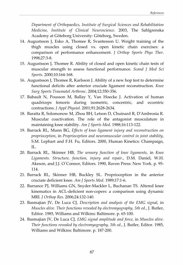
References
87
Department of Orthopaedics, Institute of Surgical Sciences and Rehabilitation Medicine, Institute of Clinical Neuroscience. 2003, The Sahlgrenska Academy at Göteborg University: Göteborg, Sweden.
14. Augustsson J, Esko A, Thomee R, Svantesson U. Weight training of the thigh muscles using closed vs. open kinetic chain exercises: a comparison of performance enhancement. J Orthop Sports Phys Ther. 1998;27:3‐8.
15. Augustsson J, Thomee R. Ability of closed and open kinetic chain tests of muscular strength to assess functional performance. Scand J Med Sci Sports. 2000;10:164‐168.
16. Augustsson J, Thomee R, Karlsson J. Ability of a new hop test to determine functional deficits after anterior cruciate ligament reconstruction. Knee Surg Sports Traumatol Arthrosc. 2004;12:350‐356.
17. Babault N, Pousson M, Ballay Y, Van Hoecke J. Activation of human quadriceps femoris during isometric, concentric, and eccentric contractions. J Appl Physiol. 2001;91:2628‐2634.
18. Baratta R, Solomonow M, Zhou BH, Letson D, Chuinard R, DʹAmbrosia R. Muscular coactivation. The role of the antagonist musculature in maintaining knee stability. Am J Sports Med. 1988;16:113‐122.
19. Barrack RL, Munn BG, Effects of knee ligament injury and reconstruction on proprioception, in Proprioception and neuromuscular control in joint stability, S.M. Lephart and F.H. Fu, Editors. 2000, Human Kinetics: Champaign, IL.
20. Barrack RL, Skinner HB, The sensory function of knee ligaments, in Knee Ligaments. Structure, function, injury and repair., D.M. Daniel, W.H. Akeson, and J.J. O´Connor, Editors. 1990, Raven Press: New York. p. 95‐114.
21. Barrack RL, Skinner HB, Buckley SL. Proprioception in the anterior cruciate deficient knee. Am J Sports Med. 1989;17:1‐6.
22. Barrance PJ, Williams GN, Snyder‐Mackler L, Buchanan TS. Altered knee kinematics in ACL‐deficient non‐copers: a comparison using dynamic MRI. J Orthop Res. 2006;24:132‐140.
23. Basmajian JV, De Luca CJ, Description and analysis of the EMG signal, in Muscles alive. Their functions revealed by electromyography, 5th ed., J. Butler, Editor. 1985, Williams and Wilkins: Baltimore. p. 65‐100.
24. Basmajian JV, De Luca CJ, EMG signal amplitude and force, in Muscles alive. Their functions revealed by electromyography, 5th ed., J. Butler, Editor. 1985, Williams and Wilkins: Baltimore. p. 187‐200.
References
88
25. Beard DJ, Kyberd PJ, Fergusson CM, Dodd CA. Proprioception after rupture of the anterior cruciate ligament. An objective indication of the need for surgery? J Bone Joint Surg Br. 1993;75:311‐315.
26. Beard DJ, Kyberd PJ, OʹConnor JJ, Fergusson CM, Dodd CA. Reflex hamstring contraction latency in anterior cruciate ligament deficiency. J Orthop Res. 1994;12:219‐228.
27. Beard DJ, Murray DW, Gill HS, Price AJ, Rees JL, Alfaro‐Adrian J, Dodd CA. Reconstruction does not reduce tibial translation in the cruciate‐deficient knee an in vivo study. J Bone Joint Surg Br. 2001;83:1098‐1103.
28. Beling J, Wolfe GA, Allen KA, Boyle JM. Lower extremity preference during gross and fine motor skills performed in sitting and standing postures. J Orthop Sports Phys Ther. 1998;28:400‐404.
29. Benoit DL, Ramsey DK, Lamontagne M, Xu L, Wretenberg P, Renstrom P. Effect of skin movement artifact on knee kinematics during gait and cutting motions measured in vivo. Gait Posture. 2006;24:152‐164.
30. Berchuck M, Andriacchi TP, Bach BR, Reider B. Gait adaptations by patients who have a deficient anterior cruciate ligament. J Bone Joint Surg Am. 1990;72:871‐877.
31. Beynnon BD, Fleming BC. Anterior cruciate ligament strain in‐vivo: a review of previous work. J Biomech. 1998;31:519‐525.
32. Beynnon BD, Fleming BC, Johnson RJ, Nichols CE, Renstrom PA, Pope MH. Anterior cruciate ligament strain behavior during rehabilitation exercises in vivo. Am J Sports Med. 1995;23:24‐34.
33. Beynnon BD, Johnson RJ, Abate JA, Fleming BC, Nichols CE. Treatment of anterior cruciate ligament injuries, part 2. Am J Sports Med. 2005;33:1751‐1767.
34. Beynnon BD, Johnson RJ, Abate JA, Fleming BC, Nichols CE. Treatment of anterior cruciate ligament injuries, part I. Am J Sports Med. 2005;33:1579‐1602.
35. Beynnon BD, Johnson RJ, Fleming BC. The science of anterior cruciate ligament rehabilitation. Clin Orthop Relat Res. 2002:9‐20.
36. Beynnon BD, Johnson RJ, Fleming BC, Stankewich CJ, Renstrom PA, Nichols CE. The strain behavior of the anterior cruciate ligament during squatting and active flexion‐extension. A comparison of an open and a closed kinetic chain exercise. Am J Sports Med. 1997;25:823‐829.
37. Beynnon BD, Uh BS, Johnson RJ, Abate JA, Nichols CE, Fleming BC, Poole AR, Roos H. Rehabilitation after anterior cruciate ligament reconstruction: a prospective, randomized, double‐blind comparison of
References
89
programs administered over 2 different time intervals. Am J Sports Med. 2005;33:347‐359.
38. Biau DJ, Tournoux C, Katsahian S, Schranz PJ, Nizard RS. Bone‐patellar tendon‐bone autografts versus hamstring autografts for reconstruction of anterior cruciate ligament: meta‐analysis. Bmj. 2006;332:995‐1001.
39. Biedert RM, Contribution of the three levels of nervous system motor control: spinal cord, lower brain, cerebral cortex, in Proprioception and neuromuscular control in joint stability, S.M. Lephart and F.H. Fu, Editors. 2000, Human Kinetics: Champaign, IL.
40. Bland JM, Altman DG. Statistical methods for assessing between two methods of clinical measurement. Lancet. 1986;feb 8:307‐310.
41. Boden BP, Dean GS, Feagin JA, Jr., Garrett WE, Jr. Mechanisms of anterior cruciate ligament injury. Orthopedics. 2000;23:573‐578.
42. Borg G. Perceived exertion as an indicator of somatic stress. Scand J Rehabil Med. 1970;2:92‐98.
43. Brandsson S, Karlsson J, Sward L, Kartus J, Eriksson BI, Karrholm J. Kinematics and laxity of the knee joint after anterior cruciate ligament reconstruction: pre‐ and postoperative radiostereometric studies. Am J Sports Med. 2002;30:361‐367.
44. Bruton A, Conway JH, Holgate ST. Reliability: what is it, and how is it measured? Physiotherapy. 2000;86:94‐99.
45. Butler DL, Noyes FR, Grood ES. Ligamentous restraints to anterior‐posterior drawer in the human knee. A biomechanical study. J Bone Joint Surg Am. 1980;62‐A:259‐270.
46. Bynum EB, Barrack RL, Alexander AH. Open versus closed chain kinetic exercises after anterior cruciate ligament reconstruction. A prospective randomized study. Am J Sports Med. 1995;23:401‐406.
47. Caborn DN, Johnson BM. The natural history of the anterior cruciate ligament‐deficient knee. A review. Clin Sports Med. 1993;12:625‐636.
48. Callaghan MJ, McCarthy CJ, Oldham JA. The reliability of surface electromyography to assess quadriceps fatigue during multi joint tasks in healthy and painful knees. J Electromyogr Kinesiol. 2007;
49. Campos GE, Luecke TJ, Wendeln HK, Toma K, Hagerman FC, Murray TF, Ragg KE, Ratamess NA, Kraemer WJ, Staron RS. Muscular adaptations in response to three different resistance‐training regimens: specificity of repetition maximum training zones. Eur J Appl Physiol. 2002;88:50‐60.
50. Carroll TJ, Riek S, Carson RG. Neural adaptations to resistance training: implications for movement control. Sports Med. 2001;31:829‐840.
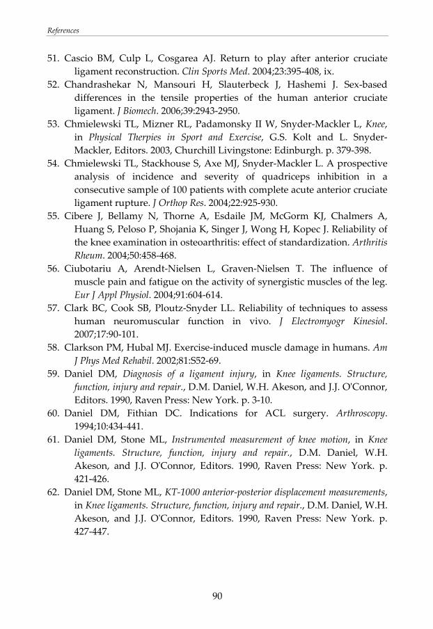
References
90
51. Cascio BM, Culp L, Cosgarea AJ. Return to play after anterior cruciate ligament reconstruction. Clin Sports Med. 2004;23:395‐408, ix.
52. Chandrashekar N, Mansouri H, Slauterbeck J, Hashemi J. Sex‐based differences in the tensile properties of the human anterior cruciate ligament. J Biomech. 2006;39:2943‐2950.
53. Chmielewski TL, Mizner RL, Padamonsky II W, Snyder‐Mackler L, Knee, in Physical Therpies in Sport and Exercise, G.S. Kolt and L. Snyder‐Mackler, Editors. 2003, Churchill Livingstone: Edinburgh. p. 379‐398.
54. Chmielewski TL, Stackhouse S, Axe MJ, Snyder‐Mackler L. A prospective analysis of incidence and severity of quadriceps inhibition in a consecutive sample of 100 patients with complete acute anterior cruciate ligament rupture. J Orthop Res. 2004;22:925‐930.
55. Cibere J, Bellamy N, Thorne A, Esdaile JM, McGorm KJ, Chalmers A, Huang S, Peloso P, Shojania K, Singer J, Wong H, Kopec J. Reliability of the knee examination in osteoarthritis: effect of standardization. Arthritis Rheum. 2004;50:458‐468.
56. Ciubotariu A, Arendt‐Nielsen L, Graven‐Nielsen T. The influence of muscle pain and fatigue on the activity of synergistic muscles of the leg. Eur J Appl Physiol. 2004;91:604‐614.
57. Clark BC, Cook SB, Ploutz‐Snyder LL. Reliability of techniques to assess human neuromuscular function in vivo. J Electromyogr Kinesiol. 2007;17:90‐101.
58. Clarkson PM, Hubal MJ. Exercise‐induced muscle damage in humans. Am J Phys Med Rehabil. 2002;81:S52‐69.
59. Daniel DM, Diagnosis of a ligament injury, in Knee ligaments. Structure, function, injury and repair., D.M. Daniel, W.H. Akeson, and J.J. OʹConnor, Editors. 1990, Raven Press: New York. p. 3‐10.
60. Daniel DM, Fithian DC. Indications for ACL surgery. Arthroscopy. 1994;10:434‐441.
61. Daniel DM, Stone ML, Instrumented measurement of knee motion, in Knee ligaments. Structure, function, injury and repair., D.M. Daniel, W.H. Akeson, and J.J. OʹConnor, Editors. 1990, Raven Press: New York. p. 421‐426.
62. Daniel DM, Stone ML, KT‐1000 anterior‐posterior displacement measurements, in Knee ligaments. Structure, function, injury and repair., D.M. Daniel, W.H. Akeson, and J.J. OʹConnor, Editors. 1990, Raven Press: New York. p. 427‐447.
References
91
63. Daniel DM, Stone ML, Sachs R, Malcom L. Instrumented measurement of anterior knee laxity in patients with acute anterior cruciate ligament disruption. Am J Sports Med. 1985;13:401‐407.
64. Dauty M, Tortellier L, Rochcongar P. Isokinetic and anterior cruciate ligament reconstruction with hamstrings or patella tendon graft: analysis of literature. Int J Sports Med. 2005;26:599‐606.
65. de Jong SN, van Caspel DR, van Haeff MJ, Saris DB. Functional assessment and muscle strength before and after reconstruction of chronic anterior cruciate ligament lesions. Arthroscopy. 2007;23:21‐28, 28 e21‐23.
66. DeCarlo M, Porter DA, Gehlsen G, Bahamonde R. Electromyographic and cinematographic analysis of the lower extremity during closed and open kinetic chain exercise. Isokinet Exerc Sci. 1992;2:24‐29.
67. Dejour H, Bonnin M. Tibial translation after anterior cruciate ligament rupture. Two radiological tests compared. J Bone Joint Surg Br. 1994;76:745‐749.
68. Dunn WR, Lyman S, Lincoln AE, Amoroso PJ, Wickiewicz T, Marx RG. The effect of anterior cruciate ligament reconstruction on the risk of knee reinjury. Am J Sports Med. 2004;32:1906‐1914.
69. Dye SF, Vaupel GL, Functional anatomy of the knee: bony geometry, static and dynamic restraints, sensory and motor innervation, in Proprioception and neuromuscular control in joint stability, S.M. Lephart and F.H. Fu, Editors. 2000, Human Kinetics: Champaign, IL.
70. Dye SF, Wojtys EM, Fu FH, Fithian DC, Gillquist I. Factors contributing to function of the knee joint after injury or reconstruction of the anterior cruciate ligament. Instr Course Lect. 1999;48:185‐198.
71. Eastlack ME, Axe MJ, Snyder‐Mackler L. Laxity, instability, and functional outcome after ACL injury: copers versus noncopers. Med Sci Sports Exerc. 1999;31:210‐215.
72. Elmlinger BS, Nyland JA, Tillett ED. Knee flexor function 2 years after anterior cruciate ligament reconstruction with semitendinosus‐gracilis autografts. Arthroscopy. 2006;22:650‐655.
73. Elmqvist LG, Lorentzon R, Johansson C, Langstrom M, Fagerlund M, Fugl‐Meyer AR. Knee extensor muscle function before and after reconstruction of anterior cruciate ligament tear. Scand J Rehabil Med. 1989;21:131‐139.
74. Engstrom B, Gornitzka J, Johansson C, Wredmark T. Knee function after anterior cruciate ligament ruptures treated conservatively. Int Orthop. 1993;17:208‐213.
References
92
75. Enoka RM. Muscle strength and its development. New perspectives. Sports Med. 1988;6:146‐168.
76. Escamilla R, Wickham R, Exercise‐based conditioning and rehabiliation, in Physical Therapies in Sport and Exercise, G.S. Kolt and L. Snyder‐Mackler, Editors. 2003, Churchill Livingstone: Edinburgh. p. 143‐164.
77. Escamilla RF, Fleisig GS, Zheng N, Barrentine SW, Wilk KE, Andrews JR. Biomechanics of the knee during closed kinetic chain and open kinetic chain exercises. Med Sci Sports Exerc. 1998;30:556‐569.
78. Fahrer H, Rentsch HU, Gerber NJ, Beyeler C, Hess CW, Grunig B. Knee effusion and reflex inhibition of the quadriceps. A bar to effective retraining. J Bone Joint Surg Br. 1988;70:635‐638.
79. Fithian DC, Paxton EW, Stone ML, Luetzow WF, Csintalan RP, Phelan D, Daniel DM. Prospective trial of a treatment algorithm for the management of the anterior cruciate ligament‐injured knee. Am J Sports Med. 2005;33:335‐346.
80. Fitzgerald GK. Open versus closed kinetic chain exercise: issues in rehabilitation after anterior cruciate ligament reconstructive surgery. Phys Ther. 1997;77:1747‐1754.
81. Fitzgerald GK, Axe MJ, Snyder‐Mackler L. A decision‐making scheme for returning patients to high‐level activity with nonoperative treatment after anterior cruciate ligament rupture. Knee Surg Sports Traumatol Arthrosc. 2000;8:76‐82.
82. Fleming B, Beynnon B, Nichols C, Johnson R, Pope M. An in vivo comparison of anterior tibial translation and strain in the anteromedial band of the anterior cruciate ligament. J Biomech. 1993;26:51‐58.
83. Fleming BC, Oksendahl H, Beynnon BD. Open‐ or closed‐kinetic chain exercises after anterior cruciate ligament reconstruction? Exerc Sport Sci Rev. 2005;33:134‐140.
84. Fleming BC, Renstrom PA, Beynnon BD, Engstrom B, Peura GD, Badger GJ, Johnson RJ. The effect of weightbearing and external loading on anterior cruciate ligament strain. J Biomech. 2001;34:163‐170.
85. Fleming BC, Renstrom PA, Ohlen G, Johnson RJ, Peura GD, Beynnon BD, Badger GJ. The gastrocnemius muscle is an antagonist of the anterior cruciate ligament. J Orthop Res. 2001;19:1178‐1184.
86. Forssblad M, Valentin A, Engstrom B, Werner S. ACL reconstruction: patellar tendon versus hamstring grafts‐‐economical aspects. Knee Surg Sports Traumatol Arthrosc. 2006;14:536‐541.
87. Frank CB, Jackson DW. The science of reconstruction of the anterior cruciate ligament. J Bone Joint Surg Am. 1997;79:1556‐1576.
References
93
88. Freedman KB, DʹAmato MJ, Nedeff DD, Kaz A, Bach BR, Jr. Arthroscopic anterior cruciate ligament reconstruction: a metaanalysis comparing patellar tendon and hamstring tendon autografts. Am J Sports Med. 2003;31:2‐11.
89. Friden T, Roberts D, Ageberg E, Walden M, Zatterstrom R. Review of knee proprioception and the relation to extremity function after an anterior cruciate ligament rupture. J Orthop Sports Phys Ther. 2001;31:567‐576.
90. Frobell RB, Lohmander LS, Roos HP. Acute rotational trauma to the knee: poor agreement between clinical assessment and magnetic resonance imaging findings. Scand J Med Sci Sports. 2007;17:109‐114.
91. Fujita I, Nishikawa T, Kambic HE, Andrish JT, Grabiner MD. Characterization of hamstring reflexes during anterior cruciate ligament disruption: in vivo results from a goat model. J Orthop Res. 2000;18:183‐189.
92. Gabriel MT, Wong EK, Woo SL, Yagi M, Debski RE. Distribution of in situ forces in the anterior cruciate ligament in response to rotatory loads. J Orthop Res. 2004;22:85‐89.
93. Garrandes F, Colson SS, Parisaux JM, Legros P. Knee isometric torque and electromyographic specificities after semitendinosus and gracilis hamstrings anterior cruciate ligament reconstruction. Am J Phys Med Rehabil. 2006;85:127‐134.
94. Georgoulis AD, Papadonikolakis A, Papageorgiou CD, Mitsou A, Stergiou N. Three‐dimensional tibiofemoral kinematics of the anterior cruciate ligament‐deficient and reconstructed knee during walking. Am J Sports Med. 2003;31:75‐79.
95. Gillquist J, Messner K. Anterior cruciate ligament reconstruction and the long‐term incidence of gonarthrosis. Sports Med. 1999;27:143‐156.
96. Gobbi A, Francisco R. Factors affecting return to sports after anterior cruciate ligament reconstruction with patellar tendon and hamstring graft: a prospective clinical investigation. Knee Surg Sports Traumatol Arthrosc. 2006;14:1021‐1028.
97. Gogia PP, Braatz JH, Rose SJ, Norton BJ. Reliability and validity of goniometric measurements at the knee. Phys Ther. 1987;67:192‐195.
98. Goradia VK, Grana WA, Pearson SE. Factors associated with decreased muscle strength after anterior cruciate ligament reconstruction with hamstring tendon grafts. Arthroscopy. 2006;22:80.
99. Graham VL, Gehlsen GM, Edwards JA. Electromyographic Evaluation of Closed and Open Kinetic Chain Knee Rehabilitation Exercises. J Athl Train. 1993;28:23‐30.
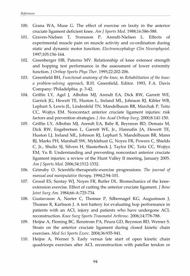
References
94
100. Grana WA, Muse G. The effect of exercise on laxity in the anterior cruciate ligament deficient knee. Am J Sports Med. 1988;16:586‐588.
101. Graven‐Nielsen T, Svensson P, Arendt‐Nielsen L. Effects of experimental muscle pain on muscle activity and co‐ordination during static and dynamic motor function. Electroencephalogr Clin Neurophysiol. 1997;105:156‐164.
102. Greenberger HB, Paterno MV. Relationship of knee extensor strength and hopping test performance in the assessment of lower extremity function. J Orthop Sports Phys Ther. 1995;22:202‐206.
103. Greenfield BH, Functional anatomy of the knee, in Rehabilitation of the knee: a problem‐solving approach, B.H. Greenfield, Editor. 1993, F.A. Davis Company: Philadelphia. p. 3‐42.
104. Griffin LY, Agel J, Albohm MJ, Arendt EA, Dick RW, Garrett WE, Garrick JG, Hewett TE, Huston L, Ireland ML, Johnson RJ, Kibler WB, Lephart S, Lewis JL, Lindenfeld TN, Mandelbaum BR, Marchak P, Teitz CC, Wojtys EM. Noncontact anterior cruciate ligament injuries: risk factors and prevention strategies. J Am Acad Orthop Surg. 2000;8:141‐150.
105. Griffin LY, Albohm MJ, Arendt EA, Bahr R, Beynnon BD, Demaio M, Dick RW, Engebretsen L, Garrett WE, Jr., Hannafin JA, Hewett TE, Huston LJ, Ireland ML, Johnson RJ, Lephart S, Mandelbaum BR, Mann BJ, Marks PH, Marshall SW, Myklebust G, Noyes FR, Powers C, Shields C, Jr., Shultz SJ, Silvers H, Slauterbeck J, Taylor DC, Teitz CC, Wojtys EM, Yu B. Understanding and preventing noncontact anterior cruciate ligament injuries: a review of the Hunt Valley II meeting, January 2005. Am J Sports Med. 2006;34:1512‐1532.
106. Grimsby O. Scientific‐therapeutic‐exercise progressions. The journal of manual and manipulative therapy. 1994;2:94‐101.
107. Grood ES, Suntay WJ, Noyes FR, Butler DL. Biomechanics of the knee‐extension exercise. Effect of cutting the anterior cruciate ligament. J Bone Joint Surg Am. 1984;66‐A:725‐734.
108. Gustavsson A, Neeter C, Thomee P, Silbernagel KG, Augustsson J, Thomee R, Karlsson J. A test battery for evaluating hop performance in patients with an ACL injury and patients who have undergone ACL reconstruction. Knee Surg Sports Traumatol Arthrosc. 2006;14:778‐788.
109. Heijne A, Fleming BC, Renstrom PA, Peura GD, Beynnon BD, Werner S. Strain on the anterior cruciate ligament during closed kinetic chain exercises. Med Sci Sports Exerc. 2004;36:935‐941.
110. Heijne A, Werner S. Early versus late start of open kinetic chain quadriceps exercises after ACL reconstruction with patellar tendon or
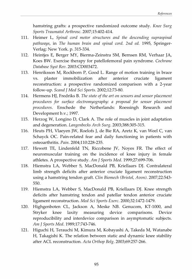
References
95
hamstring grafts: a prospective randomized outcome study. Knee Surg Sports Traumatol Arthrosc. 2007;15:402‐414.
111. Heimer L, Spinal cord motor structures and the descending supraspinal pathways, in The human brain and spinal cord. 2nd ed. 1995, Springer‐Verlag: New York. p. 315‐334.
112. Heintjes E, Berger MY, Bierma‐Zeinstra SM, Bernsen RM, Verhaar JA, Koes BW. Exercise therapy for patellofemoral pain syndrome. Cochrane Database Syst Rev. 2003:CD003472.
113. Henriksson M, Rockborn P, Good L. Range of motion training in brace vs. plaster immobilization after anterior cruciate ligament reconstruction: a prospective randomized comparison with a 2‐year follow‐up. Scand J Med Sci Sports. 2002;12:73‐80.
114. Hermens HJ, Fredriks B. The state of the art on sensors and sensor placement procedures for surface electromyography: a proposal for sensor placement procedures. Enschede the Netherlands: Roessingh Research and Development b.v.; 1997.
115. Herzog W, Longino D, Clark A. The role of muscles in joint adaptation and degeneration. Langenbecks Arch Surg. 2003;388:305‐315.
116. Heuts PH, Vlaeyen JW, Roelofs J, de Bie RA, Aretz K, van Weel C, van Schayck OC. Pain‐related fear and daily functioning in patients with osteoarthritis. Pain. 2004;110:228‐235.
117. Hewett TE, Lindenfeld TN, Riccobene JV, Noyes FR. The effect of neuromuscular training on the incidence of knee injury in female athletes. A prospective study. Am J Sports Med. 1999;27:699‐706.
118. Hiemstra LA, Webber S, MacDonald PB, Kriellaars DJ. Contralateral limb strength deficits after anterior cruciate ligament reconstruction using a hamstring tendon graft. Clin Biomech (Bristol, Avon). 2007;22:543‐550.
119. Hiemstra LA, Webber S, MacDonald PB, Kriellaars DJ. Knee strength deficits after hamstring tendon and patellar tendon anterior cruciate ligament reconstruction. Med Sci Sports Exerc. 2000;32:1472‐1479.
120. Highgenboten CL, Jackson A, Meske NB. Genucom, KT‐1000, and Stryker knee laxity measuring device comparisons. Device reproducibility and interdevice comparison in asymptomatic subjects. Am J Sports Med. 1989;17:743‐746.
121. Higuchi H, Terauchi M, Kimura M, Kobayashi A, Takeda M, Watanabe H, Takagishi K. The relation between static and dynamic knee stability after ACL reconstruction. Acta Orthop Belg. 2003;69:257‐266.
References
96
122. Hodges PW, Motor control, in Physical Therpies in Sport and Exercise, G.S. Kolt and L. Snyder‐Mackler, Editors. 2003, Churchill Livingstone: Edinburgh. p. 107‐125.
123. Hoeger WWK, Hopkins DR, Barette SL, Hale DF. Relationship between repetitions and selected percentages of one repetition maximum: a comparison between untrained and trained males and females. J Appl Sport Sci Res. 1990;4:47‐54.
124. Holden JP, Grood ES, Korvick DL, Cummings JF, Butler DL, Bylski‐Austrow DI. In vivo forces in the anterior cruciate ligament: direct measurements during walking and trotting in a quadruped. J Biomech. 1994;27:517‐526.
125. Holm I, Risberg MA, Aune AK, Tjomsland O, Steen H. Muscle strength recovery following anterior cruciate ligament reconstruction: a prospective study of 151 patients with a two‐year follow‐up. Isokinet Exerc Sci. 2000;8:57‐63.
126. Hooper DM, Morrissey MC, Drechsler W, Morrissey D, King J. Open and closed kinetic chain exercises in the early period after anterior cruciate ligament reconstruction. Improvements in level walking, stair ascent, and stair descent. Am J Sports Med. 2001;29:167‐174.
127. Hootman JM, Dick R, Agel J. Epidemiology of collegiate injuries for 15 sports: summary and recommendations for injury prevention initiatives. J Athl Train. 2007;42:311‐319.
128. Hopkins JT, Ingersoll CD, Krause BA, Edwards JE, Cordova ML. Effect of knee joint effusion on quadriceps and soleus motoneuron pool excitability. Med Sci Sports Exerc. 2001;33:123‐126.
129. Hopkins WG. Measures of reliability in sports medicine and science. Sports Med. 2000;30:1‐15.
130. Hopper DM, Goh SC, Wentworth LA, Chan DYK, Chau JHW, Wootton GJ, Strauss GR, Boyle JJW. Test‐retest reliability of knee rating scales and functional hop tests one year following anterior cruciate ligament reconstruction. Physical Therapy in Sport. 2002;3:10‐18.
131. Howell SM. Anterior tibial translation during a maximum quadriceps contraction: is it clinically significant? Am J Sports Med. 1990;18:573‐578.
132. Hsieh HH, Walker PS. Stabilizing mechanisms of the loaded and unloaded knee joint. J Bone Joint Surg Am. 1976;58‐A:87‐93.
133. Hubal MJ, Gordish‐Dressman H, Thompson PD, Price TB, Hoffman EP, Angelopoulos TJ, Gordon PM, Moyna NM, Pescatello LS, Visich PS, Zoeller RF, Seip RL, Clarkson PM. Variability in muscle size and
References
97
strength gain after unilateral resistance training. Med Sci Sports Exerc. 2005;37:964‐972.
134. Hurd WJ, Axe MJ, Snyder‐Mackler L. A 10‐Year Prospective Trial of a Patient Management Algorithm and Screening Examination for Highly Active Individuals With Anterior Cruciate Ligament Injury : Part 2, Determinants of Dynamic Knee Stability. Am J Sports Med. 2007;
135. Hurley M, Jones D, Wilson D, Newham D. Rehabilitation of quadriceps inhibited due to isolated rupture of the anterior cruciate ligament. Journal of ortopeaedic reumatology. 1992;5:145‐154.
136. Hurley MV. The effects of joint damage on muscle function, proprioception and rehabilitation. Man Ther. 1997;2:11‐17.
137. Huston L, Wojtys E, Neuromuscular performance in the ACL‐deficient knee, in Proprioception and neuromuscular control in joint stability, S.M. Lephart and F.H. Fu, Editors. 2000, Human Kinetics: Champaign, IL.
138. Häkkinen K. Neuromuscular adaptation during strength training, aging, detraining, and immobilization. Crit Rev Phys Rehabil Med. 1994;6:161‐198.
139. Ireland ML. Anterior Cruciate Ligament Injury in Female Athletes: Epidemiology. J Athl Train. 1999;34:150‐154.
140. Irrgang JJ, Neri R, The rationale for open and closed kinetic chain activities for restoration of proprioception and neuromuscular control following injury, in Proprioception and neuromuscular control in joint stability, S.M. Lephart and F.H. Fu, Editors. 2000, Human Kinetics: Champaign, IL. p. 363‐374.
141. Isaac DL, Beard DJ, Price AJ, Rees J, Murray DW, Dodd CA. In‐vivo sagittal plane knee kinematics: ACL intact, deficient and reconstructed knees. Knee. 2005;12:25‐31.
142. Isberg J, Faxen E, Brandsson S, Eriksson BI, Karrholm J, Karlsson J. Early active extension after anterior cruciate ligament reconstruction does not result in increased laxity of the knee. Knee Surg Sports Traumatol Arthrosc. 2006;14:1108‐1115.
143. Jarvela T, Kannus P, Latvala K, Jarvinen M. Simple measurements in assessing muscle performance after an ACL reconstruction. Int J Sports Med. 2002;23:196‐201.
144. Johansson H, Pedersen J, Bergenheim M, Djupsjöbacka M, Peripheral afferents of the knee: their effects on central mechanisms regulating muscle stiffness, joint stability, and proprioception and coordination, in Proprioception and neuromuscular control in joint stability, S.M. Lephart and F.H. Fu, Editors. 2000, Human Kinetics: Champaign, IL.
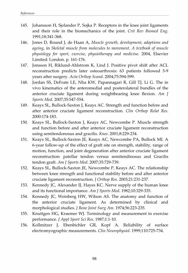
References
98
145. Johansson H, Sjolander P, Sojka P. Receptors in the knee joint ligaments and their role in the biomechanics of the joint. Crit Rev Biomed Eng. 1991;18:341‐368.
146. Jones D, Round J, de Haan A, Muscle growth, development, adaptions and ageing, in Skeletal muscle from molecules to movement. A textbook of muscle physiology for sport, exercise, physiotherapy and medicine. 2004, Elsevier Limited: London. p. 161‐176.
147. Jonsson H, Riklund‐Ahlstrom K, Lind J. Positive pivot shift after ACL reconstruction predicts later osteoarthrosis: 63 patients followed 5‐9 years after surgery. Acta Orthop Scand. 2004;75:594‐599.
148. Jordan SS, DeFrate LE, Nha KW, Papannagari R, Gill TJ, Li G. The in vivo kinematics of the anteromedial and posterolateral bundles of the anterior cruciate ligament during weightbearing knee flexion. Am J Sports Med. 2007;35:547‐554.
149. Keays SL, Bullock‐Saxton J, Keays AC. Strength and function before and after anterior cruciate ligament reconstruction. Clin Orthop Relat Res. 2000:174‐183.
150. Keays SL, Bullock‐Saxton J, Keays AC, Newcombe P. Muscle strength and function before and after anterior cruciate ligament reconstruction using semitendonosus and gracilis. Knee. 2001;8:229‐234.
151. Keays SL, Bullock‐Saxton JE, Keays AC, Newcombe PA, Bullock MI. A 6‐year follow‐up of the effect of graft site on strength, stability, range of motion, function, and joint degeneration after anterior cruciate ligament reconstruction: patellar tendon versus semitendinosus and Gracilis tendon graft. Am J Sports Med. 2007;35:729‐739.
152. Keays SL, Bullock‐Saxton JE, Newcombe P, Keays AC. The relationship between knee strength and functional stability before and after anterior cruciate ligament reconstruction. J Orthop Res. 2003;21:231‐237.
153. Kennedy JC, Alexander IJ, Hayes KC. Nerve supply of the human knee and its functional importance. Am J Sports Med. 1982;10:329‐335.
154. Kennedy JC, Weinberg HW, Wilson AS. The anatomy and function of the anterior cruciate ligament. As determined by clinical and morphological studies. J Bone Joint Surg Am. 1974;56:223‐235.
155. Knuttgen HG, Kraemer WJ. Terminology and measurement in exercise performance. J Appl Sport Sci Res. 1987;1:1‐10.
156. Kollmitzer J, Ebenbichler GR, Kopf A. Reliability of surface electromyographic measurements. Clin Neurophysiol. 1999;110:725‐734.
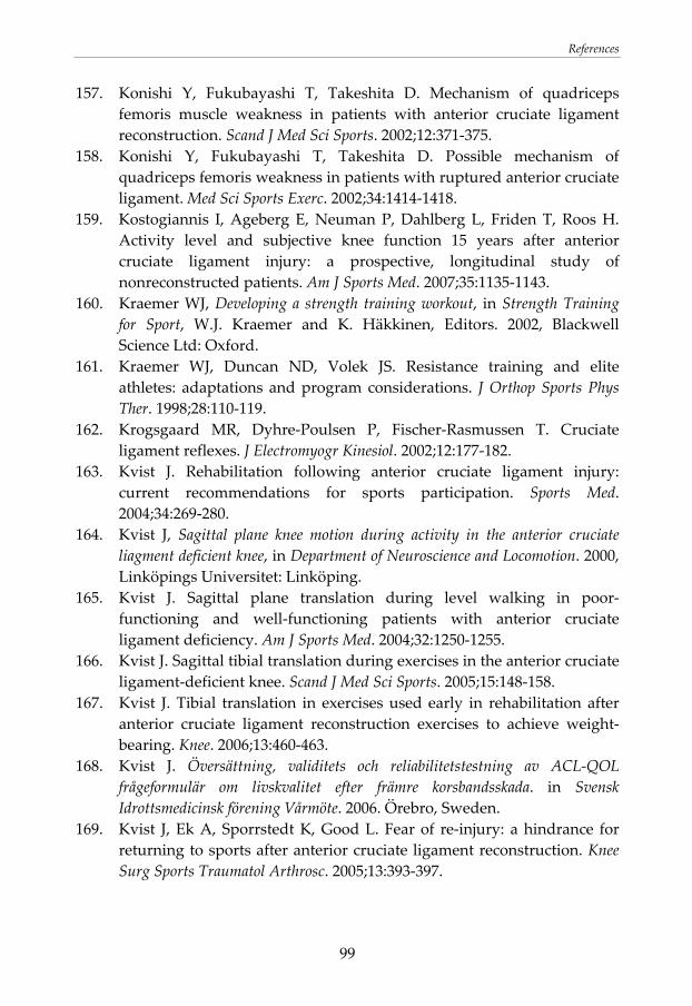
References
99
157. Konishi Y, Fukubayashi T, Takeshita D. Mechanism of quadriceps femoris muscle weakness in patients with anterior cruciate ligament reconstruction. Scand J Med Sci Sports. 2002;12:371‐375.
158. Konishi Y, Fukubayashi T, Takeshita D. Possible mechanism of quadriceps femoris weakness in patients with ruptured anterior cruciate ligament. Med Sci Sports Exerc. 2002;34:1414‐1418.
159. Kostogiannis I, Ageberg E, Neuman P, Dahlberg L, Friden T, Roos H. Activity level and subjective knee function 15 years after anterior cruciate ligament injury: a prospective, longitudinal study of nonreconstructed patients. Am J Sports Med. 2007;35:1135‐1143.
160. Kraemer WJ, Developing a strength training workout, in Strength Training for Sport, W.J. Kraemer and K. Häkkinen, Editors. 2002, Blackwell Science Ltd: Oxford.
161. Kraemer WJ, Duncan ND, Volek JS. Resistance training and elite athletes: adaptations and program considerations. J Orthop Sports Phys Ther. 1998;28:110‐119.
162. Krogsgaard MR, Dyhre‐Poulsen P, Fischer‐Rasmussen T. Cruciate ligament reflexes. J Electromyogr Kinesiol. 2002;12:177‐182.
163. Kvist J. Rehabilitation following anterior cruciate ligament injury: current recommendations for sports participation. Sports Med. 2004;34:269‐280.
164. Kvist J, Sagittal plane knee motion during activity in the anterior cruciate liagment deficient knee, in Department of Neuroscience and Locomotion. 2000, Linköpings Universitet: Linköping.
165. Kvist J. Sagittal plane translation during level walking in poor‐functioning and well‐functioning patients with anterior cruciate ligament deficiency. Am J Sports Med. 2004;32:1250‐1255.
166. Kvist J. Sagittal tibial translation during exercises in the anterior cruciate ligament‐deficient knee. Scand J Med Sci Sports. 2005;15:148‐158.
167. Kvist J. Tibial translation in exercises used early in rehabilitation after anterior cruciate ligament reconstruction exercises to achieve weight‐bearing. Knee. 2006;13:460‐463.
168. Kvist J. Översättning, validitets och reliabilitetstestning av ACL‐QOL frågeformulär om livskvalitet efter främre korsbandsskada. in Svensk Idrottsmedicinsk förening Vårmöte. 2006. Örebro, Sweden.
169. Kvist J, Ek A, Sporrstedt K, Good L. Fear of re‐injury: a hindrance for returning to sports after anterior cruciate ligament reconstruction. Knee Surg Sports Traumatol Arthrosc. 2005;13:393‐397.
References
100
170. Kvist J, Gillquist J. Anterior positioning of tibia during motion after anterior cruciate ligament injury. Med Sci Sports Exerc. 2001;33:1063‐1072.
171. Kvist J, Gillquist J. Sagittal plane knee translation and electromyographic activity during closed and open kinetic chain exercises in anterior cruciate ligament‐deficient patients and control subjects. Am J Sports Med. 2001;29:72‐82.
172. Kvist J, Good L, Tagesson S. Changes in knee motion pattern after anterior cruciate ligament injury ‐ A case report. Clin Biomech (Bristol, Avon). 2007;22:551‐556.
173. Larson RL, Prostetic replacement of knee ligaments: Overview, in The crucial ligaments. Diagnosis and treatment of ligamentous injuries about the knee, J.A. Feagin, Editor. 1988, Churchill Livingstone: New York. p. 495‐501.
174. Larsson B, Karlsson S, Eriksson M, Gerdle B. Test‐retest reliability of EMG and peak torque during repetitive maximum concentric knee extensions. J Electromyogr Kinesiol. 2003;13:281‐287.
175. Larsson B, Mansson B, Karlberg C, Syvertsson P, Elert J, Gerdle B. Reproducibility of surface EMG variables and peak torque during three sets of ten dynamic contractions. J Electromyogr Kinesiol. 1999;9:351‐357.
176. Le Veau BF. Biomechanics of human motion. 3rd edition ed. USA: W.B. Saunders Company; 1992.
177. Leeuw M, Goossens ME, Linton SJ, Crombez G, Boersma K, Vlaeyen JW. The fear‐avoidance model of musculoskeletal pain: current state of scientific evidence. J Behav Med. 2007;30:77‐94.
178. Lephart SM, Riemann MA, Fu FH, Introduction to the sensorimotor system, in Proprioception and neuromuscular control in joint stability, S.M. Lephart and F.H. Fu, Editors. 2000, Human Kinetics: Champaign, IL.
179. Li G, Rudy TW, Allen C, Sakane M, Woo SL. Effect of combined axial compressive and anterior tibial loads on in situ forces in the anterior cruciate ligament: a porcine study. J Orthop Res. 1998;16:122‐127.
180. Li G, Rudy TW, Sakane M, Kanamori A, Ma CB, Woo SL. The importance of quadriceps and hamstring muscle loading on knee kinematics and in‐situ forces in the ACL. J Biomech. 1999;32:395‐400.
181. Linko E, Harilainen A, Malmivaara A, Seitsalo S. Surgical versus conservative interventions for anterior cruciate ligament ruptures in adults. Cochrane Database Syst Rev. 2005:CD001356.
182. Liu W, Maitland ME. The effect of hamstring muscle compensation for anterior laxity in the ACL‐deficient knee during gait. J Biomech. 2000;33:871‐879.
References
101
183. Logan MC, Williams A, Lavelle J, Gedroyc W, Freeman M. Tibiofemoral kinematics following successful anterior cruciate ligament reconstruction using dynamic multiple resonance imaging. Am J Sports Med. 2004;32:984‐992.
184. Logan MC, Williams A, Lavelle J, Gedroyc W, Freeman M. What really happens during the Lachman test? A dynamic MRI analysis of tibiofemoral motion. Am J Sports Med. 2004;32:369‐375.
185. Lohmander LS, Englund PM, Dahl LL, Roos EM. The long‐term consequence of anterior cruciate ligament and meniscus injuries: osteoarthritis. Am J Sports Med. 2007;35:1756‐1769.
186. Lohmander LS, Ostenberg A, Englund M, Roos H. High prevalence of knee osteoarthritis, pain, and functional limitations in female soccer players twelve years after anterior cruciate ligament injury. Arthritis Rheum. 2004;50:3145‐3152.
187. Lorentzon R, Elmqvist LG, Sjostrom M, Fagerlund M, Fuglmeyer AR. Thigh musculature in relation to chronic anterior cruciate ligament tear: muscle size, morphology, and mechanical output before reconstruction. Am J Sports Med. 1989;17:423‐429.
188. Louie JK, Mote CD, Jr. Contribution of the musculature to rotatory laxity and torsional stiffness at the knee. J Biomech. 1987;20:281‐300.
189. Lund JP, Donga R, Widmer CG, Stohler CS. The pain‐adaptation model: a discussion of the relationship between chronic musculoskeletal pain and motor activity. Can J Physiol Pharmacol. 1991;69:683‐694.
190. Lutz GE, Palmitier RA, An KN, Chao EY. Comparison of tibiofemoral joint forces during open‐kinetic‐chain and closed‐kinetic‐chain exercises. J Bone Joint Surg Am. 1993;75:732‐739.
191. Lysholm J, Gillquist J. Evaluation of knee ligament surgery results with special emphasis on use of a scoring scale. Am J Sports Med. 1982;10:150‐154.
192. Mahfouz MR, Komistek RD, Dennis DA, Hoff WA. In vivo assessment of the kinematics in normal and anterior cruciate ligament‐deficient knees. J Bone Joint Surg Am. 2004;86‐A Suppl 2:56‐61.
193. Mangine RE, Noyes FR, DeMaio M. Minimal protection program: advanced weight bearing and range of motion after ACL reconstruction‐‐weeks 1 to 5. Orthopedics. 1992;15:504‐515.
194. Marans HJ, Jackson RW, Glossop ND, Young C. Anterior cruciate ligament insufficiency: a dynamic three‐dimensional motion analysis. Am J Sports Med. 1989;17:325‐332.
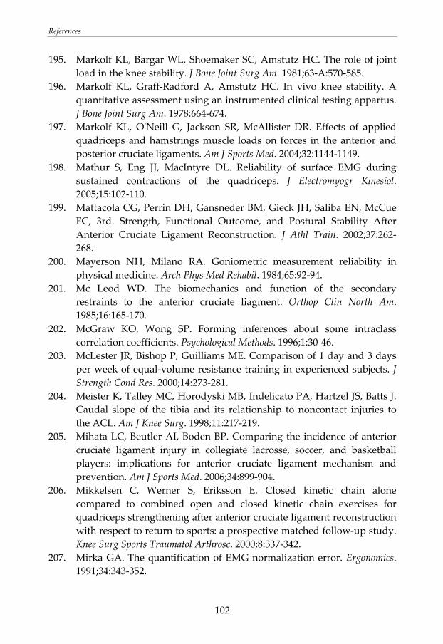
References
102
195. Markolf KL, Bargar WL, Shoemaker SC, Amstutz HC. The role of joint load in the knee stability. J Bone Joint Surg Am. 1981;63‐A:570‐585.
196. Markolf KL, Graff‐Radford A, Amstutz HC. In vivo knee stability. A quantitative assessment using an instrumented clinical testing appartus. J Bone Joint Surg Am. 1978:664‐674.
197. Markolf KL, OʹNeill G, Jackson SR, McAllister DR. Effects of applied quadriceps and hamstrings muscle loads on forces in the anterior and posterior cruciate ligaments. Am J Sports Med. 2004;32:1144‐1149.
198. Mathur S, Eng JJ, MacIntyre DL. Reliability of surface EMG during sustained contractions of the quadriceps. J Electromyogr Kinesiol. 2005;15:102‐110.
199. Mattacola CG, Perrin DH, Gansneder BM, Gieck JH, Saliba EN, McCue FC, 3rd. Strength, Functional Outcome, and Postural Stability After Anterior Cruciate Ligament Reconstruction. J Athl Train. 2002;37:262‐268.
200. Mayerson NH, Milano RA. Goniometric measurement reliability in physical medicine. Arch Phys Med Rehabil. 1984;65:92‐94.
201. Mc Leod WD. The biomechanics and function of the secondary restraints to the anterior cruciate liagment. Orthop Clin North Am. 1985;16:165‐170.
202. McGraw KO, Wong SP. Forming inferences about some intraclass correlation coefficients. Psychological Methods. 1996;1:30‐46.
203. McLester JR, Bishop P, Guilliams ME. Comparison of 1 day and 3 days per week of equal‐volume resistance training in experienced subjects. J Strength Cond Res. 2000;14:273‐281.
204. Meister K, Talley MC, Horodyski MB, Indelicato PA, Hartzel JS, Batts J. Caudal slope of the tibia and its relationship to noncontact injuries to the ACL. Am J Knee Surg. 1998;11:217‐219.
205. Mihata LC, Beutler AI, Boden BP. Comparing the incidence of anterior cruciate ligament injury in collegiate lacrosse, soccer, and basketball players: implications for anterior cruciate ligament mechanism and prevention. Am J Sports Med. 2006;34:899‐904.
206. Mikkelsen C, Werner S, Eriksson E. Closed kinetic chain alone compared to combined open and closed kinetic chain exercises for quadriceps strengthening after anterior cruciate ligament reconstruction with respect to return to sports: a prospective matched follow‐up study. Knee Surg Sports Traumatol Arthrosc. 2000;8:337‐342.
207. Mirka GA. The quantification of EMG normalization error. Ergonomics. 1991;34:343‐352.
References
103
208. Miyasaka KC, Daniel DM, Stone ML, Hirschman P. The incidence of knee ligament injuries in the general population. Am J Knee Surg. 1991;4:3‐8.
209. Moher D, Schulz KF, Altman D. The CONSORT Statement: revised recommendations for improving the quality of reports of parallel‐group randomized trials 2001. Explore (NY). 2005;1:40‐45.
210. Mohtadi N. Development and validation of the quality of life outcome measure (questionnaire) for chronic anterior cruciate ligament deficiency. Am J Sports Med. 1998;26:350‐359.
211. Moisala AS, Jarvela T, Kannus P, Jarvinen M. Muscle strength evaluations after ACL reconstruction. Int J Sports Med. 2007;28:868‐872.
212. More RC, Karras BT, Neiman R, Fritschy D, Woo SL, Daniel DM. Hamstrings‐‐an anterior cruciate ligament protagonist. An in vitro study. Am J Sports Med. 1993;21:231‐237.
213. Morrissey MC. Reflex inhibition of thigh muscles in knee injury. Causes and treatment. Sports Med. 1989;7:263‐276.
214. Morrissey MC, Drechsler WI, Morrissey D, Knight PR, Armstrong PW, McAuliffe TB. Effects of distally fixated versus nondistally fixated leg extensor resistance training on knee pain in the early period after anterior cruciate ligament reconstruction. Phys Ther. 2002;82:35‐43.
215. Morrissey MC, Harman EA, Johnson MJ. Resistance training modes: specificity and effectiveness. Med Sci Sports Exerc. 1995;27:648‐660.
216. Morrissey MC, Hooper DM, Drechsler WI, Hill HJ. Relationship of leg muscle strength and knee function in the early period after anterior cruciate ligament reconstruction. Scand J Med Sci Sports. 2004;14:360‐366.
217. Morrissey MC, Hudson ZL, Drechsler WI, Coutts FJ, Knight PR, King JB. Effects of open versus closed kinetic chain training on knee laxity in the early period after anterior cruciate ligament reconstruction. Knee Surg Sports Traumatol Arthrosc. 2000;8:343‐348.
218. Mountcastle SB, Posner M, Kragh JF, Jr., Taylor DC. Gender differences in anterior cruciate ligament injury vary with activity: epidemiology of anterior cruciate ligament injuries in a young, athletic population. Am J Sports Med. 2007;35:1635‐1642.
219. Myer GD, Paterno MV, Ford KR, Quatman CE, Hewett TE. Rehabilitation after anterior cruciate ligament reconstruction: criteria‐based progression through the return‐to‐sport phase. J Orthop Sports Phys Ther. 2006;36:385‐402.
220. Myklebust G, Maehlum S, Engebretsen L, Strand T, Solheim E. Registration of cruciate ligament injuries in Norwegian top level team
References
104
handball. A prospective study covering two seasons. Scand J Med Sci Sports. 1997;7:289‐292.
221. Myklebust G, Maehlum S, Holm I, Bahr R. A prospective cohort study of anterior cruciate ligament injuries in elite Norwegian team handball. Scand J Med Sci Sports. 1998;8:149‐153.
222. Natri A, Jarvinen M, Latvala K, Kannus P. Isokinetic muscle performance after anterior cruciate ligament surgery. Long‐term results and outcome predicting factors after primary surgery and late‐phase reconstruction. Int J Sports Med. 1996;17:223‐228.
223. Nebelung W, Wuschech H. Thirty‐five years of follow‐up of anterior cruciate ligament‐deficient knees in high‐level athletes. Arthroscopy. 2005;21:696‐702.
224. Neeter C, Gustavsson A, Thomee P, Augustsson J, Thomee R, Karlsson J. Development of a strength test battery for evaluating leg muscle power after anterior cruciate ligament injury and reconstruction. Knee Surg Sports Traumatol Arthrosc. 2006;14:571‐580.
225. Ng JK, Parnianpour M, Kippers V, Richardson CA. Reliability of electromyographic and torque measures during isometric axial rotation exertions of the trunk. Clin Neurophysiol. 2003;114:2355‐2361.
226. Nicholas JJ, Taylor FH, Buckingham RB, Ottonello D. Measurement of circumference of the knee with ordinary tape measure. Ann Rheum Dis. 1976;35:282‐284.
227. Nielsen AB, Yde J. Epidemiology of acute knee injuries: a prospective hospital investigation. J Trauma. 1991;31:1644‐1648.
228. Ninos JC, Irrgang JJ, Burdett R, Weiss JR. Electromyographic analysis of the squat performed in self‐selected lower extremity neutral rotation and 30 degrees of lower extremity turn‐out from the self‐selected neutral position. J Orthop Sports Phys Ther. 1997;25:307‐315.
229. Nisell R. Mechanics of the knee. A study of joint and muscle load with clinical applications. Acta Orthop Scand Suppl. 1985;216:1‐42.
230. Noyes FR, Barber SD, Mangine RE. Abnormal lower limb symmetry determined by function hop tests after anterior cruciate ligament rupture. Am J Sports Med. 1991;19:513‐518.
231. Noyes FR, Cummings JF, Grood ES, Walz‐Hasselfeld KA, Wroble RR. The diagnosis of knee motion limits, subluxations, and ligament injury. Am J Sports Med. 1991;19:163‐171.
232. Noyes FR, Grood E, Suntay WJ, Butler DL. The three dimensional laxity or the anterior cruciate ligament deficient knee as determined by clinical laxity tests. Iowa Orthop J. 1983;3:32‐44.
References
105
233. Noyes FR, Grood ES, Cummings JF, Wroble RR. An analysis of the pivot shift phenomenon. The knee motions and subluxations induced by different examiners. Am J Sports Med. 1991;19:148‐155.
234. Noyes FR, Grood ES, Torzilli PA. Current concepts review. The definitions of terms for motion and position of the knee and injuries of the ligaments. J Bone Joint Surg Am. 1989;71:465‐472.
235. Noyes FR, Matthews DS, Mooar PA, Grood ES. The symptomatic anterior cruciate‐deficient knee. Part II: the results of rehabilitation, activity modification, and counseling on functional disability. J Bone Joint Surg Am. 1983;65:163‐174.
236. Noyes FR, Mooar PA, Matthews DS, Butler DL. The symptomatic anterior cruciate‐deficient knee. Part I: the long‐term functional disability in athletically active individuals. J Bone Joint Surg Am. 1983;65:154‐162.
237. OʹConnor JJ. Can muscle co‐contraction protect knee ligaments after injury or repair? J Bone Joint Surg Br. 1993;75:41‐48.
238. Olsen OE, Myklebust G, Engebretsen L, Bahr R. Injury mechanisms for anterior cruciate ligament injuries in team handball: a systematic video analysis. Am J Sports Med. 2004;32:1002‐1012.
239. Oni OOA. Mechanism of injury in anterior cruciate ligament disruption. Knee. 1998;5:81‐86.
240. Ostenberg A, Roos E, Ekdahl C, Roos H. Isokinetic knee extensor strength and functional performance in healthy female soccer players. Scand J Med Sci Sports. 1998;8:257‐264.
241. Osteras H, Augestad LB, Tondel S. Isokinetic muscle strength after anterior cruciate ligament reconstruction. Scand J Med Sci Sports. 1998;8:279‐282.
242. Palastanga N, Field D, Soames R. Anatomy and human movement. Structure and function. Oxford: Butterworth‐Heinemann; 1994.
243. Palmitier RA, An KN, Scott SG, Chao EY. Kinetic chain exercise in knee rehabilitation. Sports Medicine. 1991;11:402‐413.
244. Papannagari R, Gill TJ, Defrate LE, Moses JM, Petruska AJ, Li G. In vivo kinematics of the knee after anterior cruciate ligament reconstruction: a clinical and functional evaluation. Am J Sports Med. 2006;34:2006‐2012.
245. Papastergiou SG, Koukoulias NE, Mikalef P, Ziogas E, Voulgaropoulos H. Meniscal tears in the ACL‐deficient knee: correlation between meniscal tears and the timing of ACL reconstruction. Knee Surg Sports Traumatol Arthrosc. 2007;15:1438‐1444.
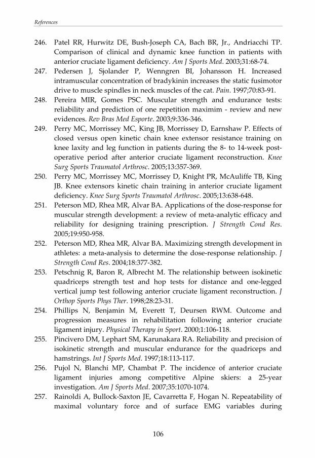
References
106
246. Patel RR, Hurwitz DE, Bush‐Joseph CA, Bach BR, Jr., Andriacchi TP. Comparison of clinical and dynamic knee function in patients with anterior cruciate ligament deficiency. Am J Sports Med. 2003;31:68‐74.
247. Pedersen J, Sjolander P, Wenngren BI, Johansson H. Increased intramuscular concentration of bradykinin increases the static fusimotor drive to muscle spindles in neck muscles of the cat. Pain. 1997;70:83‐91.
248. Pereira MIR, Gomes PSC. Muscular strength and endurance tests: reliability and prediction of one repetition maximim ‐ review and new evidences. Rev Bras Med Esporte. 2003;9:336‐346.
249. Perry MC, Morrissey MC, King JB, Morrissey D, Earnshaw P. Effects of closed versus open kinetic chain knee extensor resistance training on knee laxity and leg function in patients during the 8‐ to 14‐week post‐operative period after anterior cruciate ligament reconstruction. Knee Surg Sports Traumatol Arthrosc. 2005;13:357‐369.
250. Perry MC, Morrissey MC, Morrissey D, Knight PR, McAuliffe TB, King JB. Knee extensors kinetic chain training in anterior cruciate ligament deficiency. Knee Surg Sports Traumatol Arthrosc. 2005;13:638‐648.
251. Peterson MD, Rhea MR, Alvar BA. Applications of the dose‐response for muscular strength development: a review of meta‐analytic efficacy and reliability for designing training prescription. J Strength Cond Res. 2005;19:950‐958.
252. Peterson MD, Rhea MR, Alvar BA. Maximizing strength development in athletes: a meta‐analysis to determine the dose‐response relationship. J Strength Cond Res. 2004;18:377‐382.
253. Petschnig R, Baron R, Albrecht M. The relationship between isokinetic quadriceps strength test and hop tests for distance and one‐legged vertical jump test following anterior cruciate ligament reconstruction. J Orthop Sports Phys Ther. 1998;28:23‐31.
254. Phillips N, Benjamin M, Everett T, Deursen RWM. Outcome and progression measures in rehabilitation following anterior cruciate ligament injury. Physical Therapy in Sport. 2000;1:106‐118.
255. Pincivero DM, Lephart SM, Karunakara RA. Reliability and precision of isokinetic strength and muscular endurance for the quadriceps and hamstrings. Int J Sports Med. 1997;18:113‐117.
256. Pujol N, Blanchi MP, Chambat P. The incidence of anterior cruciate ligament injuries among competitive Alpine skiers: a 25‐year investigation. Am J Sports Med. 2007;35:1070‐1074.
257. Rainoldi A, Bullock‐Saxton JE, Cavarretta F, Hogan N. Repeatability of maximal voluntary force and of surface EMG variables during
References
107
voluntary isometric contraction of quadriceps muscles in healthy subjects. J Electromyogr Kinesiol. 2001;11:425‐438.
258. Reid A, Birmingham TB, Stratford PW, Alcock GK, Giffin JR. Hop testing provides a reliable and valid outcome measure during rehabilitation after anterior cruciate ligament reconstruction. Phys Ther. 2007;87:337‐349.
259. Reider B, Arcand MA, Diehl LH, Mroczek K, Abulencia A, Stroud CC, Palm M, Gilbertson J, Staszak P. Proprioception of the knee before and after anterior cruciate ligament reconstruction. Arthroscopy. 2003;19:2‐12.
260. Renstrom P, Arms SW, Stanwyck TS, Johnson RJ, Pope MH. Strain within the anterior cruciate ligament during hamstring and quadriceps activity. Am J Sports Med. 1986;14:83‐87.
261. Rhea MR, Alvar BA, Ball SD, Burkett LN. Three sets of weight training superior to 1 set with equal intensity for eliciting strength. J Strength Cond Res. 2002;16:525‐529.
262. Rhea MR, Alvar BA, Burkett LN, Ball SD. A meta‐analysis to determine the dose response for strength development. Med Sci Sports Exerc. 2003;35:456‐464.
263. Riederman R, Wroble RR, Grood ES, VanGinkel L, Shaffer BL. Reproducibility of the knee signature system. Am J Sports Med. 1991;19:660‐664.
264. Riemann BL, Lephart SM. The Sensorimotor System, Part I: The Physiologic Basis of Functional Joint Stability. J Athl Train. 2002;37:71‐79.
265. Riemann BL, Lephart SM. The Sensorimotor System, Part II: The Role of Proprioception in Motor Control and Functional Joint Stability. J Athl Train. 2002;37:80‐84.
266. Risberg MA, Holm I, Myklebust G, Engebretsen L. Neuromuscular Training Versus Strength Training During First 6 Months After Anterior Cruciate Ligament Reconstruction: A Randomized Clinical Trial. Phys Ther. 2007;
267. Risberg MA, Holm I, Tjomsland O, Ljunggren AE, Ekeland A. Prospective study of changes in impairment and disabilities after anterior cruciate ligament reconstruction. J Orthop Sports Phys Ther. 1999;29:400‐412.
268. Risberg MA, Lewek M, Snyder‐Mackler L. A systematic review of evidence for anterior cruciate liagement rehabilitation: how much and what type? Physical Therapy in Sport. 2004;5:125‐145.
References
108
269. Risberg MA, Mork M, Jenssen HK, Holm I. Design and implementation of a neuromuscular training program following anterior cruciate ligament reconstruction. J Orthop Sports Phys Ther. 2001;31:620‐631.
270. Roberts CS, Rash GS, Honaker JT, Wachowiak MP, Shaw JC. A deficient anterior cruciate ligament does not lead to quadriceps avoidance gait. Gait Posture. 1999;10:189‐199.
271. Ronnestad BR, Egeland W, Kvamme NH, Refsnes PE, Kadi F, Raastad T. Dissimilar effects of one‐ and three‐set strength training on strength and muscle mass gains in upper and lower body in untrained subjects. J Strength Cond Res. 2007;21:157‐163.
272. Roos EM, Roos HP, Lohmander LS, Ekdahl C, Beynnon BD. Knee Injury and Osteoarthritis Outcome Score (KOOS)‐‐development of a self‐administered outcome measure. J Orthop Sports Phys Ther. 1998;28:88‐96.
273. Roos H, Adalberth T, Dahlberg L, Lohmander LS. Osteoarthritis of the knee after injury to the anterior cruciate ligament or meniscus: the influence of time and age. Osteoarthritis Cartilage. 1995;3:261‐267.
274. Roos H, Karlsson J. Anterior cruciate ligament instability and reconstruction. Review of current trends in treatment. Scand J Med Sci Sports. 1998;8:426‐431.
275. Roos H, Ornell M, Gardsell P, Lohmander LS, Lindstrand A. Soccer after anterior cruciate ligament injury‐‐an incompatible combination? A national survey of incidence and risk factors and a 7‐year follow‐up of 310 players. Acta Orthop Scand. 1995;66:107‐112.
276. Roos PJ, Neu CP, Hull ML, Howell SM. A new tibial coordinate system improves the precision of anterior‐posterior knee laxity measurements: a cadaveric study using Roentgen stereophotogrammetric analysis. J Orthop Res. 2005;23:327‐333.
277. Ross MD, Denegar CR, Winzenried JA. Implementation of open and closed kinetic chain quadriceps strengthening exercises after anterior cruciate ligament reconstruction. J Strength Cond Res. 2001;15:466‐473.
278. Rudolph KS, Axe MJ, Buchanan TS, Scholz JP, Snyder‐Mackler L. Dynamic stability in the anterior cruciate ligament deficient knee. Knee Surg Sports Traumatol Arthrosc. 2001;9:62‐71.
279. Rudolph KS, Eastlack ME, Axe MJ, Snyder‐Mackler L. 1998 Basmajian Student Award Paper: Movement patterns after anterior cruciate ligament injury: a comparison of patients who compensate well for the injury and those who require operative stabilization. J Electromyogr Kinesiol. 1998;8:349‐362.
References
109
280. Rudolph KS, Fitzgerald K, Snyder‐Mackler L, Restoration of dynamic stability in the ACL‐deficient knee, in Proprioception and neuromuscular control in joint stability, S.M. Lephart and F.H. Fu, Editors. 2000, Human Kinetics: Champaign, IL. p. 393‐404.
281. Sakane M, Fox RJ, Woo SL, Livesay GA, Li G, Fu FH. In situ forces in the anterior cruciate ligament and its bundles in response to anterior tibial loads. J Orthop Res. 1997;15:285‐293.
282. Sale DG. Neural adaptation to resistance training. Med Sci Sports Exerc. 1988;20:S135‐145.
283. Scarvell JM, Smith PN, Refshauge KM, Galloway HR, Woods KR. Association between abnormal kinematics and degenerative change in knees of people with chronic anterior cruciate ligament deficiency: a magnetic resonance imaging study. Aust J Physiother. 2005;51:233‐240.
284. Scarvell JM, Smith PN, Refshauge KM, Galloway HR, Woods KR. Does anterior cruciate ligament reconstruction restore normal knee kinematics?: A prospective MRI analysis over two years. J Bone Joint Surg Br. 2006;88:324‐330.
285. Schultz RA, Miller DC, Kerr CS, Micheli L. Mechanoreceptors in human cruciate ligaments. A histological study. J Bone Joint Surg Am. 1984;66:1072‐1076.
286. Schutte MJ, Dabezies EJ, Zimny ML, Happel LT. Neural anatomy of the human anterior cruciate ligament. J Bone Joint Surg Am. 1987;69:243‐247.
287. Sernert N, Kartus J, Kohler K, Stener S, Larsson J, Eriksson BI, Karlsson J. Analysis of subjective, objective and functional examination tests after anterior cruciate ligament reconstruction. A follow‐up of 527 patients. Knee Surgery, Sports Traumatology, Arthroscopy. 1999;7:160‐165.
288. Shaw T. Accelerated rehabilitation following anterior cruciate ligament reconstruction. Physical Therapy in Sport. 2002;3:19‐26.
289. Shaw T, Williams MT, Chipchase LS. Do early quadriceps exercises affect the outcome of ACL reconstruction? A randomised controlled trial. Aust J Physiother. 2005;51:9‐17.
290. Shaw T, Williams MT, Chipchase LS. A review and userʹs guide to measurement of rehabilitation adherence following anterior cruciate ligament reconstruction. Physical Therapy in Sport. 2005;6:45‐51.
291. Shelbourne KD, Nitz P. Accelerated rehabilitation after anterior cruciate ligament reconstruction. Am J Sports Med. 1990;18:292‐299.
292. Shelbourne KD, Rask BP. Controversies with anterior cruciate ligament surgery and rehabilitation. Am J Knee Surg. 1998;11:136‐143.
References
110
293. Shelburne KB, Torry MR, Pandy MG. Contributions of muscles, ligaments, and the ground‐reaction force to tibiofemoral joint loading during normal gait. J Orthop Res. 2006;24:1983‐1990.
294. Shelburne KB, Torry MR, Pandy MG. Effect of muscle compensation on knee instability during ACL‐deficient gait. Med Sci Sports Exerc. 2005;37:642‐648.
295. Shoemaker SC, Markolf KL. The role of the meniscus in the anterior‐posterior stability of the loaded anterior cruciate‐deficient knee. Effects of partial versus total excision. J Bone Joint Surg Am. 1986;68:71‐79.
296. Shrout PE, Fleiss JL. Intraclass correlations: uses in assessing rater reliability. Psychological Bulletin. 1979;86:420‐428.
297. Shumway‐Cook A, Woollacott MH, Control of normal mobility, in Motor control. Theory and practical applications, J.P. Butler, Editor. 1995, Williams and Wilkins: Baltimore. p. 239‐267.
298. Simonsen EB, Magnusson SP, Bencke J, Naesborg H, Havkrog M, Ebstrup JF, Sorensen H. Can the hamstring muscles protect the anterior cruciate ligament during a side‐cutting maneuver? Scand J Med Sci Sports. 2000;10:78‐84.
299. Snyder‐Mackler L, De Luca PF, Williams PR, Eastlack ME, Bartolozzi AR, 3rd. Reflex inhibition of the quadriceps femoris muscle after injury or reconstruction of the anterior cruciate ligament. J Bone Joint Surg Am. 1994;76:555‐560.
300. Snyder‐Mackler L, Delitto A, Bailey SL, Stralka SW. Strength of the quadriceps femoris muscle and functional recovery after reconstruction of the anterior cruciate ligament. A prospective, randomized clinical trial of electrical stimulation. J Bone Joint Surg Am. 1995;77‐A:1166‐1173.
301. Snyder‐Mackler L, Fitzgerald GK, Bartolozzi AR, 3rd, Ciccotti MG. The relationship between passive joint laxity and functional outcome after anterior cruciate ligament injury. Am J Sports Med. 1997;25:191‐195.
302. Soderberg GL, Ballantyne BT, Kestel LL. Reliability of lower extremity girth measurements after anterior cruciate ligament reconstruction. Physiother Res Int. 1996;1:7‐16.
303. Soderman K, Pietila T, Alfredson H, Werner S. Anterior cruciate ligament injuries in young females playing soccer at senior levels. Scand J Med Sci Sports. 2002;12:65‐68.
304. Solomonow M, Baratta R, Zhou BH, Shoji H, Bose W, Beck C, DʹAmbrosia R. The synergistic action of the anterior cruciate ligament and thigh muscles in maintaining joint stability. Am J Sports Med. 1987;15:207‐213.
References
111
305. Solomonow M, Krogsgaard M. Sensorimotor control of knee stability. A review. Scand J Med Sci Sports. 2001;11:64‐80.
306. Sommerlath K, Gillquist J. Instrumented testing of sagittal knee laxity in stable and unstable knees. A clinical comparison of simple and computerized devices. Am J Knee Surg. 1991;4:70‐78.
307. Spencer JD, Hayes KC, Alexander IJ. Knee joint effusion and quadriceps reflex inhibition in man. Arch Phys Med Rehabil. 1984;65:171‐177.
308. Stanish W, OʹGrady P, Dillon JE, Knee ligament sprains ‐ acute and chronic, in Oxford textbook of sports medicine, M. Harries, et al., Editors. 1996, Oxford University Press: Oxford. p. 364‐384.
309. Staron RS, Karapondo DL, Kraemer WJ, Fry AC, Gordon SE, Falkel JE, Hagerman FC, Hikida RS. Skeletal muscle adaptations during early phase of heavy‐resistance training in men and women. J Appl Physiol. 1994;76:1247‐1255.
310. Steiner ME, Brown C, Zarins B, Brownstein B, Koval PS, Stone P. Measurement of anterior‐posterior displacement of the knee. A comparison of the results with instrumented devices and with clinical examination. J Bone Joint Surg Am. 1990;72:1307‐1315.
311. Sterling M, Jull G, Wright A. The effect of musculoskeletal pain on motor activity and control. J Pain. 2001;2:135‐145.
312. Stokes M, Young A. The contribution of reflex inhibition to arthrogenous muscle weakness. Clin Sci (Lond). 1984;67:7‐14.
313. Streiner DL, Norman GR, Reliability, in Health measurement scales. A practical guide to their development and use. 2nd ed. 1995, Oxford University Press Inc.: New York. p. 104‐127.
314. Suntay WJ, Grood ES, Hefzy MS, Butler DL, Noyes FR. Error analysis of a system for measuring three‐dimensional joint motion. J Biomech Eng. 1983;105:127‐135.
315. Swirtun LR, Eriksson K, Renstrom P. Who chooses anterior cruciate ligament reconstruction and why? A 2‐year prospective study. Scand J Med Sci Sports. 2006;16:441‐446.
316. Tadokoro K, Matsui N, Yagi M, Kuroda R, Kurosaka M, Yoshiya S. Evaluation of hamstring strength and tendon regrowth after harvesting for anterior cruciate ligament reconstruction. Am J Sports Med. 2004;32:1644‐1650.
317. Takeda Y, Kashiwaguchi S, Matsuura T, Higashida T, Minato A. Hamstring muscle function after tendon harvest for anterior cruciate ligament reconstruction: evaluation with T2 relaxation time of magnetic resonance imaging. Am J Sports Med. 2006;34:281‐288.
References
112
318. Tashman S, Collon D, Anderson K, Kolowich P, Anderst W. Abnormal rotational knee motion during running after anterior cruciate ligament reconstruction. Am J Sports Med. 2004;32:975‐983.
319. Tegner Y, Lysholm J. Rating systems in the evaluation of knee ligament injuries. Clin Orthop Relat Res. 1985:43‐49.
320. Theiler R, Stucki G, Schutz R, Hofer H, Seifert B, Tyndall A, Michel BA. Parametric and non‐parametric measures in the assessment of knee and hip osteoarthritis: interobserver reliability and correlation with radiology. Osteoarthritis Cartilage. 1996;4:35‐42.
321. Thomee P, Wahrborg P, Borjesson M, Thomee R, Eriksson BI, Karlsson J. A new instrument for measuring self‐efficacy in patients with an anterior cruciate ligament injury. Scand J Med Sci Sports. 2006;16:181‐187.
322. Thomee P, Wahrborg P, Borjesson M, Thomee R, Eriksson BI, Karlsson J. Self‐efficacy, symptoms and physical activity in patients with an anterior cruciate ligament injury: a prospective study. Scand J Med Sci Sports. 2007;17:238‐245.
323. Timm KE, Gennrich P, Burns R, Fyke D. The mechanical and physiological performance reliability of selected isokinetic dynamometers. Isokinet Exerc Sci. 1992;2:182‐190.
324. Tipton CM, Vailas AC, Matthes RD. Experimental studies on the influences of physical activity on ligaments, tendons and joints: a brief review. Acta Med Scand Suppl. 1986;711:157‐168.
325. Torry MR, Decker MJ, Viola RW, OʹConnor DD, Steadman JR. Intra‐articular knee joint effusion induces quadriceps avoidance gait patterns. Clin Biomech (Bristol, Avon). 2000;15:147‐159.
326. Torzilli PA, Deng X, Warren RF. The effect of joint‐compressive load and quadriceps muscle force on knee motion in the intact and anterior cruciate ligament‐sectioned knee. Am J Sports Med. 1994;22:105‐112.
327. Trees AH, Howe TE, Dixon J, White L. Exercise for treating isolated anterior cruciate ligament injuries in adults. Cochrane Database Syst Rev. 2005:CD005316.
328. Tsepis E, Vagenas G, Ristanis S, Georgoulis AD. Thigh muscle weakness in ACL‐deficient knees persists without structured rehabilitation. Clin Orthop Relat Res. 2006;450:211‐218.
329. Tyler TF, McHugh MP, Gleim GW, Nicholas SJ. The effect of immediate weightbearing after anterior cruciate ligament reconstruction. Clin Orthop Relat Res.1998:141‐148.
330. Urbach D, Nebelung W, Becker R, Awiszus F. Effects of reconstruction of the anterior cruciate ligament on voluntary activation of quadriceps
References
113
femoris a prospective twitch interpolation study. J Bone Joint Surg Br. 2001;83:1104‐1110.
331. Urbach D, Nebelung W, Weiler HT, Awiszus F. Bilateral deficit of voluntary quadriceps muscle activation after unilateral ACL tear. Med Sci Sports Exerc. 1999;31:1691‐1696.
332. Walden M, Hagglund M, Ekstrand J. High risk of new knee injury in elite footballers with previous anterior cruciate ligament injury. Br J Sports Med. 2006;40:158‐162; discussion 158‐162.
333. Verbunt JA, Seelen HA, Vlaeyen JW, Bousema EJ, van der Heijden GJ, Heuts PH, Knottnerus JA. Pain‐related factors contributing to muscle inhibition in patients with chronic low back pain: an experimental investigation based on superimposed electrical stimulation. Clin J Pain. 2005;21:232‐240.
334. Vergis A, Sagittal plane knee translation in healthy and ACL deficient subjects. A methodological study in vivo with clinical implications, in Department of Neuroscience and Locomotion. 1999, Linköpings Universitet: Linköping.
335. Vergis A, Hammerby S, Gillquist J. Fluoroscopic validation of electrogoniometrically measured femorotibial translation in healthy and ACL deficient subjects. Scand J Med Sci Sports. 2002;12:223‐229.
336. Wernbom M, Augustsson J, Thomee R. The influence of frequency, intensity, volume and mode of strength training on whole muscle cross‐sectional area in humans. Sports Med. 2007;37:225‐264.
337. Wilk KE, Escamilla RF, Fleisig GS, Barrentine SW, Andrews JR, Boyd ML. A comparison of tibiofemoral joint forces and electromyographic activity during open and closed kinetic chain exercises. Am J Sports Med. 1996;24:518‐527.
338. Wilk KE, Romaniello WT, Soscia SM, Arrigo CA, Andrews JR. The relationship between subjective knee scores, isokinetic testing, and functional testing in the ACL‐reconstructed knee. J Orthop Sports Phys Ther. 1994;20:60‐73.
339. Wilk KE, Zheng N, Fleisig G, S., Andrews JR, Clancy WG. Kinetic chain exercise: Implications for the anterior cruciate ligament patient. J Sport Rehabilitation. 1997;6:125‐143.
340. Williams GN, Barrance PJ, Snyder‐Mackler L, Axe MJ, Buchanan TS. Specificity of muscle action after anterior cruciate ligament injury. J Orthop Res. 2003;21:1131‐1137.
341. Williams GN, Buchanan TS, Barrance PJ, Axe MJ, Snyder‐Mackler L. Quadriceps weakness, atrophy, and activation failure in predicted
References
114
noncopers after anterior cruciate ligament injury. Am J Sports Med. 2005;33:402‐407.
342. Williams GN, Chmielewski T, Rudolph K, Buchanan TS, Snyder‐Mackler L. Dynamic knee stability: current theory and implications for clinicians and scientists. J Orthop Sports Phys Ther. 2001;31:546‐566.
343. Williams GN, Snyder‐Mackler L, Barrance PJ, Buchanan TS. Quadriceps femoris muscle morphology and function after ACL injury: a differential response in copers versus non‐copers. J Biomech. 2005;38:685‐693.
344. Vlaeyen JW, Kole‐Snijders AM, Boeren RG, van Eek H. Fear of movement/(re)injury in chronic low back pain and its relation to behavioral performance. Pain. 1995;62:363‐372.
345. Vlaeyen JW, Linton SJ. Fear‐avoidance and its consequences in chronic musculoskeletal pain: a state of the art. Pain. 2000;85:317‐332.
346. von Porat A, Roos EM, Roos H. High prevalence of osteoarthritis 14 years after an anterior cruciate ligament tear in male soccer players: a study of radiographic and patient relevant outcomes. Ann Rheum Dis. 2004;63:269‐273.
347. Woo SL, Hollis JM, Adams DJ, Lyon RM, Takai S. Tensile properties of the human femur‐anterior cruciate ligament‐tibia complex. The effects of specimen age and orientation. Am J Sports Med. 1991;19:217‐225.
348. Woo SL, Livesay GA, Engle C. Biomechanics of the human anterior cruciate ligament. ACL structure and role in knee motion. Orthop Rev. 1992;21:835‐842.
349. Woo SL, Vogrin TM, Abramowitch SD. Healing and repair of ligament injuries in the knee. J Am Acad Orthop Surg. 2000;8:364‐372.
350. Woo SL, Wu C, Dede O, Vercillo F, Noorani S. Biomechanics and anterior cruciate ligament reconstruction. J Orthop Surg. 2006;1:2.
351. Worrell TW, Crisp E, Larosa C. Electromyographic Reliability and Analysis of Selected Lower Extremity Muscles During Lateral Step‐Up Conditions. J Athl Train. 1998;33:156‐162.
352. Worrell TW, Karst G, Adamczyk D, Moore R, Stanley C, Steimel B, Steimel S. Influence of joint position on electromyographic and torque generation during maximal voluntary isometric contractions of the hamstrings and gluteus maximus muscles. J Orthop Sports Phys Ther. 2001;31:730‐740.
353. Wright RW, Luhmann SJ. The effect of knee effusions on KT‐1000 arthrometry. A cadaver study. Am J Sports Med. 1998;26:571‐574.
References
115
354. Yack HJ, Collins CE, Whieldon TJ. Comparison of closed and open kinetic chain exercise in the anterior cruciate ligament‐deficient knee. Am J Sports Med. 1993;21:49‐54.
355. Yang JF, Winter DA. Electromyography reliability in maximal and submaximal isometric contractions. Arch Phys Med Rehabil. 1983;64:417‐420.
356. Yoo JD, Papannagari R, Park SE, DeFrate LE, Gill TJ, Li G. The effect of anterior cruciate ligament reconstruction on knee joint kinematics under simulated muscle loads. Am J Sports Med. 2005;33:240‐246.
357. Yunes M, Richmond JC, Engels EA, Pinczewski LA. Patellar versus hamstring tendons in anterior cruciate ligament reconstruction: A meta‐analysis. Arthroscopy. 2001;17:248‐257.
358. Zakaria D, Kramer JF, Harburn KL. Reliability of non‐normalized and normalized integrated EMG during maximal isometric contractions in females. J Electromyogr Kinesiol. 1996;6:129‐135.


![PCL injury.ppt [相容模式] movie-sports/Kn… · 1. Left knee posterior cruciate ligament displaced avulsion fracture 2.Left knee Segond fractrure with posterolateral complex capsule](https://img.pdfslide.tips/doc/110x75/5f0a89e97e708231d42c20fb/pcl-c-movie-sportskn-1-left-knee-posterior-cruciate-ligament-displaced.jpg)