Embed Size (px)
Citation preview

7) 342–354www.elsevier.com/locate/yviro
Virology 364 (200
Ebola virus-like particle-induced activation of NF-κB and Erk signaling inhuman dendritic cells requires the glycoprotein mucin domain
Osvaldo Martinez, Charalampos Valmas, Christopher F. Basler ⁎
Department of Microbiology, Box 1124, Mount Sinai School of Medicine, 1 Gustave L. Levy Place, New York, NY 10029, USA
Received 22 November 2006; returned to author for revision 5 January 2007; accepted 9 March 2007Available online 16 April 2007
Abstract
Dendritic cells (DCs), important early targets of Ebola virus (EBOV) infection in vivo, are activated by Ebola virus-like particles (VLPs). Tobetter understand this phenomenon, we have systematically assessed the response of DCs to VLPs of different compositions. VLPs containing theviral matrix protein (VP40) and the viral glycoprotein (GP), were found to induce a proinflammatory response highly similar to a prototypical DCactivator, LPS. This response included the production of several proinflammatory cytokines, activation of numerous transcription factors includingNF-kappaB, the functional importance of which was demonstrated by employing inhibitors of NF-kappaB activation, and activation of ERK1/2MAP kinase. In contrast, VLPs constituted with a mutant GP lacking the heavily glycosylated mucin domain showed impaired NF-kappaB andErk activation and induced less DC cytokine production. We conclude that the GP mucin domain is required for VLPs to stimulate humandendritic cells through NF-kappaB and MAPK signaling pathways.© 2007 Elsevier Inc. All rights reserved.
Keywords: Ebola virus; Filovirus; Dendritic cell; Signaling; NF-kB; Erk; MAP kinase; Transcription factor; Cytokine; Proinflammatory; Inhibitor
Introduction
Marburg and Ebola viruses are members of the family Fi-loviridae and are the etiologic agents of severe hemorrhagicfever in infected humans. The recent outbreak of Marburghemorrhagic fever in Angola, West Africa that caused over 200deaths with a mortality rate approximating 90% underscores theseverity of filovirus infection. Ebola virus (EBOV) infectionsare also associated with a high mortality rate that ranges from40% to 90% (Mahanty and Bray, 2004). Although humaninfections have occurred sporadically, the extreme virulence ofthese viruses magnifies their importance as emerging pathogensand potential bioweapons.
Infection with the Zaire EBOV causes a rapid hemorrhagicfever that can result in death within the second week after theonset of symptoms (Mahanty and Bray, 2004). Severalobservations suggest an association between a nonfatal outcomeafter filovirus infection and the development of an effectiveadaptive immune response. For example, fatal cases of EBOV
⁎ Corresponding author. Fax: +1 212 534 1684.E-mail address: [email protected] (C.F. Basler).
0042-6822/$ - see front matter © 2007 Elsevier Inc. All rights reserved.doi:10.1016/j.virol.2007.03.020
hemorrhagic fever (EHF) show signs of an impaired immuneresponse (Bray and Geisbert, 2005; Hoenen et al., 2006),lacking the development of a cellular immune response(Sanchez et al., 2004) and in comparison with nonfatal cases,demonstrating little or no circulating antibodies against thevirus (Baize et al., 1999). Thus, a rapid and effective adaptiveimmune response may be required for recovery. Data also sug-gest that a strong well regulated innate inflammatory response isassociated with a better outcome after EBOV infection (Baizeet al., 2002; Leroy et al., 2000). Antigen presenting cells such asDCs and macrophages that act as a vital link between the earlyinnate and later adaptive immune response (Reis e Sousa, 2004)serve as early targets of EBOV infection in vivo (Geisbert et al.,2003) and likely play a prominent role in the pathogenesis ofEHF (Bray and Geisbert, 2005). These observations are thusconsistent with the idea that the earliest interactions of thevirus with the innate immune response influence the outcome ofdisease.
Ebola virus-like particles (VLPs) have been used for thestudy of early EBOV-host cell interactions. EBOV VLPs can begenerated by expression of VP40, the matrix protein of theEBOV, and, co-expression of EBOV glycoprotein (GP) with

343O. Martinez et al. / Virology 364 (2007) 342–354
VP40 yields VLPs that contain GP on their surface (Jasenoskyet al., 2001; Timmins et al., 2001). In contrast to infection withEBOV, Ebola VLPs comprised of VP40 and GP werepreviously shown to stimulate both macrophages and DCs(Bosio et al., 2004; Wahl-Jensen et al., 2005a), and based on thiscapacity, the VLPs have been proposed as vaccines againstfiloviruses (Warfield et al., 2003; Ye et al., 2006). Although it isunclear how VLPs, which lack viral genetic material andtherefore cannot productively replicate, trigger this activation;the interaction of Ebola VLPs with DCs will likely mimic theinitial interaction of DCs with infectious EBOV.
In this study, EBOV VLPs were employed to identify earlydendritic cell responses to virus infection. We have (1) carefullycharacterized which VLP components contribute to theactivation and (2) defined the signaling pathways activated bythe VLPs. Our results suggest that VLPs are similar but notidentical to a prototypical activator of DCs, lipopolysaccharide(LPS), in their capacity to induce cytokine production, activatecellular transcription factors and to activate NF-κB and MAPkinase signaling pathways. They also demonstrate that NF-κBactivation plays a critical role in both VLP and LPS induced DCactivation and that full activation of DCs requires that the GP onthe surface of the VLPs possess an intact carbohydrate-richmucin domain. These data therefore define mucin domain-dependent signaling pathways that likely contribute to EBOVpathogenesis.
Results
Incorporation of EBOV proteins into VLPs
In order to accurately define how VLPs affect DCs, we firstsystematically characterized VLPs produced in 293T cells withrespect to their biochemical make-up and morphology. Over-expression of the VP40 protein is sufficient to produce EBOVVLPs which bud from transfected cells (Jasenosky et al., 2001;Timmins et al., 2001). Moreover, co expression of additionalvirus proteins such as GP, results in their incorporation into theVLPs (Noda et al., 2002). We generated Ebola VLPs bytransfection of 293T cells with plasmids expressing VP40,VP40 and GP or a combination of VP40, VP35, VP24, VP30,NP and GP (“complete” or cVLP). Supernatants wereharvested from the mock or expression-plasmid transfected293T cells, and the VLPs within the supernatant were purifiedthrough a sucrose cushion, washed, resuspended and quanti-tated for protein content. To determine their content andmorphological characteristics, purified VLPs were subjectedto western blotting, electron microscopy and silver staining(Fig. 1).
When VLP samples were negative-stained and visualizedusing an electron microscope, all samples, with the exception of“mock” (not shown), demonstrated filamentous filovirus-likemorphology (Fig. 1A). Moreover, a representative exampleshows that the VLPs are pleimorphic (Fig. 1B). Thus, the sizeand morphological characteristics of the VLPs are similar tothose exhibited by EBOV (Geisbert and Jahrling, 1995).Equivalent amounts of VLP protein, except for mock-trans-
fected samples that contained little protein, were separated bySDS-PAGE, blotted and examined using anti-VP40, VP24,VP35, NP andGPmonoclonal antibodies. The presence of VP30was not determined since specific antibodies against VP30 werenot readily available. VP40 was detected in all VLP preparations(Fig. 1C). A ∼125 kDa form of GP was detected in VP40+GPand cVLPs. VP35, VP24, and NP were detected in the VLPsproduced from the 293T cells transfected with a combination ofall proteins (cVLP), but not in the samples that contained onlyVP40 or VP40+GP (Fig. 1C). The presence of GP in the VLPswas also documented by silver staining (Figs. 1D and E),electron microscopy, where VLPs appeared to a have GP-decorated surface (data not shown), and by ELISA, using aneutralizing monoclonal antibody against GP1 (KZ52; Mar-uyama et al., 1999) (data not shown).
To determine the relative contribution of each EBOV proteinto the contents of the VLPs, VLPs produced from 293T cellswere analyzed by SDS PAGE and silver staining. Fig. 1D lanecVLP shows the silver stained proteins from cVLPs. To identifythese proteins, individual components of the VLPs wereexcluded from each transfection, with the exception of VP40since it is required for efficient VLP production (Fig. 1D lanes-GP, -NP, -VP35, -VP30, -VP24). The exclusion of GP produceda silver stain that lacked a smear from 110–150 kDa (see lane-GP) present in other lanes while exclusion of the NP proteinlacked the sharp band found at ∼105 kDa. This banding patternis consistent with the expected sizes of the glycosylated GP andNP EBOV proteins (Fig. 1C), respectively. It is interesting tonote that bands appeared at approximately 35 kDa, but exclusionof VP35 failed to eliminate these bands (Fig. 1D, lane-VP35).Therefore, although VP24 and VP35 were present within VLPs(Fig. 1C), their contribution to the final cVLP was minimal. InFig. 1E, VP40, VP40+GP VLPs were silver stained. A smear ispresent in the VP40+GP VLP that corresponds to the expectedsize of GP and a ∼40 kDa band is present in VP40 and VP40+GP lanes (Fig. 1E) which would correspond to the expected sizeof VP40.
VP40+GP VLPs are sufficient to efficiently stimulate DCsecretion of inflammatory cytokines IL-6, IL-8, TNFα, MIP-1α,IL-12p40, RANTES and IP-10
Having demonstrated that VLPs containing different viralcomponents could be reproducibly generated, we next askedwhether different VLP preparations have different capacities tostimulate DCs. Day 5–7 monocyte-derived immature DCs thatexpressed no CD14, low MHCII, and high CD11c on their cellsurface (not shown) were used in these and subsequentexperiments. Equivalent protein concentrations of VLPs(10 μg/mL) were added to DCs. We used 10 μg/mL of VLPs,also used previously to stimulate DCs (Bosio et al., 2004),because it was subsaturating for cytokine production (not shown).Twenty-four hours post-treatment spent supernatant was testedfor the presence of IL-8, a marker of DC stimulation. As depictedin Fig. 2A, DCs treated for 24 h with LPS (10 ng/mL), VP40+GPand cVLPs, secreted similar levels of IL-8 (∼2000–2500 pg/mL)while VP40 VLPs secreted less (∼400 pg/mL). This experiment
Fig. 1. Morphology and composition of VLPs. VLPs were prepared by expressing 1) VP40, 2) VP40 and GP and 3) VP40, VP24, VP35, VP30, NP and GP (cVLPs).(A, B) VLPs were negatively stained and observed by electron microscopy. Shown in A is an example of VLP filovirus morphology. A representative VLP pictureshown in B) demonstrates the pleimorphic morphology of all the VLP preparations. All line bars represent a length of 500 nm. (C) Western blot analyses of purifiedVLP preparations using antibodies against EBOV proteins GP, NP, VP40, VP35 and VP24. (D) Silver stains were performed on SDS-PAGE-separated purified VLPs.The first lane, marked control, shows the contents of mock VLPs. In the second lane is an example of the proteins found in cVLPs. Each of the next lanes shows thecontents of cVLPs produced with exclusion of a single EBOV protein (protein excluded indicated above lane). Labeled arrows point to the indicated EBOV proteins.(E) Shows silver stain of mock, VP40 and VP40+GP VLPs.
344 O. Martinez et al. / Virology 364 (2007) 342–354
was repeated two more times and showed similar results whensupernatants were tested for the presence of both IL-8 and IL-6.It should be noted that although the absolute amounts ofcytokine varied from experiment to experiment, possibly due todifferences in DCs derived from different donors, the trendsremained the same. These results are consistent with otherstudies that demonstrated that VP40+GP VLPs could moreefficiently stimulate human DCs than VLPs that lacked GP(Wahl-Jensen et al., 2005a; Ye et al., 2006). To exclude thepossibility that the VLPs were contaminated with LPS,VP40+GP VLPs and LPS were boiled for 1 h. The resulting
samples were then used to stimulate DCs. In the case ofLPS, boiling did not inhibit or enhance LPS mediated DCcytokine secretion (not shown). However, as previouslyshown (Bosio et al., 2004), boiling of the Ebola VLPsabolished their ability to stimulate DCs (not shown). Theseresults suggest that GP makes a major contribution to thestimulatory function of the VLPs while no effect of theinternal components NP, VP35 and VP24 were detectable.
To further define the DC signaling pathways induced byVLPs, the cytokine profiles in the supernatants of VLP stimu-lated DCs were compared to the cytokine profile of a well

Fig. 2. VP40+GP VLPs are sufficient to efficiently stimulate DC secretion of inflammatory cytokines IL-6, IL-8, TNFα, MIP-1α, IL-12p40, RANTES and IP-10.(A) Equivalent protein concentrations (10 μg/mL) of VP40, VP40+GP and cVLPs along with LPS (10 ng/mL) were added to DCs. Spent media, harvested 24 hpost-treatment, was tested for the presence of IL-8. (B) Mock, VP40+GP and LPS were used to stimulate DCs for 16–18 h. Spent supernatant was tested for thepresence of 22 cytokines listed in Table 1. A select number of cytokines are shown, including all cytokines present at levels >200 pg/mL from VLP-stimulatedDC supernatants.
345O. Martinez et al. / Virology 364 (2007) 342–354
characterized inflammatory inducer, LPS. A complete list of thecytokines included in this multiplex analysis is provided inTable 1. Those cytokines not highlighted in Table 1 were eithernot detected or were detected below 200 pg/mL in thesupernatants of DCs stimulated with VLPs. Cytokines secretedby DCs stimulated by VLPs (highlighted with bold text in Table1) as well as those stimulated by LPS are shown in Fig. 2B.
VLP (VP40+GP)-stimulated DCs secreted IL-6, IL-8, MIP-1α, RANTES and IP-10 at levels that were comparable to thosesecreted by LPS-stimulated DCs (Fig. 2B). Although VP40+GP VLPs stimulated many of the same cytokines as LPS,TNFα, IL-12p40 and IL-10 were secreted at relatively higher
Table 1Cytokines detected in VLP-stimulated DC supernatants a
IL-1α IL-12p70IL-1β IL-13IL-2 IL-15IL-3 GM-CSFIL-4 IFN-γIL-5 TNFαIL-6 EOTAXINIL-7 MCP-1 b
IL-8 RANTESIL-10 MIP-1αIL-12p40 IP-10a VLP-stimulated DC supernatants were tested for the presence of the
cytokines shown in the table. Cytokines present at quantities above 200 pg/mLshown in bold and in Fig. 2B.b MCP-1 was present in supernatant of unstimulated DCs.
levels by LPS-stimulated DCs. Nevertheless, the similar patternof cytokines secretion induced by LPS and VLPs suggested thatthe two stimuli activate at least partially overlapping signalingpathways.
VLPs induce IL-6 production through an NF-κB dependentpathway
RANTES, TNFα, IL-6, MIP-1α and IL-8 are regulated bythe NF-κB signaling pathway (reviewed in Ali and Mann,2004). Therefore, we compared the kinetics of induction of NF-κB in DCs treated with VP40+GP VLPs (10 μg/mL), with LPS(10 ng/mL) or with TNFα (50 ng/mL). Total dendritic celllysates were generated and subjected to Western blotting forIκBα (Fig. 3A). NF-κB is held in the cytoplasm by IκBproteins, including IκBα. Upon stimulation by ligands thatinduce NF-κB via the classical pathway (for example by LPS orTNFα), IκBα is phosphorylated and subsequently degradedreleasing NF-κB which translocates to the nucleus and activatestranscription of a variety of genes, including the IκBα gene. Fig.3A shows that TNFα and LPS treatment of DCs induces IκBαdegradation within 10 min. However, VLP treatment induces asignificant decrease in IκBα levels only after 30 min. While theLPS and VLP induced a reduction in IκBα that lasted at least upto 1 h, the TNFα reduction in IκBα lasted up to 30 min, butbasal IkBα levels were restored by 1 h. This suggested that theNF-κB activating signal induced by VLPs had different kineticsthan the NF-κB signal induced by LPS.
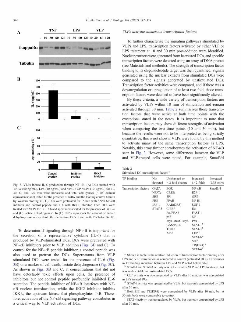
Table 2Stimulated DC transcription factors a
TF binding Notdetected
Unchanged or<2 fold change
Increased(>2 fold)
Increased(LPS only)
Transcription factors GATA EGR NF-κB Smad3/4NFATc CREB E2F-1SRE SP1 GREPRE PPAR NF-E1IRF-1 RAR(DR5) USF-1STAT-5 C/EBP Ets
Ets/PEA3 FAST-1p53 NF-1Myc-MaxC-Myb Pbx-1GAS/ISRE STAT-1 b
TFIID STAT-3 b
AP-2 CBP c
STAT-6 d
SIE e
TR(DR4) e
STAT-4 f
a Shown in table is the relative induction of transcription factor binding afterLPS and VLP stimulation as compared to control (untreated DCs). Differencesin TF binding induction between LPS and VLP noted below table.b STAT-1 and STAT-3 activity was detected after VLP and LPS treatment, but
was undetectable in unstimulated DCs.c CBP activity was downregulated by VLPs after 10 min, but was upregulated
in LPS treated DCs.d STAT-6 activity was upregulated by VLPs, but was only upregulated by LPS
after 30 min.e Both SIE and TR(DR4) were upregulated by VLPs after 10 min, but at
30 min both were comparable to control.f STAT-4 activity was upregulated by VLPs, but was only upregulated by LPS
after 30 min.
Fig. 3. VLPs induce IL-6 production through NF-κB. (A) DCs treated withTNFα (50 ng/mL), LPS (10 ng/mL) and VP40+GP VLPs (10 μg/mL) for 10,30, 60 and 120 min were harvested and total cell lysates (∼105 cellularequivalents/lane) tested for the presence of IκBα and the loading control tubulinby Western blotting. (B, C) DCs were pretreated for 15 min with SN50 NF-κBinhibitor and control peptide and 1 h with IKK2 inhibitor. Then DCs weretreated with VLPs for 12–16 h and spent media tested for the presence of B) IL-6and (C) lactate dehydrogenase. In (C) 100% represents the amount of lactatedehydrogenase released into the media from DCs treated with 1% Triton X-100.
346 O. Martinez et al. / Virology 364 (2007) 342–354
To determine if signaling through NF-κB is important forthe secretion of a representative cytokine (IL-6) that isproduced by VLP-stimulated DCs, DCs were pretreated withNF-κB inhibitors prior to VLP addition (Figs. 3B and C). Tocontrol for the NF-κB peptide inhibitor, a control peptide wasalso used to pretreat the DCs. Supernatants from VLPstimulated DCs were tested for the presence of IL-6 (Fig.3B) or a marker of cell death, lactate dehydrogenase (Fig. 3C).As shown in Figs. 3B and C, at concentrations that did nothave detectably toxic effects upon cells, the presence ofinhibitors but not control peptide profoundly inhibited IL-6secretion. The peptide inhibitor of NF-κB interferes with NF-κB nuclear translocation, while the IKK2 inhibitor inhibitsIKKβ, the upstream kinase that phosphorylates IκB. There-fore, activation of the NF-κB signaling pathway contributes ina critical way to VLP activation of DCs.
VLPs activate numerous transcription factors
To further characterize the signaling pathways stimulated byVLPs and LPS, transcription factors activated by either VLP orLPS treatment at 10 and 30 min post-addition were identified.Nuclear extracts were generated from harvestedDCs, and specifictranscription factors were detected using an array of DNA probes(see Materials and methods). The strength of transcription factorbinding to its oligonucleotide target was then quantified. Signalsgenerated using the nuclear extracts from stimulated DCs werecompared to the signals generated by unstimulated DCs.Transcription factor activities were compared, and if there was adownregulation or upregulation of at least two fold, those trans-cription factors were deemed to have been significantly altered.
By these criteria, a wide variety of transcription factors areactivated by VLPs within 10 min of stimulation and remainactivated through 30 min. Table 2 summarizes those transcrip-tion factors that were active at both time points with theexceptions stated in the notes. It is important to note thattranscription factors may show different strengths of activationwhen comparing the two time points (10 and 30 min), butbecause the results were not to be interpreted as being strictlyquantitative, this is not shown. VLPs were found by this methodto activate many of the same transcription factors as LPS.Notably, this array further corroborates the activation of NF-κBseen in Fig. 3. However, some differences between the VLPand VLP-treated cells were noted. For example, Smad3/4
347O. Martinez et al. / Virology 364 (2007) 342–354
was shown to be induced by LPS, but not by VLP stimulation(Table 2).
VLPs induce MAPK ERK1/2 signaling
VLP stimulated transcription factors are involved in a varietyof processes that include inflammation (NF-κB), proliferation(E2F-1), serum response and cell growth (SIE), hormoneresponse TR (DR4) and stress (Ets). Interestingly, MAPKmembers phosphorylate and regulate members of the Ets(Sharrocks, 2001), USF (Corre and Galibert, 2005) and STAT(Decker and Kovarik, 2000) transcription factor families.Therefore, we tested extracts of DCs stimulated 10, 30, 60and 120 min with VLP and LPS for the presence of activatedERK1/2, p38 and SAPK/JNK proteins, as evidenced by theirphosphorylation. As shown in Fig. 4, DCs stimulated with LPSfor 10 and 30 min showed ERK activation (as indicated by thepresence of phosphorylated ERK), while VLPs also inducedERK phosphorylation, but with a different kinetics. ERK phos-phorylation after VLP stimulation was seen after 30 min oftreatment. However, although activation of p38 and SAPK/JNK
Fig. 4. VLPs activate ERK1/2 in DCs. DCs treated with LPS (10 ng/mL) andVLP (VP40+GP VLPs (10 μg/mL)) for 10, 30, 60 and 120 min were harvestedand total cell lysates (∼105 cellular equivalents/lane) were tested for thepresence of phosphorylated ERK1/2 (pERK1/2), total ERK1/2, phosphorylated-p38 (pp38), total p38, phosphorylated-SAPK/JNK (pSAPK/JNK), total SAPK/JNK and tubulin. The same membranes blotted and tested for pERK1/2, pp38and pSAPK/JNK were stripped and tested for total levels of ERK1/2, p38 andSAPK/JNK, respectively. The membrane Western blotted for SAPK/JNK wasstripped and tested for levels of tubulin.
was seen after 30 min of LPS treatment, there was little p38 andno SAPK/JNK phosphorylation seen in VLP stimulated DCs(Fig. 4). Therefore, VLPs activate not only NF-κB but also ERKsignaling, following their addition to human DCs.
An intact GP mucin domain is required for DC activation
Expression of EBOV GP in human cells can have a profoundeffect on the phenotype of that cell, and these effects are partlydue to the heavily glycosylated mucin domain located in the GPectodomain (e.g. Sullivan et al., 2005; Yang et al., 2000).However, the mucin domain does not appear to be required forGP to mediate cell attachment and entry (Jeffers et al., 2002;Manicassamy et al., 2005; Yang et al., 2000). To test whetherthe mucin domain was required for stimulation of DCs, amucin-deletion GP mutant (Δ309–476) was generated. First wetested whether the GPΔmucin would be incorporated intoVLPs. 293T cells were mock, VP40, VP40+GP and VP40+GPΔmucin transfected for 48 h and the presence of VLPs wastested by Western blotting for VP40 after VLP purification.Although mock contained no VP40 protein (see “mock” lane,Fig. 5A), VP40 containing VLPs could be isolated from VP40,VP40+GP and VP40+GPΔmucin transfected 293T super-natants. Therefore, VLP production was not inhibited by the co-expression of the GPΔmucin mutant. Furthermore, electronmicroscopy performed on the VLPs demonstrated that VP40+GPΔmucin VLPs had a similar morphology to VP40+GPVLPs, including the appearance of a GP-decorated surface (notshown). To test the level of GP incorporation into VLPs,equivalent amounts of VP40, VP40+GP and VP40+ GPΔmu-cin VLPs were subjected to an anti-GP ELISA (Fig. 5B). Mockand VP40 VLPs show equivalent ODs while GP andGPΔmucin containing VLPs demonstrate equivalent ODs.Therefore, similar levels of WT and mutant GP are incorporatedinto VLPs. Equivalent amounts of GP and GPΔmucincontaining VLPs were then used to stimulate DCs (along withLPS control) for 10, 30, 60 and 120 min after which DCs wereharvested and lysates subjected to Western blot analysis forikappaBα, pERK1/2 and total ERK1/2 (Fig. 5C). The WTVLPs, as previously shown in Fig. 5, induced ikappaBαdegradation at 30 and 60 min whereas a significant decrease inikappaB was not apparent in cells treated with the VP40+GPΔmucin VLPs. Further, the moderate increase in ERKphosphorylation was more apparent in the GP wild typecontaining VLPs as compared to the mutant GP containingVLPs (Fig. 5C). Densitometry analysis on the ikappaBα,phospho ERK and control ERK bands within the Western blotconfirmed differences in the ratios of ikappaBa and phosphoERK to total ERK between VLPGP and VLPGP Δmucintreated DC lysates (not shown). Equivalent amounts of GP andGPΔmucin containing VLPs were also used to stimulate DCs(along with LPS control) for 20 h after which supernatants weretested for the presence of IL-8 (Fig. 5D). As shown in Fig. 5D,unstimulated DCs secreted relatively low levels of IL-8 similarto those secreted by VLP+GP Δmucin treated DCs, while bothLPS and VLP+GP treated DCs secreted approximately 10 foldmore IL-8 than the unstimulated control. Finally, to confirm that

Fig. 5. VLP GP mucin domain-dependent activation of NFκB and ERK1/2. (A) Western blot analyses of purified VLP preparations using antibody against EBOVprotein VP40. (B) ELISA, performed on equivalent amounts of VP40, VP40+GP, VP40+GPΔmuc VLPs using an EBOV-neutralizing anti-GP antibody (AZ52). (C)Shown is one blot of one representative experiment where equivalent protein concentrations (15 μg/mL) of VP40+GP and VP40+GPΔmuc VLPs and 10 ng/mL ofcontrol LPS were used to treat DCs for 10, 30, 60 and 120 min and the total cell lysates (∼105 cellular equivalents/lane) were tested for the presence of IκBα,phosphorylated ERK1/2 (pERK1/2) and total ERK1/2. (D) Shown is a representative experiment where equivalent protein concentrations (15 μg/mL) of VP40+GPand VP40+GPΔmuc VLPs and 10 ng/mL of control LPS were used to treat DCs (5×10⁵ cells) for 20 h and supernatant was tested for levels of IL-8. The control in thisexperiment is unstimulated DCs (unstim). (E) Association of GFP-tagged VLPs with DCs was assayed by flow cytometry. Control (top left dot plot) depicts DCsstained with isotype controls. Equivalent concentrations of (15 μg/mL) GFP-VP40+GP (bottom right dot plot), GFP-VP40+GPΔmuc (bottom left dot plot) and mockVLPs purified from GFP-transfected cells (top right dot plot) were incubated with DCs for 30 min at 37 °C, washed, stained for the DC marker CD11c and assayed byflow cytometry.
348 O. Martinez et al. / Virology 364 (2007) 342–354
deletion of the mucin domain did not alter the ability of VLPsto associate with DCs, we employed GFP-tagged VLPs. Weconstructed and expressed a GFP-VP40 chimeric protein.Expression of this stable (not shown) chimeric protein leadsto the production of fluorescent VLPs that can incorporate GP
and can be visualized by fluorescence microscopy (not shown).Equivalent (15 μg/mL) amounts of GFP-VP40+GP, GFP-VP40+GPΔmuc and mock VLPs (purified from GFP-trans-fected cells) were incubated with DCs for 30 min at 37 °C. Cellswere then washed and then tested for their association with

349O. Martinez et al. / Virology 364 (2007) 342–354
VLPs by flow cytometry. Isotype controls showed no fluo-rescence (Fig. 5E top left dot plot). CD11c+cells wereassociated with equivalent GFP fluorescence when incubatedwith GFP-VP40 +GP and GFP-VP40 +GPΔmuc VLPs(Fig. 5E, bottom dot plots) but no GFP fluorescence was seenwhen DCs were incubated with mock VLPs prepared from GFPtransfected 293Ts (Fig. 5E top right dot plot). Cumulatively,these data demonstrate that the mucin domain of GP is notrequired for association of VLPs with DCs but is required forstimulation of DCs by the VLPs.
Discussion
Our data identify novel signals triggered by EBOV VLPswithin human DCs. These studies provide molecular context toprevious observations that EBOV VLPs activate DCs andmacrophages. These results also point to specific signalingpathways that may be activated by the initial interaction ofEBOV with DCs and which may influence the outcome ofEBOV infection. Previous studies have demonstrated thatVP40+GP VLPs, but not VP40 VLPs could stimulate DCs(Bosio et al., 2004; Ye et al., 2006). In our hands, VLPscontaining the viral matrix protein, VP40 and the viralglycoprotein, GP, were found to induce a proinflammatoryresponse. The importance of GP for the DC response is expected,and fits with its ability to induce a variety of cellular responses.The EBOV GP is the viral attachment and fusion protein (Baret al., 2006; Feldmann et al., 2001), and it also exerts a variety ofadditional effects upon cells, including surface protein modula-tion (Simmons et al., 2002; Takada et al., 2000; Wahl-Jensen etal., 2005b), induction of anoikis (Alazard-Dany et al., 2006; Rayet al., 2004), and stimulation of cytokine production from humanmacrophages (Wahl-Jensen et al., 2005a) and dendritic cells(Bosio et al., 2004; Ye et al., 2006). Moreover, its vascularcytotoxicity is thought to be play an important role in virulence(Yang et al., 2000). In contrast, the presence of other viralproteins; of which NP was the most abundant, did notsubstantially alter DC responses. This observation is in spiteof the fact that other viral components might be expected tomodulate host cell responses to VLPs given their previouslydescribed functions. For example, the VP35 and VP24 proteinsinterfere with signaling pathways related to interferon responses(Basler et al., 2000, 2003; Bosio et al., 2003; Hartman et al.,2004; Reid et al., 2005, 2006). It remains unclear whether theVP35 and VP24 in “complete” VLPs reflects an inability tomodulate DC responses when introduced into cells via VLPs ortheir relatively low abundance in the VLPs.
VLPs containing the viral matrix protein (VP40) and theviral glycoprotein (GP) were found to induce a proinflammatoryresponse highly similar to a prototypical DC activator, LPS.This response included the production of several proinflamma-tory cytokines, as measured by a multiplex cytokine assay.Specifically, inflammatory cytokines IL-6, TNFα, and chemo-kines IL-8, MIP-1α, RANTES and IP-10 are all secreted byVP40+GP (Fig. 2) and cVLPs (not shown) stimulated DCs.This is consistent with previous studies that demonstrated thesecretion of the above-mentioned cytokines (Bosio et al., 2004;
Warfield et al., 2003; Ye et al., 2006) with the exception of IFNprotein-10 (IP-10), a protein which acts to attract activatedT cells and which plays a role in responses to viral infection(Dufour et al., 2002). Additionally activation of NF-κB andnumerous other transcription factors (TFs) were demonstrated,with a multiplex TF array, following either VLP or LPStreatment of DCs. TFs activated by 10 min were, for the mostpart, also stimulated after 30 min of VLP treatment (Table 2).Therefore, the results of the 10 min array corroborated theresults of the 30 min array. This also showed that VLP treatmentinduced rapid signaling. This raises the possibility that theactivation may be occurring shortly after interaction of the VLPwith receptors present at the plasma membrane of DCs. Furtherstudies are needed to clarify whether internalization of the VLPsby the immature DCs is a requirement for DC stimulation. It isinteresting to note that NF-κB was activated (>2 fold change)by VLPs, as compared to an unstimulated control, within10 min of treatment (Table 2). However, in Fig. 3, IκBαdecreased significantly only after 30 min. The reason for thisseeming discrepancy is unclear, but may reflect the fact twodifferent parameters are being measured using two differentassays. Nevertheless, NF-κB was shown to be activated byVLPs using two independent assays.
While the activation of NF-κB might have been predicted,given the role of NF-κB in proinflammatory cytokine produc-tion (Ali and Mann, 2004), it was important to demonstrateexperimentally that this activation plays a critical role in VLPinduced DC activation. This was addressed by employingpreviously described NF-κB inhibitors. Specifically, IL-6cytokine production was abolished when NF-κB inhibitorsSN50 and IKK2 inhibitors were used to pretreat DCs stimulatedwith VLPs. The SN50 peptide inhibitor blocks NF-κB nucleartranslocation while IKK2 blocks IKKβ, a component of theIKK kinase complex. Taken together, these data indicate thatVLPs induce IκBα degradation and that NF-κB activation isimportant for IL-6 secretion.
Interestingly, the transcription factor array data alsosuggested the activation of MAP kinase pathways. For example,VLP-activated TFs included TFs that regulate cell cycle controland apoptosis (EGR (Thiel and Cibelli, 2002), E2F-1 (Pardeeet al., 2004; Stevens and La Thangue, 2003)), stress andinflammatory responses (NF-κB (Ali and Mann, 2004), Ets(Sharrocks, 2001), STATs (Decker and Kovarik, 2000), USF-1(Corre and Galibert, 2005), SIE (Wang et al., 2003)) andmitogen and cytokine response (SIE, EGR (Decker et al., 2003),STATs (Decker, 1999)). Of these, Ets, SIE, STAT and USF TFfamily members can all be phosphorylated by the stress-inducedMAPKs. Consistent with the potential MAPK activated TFsdiscovered through the array, we also demonstrated that VLPscan significantly activate ERK1/2 (Fig. 4). However, unlikeLPS treatment, VLPs did not detectably activate p38 and SAPK/JNK (Fig. 4). Although both LPS and VLP treatment activatedERK1/2, they did so with different kinetics. As previouslydemonstrated in a study showing the kinetics of LPS-stimulatedDC ERK1/2 activation (Ardeshna et al., 2000), ERK1/2phosphorylation was maximal by 30 min while VLP treatmentinduced ERK1/2 activation that lasted up to 1 h (Figs. 4 and 5).

350 O. Martinez et al. / Virology 364 (2007) 342–354
Cumulatively, the cytokine, TF and signaling pathway datasuggest that VLPs activate DCs in a manner that is similar butnot identical to the prototypical DC activator, LPS.
To further address the requirements for VP40+GP inducedDC activation, the role of the GP mucin domain in DCactivation was explored. As noted above, the heavily glycosy-lated mucin domain is required for several cellular responses toGP but is not required for GP-mediated cell attachment andentry, at least for GP pseudotyped viruses (Jeffers et al., 2002;Manicassamy et al., 2005; Yang et al., 2000). AlthoughGPΔmucin was incorporated into VLPs at levels similar toGP, the VLPs containing GPΔmucin could not stimulate DCiκBα degradation and ERK phosphorylation to the same extentas VLPs containing wild type GP. The GPΔmucin VLPs alsofailed to induce IL-8 production (Fig. 5). The fact that theneutralizing antibody used in our ELISA (Fig. 5B) could bindthe GPΔmucin in the VLPs and the fact that both wild type GP-VLPs and GPΔmucin-VLPs can associate with DCs to similarlevels (Fig. 5E) suggests that GPΔmucin is in a native confor-mation and that VLPswithGPΔmucin can still infect cells. Sinceboth wild type and mutant VLPs could presumably mediatevirion entry, this would not explain the difference in signaling.Further studies are required to understand how the mucin do-main contributes to signaling leading to DC stimulation.
Although DC responses to LPS and VLPs were not identical,many similarities were noted in our studies. It should also benoted that it is difficult to exclude the possibility that a smallamount of contaminating LPS could play a role in thestimulating capacity of our VLPs, however as previouslydemonstrated (Bosio et al., 2004), VLPs, unlike LPS, lose theirstimulatory capacity when boiled (not shown). We also find thatthe minimal values obtained in the limulus amebocyte lysatesassay employed to quantify endotoxin levels (<0.03 IU/μg totalprotein, not shown and Fuller et al., 2006) were similar forGPΔmucin VLPs as compared to GP VLPs (not shown).Therefore endotoxin contamination cannot explain the differ-ences seen between the two types of VLPs. Since LPS signalsthrough TLR4, it is tempting to speculate that VLPs may alsostimulate DCs through a TLR. However, GP interacts with avariety of cell surface molecules. The identity of the primaryreceptor(s) mediating EBOV entry is controversial (Simmonset al., 2003b) and one cannot exclude signaling from thisreceptor(s) as contributing to DC activation. In addition, GP caninteract with a variety of other molecules, including C-typelectins, that are present upon DCs, monocytes and/or macro-phages (Alvarez et al., 2002; Marzi et al., 2004; Mohamadzadehet al., 2006; Simmons et al., 2003a; Takada et al., 2004). Giventhat the mucin domain is more glycosylated than other regionsof GP, it is intriguing to speculate that interactions between C-type lectins and the mucin domain might contribute to signalingwithin the DCs.
In conclusion, we show that within 1 h, EBOV GP-mediatedsignaling leads to the activation of both NF-κB and ERK1/2,and this response requires the presence of the GP mucindomain. Previous studies suggest that EBOV infection of DCsleads to poor pro-inflammatory cytokine production and DCsthat are not fully matured and fail to effectively activate a T cell
response (Bosio et al., 2003; Mahanty et al., 2003). Wehypothesize that the signals activated by EBOV VLPs representsignals that are also transduced by live EBOVand contribute tothe aberrant activation of DCs. Experiments are thus underwayto define the mechanism(s) by which these pathways areactivated by VLPs and to evaluate their significance in thecontext of EBOV infection.
Materials and methods
Cells, plasmids and antibodies
293T (human embryonic kidney) cells were grown inDulbecco's modified Eagle's medium (DMEM; Invitrogen,Carlsbad, CA) supplemented with 10% fetal calf serum(Invitrogen), 2 mM L-glutamine (Invitrogen), 100 units/mL ofpenicillin, 100 μg/mL streptomycin, 1 μg/mL ciproflaxocin at37 °C in 7% CO2. All Ebola viral genes kindly provided byViktor Volchkov (INSERM, Lyon) and Elke Mühlberger(Marburg University, Marburg) were sequenced and confirmedto correspond to the published Zaire EBOV strain Mayingasequences. EBOV VP40 was expressed using the pCDNA3 orpCAGGS vector while all the other EBOV proteins wereexpressed using pCDNA3. The GPΔmucin gene was con-structed as previously described (Manicassamy et al., 2005).Antibodies used in these studies include anti-p38, -phosphop38,-ERK1/2, -phosphoERK1/2, -SAPK/JNK, -phosphoSAPK/JNK (cell signaling technology, Danvers, MA), anti-cd11c,-CD14, -HLA-DR (BD Pharmingen, Franklin Lakes, NJ), anti-VP40 was a kind gift from Ronald Harty (University ofPennsylvania), anti-VP24 was a kind gift from Victor Volchkov(INSERM), anti-VP35, anti-GP and anti-NP were developed incollaboration with the Hybridoma Center located in the MountSinai Department of Microbiology, neutralizing anti-GP1(AZ52) was a kind gift from Erica Ollmann Saphire (TheScripps Research Institute), anti-tubulin, anti-mouse, anti-human and rabbit IgG (Sigma, St. Louis, MO), anti-IκBα(Epitomics, Burlingame, CA).
VLP production
VLPs were produced by transfecting 3 μg of expressionplasmids into 1.5×10⁶ 293T cells in six-well dishes and bytransfecting 18 μg of expression plasmids into 107 293Ts in10 cm plates. The EBOV Zaire VP40 expression plasmid wastransfected alone or in combinations with EBOV Zaire GP, NP,VP24, VP30 and VP35 expression plasmids at equal DNAconcentrations. Furthermore, to visualize fluorescent VLPs, aVP40-GFP (VP40 was cloned in frame at the N-terminus ofVP40) chimeric protein was expressed from the expressionplasmid pCAGGS. This protein could produce VLPs asdetermined by Western blotting, fluorescent microscopy andelectron microscopy (not shown). Transfections were performedusing lipofectamine 2000 (Invitrogen) at a 1:1 ratio of DNA tolipofectamine following the manufacturer's protocol. WhenVLPs were produced for silver staining, media was replaced12–16 h after 293T transfection with VP-serum free media
351O. Martinez et al. / Virology 364 (2007) 342–354
(Invitrogen). 48 h post-transfection, cells and cellular debriswere pelleted away from the harvested VLP-containing super-natant with a cell spin. Then, VLPs were centrifuged through asucrose cushion at 26000 rpm in an SW-28 rotor for 2 h at 4 °C,washed in ice-cold NTE buffer (10 mM Tris pH7.5, 100 mMNaCl, 1 mM EDTA) by centrifuging at 26000 rpm for 2 h at4 °C and then gently tapped 100 times to resuspend in 50–100 μL of NTE buffer. VLP protein content was quantitatedusing the DC protein assay (Bio-Rad, Hercules, CA). VLPswere left on ice for up to 48 h until used. VLP preparationscontained <0.03 IU (endotoxin units) per microgram of totalprotein as determined by the Limilus Amebocyte Lysate assay(Cambrex, Walkersville, MD).
Electron microscopy
10 μL of VLPs were pipetted onto 300 mesh copper gridcoated with carbon film and incubated for 15 min at RT. Gridswere then washed twice with water and negatively stained for15 s using 1% phosphotungstic acid buffered to pH 7.0 with 1 Mammonium hydroxide. Particles were then visualized using aHitachi H7000 transmission electron microscope.
Silver staining
∼1 μg of serum-free VLPs (see VLP production) wereloaded onto 4–20% gradient gels and proteins were separatedby sodium dodecyl sulfate-polyacrylamide gel electrophoresis(SDS-PAGE). Silver staining was performed using SilverQuestsilver staining kit (Invitrogen) following the manufacturer'sprotocol with the exception that half the reagent volumes wereused per assay.
Isolation and culture of human DCs
Peripheral blood mononuclear cells (PBMCs) were isolatedby Ficoll density gradient centrifugation (Histopaque; SigmaAldrich, St. Louis, MO) from buffy coats of healthy humandonors (New York Blood Center). CD14+ cells were immuno-magnetically purified using anti-human CD14 antibody-labeledmagnetic beads and iron-based Midimacs LS columns (MiltenyiBiotec, Auburn, CA). After elution from the columns, cells wereplated (0.7–1×10⁶ cells/mL) in RPMI (Invitrogen, Carlsbad,CA) supplemented with 2% human serum AB (GemCell,Gemini Bio-Products, West Sacramento, CA), 100 units/mL ofpenicillin, 100 μg/mL streptomycin, 55 μM β-mercaptoethanol,500 U/mL human granulocyte–macrophage colony-stimulatingfactor (GM-CSF; Peprotech, Rocky Hill, NJ), 500 U/mL humaninterleukin-4 (IL-4; Peprotech), 1 μg/mL ciproflaxocin(SIGMA) and incubated for 5 to 7 days at 37 °C.
Flow cytometry
Flow cytometry was performed in a Cytomic FC500Coulter station (Beckman Coulter, Miami, FL). Data wereanalyzed using WinMDI Version 2.8 (http://facs.scripps.edu/software.html).
DC stimulation
DCs plated in 96 well dishes (9×10⁴ cells/well) or 24 welldishes (0.5×10⁶ cells/well) were stimulated with 10–20 μg/mLVLP, 50 ng/mL TNFα or 10–100 ng/mL LPS (SalmonellaMinnesota R595; Alexis, San Diego, CA) for the indicatedamount of time in RPMI (Invitrogen, Carlsbad, CA) supple-mented with 1% human serum AB (GemCell), 100 units/mL ofpenicillin, 100 g/mL streptomycin, 55 μM β-mercaptoethanol.Supernatants were tested for cytokine production, while har-vested DCs were tested for MAPK and TF activation.
IL-6 and IL-8 ELISA
IL-6 and IL-8 was detected from supernatants collected 12–24 h post stimulation using capture enzyme-linked immunosor-bent assay (ELISA). The IL-6 and IL-8 (supernatant was diluted1/10 prior to IL-8 ELISA) ELISAs were performed according tothe manufacturer's protocol (eBioscience, San Diego, CA andBD Pharmingen, Franklin Lakes, NJ, respectively). Briefly forthe IL-6 ELISA, 96 well flat-bottomed Maxi-Sorp (Nunc,Rochester, NY) plates were used for capture antibody coatingand subsequent IL-6 detection. Coated plates were washed threetimes with PBS containing 0.05% Tween 20 (PBS-T) andblockedwith PBS+10%FCS (PBS-F) at room temperature (RT)for 1 h. Plates were washed three times with PBS-T, supernatantsadded and incubated for 2 h at RT followed by five washes.Detection and HRP conjugated streptavidin antibody incuba-tions (1 h at RT) were separated by five washes. After a finalseven washes, TMB substrate was added to each well andincubated for 15 min in the dark until the HRP reaction wasstopped with 1/4 volume of 1N HCl. Plates were read using anELISA plate reader (Biotek Instruments, Winooski, VT) at450 nm wavelength.
GP ELISA
Mock VLPs and VLPs (with or without GP) were adsorbedovernight onto Maxi-Sorp 96 well ELISA plates (Nunc) incoating buffer (0.05 M carbonate–bicarbonate buffer, pH 9.6)supplemented with 0.1% Triton X-100. Plates were washedthree times with PBS containing 0.05% Tween 20 (PBS-T) andblocked with PBS+10% FCS (PBS-F) at RT for 1 h. Plateswere washed three times with PBS-T. Detection (AZ52 anti-GPantibody) and HRP conjugated anti-human IgG antibodyincubations (1 h at RT) were separated by five washes. Aftera final seven washes, TMB substrate was added to each well andincubated for 15 min in the dark until the HRP reaction wasstopped with 1/4 volume of 1N HCl. Plates were read using anELISA plate reader (Biotek Instruments, Winooski, VT) at450 nm wavelength.
NF-κB inhibition and cytotoxicity assays
DCs plated in 96 well dishes (9×10⁴ cells/well) in RPMIcontaining 1% human serum were pretreated 15 min withinhibitor SN50 and control peptide SN50M at final

352 O. Martinez et al. / Virology 364 (2007) 342–354
concentration of 66 μg/mL or 1 h with IKK2 inhibitor (IKKIV) at a final concentration 2.5 μM (Calbiochem, EMDBiosciences, San Diego, CA). Spent media from 1% Triton X-100 or VLP-treated DCs (12–16 h) was tested for the presenceof IL-6 and lactate dehydrogenase. Amounts of lactatedehydrogenase (LDH) were assayed using a CytotoxicityDetection Kit (LDH) (Roche, Basel, Switzerland) followingthe manufacturer's protocol. 100% cytotoxicity was deter-mined using the amount of LDH released into the media fromDCs treated with 1% Triton X-100.
Multiplex cytokine measurements
50 μL of the stimulated DC spent media was tested for thepresence of 22 human cytokines using Beadlyte Human 22-PlexMulti-Cytokine Detection System (Upstate, Billerica, MA)following the manufacturer's protocol. Plates were read in aLuminex plate reader, and data were analyzed using softwarefrom Applied Cytometry Systems (Sacramento, CA).
Nuclear extraction
Stimulated DCs were snap-frozen in a dry-ice ethanol bath.Nuclear extractions were performed using nuclear extraction kit(Panomics, Fremont, CA) following the manufacturer's proto-col with the exception that the final resuspension volume was30 μL. Protein content in nuclear extracts was then quantitatedusing the DC protein assay (Bio-Rad).
Transcription factor array
Nuclear extractions (see above) were performed on untreatedDCs and DCs treated for 10 and 30 min with VLPs and LPS.Equivalent nuclear protein concentrations (total of 7.5 μg) wereused in the Protein/DNA array I (Panomics, Fremont, CA) todetermine transcription factor (TF) activity following themanufacturer's protocol. Very briefly, biotin-labeled probes(TransSignal Probe Mix) were added to nuclear extracts andincubated for 30 min at 15 °C. This allowed activated TFs tobind probes forming a complex. After uncomplexed probeswere washed away using spin columns, the protein/DNAcomplexes were disassociated. The column eluate containingfreed labeled probes was incubated with membranes spottedwith the probe complements. The hybridized spotted mem-branes were then washed, incubated with streptavidin–HRPO,washed again and developed by chemiluminescense. Eachtranscription factor is represented by a pair of duplicate spots onthe array. One duplicate set being a dilution of the other set.Arrays were developed using several exposure times in order toavoid signal saturation. At each exposure, films of the arrayswere scanned and the intensity of spots quantitated usingImageQuant software. The average signal of duplicate spots wasthen obtained. If duplicate spots varied more than 25%, theywere eliminated from the analyses. In order to compare thetranscription factor signal from one membrane (sample) to thecontrol (untreated DCs), signals were normalized by dividingthe sample signal with the signal of an unchanged TF (selected
from that specific exposure). To be more stringent, signals werenormalized against two TFs. According to the manufacturer,>2fold changes in signal are deemed to be significant. We chose todeem significant those signals (i.e. NF-κB) that had a greaterthan 2 fold difference as compared to the control whennormalized against both normalizing TFs. Moreover, almostall transcription factors found to be activated at the 10 min markwere also found to be significantly altered at the 30 min timepoint as compared to control. Therefore, the 10 and 30 minarrays reinforced the credibility of the transcription factorsfound to be up or down-regulated. In a few exceptional cases,TFs were upregulated at only one of the two time points, ashighlighted in Table 2.
Western blot
After the indicated time points of DC stimulation, 5×10⁵
DCs/stimuli were harvested, pelleted and snap-frozen in a dry-ice ethanol bath and stored at −80 °C. Cell pellets werethawed on ice, resuspended in 50 μL of 1× protein samplebuffer, sonicated and loaded into a 4–20% gradient or 10%gels for separation by SDS-PAGE. Proteins were transferred toa polyvinylidene difluoride (PVDF) membrane, blocked in 1%milk in TBS (50 mM Tris pH 7.5, 100 mM NaCl) for 1 h,probed overnight with appropriate antibodies diluted in 0.5%milk in TBS, washed 3 times in TBS-T (TBS+0.075%Tween-20) for 5 min/wash, probed with secondary antibodyfor 1 h and washed a final 3 times. The Western blots weredeveloped using the Western Lightning ECL kit (Perkin-Elmer, Boston, MA) and Kodak BioMax film (Kodak,Rochester, NY).
Flow cytometry
DCs were incubated with GFP-VP40 induced VLPs for30 min at 37 °C and washed twice with PBS. Cells were thenstained with an anti-CD11c (Pharmingen) for 30 min at 0 °C andwashed again. FACS was performed using a Cytomics F500machine (Beckman Coulter, Fullerton, CA) and then analyzedusing Win MDI 2.8 software.
Acknowledgments
This work was supported by funding from an NIH U19 grant(AI062623) to CFB. We would like to thank Kelley Boyd andSvetlana V. Burmakina for technical assistance with themultiplex cytokine assays and electron microscopy, respec-tively. We also want to thank Ana Fernandez-Sesma for helpfuldiscussions and Lawrence W. Leung for his critical reading ofthe manuscript.
References
Alazard-Dany, N., Volchkova, V., Reynard, O., Carbonnelle, C., Dolnik, O.,Ottmann, M., Khromykh, A., Volchkov, V.E., 2006. Ebola virus glycopro-tein GP is not cytotoxic when expressed constitutively at a moderate level. J.Gen. Virol. 87 (Pt. 5), 1247–1257.

353O. Martinez et al. / Virology 364 (2007) 342–354
Ali, S., Mann, D.A., 2004. Signal transduction via the NF-kappaB pathway: atargeted treatment modality for infection, inflammation and repair. CellBiochem. Funct. 22 (2), 67–79.
Alvarez, C.P., Lasala, F., Carrillo, J., Muniz, O., Corbi, A.L., Delgado, R., 2002.C-type lectins DC-SIGN and L-SIGN mediate cellular entry by Ebola virusin cis and in trans. J. Virol. 76 (13), 6841–6844.
Ardeshna, K.M., Pizzey, A.R., Devereux, S., Khwaja, A., 2000. The PI3 kinase,p38 SAP kinase, and NF-kappaB signal transduction pathways are involvedin the survival and maturation of lipopolysaccharide-stimulated humanmonocyte-derived dendritic cells. Blood 96 (3), 1039–1046.
Baize, S., Leroy, E.M., Georges-Courbot, M.C., Capron, M., Lansoud-Soukate,J., Debre, P., Fisher-Hoch, S.P., McCormick, J.B., Georges, A.J., 1999.Defective humoral responses and extensive intravascular apoptosis areassociated with fatal outcome in Ebola virus-infected patients. Nat. Med. 5(4), 423–426.
Baize, S., Leroy, E.M., Georges, A.J., Georges-Courbot, M.C., Capron, M.,Bedjabaga, I., Lansoud-Soukate, J., Mavoungou, E., 2002. Inflammatoryresponses in Ebola virus-infected patients. Clin. Exp. Immunol. 128 (1),163–168.
Bar, S., Takada, A., Kawaoka, Y., Alizon, M., 2006. Detection of cell–cellfusion mediated by Ebola virus glycoproteins. J. Virol. 80 (6), 2815–2822.
Basler, C.F., Wang, X., Muhlberger, E., Volchkov, V., Paragas, J., Klenk, H.D.,Garcia-Sastre, A., Palese, P., 2000. The Ebola virus VP35 protein functionsas a type I IFN antagonist. Proc. Natl. Acad. Sci. U. S. A. 97 (22),12289–12294.
Basler, C.F., Mikulasova, A., Martinez-Sobrido, L., Paragas, J., Muhlberger, E.,Bray, M., Klenk, H.D., Palese, P., Garcia-Sastre, A., 2003. The Ebola virusVP35 protein inhibits activation of interferon regulatory factor 3. J. Virol. 77(14), 7945–7956.
Bosio, C.M., Aman, M.J., Grogan, C., Hogan, R., Ruthel, G., Negley, D.,Mohamadzadeh, M., Bavari, S., Schmaljohn, A., 2003. Ebola and Marburgviruses replicate in monocyte-derived dendritic cells without inducing theproduction of cytokines and full maturation. J. Infect. Dis. 188 (11),1630–1638.
Bosio, C.M., Moore, B.D., Warfield, K.L., Ruthel, G., Mohamadzadeh, M.,Aman, M.J., Bavari, S., 2004. Ebola and Marburg virus-like particlesactivate human myeloid dendritic cells. Virology 326 (2), 280–287.
Bray, M., Geisbert, T.W., 2005. Ebola virus: the role of macrophages anddendritic cells in the pathogenesis of Ebola hemorrhagic fever. Int. J.Biochem. Cell Biol. 37 (8), 1560–1566.
Corre, S., Galibert, M.D., 2005. Upstream stimulating factors: highly versatilestress-responsive transcription factors. Pigment Cell Res. 18 (5), 337–348.
Decker, T., 1999. Introduction: STATs as essential intracellular mediators ofcytokine responses. Cell. Mol. Life Sci. 55 (12), 1505–1508.
Decker, T., Kovarik, P., 2000. Serine phosphorylation of STATs. Oncogene 19(21), 2628–2637.
Decker, E.L., Nehmann, N., Kampen, E., Eibel, H., Zipfel, P.F., Skerka, C.,2003. Early growth response proteins (EGR) and nuclear factors of activatedT cells (NFAT) form heterodimers and regulate proinflammatory cytokinegene expression. Nucleic Acids Res. 31 (3), 911–921.
Dufour, J.H., Dziejman, M., Liu, M.T., Leung, J.H., Lane, T.E., Luster, A.D.,2002. IFN-gamma-inducible protein 10 (IP-10; CXCL10)-deficient micereveal a role for IP-10 in effector T cell generation and trafficking.J. Immunol. 168 (7), 3195–3204.
Feldmann, H., Volchkov, V.E., Volchkova, V.A., Stroher, U., Klenk, H.D., 2001.Biosynthesis and role of filoviral glycoproteins. J. Gen. Virol. 82 (Pt. 12),2839–2848.
Fuller, C.L., Ruthel, G., Warfield, K.L., Swenson, D.L., Bosio, C.M., Aman,M.J., Bavari, S., 2006. NKp30-dependent cytolysis of filovirus-infectedhuman dendritic cells. Cell. Microbiol. doi:10.1111/j.1462-5822.2006.00844.x.
Geisbert, T.W., Jahrling, P.B., 1995. Differentiation of filoviruses by electronmicroscopy. Virus Res. 39 (2–3), 129–150.
Geisbert, T.W., Hensley, L.E., Larsen, T., Young, H.A., Reed, D.S., Geisbert,J.B., Scott, D.P., Kagan, E., Jahrling, P.B., Davis, K.J., 2003. Pathogenesisof Ebola hemorrhagic fever in cynomolgus macaques: evidence thatdendritic cells are early and sustained targets of infection. Am. J. Pathol.163 (6), 2347–2370.
Hartman, A.L., Towner, J.S., Nichol, S.T., 2004. A C-terminal basic amino acidmotif of Zaire ebolavirus VP35 is essential for type I interferon antagonismand displays high identity with the RNA-binding domain of anotherinterferon antagonist, the NS1 protein of influenza Avirus. Virology 328 (2),177–184.
Hoenen, T., Groseth, A., Falzarano, D., Feldmann, H., 2006. Ebola virus:unravelling pathogenesis to combat a deadly disease. Trends Mol. Med. 12(5), 206–215.
Jasenosky, L.D., Neumann, G., Lukashevich, I., Kawaoka, Y., 2001. Ebola virusVP40-induced particle formation and association with the lipid bilayer.J. Virol. 75 (11), 5205–5214.
Jeffers, S.A., Sanders, D.A., Sanchez, A., 2002. Covalent modifications of theEbola virus glycoprotein. J. Virol. 76 (24), 12463–12472.
Leroy, E.M., Baize, S., Volchkov, V.E., Fisher-Hoch, S.P., Georges-Courbot,M.C., Lansoud-Soukate, J., Capron, M., Debre, P., McCormick, J.B.,Georges, A.J., 2000. Human asymptomatic Ebola infection and stronginflammatory response. Lancet 355 (9222), 2210–2215.
Mahanty, S., Bray, M., 2004. Pathogenesis of filoviral haemorrhagic fevers.Lancet Infect. Dis. 4 (8), 487–498.
Mahanty, S., Hutchinson, K., Agarwal, S., McRae, M., Rollin, P.E., Pulendran,B., 2003. Cutting edge: impairment of dendritic cells and adaptive immunityby Ebola and Lassa viruses. J. Immunol. 170 (6), 2797–2801.
Manicassamy, B., Wang, J., Jiang, H., Rong, L., 2005. Comprehensive analysisof Ebola virus GP1 in viral entry. J. Virol. 79 (8), 4793–4805.
Maruyama, T., Rodriguez, L.L., Jahrling, P.B., Sanchez, A., Khan, A.S., Nichol,S.T., Peters, C.J., Parren, P.W., Burton, D.R., 1999. Ebola virus can beeffectively neutralized by antibody produced in natural human infection.J. Virol. 73 (7), 6024–6030.
Marzi, A., Gramberg, T., Simmons, G., Moller, P., Rennekamp, A.J.,Krumbiegel, M., Geier, M., Eisemann, J., Turza, N., Saunier, B.,Steinkasserer, A., Becker, S., Bates, P., Hofmann, H., Pohlmann, S., 2004.DC-SIGN and DC-SIGNR interact with the glycoprotein of Marburg virusand the S protein of severe acute respiratory syndrome coronavirus. J. Virol.78 (21), 12090–12095.
Mohamadzadeh, M., Coberley, S.S., Olinger, G.G., Kalina, W.V., Ruthel, G.,Fuller, C.L., Swenson, D.L., Pratt, W.D., Kuhns, D.B., Schmaljohn, A.L.,2006. Activation of triggering receptor expressed on myeloid cells-1 onhuman neutrophils by Marburg and Ebola viruses. J. Virol. 80 (14),7235–7244.
Noda, T., Sagara, H., Suzuki, E., Takada, A., Kida, H., Kawaoka, Y., 2002.Ebola virus VP40 drives the formation of virus-like filamentous particlesalong with GP. J. Virol. 76 (10), 4855–4865.
Pardee, A.B., Li, C.J., Reddy, G.P., 2004. Regulation in S phase by E2F. CellCycle 3 (9), 1091–1094.
Ray, R.B., Basu, A., Steele, R., Beyene, A., McHowat, J., Meyer, K.,Ghosh, A.K., Ray, R., 2004. Ebola virus glycoprotein-mediated anoikisof primary human cardiac microvascular endothelial cells. Virology 321(2), 181–188.
Reid, S.P., Cardenas, W.B., Basler, C.F., 2005. Homo-oligomerization facilitatesthe interferon-antagonist activity of the ebolavirus VP35 protein. Virology341 (2), 179–189.
Reid, S.P., Leung, L.W., Hartman, A.L., Martinez, O., Shaw, M.L., Carbonnelle,C., Volchkov, V.E., Nichol, S.T., Basler, C.F., 2006. Ebola virus VP24 bindskaryopherin alpha1 and blocks STAT1 nuclear accumulation. J. Virol. 80(11), 5156–5167.
Reis e Sousa, C., 2004. Toll-like receptors and dendritic cells: for whom the bugtolls. Semin. Immunol. 16 (1), 27–34.
Sanchez, A., Lukwiya,M., Bausch, D.,Mahanty, S., Sanchez,A.J.,Wagoner, K.D.,Rollin, P.E., 2004. Analysis of human peripheral blood samples from fataland nonfatal cases of Ebola (Sudan) hemorrhagic fever: cellular responses,virus load, and nitric oxide levels. J. Virol. 78 (19), 10370–10377.
Sharrocks, A.D., 2001. The ETS-domain transcription factor family. Nat. Rev.,Mol. Cell Biol. 2 (11), 827–837.
Simmons, G., Wool-Lewis, R.J., Baribaud, F., Netter, R.C., Bates, P., 2002.Ebola virus glycoproteins induce global surface protein down-modulationand loss of cell adherence. J. Virol. 76 (5), 2518–2528.
Simmons, G., Reeves, J.D., Grogan, C.C., Vandenberghe, L.H., Baribaud, F.,Whitbeck, J.C., Burke, E., Buchmeier, M.J., Soilleux, E.J., Riley, J.L.,
354 O. Martinez et al. / Virology 364 (2007) 342–354
Doms, R.W., Bates, P., Pohlmann, S., 2003a. DC-SIGN and DC-SIGNRbind Ebola glycoproteins and enhance infection of macrophages andendothelial cells. Virology 305 (1), 115–123.
Simmons, G., Rennekamp, A.J., Chai, N., Vandenberghe, L.H., Riley, J.L.,Bates, P., 2003b. Folate receptor alpha and caveolae are not required forEbola virus glycoprotein-mediated viral infection. J. Virol. 77 (24),13433–13438.
Stevens, C., La Thangue, N.B., 2003. A new role for E2F-1 in checkpointcontrol. Cell Cycle 2 (5), 435–437.
Sullivan, N.J., Peterson, M., Yang, Z.Y., Kong, W.P., Duckers, H., Nabel, E.,Nabel, G.J., 2005. Ebola virus glycoprotein toxicity is mediated by adynamin-dependent protein-trafficking pathway. J. Virol. 79 (1), 547–553.
Takada, A., Watanabe, S., Ito, H., Okazaki, K., Kida, H., Kawaoka, Y., 2000.Downregulation of beta1 integrins by Ebola virus glycoprotein: implicationfor virus entry. Virology 278 (1), 20–26.
Takada, A., Fujioka, K., Tsuiji, M., Morikawa, A., Higashi, N., Ebihara, H.,Kobasa, D., Feldmann, H., Irimura, T., Kawaoka, Y., 2004. Humanmacrophage C-type lectin specific for galactose and N-acetylgalactosaminepromotes filovirus entry. J. Virol. 78 (6), 2943–2947.
Thiel, G., Cibelli, G., 2002. Regulation of life and death by the zinc fingertranscription factor Egr-1. J. Cell. Physiol. 193 (3), 287–292.
Timmins, J., Scianimanico, S., Schoehn, G., Weissenhorn, W., 2001. Vesicularrelease of Ebola virus matrix protein VP40. Virology 283 (1), 1–6.
Wahl-Jensen, V., Kurz, S.K., Hazelton, P.R., Schnittler, H.J., Strèoher, U.,Burton, D.R., Feldmann, H., 2005a. Role of Ebola virus secretedglycoproteins and virus-like particles in activation of human macrophages.J. Virol. 79 (4), 2413–2419.
Wahl-Jensen, V.M., Afanasieva, T.A., Seebach, J., Strèoher, U., Feldmann, H.,Schnittler, H.J., 2005b. Effects of Ebola virus glycoproteins on endothelialcell activation and barrier function. J. Virol. 79 (16), 10442–10450.
Wang, J.M., Lai, M.Z., Yang-Yen, H.F., 2003. Interleukin-3 stimulation of mcl-1gene transcription involves activation of the PU.1 transcription factorthrough a p38 mitogen-activated protein kinase-dependent pathway. Mol.Cell Biol. 23 (6), 1896–1909.
Warfield, K.L., Bosio, C.M., Welcher, B.C., Deal, E.M., Mohamadzadeh, M.,Schmaljohn, A., Aman, M.J., Bavari, S., 2003. Ebola virus-like particlesprotect from lethal Ebola virus infection. Proc. Natl. Acad. Sci. U. S. A. 100(26), 15889–15894.
Yang, Z.Y., Duckers, H.J., Sullivan, N.J., Sanchez, A., Nabel, E.G., Nabel, G.J.,2000. Identification of the Ebola virus glycoprotein as the main viraldeterminant of vascular cell cytotoxicity and injury. Nat. Med. 6 (8),886–889.
Ye, L., Lin, J., Sun, Y., Bennouna, S., Lo, M., Wu, Q., Bu, Z., Pulendran, B.,Compans, R.W., Yang, C., 2006. Ebola virus-like particles produced ininsect cells exhibit dendritic cell stimulating activity and induce neutralizingantibodies. Virology.