Embed Size (px)
Citation preview
Research paper
Attenuation of Wnt/β-catenin signaling in patients with Stevens-Johnson
syndrome and toxic epidermal necrolysis
Chun-Bing Chen1,2,3,4,5,6, Wan-Chun Chang1, Ming-Ying Wu1,4, Tzu-Yang Kao1, Ying-Wen Wang1,
Chuang Wei Wang1,2,3, Chi-Ju Chen7, Wen-Hung Chung1,2,3,4,6,8 , Shih-Chi Su1,2
1 Department of Dermatology, Drug Hypersensitivity Clinical and Research Center, Chang
Gung Memorial Hospitals, Linkou, Taipei, and Keelung, Taiwan
2 Whole-Genome Research Core Laboratory of Human Diseases, Chang Gung Memorial
Hospital, Keelung, Taiwan
3 Chang Gung Immunology Consortium, Chang Gung Memorial Hospital and Chang Gung
University, Taoyuan, Taiwan
4 College of Medicine, Chang Gung University, Taoyuan, Taiwan
5 Graduate Institute of Clinical Medical Sciences, College of Medicine, Chang Gung University,
Taoyuan, Taiwan
6 Immune-Oncology Center of Excellence, Chang Gung Memorial Hospital, Linkou, Taiwan
7 Institute of Microbiology and Immunology, National Yang-Ming University, Taipei, Taiwan
8 Department of Dermatology, Xiamen Chang Gung Hospitals, China
1
2
3
4
5
6
7
8
9
10
11
12
13
14
15
16
17
18
19
1
Correspondence to:
Wen-Hung Chung, MD, PhD
Director, Drug Hypersensitivity Clinical and Research Center,
Department of Dermatology, Chang Gung Memorial Hospital
Professor, School of Medicine, Chang Gung University
5 Fusing St. Taoyuan, 33305, Taiwan
Telephone: +886-3-3281200-2216
Fax: +886-2-27191623
E-mail: [email protected]; [email protected]
Dr. Shih-Chi Su, PhD
Whole-Genome Research Core Laboratory of Human Diseases, Chang Gung Memorial
Hospital, Keelung, 204, Taiwan
Tel: 886-2-27135211#3397; Fax: 886-2-27191623; E-mail: [email protected]
Abstract: 200 words
Manuscript: 3929 words
Numbers of figures: 6
Numbers of tables: 1
Number of references: 48
Supplement: 1
Short title: Wnt/β-catenin signaling in SJS/TEN
1
2
3
4
5
6
7
8
9
10
11
12
13
14
15
16
17
18
19
20
21
22
23
24
1
Abstract:Stevens-Johnson syndrome (SJS) and toxic epidermal necrosis (TEN) are rare but life-
threatening severe cutaneous adverse reactions. Current studies have suggested that the
pathobiology of drug-mediated SJS/TEN involves a dysregulation of cellular immunity with
overwhelming activation of cytotoxic T lymphocytes. The canonical Wnt signaling pathway
plays important roles in T cell development and activation, which may provide potential
avenues for alleviating dysregulated immunity in SJS/TEN. In this study, we aimed to assess
the implication of Wnt signaling in drug-reactive T cells in SJS/TEN. We showed
downregulation of Wnt signaling components, including T cell factor 1 (TCF-1)/lymphoid
enhancer binding factor 1 (LEF-1) transcription factors, in SJS/TEN patients, suggesting that
canonical Wnt signaling is regulated during cytotoxic T cell responses in SJS/TEN. Further
analyses demonstrated that engagement of the T cell receptor by antigen encounter and
treatment of a prognostic marker of SJS/TEN, IL-15, in vitro led to the downregulation of
LEF-1 and TCF-1 expression in CD8+ T cells. Enhancement of Wnt signaling by adding the
Wnt activators attenuated ex vivo activation of drug-specific T cells from SJS/TEN patients,
indicating a functional involvement of Wnt signaling in the pathomechanism of SJS/TEN.
These findings provide additional insight into the immunopathogenesis and therapeutic
intervention of this devastating condition.
Keywords:Stevens-Johnson syndrome (SJS), toxic epidermal necrosis (TEN), cytotoxic T lymphocyte
(CTL), Wnt, T cell factor-1 (TCF-1), lymphoid enhancer binding factor 1 (LEF-1)
Short Title:
Wnt/β-catenin signaling in SJS/TEN
1
2
3
4
5
6
7
8
9
10
11
12
13
14
15
16
17
18
19
20
21
22
23
24
25
1
Abbreviations: SJS, Stevens-Johnson syndrome
TEN, toxic epidermal necrolysis
SCORTEN, severity of illness score of toxic epidermal necrolysis
RegiSCAR, Registry of Severe Cutaneous Adverse Reactions
BSA, body surface area
CTL, cytotoxic T lymphocyte
NK, natural killer
MHC, major histocompatibility complex
PBMC, peripheral blood mononuclear cell
LEF-1, lymphoid enhancer binding factor 1
LTT, lymphocyte transformation test
MPE, maculopapular exanthema
DRESS, drug reaction with eosinophilia and systemic symptoms
1
2
3
4
5
6
7
8
9
10
11
12
13
14
1
Introduction:
Stevens-Johnson syndrome(SJS) and toxic epidermal necrolysis(TEN) are considered a
spectrum of life-threatening severe cutaneous adverse reactions. SJS/TEN share similar
clinical presentations like rapidly progressing blistering exanthema and target-like macules.
The affected skin specimens typically showed subepidermal blistering, extensive apoptotic
keratinocytes and partial- or full-thickness epidermal necrosis and epidermal detachment
with a sparse dermal T-cell lymphocyte infiltrates histopathologically.[1, 2] Mucosal
involvements and skin detachments are also characteristic.[3] SJS is defined by the degree of
skin detachment less than 10% of body surface area(BSA), while TEN is defined as the area
of denuded skin greater than 30% of BSA. Besides, the cases with degree in-between(10–
30%) are diagnosed as SJS/TEN overlap.[4] Though the incidence is relative low[5, 6], they
may cause multi-organ failure and significant mortality, with the mortality rate being 10% for
patients with SJS, approximately 30% for patients with SJS/TEN overlap and almost 50% for
patients with TEN.[7, 8]
Although the underlying pathological mechanisms are not fully understood, current
pharmacogenomic studies have proposed an association between drug-induced SJS/TEN and
human leukocyte antigen(HLA) genes.[9-11] Specific HLA molecules may have higher binding
affinities for drug antigens and present the drug antigens to T cell receptor(TCR), causing a
cascade of T cell activations and aberrant immune responses directed at keratinocytes.[3,
12, 13] Though the discovery of the predisposing gene has lowered the incidence, present
therapies are mostly empirical.[14] The situation point out the fact that there are numerous
unanswered questions remain with regard to the immunological and cytotoxic signaling
pathways.
The Wnt signaling pathway is evolutionarily conserved and is implicated in a large
variety of developmental processes including specification of cell fate and maintenance of
stem cell pluripotency.[15, 16] In parallel, aberrant Wnt signaling underlies a wide range of
pathophysiological conditions.[17] This pathway utilizes T cell factor(TCF)/ lymphoid
enhancer binding factor(LEF) transcription factors and β-catenin coactivator to achieve
balanced regulation of its downstream gene expression. It is well established that several
1
2
3
4
5
6
7
8
9
10
11
12
13
14
15
16
17
18
19
20
21
22
23
24
25
26
27
28
29
30
31
1
Wnt ligands and their effector proteins are crucial for normal T cell development in thymus.
[18] Recent studies have also revealed critical requirements for Wnt signal transduction
cascade in regulation of mature T cell responses in the circulation.[19] Specifically, Wnt
pathway orchestrates the generation and persistence of functional memory CD8+ T cells,
promotes Th2 differentiation, and suppresses Th17-differentiation of activated CD4+ T cells.
[20] Activation of β-catenin facilitated CD8+ memory T cell formation, with enhanced
protective capacity and extended survival of CD4+CD25+ regulatory T cells(Tregs).[21]
The cytotoxic CD8+ T cells, that are vital in defense against pathogens and non-self
antigens(e.g. medication in adverse drug reactions), remain the key player in the
pathomechanism of SJS/TEN. Activation of drug-specific naive CD8+ T cells requires TCR
stimulation, costimulation, and proinflammatory cytokines from antigen-presenting cells
and other innate immune cells.[12] Activated T cells then undergo massive clonal expansion
and are equipped with cytokines such as interferon-γ and cytolytic molecules, including
granzyme B, perforin, and granulysin.[14] Yet, only a small portion of activated cells further
differentiates into memory CD8+ T cells, which subsequently contribute to the devastating
condition in SJS/TEN, a delayed-type hypersensitivity reaction while re-exposed to the same
culprit drug. A role of Wnt signaling pathway in promoting generation of memory CD8+ T
cells has been demonstrated based on the observation that antigen-primed human memory
CD8+ T cells expressed lower TCF-1 and LEF-1, two downstream effectors of Wnt signal, than
did naive T cells.[22] Consistently, constitutive activation of the Wnt signaling pathway was
found to reduce effector CD8+ T cell expansion.[23, 24] Another recent study confirmed
these findings and further demonstrated that forced expression of stabilized β-catenin in
naive T cells interfered with proximal TCR signaling.[25] These data suggest that modulating
the activity of Wnt pathway may be a potential niche to mitigate dysregulated immunity and
treat T cell-mediated diseases, such as SJS/TEN. However, little is known about the role of
Wnt signaling pathway in the pathobiology of SJS/TEN. In this study, we assessed the
implication of Wnt signaling in drug-reactive T cells and also determined whether the Wnt
signaling pathway is functionally associated with the disease activity in SJS/TEN.
1
2
3
4
5
6
7
8
9
10
11
12
13
14
15
16
17
18
19
20
21
22
23
24
25
26
27
28
29
1
Method:
Subject recruitment and sample collection
Patients who fulfilled the consensus diagnostic criteria for SJS/TEN were enrolled at the
Chang Gung Memorial Hospital Health System. All cases were evaluated by at least two
dermatologists, who reviewed all available photographs, histological data, and clinical
information, including type of cutaneous reactions, date of onset, and drug history, dosage,
and duration. Phenotypes were clinically assessed using the diagnostic criteria established.
[26] Plasma or serum samples, and PBMC of patients were obtained from whole blood
samples as well as blister cells within 2 days after admission(1-7 days after the index day) at
the active stage of disease course(before systemic immunomodulatory or
immunosuppressive treatment) and after complete skin reepithelization at recovered stage.
Blister fluid was collected by puncture of several blisters at the active stage while applicable.
In addition to SJS/TEN, blood samples from burn injury patients (n=11) and heathy donors
(n=11), and blister fluid from burn injury patients (n=11) were used as control groups.
Informed consent was obtained from the participants.
Quantitative RT-PCR analysis
Total RNA was extracted from cells indicated by TRIzol reagent (Invitrogen, Carlsbad,
CA) and checked for quality by the Bioanalyzer 2100 system (Agilent Technologies, CA). An
AccuScript High Fidelity 1st Strand cDNA Synthesis Kit (Stratagene) was used to prepare
cDNA. Primers were designed using Beacon Designer software. Reactions were analyzed on
a Bio-Rad iCycler iQ Multicolor Real-Time PCR Detection system using iQ SYBR Green
Supermix (Bio-Rad, Hercules, CA). Each real-time PCR reaction contained 0.5 ng/ul of cDNA
and 400 nM of each primer in a 25-ul reaction volume. The reaction was initiated at 94°C for
1.5 min, followed by 40 two-step amplification cycles consisting of 15 s of denaturation at
95°C and 45 s of annealing/elongation at 60°C. A final dissociation stage generated a melting
curve for verification of amplicon specificity. Assays were performed in triplicate.
FACS staining
Cells were subject to methanol permeabilization before examining intracellular
1
2
3
4
5
6
7
8
9
10
11
12
13
14
15
16
17
18
19
20
21
22
23
24
25
26
27
28
29
30
31
1

expression of LEF1 and TCF-1 within CD8+ T cells. LEF1 and TCF-1 were detected by indirect
staining using pretitrated mAbs REMB6 (Oncogene Research Products) and 7H3 (Upstate
Biotechnology), respectively. Briefly, 2 ug of primary mAb were added to 106 cells and
incubated for 1 h at room temperature. After washing, this is followed by staining with
corresponding 2nd Ab (DakoCytomation) for 1 h at room temperature. Cells were
subsequently washed in blocking buffer before surface staining with directly conjugated
mAbs specific for CD56 or CD8 (BD Biosciences). T he fraction of TCF1 high and LEF1 high in CD8 T
cell subsets were further analyzed and compared between the active stage and the recovery
stage in 4 SJS/TEN patients. Furthermore, to illustrate the restriction of stimulation by
specific offending drugs, we compared the expression of LEF1 and TCF-1 within CD8+ T cells
among groups stimulated by culprit drugs (including 5 SJS/TEN cases: 2 carbamazepine, 1
lam o trigine, 1 oxcarbazepine, and 1 moxifloxacin-induced SJS/TEN cases) and tolerant drugs
(including phenytoin, clonazepam, and cefazolin).
Lymphocyte activation test (LAT)
PBMCs (106/ml), obtained from the heparinized blood by density centrifugation on
Ficoll-Hypaque, were cultured in RPMI1640 medium supplemented with heat-inactivated
AB-serum, glutamine, antibiotic–antimycotic solution and nonessential amino acids. All
cultures were performed in triplicate in 96-well plates. Stimuli are the culprit drugs
(including 1 carbamazepine and 1 ketoprofen-induced SJS/TEN cases) and the pan T-cell
mitogen phytohaemagglutinin (PHA) as a positive control. After 7 days, the stimulation index
(SI) was calculated by the level of secreted granulysin of stimulated to unstimulated
cultures. The levels of granulysin were determined by home-made ELISA as described
previously, [27] whose sensitivity for granulysin is 2.5 ng/mL.
Evaluation of the circulating Wnt agonists and antagonists
Concentrations of DKK1, SOST, WIF1, and Wnt3a in sera or blister fluid were
determined by commercial ELISA(R&D systems, Minneapolis, Minnesota). All samples will be
analyzed in triplicate.
Statistical analyses
Differences in the levels of biomarkers or cell responses between groups were
1
2
3
4
5
6
7
8
9
10
11
12
13
14
15
16
17
18
19
20
21
22
23
24
25
26
27
28
29
30
31
32
1

evaluated by the Student’s t-test. The paired-sample t-test was applied in the experiments
where samples are analyzed in different time points. A p value<0.05 was considered
significant. The data were processed by using SAS statistical software(Version 9.1,2005; SAS
Institute Inc., Cary, NC).
1
2
3
4
1
Result:
Study subjects
In this study, serum samples from total 25 SJS/TEN patients were used to explore the
potential role of Wnt pathway in SJS/TEN. All the cases were collected at the Chang Gung
Memorial Hospital Health System which received referral patients from the Taiwan Severe
Cutaneous Adverse Reaction Consortium across Taiwan. The distribution of demographic
characteristics, phenotypes, and underlying conditions, and causative drugs among patients
and controls are shown in Table 1 and Supplement Table 1 , respectively . The mean age of
the study group is 52.5±18.7 years. There are 7 cases present with SJS, 6 with SJS/TEN
overlap, and 12 with TEN. In average, these patients have erythema covered more than half
of the total body surface area while blisters or detachments involved 30% of the total body
surface area (56.5 26.7 and 30.9 22.9, respectively). The mortality rate is 20.0%. For
mucosal involvement, the results show a hundred percent of oral ulcers clinically, followed
by genital ulcers and ocular involvements. The most common complications are hepatitis
and gastrointestinal bleeding (n=6, 24.0%). The presence of atypical lymphocytes in blood
draw was observed in 64.0% of the patients, and 24.0% of the patients have eosinophilia.
16.0% and 36.0% of the patients have leukocytosis and leukopenia respectively while suffer
from hypersensitivity reactions. In addition, 28.0% of the patients have thrombocytopenia.
Fever episode is also a common presentation and affects more than half of the study
group(n=13, 42.0%). Three patients suffered from permanent corneal damage and visual
impairments. As for the culprit drugs that these patients used, the offending drugs are
mainly anticonvulsant agents(n=8), antibiotics(n=7) and allopurinol(n=4).
Aberrant expression of Wnt signaling components in PBMCs and blister cells from SJS/TEN
patients as well as drug-activated T cells from SJS/TEN patients
Mounting evidence has shown that Wnt signaling pathway is involved in the regulation
of mature T cells.[22, 28-30] Upon the activation of effector T cells, components of the Wnt
signaling pathway, such as active form of β-catenin(dephosphorylated state), LEF1, TCF1,
lipoprotein receptor-related protein-5(LRP5, Wnt coreceptor), LRP6(Wnt coreceptor), are
differentially expressed. To determine the putative fluctuations in Wnt signaling during the
progression of SJS/TEN, expression levels of genes related to Wnt signaling in T cell function,
1
2
3
4
5
6
7
8
9
10
11
12
13
14
15
16
17
18
19
20
21
22
23
24
25
26
27
28
29
30
31
1
including LEF1, TCF1, LRP5, and LRP6, were assessed in blister cells from SJS/TEN patients
and compared with that in PBMCs from SJS/TEN patients and from normal subjects. We
showed that a decrease in gene expression for LEF1, TCF1, and LRP6 was detected in
SJS/TEN as compared with normal subjects(Figure 1A). Further, we examined the expression
of these genes in drug-activated T lymphocytes from SJS/TEN patients. Similar results were
obtained in T lymphocytes from SJS patients which were exposed to the causative
drug(Figure 1B). However, such downregulation was not observed for LRP5 in PBMCs,
blister cells, or drug-activated T lymphocytes from SJS/TEN.
TCF1 and LEF1 protein is downregulated in CD8+ T cells at the active stage of SJS/TEN.
To further determine whether Wnt signaling is fluctuated in CD8 T lymphocytes during
the progression of SJS/TEN, we also examined LEF1 and TCF-1 protein expression in
peripheral CD8 T cell subsets by intracellular FACS staining. As shown in Figure 2, a decrease
in the protein levels of both TCF1 and LEF1 was detected in the CD8 T cells of SJS/TEN
patients at the active stage of disease course as compared with that at recovered stage. The
portion of TCF1high and LEF1high CD8 T cells at the active stage was smaller than that at the
recovered stage, suggesting that Wnt signaling is repressed in the key effector cells involved
in SJS/TEN.
Endogenous Wnt inhibitors, DKK1 and WIF1, are increased in SJS/TEN
Given the sufficiency of LEF1/TCF1 in modulating T cell responses during drug
hypersensitivity reaction, a critical question is how this pathway is regulated in SJS/TEN.
Many secreted proteins are known to serve as endogenous regulators of Wnt signaling(e.g.
DKK1, SOST, WIF1, and Wnt3a). We, thus, tested whether these endogenous regulators of
Wnt signaling are regulated in SJS/TEN by measuring the levels of DKK1, SOST, WIF1, and
Wnt3a in the sera from SJS/TEN patients and normal subjects. We found that two
endogenous Wnt inhibitors, DKK1 and WIF1, were elevated in the sera of SJS/TEN compared
to health donors(Figure 3A). Furthermore, the levels of these endogenous Wnt regulators in
the blister fluid from the SJS/TEN and burn patients were also evaluated. The levels of DKK1
and WIF1 in blister bluid of SJS/TEN patients were higher than that in blister fluid of burn
patients(Figure 3B). These findings in part provide clues for regulation of Wnt signaling in
SJS/TEN.
1
2
3
4
5
6
7
8
9
10
11
12
13
14
15
16
17
18
19
20
21
22
23
24
25
26
27
28
29
30
31
32
1
IL-15 contributes to the attenuation of Wnt signaling in CD8+ T cells
The crosstalk between proinflammatory cytokines(e.g. IL-6, IL-8, IL-15, and TNF-a) and
Wnt signaling pathway had been demonstrated.[31, 32] We have recently shown that serum
levels of several cytokines and cytolytic proteins were elevated in SJS/TEN.[33] Of note,
among these cytokines regulated in SJS/TEN, IL-15 was shown to be associated with the
clinical severity and death of this condition.[33] Here, we demonstrated that a decrease in
the portion of TCF1high CD8 T cells was observed as PBMCs from normal subjects were
treated with IL-15 for 24 hours but not with IL-6, IL-8, or SDF-1(stromal cell-derived factor-1,
used as a non-SJS-related cytokine) (Figure 4), revealing an impact of IL-15 on altering the
activation of Wnt signaling in primary CD8+ T cells.
Drug-mediated TCR activation attenuates the Wnt signal transduction
Specific HLA types is highly associated with specific drug induced-SJS/TEN, which
implies that specific HLA molecules may have higher binding affinity for specific drug
antigens and present the drug antigens to specific TCR, further causing T cell activation and
adverse responses.[13] Thus, we further test whether drug-mediated engagement of TCR
affects Wnt signaling in SJS/TEN. For example, PBMCs of a carbamazepine(CBZ)-induced SJS
patient were stimulated with the culprit drug (CBZ) or a non-causative drug (phenytoin, PHT)
for 7 days. The expression levels of genes related to Wnt signaling involved in T cell function
were then examined in CD8+ T cells. We observed that the treatment of CBZ but not PHT
attenuated the expression levels of LEF1 and TCF-1 proteins in CD8+ T cells of CBZ-induced
SJS patients(Figure 5), suggesting that specific TCR engagement may dampen Wnt signaling
in SJS/TEN.
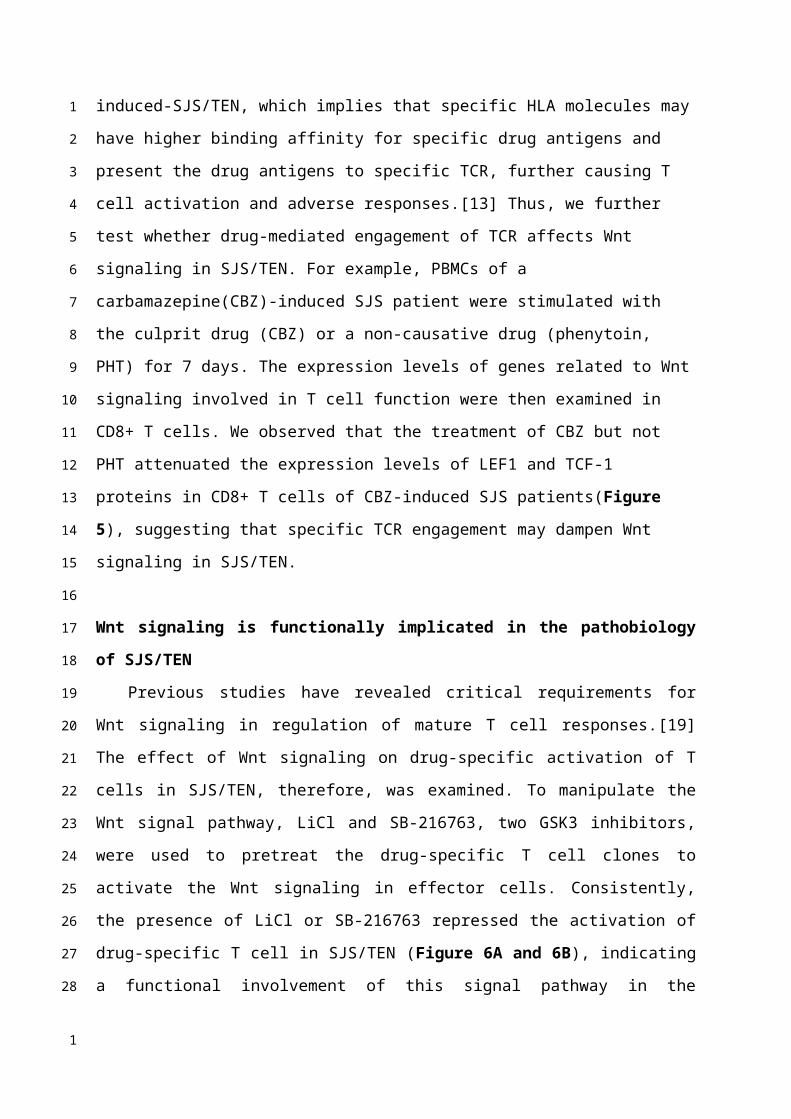
Wnt signaling is functionally implicated in the pathobiology of SJS/TEN
Previous studies have revealed critical requirements for Wnt signaling in regulation of
mature T cell responses.[19] The effect of Wnt signaling on drug-specific activation of T cells
in SJS/TEN, therefore, was examined. To manipulate the Wnt signal pathway, LiCl and SB-
216763, two GSK3 inhibitors, were used to pretreat the drug-specific T cell clones to activate
the Wnt signaling in effector cells. Consistently, the presence of LiCl or SB-216763 repressed
the activation of drug-specific T cell in SJS/TEN (Figure 6A and 6B), indicating a functional
1
2
3
4
5
6
7
8
9
10
11
12
13
14
15
16
17
18
19
20
21
22
23
24
25
26
27
28
29
30
31
32
1
involvement of this signal pathway in the pathomechanism of SJS/TEN. 1
1
Discussion:
CD8+ T cells and TCR function play a key role in the development of SJS/TEN, which is
characterized as widespread epidermal and mucosal necrosis.[34] Cytotoxic CD8 T cells that
produce cytotoxic molecules (such as granulysin and granzyme B) to cause extensive
keratinocyte death, are enriched in blister fluid samples from the skin lesions of patients
with SJS/TEN.[27, 35] These cytotoxic cells mediate the disease pathogenesis and are
correlated with the disease severity and mortality. In this study, we showed that Wnt
signaling components, including LEF1, TCF1, and LRP6, were downregulated in SJS/TEN
patients, while the serum levels of endogenous Wnt inhibitors, DKK1 and WIF1, were
elevated. These findings revealed that canonical Wnt signaling is regulated in SJS/TEN.
Further assays demonstrated that engagement of the TCR by the specific causative drug and
the treatment of a biomarker of SJS/TEN, IL-15 in vitro led to the downregulation of LEF-1
and TCF-1 expression in CD8+ T cells from SJS/TEN patients. Manipulation of Wnt signaling
by adding the Wnt activators attenuated ex vivo activation of drug-specific T cells from
SJS/TEN patients. Our data here, for the first time, suggest a functional involvement of Wnt
signaling in the pathomechanism of SJS/TEN.
The canonical Wnt signaling transduction cascade is an evolutionarily conserved and
multi-functional pathway and plays a crucial role in T cell development, differentiation, and
functionality.[36] Upon the activation of the Wnt signal pathway, naive CD8+ T cells undergo
clonal expansion and further differentiate into effector and memory precursors. The Wnt
signal pathway negatively regulates differentiated effector CD8+ T cells and positively
upregulates memory precursor CD8+ T cells through TCF-1 alone or its combination with β-
catenin.[19] In addition, a role of Wnt signaling pathway in promoting generation of
memory CD8+ T cells has been demonstrated through the observation that antigen-primed
human memory CD8+ T cells expressed lower TCF-1 and LEF-1 than did naive T cells. [22]
Consistently, constitutive activation of the Wnt signaling pathway was found to reduce
effector CD8+ T cell expansion.[23, 24] Another study also confirmed these findings and
further demonstrated that forced expression of stabilized β-catenin in naive T cells
interfered with proximal TCR signaling.[25] It is now clear that Wnt/β-catenin pathway is a
1
2
3
4
5
6
7
8
9
10
11
12
13
14
15
16
17
18
19
20
21
22
23
24
25
26
27
28
29
30
1
key signaling pathway governing CD8+ T cell differentiation. However, little is known about
the role of Wnt signaling pathway during the course of SJS/TEN.
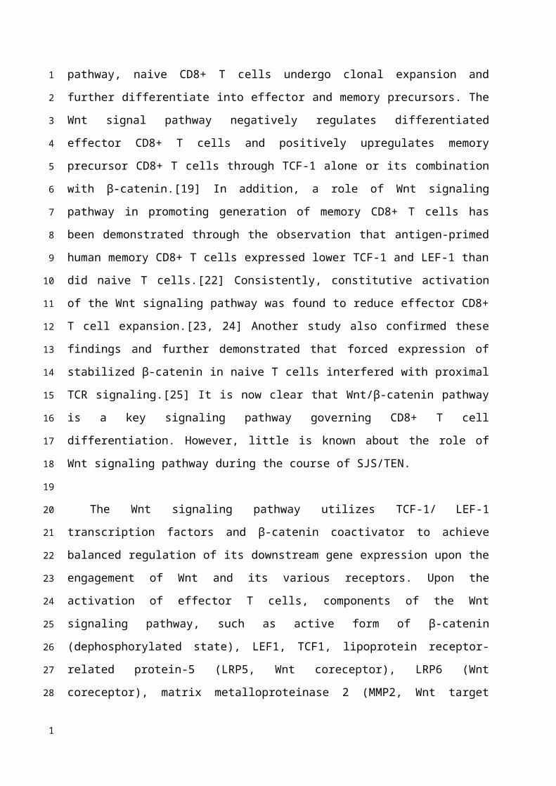
The Wnt signaling pathway utilizes TCF-1/ LEF-1 transcription factors and β-catenin
coactivator to achieve balanced regulation of its downstream gene expression upon the
engagement of Wnt and its various receptors. Upon the activation of effector T cells,
components of the Wnt signaling pathway, such as active form of β-catenin
(dephosphorylated state), LEF1, TCF1, lipoprotein receptor-related protein-5 (LRP5, Wnt
coreceptor), LRP6 (Wnt coreceptor), matrix metalloproteinase 2 (MMP2, Wnt target gene),
and MMP9 (Wnt target gene) are differentially expressed.[29, 37] In this study, we found
that genes encoding Wnt signal components, including LEF1, TCF1, and LRP6, were
downregulated in PBMC as well as in blistering cells from SJS/TEN as compared with normal
subjects. Our study further demonstrated decreased intracellular LEF1 and TCF1 protein
expression in CD8 T cell subsets of SJS/TEN patients, suggesting attenuation of Wnt signaling
in CD8 T cells in SJS/TEN. We have previously shown that a number of cytokines was
upregulated in SJS/TEN patients, of which IL-15 was significantly correlated with the disease
severity and mortality of SJS/TEN.[33] Here, we found that Wnt singals was attenuated
specifically by IL-15 but not IL-6 or IL-8. In addition to the impact from SJS-related cytokines,
Wnt signaling was dampened in CD8 T cells from SJS patients by TCR engagement with the
culprit drug antigen. Moreover, we detected that endogenous Wnt inhibitors, DKK1 and
WIF1, were elevated in the sera and bliser fluid of SJS/TEN compared to that from healthy
donors and burn patients. These findings collectively provide clues for how Wnt signal
pathway is regulated in the CD8 T cells in SJS/TEN. Furthermore, the presence of Wnt
activators attenuated ex vivo activation of drug-specific T cells from SJS/TEN patients,
indicating a functional involvement of Wnt signaling in the pathomechanism of SJS/TEN and
a reasonable therapeutic implication.
In this study, we found that the LRP5 was not simultaneously downregulated compared
with LRP6 in SJS/TEN though no statistical significance noticed. Active Wnt signaling is
initiated by binding of a Wnt protein to its receptor (Frizzeld) and one of the coreceptors
(LRP5 or LRP6). LRP5 is similar in sequence and structure to LRP6, and these two receptors
have been proposed to function largely in the same contexts and signaling pathways, but
1
2
3
4
5
6
7
8
9
10
11
12
13
14
15
16
17
18
19
20
21
22
23
24
25
26
27
28
29
30
31
32
1
there are difference between the two receptors. LRP5 usually can act as a gatekeeper for
Wnt responses to enable Wnt pathway responsiveness. [ 38 ] Comparing with LRP6, which
exhibits strong signaling activity, LRP5 was reported to be much less active in cells with Wnt
pathway activation. [ 39 ] Here, we hypothesized that LRP6 could be the main coreceptor
involved in the binding of a Wnt protein to its receptor (Frizzeld) and the coreceptors to
generate a β-catenin/TCF Wnt signal. In addition, DKK1 and WIF1 were the most significant
endogenous Wnt inhibitors comparing to SOST and Wnt3a involved in the reaction of
SJS/TEN in this study . Previous literature has also shown that DKK1 (eliciting inhibition of
LRP co-receptor function) and WIF1 (Wnt inhibitory factor 1) were both potential Wnt/β-
catenin signaling therapeutic targets for T cell responses and cytokines mediated
inflammatory autoimmune diseases. [ 40 ] Further study was needed to understand the
essential roles involved in the pathomechanism of SJS/TEN among these coreceptors and
endogenous Wnt inhibitors, and it may provide evidence for the potential biological targets
in the future.
The endogenous regulation in Wnt pathway in SJS/TEN. A previous study has
demonstrated the endogenous proteins/ligands (including LEF-1 and TCF-1) are
downregulated active Wnt signaling in effector T cells, which are the dominant immune cell
involved in SJS/TEN in our study. [ 23 ] These findings revealed consistent result between
endogenous and exogenous ligand during the activation of effector T cells. In addition, a
previous study had shown that endogenous LRP5 constitutively activates TCF/LEF-1 in the
absence of Wnt. [ 41 ] Another study also showed that removing the extracellular domain of
both LRP5 and LRP6 could lead to constitutive activation of the intracellular domain. In the
absence of exogenous Wnt 3a, full-length LRP6, but not LRP5, increased TCF/LEF-1
transcriptional activity, however both significantly potentiated Wnt 3a-induced TCF/LEF-1
activation. [ 42 ] Moreover, the intracellular domains (membrane-anchored and cytosolic) of
both LRP5 and LRP6 significantly increased TCF/LEF-1 activation in the absence of Wnt 3a,
and potentiated the Wnt 3a-induced decrease in beta-catenin phosphorylation, increase in
free beta-catenin levels and the increase in TCF/LEF-1 activity. [ 42 ] These findings have
demonstrated that LRP5 and LRP6 with and without the extracellular domain ( whether or
not they are membrane-anchored ) could still facilitate Wnt 3a-induced TCF/LEF-1 activation.
Taken together, the previous studies have shown the consistent results between exogenous
and endogenous regulation and both played important roles in modulating Wnt signaling.
1
2
3
4
5
6
7
8
9
10
11
12
13
14
15
16
17
18
19
20
21
22
23
24
25
26
27
28
29
30
31
32
1
Active Wnt signaling also plays essential roles in the activation of other immune cells,
including antigen presenting cells and Tregs, which are also known to be central to the
pathomechansim of SJS/TEN.[3, 18, 43] The canonical Wnt pathway was involved in
suppressing dendritic cell activation and cross-priming of CD8+ T cell response.[40]
Activation of Wnt signaling could potentially increase the tolerogenicity of antigen
presenting cells. On the other hand, Tregs from the acute-stage TEN patients profoundly are
impaired in their suppressive function, resulting in more severe cytotoxic consequences.[44]
Interestingly, overexpression of stabilized beta-catenin in CD4+CD25+ Treg cells led to an
increase of the survival of these Treg cells cells and could further induce an anergic
phenotype in CD4+CD25- effector T cells, which favored the suppressive effect of Treg cells
[21]. In previous studies, Wnt signals may have the potential to restore Treg-mediated
suppressive actions, which may be beneficial to reverse the overwhelming cytotoxic T
lymphocyte mediated cytotoxicity.[45] However, the role of Wnt signaling in Tregs function is
still controversial since findings from another study showed that Wnt signals favored toward
Th17 instead of Tregs lineage.[46-48] It would be of interest and further investigations are
necessary to delineate the exact role of Wnt signaling in the regulation of Tregs in SJS/TEN.
SJS/TEN are lethal severe cutaneous adverse reactions. Although the incidence has
ameliorated by identifying the genetic predisposing factors, early diagnosis and prognosis
monitoring for SJS/TEN remain a challenge for clinical physicians. Besides, no acceptable
treatment guideline for this devastating condition highlights the inadequacy of the current
therapeutic remedies. Our data here, for the first time, reveal that Wnt signaling is
functionally involved in the immunopathogenesis of SJS/TEN. These findings suggest that
enhancing the activity of Wnt pathway may be a potential strategy to modulate immunity in
SJS/TEN and provide additional insight into therapeutic aspects of this devastating condition.
1
2
3
4
5
6
7
8
9
10
11
12
13
14
15
16
17
18
19
20
21
22
23
24
25
26
27
1
Conflict of Interest
The authors declare that they have no relevant conflict of interest.
Acknowledgments
This study was supported by research grants from Ministry of Science and Technology,
Taiwan (MOST-108-2314-B-182A-006-MY3 to CB Chen, MOST-104-2314-B-182A-151-MY2 to
SC Su, and MOST-105-2628-B-010-007-MY3, MOST-106-2314-B-182A-037-MY3, to WH
Chung) and Chang Gung Memorial Hospital, Taiwan (CMRPG2H0081 and CMRPG2J0221 to
CB Chen; CIRPD1D0032, CORPG3F0041-3, OMRPG3E0041-3, and CORPG3F0061-3 to WH
Chung).
1
2
3
4
5
6
7
8
9
10
11
1
Reference:
1. Paquet, P.; Pierard, G. E., New insights in toxic epidermal necrolysis (Lyell's
syndrome): clinical considerations, pathobiology and targeted treatments revisited.
Drug Saf 2010, 33, (3), 189-212.
2. Weinborn, M.; Barbaud, A.; Truchetet, F.; Beurey, P.; Germain, L.; Cribier, B.,
Histopathological study of six types of adverse cutaneous drug reactions using
granulysin expression. Int J Dermatol 2016, 55, (11), 1225-1233.
3. Chen, C. B.; Abe, R.; Pan, R. Y.; Wang, C. W.; Hung, S. I.; Tsai, Y. G.; Chung, W. H., An
Updated Review of the Molecular Mechanisms in Drug Hypersensitivity. J Immunol
Res 2018, 2018, 6431694.
4. Roujeau, J. C.; Stern, R. S., Severe adverse cutaneous reactions to drugs. N Engl J Med
1994, 331, (19), 1272-85.
5. Rzany, B.; Correia, O.; Kelly, J. P.; Naldi, L.; Auquier, A.; Stern, R., Risk of Stevens-
Johnson syndrome and toxic epidermal necrolysis during first weeks of antiepileptic
therapy: a case-control study. Study Group of the International Case Control Study on
Severe Cutaneous Adverse Reactions. Lancet 1999, 353, (9171), 2190-4.
6. Mockenhaupt, M., The current understanding of Stevens-Johnson syndrome and
toxic epidermal necrolysis. Expert Rev Clin Immunol 2011, 7, (6), 803-13; quiz 814-5.
7. Borchers, A. T.; Lee, J. L.; Naguwa, S. M.; Cheema, G. S.; Gershwin, M. E., Stevens-
Johnson syndrome and toxic epidermal necrolysis. Autoimmun Rev 2008, 7, (8), 598-
605.
8. Roujeau, J. C.; Kelly, J. P.; Naldi, L.; Rzany, B.; Stern, R. S.; Anderson, T.; Auquier, A.;
Bastuji-Garin, S.; Correia, O.; Locati, F.; et al., Medication use and the risk of Stevens-
Johnson syndrome or toxic epidermal necrolysis. N Engl J Med 1995, 333, (24), 1600-
1
2
3
4
5
6
7
8
9
10
11
12
13
14
15
16
17
18
19
20
21
22
23
24
1

7.
9. Bharadwaj, M.; Illing, P.; Theodossis, A.; Purcell, A. W.; Rossjohn, J.; McCluskey, J.,
Drug hypersensitivity and human leukocyte antigens of the major histocompatibility
complex. Annual review of pharmacology and toxicology 2012, 52, 401-31.
10. Chen, C. B.; Hsiao, Y. H.; Wu, T.; Hsih, M. S.; Tassaneeyakul, W.; Jorns, T. P.; Sukasem,
C.; Hsu, C. N.; Su, S. C.; Chang, W. C.; Hui, R. C.; Chu, C. Y.; Chen, Y. J.; Wu, C. Y.; Hsu, C.
K.; Chiu, T. M.; Sun, P. L.; Lee, H. E.; Yang, C. Y.; Kao, P. H.; Yang, C. H.; Ho, H. C.; Lin, J.
Y.; Chang, Y. C.; Chen, M. J.; Lu, C. W.; Ng, C. Y.; Kuo, K. L.; Lin, C. Y.; Yang, C. S.; Chen,
D. P.; Chang, P. Y.; Wu, T. L.; Lin, Y. J.; Weng, Y. C.; Kuo, T. T.; Hung, S. I.; Chung, W. H.;
Taiwan Severe Cutaneous Adverse Reaction, C., Risk and association of HLA with
oxcarbazepine-induced cutaneous adverse reactions in Asians. Neurology 2017, 88,
(1), 78-86.
11. Fan, W.-L.; Shiao, M.-S.; Hui, R. C.-Y.; Su, S.-C.; Wang, C.-W.; Chang, Y.-C.; Chung, W.-H.,
HLA Association with Drug-Induced Adverse Reactions. Journal of Immunology
Research 2017, 2017, 10.
12. Su, S. C.; Chung, W. H., Update on pathobiology in Stevens-Johnson syndrome and
toxic epidermal necrolysis. Dermatol Sinica 2013, 31, (4), 175-80.
13. White, K. D.; Chung, W. H.; Hung, S. I.; Mallal, S.; Phillips, E. J., Evolving models of the
immunopathogenesis of T cell-mediated drug allergy: The role of host, pathogens,
and drug response. The Journal of allergy and clinical immunology 2015, 136, (2),
219-34.
14. Su, S. C.; Chung, W. H., Cytotoxic proteins and therapeutic targets in severe
cutaneous adverse reactions. Toxins (Basel) 2014, 6, (1), 194-210.
15. Logan, C. Y.; Nusse, R., The Wnt signaling pathway in development and disease.
1
2
3
4
5
6
7
8
9
10
11
12
13
14
15
16
17
18
19
20
21
22
23
24
1
Annual review of cell and developmental biology 2004, 20, 781-810.
16. Clevers, H., Wnt/beta-catenin signaling in development and disease. Cell 2006, 127,
(3), 469-80.
17. Clevers, H.; Nusse, R., Wnt/beta-catenin signaling and disease. Cell 2012, 149, (6),
1192-205.
18. Staal, F. J.; Luis, T. C.; Tiemessen, M. M., WNT signalling in the immune system: WNT
is spreading its wings. Nat Rev Immunol 2008, 8, (8), 581-93.
19. Xue, H. H.; Zhao, D. M., Regulation of mature T cell responses by the Wnt signaling
pathway. Annals of the New York Academy of Sciences 2012, 1247, 16-33.
20. Gattinoni, L.; Ji, Y.; Restifo, N. P., Wnt/beta-catenin signaling in T-cell immunity and
cancer immunotherapy. Clin Cancer Res 2010, 16, (19), 4695-701.
21. Ding, Y.; Shen, S.; Lino, A. C.; Curotto de Lafaille, M. A.; Lafaille, J. J., Beta-catenin
stabilization extends regulatory T cell survival and induces anergy in nonregulatory T
cells. Nat Med 2008, 14, (2), 162-9.
22. Willinger, T.; Freeman, T.; Herbert, M.; Hasegawa, H.; McMichael, A. J.; Callan, M. F.,
Human naive CD8 T cells down-regulate expression of the WNT pathway
transcription factors lymphoid enhancer binding factor 1 and transcription factor 7 (T
cell factor-1) following antigen encounter in vitro and in vivo. J Immunol 2006, 176,
(3), 1439-46.
23. Zhao, D. M.; Yu, S.; Zhou, X.; Haring, J. S.; Held, W.; Badovinac, V. P.; Harty, J. T.; Xue,
H. H., Constitutive activation of Wnt signaling favors generation of memory CD8 T
cells. J Immunol 2010, 184, (3), 1191-9.
24. Gattinoni, L.; Zhong, X. S.; Palmer, D. C.; Ji, Y.; Hinrichs, C. S.; Yu, Z.; Wrzesinski, C.;
Boni, A.; Cassard, L.; Garvin, L. M.; Paulos, C. M.; Muranski, P.; Restifo, N. P., Wnt
1
2
3
4
5
6
7
8
9
10
11
12
13
14
15
16
17
18
19
20
21
22
23
24
1
signaling arrests effector T cell differentiation and generates CD8+ memory stem
cells. Nat Med 2009, 15, (7), 808-13.
25. Driessens, G.; Zheng, Y.; Locke, F.; Cannon, J. L.; Gounari, F.; Gajewski, T. F., Beta-
catenin inhibits T cell activation by selective interference with linker for activation of
T cells-phospholipase C-gamma1 phosphorylation. J Immunol 2011, 186, (2), 784-90.
26. Auquier-Dunant, A.; Mockenhaupt, M.; Naldi, L.; Correia, O.; Schroder, W.; Roujeau, J.
C.; Reactions, S. S. G. S. C. A., Correlations between clinical patterns and causes of
erythema multiforme majus, Stevens-Johnson syndrome, and toxic epidermal
necrolysis: results of an international prospective study. Arch Dermatol 2002, 138,
(8), 1019-24.
27. Chung, W. H.; Hung, S. I.; Yang, J. Y.; Su, S. C.; Huang, S. P.; Wei, C. Y.; Chin, S. W.;
Chiou, C. C.; Chu, S. C.; Ho, H. C.; Yang, C. H.; Lu, C. F.; Wu, J. Y.; Liao, Y. D.; Chen, Y. T.,
Granulysin is a key mediator for disseminated keratinocyte death in Stevens-Johnson
syndrome and toxic epidermal necrolysis. Nat Med 2008, 14, (12), 1343-1350.
28. Wu, B.; Crampton, S. P.; Hughes, C. C., Wnt signaling induces matrix
metalloproteinase expression and regulates T cell transmigration. Immunity 2007, 26,
(2), 227-39.
29. Tiemessen, M. M.; Baert, M. R.; Kok, L.; van Eggermond, M. C.; van den Elsen, P. J.;
Arens, R.; Staal, F. J., T Cell factor 1 represses CD8+ effector T cell formation and
function. J Immunol 2014, 193, (11), 5480-7.
30. Zhou, X.; Yu, S.; Zhao, D. M.; Harty, J. T.; Badovinac, V. P.; Xue, H. H., Differentiation
and persistence of memory CD8(+) T cells depend on T cell factor 1. Immunity 2010,
33, (2), 229-40.
31. Ma, B.; Hottiger, M. O., Crosstalk between Wnt/beta-Catenin and NF-kappaB
1
2
3
4
5
6
7
8
9
10
11
12
13
14
15
16
17
18
19
20
21
22
23
24
1
Signaling Pathway during Inflammation. Front Immunol 2016, 7, 378.
32. Pyaram, K.; Chang, C.-H., Wnt/β-catenin signaling regulates IL-15-mediated terminal
maturation and interferon-gamma production of <em>invariant</em> NKT cells. The
Journal of Immunology 2016, 196, (1 Supplement), 121.14-121.14.
33. Su, S. C.; Mockenhaupt, M.; Wolkenstein, P.; Dunant, A.; Le Gouvello, S.; Chen, C. B.;
Chosidow, O.; Valeyrie-Allanore, L.; Bellon, T.; Sekula, P.; Wang, C. W.; Schumacher,
M.; Kardaun, S. H.; Hung, S. I.; Roujeau, J. C.; Chung, W. H., Interleukin-15 Is
Associated with Severity and Mortality in Stevens-Johnson Syndrome/Toxic
Epidermal Necrolysis. The Journal of investigative dermatology 2017, 137, (5), 1065-
1073.
34. Lerch, M.; Pichler, W. J., The immunological and clinical spectrum of delayed drug-
induced exanthems. Curr Opin Allergy Clin Immunol 2004, 4, (5), 411-9.
35. Chung, W. H.; Chang, W. C.; Stocker, S. L.; Juo, C. G.; Graham, G. G.; Lee, M. H.;
Williams, K. M.; Tian, Y. C.; Juan, K. C.; Jan Wu, Y. J.; Yang, C. H.; Chang, C. J.; Lin, Y. J.;
Day, R. O.; Hung, S. I., Insights into the poor prognosis of allopurinol-induced severe
cutaneous adverse reactions: the impact of renal insufficiency, high plasma levels of
oxypurinol and granulysin. Annals of the rheumatic diseases 2015, 74, (12), 2157-64.
36. Nusse, R.; Clevers, H., Wnt/beta-Catenin Signaling, Disease, and Emerging
Therapeutic Modalities. Cell 2017, 169, (6), 985-999.
37. Shi, J.; Chi, S.; Xue, J.; Yang, J.; Li, F.; Liu, X., Emerging Role and Therapeutic
Implication of Wnt Signaling Pathways in Autoimmune Diseases. J Immunol Res 2016,
2016, 9392132.
38. Goel, S.; Chin, E. N.; Fakhraldeen, S. A.; Berry, S. M.; Beebe, D. J.; Alexander, C. M.,
Both LRP5 and LRP6 receptors are required to respond to physiological Wnt ligands in
1
2
3
4
5
6
7
8
9
10
11
12
13
14
15
16
17
18
19
20
21
22
23
24
1
mammary epithelial cells and fibroblasts. J Biol Chem 2012, 287, (20), 16454-66.
39. MacDonald, B. T.; Semenov, M. V.; Huang, H.; He, X., Dissecting molecular differences
between Wnt coreceptors LRP5 and LRP6. PLoS One 2011, 6, (8), e23537.
40. Suryawanshi, A.; Tadagavadi, R. K.; Swafford, D.; Manicassamy, S., Modulation of
Inflammatory Responses by Wnt/beta-Catenin Signaling in Dendritic Cells: A Novel
Immunotherapy Target for Autoimmunity and Cancer. Front Immunol 2016, 7, 460.
41. Mao, J.; Wang, J.; Liu, B.; Pan, W.; Farr, G. H., 3rd; Flynn, C.; Yuan, H.; Takada, S.;
Kimelman, D.; Li, L.; Wu, D., Low-density lipoprotein receptor-related protein-5 binds
to Axin and regulates the canonical Wnt signaling pathway. Mol Cell 2001, 7, (4), 801-
9.
42. Mi, K.; Johnson, G. V., Role of the intracellular domains of LRP5 and LRP6 in activating
the Wnt canonical pathway. J Cell Biochem 2005, 95, (2), 328-38.
43. Bellon, T.; Blanca, M., The innate immune system in delayed cutaneous allergic
reactions to medications. Curr Opin Allergy Clin Immunol 2011, 11, (4), 292-8.
44. Takahashi, R.; Kano, Y.; Yamazaki, Y.; Kimishima, M.; Mizukawa, Y.; Shiohara, T.,
Defective regulatory T cells in patients with severe drug eruptions: timing of the
dysfunction is associated with the pathological phenotype and outcome. Journal of
immunology 2009, 182, (12), 8071-9.
45. Wang, C. W.; Yang, L. Y.; Chen, C. B.; Ho, H. C.; Hung, S. I.; Yang, C. H.; Chang, C. J.; Su,
S. C.; Hui, R. C.; Chin, S. W.; Huang, L. F.; Lin, Y. Y.; Chang, W. Y.; Fan, W. L.; Yang, C. Y.;
Ho, J. C.; Chang, Y. C.; Lu, C. W.; Chung, W. H.; and the Taiwan Severe Cutaneous
Adverse Reaction, C., Randomized, controlled trial of TNF-alpha antagonist in CTL-
mediated severe cutaneous adverse reactions. J Clin Invest 2018, 128, (3), 985-996.
46. Staal, F. J.; Arens, R., Wnt Signaling as Master Regulator of T-Lymphocyte Responses:
1
2
3
4
5
6
7
8
9
10
11
12
13
14
15
16
17
18
19
20
21
22
23
24
1
Implications for Transplant Therapy. Transplantation 2016, 100, (12), 2584-2592.
47. Chae, W. J.; Ehrlich, A. K.; Chan, P. Y.; Teixeira, A. M.; Henegariu, O.; Hao, L.; Shin, J. H.;
Park, J. H.; Tang, W. H.; Kim, S. T.; Maher, S. E.; Goldsmith-Pestana, K.; Shan, P.; Hwa,
J.; Lee, P. J.; Krause, D. S.; Rothlin, C. V.; McMahon-Pratt, D.; Bothwell, A. L., The Wnt
Antagonist Dickkopf-1 Promotes Pathological Type 2 Cell-Mediated Inflammation.
Immunity 2016, 44, (2), 246-58.
48. van Loosdregt, J.; Fleskens, V.; Tiemessen, M. M.; Mokry, M.; van Boxtel, R.;
Meerding, J.; Pals, C. E.; Kurek, D.; Baert, M. R.; Delemarre, E. M.; Grone, A.;
Koerkamp, M. J.; Sijts, A. J.; Nieuwenhuis, E. E.; Maurice, M. M.; van Es, J. H.; Ten
Berge, D.; Holstege, F. C.; Staal, F. J.; Zaiss, D. M.; Prakken, B. J.; Coffer, P. J., Canonical
Wnt signaling negatively modulates regulatory T cell function. Immunity 2013, 39,
(2), 298-310.
1
2
3
4
5
6
7
8
9
10
11
12
13
1
Table 1. Demographic and clinical characteristics of SJS/TEN patients.
Demographic data SJS/TEN (n=25)
Age, years, mean SD (range) 52.5 18.7 (3-81)
Sex ratio (M:F) 9:16
Skin (TBSA, %, mean SD (range))
Erythema 56.5 26.7 (10–100)
Blister or detachment 30.9 22.9 (1–80)
Mucosa (n, (%))
Ocular 20 (80.0)
Oral 25 (100.0)
Genital 18 (72.0)
Systemic Complications (n, (%))
Hepatitis a 7 (28.0)
Acute kidney injury b 6 (24.0)
Gastrointestinal bleeding 6 (24.0)
Pneumonitis, pneumonia, or bronchiolitis
obliterans 4 (16.0)
Blood dyscrasia (n, (%))
Eosinophilia c 6 (24.0)
Leukocytosis d 4 (16.0)
Leukopenia e 9 (36.0)
Atypical lymphocytosis f 16 (64.0)
Thrombocytopeniag 7 (28.0)
Corneal ulcer or symblepharon (n, (%)) 5 (20.0)
Fever h (n, (%)) 13 (42.0)
Mortality (n, (%)) 5 (20.0)
Underlying disease, n
Malignancy 2
Chronic kidney disease 4
12
1
Chronic liver disorder 3
Diabetes 3
Hypertension 9
Gouty 5
Epilepsy/Neuralgia 5
Rheumatoid arthritis 1
Culprit drugs, n
Anticonvulsants i 8
Antibiotics j 7
Allopurinol 4
Chlorzoxazone 1
Dapsone 1
NSAIDs 1
PPI 1
Sulfasalazine 2
SD, standard deviation; SJS, Stevens-Johnson syndrome; TEN, toxic epidermal necrolysis;
TBSA, total body surface area; NSAIDs, Non-Steroidal Anti-Inflammatory Drug; PPI, proton
pump inhibitor
a Values were 2 times greater than normal for glutamic-oxaloacetic transaminase, glutamic-
pyruvic transaminase, or total bilirubin.b The value of creatinine was 1.5 times greater than the normal value range (0.4–1.5 mg%)
after drug intake.c Eosinophils > 0.7 × 10⁹/L, or > 5%, or >10% if leukocyte < 4.0 × 10⁹/Ld The leukocyte count was greater than 11,000/μL.e The leukocyte count was less than 3,500/μL.f Abnormal lymphocytes present in blood.g The platelet count was lower than 150,000/μL.h Patients had a body temperature greater than 38.5°C.i Carbamazepine, lamotrigine, phenobarbital, and phenytoin
1
2
3
4
5
6
7
8
9
10
11
12
13
14
15
16
1
i Cephalexin, flomocef, moxifloxacin, levofloxacin, and vancomycin1
1
Figure Legends
Figure 1. Expression of Wnt signaling components is regulated in SJS/TEN and drug-
specific T cell activation. (A) Gene expression analyses of Wnt signaling components in
blister cells (BCs) and PBMCs of subjects with SJS/TEN. mRNA expression of LEF1, TCF1,
LRP5, and LRP6 was determined by real-time PCR. The respective mRNA levels were
compared among the blister cells (n=5) and PBMCs (n=5) from subjects with SJS/TEN at
active stage and control PBMCs from healthy donors (HD) (n=3). (B) Gene expression of Wnt
signaling components in drug-activated T cells from SJS/TEN patients. PBMCs obtained from
three SJS patients at active stage were cultured in RPMI1640 medium and stimulated
without (Control) and with the culprit drugs (MED), Solaxin, carbamazepine, and
vancomycin, respectively. After 7 days, the mRNA expression of LEF1, TCF1, LRP5, and LRP6
in drug-sensitive T cells, which were determined with the stimulation index (SI, calculated by
the level of secreted granulysin of stimulated to unstimulated cultures) greater than 2, was
measured by real-time PCR. *p <0.05; ** p <0.01; ***p <0.001; n.s, not significant; two-
sided Student’s t-test.
Figure 2. Intracellular LEF1 and TCF1 protein expression in CD8 T cell subsets of SJS/TEN
patients. TCF1 and LEF1 protein expression was detected in CD8 T cell subsets by
intracellular FACS staining. Black line histograms indicate TCF1 (A) or LEF1 (B) staining at the
active stage of disease course, and color line histograms represent staining at the recovered
stage. Representative flow cytometry plots show the expression levels of TCF1 (A) or LEF1
1
2
3
4
5
6
7
8
9
10
11
12
13
14
15
16
17
18
19
20
21
1
(B) in peripheral CD8 T cells of SJS/TEN patients. A representative example from one SJS/TEN
patient is shown from four individual SJS/TEN patients. (C-D) The fraction of TCF1 high CD8 T
cells and LEF1 high CD8 T cells populations are increased at the recovery stage comparing with
that at the active stage by intracellular FACS staining though no statically significance
(p=0.18 and p=0.15, respectively).
Figure 3. Endogenous Wnt inhibitors associated with SJS/TEN. (A) Serum levels of DKK1,
SOST, WIF1, and Wnt3a were examined in 10 patients with SJS/TEN and 11 healthy donors
(HD) by ELISA. (B) The levels of DKK1, SOST, WIF1, and Wnt3a in blister fluid of 9 TEN
patients and 11 burn patients were measured by ELISA. * p< 0.05; ** p< 0.01; *** p< 0.001;
two-sided Student’s t-test.
Figure 4. IL-15 contributes to the attenuation of Wnt signaling in CD8+ T cells. (A) TCF1
protein expression was detected in CD8+ T cell subsets by intracellular FACS staining after
PBMCs were treated with cytokines indicated for 24 hr. Representative flow cytometry plots
were shown from one of three independent experiments. (B-E) We observed a significant
decrease in the portion of TCF1 high CD8 T cells in PBMCs from normal subjects that were
treated with IL-15, but not with IL-6, IL-8, or SDF-1 (stromal cell-derived factor-1, used as a
non-SJS-related cytokine) . * p < 0.05; two-sided Student’s t-test .
Figure 5. Drug-specific TCR engagement attenuates the Wnt signal transduction in
SJS/TEN. PBMCs of a carbamazepine (CBZ)-induced SJS/TEN patient were stimulated with
1
2
3
4
5
6
7
8
9
10
11
12
13
14
15
16
17
18
19
20
21
22
1
the culprit drug (CBZ) or a tolerant drug (phenytoin, PHT) for 7 days. Levels of LEF1 and TCF1
protein expression were detected in CD8+ T cell subsets by intracellular FACS staining.
Representative flow cytometry plots were shown from one of five independent
experiments.
Figure 6. Activation of Wnt signaling attenuates activation of drug-specific T cells. PBMCs
of a carbamazepine (CBZ)- (A) or Ketoprofen-sensitive (B) SJS patient were obtained from
the heparinized blood by density centrifugation on Ficoll-Hypaque and stimulated with and
without the culprit drugs in the absence or presence of different concentrations of Wnt
signaling activator including LiCl (A) or SB216763 (B). After 7 days, the stimulation index (SI)
will be calculated by the level of secreted granulysin of stimulated to unstimulated cultures.
* p< 0.05; two sided Student’s t-test.
1
2
3
4
5
6
7
8
9
10
11
12
1
Figure 1. Expression of Wnt signaling components is regulated in SJS/TEN and drug-
specific T cell activation.
(A) Gene expression analyses of Wnt signaling components in blister cells (BCs) and PBMCs
1
2
3
4
1
of subjects with SJS/TEN. mRNA expression of LEF1, TCF1, LRP5, and LRP6 was determined
by real-time PCR. The respective mRNA levels were compared among the blister cells (n=5)
and PBMCs (n=5) from subjects with SJS/TEN and control PBMCs from healthy donors (HD)
(n=3). (B) Gene expression of Wnt signaling components in drug-activated T cells from
SJS/TEN patients. PBMCs obtained from three SJS/TEN patients were cultured in RPMI1640
medium and stimulated without (Control) and with the culprit drugs (MED), Solaxin,
carbamazepine, and vancomycin, respectively. After 7 days, the mRNA expression of LEF1,
TCF1, LRP5, and LRP6 in drug-sensitive T cells, which were determined with the stimulation
index (SI, calculated by the level of secreted granulysin of stimulated to unstimulated
cultures) greater than 2, was measured by real-time PCR. *p <0.05; ** p <0.01; ***p <0.001;
n.s, not significant; two-sided Student’s t-test.
1
2
3
4
5
6
7
8
9
10
11
1
Figure 2. Intracellular LEF1 and TCF1 protein expression in CD8 T cell subsets of SJS/TEN
patients.
TCF1 and LEF1 protein expression was detected in CD8 T cell subsets by intracellular FACS
staining. Black line histograms indicate TCF1 (A) or LEF1 (B) staining at the active stage of
disease course, and color line histograms represent staining at the recovered stage.
Representative flow cytometry plots show the expression levels of TCF1 (A) or LEF1 (B) in
peripheral CD8 T cells of SJS/TEN patients. A representative example from one SJS/TEN
patient is shown from four individual SJS/TEN patients. (C-D) The fraction of TCF1 high CD8 T
cells and LEF1 high CD8 T cells populations are increased at the recovery stage comparing with
1
2
3
4
5
6
7
8
9
10
1

that at the active stage by intracellular FACS staining though no statically significance
(p=0.18 and p=0.15, respectively).
1
2
1
Figure 3. Endogenous Wnt inhibitors associated with SJS/TEN.
(A) Serum levels of DKK1, SOST, WIF1, and Wnt3a were examined in 10 patients with
SJS/TEN and 11 healthy donors (HD) by ELISA. (B) The levels of DKK1, SOST, WIF1, and
Wnt3a in blister fluid of 9 TEN patients and 11 burn patients were measured by ELISA. * p<
0.05; ** p< 0.01; *** p< 0.001; two-sided Student’s t-test.
1
2
3
4
5
6
1

Figure 4. IL-15 contributes to the attenuation of Wnt signaling in CD8+ T cells.
(A) TCF1 protein expression was detected in CD8+ T cell subsets by intracellular FACS
staining after PBMCs were treated with cytokines indicated for 24 hr. Representative flow
cytometry plots were shown from one of three independent experiments. (B-E)
We observed a significant decrease in the portion of TCF1 high CD8 T cells in PBMCs from
normal subjects that were treated with IL-15, but not with IL-6, IL-8, or SDF-1 (stromal cell-
derived factor-1, used as a non-SJS-related cytokine) . * p < 0.05; two-sided Student’s t-test .
1
2
3
4
5
6
7
8
1
Figure 5. Drug-specific TCR engagement attenuates the Wnt signal transduction in
SJS/TEN.
PBMCs of a carbamazepine (CBZ)-induced SJS/TEN patient were stimulated with the culprit
drug (CBZ) or a tolerant drug (phenytoin, PHT) for 7 days. Levels of LEF1 and TCF1 protein
expression were detected in CD8+ T cell subsets by intracellular FACS staining.
Representative flow cytometry plots were shown from one of five independent
experiments.
1
2
3
4
5
6
7
8
1
Figure 6. Activation of Wnt signaling attenuates activation of drug-specific T cells.
PBMCs of a carbamazepine (CBZ)- (A) or Ketoprofen-sensitive (B) SJS patient were obtained
from the heparinized blood by density centrifugation on Ficoll-Hypaque and stimulated with
and without the culprit drugs in the absence or presence of different concentrations of Wnt
signaling activator including LiCl (A) or SB216763 (B). After 7 days, the stimulation index (SI)
will be calculated by the level of secreted granulysin of stimulated to unstimulated cultures.
* p< 0.05, two-sided Student’s t-test.
1
2
3
4
5
6
7
8
1


![[報告] 電影欣賞報告 - 奧圖瑪塔](https://img.pdfslide.tips/doc/110x75/58edf5eb1a28ab8c708b469b/-58edf5eb1a28ab8c708b469b.jpg)