Embed Size (px)
Citation preview
ECG in STEMIImportance and Challenges
Rabeea Aboufakher, MD, FACC, FSCAI
Cath Lab Director
Altru Health System
Grand Forks, ND
Disclosures
• No relevant disclosures
Overview
• Introduction
• ECG in the diagnosis of STEMI
• LBBB/paced rhythm/LVH
• Acute pericarditis/myocarditis
• Early repolarization
• Other causes of ST elevation
– Stress induced CMP
– PE
– Ventricular aneurysm

Introduction
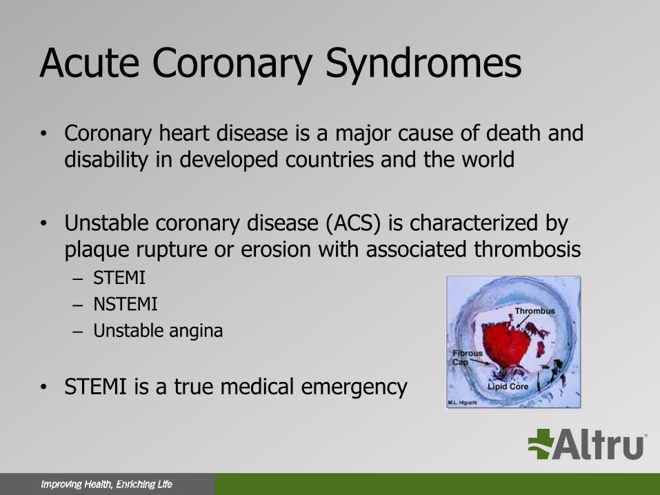
Acute Coronary Syndromes
• Coronary heart disease is a major cause of death and disability in developed countries and the world
• Unstable coronary disease (ACS) is characterized by plaque rupture or erosion with associated thrombosis
– STEMI
– NSTEMI
– Unstable angina
• STEMI is a true medical emergency
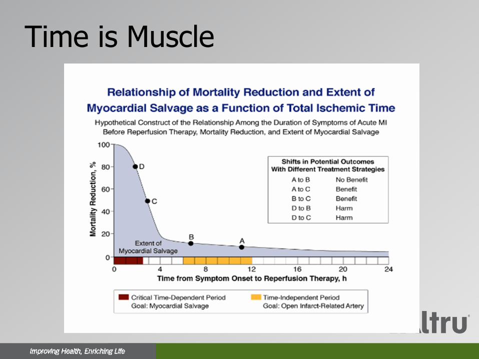
Time is Muscle
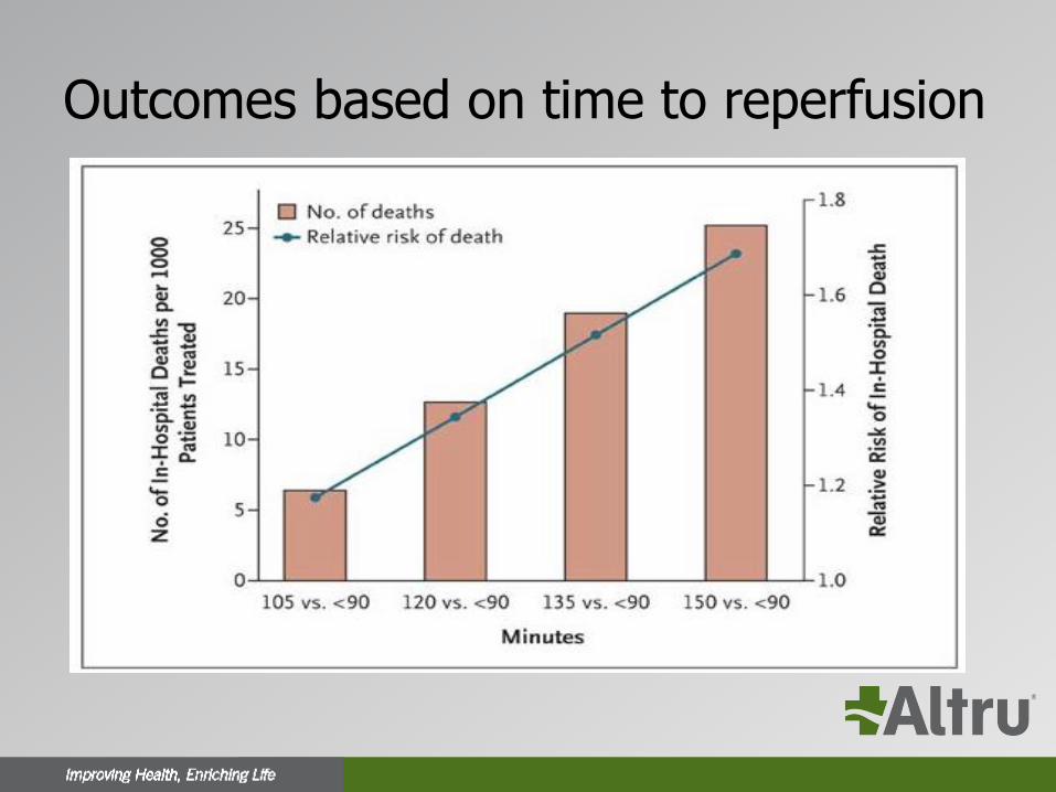
Outcomes based on time to reperfusion
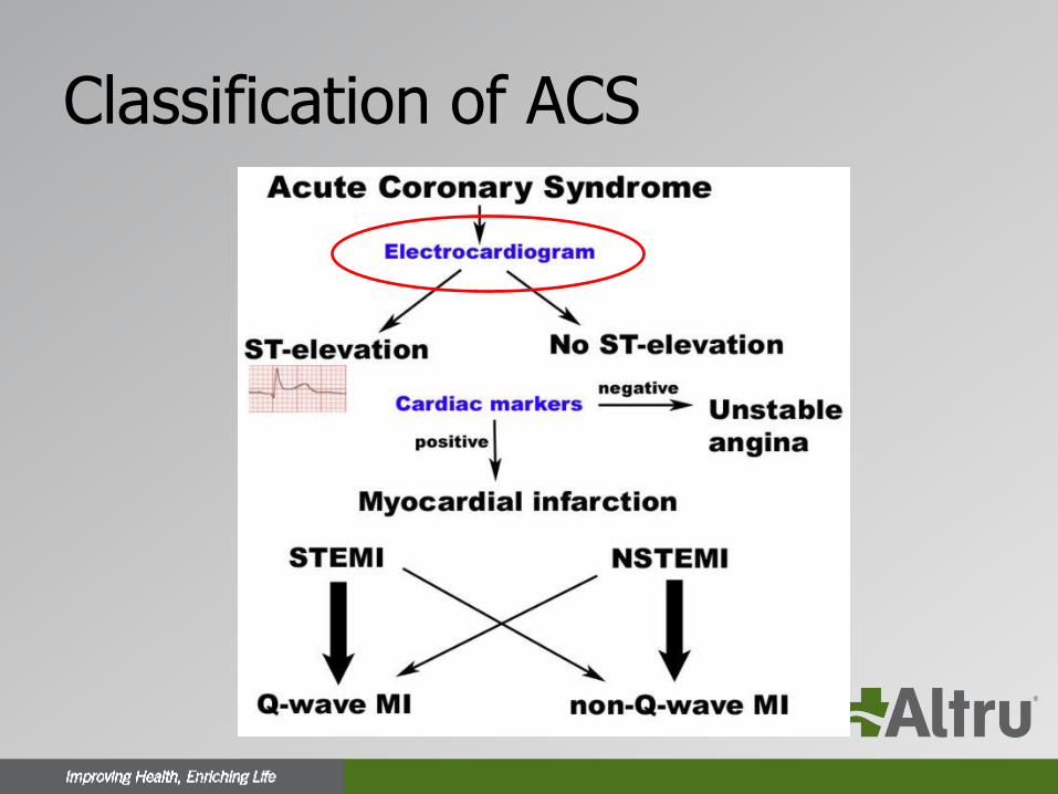
Classification of ACS
Historical Introduction
• 1895
– Einthoven, using an improved electrometer and a correction formula, distinguishes five deflections which he names P, Q, R, S and T
• 1912
– Einthoven’s triangle is described
• 1918
– Bousfield describes the spontaneous changes in the ECG during angina
Historical Introduction
• 1920
– Harold Pardee, New York, publishes the first ECG of an AMI in a human and describes the T wave as being tall and "starts from a point well up on the descent of the R wave". Pardee HEB. An electrocardiographic sign of coronary artery obstruction. Arch Int Med 1920;26:244-257
• 1930
– Sanders first describes infarction of the right ventricle
• 1942
– The augmented limb leads were added to arrive at the 12 lead ECG we use today

ECG in the Diagnosis of STEMI
ECG in STEMI
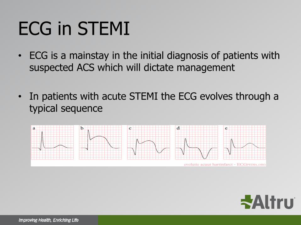
• ECG is a mainstay in the initial diagnosis of patients with suspected ACS which will dictate management
• In patients with acute STEMI the ECG evolves through a typical sequence
ECG in STEMI
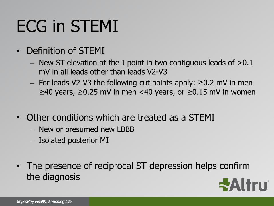
• Definition of STEMI
– New ST elevation at the J point in two contiguous leads of >0.1 mV in all leads other than leads V2-V3
– For leads V2-V3 the following cut points apply: ≥0.2 mV in men ≥40 years, ≥0.25 mV in men <40 years, or ≥0.15 mV in women
• Other conditions which are treated as a STEMI
– New or presumed new LBBB
– Isolated posterior MI
• The presence of reciprocal ST depression helps confirm the diagnosis
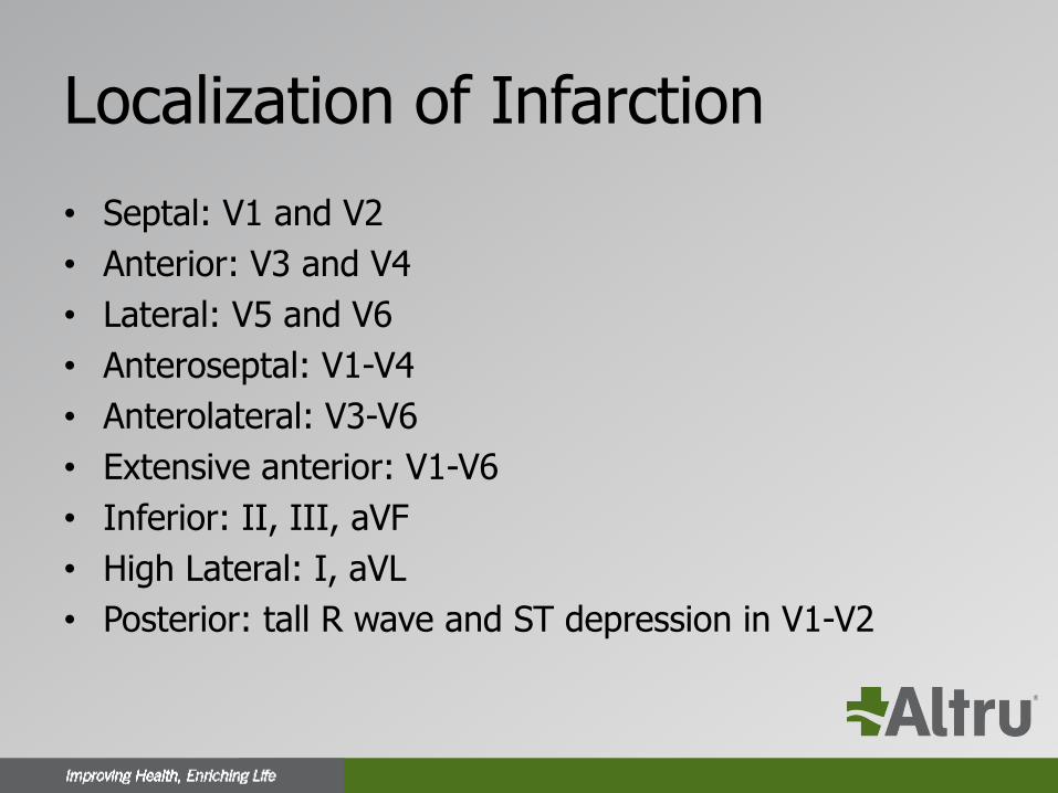
Localization of Infarction
• Septal: V1 and V2
• Anterior: V3 and V4
• Lateral: V5 and V6
• Anteroseptal: V1-V4
• Anterolateral: V3-V6
• Extensive anterior: V1-V6
• Inferior: II, III, aVF
• High Lateral: I, aVL
• Posterior: tall R wave and ST depression in V1-V2
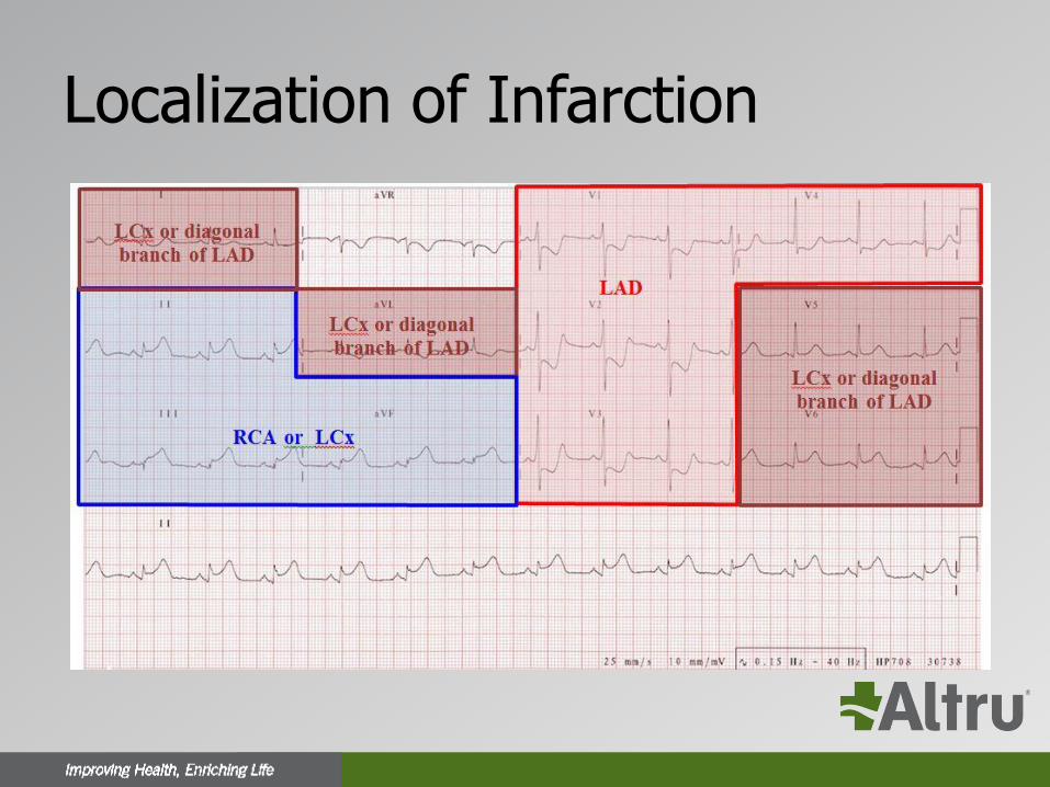
Localization of Infarction
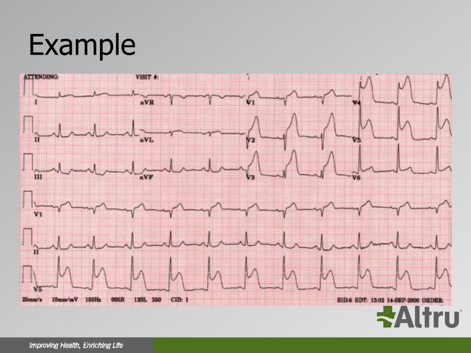
Example
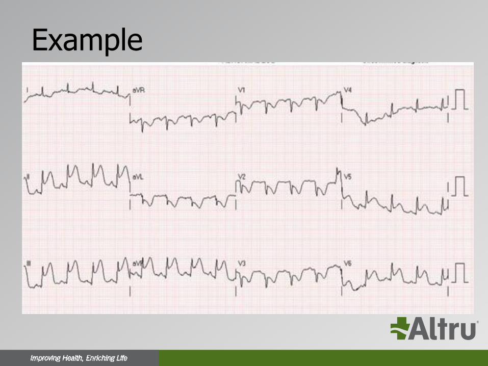
Example
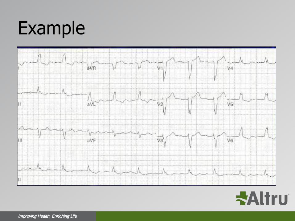
Example
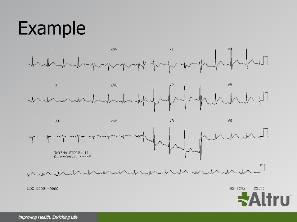
Example
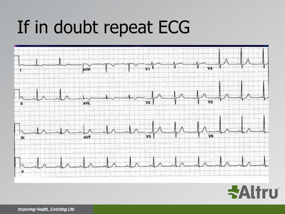
If in doubt repeat ECG
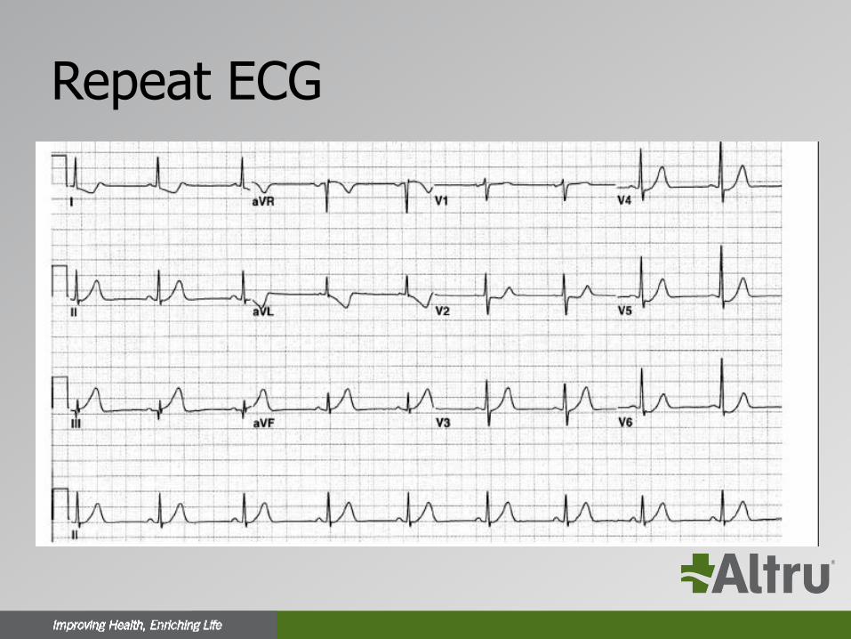
Repeat ECG
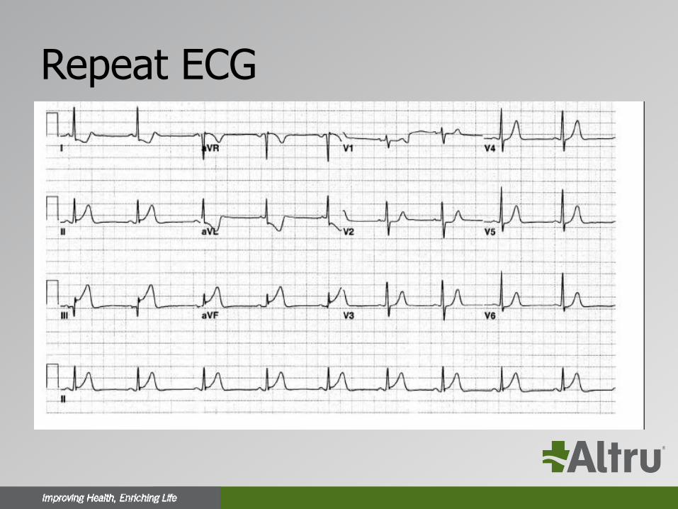
Repeat ECG
ECG Imposters
LBBB and Paced Rhythm
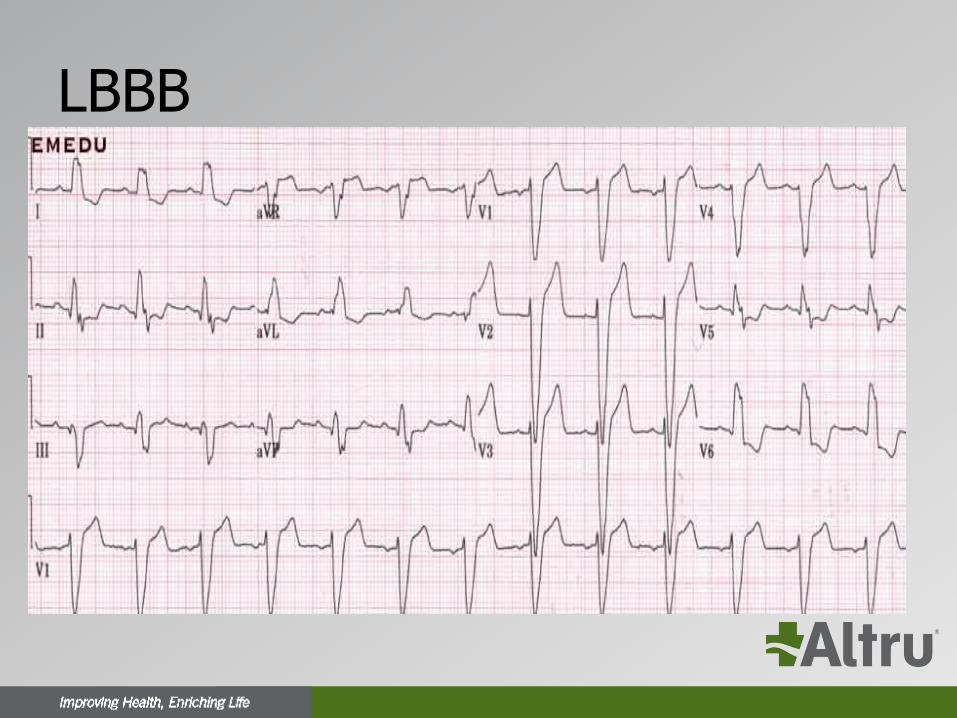
LBBB• LBBB occurs when normal electrical activity in the His-
Purkinje system is interrupted
• Most often occurs in patients with underlying heart disease
• Can be functional (rate related) and can be seen in asymptomatic individuals without structural heart disease
• Obscures ischemic changes on the ECG
LBBB
• A new LBBB in a patient with symptoms consistent with MI should be treated like a STEMI
• LBBB is associated with ST changes as part of the ECG pattern (discordant changes)
• Certain ST changes can be used to diagnose AMI in the setting of LBBB mainly called “concordant changes”
LBBB
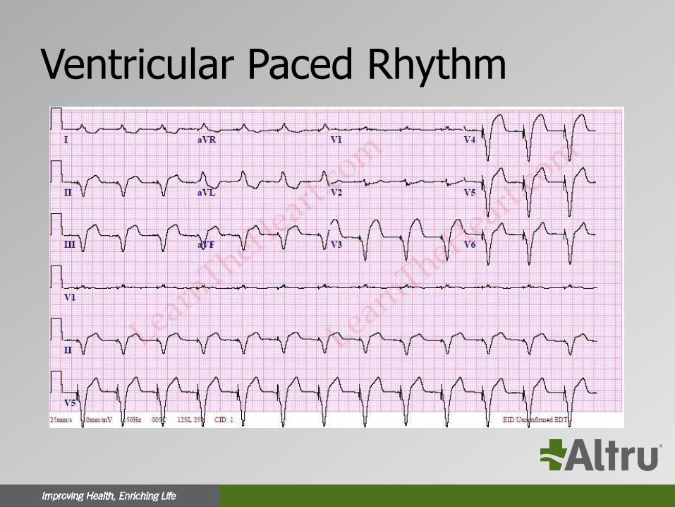
Ventricular Paced Rhythm
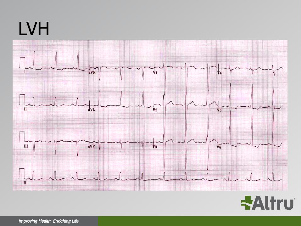
LVH

ECG Imposters
Acute Pericarditis/Myocarditis
Acute Pericarditis
• Common cause of chest pain with multiple etiologies most commonly viral or idiopathic
• Chest pain is typically pleuritic in nature and is positional
• The pain responds well to NSAIDS
• The presence of a friction rub on exam
Acute Pericarditis
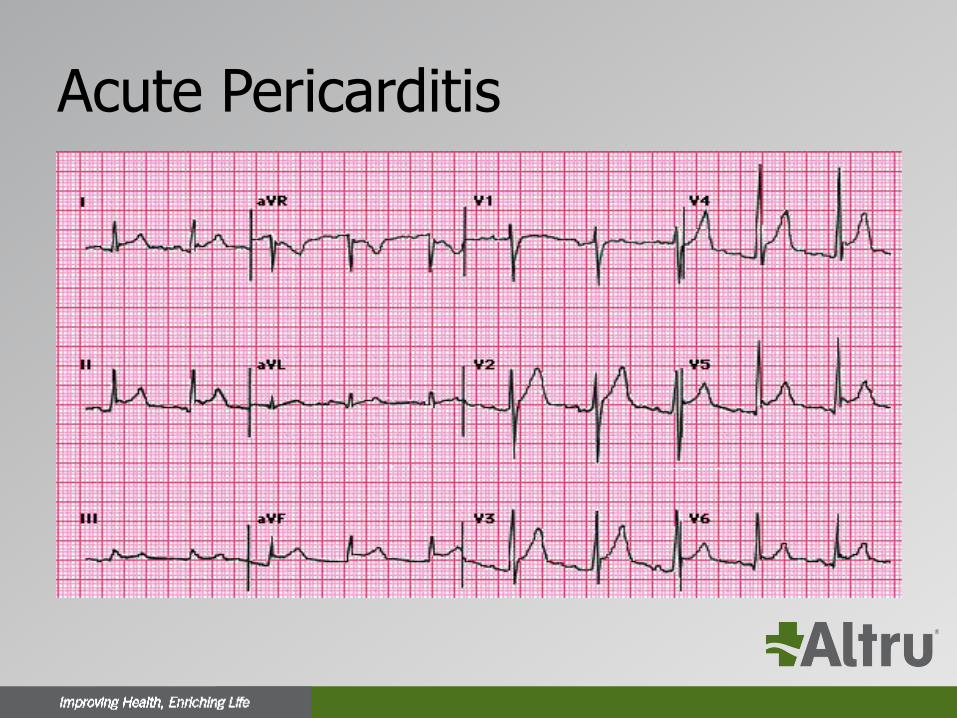
• Usually diffuse ST elevation
• Can be associated with PR depression (elevation in aVR)
• No reciprocal changes
• The morphology of the ST segment

ST morphology
Acute Pericarditis
Acute Myocarditis
• Myocarditis can occur alone or in combination with pericarditis
• Can cause similar ST changes to pericarditis
• Focal myocarditis can cause regional ST elevation even with ST depression and can be difficult to distinguish from a STEMI
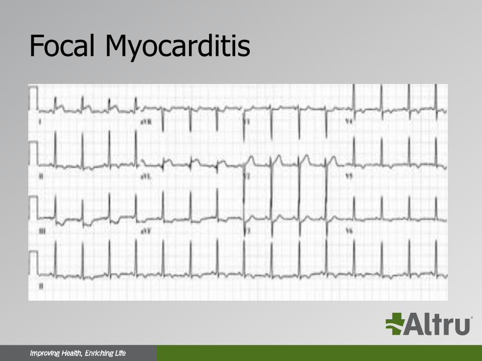
Focal Myocarditis
ECG Imposters
Normal Early Repolarization
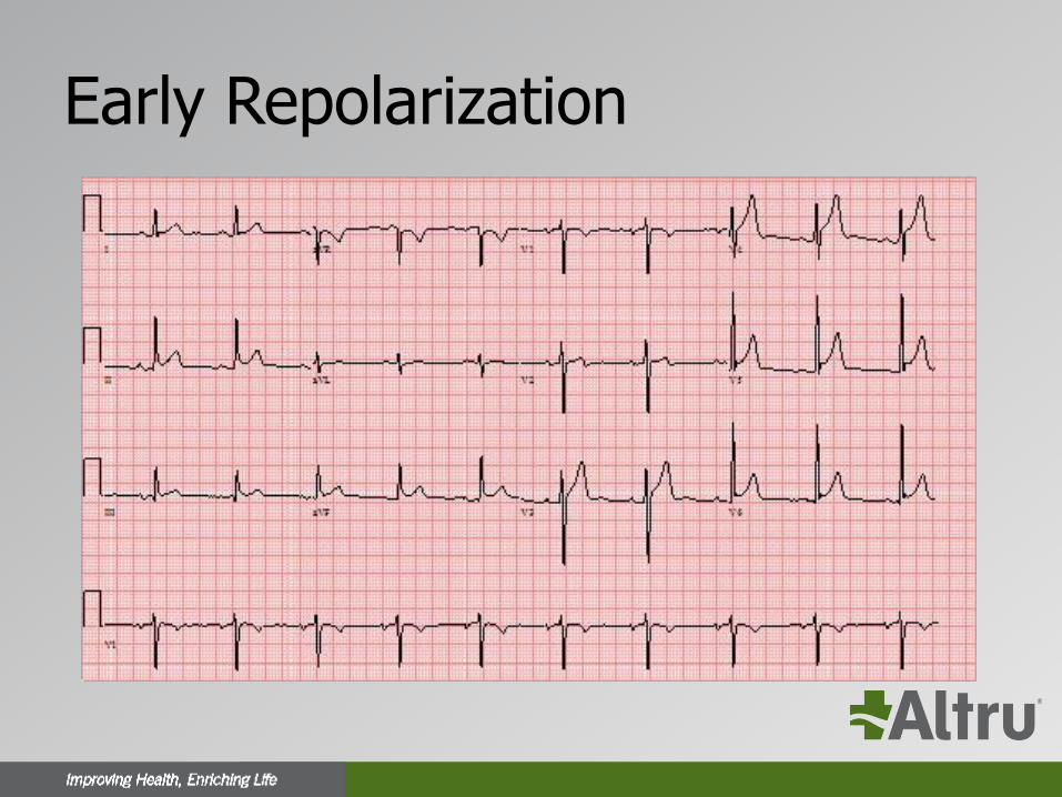
Early Repolarization
• A usually benign ECG pattern with an incidence of 5 to 13% of people so very common especially in young healthy athletes
• ST elevation (J point elevation) of 1 mm or more in 2 or more contiguous leads (usually inferior or lateral or both)
• ST morphology similar to pericarditis
• No reciprocal changes
Early Repolarization
ECG Imposters
Stress Induced Cardiomyopathy
LV Aneurysm
Pulmonary Embolism
Stress Induced Cardiomyopathy
• Also known as apical ballooning, takotsubo cardiomyopathy or broken heart syndrome
• More common than previously thought
• More common in elderly women but can occur in other groups
• An emotional or medical trigger such as loss of a loved one or severe pain or medical illness
Stress Induced Cardiomyopathy
• Patients usually have ECG changes including T wave inversions, ST depression or ST elevation
• Elevated enzymes and apical hypokinesis
• The only way to make the diagnosis at times is to perform a coronary angiogram (clean coronaries)
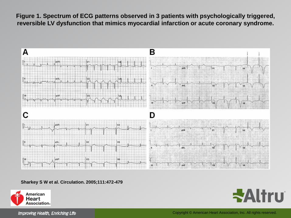
Figure 1. Spectrum of ECG patterns observed in 3 patients with psychologically triggered,
reversible LV dysfunction that mimics myocardial infarction or acute coronary syndrome.
Sharkey S W et al. Circulation. 2005;111:472-479
Copyright © American Heart Association, Inc. All rights reserved.
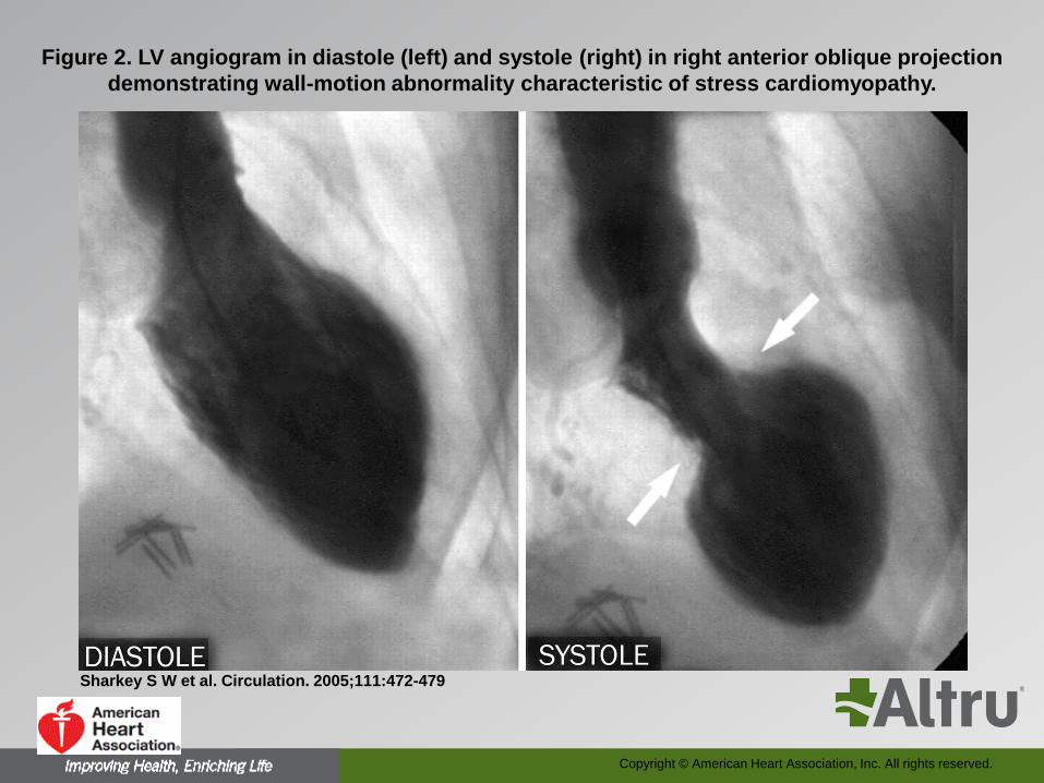
Figure 2. LV angiogram in diastole (left) and systole (right) in right anterior oblique projection
demonstrating wall-motion abnormality characteristic of stress cardiomyopathy.
Sharkey S W et al. Circulation. 2005;111:472-479
Copyright © American Heart Association, Inc. All rights reserved.
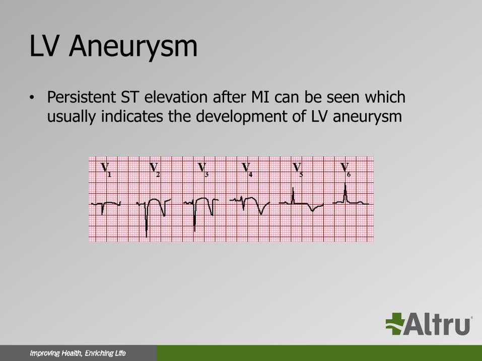
LV Aneurysm
• Persistent ST elevation after MI can be seen which usually indicates the development of LV aneurysm
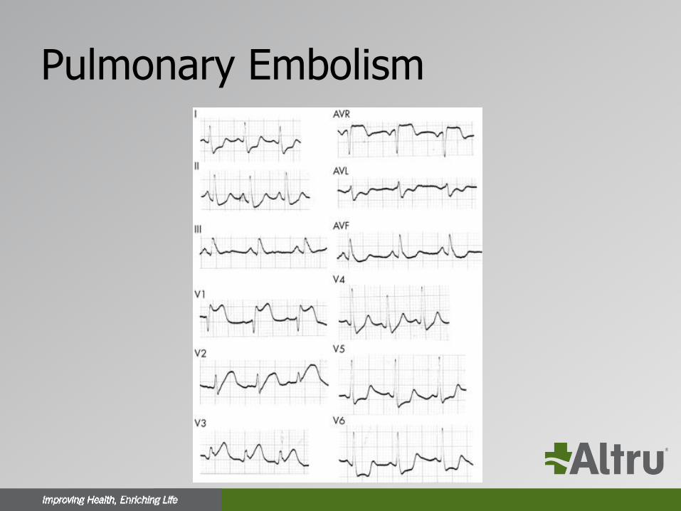
Pulmonary Embolism
In Conclusion
• ECG is the mainstay of diagnosing STEMI which is a true medical emergency
• Making the correct diagnosis promptly is life-saving
• If the clinical picture is consistent with MI and the ECG is not diagnostic serial ECG at 5-10 min intervals
• Several conditions can be associated with ST elevation on ECG most commonly LBBB, pericarditis, and early repolarization
• If in doubt call the cardiologist or activate the cath lab

Thank you
Questions??