Embed Size (px)
Citation preview
Research ArticleEdaravone, a Synthetic Free Radical Scavenger, EnhancesAlteplase-Mediated Thrombolysis
Kiyoshi Kikuchi,1,2,3,4 Kentaro Setoyama,5 Ko-ichi Kawahara,3,6 Tomoka Nagasato,7
Takuto Terashi,8 Koki Ueda,8 Kazuki Nakanishi,8 Shotaro Otsuka,8 Naoki Miura,9
Hisayo Sameshima,7 Kazuya Hosokawa,7 Yoichiro Harada,3 Binita Shrestha,3
Mika Yamamoto,3 Yoko Morimoto-Yamashita,10 Haruna Kikuchi,11 Ryoji Kiyama,12
Chinatsu Kamikokuryo,13 Salunya Tancharoen,4 Harutoshi Sakakima,6 Motohiro Morioka,2
Eiichiro Tanaka,1 Takashi Ito,3 and Ikuro Maruyama3
1Division of Brain Science, Department of Physiology, Kurume University School of Medicine, Kurume, Japan2Department of Neurosurgery, Kurume University School of Medicine, Kurume, Japan3Department of Systems Biology in Thromboregulation, Kagoshima University Graduate School of Medical and Dental Science,Kagoshima, Japan4Department of Pharmacology, Faculty of Dentistry, Mahidol University, Bangkok, Thailand5Division of Laboratory Animal Science, Natural Science Center for Research and Education, Kagoshima University,Kagoshima, Japan6Department of Biomedical Engineering, Laboratory of Functional Foods, Osaka Institute of Technology, Osaka, Japan7Research Institute, Fujimori Kogyo Co., Yokohama, Kanagawa, Japan8Course of Physical Therapy, School of Health Sciences, Faculty of Medicine, Kagoshima University, Kagoshima, Japan9Department of Veterinary Science, Laboratory of Diagnostic Imaging, Faculty of Agriculture, Kagoshima University,Kagoshima, Japan10Department of Restorative Dentistry and Endodontology, Kagoshima University Graduate School of Medical and Dental Science,Kagoshima, Japan
11Department of Psychosomatic Internal Medicine, Kagoshima University Graduate School of Medical and Dental Science,Kagoshima, Japan
12School of Health Sciences, Faculty of Medicine, Kagoshima University, Kagoshima, Japan13Department of Emergency and Critical Care Medicine, Kagoshima University Graduate School of Medical and Dental Science,Kagoshima, Japan
Correspondence should be addressed to Takashi Ito; [email protected] andIkuro Maruyama; [email protected]
Received 7 July 2017; Accepted 7 September 2017; Published 10 November 2017
Academic Editor: Adrian Doroszko
Copyright © 2017 Kiyoshi Kikuchi et al. This is an open access article distributed under the Creative Commons Attribution License,which permits unrestricted use, distribution, and reproduction in any medium, provided the original work is properly cited.
The combination of alteplase, a recombinant tissue plasminogen activator, and edaravone, an antioxidant, reportedly enhancesrecanalization after acute ischemic stroke. We examined the influence of edaravone on the thrombolytic efficacy of alteplase bymeasuring thrombolysis using a newly developed microchip-based flow-chamber assay. Rat models of embolic cerebral ischemiawere treated with either alteplase or alteplase-edaravone combination therapy. The combination therapy significantly reducedthe infarct volume and improved neurological deficits. Human blood samples from healthy volunteers were exposed toedaravone, alteplase, or a combination of alteplase and edaravone or hydrogen peroxide. Whole blood was perfused over acollagen- and thromboplastin-coated microchip; capillary occlusion was monitored with a video microscope and flow-pressuresensor. The area under the curve (extent of thrombogenesis or thrombolysis) at 30 minutes was 69.9% lower in the edaravone-alteplase- than alteplase-treated group. The thrombolytic effect of alteplase was significantly attenuated in the presence of
HindawiOxidative Medicine and Cellular LongevityVolume 2017, Article ID 6873281, 14 pageshttps://doi.org/10.1155/2017/6873281
hydrogen peroxide, suggesting that oxidative stress might hinder thrombolysis. D-dimers were measured to evaluate these effects inhuman platelet-poor plasma samples. Although hydrogen peroxide significantly decreased the elevation of D-dimers by alteplase,edaravone significantly inhibited the decrease. Edaravone enhances alteplase-mediated thrombolysis, likely by preventing oxidativestress, which inhibits fibrinolysis by alteplase in thrombi.
1. Introduction
Alteplase is the most effective and frequently used recombi-nant tissue plasminogen activator (tPA) for thrombolysis inpatients with acute ischemic stroke (AIS) [1]. Tissue plas-minogen activators are enzymes that catalyze the conversionof plasminogen to plasmin [2].
Edaravone (Radicut®; Mitsubishi Tanabe Pharma Corpo-ration, Osaka, Japan) is a low-specificity antioxidant thatscavenges various free radicals. Edaravone exhibits neurovas-cular protective effects against apoptosis, necrosis, edema,and inflammatory cytokines [3–9].
Several clinical trials have shown that edaravone-alteplase combination therapy is more effective than alte-plase alone in patients with AIS [1, 10–13]. Kimura et al.[14] presumed that these therapeutic effects occurred becauseedaravone protected the endothelium from ischemic injury,which increased the endogenous tPA levels and promotedearly recanalization [14–16]. However, the currently avail-able global clinical evidence for the efficacy of edaravone isinadequate, and more basic evidence for the efficacy of thisdrug in combination with alteplase is needed.
In the ischemic brains of rats, edaravone preventsendothelial cell damage and blood-brain barrier disruption[17, 18]. These in vivo studies showed that edaravone-alteplase combination therapy was more effective thanalteplase alone in rats with occlusion of the middle cere-bral artery by an intraluminal nylon filament [17, 18].However, no reports have described the establishment ofa model of thromboembolic clot-induced cerebral ische-mia, which would more accurately reflect clinical morbidconditions. Therefore, we used a model of thromboem-bolic clot-induced cerebral ischemia in vivo.
A recent experimental study showed that the thrombusvolume was significantly lower with edaravone-alteplasecombination treatment than with alteplase alone in a modelof helium-neon laser-induced thrombosis of rat mesentericmicrovessels [19]. Edaravone is considered to protect theendothelium, prevents new thrombus formation by enhanc-ing the expression of endothelial nitric oxide synthase,improves nitric oxide release, and inhibits the expression ofselectin [20]. An increased nitric oxide level leads to vasore-laxation, and downregulation of selectin suppresses plateletadhesion, platelet aggregation, and leukocyte adhesion [21].Therefore, edaravone is expected to accelerate early recanali-zation. However, the effects of edaravone on the blood itself,not on endothelial cells, are unknown. Current in vitro assaysof fibrinolytic reactions, such as clot-lysis tests, thromboelas-tography, and rotational thromboelastometry, are generallyperformed in the absence of blood flow; this limits their rele-vance to pathologic arterial thrombosis or physiologicalhemostasis [22, 23]. To overcome the limitations associatedwith animal models and static in vitro assays for assessing
fibrinolysis, Hosokawa et al. [24] speculated that evaluatingfibrin-rich platelet thrombus formation under shear flowcould be a useful model for studying thrombolytic processesin the arterial circulation.
The aim of the present study was to examine the mecha-nism by which edaravone promotes alteplase-mediatedthrombolysis in vitro in human blood donated by healthyvolunteers. We used the newly developed Total Thrombus-formation Analysis System (T-TAS®; Fujimori Kogyo Co.,Ltd., Tokyo, Japan) to quantify thrombolysis in bloodexposed to edaravone, alteplase, or alteplase and edaravone.We also examined the influence of hydrogen peroxide(H2O2) on alteplase-induced thrombolysis. Finally, wemeasured the concentration of D-dimers, which are fibrindegradation products, in platelet-rich plasma (PRP) sumpsolutions collected after the T-TAS assay to determinewhether edaravone-alteplase combination therapy inhibitsthrombogenesis or promotes thrombolysis.
2. Materials and Methods
The experimental protocol was approved by the InstitutionalAnimal Care and Use Committee of Kagoshima University(Kagoshima, Japan). The study protocol was approved bythe local ethics committee of Kagoshima University, andwritten informed consent was obtained from all individualsprior to their participation.
2.1. Rat Model of Thromboembolic Ischemia. Thromboem-bolic ischemia of the middle and posterior cerebral arterieswas induced in 8-week-old male Sprague–Dawley rats weigh-ing 290 to 310 g as previously described [25], with somemod-ifications. Anesthesia was induced and maintained with 2.5%to 3.0% isoflurane inhalation. After establishment of anesthe-sia, the rats were placed on the operating table in the supineposition. The rectal temperature was kept at 37°C± 1°C fromthe start of anesthesia until awakening. Initially, we per-formed a left femoral 1 cm skin incision, inserted a0.7× 1.9mm 24-gauge catheter (Angiocath®; Becton Dickin-son Co., Fukushima, Japan) into the left femoral artery undera microscope, and withdrew 0.15ml of arterial blood. Thisblood was injected into a 1.5ml microtube containing10 units of thrombin (Sawai Pharmaceutical, Osaka, Japan)in 50μl of saline and kept at room temperature for 30min.Centrifugation at 2800 rpm was performed for 2min, andthe supernatant serum was discarded. The clot was suctionedinto 4-French polyvinyl chloride tubes (Atom ExtensionTube; Atom Medical, Tokyo, Japan) with a 1ml disposablesyringe (Termosyringe®; Terumo Co., Tokyo, Japan). Underan operating microscope, the left common, external, andinternal carotid arteries were exposed through a midline inci-sion. The external carotid artery was ligated, coagulated, andcut down just proximal to the lingual and maxillary artery
2 Oxidative Medicine and Cellular Longevity
branches. All other branches of the external carotid arterywere coagulated and transected. The internal carotid arterywas then isolated to avoid damage to the vagus nerve. Thepterygopalatine artery was ligated at its origin. The internaland common carotid arteries were clamped with small aneu-rysm clips. A 24-gauge catheter (SURFLO Flash®; TerumoCo.) combined with a 1ml syringe was inserted into theinternal carotid artery via a small incision in the externalcarotid artery stump. The clot was pushed into the internalcarotid artery via a 24-gauge catheter by means of a straight-ened 1mm diameter paper clip. The thrombus occluded thedistal internal carotid artery, the proximal portion of theanterior cerebral artery, the middle cerebral artery, and theposterior cerebral arteries. To evaluate the infarction volume,neuromotor function, and hemorrhagic transformation at24 h, the 5mm clot (volume of 3.6mm3) was pushed intothe internal carotid artery (n = 8 per group). After withdraw-ing the catheter, the external carotid artery was ligated. Thetemporary clip was withdrawn, and the internal carotidartery blood flow recovered.
We did not use a cerebral blood flow (CBF) monitor.Shimamura et al. [25] reported that a CBF monitor isnot indispensable for this model because other surgicalmanipulations can be performed to establish whetherbrain injury and/or changes in intracranial pressure havebeen avoided. Additionally, dissection of the temporalmuscle causes masticatory dysfunction, leading to inade-quate nutrition. Instead, we used only rats with a neuro-logical score of 3 or 4 after awakening. The rats wereevaluated for neurological deficits after awakening and at24 h after thromboembolism. A neurological grading sys-tem with a 5-point motor function scale (0–4) was usedas previously described [26]. The scale was as follows:0 =no apparent deficits, 1 = right forelimb flexion,2 =decreased right forelimb grip when tail is pulled,3 = spontaneous movement in all directions with rightcircling only when tail is pulled, and 4= spontaneousright circling.
2.2. Drug Treatment. Three groups of rats were studied asfollows: the vehicle-injected control group, alteplase group,and edaravone-alteplase group. Edaravone and alteplase wereprovided by Mitsubishi Tanabe Pharma Corporation. Imme-diately after thromboembolism, 6mg/kg of edaravone wasadministered over a 20min period via a jugular vein catheterusing an infusion pump. At 20min after thromboembolism,3mg/kg of alteplase was administered shortly after edaravoneadministration. The alteplase group received vehicle insteadof edaravone, followed by alteplase treatment. The controlgroup received injections of vehicle instead of edaravoneand alteplase. The dose and timing of administration of edar-avone and alteplase were determined via preliminary experi-ments to maximize the effect of edaravone [5]. Therefore, thedose of alteplase was lower than that in previous reportsusing 10mg/kg [17, 18].
2.3. Measurement of Infarct Volume. After venous bloodsampling, the rats were killed and their brains excised 24 hafter thromboembolism as previously described [26].
Physiological saline was transcardially perfused beforedecapitation. The brain was carefully removed and cut intosix 2mm thick coronal sections from the frontal tip using abrain slicer. The slices were then immersed in a 1% solutionof 2,3,5-triphenyltetrazolium chloride in phosphate bufferedsaline (pH7.4) at 37°C for 10min. After staining, the sectionswere scanned to determine the ischemic infarct volume. Theinfarctions were measured using Scion Image software versusBeta 4.0.3 (Scion Corp., Frederick, MD). The total infarctarea (mm3) was multiplied by the thickness of the brainsections to obtain the infarct volume. Additionally, the pres-ence of visible hematomas or hemorrhagic transformationwas recorded.
2.4. Rat Blood Samples and Laboratory Data. At 24 h afterthromboembolism, the rats were deeply anesthetized via anintraperitoneal injection of 4% chloral hydrate (10ml/kg).Blood samples were collected from the axillary vein.
Adverse drug reactions such as renal and hepatic disor-ders are occasionally observed during edaravone treatmentin >5% of patients [27]. To evaluate adverse drug reactionsincluding renal and hepatic disorders, the serum aspartatetransaminase, serum alanine transaminase, blood urea nitro-gen, and serum creatinine levels were measured by an enzy-matic method using a Fuji DRI-CHEM Slide Kit (FujifilmMedical, Tokyo, Japan).
2.5. Human Blood Samples. Blood samples from 10 healthy,fasting Japanese volunteers (mean age, 40.3± 12.7 years)were collected in plastic tubes containing 3.2% sodiumcitrate (Terumo Co.). None of the volunteers had takenantithrombotic drugs within 2 weeks of the study. Onevolunteer regularly took an over-the-counter fish oil sup-plement (docosahexaenoic acid 300mg/day, eicosapentae-noic acid 100mg/day). Normal ranges for the T-TASanalysis have not yet been defined, but the T-TAS findingsof all volunteers’ samples lay within 95% of the median of123 healthy Japanese individuals who participated in ourpreliminary study (data not shown). Platelet-rich plasmawas prepared by centrifugation at 800 rpm for 15min,and platelet-poor plasma (PPP) was prepared by centrifu-gation at 3000 rpm for 15min.
In the experiments using whole blood and PRP samples,we selected the final concentration of alteplase (500 IU/ml)and edaravone (3μM) based on half the maximum concen-tration after administration to Japanese patients with AIS[5, 28]. In the experiments using PPP samples, we selectedthe final concentration of alteplase (50, 100, and 250 IU/ml)and edaravone (6 and 60nM) based on the findings of a pre-liminary experiment. The final concentrations of vitamin C(6μM), vitamin E (120 nM), and NAC (6μM) were selectedbased on a preliminary experiment and previous reportsand were expected to have efficacy equivalent to that of edar-avone [29]. The final concentration of H2O2 (100μM) wasbased on a previous report, recognizing that it is difficult toestimate the local concentration of reactive oxygen species(ROS) around the intravascular thrombi during AIS [5].Vitamins C and E were obtained from Kanto Kagaku Co.,Ltd. (Tokyo, Japan), NAC was obtained from Wako Pure
3Oxidative Medicine and Cellular Longevity
Chemicals (Osaka, Japan), and H2O2 was obtained fromSigma-Aldrich Japan (Tokyo, Japan).
Samples from six healthy, fasting volunteers (mean age,35.8± 7.6 years) were further selected for the experimentsin which specimens were exposed to H2O2. These 6 volun-teers were selected from the original cohort of 10 becausethey had a normal response to alteplase. Of the remainingvolunteers, one was a nonresponder, two were incompleteresponders, and one was an over-responder.
2.6. T-TAS. The thrombolytic effects of alteplase, edaravone,and the combination of alteplase and edaravone were com-pared with the controls under flow conditions using T-TASin whole blood and PRP. To quantify thrombogenesis andthrombolysis under flow conditions, the T-TAS assay wasperformed as previously described [24]. Thrombogenesisand thrombolysis were observed in the microchip using abuilt-in light microscope. An antioxidant (edaravone, vita-min C or E, or NAC) was added to the blood samples10min before the addition of alteplase. Hydrogen peroxidewas added immediately after the addition of alteplase. Assoon as alteplase or H2O2 had been administered, each sam-ple was perfused over a microchip coated with collagen andtissue thromboplastin to promote thrombosis at a flow rateof 4μl/min, corresponding to an initial wall shear rate of240 per second.
2.7. Measurement of D-Dimer Concentration in SumpSolutions of T-TAS. T-TAS sump solutions were preparedby diluting the analyzed pooled PRP samples at a 1 : 25 ratioin ethylenediaminetetraacetic acid followed by centrifugationat 800 rpm for 15min. The concentration of D-dimers wasalso measured in the sump solution using an LPIA-NV7instrument and RM73-752YLK solution (LSI MedienceCorporation, Tokyo, Japan).
2.8. Fibrinolysis Assays. Fibrinolysis assays were performed toevaluate whether edaravone enhances thrombolysis by alte-plase. Microplate-based fibrinolysis assays were performedat 37°C in flat-bottomed 96-well polystyrene plates (Corning;Sigma-Aldrich Japan) by monitoring turbidity changes(A405) using a VersaMax microplate reader (MolecularDevices Japan, Tokyo, Japan). Calcium ions accelerate theformation of a fibrin clot from fibrinogen in the presence ofthrombin. One-hundred microliter aliquots of 30% humanPPP pooled from all 10 volunteers and TBSTC (8mM Trisat pH7.4, 0.008% Tween-20, and 12mM calcium chloride)were prepared in the presence or absence of alteplase (0, 50,100, or 250 IU/ml) or edaravone (0, 6, or 60 nM; n = 5). Theconcentrations of edaravone had been determined in a pre-liminary experiment. Edaravone was added to the samples10min before alteplase.
2.9. Measurement of D-Dimer Concentration in Human PPPSamples. D-dimers were measured to determine whetheredaravone attenuates the inhibition of alteplase-inducedfibrinolysis by H2O2. Four-hundred microliter aliquots of30% human PPP pooled from all 10 volunteers and TBSTCwere prepared. After incubation at 37°C for 15min, H2O2(100μM), edaravone (60 nM), or vehicle was added. After
incubation at 37°C for 10min, alteplase (100 IU/ml), plasmin(250μg/ml), or vehicle was added. After fibrin deposition hadoccurred by incubating at 37°C for 10min, aprotinin(400KIU/ml) was added. After centrifugation at maximumspeed for 5min, the concentration of D-dimers was mea-sured using an ACL TOP automated analyzer (Instrumenta-tion Laboratory, Bedford, MA) (n = 5).
2.10. Statistical Analysis. The neurological score, infarctionvolume, hemorrhagic transformation rate, and blood testdata were analyzed using the Steel–Dwass method or Bonfer-roni–Dunn method as appropriate in multiple comparisons.
The area under the curve at 30min (AUC30) was calcu-lated to evaluate the extent of thrombogenesis or thromboly-sis. The AUC30 represents the area under the flow-pressurecurve (<80 kPa) 30min after the start of assay, as previouslydescribed [24]. The AUC30 was also used to quantify theimpairment of thrombus formation when occlusion is notachieved during an assay. Comparison between two groupswas performed using the paired t-test or Wilcoxon signed-rank test as appropriate.
Absorbance data were analyzed by repeated-measurestwo-way analysis of variance followed by Bonferroni’s test.
Data are presented as mean± standard deviation unlessotherwise indicated. Differences with P < 0 05 were consid-ered statistically significant. All analyses were performedusing SPSS Statistics (version 20; IBM Corp., Armonk, NY).
3. Results
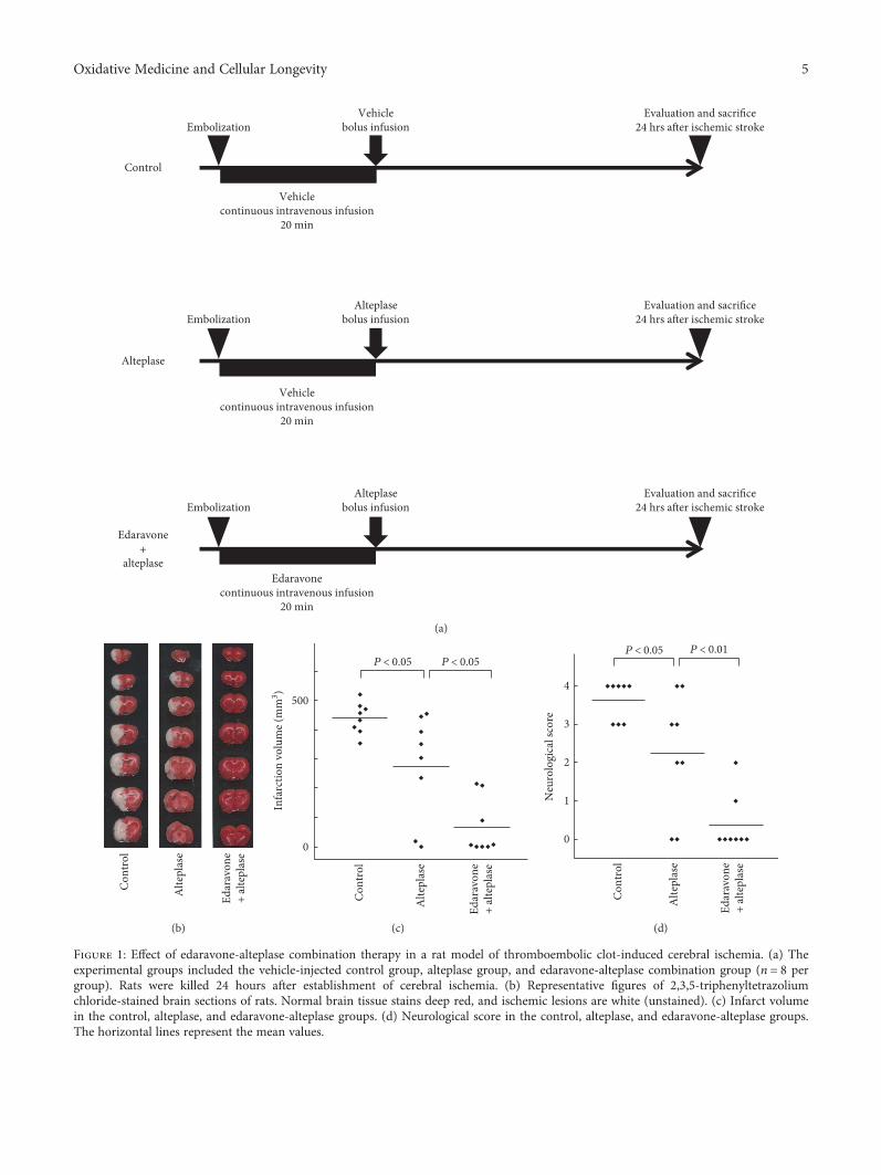
3.1. Edaravone-Alteplase Combination Reduces InfarctVolume and Improves Neuromotor Function in Rats. Weevaluated our rat thromboembolic stroke model, in whichthe distal internal carotid artery, proximal portion of theanterior cerebral artery, middle cerebral artery, and posteriorcerebral arteries were occluded by autologous thrombi(Figure 1(b)). We compared the vehicle-injected controlgroup, alteplase group, and edaravone-alteplase combinationgroup, which allowed us to investigate the therapeutic effectsof edaravone-alteplase combination therapy (Figure 1(a)).
We did not use a CBF monitor according to a previousreport [25]. The neurological score of all rats after awakeningwas 3 or 4, and the differences among the three groups of ratsthat were assigned to the different treatments were not statis-tically significant (Table 1). Therefore, we were able to inducecerebral ischemia without using a CBF monitor.
We then evaluated the infarct volume, neuromotor func-tion, hemorrhagic transformation, and adverse drug reac-tions at 24 h after ischemia in the three groups. Comparedwith the controls, alteplase significantly reduced the infarctvolume (P < 0 05) (Figures 1(b) and (c)). However, the infarctvolume was further significantly decreased in rats receivingedaravone-alteplase (P < 0 05) (Figures 1(b) and (c)). Theneurologic score in rats receiving alteplase was significantlybetter than that in the controls (P < 0 05) (Figure 1(d)).Additionally, the rats treated with edaravone-alteplaseshowed significantly better neurologic scores than the ratstreated with alteplase (P < 0 01) (Figure 1(d)). Meanwhile,the rate of hemorrhagic transformation tended to be lower
4 Oxidative Medicine and Cellular Longevity
Vehiclebolus infusion
Alteplasebolus infusion
Alteplasebolus infusion
Alteplase
Embolization
Embolization
Edaravone+
alteplase
Evaluation and sacri�ce24 hrs a�er ischemic stroke
Evaluation and sacri�ce24 hrs a�er ischemic stroke
Evaluation and sacri�ce24 hrs a�er ischemic stroke
Embolization
Control
Vehiclecontinuous intravenous infusion
20 min
Vehiclecontinuous intravenous infusion
20 min
Edaravonecontinuous intravenous infusion
20 min
(a)
Alte
plas
e
Edar
avon
e+
alte
plas
e
Cont
rol
(b)
Infa
rctio
n vo
lum
e (m
m3 )
P < 0.05 P < 0.05
Alte
plas
e
Edar
avon
e+
alte
plas
e
Cont
rol
500
0
(c)
Neu
rolo
gica
l sco
re
P < 0.05 P < 0.01
Alte
plas
e
Edar
avon
e+
alte
plas
e
Cont
rol
4
3
2
1
0
(d)
Figure 1: Effect of edaravone-alteplase combination therapy in a rat model of thromboembolic clot-induced cerebral ischemia. (a) Theexperimental groups included the vehicle-injected control group, alteplase group, and edaravone-alteplase combination group (n = 8 pergroup). Rats were killed 24 hours after establishment of cerebral ischemia. (b) Representative figures of 2,3,5-triphenyltetrazoliumchloride-stained brain sections of rats. Normal brain tissue stains deep red, and ischemic lesions are white (unstained). (c) Infarct volumein the control, alteplase, and edaravone-alteplase groups. (d) Neurological score in the control, alteplase, and edaravone-alteplase groups.The horizontal lines represent the mean values.
5Oxidative Medicine and Cellular Longevity
in the edaravone-alteplase group than in the alteplase group.However, the differences were not statistically significantamong the three groups (Table 1).
Adverse drug reactions, including renal and hepaticdisorders, were not apparent because the serum aspartatetransaminase, serum alanine transaminase, blood urea nitro-gen, and serum creatinine levels were not significantly differ-ent among the three groups (Table 1).
In conclusion, edaravone synergized with acute alteplasetreatment in this experimental thrombotic stroke model.
3.2. Characteristics of Blood Samples Obtained from HealthyVolunteers. The mean erythrocyte, leukocyte, and plateletcounts of whole blood and PRP samples are shown inTable 2. These lay within the normal ranges for healthyJapanese individuals.
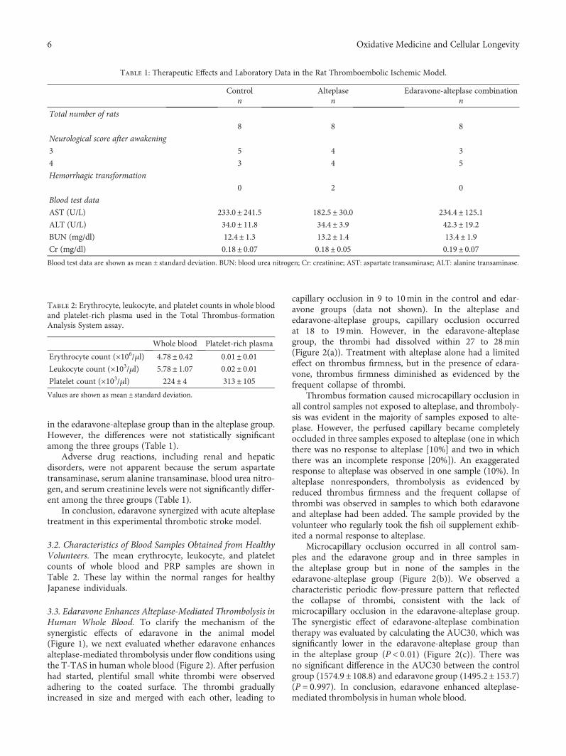
3.3. Edaravone Enhances Alteplase-Mediated Thrombolysis inHuman Whole Blood. To clarify the mechanism of thesynergistic effects of edaravone in the animal model(Figure 1), we next evaluated whether edaravone enhancesalteplase-mediated thrombolysis under flow conditions usingthe T-TAS in human whole blood (Figure 2). After perfusionhad started, plentiful small white thrombi were observedadhering to the coated surface. The thrombi graduallyincreased in size and merged with each other, leading to
capillary occlusion in 9 to 10min in the control and edar-avone groups (data not shown). In the alteplase andedaravone-alteplase groups, capillary occlusion occurredat 18 to 19min. However, in the edaravone-alteplasegroup, the thrombi had dissolved within 27 to 28min(Figure 2(a)). Treatment with alteplase alone had a limitedeffect on thrombus firmness, but in the presence of edara-vone, thrombus firmness diminished as evidenced by thefrequent collapse of thrombi.
Thrombus formation caused microcapillary occlusion inall control samples not exposed to alteplase, and thromboly-sis was evident in the majority of samples exposed to alte-plase. However, the perfused capillary became completelyoccluded in three samples exposed to alteplase (one in whichthere was no response to alteplase [10%] and two in whichthere was an incomplete response [20%]). An exaggeratedresponse to alteplase was observed in one sample (10%). Inalteplase nonresponders, thrombolysis as evidenced byreduced thrombus firmness and the frequent collapse ofthrombi was observed in samples to which both edaravoneand alteplase had been added. The sample provided by thevolunteer who regularly took the fish oil supplement exhib-ited a normal response to alteplase.
Microcapillary occlusion occurred in all control sam-ples and the edaravone group and in three samples inthe alteplase group but in none of the samples in theedaravone-alteplase group (Figure 2(b)). We observed acharacteristic periodic flow-pressure pattern that reflectedthe collapse of thrombi, consistent with the lack ofmicrocapillary occlusion in the edaravone-alteplase group.The synergistic effect of edaravone-alteplase combinationtherapy was evaluated by calculating the AUC30, which wassignificantly lower in the edaravone-alteplase group thanin the alteplase group (P < 0 01) (Figure 2(c)). There wasno significant difference in the AUC30 between the controlgroup (1574.9± 108.8) and edaravone group (1495.2± 153.7)(P = 0 997). In conclusion, edaravone enhanced alteplase-mediated thrombolysis in human whole blood.
Table 1: Therapeutic Effects and Laboratory Data in the Rat Thromboembolic Ischemic Model.
Control Alteplase Edaravone-alteplase combinationn n n
Total number of rats
8 8 8
Neurological score after awakening
3 5 4 3
4 3 4 5
Hemorrhagic transformation
0 2 0
Blood test data
AST (U/L) 233.0± 241.5 182.5± 30.0 234.4± 125.1ALT (U/L) 34.0± 11.8 34.4± 3.9 42.3± 19.2BUN (mg/dl) 12.4± 1.3 13.2± 1.4 13.4± 1.9Cr (mg/dl) 0.18± 0.07 0.18± 0.05 0.19± 0.07Blood test data are shown as mean ± standard deviation. BUN: blood urea nitrogen; Cr: creatinine; AST: aspartate transaminase; ALT: alanine transaminase.
Table 2: Erythrocyte, leukocyte, and platelet counts in whole bloodand platelet-rich plasma used in the Total Thrombus-formationAnalysis System assay.
Whole blood Platelet-rich plasma
Erythrocyte count (×106/μl) 4.78± 0.42 0.01± 0.01Leukocyte count (×103/μl) 5.78± 1.07 0.02± 0.01Platelet count (×103/μl) 224± 4 313± 105Values are shown as mean ± standard deviation.
6 Oxidative Medicine and Cellular Longevity
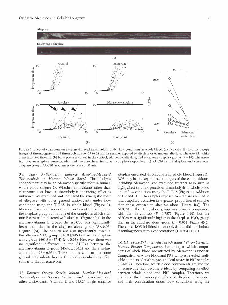
3.4. Other Antioxidants Enhance Alteplase-MediatedThrombolysis in Human Whole Blood. Thrombolysisenhancement may be an edaravone-specific effect in humanwhole blood (Figure 2). Whether antioxidants other thanedaravone also have a thrombolysis-enhancing effect isunknown. We examined and compared the synergistic effectof alteplase with other general antioxidants under flowconditions using the T-TAS in whole blood (Figure 3).Microcapillary occlusion occurred in two of the samples inthe alteplase group but in none of the samples in which vita-min E was coadministered with alteplase (Figure 3(a)). In thealteplase-vitamin E group, the AUC30 was significantlylower than that in the alteplase alone group (P < 0 05)(Figure 3(b)). The AUC30 was also significantly lower inthe alteplase-NAC group (316.8± 246.1) than the alteplasealone group (601.6± 457.4) (P < 0 05). However, there wasno significant difference in the AUC30 between thealteplase-vitamin C group (469.0± 500.1) and the alteplasealone group (P = 0 334). These findings confirm that somegeneral antioxidants have a thrombolysis-enhancing effectsimilar to that of edaravone.
3.5. Reactive Oxygen Species Inhibit Alteplase-MediatedThrombolysis in Human Whole Blood. Edaravone andother antioxidants (vitamin E and NAC) might enhance
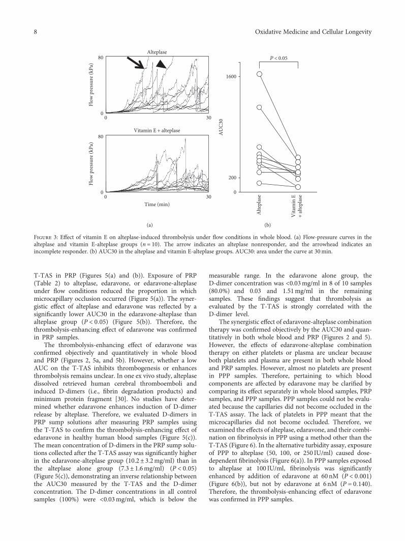
alteplase-mediated thrombolysis in whole blood (Figure 3).ROS may be the key molecular targets of these antioxidants,including edaravone. We examined whether ROS such asH2O2 affect thrombogenesis or thrombolysis in whole bloodunder flow conditions using the T-TAS (Figure 4). Additionof 100μM H2O2 to samples exposed to alteplase resulted inmicrocapillary occlusion in a greater proportion of samplesthan those exposed to alteplase alone (Figure 4(a)). TheAUC30 in the H2O2 alone group was broadly comparablewith that in controls (P = 0 787) (Figure 4(b)), but theAUC30 was significantly higher in the alteplase-H2O2 groupthan in the alteplase alone group (P < 0 05) (Figure 4(c)).Therefore, ROS inhibited thrombolysis but did not inducethrombogenesis at this concentration (100μM H2O2).
3.6. Edaravone Enhances Alteplase-Mediated Thrombolysis inHuman Plasma Components. Pertaining to which compo-nents of whole blood are affected by edaravone is unclear.Comparison of whole blood and PRP samples revealed negli-gible numbers of erythrocytes and leukocytes in PRP samples(Table 2). Therefore, which blood components are affectedby edaravone may become evident by comparing its effectbetween whole blood and PRP samples. Therefore, weexamined the thrombolytic effects of alteplase, edaravone,and their combination under flow conditions using the
Alteplase
Edaravone + alteplase
⁎
(a)
Flow
pre
ssur
e (kP
a)
Control
0
80
0 30
Edaravone
0
80
0 30
Flow
pre
ssur
e (kP
a)
Alteplase
0
80
0 30Time (min)
Edaravone + alteplase
0
80
0 30Time (min)
(b)
200
0
1600
P < 0.01
AUC3
0
Edaravone+ alterplase
Alterplase
(c)
Figure 2: Effect of edaravone on alteplase-induced thrombolysis under flow conditions in whole blood. (a) Typical still videomicroscopyimages of thrombogenesis and thrombolysis over 27 to 28min in samples exposed to alteplase or edaravone-alteplase. The asterisk (whitearea) indicates thrombi. (b) Flow-pressure curves in the control, edaravone, alteplase, and edaravone-alteplase groups (n = 10). The arrowindicates an alteplase nonresponder, and the arrowhead indicates incomplete responders. (c) AUC30 in the alteplase and edaravone-alteplase groups. AUC30: area under the curve at 30min.
7Oxidative Medicine and Cellular Longevity
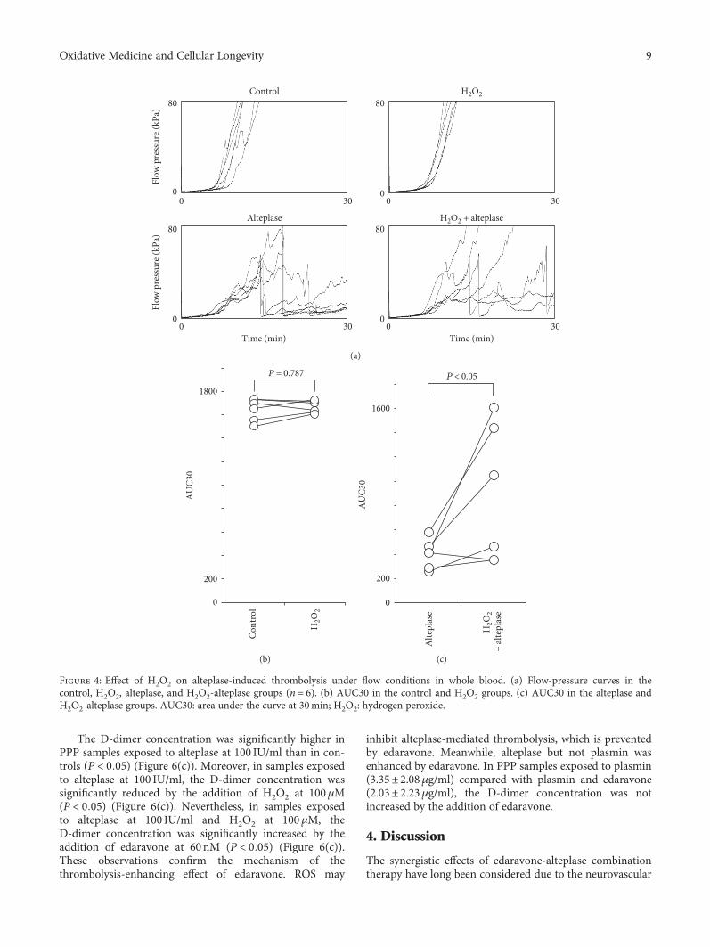
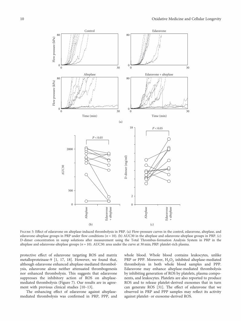
T-TAS in PRP (Figures 5(a) and (b)). Exposure of PRP(Table 2) to alteplase, edaravone, or edaravone-alteplaseunder flow conditions reduced the proportion in whichmicrocapillary occlusion occurred (Figure 5(a)). The syner-gistic effect of alteplase and edaravone was reflected by asignificantly lower AUC30 in the edaravone-alteplase thanalteplase group (P < 0 05) (Figure 5(b)). Therefore, thethrombolysis-enhancing effect of edaravone was confirmedin PRP samples.
The thrombolysis-enhancing effect of edaravone wasconfirmed objectively and quantitatively in whole bloodand PRP (Figures 2, 5a, and 5b). However, whether a lowAUC on the T-TAS inhibits thrombogenesis or enhancesthrombolysis remains unclear. In one ex vivo study, alteplasedissolved retrieved human cerebral thromboemboli andinduced D-dimers (i.e., fibrin degradation products) andminimum protein fragment [30]. No studies have deter-mined whether edaravone enhances induction of D-dimerrelease by alteplase. Therefore, we evaluated D-dimers inPRP sump solutions after measuring PRP samples usingthe T-TAS to confirm the thrombolysis-enhancing effect ofedaravone in healthy human blood samples (Figure 5(c)).The mean concentration of D-dimers in the PRP sump solu-tions collected after the T-TAS assay was significantly higherin the edaravone-alteplase group (10.2± 3.2mg/ml) than inthe alteplase alone group (7.3± 1.6mg/ml) (P < 0 05)(Figure 5(c)), demonstrating an inverse relationship betweenthe AUC30 measured by the T-TAS and the D-dimerconcentration. The D-dimer concentrations in all controlsamples (100%) were <0.03mg/ml, which is below the
measurable range. In the edaravone alone group, theD-dimer concentration was <0.03mg/ml in 8 of 10 samples(80.0%) and 0.03 and 1.51mg/ml in the remainingsamples. These findings suggest that thrombolysis asevaluated by the T-TAS is strongly correlated with theD-dimer level.
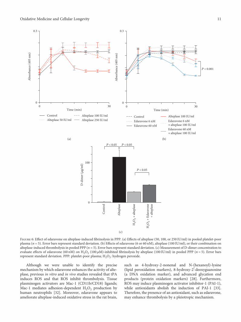
The synergistic effect of edaravone-alteplase combinationtherapy was confirmed objectively by the AUC30 and quan-titatively in both whole blood and PRP (Figures 2 and 5).However, the effects of edaravone-alteplase combinationtherapy on either platelets or plasma are unclear becauseboth platelets and plasma are present in both whole bloodand PRP samples. However, almost no platelets are presentin PPP samples. Therefore, pertaining to which bloodcomponents are affected by edaravone may be clarified bycomparing its effect separately in whole blood samples, PRPsamples, and PPP samples. PPP samples could not be evalu-ated because the capillaries did not become occluded in theT-TAS assay. The lack of platelets in PPP meant that themicrocapillaries did not become occluded. Therefore, weexamined the effects of alteplase, edaravone, and their combi-nation on fibrinolysis in PPP using a method other than theT-TAS (Figure 6). In the alternative turbidity assay, exposureof PPP to alteplase (50, 100, or 250 IU/ml) caused dose-dependent fibrinolysis (Figure 6(a)). In PPP samples exposedto alteplase at 100 IU/ml, fibrinolysis was significantlyenhanced by addition of edaravone at 60nM (P < 0 001)(Figure 6(b)), but not by edaravone at 6 nM (P = 0 140).Therefore, the thrombolysis-enhancing effect of edaravonewas confirmed in PPP samples.
Alteplase
Vitamin E + alteplase
Time (min)
80
Flow
pre
ssur
e (kP
a)Fl
ow p
ress
ure (
kPa)
00 30
0 30
80
0
(a)
P < 0.05
Alte
plas
e
Vita
min
E+
alte
plas
e
AU
C30
1600
200
0
(b)
Figure 3: Effect of vitamin E on alteplase-induced thrombolysis under flow conditions in whole blood. (a) Flow-pressure curves in thealteplase and vitamin E-alteplase groups (n = 10). The arrow indicates an alteplase nonresponder, and the arrowhead indicates anincomplete responder. (b) AUC30 in the alteplase and vitamin E-alteplase groups. AUC30: area under the curve at 30min.
8 Oxidative Medicine and Cellular Longevity
The D-dimer concentration was significantly higher inPPP samples exposed to alteplase at 100 IU/ml than in con-trols (P < 0 05) (Figure 6(c)). Moreover, in samples exposedto alteplase at 100 IU/ml, the D-dimer concentration wassignificantly reduced by the addition of H2O2 at 100μM(P < 0 05) (Figure 6(c)). Nevertheless, in samples exposedto alteplase at 100 IU/ml and H2O2 at 100μM, theD-dimer concentration was significantly increased by theaddition of edaravone at 60nM (P < 0 05) (Figure 6(c)).These observations confirm the mechanism of thethrombolysis-enhancing effect of edaravone. ROS may
inhibit alteplase-mediated thrombolysis, which is preventedby edaravone. Meanwhile, alteplase but not plasmin wasenhanced by edaravone. In PPP samples exposed to plasmin(3.35± 2.08μg/ml) compared with plasmin and edaravone(2.03± 2.23μg/ml), the D-dimer concentration was notincreased by the addition of edaravone.
4. Discussion
The synergistic effects of edaravone-alteplase combinationtherapy have long been considered due to the neurovascular
Control80
Flow
pre
ssur
e (kP
a)Fl
ow p
ress
ure (
kPa)
0
80
0
80
0
80
0
0 30 0 30
0 30 0 30
H2O2
H2O2 + alteplaseAlteplase
Time (min) Time (min)
(a)
Cont
rol
AU
C30
1800
200
0
H2O
2
P = 0.787
(b)
AU
C30
1600
200
0
H2O
2+
alte
plas
e
Alte
plas
e
P < 0.05
(c)
Figure 4: Effect of H2O2 on alteplase-induced thrombolysis under flow conditions in whole blood. (a) Flow-pressure curves in thecontrol, H2O2, alteplase, and H2O2-alteplase groups (n = 6). (b) AUC30 in the control and H2O2 groups. (c) AUC30 in the alteplase andH2O2-alteplase groups. AUC30: area under the curve at 30min; H2O2: hydrogen peroxide.
9Oxidative Medicine and Cellular Longevity
protective effect of edaravone targeting ROS and matrixmetalloproteinase-9 [1, 17, 18]. However, we found that,although edaravone enhanced alteplase-mediated thrombol-ysis, edaravone alone neither attenuated thrombogenesisnor enhanced thrombolysis. This suggests that edaravonesuppresses the inhibitory action of ROS on alteplase-mediated thrombolysis (Figure 7). Our results are in agree-ment with previous clinical studies [10–13].
The enhancing effect of edaravone against alteplase-mediated thrombolysis was confirmed in PRP, PPP, and
whole blood. Whole blood contains leukocytes, unlikePRP or PPP. Moreover, H2O2 inhibited alteplase-mediatedthrombolysis in both whole blood samples and PPP.Edaravone may enhance alteplase-mediated thrombolysisby inhibiting generation of ROS by platelets, plasma compo-nents, and leukocytes. Platelets are also reported to produceROS and to release platelet-derived exosomes that in turncan generate ROS [31]. The effect of edaravone that weobserved in PRP and PPP samples may reflect its activityagainst platelet- or exosome-derived ROS.
Control80
00 30
80
00 30
80
00 30
80
00 30
Flow
pre
ssur
e (kP
a)Fl
ow p
ress
ure (
kPa)
Alteplase
Time (min) Time (min)
Edaravone
Edaravone + alteplase
(a)
2000
AU
C30
500
0
P < 0.05
Alte
plas
e
Edar
avon
e+
alte
plas
e
(b)
18
2
0
D-d
inne
r (m
g/m
l)
Alte
plas
e
P < 0.05
Edar
avon
e+
alte
plas
e
(c)
Figure 5: Effect of edaravone on alteplase-induced thrombolysis in PRP. (a) Flow-pressure curves in the control, edaravone, alteplase, andedaravone-alteplase groups in PRP under flow conditions (n = 10). (b) AUC30 in the alteplase and edaravone-alteplase groups in PRP. (c)D-dimer concentration in sump solutions after measurement using the Total Thrombus-formation Analysis System in PRP in thealteplase and edaravone-alteplase groups (n = 10). AUC30: area under the curve at 30min; PRP: platelet-rich plasma.
10 Oxidative Medicine and Cellular Longevity
Although we were unable to identify the precisemechanism by which edaravone enhances the activity of alte-plase, previous in vitro and in vivo studies revealed that tPAinduces ROS and that ROS inhibit thrombolysis. Tissueplasminogen activators are Mac-1 (CD11b/CD18) ligands;Mac-1 mediates adhesion-dependent H2O2 production byhuman neutrophils [32]. Moreover, edaravone appears toameliorate alteplase-induced oxidative stress in the rat brain,
such as 4-hydroxy-2-nonenal and N-(hexanoyl)-lysine(lipid peroxidation markers), 8-hydroxy-2′-deoxyguanosine(a DNA oxidation marker), and advanced glycation endproducts (protein oxidation markers) [28]. Furthermore,ROS may induce plasminogen activator inhibitor-1 (PAI-1),while antioxidants abolish the induction of PAI-1 [33].Therefore, the presence of an antioxidant, such as edaravone,may enhance thrombolysis by a pleiotropic mechanism.
Control
0.3A
bsor
banc
e (40
5 nm
)
00 30
Time (min)
Alteplase 50 IU/mlAlteplase 100 IU/mlAlteplase 250 IU/ml
(a)
Abs
orba
nce (
405
nm)
Time (min)
P < 0.001
0.3
00 30
ControlEdaravone 6 nM Edaravone 6 nM
+ alteplase 100 IU/mlEdaravone 60 nM+ alteplase 100 IU/ml
Edaravone 60 nM
Alteplase 100 IU/ml
(b)
D-d
imer
(𝜇g/
ml)
P < 0.05 P < 0.05
P < 0.05
100
0
H2O
2 + al
tepl
ase
H2O
2 + E
dara
vone
+ al
tepl
ase
Cont
rol
Alte
plas
e
(c)
Figure 6: Effect of edaravone on alteplase-induced fibrinolysis in PPP. (a) Effects of alteplase (50, 100, or 250 IU/ml) in pooled platelet-poorplasma (n = 5). Error bars represent standard deviation. (b) Effects of edaravone (6 or 60 nM), alteplase (100 IU/ml), or their combination onalteplase-induced thrombolysis in pooled PPP (n = 5). Error bars represent standard deviation. (c) Measurement of D-dimer concentration toevaluate effects of edaravone (60 nM) on H2O2 (100 μM)-inhibited fibrinolysis by alteplase (100 IU/ml) in pooled PPP (n = 5). Error barsrepresent standard deviation. PPP: platelet-poor plasma; H2O2: hydrogen peroxide.
11Oxidative Medicine and Cellular Longevity
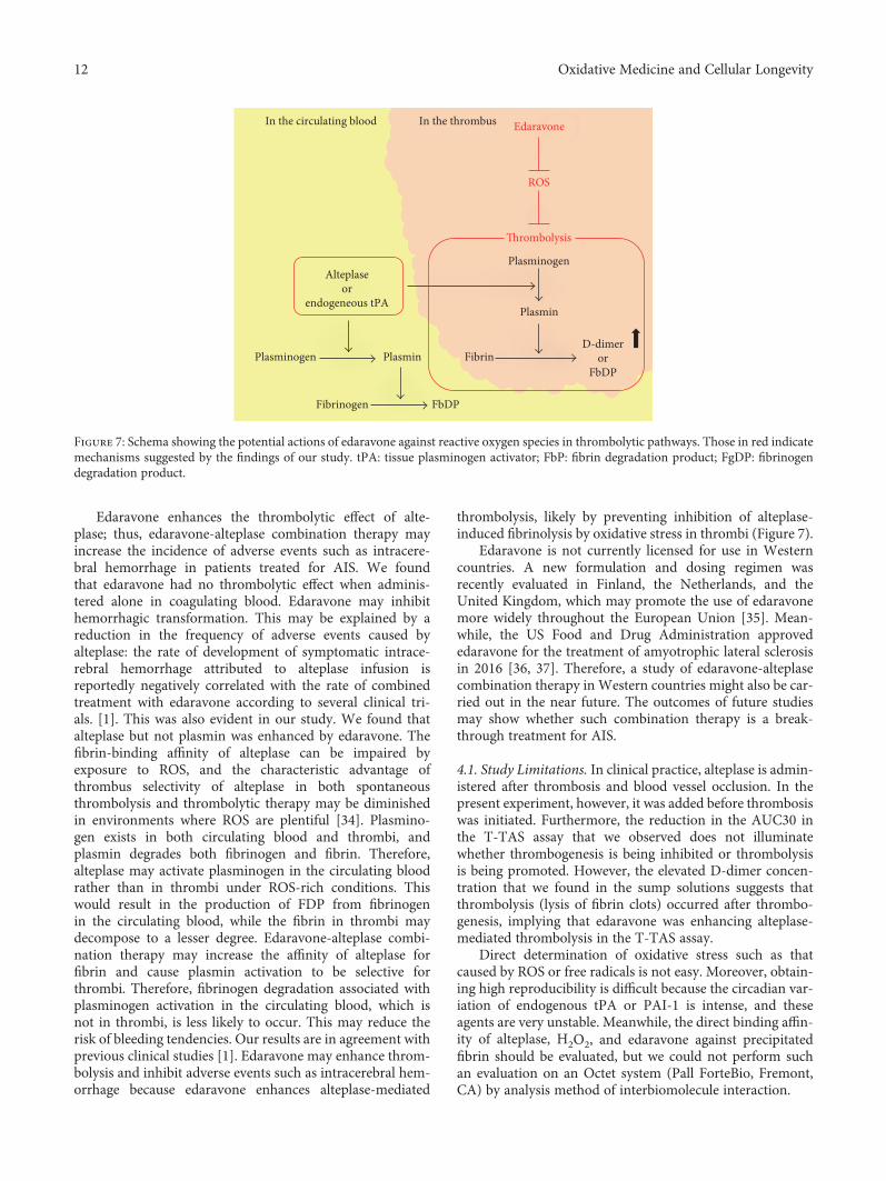
Edaravone enhances the thrombolytic effect of alte-plase; thus, edaravone-alteplase combination therapy mayincrease the incidence of adverse events such as intracere-bral hemorrhage in patients treated for AIS. We foundthat edaravone had no thrombolytic effect when adminis-tered alone in coagulating blood. Edaravone may inhibithemorrhagic transformation. This may be explained by areduction in the frequency of adverse events caused byalteplase: the rate of development of symptomatic intrace-rebral hemorrhage attributed to alteplase infusion isreportedly negatively correlated with the rate of combinedtreatment with edaravone according to several clinical tri-als. [1]. This was also evident in our study. We found thatalteplase but not plasmin was enhanced by edaravone. Thefibrin-binding affinity of alteplase can be impaired byexposure to ROS, and the characteristic advantage ofthrombus selectivity of alteplase in both spontaneousthrombolysis and thrombolytic therapy may be diminishedin environments where ROS are plentiful [34]. Plasmino-gen exists in both circulating blood and thrombi, andplasmin degrades both fibrinogen and fibrin. Therefore,alteplase may activate plasminogen in the circulating bloodrather than in thrombi under ROS-rich conditions. Thiswould result in the production of FDP from fibrinogenin the circulating blood, while the fibrin in thrombi maydecompose to a lesser degree. Edaravone-alteplase combi-nation therapy may increase the affinity of alteplase forfibrin and cause plasmin activation to be selective forthrombi. Therefore, fibrinogen degradation associated withplasminogen activation in the circulating blood, which isnot in thrombi, is less likely to occur. This may reduce therisk of bleeding tendencies. Our results are in agreement withprevious clinical studies [1]. Edaravone may enhance throm-bolysis and inhibit adverse events such as intracerebral hem-orrhage because edaravone enhances alteplase-mediated
thrombolysis, likely by preventing inhibition of alteplase-induced fibrinolysis by oxidative stress in thrombi (Figure 7).
Edaravone is not currently licensed for use in Westerncountries. A new formulation and dosing regimen wasrecently evaluated in Finland, the Netherlands, and theUnited Kingdom, which may promote the use of edaravonemore widely throughout the European Union [35]. Mean-while, the US Food and Drug Administration approvededaravone for the treatment of amyotrophic lateral sclerosisin 2016 [36, 37]. Therefore, a study of edaravone-alteplasecombination therapy in Western countries might also be car-ried out in the near future. The outcomes of future studiesmay show whether such combination therapy is a break-through treatment for AIS.
4.1. Study Limitations. In clinical practice, alteplase is admin-istered after thrombosis and blood vessel occlusion. In thepresent experiment, however, it was added before thrombosiswas initiated. Furthermore, the reduction in the AUC30 inthe T-TAS assay that we observed does not illuminatewhether thrombogenesis is being inhibited or thrombolysisis being promoted. However, the elevated D-dimer concen-tration that we found in the sump solutions suggests thatthrombolysis (lysis of fibrin clots) occurred after thrombo-genesis, implying that edaravone was enhancing alteplase-mediated thrombolysis in the T-TAS assay.
Direct determination of oxidative stress such as thatcaused by ROS or free radicals is not easy. Moreover, obtain-ing high reproducibility is difficult because the circadian var-iation of endogenous tPA or PAI-1 is intense, and theseagents are very unstable. Meanwhile, the direct binding affin-ity of alteplase, H2O2, and edaravone against precipitatedfibrin should be evaluated, but we could not perform suchan evaluation on an Octet system (Pall ForteBio, Fremont,CA) by analysis method of interbiomolecule interaction.
In the circulating blood In the thrombus Edaravone
ROS
�rombolysis
PlasminogenAlteplase
orendogeneous tPA
Plasminogen
Plasmin
Plasmin Fibrin
Fibrinogen
D-dimeror
FbDP
FbDP
Figure 7: Schema showing the potential actions of edaravone against reactive oxygen species in thrombolytic pathways. Those in red indicatemechanisms suggested by the findings of our study. tPA: tissue plasminogen activator; FbP: fibrin degradation product; FgDP: fibrinogendegradation product.
12 Oxidative Medicine and Cellular Longevity
5. Conclusions
We investigated the effect of edaravone on alteplase-inducedthrombolysis using a rat model of thromboembolic clot-induced cerebral ischemia (severe cardioembolic cerebralinfarction in vivo). Moreover, we examined the mechanismby which edaravone promotes alteplase-mediated thrombol-ysis in vitro in human blood donated by healthy volunteersusing a newly developed microchip-based flow-chamberassay (the Total Thrombus-formation Analysis System) toperform a quantitative analysis under flow conditions. Weshowed that edaravone is an enhancer of alteplase, althoughprevious reports have only shown the synergistic effect ofedaravone as a neuroprotectant. The thrombolytic effect ofalteplase was significantly attenuated in the presence ofhydrogen peroxide, suggesting that oxidative stress mighthinder thrombolysis. Edaravone alone did not influencethrombosis or thrombolysis. Edaravone enhances alteplase-mediated thrombolysis in vitro, likely by acting as an antiox-idant to prevent free radical-related inhibition of alteplaseactivity on thrombi. Furthermore, edaravone significantlyattenuated inhibition of alteplase-induced fibrinolysis byhydrogen peroxide as shown by the measurement of plasmaD-dimers in human platelet-poor plasma.
Disclosure
Tomoka Nagasato, Hisayo Sameshima, and KazuyaHosokawa are employees of Fujimori Kogyo Co., Ltd.
Conflicts of Interest
The authors declare that they have no conflicts of interest.
Authors’ Contributions
Kiyoshi Kikuchi conducted the study, analyzed the data,and wrote the manuscript. Ketaro Setoyama, Ko-ichiKawahara, Tomoka Nagasato, Takuto Terashi, Koki Ueda,Kazuki Nakanishi, Shotaro Otsuka, Naoki Miura, HisayoSameshima, Kazuya Hosokawa, Yoichiro Harada, BinitaShrestha, Mika Yamamoto, Yoko Morimoto-Yamashita,Haruna Kikuchi, Ryoji Kiyama, Chinatsu Kamikokuryo,Salunya Tancharoen, Harutoshi Sakakima, MotohiroMorioka, Eiichiro Tanaka, and Takashi Ito all helped withdata analysis. Ikuro Maruyama designed the study andanalyzed the data.
Acknowledgments
This study was supported by grants from the MiharaCerebrovascular Disorder Research Promotion Fund (toIkuro Maruyama), JSPS KAKENHI (Grant no. JP16K10746to Kiyoshi Kikuchi), the General Insurance Association ofJapan (to Kiyoshi Kikuchi), and ZENKYOREN (NationalMutual Insurance Federation of Agricultural Cooperatives)of Japan (to Kiyoshi Kikuchi). The authors thank Dr. ArataAbe, Dr. Norihito Shimamura, Ms. Akiko Katano, and Ms.Nobue Uto for their invaluable technical assistance. Alteplaseand edaravone were kind gifts from Mitsubishi Tanabe
Pharma Corporation, Osaka, Japan. The authors thankEdanz Group (http://www.edanzediting.com) for editingthe draft of this manuscript.
References
[1] K. Kikuchi, E. Tanaka, Y. Murai, and S. Tancharoen, “Clinicaltrials in acute ischemic stroke,” CNS Drugs, vol. 28, no. 10,pp. 929–938, 2014.
[2] I. Gravanis and S. E. Tsirka, “Tissue-type plasminogenactivator as a therapeutic target in stroke,” Expert Opinionon Therapeutic Targets, vol. 12, no. 2, pp. 159–170, 2008.
[3] E. Kamogawa and Y. Sueishi, “Amultiple free-radical scaveng-ing (MULTIS) study on the antioxidant capacity of a neuro-protective drug, edaravone as compared with uric acid,glutathione, and trolox,” Bioorganic & Medicinal ChemistryLetters, vol. 24, no. 5, pp. 1376–1379, 2014.
[4] K. Kikuchi, K.-i. Kawahara, N. Miyagi et al., “Edaravone: anew therapeutic approach for the treatment of acute stroke,”Medical Hypotheses, vol. 75, no. 6, pp. 583–585, 2010.
[5] K. Kikuchi, K. Kawahara, S. Tancharoen et al., “The freeradical scavenger edaravone rescues rats from cerebralinfarction by attenuating the release of high-mobility groupbox-1 in neuronal cells,” Journal of Pharmacology andExperimental Therapeutics, vol. 329, no. 3, pp. 865–874,2009.
[6] K. Kikuchi, N. Takeshige, N. Miura et al., “Beyond free radicalscavenging: beneficial effects of edaravone (Radicut) in variousdiseases (review),” Experimental and Therapeutic Medicine,vol. 3, no. 1, pp. 3–8, 2012.
[7] K. Kikuchi, S. Tancharoen, F. Matsuda et al., “Edaravoneattenuates cerebral ischemic injury by suppressing aquapo-rin-4,” Biochemical and Biophysical Research Communica-tions, vol. 390, no. 4, pp. 1121–1125, 2009.
[8] K. Kikuchi, H. Uchikado, M. Morioka, Y. Murai, andE. Tanaka, “Clinical neuroprotective drugs for treatmentand prevention of stroke,” International Journal of Molecu-lar Sciences, vol. 13, no. 6, pp. 7739–7761, 2012.
[9] E. Tanaka, S. Niiyama, S. Sato, A. Yamada, and H. Higashi,“Arachidonic acid metabolites contribute to the irreversibledepolarization induced by in vitro ischemia,” Journal ofNeurophysiology, vol. 90, no. 5, pp. 3213–3223, 2003.
[10] T. Wada, H. Yasunaga, R. Inokuchi et al., “Effects of edaravoneon early outcomes in acute ischemic stroke patients treatedwith recombinant tissue plasminogen activator,” Journal ofthe Neurological Sciences, vol. 345, no. 1-2, pp. 106–111,2014.
[11] S. Kono, K. Deguchi, N. Morimoto et al., “Tissue plasminogenactivator thrombolytic therapy for acute ischemic stroke in 4hospital groups in Japan,” Journal of Stroke & CerebrovascularDiseases, vol. 22, no. 3, pp. 190–196, 2013.
[12] S. Kono, K. Deguchi, N. Morimoto et al., “Intravenousthrombolysis with neuroprotective therapy by edaravonefor ischemic stroke patients older than 80 years of age,”Journal of Stroke & Cerebrovascular Diseases, vol. 22,no. 7, pp. 1175–1183, 2013.
[13] Y. Miyaji, S. Yoshimura, N. Sakai et al., “Effect of edaravoneon favorable outcome in patients with acute cerebral largevessel occlusion: subanalysis of RESCUE-Japan Registry,”Neurologia Medico-Chirurgica, vol. 55, no. 3, pp. 241–247,2015.
13Oxidative Medicine and Cellular Longevity
[14] K. Kimura, J. Aoki, Y. Sakamoto et al., “Administration ofedaravone, a free radical scavenger, during t-pa infusion canenhance early recanalization in acute stroke patients—a pre-liminary study,” Journal of the Neurological Sciences, vol. 313,no. 1-2, pp. 132–136, 2012.
[15] J. J. Emeis, “Regulation of the acute release of tissue-typeplasminogen activator from the endothelium by coagulationactivation products,” Annals of the New York Academy ofSciences, vol. 667, pp. 249–258, 1992.
[16] J. Aoki, K. Kimura, N. Morita et al., “YAMATO Study(tissue-type plasminogen activator and edaravone combi-nation therapy),” Stroke, vol. 48, no. 3, pp. 712–719, 2017.
[17] K. Yagi, K. T. Kitazato, M. Uno et al., “Edaravone, a free radicalscavenger, inhibits MMP-9-related brain hemorrhage in ratstreated with tissue plasminogen activator,” Stroke, vol. 40,no. 3, pp. 626–631, 2009.
[18] T. Yamashita, T. Kamiya, K. Deguchi et al., “Dissociation andprotection of the neurovascular unit after thrombolysis andreperfusion in ischemic rat brain,” Journal of Cerebral BloodFlow & Metabolism, vol. 29, no. 4, pp. 715–725, 2009.
[19] T. Yamashita, T. Sato, K. Sakamoto, H. Ishii, and J. Yamamoto,“The free-radical scavenger edaravone accelerates thromboly-sis with alteplase in an experimental thrombosis model,”Thrombosis Research, vol. 135, no. 6, pp. 1209–1213, 2015.
[20] T. Yamashita, M. Shoge, E. Oda et al., “The free-radicalscavenger, edaravone, augments NO release from vascular cellsand platelets after laser-induced, acute endothelial injuryin vivo,” Platelets, vol. 17, no. 3, pp. 201–206, 2006.
[21] K. Sakamoto, T. Yamashita, H. Yamanishi, and J. Yamamoto,“The effect of edaravone to leukocyte rolling and adhesionmolecule expression in microvessels,” Health, vol. 5, no. 3,pp. 402–408, 2013.
[22] A. O. Spiel, F. B. Mayr, C. Firbas, P. Quehenberger, andB. Jilma, “Validation of rotation thrombelastography in amodel of systemic activation of fibrinolysis and coagulationin humans,” Journal of Thrombosis and Haemostasis, vol. 4,no. 2, pp. 411–416, 2006.
[23] L. Traby, M. Kollars, L. Eischer, S. Eichinger, and P. A. Kyrle,“Prediction of recurrent venous thromboembolism by clotlysis time: a prospective cohort study,” PLoS One, vol. 7,no. 12, article e51447, 2012.
[24] K. Hosokawa, T. Ohnishi, T. Kondo et al., “A novel automatedmicrochip flow-chamber system to quantitatively evaluatethrombus formation and antithrombotic agents under bloodflow conditions,” Journal of Thrombosis and Haemostasis,vol. 9, no. 10, pp. 2029–2037, 2011.
[25] N. Shimamura, N. Matsuda, and K. Kakuta, “A model of ratembolic cerebral infarction with a quantifiable, autologousarterial blood clot,” Translational Stroke Research, vol. 4,no. 5, pp. 564–570, 2013.
[26] S. Otsuka, H. Sakakima, M. Sumizono, S. Takada, T. Terashi,and Y. Yoshida, “The neuroprotective effects of precondition-ing exercise on brain damage and neurotrophic factors afterfocal brain ischemia in rats,” Behavioural Brain Research,vol. 303, pp. 9–18, 2016.
[27] K. Kikuchi, S. Tancharoen, N. Takeshige et al., “The efficacy ofedaravone (radicut), a free radical scavenger, for cardiovascu-lar disease,” International Journal of Molecular Sciences,vol. 14, no. 7, pp. 13909–13930, 2013.
[28] V. Lukic-Panin, K. Deguchi, T. Yamashita et al., “Free radicalscavenger edaravone administration protects against tissue
plasminogen activator induced oxidative stress and bloodbrain barrier damage,” Current Neurovascular Research,vol. 7, no. 4, pp. 319–329, 2010.
[29] T. Watanabe, M. Tanaka, K. Watanabe, Y. Takamatsu, andA. Tobe, “Research and development of the free radicalscavenger edaravone as a neuroprotectant,” YakugakuZasshi–Journal of the Pharmaceutical Society of Japan,vol. 124, no. 3, pp. 99–111, 2004, (in Japanese).
[30] V. J. Marder, A. Blinc, T. Gruber et al., “Comparison of plasminwith recombinant tissue-type plasminogen activator in lysis ofcerebral thromboemboli retrieved from patients with acuteischemic stroke,” Stroke, vol. 42, no. 8, pp. 2222–2228, 2011.
[31] M. Janiszewski, A. O. Do Carmo, M. A. Pedro, E. Silva,E. Knobel, and F. R. Laurindo, “Platelet-derived exosomesof septic individuals possess proapoptotic NAD(P)H oxidaseactivity: a novel vascular redox pathway,” Critical CareMedicine, vol. 32, no. 3, pp. 818–825, 2004.
[32] C. Cao, D. A. Lawrence, Y. Li et al., “Endocytic receptor LRPtogether with tPA and PAI-1 coordinates Mac-1-dependentmacrophage migration,” EMBO Journal, vol. 25, no. 9,pp. 1860–1870, 2006.
[33] M. Mishina, Y. Komaba, S. Kobayashi et al., “Administra-tion of free radical scavenger edaravone associated withhigher frequency of hemorrhagic transformation in patientswith cardiogenic embolism,” Neurologia Medico-Chirurgica,vol. 48, no. 7, pp. 292–297, 2008.
[34] Y. H. Feng and G. Hart, “In vitro oxidative damage totissue-type plasminogen activator: a selective modificationof the biological functions,” Cardiovascular Research,vol. 30, no. 2, pp. 255–261, 1995.
[35] M. Kaste, S. Murayama, G. A. Ford, D. W. J. Dippel, M. R.Walters, and T. Tatlisumak, “Safety, tolerability and pharma-cokinetics of MCI-186 in patients with acute ischemic stroke:new formulation and dosing regimen,” CerebrovascularDiseases, vol. 36, no. 3, pp. 196–204, 2013.
[36] O. Hardiman and L. H. van den Berg, “Edaravone: a newtreatment for ALS on the horizon?,” The Lancet Neurology,vol. 16, no. 7, pp. 490-491, 2017.
[37] Edaravone (MCI-186) ALS 19 Study Group, “Safety andefficacy of edaravone in well defined patients with amyo-trophic lateral sclerosis: a randomised, double-blind,placebo-controlled trial,” The Lancet Neurology, vol. 16,no. 7, pp. 505–512, 2017.
14 Oxidative Medicine and Cellular Longevity

Submit your manuscripts athttps://www.hindawi.com
Stem CellsInternational
Hindawi Publishing Corporationhttp://www.hindawi.com Volume 2014
Hindawi Publishing Corporationhttp://www.hindawi.com Volume 2014
MEDIATORSINFLAMMATION
of
Hindawi Publishing Corporationhttp://www.hindawi.com Volume 2014
Behavioural Neurology
EndocrinologyInternational Journal of
Hindawi Publishing Corporationhttp://www.hindawi.com Volume 2014
Hindawi Publishing Corporationhttp://www.hindawi.com Volume 2014
Disease Markers
Hindawi Publishing Corporationhttp://www.hindawi.com Volume 2014
BioMed Research International
OncologyJournal of
Hindawi Publishing Corporationhttp://www.hindawi.com Volume 2014
Hindawi Publishing Corporationhttp://www.hindawi.com Volume 2014
Oxidative Medicine and Cellular Longevity
Hindawi Publishing Corporationhttp://www.hindawi.com Volume 2014
PPAR Research
The Scientific World JournalHindawi Publishing Corporation http://www.hindawi.com Volume 2014
Immunology ResearchHindawi Publishing Corporationhttp://www.hindawi.com Volume 2014
Journal of
ObesityJournal of
Hindawi Publishing Corporationhttp://www.hindawi.com Volume 2014
Hindawi Publishing Corporationhttp://www.hindawi.com Volume 2014
Computational and Mathematical Methods in Medicine
OphthalmologyJournal of
Hindawi Publishing Corporationhttp://www.hindawi.com Volume 2014
Diabetes ResearchJournal of
Hindawi Publishing Corporationhttp://www.hindawi.com Volume 2014
Hindawi Publishing Corporationhttp://www.hindawi.com Volume 2014
Research and TreatmentAIDS
Hindawi Publishing Corporationhttp://www.hindawi.com Volume 2014
Gastroenterology Research and Practice
Hindawi Publishing Corporationhttp://www.hindawi.com Volume 2014
Parkinson’s Disease
Evidence-Based Complementary and Alternative Medicine
Volume 2014Hindawi Publishing Corporationhttp://www.hindawi.com
![ORIGINAL ARTICLE Open Access β-Keto esters from ketones ...tory and antiphlogistic properties. Especially, a pyrazolone derivative (edaravone) [3] acts as a radical scavenger to interrupt](https://img.pdfslide.tips/doc/110x75/608fba6ac49a6d7592273fd2/original-article-open-access-keto-esters-from-ketones-tory-and-antiphlogistic.jpg)