Embed Size (px)
Citation preview
RESEARCH Open Access
Effect of physicochemical characterdifferences on the genotoxic potencyof kaolinTatsuya Kato1, Tatsushi Toyooka2,4, Yuko Ibuki2, Shuichi Masuda2, Masatoshi Watanabe3 and Yukari Totsuka1*
Abstract
Background: Kaolin is white clay mineral with the chemical composition Al2Si2O5(OH)4, and many varieties ofkaolins having different crystal structures are utilized in industrial, cosmetic and medical fields. To evaluate theeffect of physicochemical character differences on the genotoxicity of kaolin, two types of kaolin, kaolin-S withsmooth, sphere-shaped crystals, and kaolin-P with clusters of thin pseudohexagonal plates, were used in the study.
Results: ICR mice were intratracheally instilled with the kaolins (0.05 and 0.2 mg/mouse), and comet assay wasperformed on their lungs. Both kaolins showed DNA damage in the lungs of the mice, however the DNAdamaging potency was much higher with kaolin-P than that with kaolin-S.In order to clarify the mechanisms for the different genotoxic potency, we examined the incorporation rate andROS generation of these two types of kaolin in alveolar epithelial A549 and macrophage-like RAW264 cells, usingflow cytometric (FCM) analysis. Kaolin-P showed a higher incorporation rate into the mammalian cells and ROSgeneration than that of kaolin-S. Especially, RAW264 cells aggressively incorporated kaolins, and generated ROS,whereas almost no ROS generation was observed in A549 cells. In addition, inflammatory cytokines were quantified,using the ELISA method, to understand further genotoxic potency differences of kaolins. Concentrations ofinterleukin-1β (IL-1β) and tumor necrosis factor-α (TNF-α) in the media were increased by exposure to bothkaolins, but in the case of kaolin-P, these inflammatory cytokines were significantly elevated. Based on thesefindings, differences of genotoxic potency may contribute to incorporation rates into immune cells. Furthermore, it islikely that immune cells and epithelial cells might closely interact with each other for the appearance of genotoxocityin vivo. In order to clarify the interaction between epithelial and immune cells, A549 and RAW264 were co-culturedand RAW264 cells only were exposed to kaolins, then subsequently A549 was applied to FCM analysis and cometassay. DNA damage observed in the A549 cells markedly increased in the presence of kaolin-exposed RAW264 cellscompared to the single culture.
Conclusion: From these observations, it is suggested that mechanisms of kaolin genotoxicity against epithelial cells arethrough the activation of macrophage cells. Therefore, it is thought that interactions between epithelial and immunecells would be very important for evaluation of the genotoxicity of fine particulate matter. We also showed here thatco-culture models of epithelial and immune cells could be used as suitable models for evaluation of lung genotoxicityof fine particulate matter, including nanomaterials, as in vivo mimicking systems.
Keywords: Kaolin, Genotoxicity, Co-culture
* Correspondence: [email protected] of Carcinogenesis and Cancer Prevention, National Cancer CenterResearch Institute, 1-1 Tsukiji 5-chome, Chuo-ku, Tokyo 104-0045, JapanFull list of author information is available at the end of the article
© The Author(s). 2017 Open Access This article is distributed under the terms of the Creative Commons Attribution 4.0International License (http://creativecommons.org/licenses/by/4.0/), which permits unrestricted use, distribution, andreproduction in any medium, provided you give appropriate credit to the original author(s) and the source, provide a link tothe Creative Commons license, and indicate if changes were made. The Creative Commons Public Domain Dedication waiver(http://creativecommons.org/publicdomain/zero/1.0/) applies to the data made available in this article, unless otherwise stated.
Kato et al. Genes and Environment (2017) 39:12 DOI 10.1186/s41021-017-0075-y
BackgroundKaolin is a naturally occurring white clay mineral withthe chemical composition Al2Si2O5(OH)4. A largeamount of kaolin is primarily used in the paper industryboth as a filler and as a coating for paper. Other applica-tions of kaolin include use in the ceramics industry, cos-metics, and pharmaceuticals [1]. We have previouslyreported that kaolin showed genotoxic effects in in vitroand in vivo assay systems [2, 3]. Recently, there havebeen many reports that toxicity induced by fine particlesis influenced by physicochemical differences such as size[4–9]. The chemical structure of kaolin is a two-layersilicate and is known to consist of a silica tetrahedrallayer covalently bonded to an alumina octahedral layerthrough an apical oxygen atom [1]. Kaolinite is found aspseudo-hexagonal triclinic crystals and generally inter-acts between face-face, face-edge, and edge-edge surfacesto make an aggregate form [1]. Therefore, various sizes,zeta-potentials, and surface structures exist in the indus-trial mineral commodity of kaolins, and they are widelydistributed for many intended uses. However the influ-ences of physicochemical differences of kaolins on geno-toxicity are not fully understood, yet. To verify thegenotoxic effects of two kinds of kaolins representingdifferent surface structures (one is smooth, sphere-shaped crystals named kaolin-S, and the other is clustersof thin pseudohexagonal plates named kaolin-P), here,we examined the DNA damaging potency with cometassay in in vivo and in vitro. The incorporation intomammalian cells and their ROS generation using flowcytometric (FCM) analysis were also investigated. In thepresent study, both kaolins showed DNA damage in themice of lungs, however the DNA damaging potency wasmuch higher for kaolin-P than that for kaolin-S. Thispotent genotoxicity of kaolin-P was also supported bythe results obtained from analysis of the incorporationrate, ROS production and inflammatory cytokine gener-ation. Furthermore, aiming to examine the mechanismsinvolved in the appearance of genotoxic potential differ-ences in vivo induced by kaolin-S and -P, we conductedgenotoxicity analysis using a co-culture system with lungepithelial A549 and macrophage-like RAW264 cells asan in vivo mimicking system. In the present study, pos-sible mechanisms and importance of cell-cell interactionsfor genotoxicty induced by kaolin are also discussed.
MethodsMaterialsKaolin-S and kaolin-P were purchased from TakeharaChemical Industrial Co., Ltd. (Okayama, Japan) andMineral and Pigment Solutions Inc. (NJ, USA), respect-ively. Kaolin-S is used for medical and pharmaceuticalproducts and kaolin-P is used for industrial products.Both kaolins were suspended in saline (Otsuka
Pharmaceutical Co. Ltd., Tokyo, Japan) containing 0.05%of Tween 80 (Nacalai Tesque, Kyoto, Japan) by sonic-ation for 15–20 min, at a concentration of 2 mg/mL, asa stock solution. Crystal appearance observed under ascanning electron microscope (SEM) was done by A-KIT Corporation (Gifu, Japan). The size distributions ofkaolins used in the present study was analyzed by dy-namic light scattering (DLS) using FPAR-1000 (OtsukaElectronics Co., Ltd., Osaka) as described previously [2].Physical characterization of particles such as zeta-poten-tial was done by UBE Scientific Analysis Laboratory, Inc.(Yamaguchi, Japan). Type I agarose, low melting pointagarose, dimethyl sulfoxide and Triton X-100 werebought from Sigma-Aldrich. Ethidium bromide was ob-tained from Merck (Darmstadt, Germany). Other chemi-cals were purchased from Wako Pure Chemical Industries(Osaka, Japan).
AnimalsMale ICR mice (8 weeks old) were purchased from JapanSLC (Shizuoka, Japan) and were acclimatized for 1 week.Food (CE-2 commercial diet: Japan Clea Co., Tokyo,Japan) and water were given freely. A conventional roomwas air conditioned at 23 °C with a light/dark (12 h/12 h)cycle. After quarantine for one week the experiments wereconducted according to the “Guidelines for Animal Exper-iments in the National Cancer Center”.
Evaluation of in vivo genotoxicity of kaolinsIn order to evaluate the in vivo genotoxicity, alkalinecomet assay on the lungs of mice was performed. Twodoses of kaolins (0.05 and 0.2 mg per animal) were sus-pended in 0.1 mL of 0.05% Tween 80, then intratrache-ally instilled to mice (n = 5 for each dose) using apolyethylene tube under anesthesia of 4% halothane(Takeda Chemical, Osaka, Japan). The control mice (n = 5)were instilled intratracheally with 0.1 mL of the solventalone, and non-treated mice were prepared to confirm theeffect of Tween 80. Mice were sacrificed at 3 h after kaolininstillation and lungs were removed then subsequentlyminced and suspended with chilled homogenizing bufferand homogenized gently with a Dounce-type homogenizeron ice. Ten microliters of each cell suspension were mixedwith 90 μl of 0.5% low melting point agarose and the mix-ture was spread on an MAS coated glass slide (MatsunamiGlass Ind., Ltd, Osaka, Japan). The slide was immersed inlysing solution (2.8 M NaCl, 0.1 M EDTA-2Na, 0.01 MTris-base, 0.2 M NaOH, 10% dimethyl sulfoxide, and 1%Triton X-100, and pH 10.0) and refrigerated at 4 °C over-night. Then, the slides were immersed in alkaline electro-phoresis buffer (0.3 M NaOH, 1 mM EDTA-2NA) at 4 °Cfor 15 min to allow for DNA unwinding. Electrophoresiswas performed for 15 min at 4 °C (25 V, 300 mA). Theslides were neutralized with Tris buffer (0.4 M Tris-base,
Kato et al. Genes and Environment (2017) 39:12 Page 2 of 10
pH 7.5) at room temperature for 5 min, and dehy-drated with ethanol to fix. The cells on the slide werestained with SYBR Gold® (10,000 × dilution; MolecularProbes, Eugene, OR) for 10 min, and washed withdistilled water 3 times. After drying, comet imageswere analyzed using a fluorescence microscope (mag-nification 200×) equipped with CCD camera. Twoslides (50 cells/slide) were prepared per mouse, and100 cells were examined per mouse (Finally, 500cells/10 slides were prepared from each treatmentgroup). The percentage tail intensity was measuredusing Comet Assay IV (Perceptive Instruments, Ltd.,Haverhill, UK). For evaluation of DNA damage, theslides were randomised and coded so that the treatmentgroup was blinded to the scorer. The frequency of hedge-hogs was not included in the data.
Cell culture and treatment with kaolinA549 (RIKEN Cell Bank, Tsukuba, Japan) and RAW264(RIKEN Cell Bank) were maintained in MEM (NacalaiTesque, Kyoto, Japan) supplemented with 10% fetal bo-vine serum, penicillin (100 U/mL) and streptomycin(0.1 mg/mL). The cells were cultured at 37 °C in 5%CO2. All experiments were performed with exponentiallygrowing cells.Kaolin stock solution was diluted with MEM at
suitable concentrations, and sonicated for 10 min.A549 and RAW264 cells were treated with each kao-lin at specific concentrations (20–200 μg/mL) and in-cubated for a predetermined time (30–180 min).After treatment, kaolin was removed and cells wereharvested for each assay.
Analysis of incorporation rates of kaolinThe incorporation rates of kaolin into mammalian cellswere measured using the FCM analysis developed inprevious reports [4, 10]. Briefly, A549 and RAW264treated with several conditions of kaolin were trypsinizedand suspended in fresh culture medium. Propidium iod-ide (PI; 0.5 μg/mL) was added to each cell suspension todetermine the cell survival rates. The numbers of cellsincorporating kaolin was analyzed with FACSCaliber(Becton Dickinson, Mountain View, CA). In the FCManalysis, forward-scattered (FS) light indicating cell sizeand side-scattered (SS) light indicating intracellular com-plexity were observed. The cells increasing SS were con-sidered as material-incorporating cells, and the changein SS was an index of incorporation rates, as describedpreviously [4, 10].
Determination of intercellular ROSIntercellular ROS in the mammalian cells were mea-sured using FCM with 2′,7′-dichlorodihydrofluorescindiacetate (DCFH-DA) as described by Könczöl et al.
with minor modifications [11]. DCFH-DA that has non-fluorescence can permeate the cell membrane, wheresubsequently it is hydrolyzed by unspecific esterases andconverted into DCF that fluoresces in the presence ofROS. DCFH-DA was added into cell suspensions treatedwith kaolin at a final concentration of 20 μM, and incu-bated at 37 °C for 30 min. The cell suspensions were ap-plied to FCM analysis, and the fluorescence wasdetected at an excitation wavelength of 485 nm and anemission wavelength of 530 nm. The numbers of cellsincreasing the fluorescence level were measured as theROS generating cells.
In vitro comet assayDNA damaging potency of kaolin against mammaliancells was employed by comet assay using the same proced-ure described above (See in “Evaluation of in vivo geno-toxicity of kaolins”) with some modification [2]. Briefly,ten μl of 8 × 10³ cells/mL cell suspension was mixed with90 μl of 0.5% low melting point agarose and the mixturewas spread on the MAS coated glass slide. Afterimmersion in lysing solution, slides were incubated for30 min at 37 °C with or without formadidopyrimidine-DNA glycosylase (FPG) protein (Sigma-Aldrich, St Louis,MO) in order to confirm oxidative DNA damage [12].Thereafter, each slide was then placed in alkaline electro-phoresis buffer to allow for DNA unwinding. After elec-trophoresis, the slides were neutralized and dehydrated,then the cells were stained with SYBR Gold®. DNA dam-age was measured by the same procedure for in vivocomet assay described above.
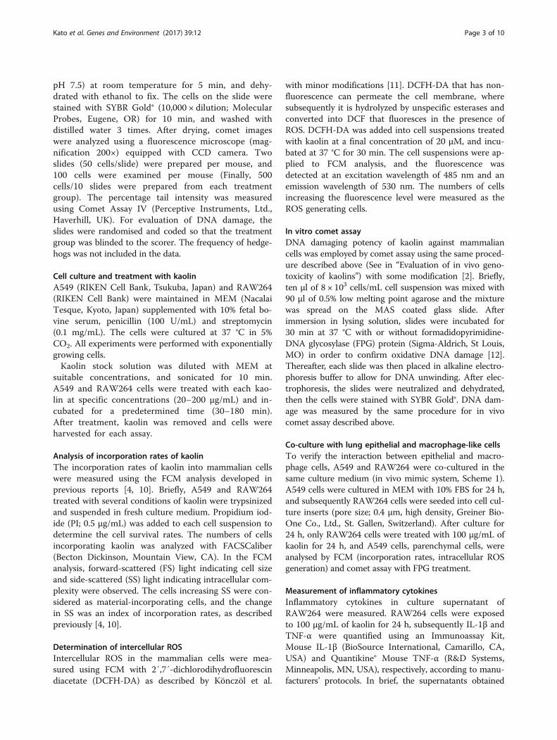
Co-culture with lung epithelial and macrophage-like cellsTo verify the interaction between epithelial and macro-phage cells, A549 and RAW264 were co-cultured in thesame culture medium (in vivo mimic system, Scheme 1).A549 cells were cultured in MEM with 10% FBS for 24 h,and subsequently RAW264 cells were seeded into cell cul-ture inserts (pore size; 0.4 μm, high density, Greiner Bio-One Co., Ltd., St. Gallen, Switzerland). After culture for24 h, only RAW264 cells were treated with 100 μg/mL ofkaolin for 24 h, and A549 cells, parenchymal cells, wereanalysed by FCM (incorporation rates, intracellular ROSgeneration) and comet assay with FPG treatment.
Measurement of inflammatory cytokinesInflammatory cytokines in culture supernatant ofRAW264 were measured. RAW264 cells were exposedto 100 μg/mL of kaolin for 24 h, subsequently IL-1β andTNF-α were quantified using an Immunoassay Kit,Mouse IL-1β (BioSource International, Camarillo, CA,USA) and Quantikine® Mouse TNF-α (R&D Systems,Minneapolis, MN, USA), respectively, according to manu-facturers’ protocols. In brief, the supernatants obtained
Kato et al. Genes and Environment (2017) 39:12 Page 3 of 10
from culture media of RAW264 with or without kaolintreatment were transferred into 96 well plate and mixedwith same volume of incubation buffer (for IL-1β) or assaydiluent RD1-63 (for TNF-α), respectively. For detection ofIL-1β, biotinylated anti-IL-1β solution was added and in-cubated for 90 min at 37 °C. The absorbance at 450 nmwas measured after reaction with streptavidin-hoseradishperoxidase (HRP) and subsequent stabilized chromogen.In the case of TNF-α detection, monoclonal antibody formouse TNF-α conjugated with HRP was subsequentlyreacted for 120 min and stabilized chromogen for 30 minin dark condition. Optical density at 450 nm (measuring)and 550 nm (correction) were measured and amounts ofTNF-α were calucurated.
Statistical analysisThe data from all studies except for comet assay areexpressed as the mean ± standard deviations. The datawere statistically compared using the Student’s t-test.
The data obtained from comet assay are expressed asmean ± standard errors. To test for significant differ-ences of % tail intensity in the comet assay between agroup treated with materials and an untreated group,Dunnett’s test after one-way ANOVA was used to evalu-ate the differences; p values lower than 0.05 were con-sidered to indicate statistical significance.
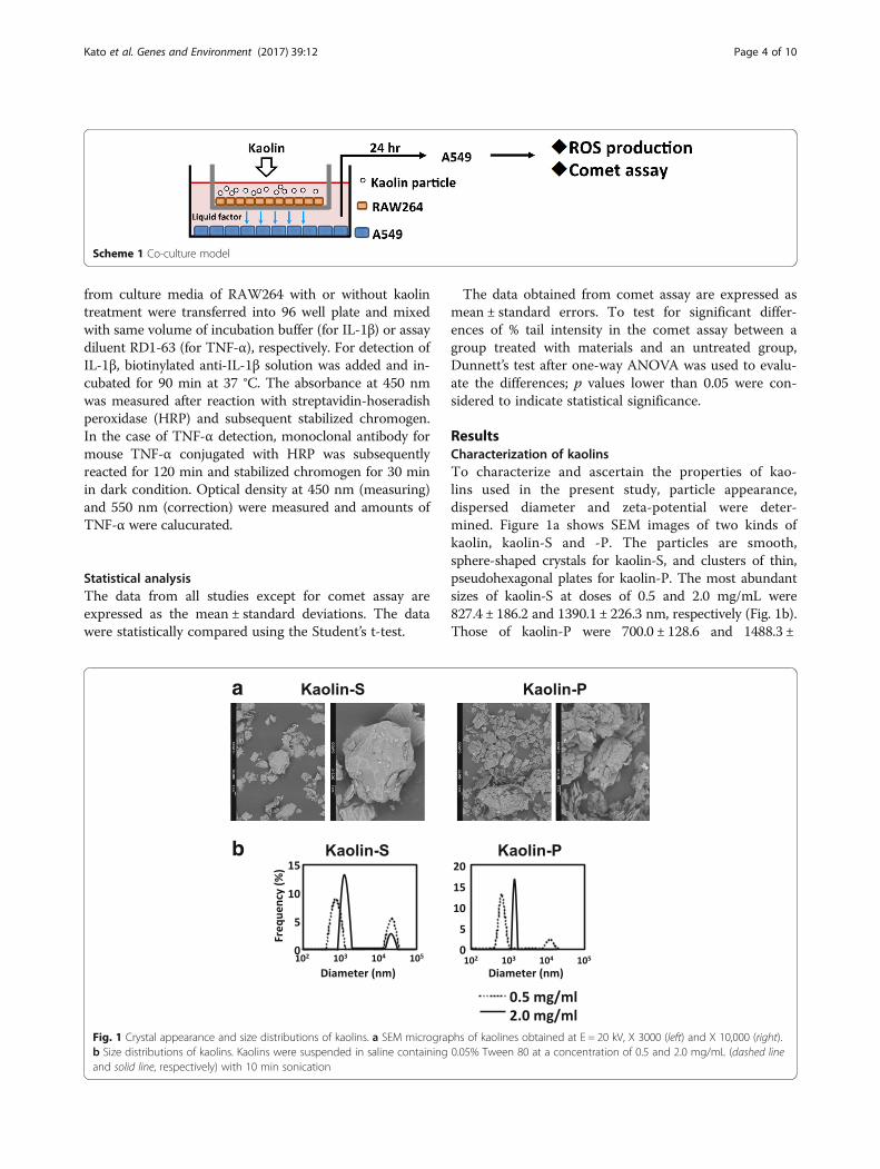
ResultsCharacterization of kaolinsTo characterize and ascertain the properties of kao-lins used in the present study, particle appearance,dispersed diameter and zeta-potential were deter-mined. Figure 1a shows SEM images of two kinds ofkaolin, kaolin-S and -P. The particles are smooth,sphere-shaped crystals for kaolin-S, and clusters of thin,pseudohexagonal plates for kaolin-P. The most abundantsizes of kaolin-S at doses of 0.5 and 2.0 mg/mL were827.4 ± 186.2 and 1390.1 ± 226.3 nm, respectively (Fig. 1b).Those of kaolin-P were 700.0 ± 128.6 and 1488.3 ±
a
b
Fig. 1 Crystal appearance and size distributions of kaolins. a SEM micrographs of kaolines obtained at E = 20 kV, X 3000 (left) and X 10,000 (right).b Size distributions of kaolins. Kaolins were suspended in saline containing 0.05% Tween 80 at a concentration of 0.5 and 2.0 mg/mL (dashed lineand solid line, respectively) with 10 min sonication
Scheme 1 Co-culture model
Kato et al. Genes and Environment (2017) 39:12 Page 4 of 10
83.7 nm, respectively (Fig. 1b). The size distributions ofthese two kaolins were not different from each other.Moreover, the zeta-potentials were −8.29 mV for kaolin-Sand −21.73 mV for kaolin-P.
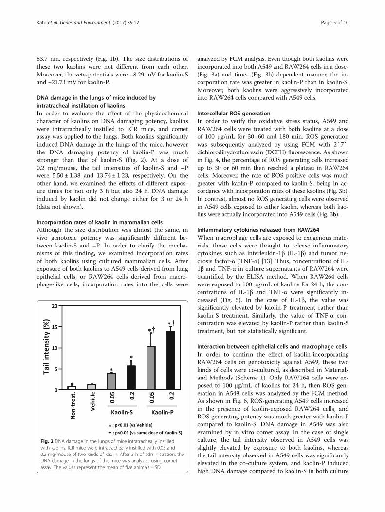
DNA damage in the lungs of mice induced byintratracheal instillation of kaolinsIn order to evaluate the effect of the physicochemicalcharacter of kaolins on DNA damaging potency, kaolinswere intratracheally instilled to ICR mice, and cometassay was applied to the lungs. Both kaolins significantlyinduced DNA damage in the lungs of the mice, howeverthe DNA damaging potency of kaolin-P was muchstronger than that of kaolin-S (Fig. 2). At a dose of0.2 mg/mouse, the tail intensities of kaolin-S and –Pwere 5.50 ± 1.38 and 13.74 ± 1.23, respectively. On theother hand, we examined the effects of different expos-ure times for not only 3 h but also 24 h. DNA damageinduced by kaolin did not change either for 3 or 24 h(data not shown).
Incorporation rates of kaolin in mammalian cellsAlthough the size distribution was almost the same, invivo genotoxic potency was significantly different be-tween kaolin-S and –P. In order to clarify the mecha-nisms of this finding, we examined incorporation ratesof both kaolins using cultured mammalian cells. Afterexposure of both kaolins to A549 cells derived from lungepithelial cells, or RAW264 cells derived from macro-phage-like cells, incorporation rates into the cells were
analyzed by FCM analysis. Even though both kaolins wereincorporated into both A549 and RAW264 cells in a dose-(Fig. 3a) and time- (Fig. 3b) dependent manner, the in-corporation rate was greater in kaolin-P than in kaolin-S.Moreover, both kaolins were aggressively incorporatedinto RAW264 cells compared with A549 cells.
Intercellular ROS generationIn order to verify the oxidative stress status, A549 andRAW264 cells were treated with both kaolins at a doseof 100 μg/mL for 30, 60 and 180 min. ROS generationwas subsequently analyzed by using FCM with 2′,7′-dichlorodihydrofluorescin (DCFH) fluorescence. As shownin Fig. 4, the percentage of ROS generating cells increasedup to 30 or 60 min then reached a plateau in RAW264cells. Moreover, the rate of ROS positive cells was muchgreater with kaolin-P compared to kaolin-S, being in ac-cordance with incorporation rates of these kaolins (Fig. 3b).In contrast, almost no ROS generating cells were observedin A549 cells exposed to either kaolin, whereas both kao-lins were actually incorporated into A549 cells (Fig. 3b).
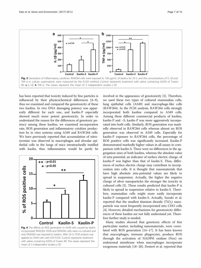
Inflammatory cytokines released from RAW264When macrophage cells are exposed to exogenous mate-rials, those cells were thought to release inflammatorycytokines such as interleukin-1β (IL-1β) and tumor ne-crosis factor-α (TNF-α) [13]. Thus, concentrations of IL-1β and TNF-α in culture supernatants of RAW264 werequantified by the ELISA method. When RAW264 cellswere exposed to 100 μg/mL of kaolins for 24 h, the con-centrations of IL-1β and TNF-α were significantly in-creased (Fig. 5). In the case of IL-1β, the value wassignificantly elevated by kaolin-P treatment rather thankaolin-S treatment. Similarly, the value of TNF-α con-centration was elevated by kaolin-P rather than kaolin-Streatment, but not statistically significant.
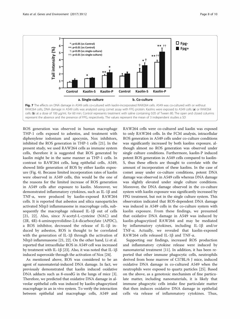
Interaction between epithelial cells and macrophage cellsIn order to confirm the effect of kaolin-incorporatingRAW264 cells on genotoxicity against A549, these twokinds of cells were co-cultured, as described in Materialsand Methods (Scheme 1). Only RAW264 cells were ex-posed to 100 μg/mL of kaolins for 24 h, then ROS gen-eration in A549 cells was analyzed by the FCM method.As shown in Fig. 6, ROS-generating A549 cells increasedin the presence of kaolin-exposed RAW264 cells, andROS generating potency was much greater with kaolin-Pcompared to kaolin-S. DNA damage in A549 was alsoexamined by in vitro comet assay. In the case of singleculture, the tail intensity observed in A549 cells wasslightly elevated by exposure to both kaolins, whereasthe tail intensity observed in A549 cells was significantlyelevated in the co-culture system, and kaolin-P inducedhigh DNA damage compared to kaolin-S in both culture
Fig. 2 DNA damage in the lungs of mice intratracheally instilledwith kaolins. ICR mice were intratracheally instilled with 0.05 and0.2 mg/mouse of two kinds of kaolin. After 3 h of administration, theDNA damage in the lungs of the mice was analyzed using cometassay. The values represent the mean of five animals ± SD
Kato et al. Genes and Environment (2017) 39:12 Page 5 of 10

systems (Fig. 7). Moreover, to determine the oxidativeDNA damage, comet assay with formamidopyramidine-DNA glycosylase (FPG) protein was performed. DNAdamage induced by both kaolins did not increase withFPG treatment under single-culture conditions (Fig. 7a).These results suggest that these kaolins were not capableof inducing ROS-dependent DNA damage in A549 cells,in a single-culture system. On the other hand, the tailintensity was largely increased in the presence of FPG
treatment with both kaolins under co-culture conditions,therefore it is suggested that kaolin may induce oxidativeDNA damage in epithelial cells through activation ofmacrophages.
DiscussionWe found that physicochemical characters, such as sur-face structure and zeta-potential, but not size, were differ-ent between two kinds of kaolin (kaolin-P and kaolin-S). It
a
b
Fig. 3 FCM analysis of incorporation rates of kaolins into mammalian cells. A549 and RAW264 cells were treated with two kinds of kaolin atseveral doses (20, 50, 100 and 200 μg/mL) for 30, 60 and 180 min. FCM analysis was applied to the cells to examine the incorporation rates.a Dose-dependent incorporation of kaolins (time duration was 180 min). b Time-dependent incorporation of kaolins (dose was 100 μg/mL)
Fig. 4 ROS generation in kaolin-exposed cells. A549 and RAW264 cells were exposed to two kinds of kaolin at a dose of 100 μg/mL for 30, 60and 180 min, and subsequently, ROS generating cells were determined by using FCM with DCFH-DA
Kato et al. Genes and Environment (2017) 39:12 Page 6 of 10
has been reported that toxicity induced by fine particles isinfluenced by their physicochemical differences [4–9],thus we examined and compared the genotoxicity of thesetwo kaolins. In vivo DNA damaging potency was appar-ently different for each one, and kaolin-P especiallyshowed much more potent genotoxicity. In order tounderstand the reason for the differences of genotoxic po-tency among these kaolins, we examined incorporationrate, ROS generation and inflammatory cytokine produc-tion by in vitro systems using A549 and RAW264 cells.We have previously reported that accumulation of nitro-tyrosine was observed in macrophages and alveolar epi-thelial cells in the lungs of mice intratracheally instilledwith kaolin, thus inflammation would be partly be
involved in the appearance of genotoxicity [3]. Therefore,we used these two types of cultured mammalian cells,lung epithelial cells (A549) and macrophage-like cells(RAW264). In the FCM analysis, RAW264 cells stronglyincorporated both kaolins compared to A549 cells.Among these different commercial products of kaolins,kaolin-P and –S, kaolin-P was more aggressively incorpo-rated into both cells. Similarly, ROS generation was mark-edly observed in RAW264 cells whereas almost no ROSgeneration was observed in A549 cells. Especially forkaolin-P exposure to RAW264 cells, the percentage ofROS positive cells was significantly increased. Kaolin-Pdemonstrated markedly higher values in all assays in com-parison with kaolin-S. There were no differences in the ag-gregation sizes of both kaolins, whereas the absolute valueof zeta-potential, an indicator of surface electric charge, ofkaolin-P was higher than that of kaolin-S. Thus, differ-ences of surface electric charge may contribute to incorp-oration into cells. It is thought that nanomaterials thathave high absolute zeta-potential values are likely tospread in suspension. Actually, the higher the negativecharge of silver nanoparticles the stronger the toxicity incultured cells [5]. These results predicted that kaolin-P islikely to spread in suspension relative to kaolin-S. There-fore, mammalian cells might more easily incorporatekaolin-P compared with kaolin-S. Actually, Suzuki et al.reported that the smallest titanium dioxide (TiO2) nano-particle was most frequently incorporated into CHO cells[4]. However, detailed mechanisms for genotoxicity differ-ences of these kaolins are not fully understand yet. There-fore further study is needed.Many studies showed that genotoxic effects of fine
particulate matter, including nanomaterials, were corre-lated with ROS generation [14–17]. It has been knownthat macrophages, immune phagocytes, produce ROSthrough the activation of NADPH oxidase (Nox) onendosomal membrane when macrophages incorporateexogenous materials [18–20]. Dostert et al. reported that
a b
Fig. 5 Generation of inflammatory cytokines. RAW264 cells were exposed to 100 μg/mL of kaolins for 24 h, and the concentrations of IL-1β andTNF-α in culture supernatants were measured by the ELISA method. Control represents treatment with saline containing 0.05% of Tween80. a IL-1β. b TNF-α. The values represent the mean of 3 independent studies ± SD
Fig. 6 The effects on ROS generation in A549 cells caused by kaolin-incorporated RAW264. A549 and RAW264 cells were co-cultured andonly RAW264 was exposed to kaolins. After 24 h, FCM analysis wasapplied to A549 cells with DCFH-DA. Control represents treatmentwith saline containing 0.05% of Tween 80. The values represent themean of 3 independent studies ± SD
Kato et al. Genes and Environment (2017) 39:12 Page 7 of 10
ROS generation was observed in human macrophageTHP-1 cells exposed to asbestos, and treatment withdiphenylene iodonium and apocynin, Nox inhibitors,inhibited the ROS generation in THP-1 cells [21]. In thepresent study, we used RAW264 cells as immune systemcells, therefore it is suggested that ROS generated bykaolin might be in the same manner as THP-1 cells. Incontrast to RAW264 cells, lung epithelial cells, A549,showed little generation of ROS by either kaolin expos-ure (Fig. 4). Because limited incorporation rates of kaolinwere observed in A549 cells, this would be the one ofthe reasons for the limited increase of ROS generationin A549 cells after exposure to kaolin. Moreover, wedemonstrated inflammatory cytokines, such as IL-1β andTNF-α, were produced by kaolin-exposed RAW264cells. It is reported that asbestos and silica nanoparticlesactivated Nlrp3 inflammasome in macrophage cells, sub-sequently the macrophage released IL-1β out of cells[21, 22]. Also, since N-acetyl-L-cysteine (NAC) and(2R, 4R)-4-aminopyrrolidine-2,4-dicarboxylate (APDC),a ROS inhibitor, decreased the release of IL-1β in-duced by asbestos, ROS is thought to be correlatedwith the generation of IL-1β through the activation ofNlrp3 inflammasome [21, 22]. On the other hand, Li et al.reported that intracellular ROS in A549 cell was increasedby treatment with IL-1β [23]. Also, it was noted that IL-1βinduced superoxide through the activation of Nox [24].As mentioned above, ROS was considered to be an
agent of nanomaterial-induced DNA damage. In fact, wepreviously demonstrated that kaolin induced oxidativeDNA adducts such as 8-oxodG in the lungs of mice [3].Therefore, we predicted that oxidative DNA damage in al-veolar epithelial cells was induced by kaolin-phagocytizedmacrophage in an in vivo system. To verify the interactionbetween epithelial and macrophage cells, A549 and
RAW264 cells were co-cultured and kaolin was exposedto only RAW264 cells. In the FCM analysis, intracellularROS generation in A549 cells under co-culture conditionswas significantly increased by both kaolins exposure, al-though almost no ROS generation was observed undersingle culture conditions. Furthermore, kaolin-P inducedpotent ROS generation in A549 cells compared to kaolin-S, thus these effects are thought to correlate with theamount of incorporation of these kaolins. In the case ofcomet assay under co-culture conditions, potent DNAdamage was observed in A549 cells whereas DNA damagewas slightly elevated under single culture conditions.Moreover, the DNA damage observed in the co-culturesystem with kaolin exposure was significantly increased byFPG treatment, but not in the single culture system. Thisobservation indicated that ROS-dependent DNA damagewas induced in A549 cells in the co-culture system withkaolin exposure. From these findings, we presumedthat oxidative DNA damage in A549 was induced bykaolin-phagocytized RAW264 and may be mediatedby inflammatory cytokines, including IL-1β and/orTNF-α. Actually, we revealed that kaolin-exposedRAW264 cells released IL-1β and TNF-α.Supporting our findings, increased ROS production
and inflammatory cytokine release were induced bynanomaterial treatment [11]. In addition, it has been re-ported that other immune phagocytic cells, neutrophilsderived from bone marrow of C57BL/6 J mice, inducedoxidative DNA damage in co-cultured A549 when theneutrophils were exposed to quartz particles [25]. Basedon the above, as a genotoxic mechanism of fine particu-late matter, including nanomaterials, it is likely thatimmune phagocytic cells intake fine particulate matterthat then induces oxidative DNA damage in epithelialcells via release of inflammatory cytokines. Thus,
Fig. 7 The effects on DNA damage in A549 cells co-cultured with kaolin-incorporated RAW264 cells. A549 was co-cultured with or withoutRAW264 cells, DNA damage in A549 cells was analyzed using comet assay with FPG protein. Kaolins were exposed to A549 cells (a) or RAW264cells (b) at a dose of 100 μg/mL for 60 min. Control represents treatment with saline containing 0.05 of Tween 80. The open and closed columnsrepresent the absence and the presence of FPG, respectively. The values represent the mean of 3 independent studies ± SD
Kato et al. Genes and Environment (2017) 39:12 Page 8 of 10
considering the genotoxicity of fine particulate matter,interactions between epithelial and immune cellswould be very important. However, most reports de-scribing in vitro genotoxicity of nanomaterials usedsingle culture systems. It is thought that single cul-ture systems were not sufficient to evaluate the geno-toxicity of nanomaterials. We showed in the presentstudy, using a co-culture of epithelial and immunecells could be used as a suitable model for evaluatinglung genotoxicity of nanomaterials as an in vivo mim-icking system.
ConclusionsWe have demonstrated that two kinds of commercialproducts of kaolin with different physicochemical charac-ters, such as surface structure and zeta-potential, revealeddifferent genotoxic potency in vivo. Based on in vitro ana-lysis, this genotoxic potency might be influenced by easeof uptake into immune cells. Mechanisms of kaolin geno-toxicity against epithelial cells are suggested to be throughthe activation of macrophage cells. Namely, immunephagocytic cells ingested kaolin then induced oxidativeDNA damage in epithelial cells via release of inflammatorycytokines, such as IL-1β and TNF-α. Therefore, it isthought that interactions between epithelial and immunecells would be very important for evaluation of the geno-toxicity of fine particulate matter. We also showed here,co-culture models of epithelial and immune cells could beused as suitable models for evaluation of lung genotoxicityof fine particulate matter, including nanomaterials, as invivo mimicking systems.
AcknowledgmentsWe thank Mr Naoaki Uchiya for his excellent technical assistance.
FundingThis study was supported by Research on Global Health Issues (U.S.-JapanCooperative Medical Sciences Program) from the Japan Agency for MedicalResearch and Development, AMED, for Research on Risk of ChemicalSubstances from the Ministry of Health, Labour, and Welfare of Japan. Thestudy was also supported by a grant from the Japan Chemical IndustryAssociation (JCIA) Long-range Research Initiative (LRI).
Availability of data and materialsAll data generated or analyzed during this study are included in thispublished article.
Authors’ contributionsTK performed the comet assay, FCM analyses for incorporation and ROSgeneration, ELISA for production of inflammatory cytokines and drafted themanuscript. TT carried out the FCM analyses for incorporation. Analyses ofsize distribution and agglomeration state of particles were conducted byMW. YI, SM and YT conceived and supervised the study. All authors read andapproved the final manuscript.
Competing interestsThe authors declare that they have no competing interests.
Consent for publicationNot applicable.
Ethics approvalAll animal work was conducted in accordance with the “Guidelines forAnimal Experiments in the National Cancer Center” and approved by theCommittee for Ethics of Animal Experimentation of the National CancerCenter.
Author details1Division of Carcinogenesis and Cancer Prevention, National Cancer CenterResearch Institute, 1-1 Tsukiji 5-chome, Chuo-ku, Tokyo 104-0045, Japan.2Graduate School of Food and Nutritional Sciences, University of Shizuoka,52-1, Yada, Shizuoka 422-8526, Japan. 3Division of Materials Science andEngineering, Graduate School of Engineering, Yokohama National University,Hodogaya-ku, Yokohama, Japan. 4Present Address: National Institute ofOccupational Safety and Health, Nagao 6-21-1, Tama-Ku, Kawasaki 214-8585,Japan.
Received: 23 September 2016 Accepted: 3 February 2017
References1. Gupta V, Hampton MA, Stokes JR, Nguyen AV, Miller JD. Particle interactions
in kaolinite suspensions and corresponding aggregate structures. J ColloidInterface Sci. 2011;359:95–103.
2. Totsuka Y, Higuchi T, Imai T, Nishikawa A, Nohmi T, Kato T, Masuda S, KinaeN, Hiyoshi K, Ogo S, Kawanishi M, Yagi T, Ichinose T, Fukumori N, WatanabeM, Sugimura T, Wakabayashi K. Genotoxicity of nano/microparticles ininvitro micronuclei, in vivo comet and mutation assay systems. Part FibereToxicol. 2009;6:23.
3. Totsuka Y, Kato T, Masuda S, Ishino K, Matsumoto Y, Goto S, Kawanishi M,Yagi T, Wakabayashi K. In vitro and in vivo genotoxicity induced byfullerene (C60) and kaolin. Genes Environ. 2011;1:14–20.
4. Suzuki H, Toyooka T, Ibuki Y. Simple and easy method to evaluate uptakepotential of nanoparticles in mammalian cells using a flow cytometric lightscatter analysis. Environ Sci Technol. 2007;41:3018–24.
5. Kaur J, Tiloo K. Evaluating cell specific cytotoxicity of differentially chargedsilver nanoparticles. Food Chem Toxicol. 2012;51C:1–14.
6. Gliga AR, Skoglund S, Wallinder IO, Fadeel B, Karlsson HL. Size-dependentcytotoxicity of silver nanoparticles in human lung cells: the role of cellularuptake, agglomeration and Ag release. Part Fibre Toxicol. 2014;11:11.
7. Battal D, Çelik A, Güler G, Aktaş A, Yildirimcan S, Ocakoglu K, ÇömelekoǧluÜ. SiO2 Nanoparticule-induced size-dependent genotoxicity - an in vitrostudy using sister chromatid exchange, micronucleus and comet assay.Drug Chem Toxicol. 2015;38:196–204.
8. Sung JH, Park SJ, Jeong MS, Song KS, Ahn KS, Ryu HR, Lee H, Song MR, ChoMH, Kim JS. Physicochemical analysis and repeated-dose 90-days oraltoxicity study of nanocalcium carbonate in Sprague-Dawley rats.Nanotoxicology. 2015;9:603–12.
9. Xiong S, George S, Yu H, Damoiseaux R, France B, Ng KW, Loo JS. Sizeinfluences the cytotoxicity of poly (lactic-co-glycolic acid) (PLGA) andtitanium dioxide (TiO(2)) nanoparticles. Arch Toxicol. 2013;87:1075–86.
10. Ibuki Y, Toyooka T. Nanoparticle uptake measured by flow cytometry.Methods Mol Biol. 2012;926:157–66.
11. Könczöl M, Ebeling S, Goldenberg E, Treude F, Gminski R, Gieré R, GrobétyB, Rothen-Rutishauser B, Merfort I, Mersch-Sundermann V. Cytotoxicity andgenotoxicity of size-fractionated iron oxide (magnetite) in A549 humanlung epithelial cells: role of ROS, JNK, and NF-κB. Chem Res Toxicol. 2011;24:1460–75.
12. Toyoizumi T, Sekiguchi H, Takabayashi F, Deguchi Y, Masuda S, Kinae N.Induction effect of coadministration of soybean isoflavones and sodiumnitrite on DNA damage in mouse stomach. Food Chem Toxicol. 2010;48:2585–91.
13. Scherbart AM, Langer J, Bushmelev A, van Berlo D, Haberzettl P, vanSchmidt AM, Rose CR, Schins RP, Albrecht C. Contrasting macrophageactivation by fine and ultrafine titanium dioxide particles is associated withdifferent uptake mechanisms. Part Fibre Toxicol. 2011;8:31.
14. Rubio L, El Yamani N, Kazimirova A, Dusinska M, Marcos R. Multi-walledcarbon nanotubes (NM401) induce ROS-mediated HPRT mutations inChinese hamster lung fibroblasts. Environ Res. 2016;146:185–90.
15. Platel A, Carpentier R, Becart E, Mordacq G, Betbeder D, Nesslany F.Influence of the surface charge of PLGA nanoparticles on their in vitro
Kato et al. Genes and Environment (2017) 39:12 Page 9 of 10

genotoxicity, cytotoxicity, ROS production and endocytosis. J Appl Toxicol.2016;36:434–44.
16. Kansara K, Patel P, Shah D, Shukla RK, Singh S, Kumar A, Dhawan A. TiO2nanoparticles induce DNA double strand breaks and cell cycle arrest inhuman alveolar cells. Environ Mol Mutagen. 2015;56:204–17.
17. Martin LD, Krunkosky TM, Dye JA, Fischer BM, Jiang NF, Rochelle LG, AkleyNJ, Dreher KL, Adler KB. The role of intracellular redox imbalance innanomaterial induced cellular damage and genotoxicity: a review. EnvironMol Mutagen. 2015;56:111–24.
18. Kermanizadeh A, Chauché C, Brown DM, Loft S, Møller P. The role ofreactive oxygen and nitrogen species in the response of airway epitheliumto particulates. Environ Health Perspect. 1997;105 Suppl 5:1301–7.
19. Forman HJ, Torres M. Reactive oxygen species and cell signaling: respiratoryburst in macrophage signaling. Am J Respir Crit Care Med. 2002;166:S4–8.
20. Forman HJ, Torres M. Signaling by the respiratory burst in macrophages.IUBMB Life. 2001;51:365–71.
21. Dostert C, Pétrilli V, Van Bruggen R, Steele C, Mossman BT, Tschopp J. Innateimmune activation through Nalp3 inflammasome sensing of asbestos andsilica. Science. 2008;320:674–7.
22. Yazdi AS, Guarda G, Riteau N, Drexler SK, Tardivel A, Couillin I, Tschopp J.Nanoparticles activate the NLR pyrin domain containing 3 (Nlrp3)inflammasome and cause pulmonary inflammation through release of IL-1αand IL-1β. Proc Natl Acad Sci U S A. 2010;107:19449–54.
23. Li WJ, Wang TK. Calcitonin gene-related peptide inhibits interleukin-1beta-induced interleukin-8 secretion in human type II alveolar epithelial cells.Acta Pharmacol Sin. 2006;27:1340–5.
24. Brigelius-Flohé R, Banning A, Kny M, Böl GF. Redox events in interleukin-1signaling. Archives of Biochemistry and Biophysics. 2004;423(1):66–73.
25. van Berlo D, Wessels A, Boots AW, Wilhelmi V, Scherbart AM, Gerloff K, vanSchooten FJ, Albrecht C, Schins RP. Neutrophil-derived ROS contribute tooxidative DNA damage induction by quartz particles. Free Radical Biologyand Medicine. 2010;49(11):1685–1693.
• We accept pre-submission inquiries
• Our selector tool helps you to find the most relevant journal
• We provide round the clock customer support
• Convenient online submission
• Thorough peer review
• Inclusion in PubMed and all major indexing services
• Maximum visibility for your research
Submit your manuscript atwww.biomedcentral.com/submit
Submit your next manuscript to BioMed Central and we will help you at every step:
Kato et al. Genes and Environment (2017) 39:12 Page 10 of 10