Embed Size (px)
Citation preview
- 31 -
Effect of Quercetin and DMSO on Skeletal Myogenesis fromC2C12 Skeletal Muscle Cells with Special Reference to PKB/AktActivity, Myogenin and Bcl-2 Expression
Arkadiusz Orzechowski(1), Katarzyna Grzelkowska, Wojciech Karlik and TomaszMotyl
(1) Department of Physiology, Biochemistry, Pharmacology and Toxicology, Fac-ulty of Veterinary Medicine, Warsaw Agricultural University, Warsaw, Poland
AbstractConflicting data regarding the effect of antioxidants on skeletal myogenesis prompted us tostudy the action of superoxide anion and hydroxyl radical scavengers on differentiating mu-rine C2C12 myoblasts. The onset of myotube formation was delayed by quercetin andDMSO while DNA synthesis was stimulated in response to elevated doses of both factors.Cell viability measured by MTT assay was inhibited either by 100 µM quercetin or 1% or2% of DMSO whereas elevated number of apoptotic cells was detected at the same time.Muscle cell differentiation retarded by quercetin or DMSO was reflected by delay in myo-genin expression and lowered distribution of myotubes with low (< 10) number of cellmyonuclei. For large myotubes (> 10) low scores for DMSO, and high scores for quercetinwere observed. Based on phosphorylation status, both antioxidants delayed PKB activationand PKB-dependent differentiation, as well as antiapoptotic effect of PKB. Bcl-2 antia-poptotic protein level was elevated earlier for control than for experimental treatment. Mu-scle creatine kinase activity reflected the reduced rate of myogenesis. In conclusion, pro-mitogenic activity of quercetin and DMSO disturbs differentiation programme of myoblastsand might explain why more apoptotic cells was found after high doses of both factors. Incontrast to DMSO, quercetin-induced delay in myogenesis may result in larger musclemass. Results of this study support the idea that muscle differentiation can be regulated byscavengers of superoxide anion and hydroxyl radical.Key words: antioxidants, apoptosis, Bcl-2, muscle differentiation, myogenin, PKB.
Basic Appl Myol 11 (1): 31-44, 2001
Muscle growth results from hypertrophying myofi-bers, which increase their myonuclear number by re-cruitment of satellite cells. Determined myoblasts, underconditions that favour differentiation, align and fuse toform multinucleated myotubes [44]. The mechanismsunderlying the recruitment of satellite cells to hyper-plastic or hypertrophic growth have not been estab-lished. It is widely accepted, that high proliferation ratedelays the myogenesis and results in higher musclemass [8, 13, 37]. The higher the rate of cell multiplica-tion the higher the mass of growing muscle. Somegrowth factors and oncogenes as well as agents presentintracellularly or in the extracellular matrix (ECM) alsocan repress the activity of myogenin and subsequentlyinhibit myogenesis [5, 31, 51]. However, there is alsoevidence that accelerated mitogenesis in the circum-stances the promote enhanced muscle differentiation
impairs the survival of muscle precursor cells [53, 54].With regard to transduction of signals carrying orders tosurvive, proliferate or differentiate attention is put onprotein kinase B a product of Akt gene (PKB/Akt). PKBprotects against cell death either through downstreamphosphorylation of Bad protein, subsequent release ofBclXL or Bcl-2 from heterodimers with Bad, sequestra-tion of Bax forming heterodimers with BclXL or Bcl-2,and consequent inhibition of pore formation in the mito-chondrial membrane which unable the release of cyto-chrome C (cyt C) and apoptosis inducing factor(AIF)[36] or by phosphorylation-dependent inactivationof caspase 9 [15]. Two potent antagonists of reactiveoxygen species (ROS) action in muscle cells, namely,quercetin and DMSO were evaluated in order to find theimportance of these antioxidants to myogenesis. Quer-cetin biological activities are pleiotropic, opposite to
Effect of quercetin and DMSO
- 32 -
ROS, and include cell specific inhibition of ubiquitousenzymes (xanthine oxidase, tyrosine kinases, ornithinedecarboxylase (ODC), PI-3K, MAPK-s, calmodulin(CaM), lipooxygenase (LOX) and cyclooxygenase(COX) [10, 16, 17, 56]. The role of antioxidants in celltransformation is uncertain, although several groups ofauthors have reported that, for instance, once activatedby specific stimuli (inflammatory cytokines, ROS) he-patic stellate cells differentiate into myofibroblast-likecells, while antioxidants including quercetin inactivatethis process [22, 23]. DMSO was well recognized for itsability to modulate the expression of various genesnegatively [7, 32] or positively inducing muscle cell dif-ferentiation [2, 42]. However, up to date no data areavailable to elucidate quercetin biological activity inrelation to muscle gene expression. According to studieson digestion and absorption, quercetin is the main die-tary flavonoid consumed by Caucasians, so its relevancefor muscle development would be of great value [11]. Inrats fed diet enriched with 0.5% w/w blood plasmaquercetin may rise up to 100 µM [29], but in humanbeings who consume approximately 50-80 mg of quer-cetin per day, the plasma level generally does not ex-ceed 1 µM [20].
Another equally important issue is the understandingof abrogated myogenesis brought about by DMSO. Mu-noz-Canoves et al. [34] showed that DMSO inhibitedmyogenesis in cultured C2C12 myoblasts by repressingurokinase type plasminogen activator (uPA) gene ex-pression. Unfortunately, studies of Munoz-Canoves etal. [34] were not verified by the examination of cell sur-vival. DMSO is a skin permeable popular vehicle andcompound used by body builders for topical treatmentof the muscle pain and stiffness. It is also present in theanti-inflammatory and anti-rheumatoid medicaments.The issue whether and how DMSO influences musclecell differentiation is a therefore a merit of considerableimportance.
Moreover, with regard to muscle cell differentiation itshould be pointed out that delayed myogenesis with ex-tended period of cell multiplication but not apoptosismight be a characteristic feature of increased musclemass. The aim of this paper was to provide a descriptionof the relation between the cell growth, formation ofmyotubes, cell viability and apoptosis of cultured murineC2C12 muscle cells. Concomitantly, we scrutinized theexpression of myogenin, Bcl-2 protein, PKB and MCKactivity during differentiation. In this study we demon-strated that antioxidants quercetin and DMSO delay theexpression of molecular and metabolic markers of differ-entiation. The results of our study suggest that superoxideanion (quercetin) and hydroxyl radical (DMSO) scaven-gers retard the onset of muscle cell fusion but, at least, inthe case of quercetin this effect was transient and onceinitiated myogenesis was significantly accelerated by
quercetin (larger myotubes). Both agents caused markeddelay in PKB activation which was hardly followed byreduced rate of apoptosis; furthermore antiapoptoticproperties of quercetin and DMSO diminished with theincreasing dose of each factor. Every chosen biochemicalindex of muscle cell differentiation (myogenin, MCK)confirmed pictures obtained from microscopic observa-tions (myotubes formed).
A deeper understanding of the role of phytochemicalsand thiols in the mechanism of muscle growth, devel-opment and regeneration may have important implica-tions for the prevention and treatment of neuromusculardisorders. Further studies are needed to address theabove-mentioned effects and ascertain whether thephysicochemical properties other then antioxidant causedelayed myogenesis.
Materials and Methods
Reagents
All reagents were cell culture tested, of high purity,and unless otherwise stated they were purchased fromSigma-Aldrich Chemical Co., plastics were from Corn-ing-Costar, while sera, media and antibiotics were fromGibco Life Technologies. Phosphate buffered saline(PBS) and ultra pure agarose were obtained from GibcoBRL. Primary rabbit polyclonal anti-Myogenin IgG an-tibody (M-225, sc-576), rabbit anti-Bcl-2 IgG antibody(N-19; sc-492), and goat anti-Akt-1 IgG antibody (C-20;sc-1618) as well as fluorescein conjugated secondarygoat anti-rabbit IgG antibody for cytoimmunofluores-cent studies were obtained from Santa Cruz Biotechnol-ogy (Santa Cruz, CA, USA). Sodium dodecyl sulphate(SDS) 10% (w/v), Sequi-Blot PVDF Membrane 0.2 µmand all reagents for immunoblotting were obtained fromBio-Rad Laboratories (CA, USA). Tris, EDTA, NaCl,proteinase K, RNAse A from bovine pancreas andLambda DNA EcoR I HindIII Digest were obtainedfrom Sigma Chemical Co. (St. Louis, MO, USA). Otherreagents were purchased as stated in the description ofthe respective methods (see below). Protein content wasassayed both by Lowry method (Sigma Chemical Co.St. Louis, MO, USA) and the bicinchoninic acid method(BCA, Pierce Chemical Co., Rockford, IL, USA).
Cell culture
Murine C2C12 myoblastic cell line (satellite cellsfrom thigh muscle) purchased from European Collectionof Animal Cell Cultures (ECACC) was maintained inexponential phase of growth (20% (v/v) FCS/DMEMwith Glutamax) designed as GM (growth medium) sup-plied with antibiotic-antimycotic mixture (Penicillin Gsodium salt 50 IU/mL, Streptomycin sulphate 50µg/mL; Gentamycin sulphate 20 µg/ml; Anti PPLOagent - Tylocine base 6 µg/mL, Fungizone - Amphoteri-cin B 1 µg/ml), in a controlled humidified air atmos-
Effect of quercetin and DMSO
- 33 -
phere supplied with 5% CO2, at 37°C in a multiwell ortissue culture Petri dishes (Corning-Costar Inc. U.S.A.).
Experimental procedure
Every other day the cells were washed twice with phos-phate buffered saline (PBS) and medium was changeduntil they reached 100% confluence. Confluent cells(myoblasts of the same cell density fully covering surfacedish) were then guided to post mitotic status, and differ-entiation and fusion were initiated by replacing GM with2% (v/v) horse serum HS/DMEM designed as DM (dif-ferentiating medium). In the above mentioned conditionsC2C12 myoblasts easily and fully differentiate intomyotubes, therefore we could follow up modifications ofdifferentiation process during 5 subsequent days. Duringthe study freshly prepared media without or with the ex-perimental factors were changed every 24 hours. Querce-tin dissolved in DMSO (1, 10, 100 µM) or DMSO (0.1, 1,2% v/v) were added to the medium. With regard to quer-cetin, the lowest DMSO concentration present (0.1% v/v)played the role of the control system, while for DMSOtreatments fresh DM medium became the control systemadditionally shown on figures.
Cell viability
Assessment of cell viability based on mitochondrialfunction was assayed by the ability of cells to convertsoluble MTT (3-(4,5-dimethylthiazol-2-yl)-2-5-diphenyl-tetrazolium bromide) into an insoluble purple formazanreaction product with minor modifications to protocoldescribed by Jacobson et al. [21]. The GM medium wasreplaced with DM medium with or without experimen-tal factors (changed every 24 h), and for this assay dur-ing the last 4 h of incubation time of each day these me-dia were replaced with MTT solution (0.5 mg/mL inDMEM without phenol red, sterilised by filtration).MTT solution was then aspirated and formazan was dis-solved by addition of 100 µL dimethylsulfoxide(DMSO). The absorbance (570-630 nm) was measuredwith ELISA Reader type ELx808, BIO-TEK Instru-ments (U.S.A) and % survival was defined as ((experi-mental-blank)/(control-blank)) x 100, where the blankwas the value obtained from wells containing DMSOwithout cells. In all cases, the cells were examined un-der phase-contrast microscopy before application ofMTT to visually assess the degree of cell death. Percentviability (MTT conversion into purple formazan incomparison with control value of 2% HS/DMEM forDMSO or 0.1% DMSO in 2% HS/DMEM for querce-tin) indicates cell viability (mitochondrial respiration oractivity of mitochondrial dehydrogenases).
Apoptosis
Cytotoxicity with resultant cell death was monitored bymicroscopic observations (Olympus BX-20). Apoptosiswas evaluated by in situ uptake of bisbenzimide (HO
33342) and propidium iodide (PI) as described by Abu-Shakra et al. [1]. Percentage of apoptotis-apoptotic index(the number of apoptotic nuclei expressed per total num-ber of nuclei) was calculated. Multiwell (6) dishes wereused to grow myoblasts on cover slips coated with a 10%gelatine film. Experimental media were added, and at theend of each following 24 hours of study fluorochromes(HO 33342 or PI) were added. Firstly, for the last 30 min.bisbenzimide HO 33342 (stock solution of 25 mg/mL inH2O) was introduced to give final concentration of 0.3mg/mL, then for the final 5 min. propidium iodide (stocksolution of 10 mg/mL in PBS) was added to give workingsolution of 5 µg/mL. Media were aspirated, cells gentlywashed with ice cold PBS, and mounted on slides usingmounting medium anti-fading solution (DAKO, Den-mark). Since PI does not enter cells that are alive, deadcells (late apoptotic and necrotic) were stained with thisfluorochrome. On the other hand, HO 33342 penetratesevery cell and also stains nuclear DNA. Fluorescent mi-croscope BX-60 Olympus equipped with a PM20 auto-matic photomicrograph system was used for photo-graphic recording. In the ultraviolet light at least onethousand nuclei in total were counted in ten (or more ifnecessary) randomly chosen visual fields per each slideand cells were qualified as follows: regular oval shapedblue nuclei (alive cells); condensed white-blue nuclei(apoptotic). In the same visual field the excitation ofpropidium iodide light led to the appearance of red nucleiof dead cells, mainly apoptotic.
Proliferation and protein assays
Simultaneous labelling with (3H)-methyl-thymidineand (14C)-leucine (Amersham PLC, 1 µCi/mL, 5Ci/mmol) was used every day during 5 days (24 h oftreatment) in order to determine changes in both theproliferation assay (mitogenicity) and protein synthesis(anabolism). When cells became fully confluent, cul-tures were washed twice with PBS and this day was setas day “0”. Control and experimental media with quer-cetin and DMSO were poured into multiwell (24)plates. A volume of 10 µL of mixture of 30 µCi/mL ofeach label, to give final concentration of 1 µCi/mL ofthe radioisotope mixture was added to the one of theplates and the plates were transferred to the incubatoras described for cell culture. Total label time was 24hours. Every day cells were washed twice with PBSand immersed with fresh media, whereas one plate wassupplemented with a radioactive label. After 24 h in-cubation with the label the cells were subject to fixa-tion. The wells were poured with 0.5 mL of TCA (10%w/v) to precipitate protein and DNA. After overnightincubation at 4°C, TCA solution was aspirated and thecells were washed for 5 min with 70% v/v methanolfollowed by a 5 min wash with 90% v/v methanol. Thecells were dissolved in 0.25 mL of 0.5 M NaOH sup-plied with 0.2% v/v Triton X-100, kept in the incuba-
Effect of quercetin and DMSO
- 34 -
tor for 24-48 h (37°C, 95% humidity). The completionwhether the cells were dissolved was controlled micro-scopically. Soluble cell solution was neutralised with 5M formic acid, mixed with scintillation cocktail(Aquasol, New England Nuclear), vigorously agitated,and counted in Packard TRI-CARB 1600 CA β-counter. The results obtained in d.p.m. were expressedin arbitrary units as % values (experimental values atthe following days divided by the control value at day1. of experiment multiplied by 100).
Myotube formation
Cell cultures were also analysed morphologically afterfixing and staining with Giemsa reagent. Mononuclearmyocytes (spindle shaped) and multinuclear (3-10 myo-nuclei per myotube) as well as (10 > myonuclei permyotube) myotubes were identified with a contrast-phase microscope (Olympus BX-20) in 10 randomlychosen microscopic fields and photographed. Since thenumber of cells does not increase in confluent culturesmyotube indices (3 > 10 myonuclei per myotube) and(10 myonuclei > per myotube) were calculated by scor-ing the number of myotubes containing certain numberof myonuclei present per microscopic field.
Creatine kinase activity
Muscle creatine kinase (MCK) activity in cell lysateswas determined using CK assay kit (Sigma Diagnostics,St. Lois, MO, USA) with N-acetyl cysteine included asthe reagent to stimulate MCK activity.
Immunoblotting
Immunoblotting was performed as follows: an ali-quot of ice-cold extraction buffer containing: 50 mMTris-acetate, 50 mM NaF, 2.5 mM EDTA, 1 mMEGTA, 5 mM sodium pyrophosphate, 5 mM β-glycerophosphate, 1 mM sodium orthovanadate(Na3VO4), 2 mM dithioerytreitol (DTT), 1 mM ben-zamidine, 4 µg leupeptin, and 1% (v/v) SDS, pH 7.2)was added to a tube and the cellular suspension wasdissolved by repetitive triturating with a pipette tip andleft to stay for 10 min at room temperature. Viscoussolution was then transferred to a fresh tube, frozen inliquid nitrogen and stored at -70°C until used. Westernblot analysis was carried out using equal amounts ofprotein (100 µg) subjected to SDS-PAGE under re-ducing conditions. Electrotransfer of proteins to PVDFmembranes (0.2 µm) was performed for 1 h at 100 Vand followed by blocking in TBS buffer (20 mM Tris,500 mM NaCl, pH 7.5) supplemented with 5% non-fatpowdered milk. The membranes were then probed withprimary antibody - rabbit polyclonal anti-Myogenin (1µg/ml), rabbit polyclonal anti-Bcl-2 (1 µg/ml, ) andgoat polyclonal anti-Akt-1 (1 µg/ml) for 16 h at 4°C,washed three times in TBS containing 0.05% Tween-20 and were incubated with goat anti-rabbit or anti-
goat antibody conjugated with horseradish peroxidase.The blots were developed using the enhanced chemi-luminescence (ECL) detection system (Amersham) ac-cording to the manufacturer’s protocol. The mem-branes were scanned and analysed using a JX-330Sharp scanner and densitometry of bands was per-formed using Diversity OneTM version 1.3 software(pdi, New York, NY, USA).
Cytoimmunofluorescence studies
The presence and intracellular location of certainregulatory proteins was based on the immunocyto-chemical detection performed by two-step reaction. In6 well (35 mm diameter) tissue culture dishes cellsgrown on cover slips coated with 10% gelatine werefixed with methanol free 1% (w/v) formaldehyde inPBS for 15 min (37°C, 100% humidity), washed twicewith PBS and transferred into 70% ethanol (stored upto 48 h at -20°C). Subsequently, the cells were washedtwice with PBS supplemented with 1% (w/v) bovineserum albumin (PBS-BSA). The cells were than im-mersed under 100 µl of sterile primary rabbit anti-Myogenin or anti-Bcl-2 IgG antibody solution (10µg/ml w/v, Santa Cruz, CA, USA) in 1% PBS-BSAand incubated for 1.5 h (37°C, 100% humidity). Rabbitanti-IgG antibody (Santa Cruz, CA, USA) served asisotype negative control. The cells were washed twicewith PBS, immersed under 100 µl of secondary FITC-conjugated goat-anti rabbit IgG diluted 1:100 v/v(Santa Cruz, CA, USA) and incubated again for 1.5 h(37°C, 100% humidity). After a subsequent doublewash with PBS cover slips were drained and mountedupside down on microscopic slides covered with adrop of mounting medium (DAKO Corp. Denmark).Sometimes the myonuclei were labelled with Actino-mycin D (5 µg/ml H2O2, 10 min, 4°C) before micro-scopic evaluation. The cells were observed under afluorescent microscope and photographed (fluorescentmicroscope BX-60 Olympus equipped with the PM20automatic photomicrograph system).
Oligonucleosomal fragmentation of DNA
The method for agarose-gel electrophoresis of wholecells described by Wolfe et al. [55] was adopted withminor modifications. Briefly, approximately 1 x 10⁶ ofcells grown on tissue culture Petri dishes (35 mm di-ameter) was aseptically harvested with rubber police-man in PBS-D, monolayer and medium transferredinto sterile Eppendorf tubes and centrifuged (800 g, 10min, 4°C), the supernatant was aspired, and the cellpellet after wash with PBS and concomitant centrifu-gation was held in sterile Eppendorf tubes until assay(4°C). The cells were resuspended by triturating in 15µL of sterile deionized water, vortexed for 1 sec. Ap-proximately 6 µL of RNAse A solution (10 mg/mL in10 mM Tris, 15 mM sodium acetate, pH 7.5, pre-
Effect of quercetin and DMSO
- 35 -
heated for 15 min in 95°C) was added to each tube, thesamples were mixed by spin and incubated at 56°C for1 h. Afterwards, 9 µL of loading buffer (12% Ficoll400 in TAE, 0.25% bromophenol blue and 0.25% xy-lene cyan blue) was added and the samples were spunagain. While running digestion, the gels were preparedby dissolving 1.8 g agarose in TAE buffer 1.8% (w/v)(10 mM Tris, 10 mM sodium acetate, 1 mM EDTA)and melted it in a microwave oven. 1 ml of 10% SDS(w/v) was added to liquid gel to give the final contentof 2% followed by addition of 20 µL of proteinase K(20 mg/ml) after cooling to 55°C. The solution wasthen immediately poured into the gap above the wells.Each sample was transferred into each well. 1 µL of λDNA EcoR I HindIII Digest (125-21,226 bp) mixedwith 10 µL TAE and 5 µL of loading buffer was ap-plied to each gel to provide a size marker. Electropho-resis of DNA was initially performed with 2V/cm (20Volts) for 1 h, followed by 3 h with 8V/cm (80 Volts)with TAE as running buffer. After electrophoresis thegels were stained for 20 min with ethidium bromide (1µg/mL) followed by 5 min washing with deionizedwater. The gels were then UV illuminated and photo-graphed by Biometra BioDoc II video imaging system(U.S.A.).
Statistical analysis
Value on day 1. in the control group for each seriesof treatments was set as 100%. All remaining data(unless otherwise stated) were expressed as % of thisvalue (% of control). Since quercetin was dissolved inDMSO (present as 0.1% v/v of experimental medium),0.1% of DMSO is to be chosen as a control group forquercetin. The statistical analysis was performed byone-way analysis of variance. Determination of sig-nificance between the differences of the means wascarried out with Tukey’s multiple range test. In orderto compare the treatment means at the same time
points the results were analysed by two-way analysisof variance. The results were expressed as the meansand SEM with a value of P < 0.05 taken as significant,P < 0.01 as highly significant and P < 0.001 as veryhighly significant.
Results
Proliferation assay and protein synthesis
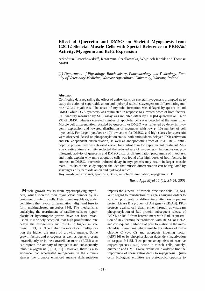
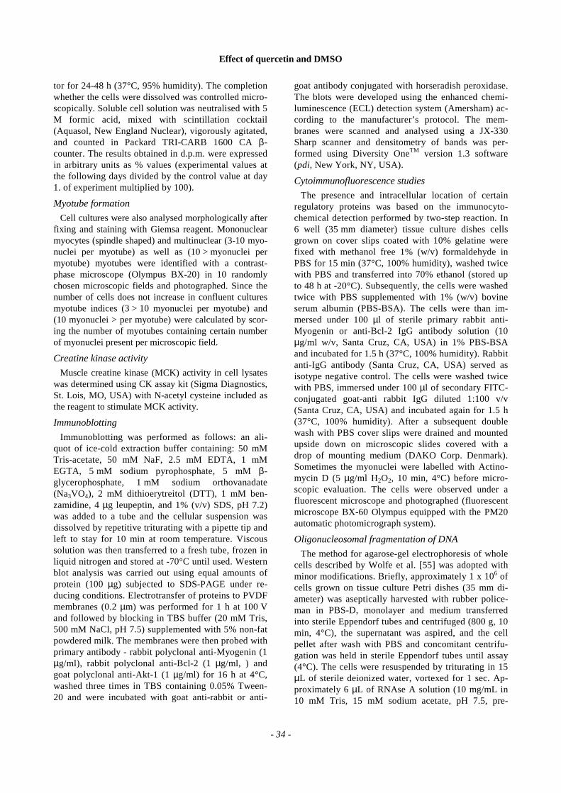
Simultaneous determination of both DNA synthesisand protein synthesis provide indirect informationabout the rate of cell growth occurring during tissuedevelopment [38]. In the control system (DM) theslope of curve representing time-course for multiplic-ity (proliferation assay - DNA synthesis) was falling(by 74% in extreme; P < 0.001, Fig. 1a), whereas theslope of curve illustrating time-course of protein syn-thesis was elevating (by 70% in extreme; P < 0.001;Fig. 1b).
Quercetin and DMSO, regardless of the dose used,were apparently mitogenic (P < 0.001). However,DNA synthesis rate was not in direct proportion to theconcentration of DMSO or quercetin (Fig. 1a). Mito-genicity stimulated by quercetin, in extremes,amounted to 50%, 58%, and 12% for 1, 10 and 100µM, respectively, whereas for DMSO reached 20%, 0,40% for 0.1%, 1% and 2%, respectively. Also proteinsynthesis did not directly respond to the dose of ex-perimental factors (P < 0.001). For quercetin, proteinformation was inhibited by 12% after 1 µM (P < 0.05)and by 60% after 100 µM (P < 0.001), except stimula-tion by 80% after 10 µM of quercetin (Fig 1b;P < 0.01). This relation was especially evident forDMSO, where protein synthesis rate did not differfrom control after 0.1% DMSO, whereas it was inhib-ited after 1% and 2% of DMSO, respectively (Fig. 1b;P < 0.001).
Figure 1. Effects of various concentrations of quercetin or DMSO on DNA synthesis (proliferation assay) (a) or pro-tein synthesis (b). Confluent murine C2C12 myoblasts grown on multiwell (24) plates were incubated in DMEMsupplemented with 2% HS (DM) in the absence or presence of quercetin (1, 10, 100 µM) or DMSO (0.1, 1, 2%v/v) for 5 days. At each time point (day 1., 2., 3., 4., 5.), cells from three wells of multiwell plate per each treat-ment taken and radioactivity (d.p.m.) of TCA precipitable fractions of (3H)-thymidine labeled DNA and (12C)-leucine labeled protein were assessed as indicated. The experiment was repeated at least twice with similar re-sults. Values are means with SEM.
Effect of quercetin and DMSO
- 36 -
Myotube formation
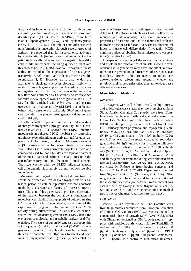
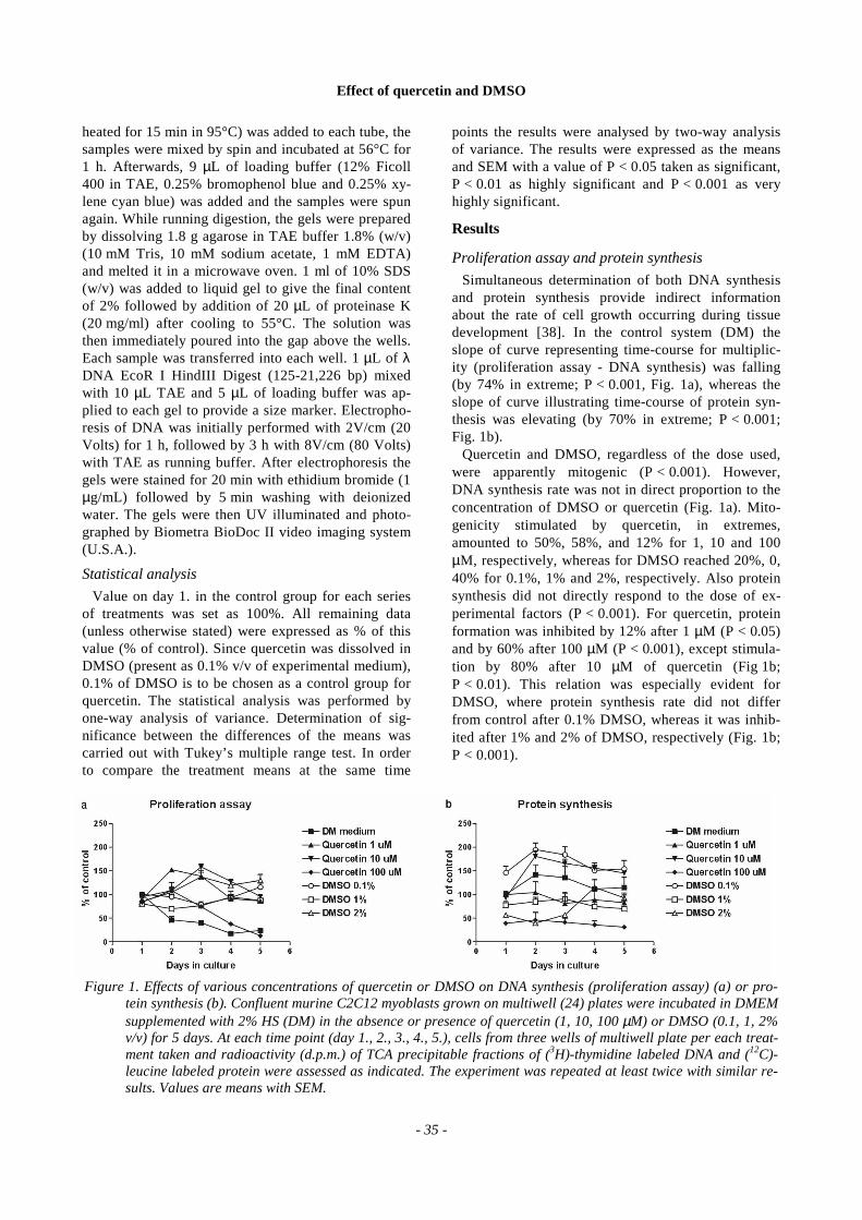
Since the intensity of myotube formation may differ,we decided to study the effect of each of the experi-mental agent on the presence of either myotubes with3-10 myonuclei (initial step of fusion) or myotubeswith 10 or more myonuclei (late step of fusion), sepa-rately.
Both quercetin and DMSO were observed to delay theonset of myotube formation ( < 10 myonuclei) in com-parison to controls (Fig. 2a, P < 0.001). Also the numberof myotubes formed after the replacement of GM withDM was higher in controls than in the experimentalcultures (P < 0.001). While quercetin was shown to ac-celerate the formation of already existing myotubes(10 > myonuclei) manifested by more numerous largemyotubes (Fig. 2b; increase by 250%; P < 0.001),DMSO was found to maintain inhibitory effect until thevery end of experiment (Fig. 2b; decrease by 250%;P < 0.001). Furthermore, during 2% DMSO treatmentno myotube with more than 10 myonuclei was found allover the experimental period. Both effects were time-dependent (Fig. 2).
Myogenin expression
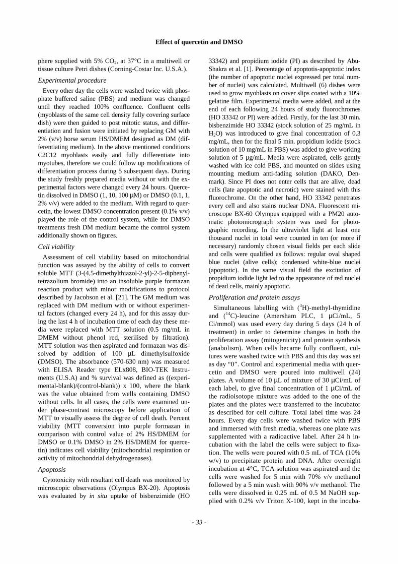
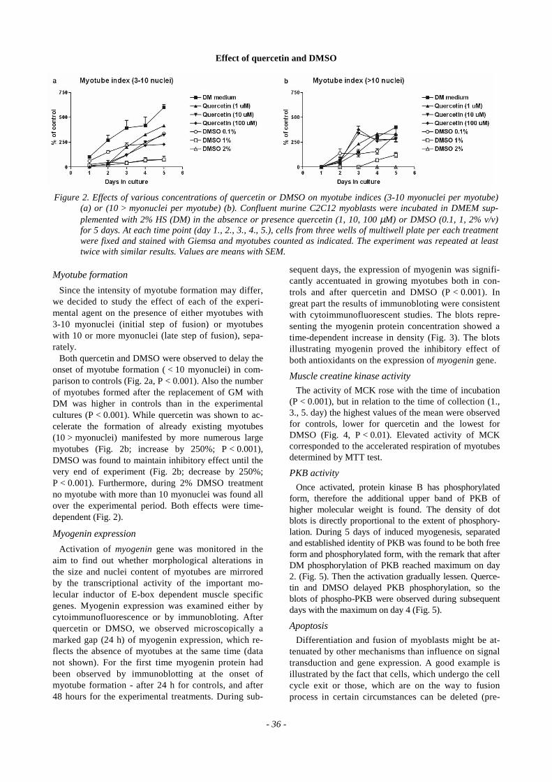
Activation of myogenin gene was monitored in theaim to find out whether morphological alterations inthe size and nuclei content of myotubes are mirroredby the transcriptional activity of the important mo-lecular inductor of E-box dependent muscle specificgenes. Myogenin expression was examined either bycytoimmunofluorescence or by immunobloting. Afterquercetin or DMSO, we observed microscopically amarked gap (24 h) of myogenin expression, which re-flects the absence of myotubes at the same time (datanot shown). For the first time myogenin protein hadbeen observed by immunoblotting at the onset ofmyotube formation - after 24 h for controls, and after48 hours for the experimental treatments. During sub-
sequent days, the expression of myogenin was signifi-cantly accentuated in growing myotubes both in con-trols and after quercetin and DMSO (P < 0.001). Ingreat part the results of immunobloting were consistentwith cytoimmunofluorescent studies. The blots repre-senting the myogenin protein concentration showed atime-dependent increase in density (Fig. 3). The blotsillustrating myogenin proved the inhibitory effect ofboth antioxidants on the expression of myogenin gene.
Muscle creatine kinase activity
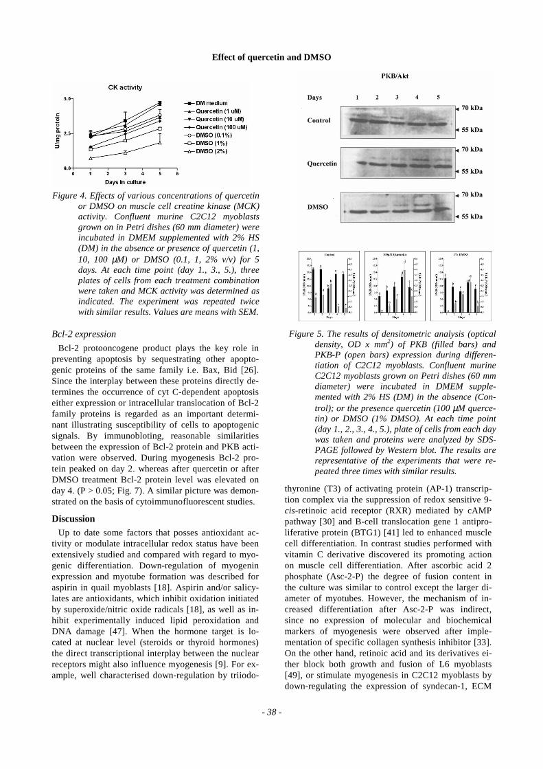
The activity of MCK rose with the time of incubation(P < 0.001), but in relation to the time of collection (1.,3., 5. day) the highest values of the mean were observedfor controls, lower for quercetin and the lowest forDMSO (Fig. 4, P < 0.01). Elevated activity of MCKcorresponded to the accelerated respiration of myotubesdetermined by MTT test.
PKB activity
Once activated, protein kinase B has phosphorylatedform, therefore the additional upper band of PKB ofhigher molecular weight is found. The density of dotblots is directly proportional to the extent of phosphory-lation. During 5 days of induced myogenesis, separatedand established identity of PKB was found to be both freeform and phosphorylated form, with the remark that afterDM phosphorylation of PKB reached maximum on day2. (Fig. 5). Then the activation gradually lessen. Querce-tin and DMSO delayed PKB phosphorylation, so theblots of phospho-PKB were observed during subsequentdays with the maximum on day 4 (Fig. 5).
Apoptosis
Differentiation and fusion of myoblasts might be at-tenuated by other mechanisms than influence on signaltransduction and gene expression. A good example isillustrated by the fact that cells, which undergo the cellcycle exit or those, which are on the way to fusionprocess in certain circumstances can be deleted (pre-
Figure 2. Effects of various concentrations of quercetin or DMSO on myotube indices (3-10 myonuclei per myotube)(a) or (10 > myonuclei per myotube) (b). Confluent murine C2C12 myoblasts were incubated in DMEM sup-plemented with 2% HS (DM) in the absence or presence quercetin (1, 10, 100 µM) or DMSO (0.1, 1, 2% v/v)for 5 days. At each time point (day 1., 2., 3., 4., 5.), cells from three wells of multiwell plate per each treatmentwere fixed and stained with Giemsa and myotubes counted as indicated. The experiment was repeated at leasttwice with similar results. Values are means with SEM.
Effect of quercetin and DMSO
- 37 -
mature mitosis or mitotic catastrophe). During apopto-sis cell membrane remains intact, chromatin is con-densed and finally cell blabbing occurs with the cellfragmentation to apoptotic bodies. Regardless of theroute of cell elimination, the essential question iswhether dying cells are those, which are currentlyforming myotubes, or others, which are not involved inthe fusion process. Observations performed on cellssimultaneously stained in situ with HO33342 and PIenable identification of floating cells, mononuclearcells and myonuclei marked with dense chromatin, asymptom of apoptotic cell death. The apoptotic index(AI) was calculated, and a photographic record per-formed to visualize the necrobiology of the musclecells. Assuming the lack of injury, the cells might dis-
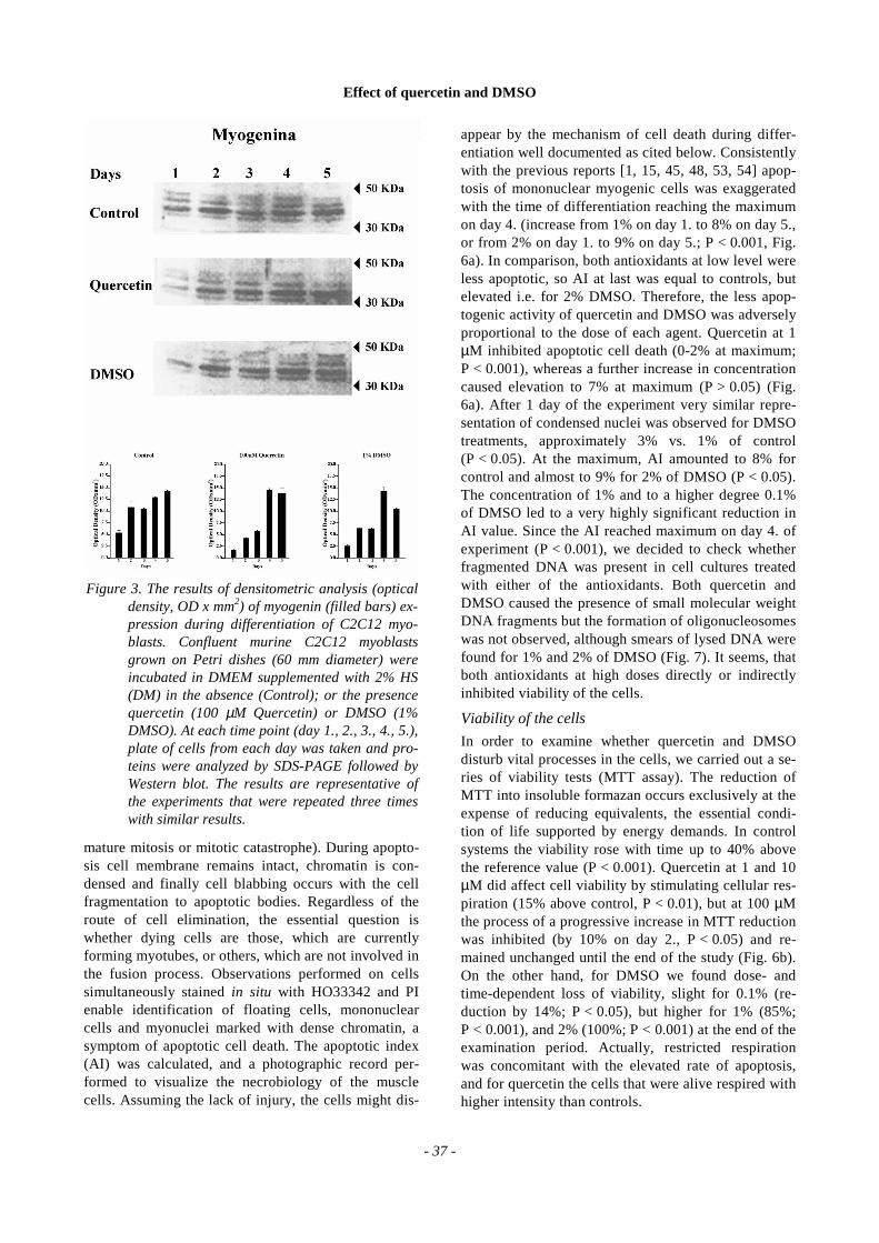
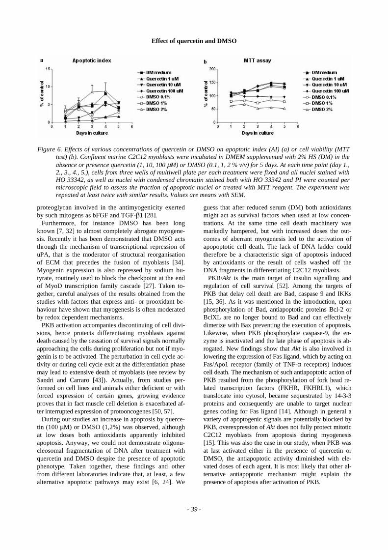
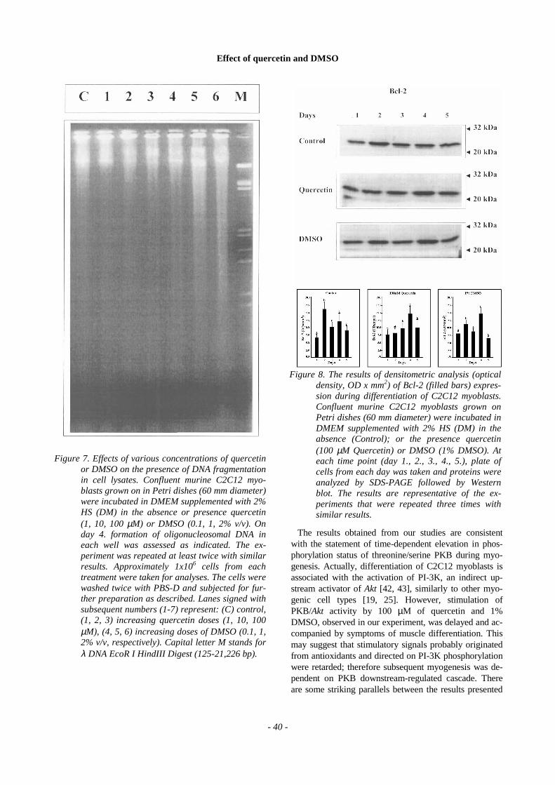
appear by the mechanism of cell death during differ-entiation well documented as cited below. Consistentlywith the previous reports [1, 15, 45, 48, 53, 54] apop-tosis of mononuclear myogenic cells was exaggeratedwith the time of differentiation reaching the maximumon day 4. (increase from 1% on day 1. to 8% on day 5.,or from 2% on day 1. to 9% on day 5.; P < 0.001, Fig.6a). In comparison, both antioxidants at low level wereless apoptotic, so AI at last was equal to controls, butelevated i.e. for 2% DMSO. Therefore, the less apop-togenic activity of quercetin and DMSO was adverselyproportional to the dose of each agent. Quercetin at 1µM inhibited apoptotic cell death (0-2% at maximum;P < 0.001), whereas a further increase in concentrationcaused elevation to 7% at maximum (P > 0.05) (Fig.6a). After 1 day of the experiment very similar repre-sentation of condensed nuclei was observed for DMSOtreatments, approximately 3% vs. 1% of control(P < 0.05). At the maximum, AI amounted to 8% forcontrol and almost to 9% for 2% of DMSO (P < 0.05).The concentration of 1% and to a higher degree 0.1%of DMSO led to a very highly significant reduction inAI value. Since the AI reached maximum on day 4. ofexperiment (P < 0.001), we decided to check whetherfragmented DNA was present in cell cultures treatedwith either of the antioxidants. Both quercetin andDMSO caused the presence of small molecular weightDNA fragments but the formation of oligonucleosomeswas not observed, although smears of lysed DNA werefound for 1% and 2% of DMSO (Fig. 7). It seems, thatboth antioxidants at high doses directly or indirectlyinhibited viability of the cells.
Viability of the cells
In order to examine whether quercetin and DMSOdisturb vital processes in the cells, we carried out a se-ries of viability tests (MTT assay). The reduction ofMTT into insoluble formazan occurs exclusively at theexpense of reducing equivalents, the essential condi-tion of life supported by energy demands. In controlsystems the viability rose with time up to 40% abovethe reference value (P < 0.001). Quercetin at 1 and 10µM did affect cell viability by stimulating cellular res-piration (15% above control, P < 0.01), but at 100 µMthe process of a progressive increase in MTT reductionwas inhibited (by 10% on day 2., P < 0.05) and re-mained unchanged until the end of the study (Fig. 6b).On the other hand, for DMSO we found dose- andtime-dependent loss of viability, slight for 0.1% (re-duction by 14%; P < 0.05), but higher for 1% (85%;P < 0.001), and 2% (100%; P < 0.001) at the end of theexamination period. Actually, restricted respirationwas concomitant with the elevated rate of apoptosis,and for quercetin the cells that were alive respired withhigher intensity than controls.
Figure 3. The results of densitometric analysis (opticaldensity, OD x mm2) of myogenin (filled bars) ex-pression during differentiation of C2C12 myo-blasts. Confluent murine C2C12 myoblastsgrown on Petri dishes (60 mm diameter) wereincubated in DMEM supplemented with 2% HS(DM) in the absence (Control); or the presencequercetin (100 µM Quercetin) or DMSO (1%DMSO). At each time point (day 1., 2., 3., 4., 5.),plate of cells from each day was taken and pro-teins were analyzed by SDS-PAGE followed byWestern blot. The results are representative ofthe experiments that were repeated three timeswith similar results.
Effect of quercetin and DMSO
- 38 -
Bcl-2 expression
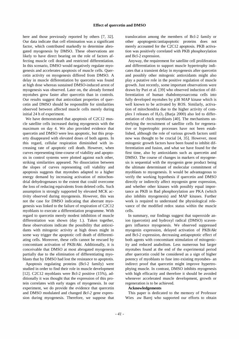
Bcl-2 protooncogene product plays the key role inpreventing apoptosis by sequestrating other apopto-genic proteins of the same family i.e. Bax, Bid [26].Since the interplay between these proteins directly de-termines the occurrence of cyt C-dependent apoptosiseither expression or intracellular translocation of Bcl-2family proteins is regarded as an important determi-nant illustrating susceptibility of cells to apoptogenicsignals. By immunobloting, reasonable similaritiesbetween the expression of Bcl-2 protein and PKB acti-vation were observed. During myogenesis Bcl-2 pro-tein peaked on day 2. whereas after quercetin or afterDMSO treatment Bcl-2 protein level was elevated onday 4. (P > 0.05; Fig. 7). A similar picture was demon-strated on the basis of cytoimmunofluorescent studies.
Discussion
Up to date some factors that posses antioxidant ac-tivity or modulate intracellular redox status have beenextensively studied and compared with regard to myo-genic differentiation. Down-regulation of myogeninexpression and myotube formation was described foraspirin in quail myoblasts [18]. Aspirin and/or salicy-lates are antioxidants, which inhibit oxidation initiatedby superoxide/nitric oxide radicals [18], as well as in-hibit experimentally induced lipid peroxidation andDNA damage [47]. When the hormone target is lo-cated at nuclear level (steroids or thyroid hormones)the direct transcriptional interplay between the nuclearreceptors might also influence myogenesis [9]. For ex-ample, well characterised down-regulation by triiodo-
thyronine (T3) of activating protein (AP-1) transcrip-tion complex via the suppression of redox sensitive 9-cis-retinoic acid receptor (RXR) mediated by cAMPpathway [30] and B-cell translocation gene 1 antipro-liferative protein (BTG1) [41] led to enhanced musclecell differentiation. In contrast studies performed withvitamin C derivative discovered its promoting actionon muscle cell differentiation. After ascorbic acid 2phosphate (Asc-2-P) the degree of fusion content inthe culture was similar to control except the larger di-ameter of myotubes. However, the mechanism of in-creased differentiation after Asc-2-P was indirect,since no expression of molecular and biochemicalmarkers of myogenesis were observed after imple-mentation of specific collagen synthesis inhibitor [33].On the other hand, retinoic acid and its derivatives ei-ther block both growth and fusion of L6 myoblasts[49], or stimulate myogenesis in C2C12 myoblasts bydown-regulating the expression of syndecan-1, ECM
Figure 4. Effects of various concentrations of quercetinor DMSO on muscle cell creatine kinase (MCK)activity. Confluent murine C2C12 myoblastsgrown on in Petri dishes (60 mm diameter) wereincubated in DMEM supplemented with 2% HS(DM) in the absence or presence of quercetin (1,10, 100 µM) or DMSO (0.1, 1, 2% v/v) for 5days. At each time point (day 1., 3., 5.), threeplates of cells from each treatment combinationwere taken and MCK activity was determined asindicated. The experiment was repeated twicewith similar results. Values are means with SEM.
Figure 5. The results of densitometric analysis (opticaldensity, OD x mm2) of PKB (filled bars) andPKB-P (open bars) expression during differen-tiation of C2C12 myoblasts. Confluent murineC2C12 myoblasts grown on Petri dishes (60 mmdiameter) were incubated in DMEM supple-mented with 2% HS (DM) in the absence (Con-trol); or the presence quercetin (100 µM querce-tin) or DMSO (1% DMSO). At each time point(day 1., 2., 3., 4., 5.), plate of cells from each daywas taken and proteins were analyzed by SDS-PAGE followed by Western blot. The results arerepresentative of the experiments that were re-peated three times with similar results.
Effect of quercetin and DMSO
- 39 -
proteoglycan involved in the antimyogenicity exertedby such mitogens as bFGF and TGF-β1 [28].
Furthermore, for instance DMSO has been longknown [7, 32] to almost completely abrogate myogene-sis. Recently it has been demonstrated that DMSO actsthrough the mechanism of transcriptional repression ofuPA, that is the moderator of structural reorganisationof ECM that precedes the fusion of myoblasts [34].Myogenin expression is also repressed by sodium bu-tyrate, routinely used to block the checkpoint at the endof MyoD transcription family cascade [27]. Taken to-gether, careful analyses of the results obtained from thestudies with factors that express anti- or prooxidant be-haviour have shown that myogenesis is often moderatedby redox dependent mechanisms.
PKB activation accompanies discontinuing of cell divi-sions, hence protects differentiating myoblasts againstdeath caused by the cessation of survival signals normallyapproaching the cells during proliferation but not if myo-genin is to be activated. The perturbation in cell cycle ac-tivity or during cell cycle exit at the differentiation phasemay lead to extensive death of myoblasts (see review bySandri and Carraro [43]). Actually, from studies per-formed on cell lines and animals either deficient or withforced expression of certain genes, growing evidenceproves that in fact muscle cell deletion is exacerbated af-ter interrupted expression of protooncogenes [50, 57].
During our studies an increase in apoptosis by querce-tin (100 µM) or DMSO (1,2%) was observed, althoughat low doses both antioxidants apparently inhibitedapoptosis. Anyway, we could not demonstrate oligonu-cleosomal fragmentation of DNA after treatment withquercetin and DMSO despite the presence of apoptoticphenotype. Taken together, these findings and otherfrom different laboratories indicate that, at least, a fewalternative apoptotic pathways may exist [6, 24]. We
guess that after reduced serum (DM) both antioxidantsmight act as survival factors when used at low concen-trations. At the same time cell death machinery wasmarkedly hampered, but with increased doses the out-comes of aberrant myogenesis led to the activation ofapopoptotic cell death. The lack of DNA ladder couldtherefore be a characteristic sign of apoptosis inducedby antioxidants or the result of cells washed off theDNA fragments in differentiating C2C12 myoblasts.
PKB/Akt is the main target of insulin signalling andregulation of cell survival [52]. Among the targets ofPKB that delay cell death are Bad, caspase 9 and IKKs[15, 36]. As it was mentioned in the introduction, uponphosphorylation of Bad, antiapoptotic proteins Bcl-2 orBclXL are no longer bound to Bad and can effectivelydimerize with Bax preventing the execution of apoptosis.Likewise, when PKB phosphorylate caspase-9, the en-zyme is inactivated and the late phase of apoptosis is ab-rogated. New findings show that Akt is also involved inlowering the expression of Fas ligand, which by acting onFas/Apo1 receptor (family of TNF-α receptors) inducescell death. The mechanism of such antiapoptotic action ofPKB resulted from the phosphorylation of fork head re-lated transcription factors (FKHR, FKHRL1), whichtranslocate into cytosol, became sequestrated by 14-3-3proteins and consequently are unable to target nucleargenes coding for Fas ligand [14]. Although in general avariety of apoptogenic signals are potentially blocked byPKB, overexpression of Akt does not fully protect mitoticC2C12 myoblasts from apoptosis during myogenesis[15]. This was also the case in our study, when PKB wasat last activated either in the presence of quercetin orDMSO, the antiapoptotic activity diminished with ele-vated doses of each agent. It is most likely that other al-ternative antiapoptotic mechanism might explain thepresence of apoptosis after activation of PKB.
Figure 6. Effects of various concentrations of quercetin or DMSO on apoptotic index (AI) (a) or cell viability (MTTtest) (b). Confluent murine C2C12 myoblasts were incubated in DMEM supplemented with 2% HS (DM) in theabsence or presence quercetin (1, 10, 100 µM) or DMSO (0.1, 1, 2 % v/v) for 5 days. At each time point (day 1.,2., 3., 4., 5.), cells from three wells of multiwell plate per each treatment were fixed and all nuclei stained withHO 33342, as well as nuclei with condensed chromatin stained both with HO 33342 and PI were counted permicroscopic field to assess the fraction of apoptotic nuclei or treated with MTT reagent. The experiment wasrepeated at least twice with similar results. Values are means with SEM.
Effect of quercetin and DMSO
- 40 -
The results obtained from our studies are consistentwith the statement of time-dependent elevation in phos-phorylation status of threonine/serine PKB during myo-genesis. Actually, differentiation of C2C12 myoblasts isassociated with the activation of PI-3K, an indirect up-stream activator of Akt [42, 43], similarly to other myo-genic cell types [19, 25]. However, stimulation ofPKB/Akt activity by 100 µM of quercetin and 1%DMSO, observed in our experiment, was delayed and ac-companied by symptoms of muscle differentiation. Thismay suggest that stimulatory signals probably originatedfrom antioxidants and directed on PI-3K phosphorylationwere retarded; therefore subsequent myogenesis was de-pendent on PKB downstream-regulated cascade. Thereare some striking parallels between the results presented
Figure 7. Effects of various concentrations of quercetinor DMSO on the presence of DNA fragmentationin cell lysates. Confluent murine C2C12 myo-blasts grown on in Petri dishes (60 mm diameter)were incubated in DMEM supplemented with 2%HS (DM) in the absence or presence quercetin(1, 10, 100 µM) or DMSO (0.1, 1, 2% v/v). Onday 4. formation of oligonucleosomal DNA ineach well was assessed as indicated. The ex-periment was repeated at least twice with similarresults. Approximately 1x10⁶ cells from eachtreatment were taken for analyses. The cells werewashed twice with PBS-D and subjected for fur-ther preparation as described. Lanes signed withsubsequent numbers (1-7) represent: (C) control,(1, 2, 3) increasing quercetin doses (1, 10, 100µM), (4, 5, 6) increasing doses of DMSO (0.1, 1,2% v/v, respectively). Capital letter M stands forλ DNA EcoR I HindIII Digest (125-21,226 bp).
Figure 8. The results of densitometric analysis (opticaldensity, OD x mm2) of Bcl-2 (filled bars) expres-sion during differentiation of C2C12 myoblasts.Confluent murine C2C12 myoblasts grown onPetri dishes (60 mm diameter) were incubated inDMEM supplemented with 2% HS (DM) in theabsence (Control); or the presence quercetin(100 µM Quercetin) or DMSO (1% DMSO). Ateach time point (day 1., 2., 3., 4., 5.), plate ofcells from each day was taken and proteins wereanalyzed by SDS-PAGE followed by Westernblot. The results are representative of the ex-periments that were repeated three times withsimilar results.
Effect of quercetin and DMSO
- 41 -
here and those previously reported by others [7, 32].Our data indicate that cell elimination was a significantfactor, which contributed markedly to determine abro-gated myogenesis by DMSO. These observations arelikely to have direct bearing on the role of factors af-fecting muscle cell death and restricted differentiation.In this scenario, DMSO would negatively regulate myo-genesis and accelerates apoptosis of muscle cells. Quer-cetin activity on myogenesis differed from DMSO. Adelay in muscle differentiation by quercetin was foundat high dose whereas sustained DMSO-induced arrest ofmyogenesis was observed. Later on, the already formedmyotubes grew faster after quercetin than in controls.Our results suggest that antioxidant properties of quer-cetin and DMSO should be responsible for similaritiesobserved between affected muscle cells merely duringinitial 24 h of experiment.
We have demonstrated that apoptosis of C2C12 mus-cle satellite cells increased during myogenesis with themaximum on day 4. We also provided evidence thatquercetin and DMSO were less apoptotic, but this prop-erty disappeared with elevated doses of both agents. Inthis regard, cellular respiration diminished with in-creasing rate of apoptotic cell death. However, whencurves representing time-course of viability and apopto-sis in control systems were plotted against each other,striking similarities appeared. No dissociation betweenthe slopes of curves representing cell viability andapoptosis suggests that myotubes adapted to a higherenergy demand by increasing activation of mitochon-drial dehydrogenases to the extent that could overcomethe loss of reducing equivalents from deleted cells. Suchassumption is strongly supported by elevated MCK ac-tivity observed during myogenesis. However, this wasnot the case for DMSO indicating that aberrant myo-genesis was linked to the failure of respiration of C2C12myoblasts to execute a differentiation programme. Withregard to quercetin merely modest inhibition of muscledifferentiation was shown (day 1.). Taken together,these observations indicate the possibility that antioxi-dants with mitogenic activity at high doses might insome way trigger the apoptotic cell death of differenti-ating cells. Moreover, these cells cannot be rescued byconcomitant activation of PKB/Akt. Additionally, it isconceivable that DMSO at most abrogated myogenesispartially due to the elimination of differentiating myo-blasts that by DMSO had lost the resistance to apoptotis.
Apoptosis regulating proteins (Bcl-2 family) werestudied in order to find their role in muscle development[12]. C2C12 myoblasts were Bcl-2 positive (15%), ad-ditionally it was thought that the expression of this pro-tein correlates with early stages of myogenesis. In ourexperiment, we do provide the evidence that quercetinand DMSO modulated and changed Bcl-2 gene expres-sion during myogenesis. Therefore, we suppose that
translocation among the members of Bcl-2 family orother apoptogenic/antiapoptotic proteins does notmerely accounted for the C2C12 apoptosis. PKB activa-tion was positively correlated with PKB phosphorylationand Bcl-2 expression.
Anyway, the requirement for satellite cell proliferationand differentiation to support muscle hypertrophy indi-cates that a transient delay in myogenesis after quercetinand possibly other mitogenic antioxidants might alsoplay a putative role in the positive regulation of musclegrowth. Just recently, some important observations weredrawn by Puri et al. [39] who observed induction of dif-ferentiation of human rhabdomyosarcoma cells intofully developed myotubes by p38 MAP kinase which iswell known to be activated by ROS. Similarly, activa-tion of mitochondria due to the higher activity of com-plex I releases of H2O2 (Barja 2000) also led to differ-entiation of chick myoblasts [40]. The mechanisms un-derlying the recruitment of satellite cells for regenera-tive or hypertrophic processes have not been estab-lished, although the role of various growth factors untilnow was thought to be crucial [4, 35]. However, mostmitogenic growth factors have been found to inhibit dif-ferentiation and fusion, and what we have found for thefirst time, also by antioxidants such as quercetin andDMSO. The course of changes in markers of myogene-sis is sequential with the myogenin gene product beingthe ultimate determinant of molecular commitment ofmyoblasts to myogenesis. It would be advantageous toverify the working hypothesis if quercetin and DMSOdirectly or indirectly affect myogenin gene expression,and whether other kinases with possibly equal impor-tance as PKB in Bad phosphorylation are PKA (whichalso inhibits myogenesis) and MAP kinases. Furtherwork is required to understand the physiological rele-vance of the modified redox status within the musclecells.
In summary, our findings suggest that superoxide an-ion (quercetin) and hydroxyl radical (DMSO) scaven-gers influence myogenesis. We observed suppressedmyogenin expression, delayed activation of PKB/Aktand Bcl-2 expression, decreasing antiapoptotic effect ofboth agents with concomitant stimulation of mitogenic-ity and reduced anabolism. Less numerous but largermyotubes found at the end of the experimental periodafter quercetin could be considered as a sign of higherpotency of myoblasts to fuse into existing myotubes- anindirect proof that quercetin might improve hypertro-phying muscle. In contrast, DMSO inhibits myogenesiswith high efficacity and therefore it should be avoidedwhenever accelerated muscle development, growth orregeneration is to be achieved.
AcknowledgementsThis paper is dedicated to the memory of Professor
Wies�aw Barej who supported our efforts to obtain
Effect of quercetin and DMSO
- 42 -
funds necessary to complete this study. We thank Mrs.Justyna Olczak and Mrs. Teresa Hass for their contri-bution to microscopic evaluation of myotube forma-tion and biochemical analyses. This work was sup-ported by the grant No. 5 PO6K 029 15 from the Pol-ish State Committee of Sciences.
Address correspondence to:
Arkadiusz Orzechowski, Department of Physiology,Biochemistry, Pharmacology and Toxicology, Facultyof Veterinary Medicine, Warsaw Agricultural Univer-sity, Nowoursynowska 166, 02-787 Warsaw, Poland,tel. and fax +48 022 847 24 52, Email [email protected]..
References
[1] Abu-Shakra S, Alhalabi MS, Nachtmaan FC,Schemidt RA, Brusilow WSA: Anabolic steroidsinduce injury and apoptosis of differentiatedskeletal muscle. J Neurosci Res 1997; 47: 186-197. [2] Angello JS, Stern HM, Hauschka SD: P19 embryo-nal carcinoma cells: a model system for studyingneural tube induction of skeletal myogenesis. DevBiol 1997; 192: 93-98. [3] Barja G: Mitochondrial oxygen radical generationand leak: sites of production in states 4 and 3, organspecifity, and relation to aging and longevity. JBioenerg Biomembr 2000; 31 (4): 347-366. [4] Bass J, Oldham J, Sharma M, Kambadur R: Growthfactors controlling muscle development. Dom AnimEndocrinol 1999; 17: 191-197. [5] Benezra R, Davis RL, Lockshon D, Turner DL,Weintraub H: The protein Id: A negative regulatorof helix-loop-helix DNA binding proteins. Cell1990; 61: 49-59. [6] Benson RSP, Dive C, Watson AJM: Comparative ef-fects of Bcl-2 over-expression and ZVAD.FMK treat-ment on dexamethasone and VP16-induced apoptosisin CEM cells. Cell Death Differ 1998; 5: 432-439. [7] Blau HM, Epstein ChJ: Manipulation of myogene-sis in vitro: Reversible inhibition by DMSO. Cell1979; 17: 95-108. [8] Cassar-Malek I, Langlois N, Picard B, Geay Y:Regulation of bovine satellite cell proliferation anddifferentiation by insulin and triiodothyronine. DomAnim Endocrinol 1999; 17: 373-388. [9] Cassar-Malek I, Marchal S, Rochard P, Casas F,Wrutniak C, Samarut J, Cabello G: Induction of c-erbA-AP-1 interactions and c-erb A transcriptionalactivity in myoblasts by XRX. J Biol Chem 1996;271: 11392-11399. [10] Cos P, Ying L, Calomme M, Hu JP, Cimanga K,Poel BV, Pieters L, Vlietinck AJ, Berghe DV:Structure-activity relationship and classification offlavonoids as inhibitors of xanthine oxidase and su-peroxide scavengers. J Nat Prod 1998; 61 (1): 71-76.
[11] De Vries JHM, Janssen PLTMK, Hollman PCH, VanStaveren WA, Katan MB: Consumption of quercetinand kaempferol in free-living subjects eating a varietyof diets. Cancer Lett 1997; 114: 141-144. [12] Dominov JA, Dunn JJ, Miller JB: Bcl-2 expressionidentifies an early stage of myogenesis and pro-motes clonal expansion of muscle cells. J Cell Biol1998; 142: 537-544. [13] Doumit ME, Cook DR, Merkel RA: Testosteroneup-regulates androgen receptors and decreases dif-ferentiation of porcine myogenic satellite cells invitro. Endocrinol 1996; 137 (4): 1385-1394. [14] Ferrigno P, Silver PA: Regulated nuclear localiza-tion of stress-responsive factors: how the nucleartrafficking of protein kinases and transcription fac-tors contributes to cell survival. Oncogene 1999;18: 6129-6134. [15] Fujio Y, Guo K, Mano T, Mitsuuchi Y, Testa JR,Walsh K: Cell cycle withdrawal promotes myogenicinduction of Akt, a positive modulator myocyte sur-vival. Mol Cell Biol 1999; 19 (7): 5073-5082. [16] Gamet-Payrastre L, Manenti S, Gratacap M-P, Tul-liez J, Chap H, Payrastre B: Flavonoids and the in-hibition of PKC and PI-3 kinase. Gen Pharmacol1999; 32: 279-286. [17] Gryglewski RJ, Korbut R, Robak J, Swies J: On themechanisms of antithrombotic action of flavonoids.Biochem Pharmacol 1987; 36: 317-322. [18] Hase H, Hirayama E, Jeman K: Further investiga-tion of some inhibitors on myogenic differentiation:mechanism of inhibition with HMBA on quailmyoblasts transformed with Rous sarcoma virus.Cell Struct Func 1996; 21: 17-25. [19] Hayashi K, Takahashi M, Kimura K, Nishida W, SagaH, Sobue K: Changes in the balance of phosphoinosi-tide 3-kinase/protein kinase (Akt) and the mitogen-activated protein kinases (ERK/p38MAPK) determinea phenotype of visceral and vascular smooth musclecells. J Cell Biol 1999; 145 (4): 727-740. [20] Hollman PCH, Vantrijp JMP, Mengelers MJB,Devries JHM, Katan MB: Bioavailability of thedietary antioxidant flavonol quercetin in man. Can-cer Lett 1997; 260: 10-13. [21] Jacobson MD, Burne JF, Raff MC: Programmedcell death and Bcl-2 protection in the absence of anucleus. EMBO J 1994; 13 (8): 1899-1910. [22] Kawada N, Kuroki T, Kobayashi K, Inoue M, KanedaK: Inhibition of myofibroblastic transformation ofcultured rat hepatic stellate cells by methylxanthinesand dibutyryl-cAMP. Dig Dis Sci 1996; 41: 1022-1029. [23] Kawada N, Seki S, Inoue M, Kuroki T: Effect of anti-oxidants, resveratrol, quercetin, and N-acetylcysteine,on the functions of cultured rat hepatic stellate cellsand Kupfer cells. Hepatology 1998; 27: 1265-1274.
Effect of quercetin and DMSO
- 43 -
[24] Kessel D, Luo Y: Photodynamic therapy: a mito-chondrial inducer of apoptosis. Cell Death Differ1999; 6: 28-35. [25] Kim JM, Yoon M, Kim J, Kim SS, Kang I, Ha J,Kim SS: Phosphatidylinositol 3-kinase regulates dif-ferentiation of H9c2 cardiomyoblasts mainly throughthe protein kinase B/Akt-independent pathway. ArchBiochem Biophys 1999; 367 (1): 67-73. [26] Kroemer G: The proto-oncogene Bcl-2 and its role inregulating apoptosis. Nat Med 1997; 3 (6): 614-620. [27] Larrain J, Alvarez J, Hassell JR., Brandan E: Ex-pression of perlecan, a proteoglycan that binds my-ogenic inhibitory basic fibroblast growth factor, isdown regulated during skeletal muscle differentia-tion. Exp Cell Res 1997a; 234: 405-412. [28] Larrain J, Cizmeci-Smith G, Troncoso V, Stahl RC,Carey DJ, Brandan E: Syndecan-1 expression isdown-regulated during myoblast terminal differen-tiation. J Biol Chem 1997b; 272 (29): 18418-18424. [29] Manach C, Morand C, Demigne C, Texier O, Re-gerat F, Remesy C: Bioavailability of rutin andquercetin in rats. FEBS Lett 1997: 409: 12-16. [30] Marchal S, Cassar-Malek I, Rodier A, Wrutniak C,Cabello G: Mécanismes moléculaires impliquesdans l’activité myogenique de la triiodothyronine(T3). Medecine/Sciences 1996; 12: 1065-1076. [31] Melo F, Carey DJ, Brandan E: Extracellular matrix isrequired for skeletal muscle differentiation but notmyogenin expression. J Cell Biochem 1996; 62: 227-239. [32] Miranda AF, Nette EG, Khan S, Brockbank K,Schonberg M: Alteration of myoblast phenotypeby dimethylsulfoxide. Proc Natl Acad Sci USA1978; 83: 8511-8515. [33] Mitsumoto Y, Liu Z, Klip A: A long-lasting vita-min C derivative, ascorbic acid-2-phosphate, in-creases myogenin gene expression and promotesdifferentiation in L6 muscle cells. Biochem BiophysRes Commun 1994; 199 (1): 394-402. [34] Munoz-Canovez P, Miralles F, Baiget M, Felez J:Inhibition of urokinase plasminogen activator(uPA) abrogates myogenesis in vitro. ThrombHaemost 1997; 77 (3): 526-534. [35] Olson EN: Interplay between proliferation and dif-ferentiation within the myogenic lineage. Dev Biol1992: 154: 261-272. [36] Pastorino JG, Tafani M, Farber JL: Tumor necro-sis factor induces phosphorylation and transloca-tion of Bad through a phosphatidylinositide-3-OHkinase-dependent pathway. J Biol Chem 1999; 274(27): 19411-19416. [37] Picard B, Depreux F, Geay Y: Muscle differentiationof normal and double-muscled bovine foetal myoblastsin primary culture. Basic Appl Myol 1998; 8: 197-203.
[38] Preising P: What makes cells grow larger and howdoes they do it? Renal hypertrophy revisited. ExpNephrol 1999; 7: 273-283. [39] Puri PL, Wu Z, Zhang P, Wood LD, Bhakta KS,Han J, Feramisco JR, Karin M, Wang JYJ: Induc-tion of terminal differentiation by constitutive acti-vation of p38 MAP kinase in human rhabdomyo-sarcoma cells. Gen Dev 2000; 14: 574-584. [40] Rochard P, Rodier A, Casas F, Cassar-Malek I,Marchal-Victorion S, Daury L, Wrutniak C, Ca-bello G: Mitochondrial activity is involved in theregulation of myoblast differentiation through my-ogenin expression and activity of myogenic factors.J Biol Chem 2000; 275 (4): 2733-2744. [41] Rodier A, Marchal-Victorion S, Rochard P, CasasF, Cassar-Malek I, Rouault J-P, Magaud J-P, Ma-son DY, Wrutniak C, Cabello G: BTG1: a triiodo-thyronine target involved in the myogenic influenceof the hormone. Exp Cell Res 1999; 249: 337-348. [42] Rudnicki MA, McBurney MW: Cell culture meth-ods and induction of differentiation of embryonalcarcinoma cell lines, in Robertson EJ (ed): Terato-carcinoma and embryonic stem cells. A practicalapproach. IRL Press, 1987, pp 19-50. [43] Sandri M, Carraro U: Apoptosis of skeletal musclesduring development and disease. The Int J Biochem& Cell Biol 1999; 31: 1373-1390. [44] Santander RG, Lobo MVT, Alonso MFJ,Cuadrado GM: Fusion mechanism of the myo-blasts in the myotome of the chick embryo. HistolHistopath 1993; 8: 471-490. [45] Sarbassov D, Jones LG, Petersen CA: Extracellularsignal-regulated kinase-1 and -2 respond differentlyto myogenic and differentiative signaling pathwaysin myoblasts. Mol Endocrinol 1997; 11: 2038-2047. [46] Sarbassov D, Petersen CA: Insulin receptor sub-strate-1 and phosphatidylinositol 3-kinase regulatekinase-dependent and -independent signallingpathways during myogenic differentiation. Mol En-docrinol 1998; 12: 1870-1878. [47] Shi X, Ding M, Dong Z, Chen F, Ye J, Wang S,Leonard SS, Castranova V, Vallyathen V: Antioxi-dant properties of aspirin: characterization of theability of aspirin to inhibit silica-induced lipid per-oxidation, and TNF-α production. Mol Cell Bio-chem 1999; 199: 93-102. [48] Shiio Y, Sawada J, Handa H, Yamamoto T, InoueJ: Activation of the retinoblastoma gene expressionby Bcl-3: implication for muscle cell differentia-tion. Oncogene 1996; 12: 1837-1845. [49] Shin YJ, Woo JH, Chung CH, Kim HS: Retinoic acidand its geometrical isomers block both growth and fu-sion of L6 myoblasts by modulating the expression ofprotein kinase A. Mol Cells 2000; 10 (2): 162-168.
Effect of quercetin and DMSO
- 44 -
[50] Tsai CC, Saffiz JE, Billadello JJ: Expression of theGs protein α-subunit disrupts the normal programof differentiation in cultured murine myogeniccells. J Clin Invest 1997; 99 (1): 67-76. [51] Vaidya TB, Rhodes SJ, Taparowsky EJ, KoniecznySR: Fibroblast growth factor and transforming growthfactor β repress transcription of the myogenic regula-tory gene MyoD1. Mol Cell Biol 1989; 9: 3576-3579. [52] Vanhaesebroeck B, Waterfield MD: Signaling bydistinct classes of phosphoinositide 3-kinases. ExpCell Res 1999; 253: 239-254. [53] Wang J, Guo K, Wills KN, Walsh K: Rb functionsto in inhibit apoptosis during myocyte differentia-tion. Cancer Res 1997; 57: 359-354. [54] Wang J, Walsh K: Resistance to apoptosis con-ferred by Cdk inhibitors during myocyte differen-tiation. Science 1996; 273: 359-354.
[55] Wolfe JT, Pringle JH, Cohen GM: Assays for themeasurement of DNA fragmentation duringapoptosis, in Cotter TG, Martin SJ (eds): Tech-niques in apoptosis. A user guide. London, Port-land Press Ltd, 1996, pp 58-60. [56] Yu R, Jiao J-J, Duh J-L, Gudehithlu K, Tan T-H,Kong ANT: Activation of mitogen-activated proteinkinases by green tea polyphenols: potential signal-ling pathways in the regulation of antioxidant-responsive element-mediated phase II enzyme geneexpression. Carcinogenesis 1997; 18 (2): 451-456. [57] Zhang P: The cell cycle and development: redun-dant roles of cell cycle regulators. Curr Opin CellBiol 1999; 11: 655-662.