Embed Size (px)
Citation preview
TOUCH MEDICAL MEDIA94
Review Intraocular Lenses
Effective Ocular Biometry and Intraocular Lens Power CalculationMagdalena Turczynowska,1 Katarzyna Koźlik-Nowakowska,2 Magdalena Gaca-Wysocka2 and Andrzej Grzybowski2,3
1. Department of Ophthalmology, Stefan Żeromski Specialist Municipal Hospital, Cracow, Poland; 2. Department of Ophthalmology, Poznan City Hospital, Poland; 3. Department of Ophthalmology, University of Warmia and Mazury, Olsztyn, Poland
S ince the introduction of phacoemulsification, cataract surgery has evolved remarkably. The use of premium intraocular lenses (IOLs) (aspheric, toric, multifocal), refractive lens exchange and patients after refractive surgery procedures require extremely precise clinical measurements and IOL calculation formulas to achieve desired postoperative refraction. For many years, ultrasound biometry
has been the standard for measurement of ocular parameters. The introduction of optical biometry (fast and non-invasive) has replaced ultrasound methods and is now considered as the clinical standard for ocular biometry. Recently, several modern optical instruments have been commercially launched and there are new methods available, including the empirical, analytical, numerical or combined methods to determine IOL power. The aim of this review is to present current techniques of ocular biometry and IOL power calculation formulas, which will contribute to achieve highly accurate refractive outcomes.
Keywords
Biometry, ocular biometry, optical biometry, optical biometry devices, intraocular lenses, IOLs, IOL power calculation, IOL power calculation formulas
Disclosure: Magdalena Turczynowska, Katarzyna Koźlik-Nowakowska, Magdalena Gaca-Wysocka and Andrzej Grzybowski have nothing to disclose in relation to this article. No funding was received in the publication of this article. This study involves a review of the literature and did not involve any studies with human or animal subjects performed by any of the authors.
Authorship: All named authors meet the International Committee of Medical Journal Editors (ICMJE) criteria for authorship of this manuscript, take responsibility for the integrity of the work as a whole, and have given final approval to the version to be published.
Open Access: This article is published under the Creative Commons Attribution Noncommercial License, which permits any non-commercial use, distribution, adaptation and reproduction provided the original author(s) and source are given appropriate credit.
Received: 16 August 2016
Accepted: 18 September 2016
Citation: European Ophthalmic Review, 2016;10(2):94–100
Corresponding Author: Magdalena Turczynowska, Department of Ophthalmology, Stefan Żeromski Specialist Municipal Hospital in Kraków, os. Na Skarpie 66, 31-913 Kraków, Poland. E: [email protected]
Cataract surgery is currently the most frequently performed surgical technique worldwide. Since
the introduction of phacoemulsification by Kelman in 1967, surgical technology and construction of
implanted intraocular lenses (IOLs) have undergone considerable improvement. Small, sutureless
incisions and the use of foldable intraocular lenses reduced the incidence of complications and
surgically induced astigmatism.1,2 Furthermore, the use of premium intraocular lenses (aspheric,
toric, multifocal or a combination) allows the patient to become fully spectacle-independent.3
The improvement of surgical treatment results in rising expectations of patients. The key issue is
to achieve the desired refractive outcome. Essential for this purpose are precise measurements
of the eye and selection of the optimal IOL calculation formula. The aim of this article is to present
current techniques of ocular biometry and IOL power calculation formulas, which will contribute
to achieve highly accurate refractive outcomes.
Ocular biometryThe first step to achieve satisfactory postoperative refractive outcome is accurate ocular
biometry. Biometry enables the measurement of the various dimensions of the eye, including
axial length (AL), anterior chamber depth (ACD), lens thickness (LT) or central corneal thickness
(CCT). These values, together with the keratometry are essential for the IOL power calculation.
Precision of measurements is crucial, as a 0.1 mm error in AL results in a refractive error of
about 0.27 diopter (D).4
Ultrasound biometryFor many years, the only way to measure the AL of the eye was with ultrasound biometry. This
technique measures the distance from the surface of the corneal apex to the internal limiting
membrane (ILM). Good alignment along the ocular axis is important and that requires patient
cooperation (which can be difficult in children or patients with mental disorders). In cases where
a probe has direct contact with the cornea, there is a risk of a corneal damage or infection.
Therefore, a topical anaesthetic and proper disinfection of the probe are required. Occurring
inter-individual differences are highly dependent on the pressure exerted on the eye by
the ultrasound probe. High pressure results in corneal indentation and shortening of the AL.
Immersion ultrasound minimises the indentation of the cornea as it uses a saline-filled shell between
the probe and the eye. Clinical studies have shown that immersion biometry is more accurate and
more reliable than ultrasound biometry performed in contact mode.5–8 A limitation of ultrasound
biometry is low image resolution, as a consequence of using a long, low-resolution wavelength
(10 MHz) to measure small dimensions. In addition, differences in retinal thickness near the fovea
or the presence of other macular pathologies contribute to inconsistent measurements.9,10
Optical biometryThe introduction of optical biometry has steadily replaced ultrasound methods and is now
considered the clinical standard for ocular biometry. The results are comparable to those achieved
Grzybowski FINAL.indd 94 17/01/2017 16:54
DOI: https://doi.org/10.17925/EOR.2016.10.02.94
95EUROPEAN OPHTHALMIC REVIEW
Effective Ocular Biometry and Intraocular Lens Power Calculation
by immersion ultrasound biometry,11 but this new method is fast, easy
to reproduce by different examiners, non-invasive and non-contact.
Repeatability and reproducibility of measurements obtained using this
technique are high and the results are less dependent on operators’
skills. However, it is difficult to obtain a measurement in the presence
of a dense cataract or other opacities such as corneal scar and vitreous
haemorrhage. Optical biometry measures the distance from the corneal
surface to the retinal pigment epithelium (RPE). It may be associated
with overestimation of measurements of about 0.15–0.5 mm.12 Optical
biometry can also be successfully performed in pseudophakic or silicone
oil-filled eyes. Furthermore, in high myopic eyes, due to the presence
of posterior staphyloma, it may give better results than conventional
ultrasound techniques for measuring the AL.
Optical biometry devicesNew optical biometry devices provide measurements not only of AL
but also other important variables, such as: keratometry, ACD, LT, CCT,
pupil size (PS) or white-to-white distance (WTW). To measure the AL of
the eye, currently available devices use different technologies. IOLMaster
500 (Carl Zeiss Meditec, Jena, Germany), AL-Scan (Nidek, Aichi, Japan)
and Pentacam AXL (Oculus, Menlo Park, California, US) use partial
coherence interferometry (PCI) technology. Lenstar LS 900 (Haag-Streit,
Koeniz, Switzerland), Aladdin (Topcon, Tokyo, Japan), Galilei G6 (Ziemer,
Port, Switzerland) and OA-2000 (Tomey GmbH, Nürnberg, Germany)
use optical low-coherence interferometry (OLCR). Swept source OCT
(ss-OCT), used by the IOLMaster 700 (Carl Zeiss Meditec, Jena, Germany)
and ARGOS (Movu, Santa Clara, California, US) devices, is the newest
technology to be implemented in biometry.
The IOLMaster 500 was the first optical biometer and was introduced
in autumn 1999. The device is based on the PCI principle and measures
AL using infrared light (λ=780 nm) of short coherence emitted by
semiconductor laser diode. Furthermore, it measures keratometry,
analysing the anterior corneal curvature at six reference points at
approximately 2.3 mm optical zone. The ACD is measured using slit-lamp
illumination and is defined as a distance from the corneal epithelium and
to the anterior lens surface. WTW is obtained by analysing the image of the
iris using an infrared light source (wavelength 880 nm). All measurements
are performed simultaneously. IOLMaster 500 is currently considered as
a gold-standard biometer.13–15 Its repeatability and reproducibility have
been assessed in several studies.16–19
AL-Scan uses an 830 nm infrared laser diode for AL measurement with
PCI. It also measures keratometry (K) at 36 measurement points in two
circles with diameters of 2.4 mm and 3.3 mm, reflected from the corneal
surface. WTW and PS are obtained by analysing the image of the iris
and fitting the best circle with the lowest error square to the detected
edge. ACD and CCT are measured with an incorporated Scheimpflug
camera with a 470 nm monochromatic light. The device was introduced
for clinical practice in Europe in 2012. Srivannaboon et al.20 compared
the repeatability and reproducibility of ocular biometry and IOL power
obtained with AL-Scan and IOLMaster 500. AL-Scan provided excellent
repeatability and reproducibility for all measured parameters (AL, K,
ACD and WTW). Agreement with the IOLMaster 500 was good except
for the WTW. This can be caused by different algorithms used by these
devices for edge detection around iris image. Furthermore, the light
source used for WTW measurements is different: AL-Scan uses a green
light source (wavelength 525 nm) and IOLMaster uses an infrared light
source (wavelength 880 nm). Kaswin et al.21 evaluated the agreement in
AL, K, ACD measurements and IOL power calculations with AL-Scan and
IOLMaster 500. They reported excellent correlation in AL measurements
and K readings as well as good agreement in ACD measurements
between these two biometers. The IOL power calculations were also
highly comparable between these devices.
The Pentacam AXL device consists of a Scheimpflug camera which
rotates around the eye and a PCI-based optical biometer. It was
introduced in autumn 2015. In addition to anterior segment tomography,
ACD, CCT and WTW measurements, corneal topography, anterior and
posterior corneal surface and spherical aberrations, it also has integrated
AL measurement. Calculation of toric IOLs is based on the total corneal
refractive power and it takes into account the influence of the posterior
corneal surface. To our knowledge, no study has yet evaluated the
repeatability, reproducibility and accuracy of biometry measurements
obtained using this device.
The Lenstar LS 900 biometer is based on OLCR. Using a 820 µm
superluminescent diode as light source, it allows the measurement of
the AL, CCT, LT and ACD. The retinal thickness can also be determined
from the scans, but this requires subjective alignment of a cursor. It also
uses 950 µm light to assess by image analysis central corneal curvature
using two rings of diameter 1.65 mm and 2.30 mm of 16 light spot each.
WTW and PS are obtained by fitting the best circle with the lowest error
square to the detected edge. Optional T-cone module complements
this device with a Placido topography of the central 6 mm corneal zone.
Several studies confirmed Lenstar’s repeatability, reproducibility and
agreement with other biometry devices. Generally, Lenstar provided
results that correlated very well with those of the IOLMaster. Excellent
agreement has been shown between the AL measurements taken by
Lenstar and IOLMaster,22–25 but only good22 or moderate24 agreement
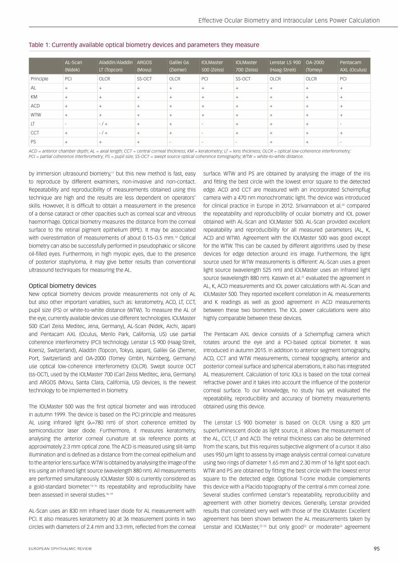
Table 1: Currently available optical biometry devices and parameters they measure
AL-Scan
(Nidek)
Aladdin/Aladdin
LT (Topcon)
ARGOS
(Movu)
Galilei G6
(Ziemer)
IOLMaster
500 (Zeiss)
IOLMaster
700 (Zeiss)
Lenstar LS 900
(Haag-Streit)
OA-2000
(Tomey)
Pentacam
AXL (Oculus)
Principle PCI OLCR SS-OCT OLCR PCI SS-OCT OLCR OLCR PCI
AL + + + + + + + + +
KM + + + + + + + + +
ACD + + + + + + + + +
WTW + + + + + + + + +
LT - - / + + + - + + + -
CCT + - / + + + - + + + +
PS + + + - - - + + -
ACD = anterior chamber depth; AL = axial length; CCT = central corneal thickness; KM = keratometry; LT = lens thickness; OLCR = optical low-coherence interferometry; PCI = partial coherence interferometry; PS = pupil size; SS-OCT = swept source optical coherence tomography; WTW = white-to-white distance.
Grzybowski FINAL.indd 95 17/01/2017 16:54
96 EUROPEAN OPHTHALMIC REVIEW
Review Intraocular Lenses
between these two devices in ACD measurements. In some cases, small
but statistically significant differences in K and ACD measurements
were reported.24 However, in a few studies, the AL measurements taken
by Lenstar were slightly higher than the IOLMaster measurements,
but the differences were not clinically significant.22,23 The Lenstar was
unable to take measurements due to lens opacities in a similar number
of patients to the IOLMaster.23
Aladdin is an optical biometer based on OLCR with an 830 nm super
luminescent diode and Placido topography system. It allows to perform
eight measurements in one acquisition: AL, keratometry, corneal
topography, ACD, pupillometry, WTW, CCT and LT, although the last two
parameters (measured by OLCR) are available only on the Aladdin LT.
Pupillometry can be measured in three modes: dynamic, photopic and
mesopic. Corneal topography is based on the reflection of 24 Placido
disc rings with a diameter of 8.0 mm. Topography-based keratometry is
obtained by analysing approximately 1,024 data points of four dedicated
Placido rings whose diameters range between 2.4 mm and 3.4 mm.
Aladdin provides also Zernike analysis and keratoconus screening. In
several studies, Aladdin provided good agreement and repeatability
compared with the IOLMaster. According to Huang et al.,28 repeatability
and reproducibility for AL, ACD and K measurements was found to be
excellent. However, the precision of WTW measurements was lower in
eyes with cataract. In addition, Aladdin is equipped with Placido-disc
corneal topographer and can provide information that is not available
on the IOLMaster, such as corneal map and corneal asphericity,
which were recently shown to influence the IOL power refractive
prediction error.29
OA-2000 combines Placido-disc topography and OLCR biometry. It
measures AL, CCT, LT and ACD using the OLCR technique. Corneal
curvature is measured by Placido-disc topography with nine rings
each 256 points in a 5.5 mm zone projected onto the cornea. It also
measures WTW and PS. Goebels et al.30 compared the OA-2000 device
with the Lenstar and IOLMaster. In this study, the OA-2000 biometer
generated the most accurate results that correlated very well with the
measurements obtained by Lenstar and IOLMaster. Excellent correlation
among all three devices was shown for AL measurements. Although
three different techniques to achieve K values were used, the correlation
between the different devices was very high. For ACD measurements,
good correlation was found, with the highest correlation between OA-
2000 and Lenstar devices (both use OLCR). The ACD values were highest
with the OA-2000 and lowest with the IOLMaster. All differences were
statistically, but not clinically, significant.
Galilei G6 combines OLCR optical biometry, dual-Scheimpflug imaging and
Placido-disc topography measures AL, LT, ACD, CCT, corneal topography,
PS and WTW. In addition to biometry, Galilei G6 provides high-definition
pachymetry, total corneal wavefront, curvature and astigmatism data
of the anterior and posterior cornea – complete data required to plan
cataract or refractive surgery. Shin et al.31 compared Galilei G6 with
Lenstar biometer. All parameters measured by the Galilei G6 were highly
repeatable. There were no statistically significant differences between K
and ACD measurements obtained by these two devices, however, the
measurements for AL, LT and WTW were significantly different. The K,
AL, ACD, LT and WTW showed good correlations (all p<0.001), however,
the agreements of LT and WTW were not good between the two devices.
The IOL powers using the SRK/T, Holladay 1, Hoffer Q and Haigis formulas
were compared – they did not show statistically significant differences (all
p>0.05), however, agreements between the IOL powers were not strong.
IOLMaster 700 is a biometry device based on swept-source OCT
technology that enables full-eye length tomography, providing good
fixation control. It uses high-frequency 1,055 nm tunable laser source
for AL, LT, ACD and CCT measurements. Keratometry measurements
are distance-independent. Light is projected onto the cornea at three
zones (1.5, 2.5 and 3.5 mm). PS and WTW are obtained using an LED
light source. Furthermore, the device provides a 1.0 mm horizontal scan
of the retina to ensure that the measurements are on the visual axis by
using the presence of foveal pit.32 According to Srivannaboon et al.,32
the measurement speed of IOLMaster 700 was statistically significantly
faster than IOLMaster 500 (p<0.05). In several studies, IOLMaster 700
showed very high repeatability and reproducibility and good agreement
with IOLMaster 500 and Lenstar,32–34 although repeatability and
reproducibility of ACD measurements obtained by IOLMaster 700 were
better than those from the IOLMaster 500.32 In addition, studies showed
that IOLMaster 700 penetrated the opaque media better and measured
the AL with fewer dropouts compared with the Lenstar and IOLMaster 500
even in dense cataracts.32,34
Argos biometer uses a 1,060 nm wavelength and 20 nm bandwidth
swept-source technology to collect two-dimensional OCT data
of the full eye (SS-OCT). It measures AL, LT, ACD and CCT with
ss-OCT. Keratometry values are generated by illumination from a ring of
16 infrared LEDs. In addition, the device measures PS by analysing the
two-dimensional OCT image. Shammas et al.35 evaluated the repeatability
and reproducibility of the measurements obtained with the Argos
biometer and compared them with the results obtained with the
IOLMaster 500 and the Lenstar LS 900 biometers. The study showed
high repeatability and reproducibility of measurements obtained by
Argos biometer. AL measurements with the new SS-OCT biometer were
comparable to PCI and OLCR measurements, with a faster and higher
acquisition rate, even in the presence of a dense nuclear or posterior
subcapsular cataract.
Optical measurements differ from measurements obtained by
ultrasound methods. Therefore, individual optimisation of constants
is necessary. This can be achieved by thorough analysis of pre- and
postoperative clinical data. In October 1999, an independent group of
scientists and users, working in the field of optical biometry founded
the User Group for Laser Interference Biometry (ULIB). One of the
most important purposes of this group is the optimisation of lens
constants for the IOL power calculation. The results of this optimisation
are published on the ULIB website (http://ocusoft.de/ulib/index.htm).
Optimised IOL constants are currently available for the IOLMaster
500 and 700, Lenstar LS 900, AL-Scan, Aladdin and recently also for
Pentacam AXL.
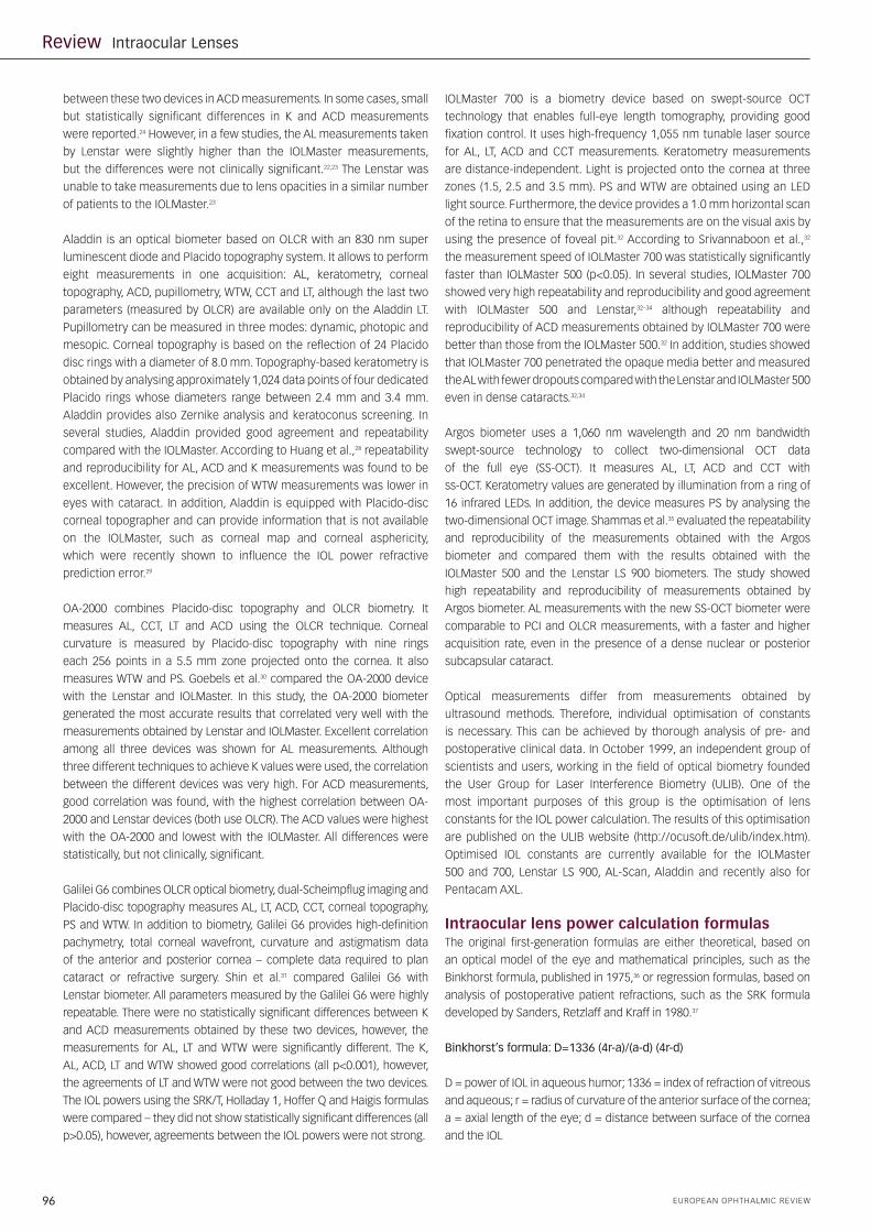
Intraocular lens power calculation formulas The original first-generation formulas are either theoretical, based on
an optical model of the eye and mathematical principles, such as the
Binkhorst formula, published in 1975,36 or regression formulas, based on
analysis of postoperative patient refractions, such as the SRK formula
developed by Sanders, Retzlaff and Kraff in 1980.37
Binkhorst’s formula: D=1336 (4r-a)/(a-d) (4r-d)
D = power of IOL in aqueous humor; 1336 = index of refraction of vitreous
and aqueous; r = radius of curvature of the anterior surface of the cornea;
a = axial length of the eye; d = distance between surface of the cornea
and the IOL
Grzybowski FINAL.indd 96 17/01/2017 16:54
97EUROPEAN OPHTHALMIC REVIEW
Effective Ocular Biometry and Intraocular Lens Power Calculation
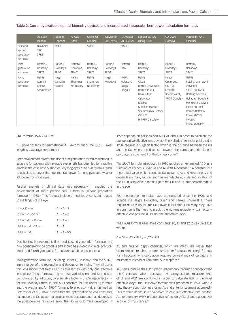
SRK formula: P=A-2.5L-0.9K
P = power of lens for emmetropia; A = A-constant of the IOL; L = axial
length; K = average keratometry
Refractive outcomes after the use of first-generation formulas were quite
accurate for patients with average eye length, but often led to refractive
errors in the case of very short or very long eyes.38 The SRK formula tends
to calculate stronger than optimal IOL power for long eyes and weaker
IOL power for short eyes.
Further analysis of clinical data was necessary. It enabled the
development of more precise SRK II formula (second-generation
formula) in 1988.39 This formula include a modified A constant, related
to the length of the eye:
if AL<20 mm A1 = A + 3
21 mm>AL≥20 mm A1 = A + 2
22 mm>AL ≥ 21 mm A1 = A + 1
24.5 mm>AL≥22 mm A1 = A
24.5 mm>AL A1 = A – 0.5
Despite this improvement, first- and second-generation formulas are
now considered to be obsolete and should be avoided in clinical practice.
Third- and fourth-generation formulas should be chosen instead.
Third-generation formulas, including Hoffer Q, Holladay1 and the SRK/T,
are a merger of the regression and theoretical formulas. They all use a
thin-lens model that treats IOLs as thin lenses with only one effective
lens plane. These formulas rely on two variables (AL and K) and can
be optimised by adjusting by a suitable factor – the ‘surgeon factor’ –
for the Holladay1 formula, the ACD-constant for the Hoffer Q formula
and the A-constant for SRK/T formula. Terzi et al.,40 Haigis41 as well as
Petermeier et al.,42 have proven that the optimisation of lens constants
has made the IOL power calculation more accurate and has decreased
the postoperative refractive error. The Hoffer Q formula developed in
1992 depends on personalised ACD, AL and K in order to calculate the
postoperative effective lens power.43 The Holladay1 formula, published in
1988, requires a surgeon factor, which is the distance between the iris
and the IOL, where the distance between the cornea and iris plane is
calculated as the height of the corneal curve.44
The SRK/T formula introduced in 1990 requires an estimated ACD as a
function of corneal curvature and AL with A-constant.45 A constant is a
theoretical value, which connects IOL power to AL and keratometry and
depends on many factors such as manufacturer, style and location of
the IOL. It is specific to the design of the IOL and its intended orientation
in the eye.
Fourth-generation formulas have promulgated since the 1990s and
include the Haigis, Holladay2, Olsen and Barrett Universal II. These
require more variables for IOL power calculation. One thing they have
in common is the need to predict the non-measurable, virtual factor –
effective lens position (ELP), not the anatomical one.
The Haigis formula uses three constants: a0, a1 and a2 to calculate ELP,
where:
d = a0 + (a1 × ACD) + (a2 × AL)
AL and anterior depth chamber, which are measured, rather than
estimated, are required, in contrast to other formulas. The Haigis formula
for intraocular lens calculation requires corneal radii of curvature in
millimeters instead of keratometry in diopters.46
In Olsen’s formula, the ELP is predicted primarily through a concept called
the C constant, where accurate, ray tracing-assisted measurements
of LT and ACD are combined in order to calculate ELP in the most
effective way.47 The Holladay2 formula was proposed in 1993, when a
new theory about biometry using AL and anterior segment appeared.48
This formula needs seven variables to calculate effective lens positon:
AL, keratometry, WTW, preoperative refraction, ACD, LT and patient age,
in order of importance.49
Table 2: Currently available optical biometry devices and incorporated intraocular lens power calculation formulas
AL-Scan
(Nidek)
Aladdin
(Topcon)
ARGOS
(Movu)
Galilei G6
(Ziemer)
IOLMaster
500 (Zeiss)
IOLMaster
700 (Zeiss)
Lenstar LS 900
(Haag-Streit)
OA-2000
(Tomey)
Pentacam AXL
(Oculus)
First and
second
generation
formulas:
Binkhorst
SRK
SRK II
SRK II SRK II SRK II
Third
generation
formulas:
HofferQ,
Holladay1,
SRK/T
HofferQ,
Holladay1,
SRK/T
HofferQ,
Holladay1,
SRK/T
HofferQ,
Holladay1,
SRK/T
HofferQ
Holladay1
SRK/T
HofferQ,
SRK/T
HofferQ,
Holladay1,
SRK/T
HofferQ,
Holladay1,
SRK/T
HofferQ,
Holladay1,
SRK/T
Fourth
generation
formulas:
Haigis
Camellin-
Calossi
Shammas PL
Haigis
Camellin-
Calossi
Haigis
Shammas
No-History
Haigis
Shammas
No-History
Haigis
Holladay2
Haigis
Holladay2
Haigis-L
Haigis-T
Haigis
Olsen
Barrett Universal II,
Barrett True-K,
Barrett Toric
Calculator
Masket,
Modified Masket,
Shammas No-History
OKULIX
Hill-RBF Calculator
Haigis
Optimized,
OKULIX
Easy IOL
Shammas PL,
SRK/T Double K
Haigis
PotvinShammasHill
PotvinHill
SRK/T Double K
HofferQ Double K
Holladay1 Double K
Meridional analysis
based on Total
Cornea Refraktiv
Power (TCRP)
OKULIX
Phaco Optics®
Grzybowski FINAL.indd 97 17/01/2017 16:54
98 EUROPEAN OPHTHALMIC REVIEW
Review Intraocular Lenses
Barrett Universal II is a thick lens formula, in which ELP is characterised
by LF (lens factor) and anatomic chamber depth. The LF is influenced by:
keratometry, AL, ACD, LT and WTW, in order of importance. This formula
notifies the change in planes, which is connected with different IOL
powers. It recognises the negative value of lens factor in the presence of
negative-powered type of IOL, which has to be taken into consideration
when calculating the ELP.50
A major challenge is IOL power calculation in patients who have undergone
refractive surgery, as it is difficult to measure the true corneal power
and estimate the ELP. After myopic refractive surgery (photorefractive
keratectomy [PRK], LASIK, radial keratotomy [RK]), both keratometry and
corneal topography tend to overestimate corneal power. This problem
can be remedied by double-K modifications of third-generation formulas:
SRK/T, Hoffer Q or Holladay1. Double-K methods use the preoperative K
values for the ELP calculation and the postoperative K values for the IOL
power determination.51 One of the drawbacks of these methods is ELP
calculation dependence on the central corneal power.
Other formula used for IOL power prediction after LASIK or PRK is the
Masket method. This formula omits the double K step required by other
pre-LASIK/PRK K-dependent methods and simply adjusts the power of
the IOL using the knowledge of the surgically induced refractive change.
It is particularly useful when corneal power before refractive surgery is
unavailable, but the refractive change is known (even if uncertain).52 More
reliable methods of determining IOL power after refractive surgery do
not rely on historical data, which may be inaccurate or unavailable, for
example, Shammas no-history, Haigis-L and Camellin-Calossi.
The Shammas no-history formula was first published in 2007. It is a
post-LASIK modification of a previously described formula, in which the
average corneal power, K, is replaced by the corrected mean corneal
power, Kc and where Kc=1.14 Kpost-6.8, with Kpost being the post-LASIK
K-readings in diopters.53
The Haigis-L formula, designed in 2008, using corneal radius measured
in mm generates a corrected corneal radius, which is then used by the
regular Haigis formula to calculate the IOL power.54
The Camellin-Calossi formula, first published in 2006, is one of the
most recent formulas used commonly for calculating IOL power in
eyes which have undergone refractive surgery. This formula is based on
modified Binkhorst II formula and empirically adjusts corneal power and
calculates ELP regardless of corneal keratometry (K). According to Suto
et al., the Camellin-Calossi formula can be also used for calculating IOL
in normal cataractous eyes and its accuracy is equivalent to common
IOL formulas: SRK/T and Haigis.55
There are also formulas designed exclusively for particular devices, for
example, PotvinShammasHill and PotvinHill formulas. They use data from
the Pentacam device, specifically the true net power in a 4.0 mm zone
centered on the corneal apex, to calculate IOL power in post-myopic
LASIK eyes (PotvinShammasHill formula) and after radial keratotomy
(PotvinHill formula).56
Recently, a new software using numerical ray tracing for IOL power
calculation became available (e.g., Okulix, EasyIOL, PhcoOptics). The
accuracy of numerical ray tracing is independent of AL. Therefore, very
long or very short eyes can gain the most from the higher accuracy of
this approach. For average-size eyes, however, the results of ray-tracing
methods were as accurate as theoretical thin-lens formulas.57,58
One of the most recent calculation methods was released in June 2016:
the Hill-RBF on-line calculator. It is an advanced, self-validating method
using artificial intelligence and pattern recognition to select an IOL for a
patient. The calculator is entirely data-driven and is independent of the
limitations of theoretical vergence formulas. It has been optimised for
use with the Lenstar LS 900, but may also be used with data from other
optical biometers.59
It has not been proven that any of the recent formulas are better than
the others, however, first and second generation formulas such as
SRK II should not be used any longer, as they have minimal theoretical
value.47 The new formulas are more precise than previous ones, but
their advantage can be noticed clearly in IOL calculation of non-
typical eyes.60 Modern IOL power calculations have similar outcomes
in eyes with average AL but they are less accurate in eyes with long
or short AL.43
Apparently, there is no multipurpose formula for every type of eye, the
use of a particular formula depends on several parameters, such as the
eye’s AL, astigmatism, previous refractive surgery and differs in phakic
and pseudophakic eyes.51 According to Wang et al., the Haigis, Hoffer Q,
Holladay1 and SRK/T formulas are equally accurate for calculating the IOL
power in phakic eyes between 22 mm and 24.5 mm length.61
In a group of patients with AL below 22 mm, getting a precise
postoperative refraction is more difficult than amongst other patients,
as short eyes usually need a high-power intraocular lens. In 2012, Day
et al.62 showed that the Hoffer Q formula had the lowest absolute mean
error in eyes with AL from 20.00 to 20.99 mm and Hoffer Q and Holladay1
formulas had made more accurate calculations than SRK/T formula. Carifi
et al.63 compared the refractive results among various formulas (Hoffer
Q, Holladay1, Holladay2, Haigis, SRK-T and SRK-II) in patients undergoing
phacoemulsification cataract surgery with a single highly powerful IOL
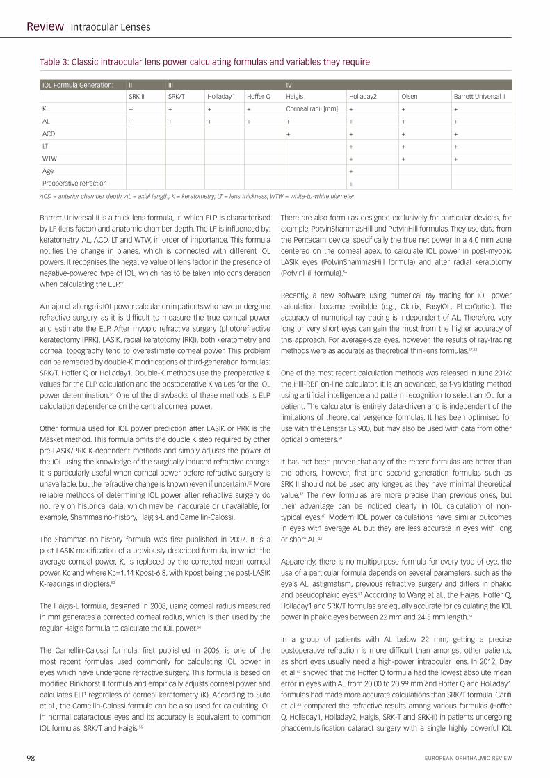
Table 3: Classic intraocular lens power calculating formulas and variables they require
IOL Formula Generation: II III IV
SRK II SRK/T Holladay1 Hoffer Q Haigis Holladay2 Olsen Barrett Universal II
K + + + + Corneal radii [mm] + + +
AL + + + + + + + +
ACD + + + +
LT + + +
WTW + + +
Age +
Preoperative refraction +
ACD = anterior chamber depth; AL = axial length; K = keratometry; LT = lens thickness; WTW = white-to-white diameter.
Grzybowski FINAL.indd 98 17/01/2017 16:54
99EUROPEAN OPHTHALMIC REVIEW
Effective Ocular Biometry and Intraocular Lens Power Calculation
implanted in the capsular bag (range of powers +35.0 to +40.0 D). The
study showed that none of the latest-generation formulas (Hofer Q,
Haigis, Holladay1 and Holladay2) significantly outperformed the others
(p=0.245). However, the SRK formulas yielded less accurate predictions
in these cases. The authors suggested that the SRK/T formula should not
been used in IOL power calculation in eyes with AL shorter than 22 mm.
Currently, the most recommended formulas for IOL power calculation for
short eyes are the third-generation formula Hoffer Q64,65,43 and the fourth-
generation formula Holladay2.66 According to the study conducted in 2014
by Eom et al.,43 the Hoffer Q and Haigis formulas are similarly accurate in
calculating IOL power in eyes with short AL, but the Haigis formula is more
precise in eyes with ACD <2.4 mm. Similar outcomes were presented by
Maclaren et al.67 in 2007, when they showed that the Haigis and Hoffer Q
formulae performed well in eyes with long AL when using conventional
biometry methods and phacoemulsification.
There are also difficulties in choosing the most appropriate IOL power
for patients with high myopia. The main problem is staphyloma,
which makes the measurement of AL harder than usual, as well as
restricted access to IOL power calculation formulas for those patients.
It was suggested that there are no significant differences in IOL power
calculation in patients with AL >26 mm using Haigis, HofferQ, SRK/T
formulas, but it has been shown that SRK/T formula has the lowest
mean error.68 Aristodemou et al.69 has also shown that the most suitable
formula for eyes longer than 27 mm is SRK/T. In 2015 it was shown
that Barrett Universal II, one of the most recent published formulas, is
more accurate than other known formulas in long eyes with AL greater
than 26 mm.70
Patients’ requirements concerning visual effect after cataract surgery are
rising. In order to increase the accuracy of IOL power calculation and
postoperative refractive outcome, an ideal calculation formula has been
searched for for many years. A multipurpose formula, which can be used
in every eye’s AL, is still to be found.
ConclusionThe latest biometry technologies and modern IOL power calculation
formulas have significantly improved refractive outcomes after cataract
surgery. Well-calibrated devices, using optical rather than ultrasound
biometry, optimised IOL constants and properly selected last-generation
IOL power calculation formulas that fit to a particular patient can provide
excellent refractive outcomes. q
1. George R, Rupauliha P, Sripriya AV, et al., Comparison of endothelial cell loss and surgically induced astigmatism following conventional extracapsular cataract surgery, manual small-incision surgery and phacoemulsification, Ophthalmic Epidemiol, 2005;12:293–7.
2. Zheng L, Merriam JC, Zaider M, Astigmatism and visual recovery after ‘large incision’ extracapsular cataract surgery and ‘small’ incisions for phacoemulsification, Trans Am Ophthalmol Soc, 1997;95:387–410.
3. Javitt J, Steinert R, Cataract extraction with multifocal intraocular lens implantation: a multinational clinical trial evaluating clinical, functional and quality-of-life outcomes, Ophthalmology, 2000;107:2040–8.
4. Basic and Clinical Science Course, Section 3: Clinical Optics. (2011-2012 ed.) American Academy of Ophthalmology. pp. 211–223.
5. Ossoinig KC, Standardized echography: basic principles, clinical applications and results. Int Ophthalmol Clin, 1979;19:127–210.
6. Schelenz J, Kammann J, Comparison of contact and immersion technique for axial length measurement and implant power calculation, J Cataract Refract Surg, 1989;15:425–8.
7. Shamma’s HJF, A comparison of immersion and contact techniques for axial length measurements, J Am Intraocul Implant Soc, 1984;10:444–7.
8. Olsen T, Nielsen PJ, Immersion versus contact technique in the measurement of axial length by ultrasound, Acta Ophthalmol, 1989;67:101–2.
9. Lee AC, Qazi MA, Pepose JS, Biometry and intraocular lens power calculation, Curr Opin Ophthalmol, 2008;19:13–7.
10. Frings A, Dulz S, Skevas C, et al.,Postoperative refractive error after phacovitrectomy for epiretinal membrane with and without macular oedema, Graefes Arch Clin Exp Ophthalmol, 2015;253:1097–104.
11. Haigis W, Lege B, Miller N, Schneider B, Comparison of immersion ultrasound biometry and partial coherence interferometry for intraocular lens calculation according to Haigis, Graefes Arch Clin Exp Ophthalmol, 2000;238:765–73.
12. Grzybowski A, Gaca-Wysocka M, Current knowledge of the lens, Przegląd Okulistyczny, 2014:4:1–4.
13. Drexler W, Findl O, Menapace R, et al., Partial coherence interferometry: a novel approach to biometry in cataract surgery, Am J Ophthalmol, 1998;126:524–34.
14. Packer M, Fine IH, Hoffman RS, Immersion A-scan compared with partial coherence interferometry: outcomes analysis, J Cataract Refract Surg, 2002 Feb;28:239–42.
15. Bhatt AB, Schefler AC, Feuer WJ, et al., Comparison of predictions made by the intraocular lens master and ultrasound biometry, Arch Ophthalmol, 2008;126:929–33.
16. Kielhorn I, Rajan MS, Tesha PM, et al., Clinical assessment of the Zeiss IOLMaster, J Cataract Refract Surg, 2003;29:518–22.
17. Lam AK, Chan R, Pang PC, The repeatability and accuracy of axial length and anterior chamber depth measurements from the IOLMaster™, Ophthalmic Physiol Opt, 2001;21 477–83.
18. Sheng H, Bottjer CA, Bullimore MA, Ocular component measurement using the Zeiss IOLMaster, Optom Vis Sci, 2004;81:27–34.
19. Santodomingo-Rubido J, Mallen EA, Gilmartin B, Wolffsohn JS, A new non-contact optical device for ocular biometry, Br J Ophthalmol, 2002;86:458–62.
20. Srivannaboon S, Chirapapaisan C, Chonpimai P, Koodkaew S, Comparison of ocular biometry and intraocular lens power using a new biometer and a standard biometer, J Cataract Refract Surg, 2014;40:709–15.
21. Kaswin G, Rousseau A, Mgarrech M, et al., Biometry and intraocular lens power calculation results with a new optical biometry device: comparison with the gold standard, J Cataract Refract Surg, 2014;40:593–600.
22. Hoffer KJ, Shammas HJ, Savini G, Comparison of 2 laser instruments for measuring axial length, J Cataract Refract Surg, 2010;36:644–8
23. Buckhurst PJ, Wolffsohn JS, Shah S, et al., A new optical low coherence reflectometry device for ocular biometry in cataract patients, Br J Ophthalmol, 2009;93:949–53
24. Holzer MP, Mamusa M, Auffarth GU, Accuracy of a new partial coherence interferometry analyser for biometric measurements, Br J Ophthalmol, 2009;93:807–10
25. Rohrer K, Frueh BE, Wälti R, et al., Comparison and evaluation of ocular biometry using a new noncontact optical low-coherence reflectometer, Ophthalmology, 2009;116:2087–92
26. Mandal P, Berrow EJ, Naroo SA, et al., Validity and repeatability of the Aladdin ocular biometer, Br J Ophthalmol, 2014;98:256–8.
27. Hoffer KJ, Shammas HJ, Savini G, Huang J, Multicenter study of optical low-coherence interferometry and partial-coherence interferometry optical biometers with patients from the United States and China, J Cataract Refract Surg, 2016;42:62–7.
28. Huang J, Savini G, Wu F, et al., Repeatability and reproducibility of ocular biometry using a new noncontact optical low-coherence interferometer, J Cataract Refract Surg, 2015;41:2233–41.
29. Savini G, Hoffer KJ, Barboni P, Influence of corneal asphericity on the refractive outcome of intraocular lens implantation in cataract surgery, J Cataract Refract Surg, 2015;41:785–9.
30. Goebels S, Pattmöller M, Eppig T, et al., Comparison of 3 biometry devices in cataract patients, J Cataract Refract Surg, 2015;41:2387–93.
31. Shin MC, Chung SY, Hwang HS, Han KE, Comparison of Two Optical Biometers, Optom Vis Sci, 2016;93:259–65.
32. Srivannaboon S, Chirapapaisan C, Chonpimai P, Loket S, Clinical comparison of a new swept-source optical coherence tomography-based optical biometer and a time-domain optical coherence tomography-based optical biometer, J Cataract Refract Surg, 2015;41:2224–32.
33. Kunert KS, Peter M, Blum M, et al., Repeatability and agreement in optical biometry of a new swept-source optical coherence tomography-based biometer versus partial coherence interferometry and optical low-coherence reflectometry, J Cataract Refract Surg, 2016;42:76–83.
34. Kurian M, Negalur N, Das S, et al., Biometry with a new swept-source optical coherence tomography biometer: Repeatability and agreement with an optical low-coherence reflectometry device, J Cataract Refract Surg, 2016;42:577–81.
35. Shammas HJ, Ortiz S, Shammas MC, et al., Biometry measurements using a new large-coherence-length swept-source optical coherence tomographer, J Cataract Refract Surg, 2016;42:50–61
36. Binkhorst RD, The optical design of intraocular lens implants, Ophthalmic Surg, 1975;6:17–31.
37. Sanders DR, Kraff MC, Improvement of intraocular lens power calculation using empirical data, J Am Intraocul Implant Soc, 1980;6:263–7.
38. Holladay JT, Prager TC, Chandler TY, et al., A three-part system for refining intraocular lens power calculations, J Cataract Refract Surg, 1988;14:17–24.
39. Sanders DR, Retzlaff J, Kraff MC, Comparison of the SRK II formula and other second generation formulas, J Cataract Refract Surg, 1988;14:136–41.
40. Terzi E, Wang L, Kohnen T, Accuracy of modern intraocular lens power calculation formulas in refractive lens exchange
for high myopia and high hyperopia, J Cataract Refract Surg, 2009;35:1181–9.
41. Haigis W, Intraocular lens calculation in extreme myopia, J Cataract Refract Surg, 2009;35:906–11.
42. Petermeier K, Gekeler F, Messias A, et al., Intraocular lens power calculation and optimized constants for highly myopic eyes, J Cataract Refract Surg, 2009;35:1575–81.
43. Eom Y, Kang S, Song J, et al., Comparison of Hoffer Q and Haigis Formulae for Intraocular Lens Power Calculation According to the Anterior Chamber Depth in Short Eyes, Am J Ophthalmol, 2014;157:818–24.
44. Ghanem A, El-Sayed H, Accuracy of intraocular lens power calculation in high myopia, Oman J Ophthalmol, 2010;3:126–30.
45. Retzlaff JA, Sanders DR, Kraff MC, Development of the SRK/T intraocular lens implant power calculation formula, J Cataract Refract Surg, 1990;16:333–40.
46. Haigis W, Challenges and approaches in modern biometry and IOL calculation, Saudi J Ophthalmol, 2012;26:7–12.
47. Hirnshall N, Findi O, Intraocular Lens Power Calculation - Still Searching for the Holy Grail, Eur Ophthalmic Rev, 2015;9;13–6
48. Trivedi RH, Wilson ME, Reardon W, Accuracy of the Holladay 2 intraocular lens formula for pediatric eyes in the absence of preoperative refraction, J Cataract Refract Surg, 37:1239–43.
49. Mahdavi S, Holladay J, IOLMaster 500 and integration of the Holladay2 Formula for intraocular lens calculations, Eur Ophthalmic Rev, 2011;5:134–5
50. Barrett G, A formula for all seasons. The Barrett Universal II and True K and the Barrett Toric Calculator provide the best results for toric, post refractive and standard cataract patients, Supplement to: Cataract & Refractive Surgery Today Europe, October 2014.
51. Aramberri J, Intraocular lens power calculation after corneal refractive surgery: double-K method, J Cataract Refract Surg, 2003;29:2063–8.
52. Masket S, Masket SE, Simple regression formula for intraocular lens power adjustment in eyes requiring cataract surgery after excimer laser photoablation, J Cataract Refract Surg, 2006;32:430–4.
53. Shammas HJ, Shammas MC, No-history method of intraocular lens power calculation for cataract surgery after myopic laser in situ keratomileusis, J Cataract Refract Surg, 2007;33:31–6.
54. Haigis W, Intraocular lens calculation after refractive surgery for myopia: Haigis-L formula, J Cataract Refract Surg, 2008;34:1658–63.
55. Suto C, Shimamura E, Watanabe I, Comparison of 2 optical biometers and evaluation of the Camellin-Calossi Intraocular lens formula for normal cataractous eyes, J Cataract Refract Surg, 2015;41:2366–72.
56. Potvin R, Hill W, New algorithm for intraocular lens power calculations after myopic laser in situ keratomileusis based on rotating Scheimpflug camera data, J Cataract Refract Surg, 2015;41:339–47.
57. Preussner PR, Wahl J, Lahdo H, et al., Ray tracing for intraocular lens calculation, J Cataract Refract Surg, 2002;28:1412–9.
58. Jin H, Rabsilber T, Ehmer A, et al., Comparison of ray-tracing method and thin-lens formula in intraocular lens power calculations, J Cataract Refract Surg, 2009;35:650–62.
59. Hill-RBF Calculator, IOL Power Calculations for Cataract Surgery. Available at: www.rbfcalculator.com (accessed 15 September 2016).
60. Section 03: Clinical optics. In: Basic and clinical science (Polish edition), Poland: Elsevier Urban & Partner 2008-2009 pg 236–238, 325,328, 329.
Grzybowski FINAL.indd 99 17/01/2017 16:54

100 EUROPEAN OPHTHALMIC REVIEW
Review Intraocular Lenses
61. Wang J, Chang S, Optical biometry intraocular lens power calculation using different formulas in patients with different axial lengths, Int J Ophthalmol, 2013;6:150–4.
62. Day A, Foster P, Stevens J, Accuracy of intraocular lens power calculation in eyes with axial length <22.00 mm, Clin Experiment Ophthalmol, 2012;40:855–62.
63. Carifi G, Aiello F, Zygoura V, et al., Accuracy of the refractive prediction determined by multiple currently available intraocular lens power calculation formulas in small eyes, Am J Ophthalmol, 2015;159:577–83.
64. Hoffer KJ, The Hofer Q formula: a comparison of theoretic and
regression formulas, J Cataract Refract Surg, 1993;19;700–712; errata 1994;20;1677.
65. Olsen T, Calculation of intraocular lens power: a review, Acta Ophthalmol Scand, 2007;85:472–85.
66. Hoffer KJ, Clinical results using the Holladay 2 intraocular lens power formula, J Cataract Refract Surg, 2000;26:1233–7.
67. Maclaren R, Natkunarajah M, Riaz Y et al., Biometry and Formula Accuracy With Intraocular Lenses Used for Cataract Surgery in Extreme Hyperopia, Am J Ophthalmol, 2007;143: 920–31.
68. El-Nafees R, Moawad A, Kishk H, Gaafar W, Intra-ocular lens power calculation in patients with high axial myopia before cataract surgery, Saudi J Ophthalmol, 2010;24:77–80.
69. Aristodemou P, Knox Cartwright NE, Sparrow JM, Johnston RL, Formula choice: Hoffer Q, Holladay 1, or SRK/T and refractive outcomes in 8108 eyes after cataract surgery with biometry by partial coherence interferometry, J Cataract Refract Surg, 2011;37:63–71.
70. Chong EW, Mehta JS, High myopia and cataract surgery, Curr Opin Ophthalmol, 2016;27:45–50.
Grzybowski FINAL.indd 100 17/01/2017 16:54