Embed Size (px)
Citation preview

Effects of Mineral Fibers on the Expression ofGenes Whose Product May Play a Role inFiber PathogenesisTohru Tsuda,l Yasuo Morimoto,l Hiroshi Yamato,l HiromiNakamura,' Hajime Hori,2 Nobuhiko Nagata,3 MasamitsuKido,3 Toshiaki Higashi,1 and Isamu Tanaka11'nstitute of Industrial Ecological Sciences, 2School of Health Sciences,and 3Department of Respiratory Disease, University of Occupational andEnvironmental Health, Kitakyushu, Japan
To determine which factors are useful for the risk assessment of man-made fibers, we examinedthe gene expression of proinflammatory cytokines, growth factors, manganese superoxidedismutase (MnSOD), and inducible nitric oxide synthase (iNOS) in mineral fiber-exposed rats bymeans of reverse transcription-polymerase chain reaction (RT-PCR). Male Wistar rats received a
single intratracheal instillation of either saline (control) or two types of fibers (2 mg of UnionInternationale Centre le Cancer (UICC) chrysotile or alumina silicate refractory ceramic fiber[RCFI). Expression of interleukin-la (IL-la), interleukin-6 (IL-6), tumor necrosis factor alpha (TNF-a), platelet-deriving growth factor-A, (PDGF-A), platelet-deriving growth factor-B (PDGF-B),transforming growth factor 13 (TGF-01), basic fibroblast growth factor (bFGF), MnSOD, and iNOSmRNA from lung and lipopolysaccharide (LPS)-stimulated alveolar macrophages (AM) wereassessed by RT-PCR. Among these factors, IL-la, TNF-a, IL-6, bFGF, and iNOS would be thepossible parameters for the risk assessment of fibers. In a follow-up study, we investigated thetime course (3 days, 1 week, 1 month, and 3 months) of expression of IL-1 a and TNF-a by LPS-stimulated AM exposed to mineral fibers in vivo. Male Wistar rats were instilled intratracheallywith saline or fibers (2 mg of Union Internationale Contre le Cancer UICC crocidolite or potassiumoctatitanate whisker [TWI). The expression of IL-la mRNA by fibers was greatest in TW,crocidolite, chrysotile, and RCF-instilled rat AM, in that order. The increase of IL-la and TNF-amRNA in AM peaked at 1 month and 3 days after exposure to crocidolite or TW, respectively.The expression of IL-1a by fibers (crocidolite, chrysotile, TW, and RCF) may be a good indicator ofthe pathologic potential of fibers. Environ Health Perspect 105(Suppl 5):1173-1178 (1997)
Key words: man-made fiber, asbestos, risk assessment, RT-PCR, cytokine
Introduction
Recently, various types of man-made fibers Asbestos fibers and man-made fibers(MMF) have been developed as substitutes deposited in the lung lead to an activation offor asbestos; the demand for these products alveolar macrophages (AM). AM can releaseis increasing. Some of these fibers are factors such as tumor necrosis factor a (TNF-thought to possess the same adverse biologi- a), interleukin-la (IL-la), interleukin-6 (IL-cal effects as asbestos because of their similar 6), and basic fibroblast growth factor (bFGF)physiochemical properties (1). that augment cellular inflammation (2).
This paper is based on a presentation at The Sixth International Meeting on the Toxicology of Natural and Man-Made Fibrous and Non-Fibrous Particles held 15-18 September 1996 in Lake Placid, New York. Manuscriptreceived at EHP26 March 1997; accepted 23 April 1997.
Address correspondence to Dr. T. Tsuda, Department of Occupational Pneumology, Institute of IndustrialEcological Sciences, University of Occupational and Environmental Health, Japan, 1-1 Iseigaoka, Yahata-nishi,Kitakyushu, Japan 807. Telephone: 81 93 691 7466. Fax: 81 93 691 4284. E-mail: [email protected]
Abbreviations used: AM, alveolar macrophage(s); BAL, bronchoalveolar lavage; BALF, bronchoalveolarlavage fluid; bFGF, basic fibroblast growth factor; IL-6, interleukin-6; iNOS, inducible nitric oxide synthase; LPS,lipopolysaccharide; MMF, man-made fiber(s); MnSOD, manganese superoxide dismutase; NIH, NationalInstitutes of Health; PDGF-A, platelet-deriving growth factor A; PDGF-B, platelet-deriving growth factor B; RCF,refractory fiber(s); RT-PCR, reverse transcription-polymerase chain reaction; TGF-1l, transforming growth fac-tor 13; TNF-a, tumor necrosis factor alpha; TW, potassium octatitanate whisker; UICC, Union InternationaleContre le Cancer; UV, ultraviolet.
Release of oxidants by these cells may leadto lung injury (3). Manganese superoxidedismutase (MnSOD) and inducible nitricoxide synthase (iNOS) are biomarkers forsuperoxide (4). Growth factors likeplatelet-derived growth factor A (PDGF-A),platelet-derived growth factor B (PDGF-B),and transforming growth factor P1 (TGF-1) signal interstitial fibroblasts to replicateand modulate their production of connec-tive tissue proteins (5). Fibroblast growthfactors modulate potent growth of cells andneovascularization (6).
These internal tissue cells and cytokinecascades could account for the chronicnature of the inflammation. The accumula-tion of inflammatory cells, fibroblasts, andconnective tissue matrices leads to lungremodeling such as thickening of alveolarand bronchiole walls.
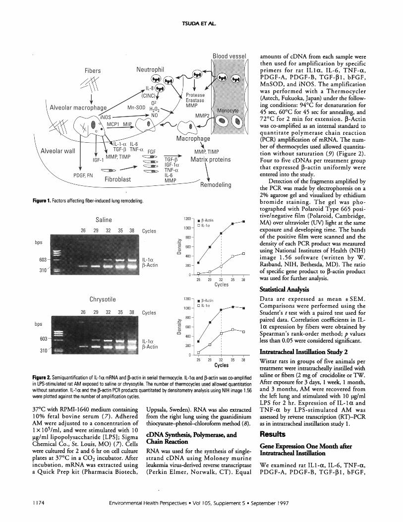
Even though the mechanism is notcompletely understood, evidence suggeststhat various factors are related to eachfibrotic process in the lung (Figure 1). It isimportant to investigate among these factorsparameters useful for the risk assessment ofMMF and the kinetics of their expression inthe process of the lung remodeling.
Materials and MethodsFiber Preparation
The fibers used in this study were UnionInternationale Contre le Cancer (UICC)crocidolite asbestos (crocidolite), UICCchrysotile asbestos ([chrysotile]; potassiumoctatitanate whisker (TW), and alumina sili-cate refractory ceramic fibers (RCF) (1). Thecrocidolite preparation, measured usingscanning electron microscopy, had a geomet-ric mean diameter of ( 0.20 pm (SD 1.5)and a geometric mean length of 1.3 pm (SD2.3). For chrysotile, geometric mean diame-ter and geometric mean length were 0.085pm (SD 1.4) and 0.7 pm (SD 1.9), respec-tively. For TW, they were 0.41 pm (SD 1.5)and 2.8 pm (SD 2.0), respectively. For RCF,they were 1.2 pm (SD 1.7) and 9.6 pm(SD 1.9), respectively.
Intratracheal Instillation Study 1Ten-week-old male Wistar rats, in groups offive per treatment, were intratracheallyinstilled with saline or fiber (2 mg chrysotileor RCF). One month after tracheal instilla-tion, bronchoalveolar lavage (BAL) was per-formed using the left lung. The cellsrecovered from BAL were plated in tissue cul-ture plates and allowed to attach for 1 hr at
Environmental Health Perspectives * Vol 105, Supplement 5 * September 1997
.l
1 173
TSUDA ET AL.
Figure 1. Factors affecting fiber-induced lung remodeling.
Saline26 29 32 35 38 Cycles
bps ,
IL-la,-Actin
Chrysotile
Cycles
IL-laP-Actin
Cycles
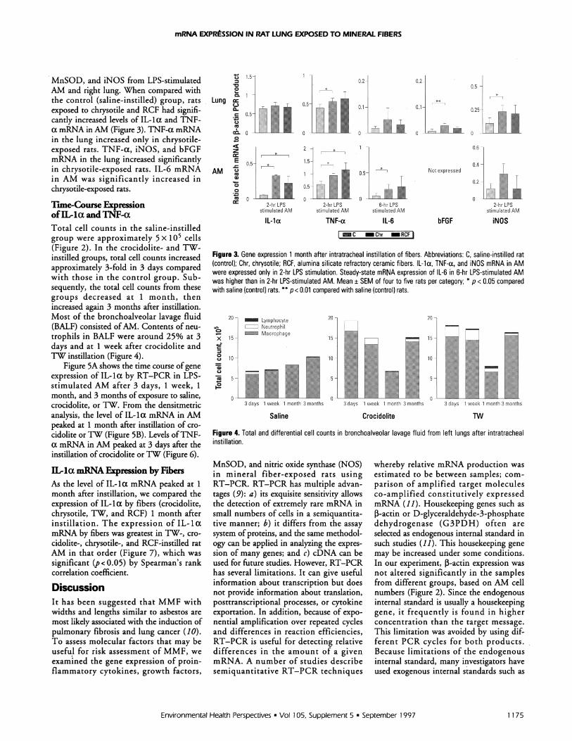
Figure 2. Semiquantification of IL-la mRNA and 3-actin in serial thermocycle. IL-la and P-actin was co-amplifiedin LPS-stimulated rat AM exposed to saline or chrysoytile. The number of thermocycles used allowed quantitationwithout saturation. IL-i a and the 1-actin PCR products quantitated by densitometry analysis using NIH image 1.56were plotted against the number of amplification cycles.
37°C with RPMI-1640 medium containing10% fetal bovine serum (7). AdheredAM were adjusted to a concentration of1 x 105/ml, and were stimulated with 10pg/ml lipopolysaccharide [LPS]; SigmaChemical Co., St. Louis, MO) (7). Cellswere cultured for 2 and 6 hr on cell cultureplates at 37°C in a CO2 incubator. Afterincubation, mRNA was extracted usinga Quick Prep kit (Pharmacia Biotech,
Uppsala, Sweden). RNA was also extractedfrom the right lung using the guanidiniumthiocyanate-phenol-chloroform method (8).
cDNA Synthesis, Polymerase, andChain ReactionRNA was used for the synthesis of single-strand cDNA using Moloney murineleukemia virus-derived reverse transcriptase(Perkin Elmer, Norwalk, CT). Equal
amounts of cDNA from each sample werethen used for amplification by specificprimers for rat ILIa, IL-6, TNF-a,PDGF-A, PDGF-B, TGF-f13, bFGF,MnSOD, and iNOS. The amplificationwas performed with a Thermocycler(Astech, Fukuoka, Japan) under the follow-ing conditions: 94°C for denaturation for45 sec, 60°C for 45 sec for annealing, and72°C for 2 min for extension. ,-Actinwas co-amplified as an internal standard toquantitate polymerase chain reaction(PCR) amplification of mRNA. The num-ber of thermocycles used allowed quantita-tion without saturation (9) (Figure 2).Four to five cDNAs per treatment groupthat expressed 1-actin uniformly wereentered into the study.
Detection of the fragments amplified bythe PCR was made by electrophoresis on a2% agarose gel and visualized by ethidiumbromide staining. The gel was pho-tographed with Polaroid Type 665 posi-tive/negative film (Polaroid, Cambridge,MA) over ultraviolet (UV) light at the sameexposure and developing time. The bandsof the positive film were scanned and thedensity of each PCR product was measuredusing National Institutes of Health (NIH)image 1.56 software (written by W.Rasband, NIH, Bethesda, MD). The ratioof specific gene product to J-actin productwas used for further analysis.
Statistcl AnalysisData are expressed as mean ± SEM.Comparisons were performed using theStudent's t test with a paired test used forpaired data. Correlation coefficients in IL-la expression by fibers were obtained bySpearman's rank-order method; p valuesless than 0.05 were considered significant.
Inratracheal Instillation Study 2Wistar rats in groups of five animals pertreatment were intratracheally instilled withsaline or fibers (2 mg of crocidolite or TW.After exposure for 3 days, 1 week, 1 month,and 3 months, AM were recovered fromthe left lung and stimulated with 10 pg/mlLPS for 2 hr. Expression of IL-la andTNF-a by LPS-stimulated AM wasassessed by reverse transcription (RT)-PCRas in intratracheal instillation study 1.
ResultsGene Expression One Month afterIntratracheal Instillaton
We examined rat ILl-a, IL-6, TNF-a,PDGF-A, PDGF-B, TGF-31, bFGF,
Environmental Health Perspectives * Vol 105, Supplement 5 * September 1997
603-
310 /
bps
603-
310'
1 174
mRNA EXPRtSSION IN RAT LUNG EXPOSED TO MINERAL FIBERS
MnSOD, and iNOS from LPS-stimulatedAM and right lung. When compared withthe control (saline-instilled) group, ratsexposed to chrysotile and RCF had signifi-cantly increased levels of IL-la and TNF-a mRNA in AM (Figure 3). TNF-a mRNAin the lung increased only in chrysotile-exposed rats. TNF-a, iNOS, and bFGFmRNA in the lung increased significantlyin chrysotile-exposed rats. IL-6 mRNAin AM was significantly increased inchrysotile-exposed rats.
rlme-Cou ExpressionofIL-la and TNF-aTotal cell counts in the saline-instilledgroup were approximately 5 x 10⁵ cells(Figure 2). In the crocidolite- and TW-instilled groups, total cell counts increasedapproximately 3-fold in 3 days comparedwith those in the control group. Sub-sequently, the total cell counts from thesegroups decreased at 1 month, thenincreased again 3 months after instillation.Most of the bronchoalveolar lavage fluid(BALF) consisted ofAM. Contents of neu-trophils in BALF were around 25% at 3days and at 1 week after crocidolite andTW instillation (Figure 4).
Figure 5A shows the time course of geneexpression of IL-la by RT-PCR in LPS-stimulated AM after 3 days, 1 week, 1month, and 3 months of exposure to saline,crocidolite, or TW. From the densitmetricanalysis, the level of IL-la mRNA in AMpeaked at 1 month after instillation of cro-cidolite orTW (Figure SB). Levels ofTNF-a mRNA in AM peaked at 3 days after theinstillation of crocidolite orTW (Figure 6).
IL-la mRNA Expression by FibersAs the level of IL-la mRNA peaked at 1month after instillation, we compared theexpression of IL-la by fibers (crocidolite,chrysotile, TW, and RCF) 1 month afterinstillation. The expression of IL-lamRNA by fibers was greatest in TW-, cro-cidolite-, chrysotile-, and RCF-instilled ratAM in that order (Figure 7), which wassignificant (p < 0.05) by Spearman's rankcorrelation coefficient.
DiscussionIt has been suggested that MMF withwidths and lengths similar to asbestos aremost likely associated with the induction ofpulmonary fibrosis and lung cancer (10).To assess molecular factors that may beuseful for risk assessment of MMF, weexamined the gene expression of proin-flammatory cytokines, growth factors,
1.2
co
A M5-1.5
,J0.4AM 0Not expressed
14- ~~ ~ ~ ~~i0.20 ~~0.5 j-
2-hr LPS 2-hr LPS 6-hr LPS 2-hr LPSstimulated AM stimulatedAM stmulated AMstimulated AM
IL-la TNF-a IL-6 bFGF iNOS
Figure 3. Gene expression 1 month after intratracheal instillation of fibers. Abbreviations: C, saline-instilled rat(control); Chr, chrysotile; RCF, alumina silicate refractory ceramic fibers. IL-la, TNF-a, and iNOS mRNA in AMwere expressed only in 2-hr LPS stimulation. Steady-state mRNA expression of IL-6 in 6-hr LPS-stimulated AMwas higher than in 2-hr LPS-stimulated AM. Mean ± SEM of four to five rats per category; * p < 0.05 comparedwith saline (control) rats. ** p< 0.01 compared with saline (control) rats.
20- Lymphocyte 20- 20-Ln NeutrophilCD ~~~Macrophage
x 15 15 15
0 c, 10- 10- 10-
5 5- 5-
0
3 days 1 week 1 month 3 months 3 days 1 week 1 month 3 months 3 days 1 week 1 month 3 months
Saline Crocidolite TW
Figure 4. Total and differential cell counts in bronchoalveolar lavage fluid from left lungs after intratrachealinstillation.
MnSOD, and nitric oxide synthase (NOS)in mineral fiber-exposed rats usingRT-PCR. RT-PCR has multiple advan-tages (9): a) its exquisite sensitivity allowsthe detection of extremely rare mRNA insmall numbers of cells in a semiquantita-tive manner; b) it differs from the assaysystem of proteins, and the same methodol-ogy can be applied in analyzing the expres-sion of many genes; and c) cDNA can beused for future studies. However, RT-PCRhas several limitations. It can give usefulinformation about transcription but doesnot provide information about translation,posttranscriptional processes, or cytokineexportation. In addition, because of expo-nential amplification over repeated cyclesand differences in reaction efficiencies,RT-PCR is useful for detecting relativedifferences in the amount of a givenmRNA. A number of studies describesemiquantitative RT-PCR techniques
whereby relative mRNA production wasestimated to be between samples; com-parison of amplified target moleculesco-amplified constitutively expressedmRNA (11). Housekeeping genes such asP-actin or D-glyceraldehyde-3-phosphatedehydrogenase (G3PDH) often areselected as endogenous internal standard insuch studies (11). This housekeeping genemay be increased under some conditions.In our experiment, P-actin expression wasnot altered significantly in the samplesfrom different groups, based on AM cellnumbers (Figure 2). Since the endogenousinternal standard is usually a housekeepinggene, it frequently is found in higherconcentration than the target message.This limitation was avoided by using dif-ferent PCR cycles for both products.Because limitations of the endogenousinternal standard, many investigators haveused exogenous internal standards such as
Environmental Health Perspectives * Vol 105, Supplement 5 * September 1997 1 175

B0.2 -
IL-la (623 bp)tin (357 bp) .'
Co0
d 0.1-
IL-laJ-Actin °
0
Co1
0
IL-la3-Actin
3 days 1 week 1 month 3 months
Figure 5. Time-course of expression of IL-la mRNA in LPS-stimulated rat AM exposed to mineral fibers. (A) Ethidium bromide staining of PCR products separated in 2%agarose gel. The 623-bp products for IL-la and the 357-bp products for f-actin are indicated. LPS- stimulated AM from four to five rats per treatment per time point wereexamined. (B) Levels of IL-1 a mRNA in AM exposed to mineral fibers. Results are expressed as the ratio of IL-1 a to P-actin (mean ± SEM).
To find more sensitive and specificTW parameters of lung remodeling by fibers,
many factors that may contribute to the
2o0.4 crocidolte \process must be considered. RT-PCR may0.4 -4 Crocidolitebe a suitable method for surveying possiblez VL\ w K) , parameters of fiber-induced lung remodeling.I--
0.2- Salin We chose chrysotile and RCF for intra-.2 1 tracheal instillation study 1. Thoughcc chrysotile can induce lung cancer (fibrosis
o - and mesothelioma), it is one of the least3days 1 week 1 month 3 months toxic asbestos fibers. There is considerable
Figure 6. Levels of TNFa mRNA in exposed to mineral evidence for the fibrogenicity and carcino-fibers. Results are expressed in ratio of TNF-a to 5 genicity of RCF in laboratory animals, com-actin(mean+SEM). pared to that for other MMF. (13). For
intratracheal study 2, we chose more fibro-0.2 genic fibers, UICC crocidolite and TW, to
detect time-course change of gene expres-C., sion. TW produces marked pulmonaryXF 0.15- fibrosis in rats with long-term exposure (14).
In the previous study, unstimulated AM0.1o _ T obtained from rats treated with chrysotile
did not significantly enhance steady-state0 levels of IL-la mRNA (data not shown).
'r 0.05-co T * - - - AM harvested from rat lung did not express
IL-1 protein, and LPS treatment of quies-C Cro cent cells (after 24-hr in vitro culture)c CroChr 1W RCF induced low-level expression of IL-la and
Figure 7. Levels of IL-la mRNA by fiber. Abbreviations: IL-13 (15). Short-term inhalation of RCFC, control, Cro, crocidolite; Chr, chrysotile; TW: potas- resulted in markedly increased IL-10 pro-
sium octatitanate whisker; RCF; alumina silicate tein expression after stimulation with LPSceramic fibers. Results are expressed in ratio of IL-la (15). In vivo exposure of AM to LPSto fP-actin (mean ± SEM). increased proinflammatory cytokine
mRNA, although the kinetics of upregula-synthetic RNA sequence or a synthetic tion varied (16). For these reasons, we
DNA sequence that is not present in the tar- examined mRNA expression in 2 and 6 hrget sample (12). Nevertheless, the house- LPS-stimulated AM. As a cautionary note,
keeping gene is very useful in controlling for results from ex vivo LPS-stimulated AMdifferences in RNA loading and for assess- may not necessarily indicate a role for a
ing differences in the quality ofRNA (12). stimulated cytokine in the pathogenesis of
inflammation associated with exposure to
fibers in vivo.In intratracheal study 1, AM exposed to
chrysotile or RCF were found to haveupregulated IL-la, TNF-a and IL-6mRNA transcripts in response to LPS.These are proinflammatory cytokines withboth inflammatory and fibrogenic activitiessuch as attraction of inflammatory cells,production of superoxide and collagenases,and proliferation of fibroblasts (17). TNF-a mRNA and protein have been detectedin the lung from patients with idiopathicpulmonary fibrosis (18) and in lungs frommice with pulmonary fibrosis elicited byexposure to bleomycin or silica (19).Increased release of IL-I from AM has beenreported after asbestos exposure by inhala-tion or intratracheal instillation (20). Ininflammatory reactions, IL-6 could act as
not only a proinflammatory cytokinebecause of its ability to induce the expres-sion of cellular adhesion molecules on
monocytes and the facilitation of theirinfiltration into the lung, but also as an
antiinflammatory cytokine that inhibits theproduction of TNF and IL-1 (21). Anincrease has been reported in IL-6 releasedby bronchoalveolar cells from rats treatedwith asbestos or coal mineral dust (22).
Oxidants produced by inflammatorycells are thought to lead to lung injury inpulmonary fibrosis. Nitric oxide synthase(NOS) produces reactive species such as
nitric oxide (NO0) and peroxynitrite anion.NO and peroxynitrate are also cytotoxic to
host parenchymal cells (4). In our model,levels of iNOS mRNA increased in-lungsexposed to chrysotile.
Environmental Health Perspectives * Vol 105, Supplement 5 * September 1997
A
TSUDA ET AL.
I
ker
3 days 1 week 1 month 3 months
P-Aci
1 176

mRNA EXPRESSION IN RAT LUNG EXPOSED TO MINERAL FIBERS
Among the FGFs, bFGF stimulatedthe replication of endothelial cells in vitroand new microvessel growth in vivo (23).Asbestos exposure induced lavaged cells tosecrete a fibroblast growth factor from 1to 24 weeks after exposure in rats (24). Inlungs exposed to chrysotile, we foundincreased levels of bFGF mRNA. Basedon these results, IL-la in the AM, TNF-ain the AM and the lung, IL-6 in the AM,and iNOS and bFGF in the lung wouldbe the possible parameters of risk assess-ment of man-made fibers in this model.Accordingly, we set out to further investi-gate the time course of expression of IL-la and TNF-a mRNA from exposed tocrocidolite and TW.
As previously reported for rats (24),total cell count of BALF at 1 month afterchrysotile or TW instillation was almost thesame as the control group and increased at3 months in the present study. Crocidoliteor TW instillation resulted in pulmonaryinflammation as evidenced by increased
numbers of BALF neutrophils and macro-phages at 3 days and 1 week after theexposure. Consistent with the acute inflam-mation, levels of TNF-a mRNA weregreatest at 3 days after the exposure anddecreased thereafter. In contrast to TNF-a,levels of IL-la mRNA peaked at 1 monthafter crocidolite orTW expossure.
There have been many attempts topredict the toxicity of mineral fibers basedon cytotoxic potentials of fibers using avariety of cell types in vitro. In an in vitrostudy using AM, TW caused the highestlevel of TNF-a production among fibers(1). This is consistent with the presentstudy on TNF-a mRNA expression 3days after instillation. TNF-a mRNAexpression in TW-instilled animals washigher than that in crocidolite-instilledanimals. Lee et al. (25) reported that cro-cidolite was the most potent fibrogenicagent and was 10 times more fibrogenicthan potassium octatitanate (Fybex) interms of exposure concentration In our
study, the expression of IL-la by fiberchallenge (TW > crocidolite > chrysotile >RCF) may correlate with the reportedpathologic potential of fibers (25,26).Accordingly, our approach may be usefulfor evaluating the potential toxicity ofnewly developed man-made fiber. Furtherinvestigations using other fibers are neces-sary to confirm the general applicability ofthe method.
Both TNF-a and IL-la are proinflam-matory cytokines. It is necessary to demon-strate that acute inflammation completelypredicts the chronic change induced byfibers. As TNF-a plays a key role in lungremodeling (27), further investigations onthe correlation between proinflammatorycytokines and the order of the toxicity offibrous materials are also required. Alongthis line, correlations between gene expres-sion and pathologic changes induced byfibrous materials may prove to be a power-ful approach for assessing health risks dueto fiber exposure.
REFERENCES
1. Fujino A, Hori H, Higahi T, Morimoto Y, Tanaka I, Kaji H.In-vitro biological study evaluates the toxic potentials of fibrousmaterials. Int J Occup Environ Health 1:21-28 (1995).
2. Driscoll KE, Maurern JK, Hassenbein D, Carter J, JansenMWY, Mossman BT, Osier M, Oberdorster G. Contributionof macrophage-derived cytokines and cytokine networks tomineral dust-induced lung inflammation. In: Toxic andCarcinogenic Effects of Solid Particles in Respiratory Tract(Mohr U, Dungworth DL, Mauderly JL, Orberdorster G, eds).Washington:ILSI Press, 1994;170-190.
3. Ward PA, Mulligan MS. New insights into mechanisms ofoxyradical and neutrophil mediated lung injury. KlinWochenschr 69:1009-1011 (1991).
4. Warner RL, Paine R 3rd, Christensen PJ, Marletta MA,Richards MK, Wilcoxen SE, Ward PA. Lung sources andcytokine requirements for in vivo expression of inducible nitricoxide synthase. Am J Respir Cell Mol Biol 12(6):649-661(1995).
5. Vanhee D, Gosset P, Wallaert B, Voisin C, Tonnel AB.Mechanisms of fibrosis in coal workers' pneumoconiosis.Increased production of platelet-derived growth factor, insulin-like growth factor type 1, and transforming growth factor andrelationship to disease severity. Am J Respir Crit Care Med150:1049-1055 (1994).
6. Folkman J, Klagsbrun M. Angiogenic factors. Science 235:442-447 (1987).
7. Jordana M, Richards C, Irving LB, Gavldie J. Spontaneous invitro release of alveolar-macrophage cytokines after the intratra-cheal instillation of bleomycin in rats. Am Rev Respir Dis137:1135-1140 (1988).
8. Chomczynski P, Sacchi N. Single step method of RNA isola-tion by acid guanidium thicyanate-phenol-chloroform extrac-tion. Anal Biochem 162:156-159 (1987).
9. Weisner RJ, Zac R. Quantitative approaches for studying geneexpression. Am J Physiol 260:L179-L188 (1991).
10. Pott F. Testing the carcinogenicity of fibers in laboratory ani-mals: results and conclusions. In: Fiber Toxicology (WarheitDB, ed.) San Diego:Academic Press, 1993; 395-424.
11. Mohler KM, Butler LD. Quantification of cytokine mRNAlevels utilizing the reverse transcriptase-polymerase chain reac-tion following primary antigen specific sensitization in vivo.Molec Immunol 28:437-447 (1991).
12. Zamorano PL, Mahesh VB, Brann DW. Quantative RT-PCRfor neuroendocrine studies. Neuroendocrinology 63:397-407(1996).
13. Hesterberg TW, Miiller WC, Thevenaz P, Anderson R.Chronic inhalation studies of man-made vitreous fibres: charac-terization of fibres in the exposure aerosol and lungs. AnnOccup Hyg 5:637-653 (1995).
14. Lee KP, Barras CE, Griffith FD, Waritz RS. Pulmonaryresponse and transmigration of inorganic fibers by inhalationexposure. Am J Pathol 102:314-323 (1981).
15. Miller K, Hudpith BN, Meredith C. Secretory and accessorycell function of alveloar macrophage. Environ Health Perspect97:85-89 (1992).
16. Xing Z, Jordana M, Kirpalani H, Driscoll KE, Schall TJ,Gauldie J. Cytokine expression by neutrophil and macrophagesin vzvo: endotoxin induces tumor necrosis factor-a, macrophageinflammatory protein-2, interleukin-1I , and interleukin-6 butnot RANTES or transforming growth factor-P31 mRNA expres-sion in acute lung inflammation. Am J Respir Cell Mol Biol10:148-153 (1994).
17. Strieter RM, Phan SH, Showell HJ, Remick DG, Lynch JP,Genord M, Raiford C, Eskandari M, Marks RM, Kunkel SL.Monokine-induced neutrophil chemotactic factor gene expres-sion in human fibroblasts. J Biol Chem 264(18):10621-10626(1989).
18. Zhang Y, Lee TC, Guillemin B, Yu MC, Rom WN. EnhancedIL-1i and tumor necrosis factor-a release and messenger RNAexpression in macrophages from idiopathic pulmonary fibrosis orafter asbestos exposure. J Immunol 150(9):4188-4196 (1993).
19. Piguet PF,Vesin C. Treatment by human recombinant solubleTNF receptor of pulmonary fibrosis induced by bleomycin orsilica in mice. Eur Respir J 7(3):515-518 (1994).
20. Lemaire I, Beaudoin H, Masse S, Grondin C. Alveolarmacrophage stimulation of lung fibroblast growth in asbestos-
Environmental Health Perspectives * Vol 105, Supplement 5 * September 1997 1177

TSUDA ETAL
induced pulmonary fibrosis. Am J Pathol 122:205-211 (1986).21. Schindler R, Mancilla J, Endres S, Ghorbani R, Clark SC,
Dinarello CA. Correlations and interactions in the productionof interleukin-6 (IL-6), IL-1, and tumor necrosis factor (TNF)in human blood mononuclear cells: IL-6 suppresses IL-1 andTNF. Blood 75(1):40-47 (1990).
22. Gosset P, Lassalle P, Vanhee D, Wallaert B, Aerts C, Voisin C,Tonnel AB. Production of tumor necrosis factor-a and inter-leukin-6 by human alveolar macrophages exposed in vitro to coalmine dust. Am J Respir Cell Mol Biol 5(5):431-436 (1991).
23. Shing Y, Folkman J, Haudenschild C, Lund D, Crum R,Klagsbrun M. Angiogenesis is stimulated by a tumor-derivedendothelial cell growth factor. J Cell Biochem 29:275-287(1985).
24. Lemaire I, Beaudoin H, Dubois C. Cytokene regulation of
lung fibroblast proliferation. Am Rev Respir Dis 134:653-658(1986).
25. Lee KP, Barras CE, Griffith FD, Waritz RS, Lapin CA.Comparative pulmonary response to inhaled inorganic fiberswith asbestos and fiberglass. Environ Res 24:167-191(1981).
26. Hesterberg T, Chase G, Versen R, Anderson R. Studies toassess the carcingenic potential of man-made viterous fibers. In:Toxicology of Industrial Compounds (Thomas H, Hess R,Waechter F, eds). Bristol, PA:Taylor and Francis, 1995;93-117.
27. Miyazaki Y, Araki K, Vesin C, Garcia I, Kapanci Y, WhitsettJA. Expression of a tumor necrosis factor-a transgene inmurine lung causes lymphocytic and fibrosing alveolitis. Amouse model of progressive pulmonary fibrosis. J Clin Invest96:250-259 (1995).
1178 Environmental Health Perspectives * Vol 105, Supplement 5 * September 1997