Embed Size (px)
Citation preview
Efficacy estimation of erythropoiesis-stimulating agents using erythropoietin-deficiency anemic mice
by Norio Suzuki, Yusuke Sasaki, Koichiro Kato, Shun Yamazaki, Mitsue Kurasawa, Keigo Yorozu, Yasushi Shimonaka, and Masayuki Yamamoto
Haematologica 2016 [Epub ahead of print]
Citation: Suzuki N, Sasaki Y, Kato K, Yamazaki S, Kurasawa M, Yorozu K, Shimonaka Y, and Yamamoto M. Efficacy estimation of erythropoiesis-stimulating agents using erythropoietin-deficiencyanemic mice. Haematologica. 2016; 101:xxxdoi:10.3324/haematol.2015.140814
Publisher's Disclaimer.E-publishing ahead of print is increasingly important for the rapid dissemination of science.Haematologica is, therefore, E-publishing PDF files of an early version of manuscripts thathave completed a regular peer review and have been accepted for publication. E-publishingof this PDF file has been approved by the authors. After having E-published Ahead of Print,manuscripts will then undergo technical and English editing, typesetting, proof correction andbe presented for the authors' final approval; the final version of the manuscript will thenappear in print on a regular issue of the journal. All legal disclaimers that apply to thejournal also pertain to this production process.
Copyright 2016 Ferrata Storti Foundation.Published Ahead of Print on May 31, 2016, as doi:10.3324/haematol.2015.140814.
Suzuki et al
1
Efficacy estimation of erythropoiesis-stimulating agents using
erythropoietin-deficiency anemic mice
Norio Suzuki1*, Yusuke Sasaki2, Koichiro Kato1, Shun Yamazaki1,3,
Mitsue Kurasawa2, Keigo Yorozu2, Yasushi Shimonaka2, and Masayuki Yamamoto3*
1 Division of Oxygen Biology, Tohoku University Graduate School of Medicine, Sendai,
Japan; 2 Product Research Department, Chugai Pharmaceutical Co., Ltd., Kamakura, Japan; 3 Department of Medical Biochemistry, Tohoku University Graduate School of
Medicine, Sendai, Japan
*Corresponding author:
Norio Suzuki and Masayuki Yamamoto
Division of Oxygen Biology, Tohoku University Graduate School of Medicine, 2-1
Seiryo-machi, Aoba-ku, Sendai 980-8575, Japan
E-mail: [email protected]
Tel: +81-22-717-8206 / Fax: +81-22-717-8090
Manuscript information:
1,463 words in the text; 3 Figures (2 color figures); 15 references;
1 Supplementary Appendix (Methods, 3 Figures and 1 Table)
Suzuki et al
2
Erythropoietin (EPO) is an essential growth factor for red blood cell (RBC) production,
and it is mainly produced by renal EPO-producing (REP) cells in the kidneys in an
anemia/hypoxia-inducible manner.1,2 Erythropoiesis-stimulating agents (ESAs),
including recombinant human EPO (rHuEPO), have been used to treat EPO-deficiency
anemia in kidney disease patients for a quarter century.3,4 Due to the short plasma
half-life of rHuEPO (approximately 1 day after subcutaneous injection), renal anemia
patients require rHuEPO injections every 2 or 3 days to maintain their RBC count at
non-anemic levels.3 Recently, long-acting ESAs, Darbepoetin alpha (DA, genetically
modified EPO) and continuous EPO receptor (EPOR) activator (C.E.R.A., chemically
modified rHuEPO), have been developed, with plasma half-lives of approximately 2
days and 5 days, respectively, after subcutaneous injection.3,4 Because of the lack of
suitable animal models of EPO-deficiency anemia, it has been difficult to elucidate the
detailed profiles of ESA-induced erythropoiesis in vivo. We recently generated a
genetically modified mouse model of EPO-deficiency anemia, inherited super anemia
mouse/mice (ISAM, EpoGFP/GFP:Tg3.3K-EpoE3 genotype).5 Using ISAM, we were able to
obtain comparable measurements of the efficacies of 3 ESAs and demonstrated that the
efficacies of these agents on erythropoiesis and iron metabolism depend on their plasma
half-lives.
ISAM exhibit severe normocytic-normochromic anemia due to the loss of
renal EPO production.5 The EPO-deficiency anemia in ISAM begins approximately 2
weeks after birth, when the major site of EPO production switches from the liver to the
kidneys.5,6 We first confirmed that the hematocrit values and hemoglobin concentrations
Suzuki et al
3
in the peripheral blood of ISAM were decreased to half of those in control mice at 4
weeks of age (Online Supplementary Figure S1A). Iron concentrations in the serum and
liver of ISAM were higher than those in control mice, suggesting that iron usage for
erythropoiesis was suppressed due to anemia in ISAM. Consistently, the unsaturated
iron binding capacity of transferrin (UIBC) in the peripheral blood was decreased, and
the serum level of hepcidin, a peptide hormone that inhibits iron entry into circulation,7.8
was increased in ISAM. Additionally, we found that EPO deficiency seems to be
indirectly related to both systemic hypoxia and cardiomegaly through chronic severe
anemia in mature ISAM (Online Supplementary Figure S1B, C).
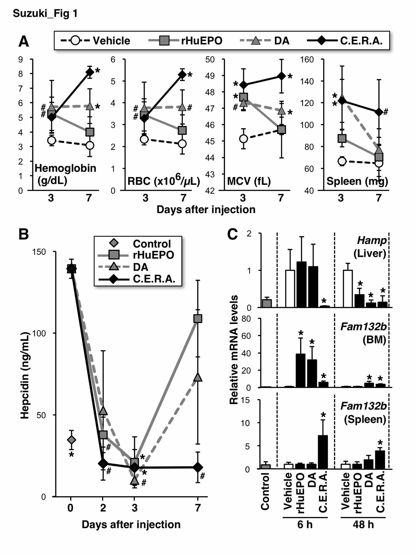
Three representative ESAs (rHuEPO, DA and C.E.R.A.) were subcutaneously
injected into ISAM at 3.0 µg of EPO-peptide weight per 1-kg body weight (BW), a
comparable dose as in clinical use. Three days after single injections of each ESA
(day-3), the hemoglobin and RBC concentrations in the peripheral blood of ISAM were
increased, as were the mean corpuscular volume (MCV) of RBCs and the weight of the
spleen, the major site of EPO-inducible erythropoiesis in mice (Figure 1A).9 These data
indicate that these 3 ESAs similarly induce erythropoiesis in ISAM on day-3. Although
C.E.R.A. further increased the levels of hemoglobin and RBC during the last 4 days of
observation, the erythropoietic effects of rHuEPO or DA were eliminated on day-7
(Figure 1A). Thus, the profiles of erythropoietic induction by ESAs largely depend on
their plasma half-lives.
The elevated concentration of serum hepcidin in ISAM was strongly
decreased to levels less than those in normal mice on day-3 (Figure 1B). At day-7,
Suzuki et al
4
C.E.R.A. continued to suppress hepcidin levels, whereas hepcidin levels re-increased in
ISAM injected with DA or rHuEPO. The circulating hepcidin concentration is
fundamentally regulated at the gene (Hamp) transcription level in hepatocytes.7,8,10 Each
ESA significantly suppressed Hamp mRNA expression in ISAM livers 48 hours after
administration (Figure 1C). At 6 hours after administration, C.E.R.A. dramatically
decreased Hamp mRNA levels, whereas rHuEPO or DA administration did not change
the induced Hamp levels. Hepatic Hamp expression is strongly suppressed by
erythroferrone, which is secreted by erythroblasts immediately after EPO
stimulation.7,11 In the hematopoietic organs of ISAM, erythroferrone (Fam132b) gene
expression was induced in 6 hours after C.E.R.A. administration, and the induced levels
were maintained 48 hours after administration (Figure 1C). DA administration induced
Fam132b mRNA expression in the bone marrow of ISAM 6 and 48 hours after
administration, suggesting different organ distributions of ESAs. In fact, rHuEPO
induced Fam132b expression in the bone marrow of ISAM 6 hours after administration
but not in the bone marrow and spleen 48 hours after administration (Figure 1C). These
data suggest that EPO gradually and persistently suppresses hepatic hepcidin production
through the quick and transient induction of erythroblastic erythroferrone production
because rHuEPO decreases the elevated hepcidin levels in ISAM on day-2 and day-3
but not at 6 hours after administration (see Figure 1C). Although the mechanism of
erythroferrone-mediated hepcidin suppression is unknown, the efficacies of shorter
half-life ESAs on hepcidin suppression are weaker than that of C.E.R.A. by the
single-dose injection.
Suzuki et al
5
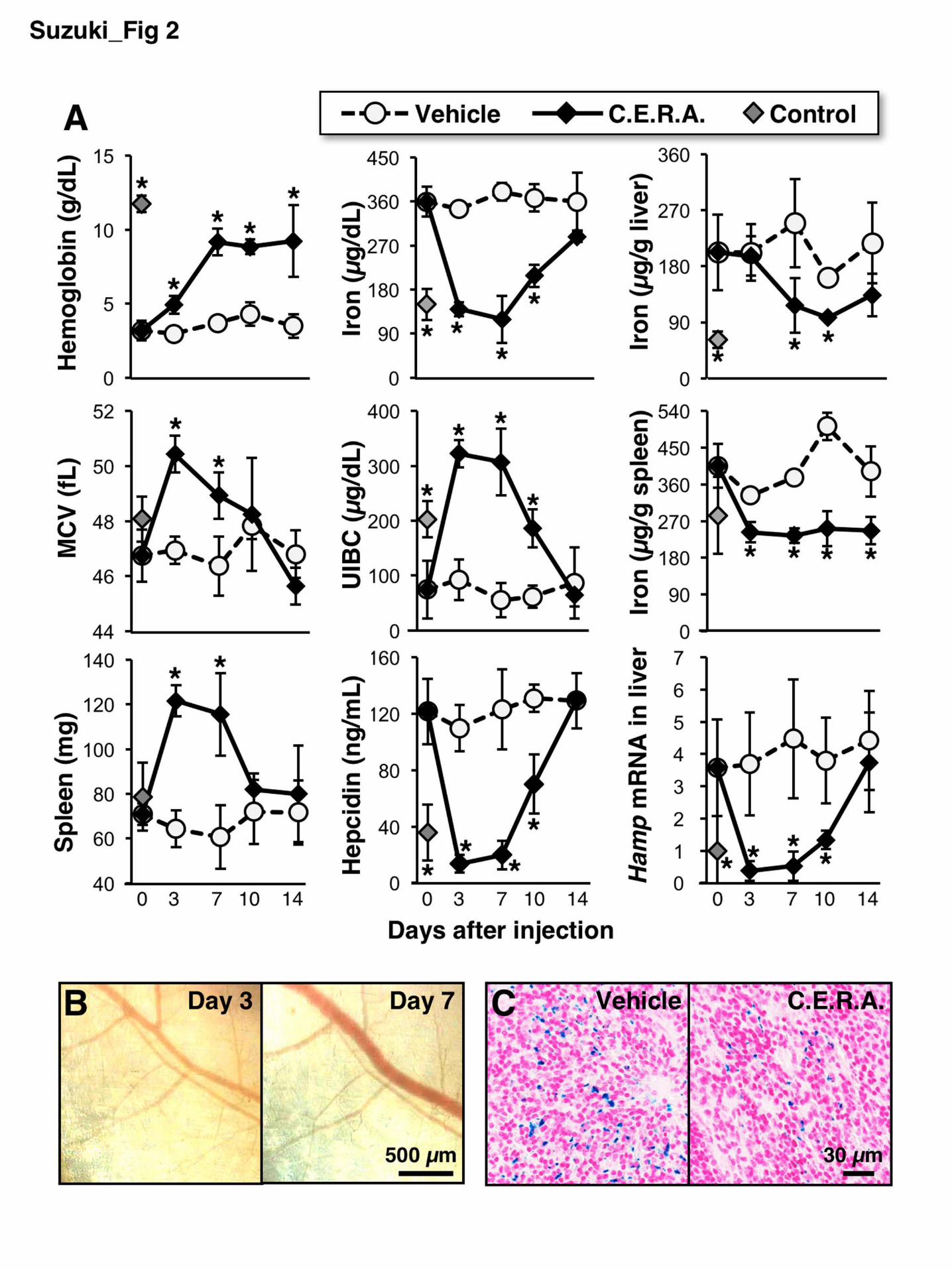
The hemoglobin levels of ISAM was continuously increased for 7 days after
administration, and the increased level was maintained for an additional 7 days (Figure
2A). Indeed, fixed-point images of the ISAM back skin showed that the thin blood
vessels of ISAM were filled with RBCs on day-7 (Figure 2B). Because both the MCV
and spleen weights returned to the basal levels of the untreated ISAM at day-10 (Figure
2A), we concluded that C.E.R.A. continuously stimulates erythropoiesis for one week
after administration.
C.E.R.A. dramatically reduced serum iron concentrations to the level of
normal mice at day-3 (Figure 2A). Because of the rapid reduction in serum iron
concentrations, the UIBC of ISAM was higher than that of control mice on day-3 and
day-7. The altered levels of both serum iron and UIBC returned to untreated-ISAM
levels on day-7. The accumulated iron in the spleens and livers of ISAM were decreased
on day-3 and day-7, respectively (Figure 2A). Berlin blue staining also revealed
decreased iron deposits in the ISAM spleens on day-7 (Figure 2C). Because the splenic
iron was used before the hepatic iron was used, local splenic iron storage may be
predominantly utilized for erythropoiesis instead of hepatic iron. The reduced serum
hepcidin concentrations and hepatic Hamp mRNA expression in ISAM treated with
C.E.R.A. were comparable to those of control mice between day-3 and day-7 and
re-increased to their original levels at day-14 (Figure 2A).
We then investigated the cardiomegaly and systemic hypoxia in ISAM after
C.E.R.A. administration. ISAM hearts were enlarged due to severe anemia at 12 weeks
of age, and C.E.R.A. administration decreased the size and weight within 7 days (Figure
Suzuki et al
6
3A,B). The anemia phenotype was reversed 28 days after a single dose of C.E.R.A.
(Online Supplementary Figure S2A), and cardiomegaly developed again in ISAM
(Figure 3C). These data indicate that severe anemia reversibly causes cardiomegaly in
mice. A systemic hypoxic milieu emerged as a result of the severe anemia in ISAM, and
the expression levels of the hypoxia-inducible genes Egln3 and Slc2a312 were
significantly higher in the hearts and kidneys, respectively, of vehicle-treated ISAM
than in those of the control mice (Figure 3D). The induced gene expression levels were
reduced to the levels of control mice on day-10 (Figure 3D). These therapeutic effects
were similarly observed in rHuEPO-treated ISAM when the hemoglobin level was
increased to the normal range by 4-time injections for 7 days (Online Supplementary
Figure S3).
In the ISAM-REC mice (EpoGFP/GFP:Tg3.3K-EpoE3:Rosa26LSL-tdTomato:TgEpoCre
genotype), tdTomato expression permanently labels all REP cells, and EpoGFP
expression is a marker for REP cells in which the transcription of the Epo allele is
activated.5 To characterize REP cells under stable non-anemic conditions, C.E.R.A. was
injected into ISAM every week for 4 weeks. The hematocrit values of ISAM remained
in the normal range after the second injection (Online Supplementary Figure S2B), and
EpoGFP mRNA expression was dramatically decreased, whereas the expression of
REP-cell markers (tdTomato and Pdgfrb) were unaffected on day-28 (Figure 3E).13 The
expression of the hypoxia-inducible Adm (Adrenomedullin) gene in ISAM kidneys was
significantly reduced by the weekly C.E.R.A. administration,12 indicating that the
hypoxic milieu of the ISAM kidneys was ameliorated. Analyses of tissue sections from
Suzuki et al
7
the ISAM-REC kidneys also demonstrated that the increased expression of EpoGFP in
the REP cells of ISAM disappeared after the weekly C.E.R.A. administration, without a
loss of tdTomato-positive REP cells (Figure 3F). These results demonstrate that Epo
transcription is activated in the REP cells, which sense hypoxia/anemia, and that the
total number of REP cells is stable in the kidneys regardless of the oxygen conditions.
This study proposes that ISAM provides a remarkable experimental system to
assess ESA efficacy and to elucidate the in vivo mechanisms of erythropoiesis that are
linked to iron metabolism. The increased serum and tissue iron levels, which are
considered the source of cytotoxic hydroxyl radicals,14 were decreased immediately and
sustainably after C.E.R.A. administration. Both the hypoxic milieu and cardiomegaly in
ISAM were ameliorated by ESA administration. Renal anemia in chronic kidney
diseases is often linked to chronic heart failure (cardio-renal-anemia syndrome,
CRAS).15 We propose that ESA may interfere with the CRAS linkage by inducing
erythropoiesis, and ISAM may help elucidate the molecular basis of CRAS.
Suzuki et al
8
Acknowledgements: We thank Atsuko Konuma, Aina Fukuda (Tohoku University) and
Yukari Matsuo-Tezuka (Chugai Pharmaceutical) for their technical support. We are also
grateful to Sakura Motion Picture Co., Ltd., the Biomedical Research Core, and the
Center for Laboratory Animal Research of Tohoku University for technical support.
Funding: This work was supported in part by Grants-in-Aid from MEXT/JSPS
KAKENHI (Grant Numbers 26111002 and 24249015 for MY; 26116702 and 25670157
for NS), the Platform for Drug Discovery, Informatics, and Structural Life Science from
MEXT, Japan (MY and NS), SENSHIN Medical Research Foundation (NS), Japan
Anti-Doping Agency (NS and MY) and Senri Life Science Foundation (NS). The
funders had no role in the study design, data collection and analysis, the decision to
publish or the preparation of the manuscript.
Suzuki et al
9
References
1. Suzuki N, Yamamoto M. Roles of renal erythropoietin-producing (REP) cells in the
maintenance of systemic oxygen homeostasis. Pflugers Arch. 2015;468(1):3-12.
2. Souma T, Nezu M, Nakano D, et al. Erythropoietin synthesis in renal
myofibroblasts is restored by activation of hypoxia signaling. J Am Soc Nephrol.
2015;27(2):428-438.
3. Macdougall IC, Robson R, Opatrna S, et al. Pharmacokinetics and
pharmacodynamics of intravenous and subcutaneous continuous erythropoietin
receptor activator (C.E.R.A.) in patients with chronic kidney disease. Clin J Am
Soc Nephrol. 2006;1(6):1211-1215.
4. Jelkmann W. The ESA scenario gets complex: from biosimilar epoetins to activin
traps. Nephrol Dial Transplant. 2015;30(4):553-559.
5. Yamazaki S, Souma T, Hirano I, et al. A mouse model of adult-onset anaemia due
to erythropoietin deficiency. Nat Commun. 2013;4:1950.
6. Suzuki N, Obara N, Pan X, et al. Specific contribution of the erythropoietin gene 3'
enhancer to hepatic erythropoiesis after late embryonic stages. Mol Cell Biol.
2011;31(18):3896-3905.
7. Koury MJ, Haase VH. Anaemia in kidney disease: harnessing hypoxia responses
for therapy. Nat Rev Nephrol. 2015;11(7):394-410.
8. Gutschow P, Schmidt PJ, Han H, et al. A competitive enzyme-linked
immunosorbent assay specific for murine hepcidin-1: correlation with hepatic
mRNA expression in established and novel models of dysregulated iron
homeostasis. Haematologica. 2015;100(2):167-177.
9. Xiang J, Wu DC, Chen Y, Paulson RF. In vitro culture of stress erythroid
progenitors identifies distinct progenitor populations and analogous human
progenitors. Blood. 2015;125(11):1803-1812.
10. Sasaki Y, Noguchi-Sasaki M, Yasuno H, Yorozu K, Shimonaka Y. Erythropoietin
stimulation decreases hepcidin expression through hematopoietic activity on bone
marrow cells in mice. Int J Hematol. 2012;96(6):692-700.
11. Kuhrt D, Wojchowski DM. Emerging EPO and EPO receptor regulators and signal
transducers. Blood. 2015;125(23):3536-3541.
Suzuki et al
10
12. Tausendschön M, Rehli M, Dehne N, et al. Genome-wide identification of
hypoxia-inducible factor-1 and -2 binding sites in hypoxic human macrophages
alternatively activated by IL-10. Biochim Biophys Acta. 2015;1849(1):10-22.
13. Pan X, Suzuki N, Hirano I, Yamazaki S, Minegishi N, Yamamoto M. Isolation and
characterization of renal erythropoietin-producing cells from genetically produced
anemia mice. PLoS One. 2011;6(10):e25839.
14. Arosio P, Levi S. Cytosolic and mitochondrial ferritins in the regulation of cellular
iron homeostasis and oxidative damage. Biochim Biophys Acta.
2010;1800(8):783-792.
15. Attanasio P, Ronco C, Anker SD, Cicoira M, von Haehling S. Role of iron
deficiency and anemia in cardio-renal syndromes. Semin Nephrol.
2012;32(1):57-62.
Suzuki et al
11
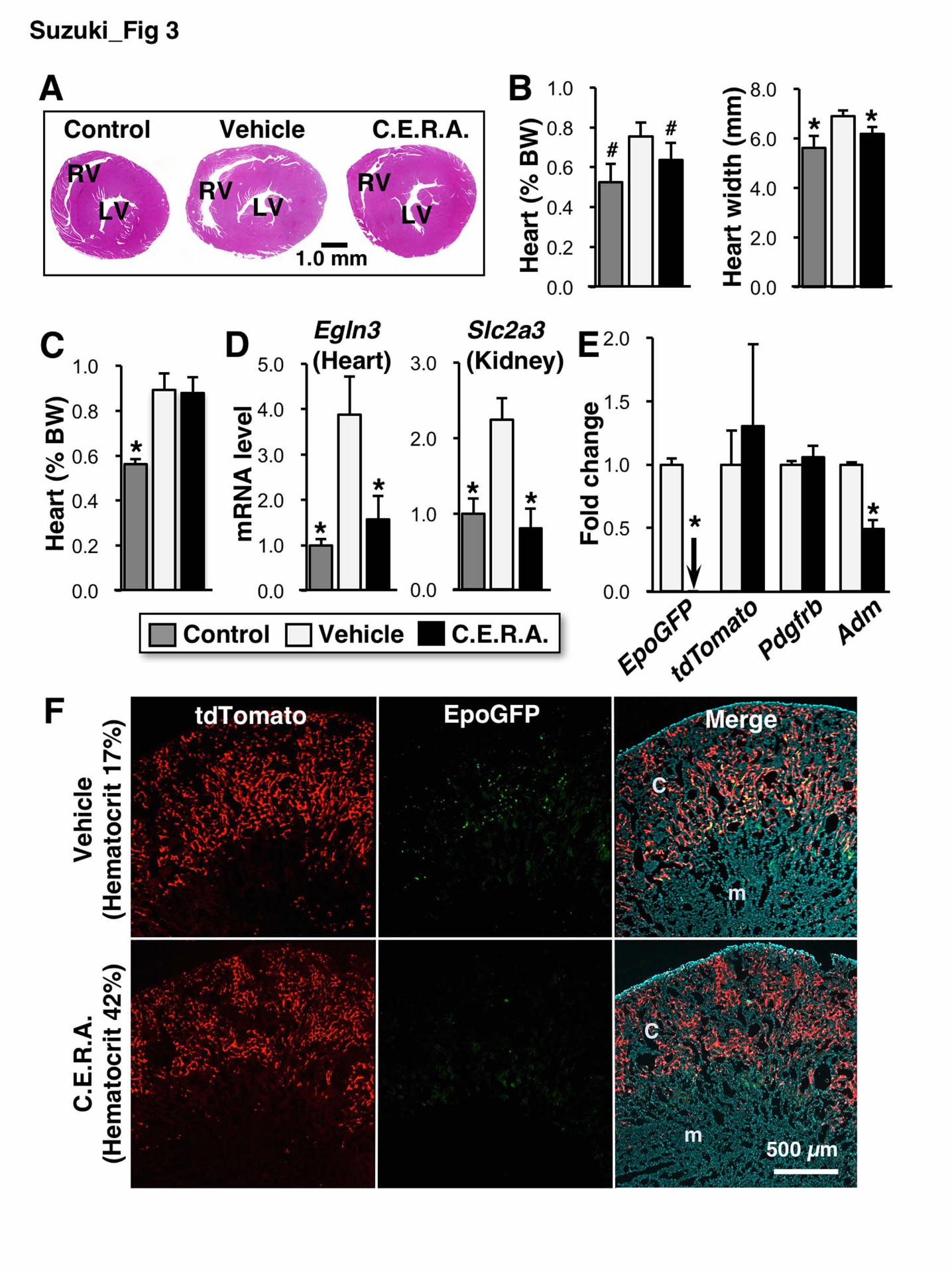
Figure legends
Figure 1. ESA administration stimulates erythropoiesis and induces erythroferrone
expression in hematopoietic organs, followed by the suppression of hepcidin production
in ISAM. (A) rHuEPO, DA or C.E.R.A. was subcutaneously injected into 12- to
14-week-old ISAM at a 3.0 µg/kg BW dose on day-0, and the hemoglobin
concentration, red blood cell (RBC) count and mean corpuscular volume (MCV) in the
peripheral blood were measured 3 and 7 days after injection. The spleen weights were
also measured. n=4–6. *P<0.01, #P<0.05 compared with vehicle-treated mice (white
circles) using Dunnett’s test at each time point. (B) The serum hepcidin concentrations
were measured 2, 3 and 7 days after administration of each ESA. Data from the
untreated control mice are also shown. n=3 for each point. *P<0.01 compared with
ISAM on day 0 using nonparametric Steel test. (C) At 6 and 48 hours after the
injection of ESAs into ISAM, the expression levels of Hamp (hepcidin) and Fam132b
(erythroferrone) mRNA were measured in the livers and hematopoietic organs (bone
marrow [BM] and spleen), respectively. Data from untreated control mice are also
shown. n=3 for each group. *P<0.01 compared with the vehicle-treated samples using
Student’s t test at each time point.
Figure 2. A single dose of C.E.R.A. continuously induces erythropoiesis and iron
utilization in ISAM. (A) C.E.R.A. was subcutaneously injected at a 3.0 µg/kg BW
dose into ISAM on day-0. Changes in the levels of the indicated parameters were
Suzuki et al
12
measured. n=3 for each point. Data from untreated control mice are also shown.
*P<0.01 compared with vehicle-treated ISAM at each time point using Student’s t test.
(B) Fixed-point observation of the inside of the back skin of a living ISAM 3 and 7 days
after C.E.R.A. administration. Red blood cells filled the vascular networks of ISAM 7
days after injection. Scale bar, 500 µm. (C) Berlin blue staining of the spleen sections
from ISAM 7 days after the injection of vehicle or C.E.R.A. shows a decrease in
hemosiderin deposition (blue) following C.E.R.A. administration. Scale bar, 30 µm.
Figure 3. C.E.R.A. administration improves the cardiomegaly and hypoxic milieu of
ISAM. (A) Hematoxylin-eosin staining of ISAM heart sections 7 days after C.E.R.A.
or vehicle administration. An image from an untreated control mouse is also shown. LV,
left ventricle; RV, right ventricle. (B) The weight (% body weight, BW) and maximum
width of the ISAM hearts were measured 7 days after C.E.R.A. or vehicle
administration. The data from the untreated control mice are also shown. n=3 for each
group. (C) The weights of ISAM hearts were measured at 28 days after C.E.R.A. or
vehicle administration in ISAM with recurring anemia. The data from the untreated
control mice are also shown. n=3 for each group. (D) mRNA expression levels of the
Egln3 (Phd3) gene in the heart and the Slc2a3 (Glut3) gene in the kidney were
measured 10 days after C.E.R.A. or vehicle injection in ISAM. Data from untreated
control mice are also shown. Male mice at 12 to 16 weeks of age were analyzed. n=3
for each group. (E) Changes in the mRNA expression of the indicated genes were
examined in the kidneys of ISAM-REC mice injected with C.E.R.A. or vehicle every
Suzuki et al
13
week for 28 days. n=3 for each group. The arrow indicates an undetectable level.
*P<0.01, #P<0.05 compared with vehicle-treated ISAM-REC mice using Student’s t test.
(F) EpoGFP (green) and tdTomato (red) fluorescence was detected in kidney sections
from ISAM-REC mice injected with C.E.R.A. or vehicle every week for 28 days. The
right panels are the merged images of EpoGFP and tdTomato expression, with DAPI
counterstaining. c, cortex; m, medulla.
- S 1 -
Supplementary Appendix “Efficacy estimation of erythropoiesis-stimulating agents using erythropoietin-
deficiency anemic mice” by Norio Suzuki, et al. Methods Mice Inherited super anemia mouse/mice (ISAM, EpoGFP/GFP:Tg3.3K-EpoE genotype) and ISAM-REC mice (EpoGFP/GFP:Tg3.3K-EpoE3:Rosa26LSL-tdTomato:TgEpoCre genotype)1,2 were backcrossed with the C57Black/6 strain more than 6 times, and male mice were used for experiments. In the ISAM-REC mice, a GFP cDNA is homozygously knocked into the Epo gene (EpoGFP), and EpoGFP expression in REP cells is strongly activated by severe anemia. Additionally, anemia-activated EpoCre transgene expression efficiently causes recombination of the Rosa26LSL-tdTomato locus and induces the expression of the tdTomato fluorescent protein from the recombined Rosa26LSL-tdTomato locus in REP cells permanently.2 All mice were maintained under the Regulations for Animal Experiments and Related Activities of Tohoku University. Blood analysis Peripheral blood (0.2–0.3 mL) was collected from the mouse heart or submandibular vein into a 1.5-mL tube containing 5.0 µL of 0.5 M EDTA. To measure long-term changes of the hematocrit values in living mice, approximately 60 µL of peripheral blood was taken weekly from the tail using a heparinized microtube (Drummond) followed by centrifugation. The effect of this weekly small-volume phlebotomy was negligible. Measurement of iron indices Serum iron levels were measured using an automatic biochemistry analyzer (TBA-2000FR, Toshiba). Serum hepcidin levels were measured by a sensitive liquid chromatography/electrospray ionization tandem mass spectrometry method using a Triple Quad 5500 system (AB Sciex) equipped with a Prominence UFLCXR system (Shimadzu) as previously reported (the lower limit of quantitation is 10 ng/mL).3,4 The hepatic and splenic iron contents were measured via inductively coupled plasma atomic emission spectroscopy using an Optima 8000 spectrometer (PerkinElmer).
- S 2 -
Erythropoiesis stimulating agents (ESAs) rHuEPO (epoetin beta, Epogen, Chugai Pharmaceutical), Darbepoetin alpha (DA, NESP, Kyowa Hakko-Kirin) and C.E.R.A. (epoetin beta pegol, Mircera, Chugai Pharmaceutical) were reconstituted with PBS containing 0.02% Tween 80. Each ESA was subcutaneously injected at a dose of 3.0 µg/kg body weight (BW). The dose corresponds to the peptide weight of each ESA, excluding their carbohydrate chains, to compare the efficacies of the ESAs at the same molar concentrations. DA contains 2 additional glycans by genetic modification compared with rHuEPO, and C.E.R.A. is a methoxy polyethylene glycol (PEG)-conjugated rHuEPO. These carbohydrate modifications reduce the affinities between ESAs and EPOR, which determine the plasma half-lives of ESAs because EPO-EPOR complexes are degraded following their endocytosis into erythroid cells after signal transduction.5 PEG may also affect the stability of glycans in ESAs, and asialoglycans of ESAs may be associated with ESA half-life through degradation in hepatocytes after the capture of asialo-ESAs by asialoglycoprotein receptors.6 Reverse transcription quantitative PCR (RT-qPCR) Total RNA was extracted using ISOGEN (Nippon Gene). cDNAs was synthesized using a SuperScript III system (Invitrogen). Quantitative PCR (qPCR) was performed with the primers listed in Table 1 using the FastStart reagent (Roche). Hprt mRNA expression levels were used as an internal control for the qPCR experiments. Dorsal chamber window and surface PO2 measurement A dorsal chamber window was prepared in the mouse back skin as previously described.7,8 To monitor the oxygen concentration inside the back skin, oxygen sensor foil (PreSens, Germany) was placed between the hole and the cover glass, and the surface oxygen tension was measured using a VisiSens A1 detector camera and imaging software (PreSens).9 Histological analyses Heart size was measured as the maximum width using a slide caliper. Sections (4 μm thickness) were prepared from paraffin-embedded formalin-fixed organs. Hemosiderin
- S 3 -
deposition was assessed using Berlin blue staining.3 The heart sections were stained with hematoxylin-eosin (Muto). To detect fluorescent protein expression, frozen kidney sections (10 µm thickness), which were fixed in 4% paraformaldehyde for 4 hours at 4 ºC, were observed after DAPI counterstaining using a BZ9000 microscope (Keyence). Statistics
The data are presented as the means ± standard deviation (SD). The P values were calculated using two-tailed, unpaired Student’s t tests. Dunnett’s test or the nonparametric Steel test were also used for multiple comparison.
- S 4 -
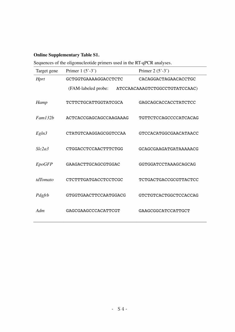
Online Supplementary Table S1. Sequences of the oligonucleotide primers used in the RT-qPCR analyses.
Target gene Primer 1 (5’-3’) Primer 2 (5’-3’)
Hprt GCTGGTGAAAAGGACCTCTC CACAGGACTAGAACACCTGC
(FAM-labeled probe: ATCCAACAAAGTCTGGCCTGTATCCAAC)
Hamp TCTTCTGCATTGGTATCGCA GAGCAGCACCACCTATCTCC
Fam132b ACTCACCGAGCAGCCAAGAAAG TGTTCTCCAGCCCCATCACAG
Egln3 CTATGTCAAGGAGCGGTCCAA GTCCACATGGCGAACATAACC
Slc2a3 CTGGACCTCCAACTTTCTGG GCAGCGAAGATGATAAAAACG
EpoGFP GAAGACTTGCAGCGTGGAC GGTGGATCCTAAAGCAGCAG
tdTomato CTCTTTGATGACCTCCTCGC TCTGACTGACCGCGTTACTCC
Pdgfrb GTGGTGAACTTCCAATGGACG GTCTGTCACTGGCTCCACCAG
Adm GAGCGAAGCCCACATTCGT GAAGCGGCATCCATTGCT
- S 5 -
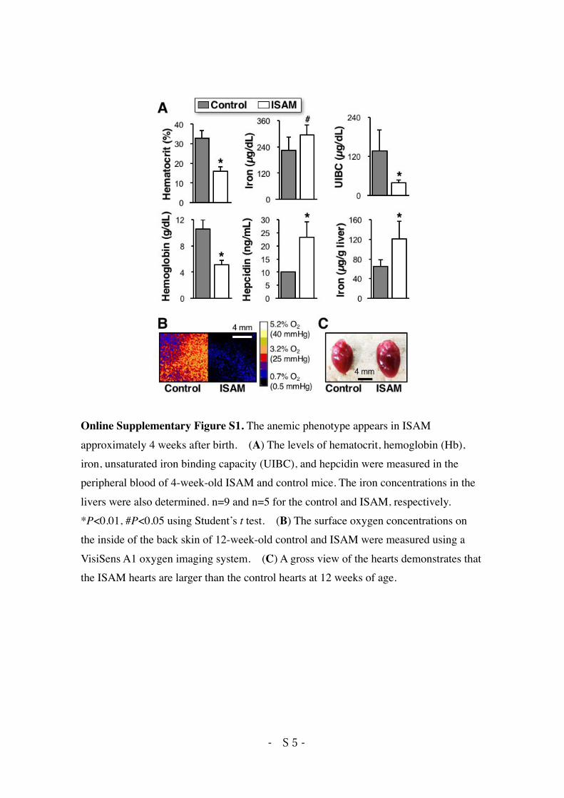
Online Supplementary Figure S1. The anemic phenotype appears in ISAM approximately 4 weeks after birth. (A) The levels of hematocrit, hemoglobin (Hb), iron, unsaturated iron binding capacity (UIBC), and hepcidin were measured in the peripheral blood of 4-week-old ISAM and control mice. The iron concentrations in the livers were also determined. n=9 and n=5 for the control and ISAM, respectively. *P<0.01, #P<0.05 using Student’s t test. (B) The surface oxygen concentrations on the inside of the back skin of 12-week-old control and ISAM were measured using a VisiSens A1 oxygen imaging system. (C) A gross view of the hearts demonstrates that the ISAM hearts are larger than the control hearts at 12 weeks of age.
- S 6 -
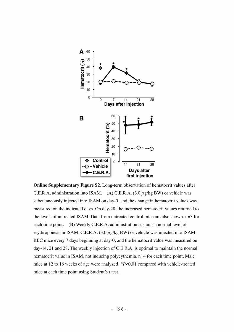
Online Supplementary Figure S2. Long-term observation of hematocrit values after C.E.R.A. administration into ISAM. (A) C.E.R.A. (3.0 µg/kg BW) or vehicle was subcutaneously injected into ISAM on day-0, and the change in hematocrit values was measured on the indicated days. On day-28, the increased hematocrit values returned to the levels of untreated ISAM. Data from untreated control mice are also shown. n=3 for each time point. (B) Weekly C.E.R.A. administration sustains a normal level of erythropoiesis in ISAM. C.E.R.A. (3.0 µg/kg BW) or vehicle was injected into ISAM-REC mice every 7 days beginning at day-0, and the hematocrit value was measured on day-14, 21 and 28. The weekly injection of C.E.R.A. is optimal to maintain the normal hematocrit value in ISAM, not inducing polycythemia. n=4 for each time point. Male mice at 12 to 16 weeks of age were analyzed. *P<0.01 compared with vehicle-treated mice at each time point using Student’s t test.
- S 7 -
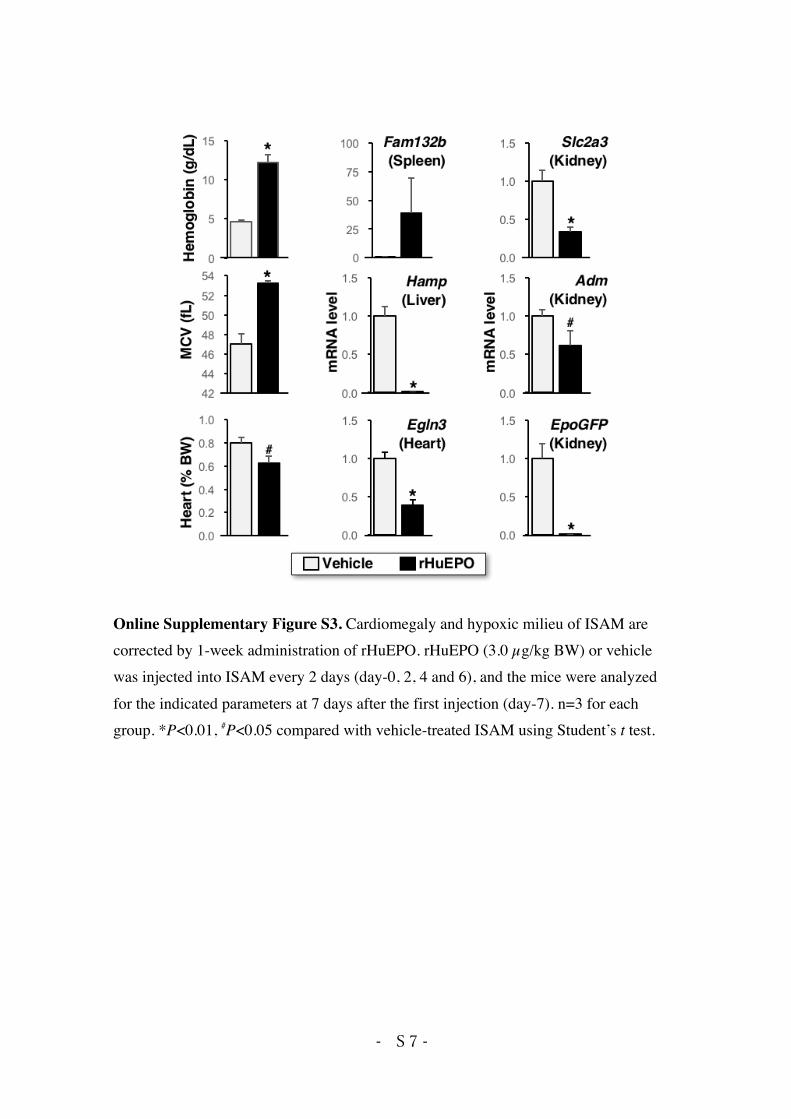
Online Supplementary Figure S3. Cardiomegaly and hypoxic milieu of ISAM are corrected by 1-week administration of rHuEPO. rHuEPO (3.0 µg/kg BW) or vehicle was injected into ISAM every 2 days (day-0, 2, 4 and 6), and the mice were analyzed for the indicated parameters at 7 days after the first injection (day-7). n=3 for each group. *P<0.01, #P<0.05 compared with vehicle-treated ISAM using Student’s t test.
- S 8 -
References for Supplementary Appendix 1. Suzuki N, Yamamoto M. Roles of renal erythropoietin-producing (REP) cells in the
maintenance of systemic oxygen homeostasis. Pflugers Arch. 2015;468(1):3-12. 2. Yamazaki S, Souma T, Hirano I, et al. A mouse model of adult-onset anaemia due to
erythropoietin deficiency. Nat Commun. 2013;4:1950. 3. Sasaki Y, Noguchi-Sasaki M, Yasuno H, Yorozu K, Shimonaka Y. Erythropoietin
stimulation decreases hepcidin expression through hematopoietic activity on bone marrow cells in mice. Int J Hematol. 2012;96(6):692-700.
4. Murao N, Ishigai M, Yasuno H, Shimonaka Y, Aso Y. Simple and sensitive quantification of bioactive peptides in biological matrices using liquid chromatography/selected reaction monitoring mass spectrometry coupled with trichloroacetic acid clean-up. Rapid Commun Mass Spectrom. 2007;21(24):4033-4038.
5. Gross AW, Lodish HF. Cellular trafficking and degradation of erythropoietin and novel erythropoiesis stimulating protein (NESP). J Biol Chem. 2006;281(4):2024-2032.
6. Mi Y, Lin A, Fiete D, Steirer L, Baenziger JU. Modulation of mannose and asialoglycoprotein receptor expression determines glycoprotein hormone half-life at critical points in the reproductive cycle. J Biol Chem. 2014;289(17):12157-12167.
7. Palmer GM, Fontanella AN, Shan S, et al. In vivo optical molecular imaging and analysis in mice using dorsal window chamber models applied to hypoxia, vasculature and fluorescent reporters. Nat Protoc. 2011;6(9):1355-1366.
8. Laschke MW, Vollmar B, Menger MD. The dorsal skinfold chamber: window into the dynamic interaction of biomaterials with their surrounding host tissue. Eur Cell Mater. 2011;22:147-167.
9. Souma T, Nezu M, Nakano D, et al. Erythropoietin synthesis in renal myofibroblasts is restored by activation of hypoxia signaling. J Am Soc Nephrol. 2015;27(2):428-438.