Embed Size (px)
Citation preview
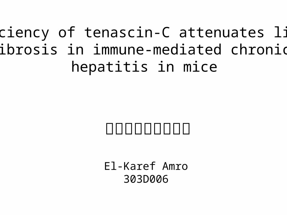
El-Karef Amro 303D006
三重大学医学部病理
Deficiency of tenascin-C attenuates liver fibrosis in immune-mediated chronic
hepatitis in mice
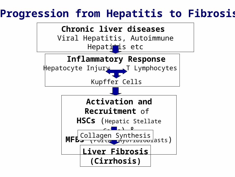
Chronic liver diseases Viral Hepatitis, Autoimmune Hepatitis etc
Progression from Hepatitis to Fibrosis
Activation and Recruitment of HSCs (Hepatic Stellate Cells) &MFBs (Portal Myofibroblasts)
Liver Fibrosis(Cirrhosis)
T LymphocytesHepatocyte Injury
Kupffer Cells
Inflammatory Response
Collagen Synthesis
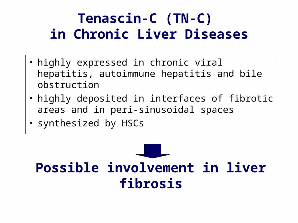
Tenascin-C (TN-C) in Chronic Liver Diseases
• highly expressed in chronic viral hepatitis, autoimmune hepatitis and bile obstruction
• highly deposited in interfaces of fibrotic areas and in peri-sinusoidal spaces
• synthesized by HSCs
Possible involvement in liver fibrosis
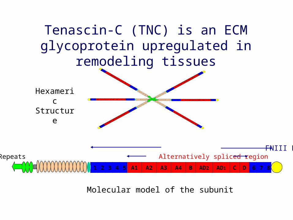
1 2 3 4 5 A1 A2 A3 A4 B AD2 AD1 C D 6 7 8
FNIII Repeat TA EGFL Repeats Alternatively spliced region FG
876DCAD1AD2BA4A3A2A154321 876DCAD1AD2BA4A3A2A154321
8 7 6 D C AD1 AD2 B A4 A3 A2 A1 5 4 3 2 18 7 6 D C AD1 AD2 B A4 A3 A2 A1 5 4 3 2 1
Tenascin-C (TNC) is an ECM glycoprotein upregulated in remodeling tissues
Hexameric Structure
Molecular model of the subunit
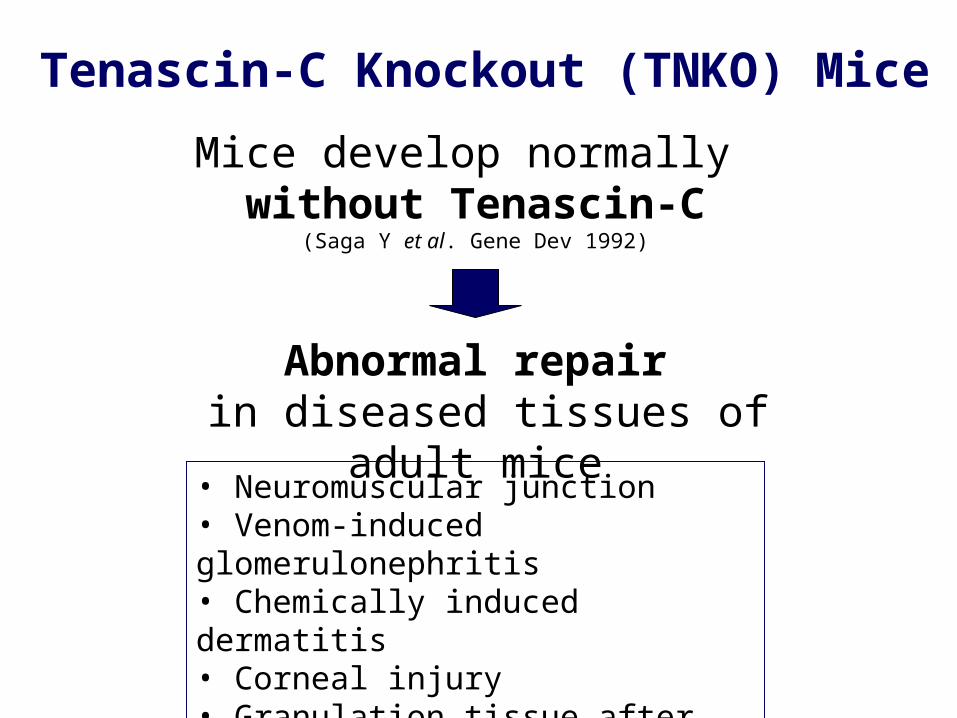
Mice develop normally without Tenascin-C
(Saga Y et al. Gene Dev 1992)
Tenascin-C Knockout (TNKO) Mice
Abnormal repair in diseased tissues of adult mice
• Neuromuscular junction• Venom-induced glomerulonephritis• Chemically induced dermatitis• Corneal injury• Granulation tissue after cardiac injury
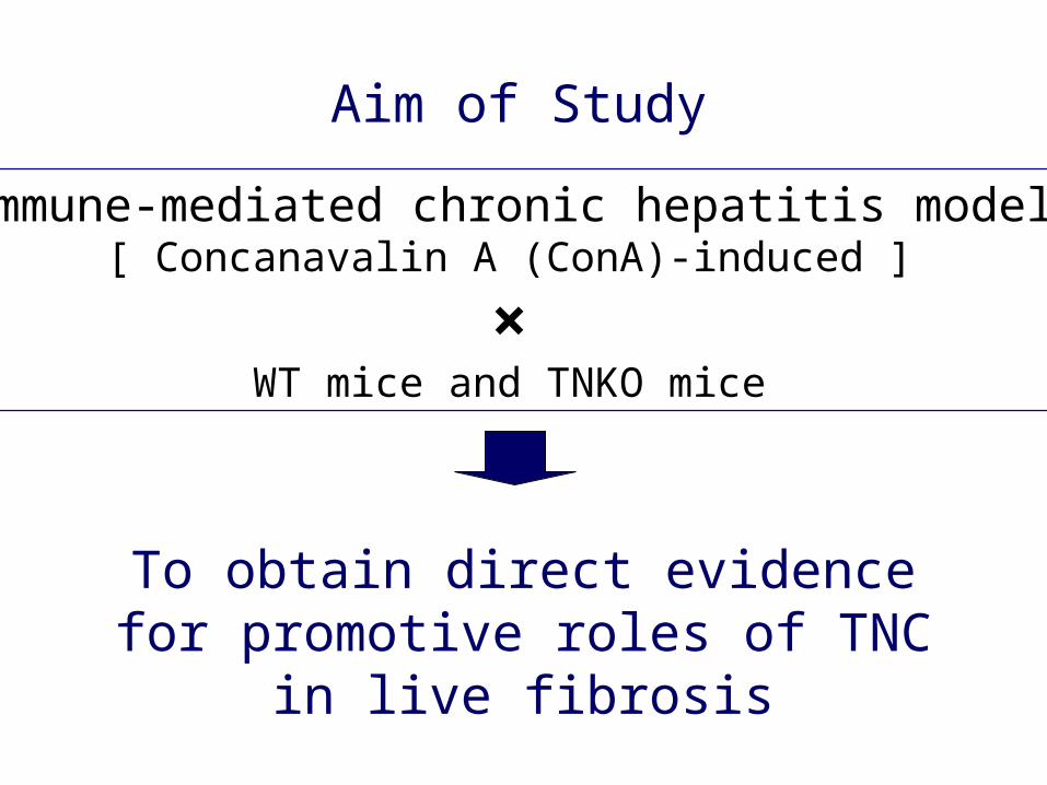
Aim of Study
Immune-mediated chronic hepatitis model[ Concanavalin A (ConA)-induced ]
×WT mice and TNKO mice
To obtain direct evidence for promotive roles of TNC in live fibrosis
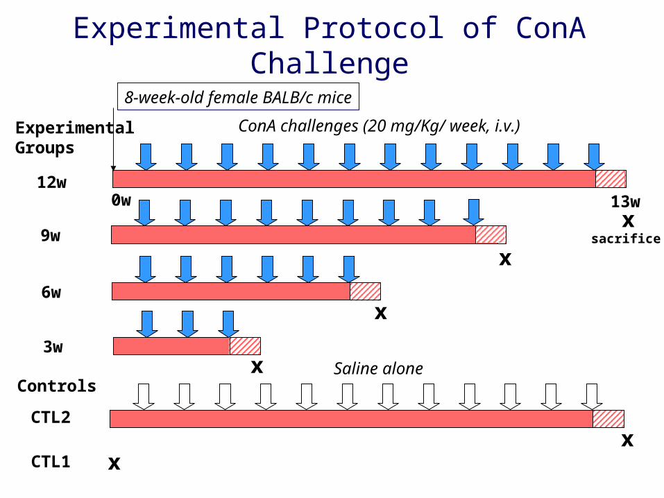
Experimental Protocol of ConA Challenge
sacrifice
ExperimentalGroups
8-week-old female BALB/c mice
0w 13w
ConA challenges (20 mg/Kg/ week, i.v.)
12w
9w
6w
3wSaline alone
CTL2
CTL1
x
x
x
x
xx
Controls
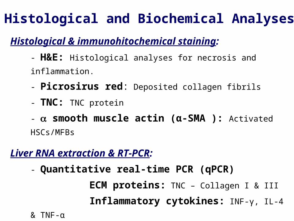
Histological & immunohitochemical staining:
- H&E: Histological analyses for necrosis and inflammation.
- Picrosirus red: Deposited collagen fibrils
- TNC: TNC protein
- smooth muscle actin (α-SMA ): Activated HSCs/MFBs
Liver RNA extraction & RT-PCR:
- Quantitative real-time PCR (qPCR)
ECM proteins: TNC – Collagen I & III
Inflammatory cytokines: INF-γ, IL-4 & TNF-α
Fibrogenic cytokine: TGF-β
Histological and Biochemical Analyses
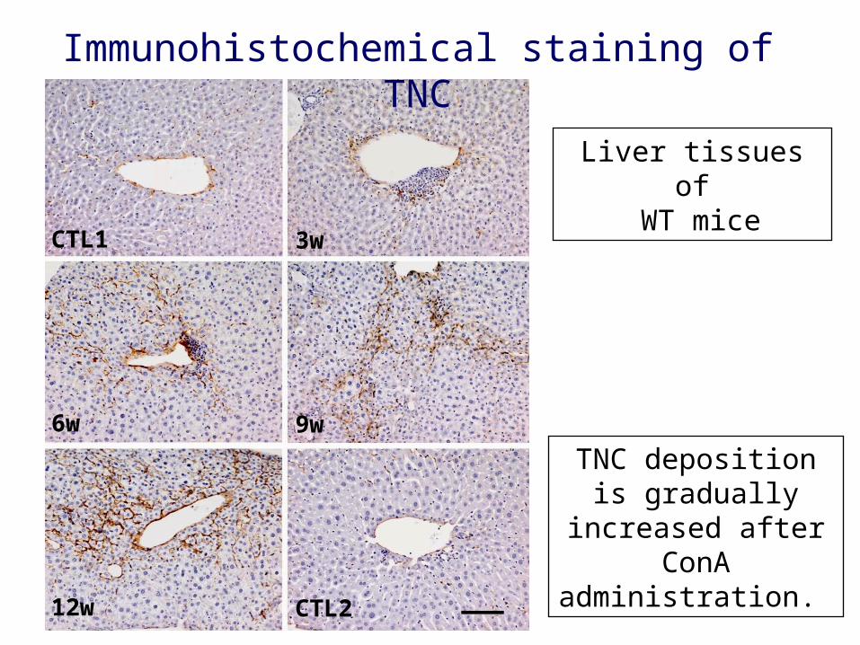
CTL1
6w
12w CTL2
3w
9w
Immunohistochemical staining of TNC
TNC deposition is gradually increased
after ConA administration.
Liver tissues of WT mice
0
1
2
3
4
5
6
CTL1 3w 6w 9w 12w CTL2T
NC
mR
NA
(fo
lds)
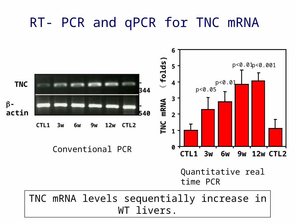
p<0.05
RT- PCR and qPCR for TNC mRNA
TNC mRNA levels sequentially increase in WT livers.
Quantitative real time PCR
-actin
TNC
- 540
- 344
CTL1 3w 6w 9w 12w CTL2
Conventional PCR
p<0.01
p<0.01 p<0.001
CTL2
12w
WT TNKO
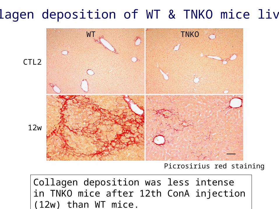
Collagen deposition of WT & TNKO mice livers
Picrosirius red staining
Collagen deposition was less intense in TNKO mice after 12th ConA injection (12w) than WT mice.
0
5
10
15
20
25
CTL1 3w 6w 9w 12w CTL2
Fib
rotic
are
a (%
)
0
1
2
3
4
5
6
7
CTL1 3w 6w 9w 12w CTL2
ns
Fib
rotic
are
a (%
)
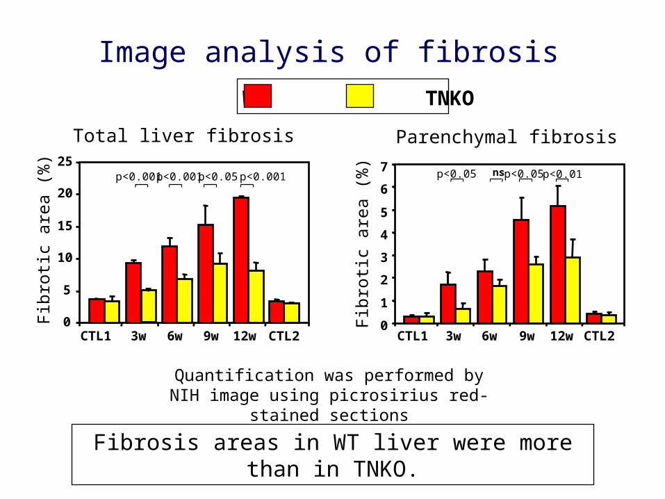
Image analysis of fibrosis
Total liver fibrosis Parenchymal fibrosis
Fibrosis areas in WT liver were more than in TNKO.
Quantification was performed by NIH image using picrosirius red-stained sections
WT TNKO
p<0.01p<0.001 p<0.001p<0.001p<0.05 p<0.05 p<0.05
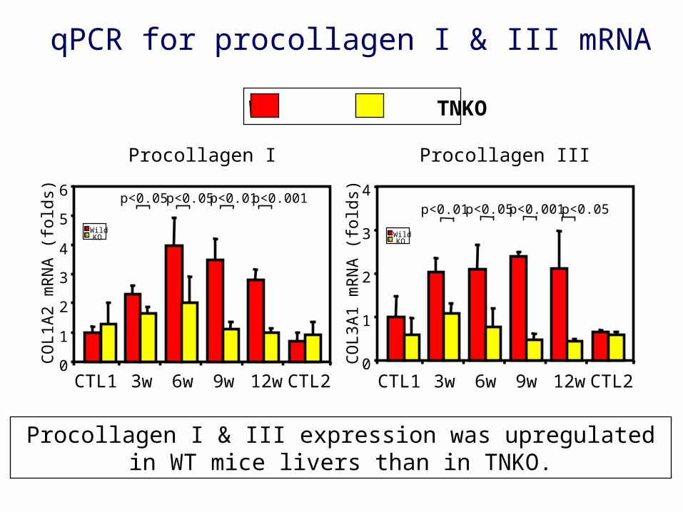
Procollagen I Procollagen III
qPCR for procollagen I & III mRNA
WildKO Wild
KO
0
1
2
3
4
CO
L3A
1 m
RN
A (
fold
s)
CTL1 3w 6w 9w 12w CTL20
1
2
3
4
5
6
CTL1 3w 6w 9w 12w CTL2
CO
L1A
2 m
RN
A (
fold
s)
Procollagen I & III expression was upregulated in WT mice livers than in TNKO.
WT TNKO
p<0.001 p<0.05 p<0.05 p<0.01 p<0.01 p<0.05 p<0.001 p<0.05
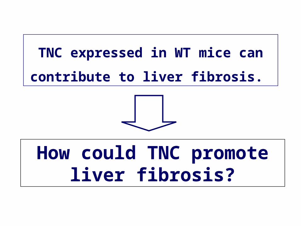
TNC expressed in WT mice can
contribute to liver fibrosis.
How could TNC promote liver fibrosis?
TNKO
PT
PT
CV
CV
WT
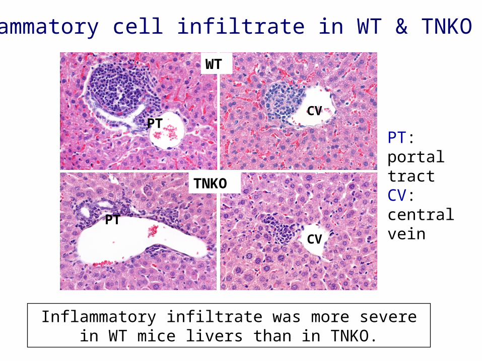
Inflammatory cell infiltrate in WT & TNKO mice
PT: portal tract CV: central vein
Inflammatory infiltrate was more severe in WT mice livers than in TNKO.
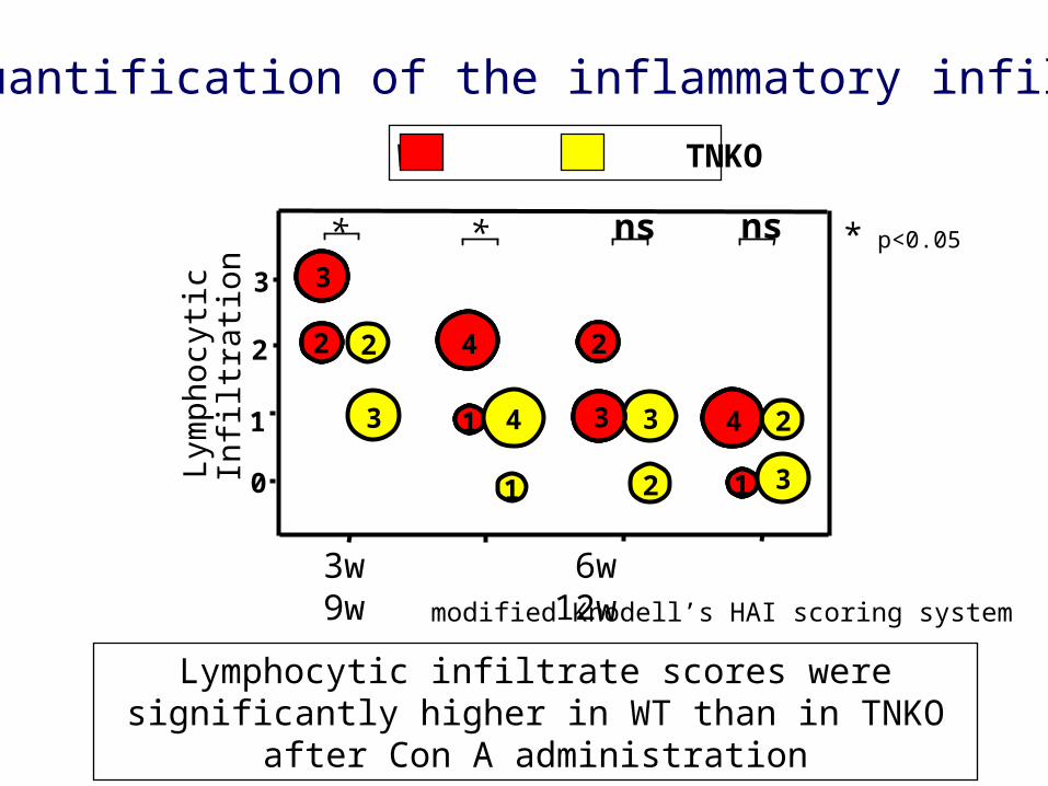
Semi-quantification of the inflammatory infiltrate
Lymphocytic infiltrate scores were significantly higher in WT than in TNKO after Con A administration
modified Knodell’s HAI scoring system
Lym
phoc
ytic
Infil
trat
ion
3w 6w 9w 12w
3
2
1
0
3
1
2 2
1 3
3
2 4
43 24
2 1 3
* ns* ns
WT TNKO
* p<0.05

qPCR of inflammatory cytokines mRNA
Cytokines were significantly upregulated in WT than in
TNKO.
WT TNKO
02468
1012
02468
1012
CTL1 3w 6w 9w 12w CTL2
INF-
γm
RN
A
(fo
ld v
alu
e) *** *** *
0
2
4
6
8
CTL1 3w 6w 9w 12w CTL2
IL-4
mR
NA
(f
old
val
ue
) * * **
02468
1012
CTL1 3w 6w 9w 12w CTL2
TN
F-
mR
NA
(f
old
val
ue
) ****** **
* p<0.05
** p<0.01
*** p<0.001
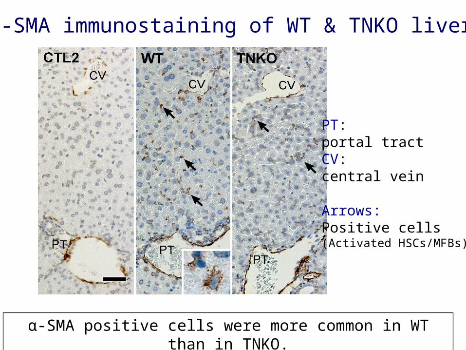
α-SMA immunostaining of WT & TNKO liver
α-SMA positive cells were more common in WT than in TNKO.
PT: portal tract CV: central vein
Arrows:Positive cells(Activated HSCs/MFBs)
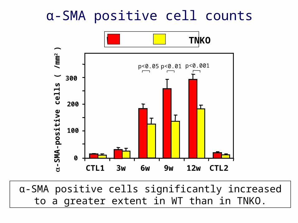
α-SMA positive cell counts
100
200
-S
MA
-po
siti
ve c
ells
( /
mm
2 )
0
300
CTL1 3w 6w 9w 12w CTL2
p<0.05
α-SMA positive cells significantly increased to a greater extent in WT than in TNKO.
WT TNKO
p<0.01 p<0.001
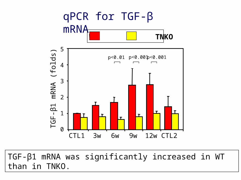
0
1
2
3
4
5
CTL1 3w 6w 9w 12w CTL2
TG
F-β
1 m
RN
A (
fold
s)
qPCR for TGF-β mRNA
TGF-β1 mRNA was significantly increased in WT than in TNKO.
WT TNKO
p<0.001 p<0.001p<0.01
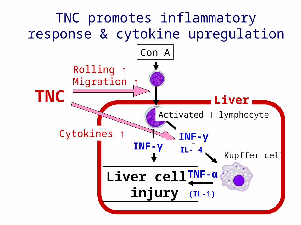
Con A
Kupffer cell
Liver cell injury
Activated T lymphocyte
TNF-α
INF-γIL- 4
TNC promotes inflammatory response & cytokine upregulation
LiverTNC
INF-γ
Rolling ↑Migration ↑
Cytokines ↑
(IL-1)
Kupffer cell
Liver cell injury
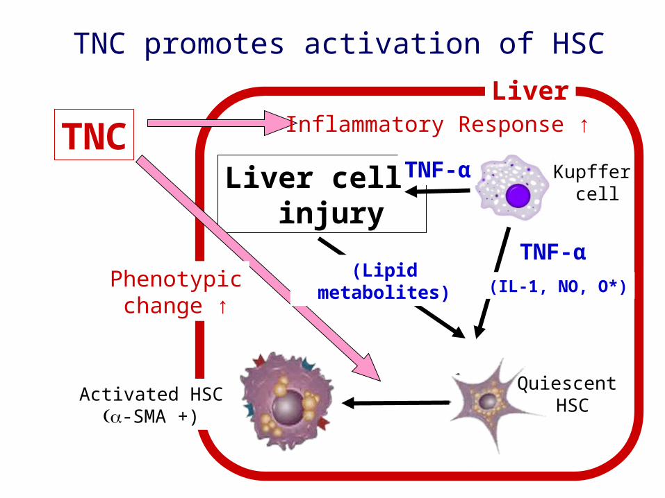
Liver
TNC Inflammatory Response ↑
TNF-α
(IL-1, NO, O*)
Quiescent HSC
(Lipid metabolites)
TNF-α
Activated HSC-SMA +)
Phenotypicchange ↑
TNC promotes activation of HSC
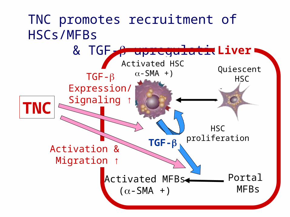
TNC promotes recruitment of HSCs/MFBs & TGF- upregulation
TNC
Quiescent HSC
Activated HSC-SMA +)TGF-
Expression/Signaling ↑
Portal MFBs
Activation & Migration ↑
Activated MFBs(-SMA +)
TGF-HSC proliferation
Liver
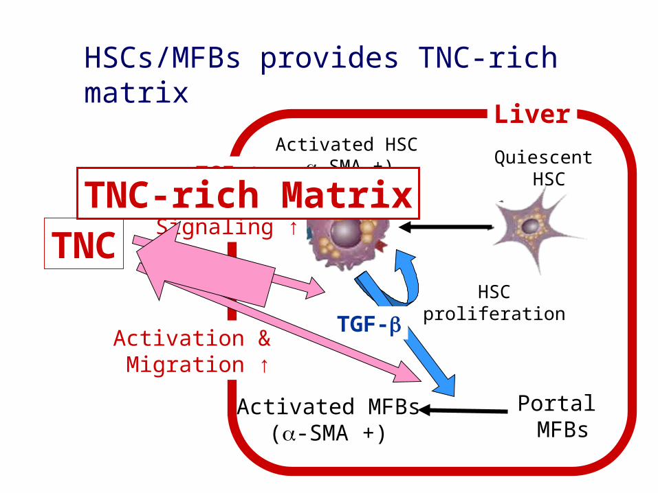
HSCs/MFBs provides TNC-rich matrix
TNC
Quiescent HSC
Activated HSC-SMA +)TGF-
Expression/Signaling ↑
Portal MFBs
Activation & Migration ↑
Activated MFBs(-SMA +)
TGF-HSC proliferation
Liver
TNC-rich Matrix
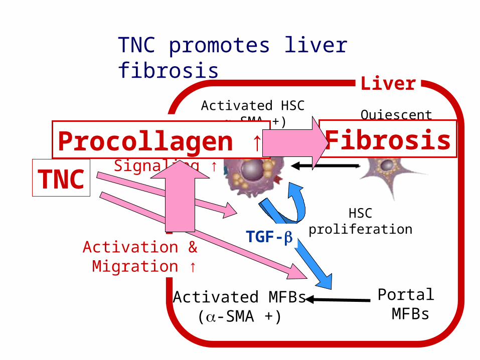
TNC promotes liver fibrosis
TNC
Quiescent HSC
Activated HSC-SMA +)TGF-
Expression/Signaling ↑
Portal MFBs
Activation & Migration ↑
Activated MFBs(-SMA +)
TGF-HSC proliferation
Liver
Procollagen ↑ Fibrosis
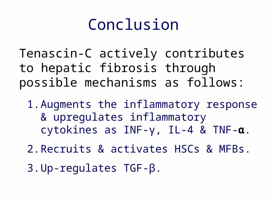
Tenascin-C actively contributes to hepatic fibrosis through possible mechanisms as follows:
1. Augments the inflammatory response & upregulates inflammatory cytokines as INF-γ, IL-4 & TNF-α.
2. Recruits & activates HSCs & MFBs.
3. Up-regulates TGF-β.
Conclusion