Embed Size (px)
Citation preview
TitleELECTROMYOGRAPHIC STUDIES ON CROSSEDANASTOMOSIS BETWEEN THE TIBIAL AND THEFIBULAR NERVE IN DOGS
Author(s) NISHIMURA, SHOZO
Citation 日本外科宝函 (1958), 27(4): 919-932
Issue Date 1958-07-01
URL http://hdl.handle.net/2433/206663
Right
Type Departmental Bulletin Paper
Textversion publisher
Kyoto University

919
ELECTROMYOGRAPHIC STUDIES ON CROSSED ANASTOMOSIS BETWEEN THE TIBIAL AND THE FIBULAR NERVE IN DOGS
by
Sttozo N !SHIMURA
From the !st Surgical Division, Kyoto University Medical School (Director: Prof. Dr. CmsA~唱 ARAKr)
Received for publication Apr. 19, 1958
INTRODUCTION
In relation to the problem whether permanent functional disturbances are left
or not after cutting two functionally different nerves and suturing them crosswise,
since LETIEV ANT in 1873, RA w A, GuNN, S1cK and SANGER, MENASSE, KENNEDY, OsBo・
RNE and KILVINGTON, SHIBUYA, NoRIOKA, BARRON, SPERRY and so on have reported
on the results of the suture behveen heteronymous or heterogenous nerves, both in human beings and animals. There has not 3モtbeen, however, any unanimous
opinion about the recovery of normal function after operation. The views present-
ed so far can be divided into the following three.
1) A school which admits the satisfactor;; functional recovery occurring rather
early in the postoperative period (RAwA, KENNEDY, OSBORNE and KILVINGTON, NoR10-
KA, BARRON and ARIZA w A) .
2) Opinion of non-functional recovery (SPERRY and ¥VATANABE).
3) Opinion by which the satisfactor~· recovery can take place only by the
repeated exercises after the nerve regeneration (SPERRY).
ARIZAWA, a former member of our laborator~· . in 1952, using 33 rabbits and
3 dogs, practiced the crossed anastomosis of the tibial with the fibular nerve and observed the restoration of normal functions within 6 months. He considered it
as being the result of the functional compensation bγthe cerebrum and the spinal cord.
In 1954, Isttn in our laboratory, carrying out the crossed anastomosis between a spinal and the vagal nerve in the cervical region in cats, reported that the
nerve regeneration was surely recognized b~’ either histological stud~· or b;; electri-cal stimulation, but the function regained was not normal.
In 1955, ¥V ATANABE, another member of our laboratory, practiced the crossed
anastomosis between the tibial and the fibular nerve at the upper level of the
thigh in dogs, and found the postoperative functional changes to be divided into
three stadiums. In 3~6 months after operation the walk of the animals became
seemingly normal, but after 6 months th町ア walked abnormally again, and no
restoration took place by the end of 12th postoperative month when they were killed.
Because his observations, however, were gross and clinical, the detailed physio-logical mechanism going on during the recovery was almost unknown.

920 日本外科宝函第27巻第4号
In 1947, GoLSETH and F1zzELL, performing the suture immediately after cutting
sciatic nerve in cats, observed the electromyogram with the progr田sof restoration. And he proved that the electromyographic restoration and functional recovery occurred hand in hand.
In 1952, KATO obtained the similar results, after the sciatic nerve suture in rabbits.
In the present report, I studied electromyographic;illy the regenerative procei:s
of crossed nerve anastomosis and also the recover~· of reflexes.
》IATERIALS AND METHODS OF EXPERIMENTS
(1) Experimental Animals Twenty-two adult dogs of about 10 kg body weight. Completely crossed
anastomosis in 19 dogs and partiallJ’crossed anastomosis in 3. (2) Experimental Equipments The electrode used for E. M. G. was a coaxial needle made from a subcutaneous
injection needle of 1/4 mm in diameter in which an enamel insulated cupreous wire of 100μ diameter was enclosed and fixed.
The electromyographic apparatus were the E. M. G. 52・5type manufactured by
Tokyo Denki-Seiki & Co. Ltd., with the frequency characteristics adjusted and the 2-beam E. l¥I. G. apparatus by Sanei-Sokuki & Co. Ltd. In order to reduce induced alternating current at the time of observation of E. l¥I. G., a pedestal, on which a dog was placed, was made, with insulators attached under its legs.
(3) MethoJs of Experiments A) Surgical Operations a) Narcosis: After the basal narcosis with 4% narcopon scopolamine, the
intraperitoneal injection of 10% isomytal sodium solution 0.3cc pro kg was done. The operations were carried out under this isomytal general anesthesia.
b) (正impletclycrossed anastomosis of tibial and fibular nerves: A skin incis-ion of about 5 cm length was delivered on the posterior surface of the right upper thigh. Between :¥I. biceps femoris and M. semitendineus, we could easily白ndthe bifurcation of the sciatic nerve into the tibial and the fibular nerve. The both nerves, after fullJ・ separated from the surroundmgs, were cut at the point about 4 cm peripheral from the bifurcation, and the central end of the fibular nerve and the per匂heralend of the tibial nerve were suturecl with each other, and simultane-ously the central end of the tibial nerve and the peripheral end of the fibular
nerve were also sutured. The nerve suture was done following the method of
TAKETOMo and ¥VATANABE; the arterial tube preserved in 70% alcohol, was used for the intubation of the united nerve ends, and no suturing gut was passed through the nerve ends (Fig. 1 a).
c) Partially crossed anastomosis: In the same way as in the completely crossed anastomosis, the both nerves were exposed. And they were slivered respectively in half. Each half of them was cut and the central end of the one was sutured with the peripheral end of the other (Fig. 1. b).
d) Unilateral decortication of motor areas : In the three dogs, which survived for more than one year and 4 months after the completely crossed anastomosis

ELECTROMYOGRAPHIC STUDIES ON CROSSED ANASTOMOSIS 921
a. T. ph
T. F
b.
T. Fig. 1 Schematical illustration of crossed nerve
anastomosis. (From WATANABE, Arch. J ap. Chir. 24, 135, 1955)
F.
and of which the functional recoYCry
was satisfactory, I performed the uni-
lateral decortication of the contralateral
motor areas. As controls, the similar
operation was carried out in 3 health~·
adult dogs. The motor areas of clogs
are considered to be situated in GFi
sigmoideus anterior et posterior and
Gyri proreus and coronalis. By the left
frontoparietal craniotomy the left fron-
tal and the parietal lobe were full~· exposed. By giving them electric stimu-
lations of 60=, 10 v, and obtaining
the motor responses, I confirmed the
motor areas and decorticated them wide-
ly enough. F.
B) Postoperative Neurological Ch-
anges (Clinical S~·mptoms)
1) Standing posture. 2) Walking, a : Completely crossed anastomosis b : Partially crossed anastomosis T: N. tibialis particular!≫ crippling. The test was F: N. fibularis p : Arterial tube made not onb’ in slow!)・walkingbut
in rapid running. 3) Prompt and unprepared action ; the stepping of hungry dogs when they hun句yup to approach the feed, or the postures of the right hindleg
and foot when dogs spring up旬 thefeed above the head. 4) Trophic disturbanc-
es: shedding of the hair, deformity of nail, hemorrhage and ulcer formation are
often seen on the dorsal surface of the right foot. And the atrophy of crural and plantar muscles is also found.
C) Electromyogram On the insulated pedestal above mentioned, a dog was placed and the three
extremities except the right lower were fixed. Inserting an electrode into a muscle with no anesthesia, I observed E. M. G. by means of oscilloscope with Brown tube and took photo~raphs if necessary. The examined muscles were l¥I. tibialis anterior, i¥I. gastorocnemius and l¥I. plantaris. The time intervals of the E. l¥L G. examina-tions were 1~10 days at the biginning, 20 cla;.芯 after100 da)’s and 30 days after 180 da~·s. The dogs in which the E. l¥L G. study was made, were 19 in the group of completely crossed anastomosis. Among them, 6 suni\℃<l for more than one year, the maximum survival being for one year and 6 months. Also the E. l¥I. G. study was performed in 3 dogs with partially crossed anastomosis. The maximum survival was for one ~·ear and 4 months.
D) Reflex 1) Flexion reflex. In dogs, when the gluteal region or the lateral surface of
the thigh is stimulated, the contraction of the anterior tibial muscle with the dorsal flexion of the foJt occurs on the same side. At the time of this flexion reflex, the reflex waves in the E. :.¥I. G. of the anterior tibial muscle or the gastrocnemius in cases of crossed anastomosis were observed.
922 日本外科宝函第27巻第4号
2) Achilles tendon reflex. Insertin広 anelectrode into the gastrocnemius and also in the anterior tibial muscle in cases of crossed anastomosis, I observed the
reflex wave in the E. M. G. which was elicited by the knock on Achilles tendon.
E) Anatomical and Histological Researches After the animals were slaughtered, their cerebrum (decorticated), the nerv田
at the sites of crossed anastomosis and the muscles under their innervation were observed with the naked eyes. And the nerves at the site of crossed anastomosis were subjected to the histological examination after the transverse section and Wei-
gertstaining. The muscles were stained by hematoxylin-eosin.
RESULTS OF EXPERIMENTS
川、…
f
MV
川、円》hμ川
ぬ
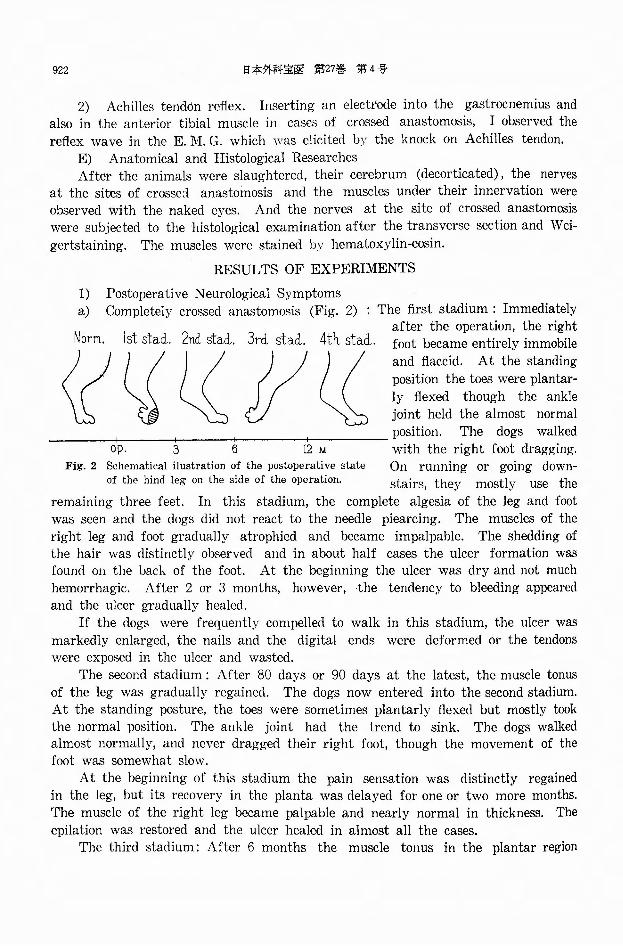
1) Postoperative Neurological Symptoms a) Completely crossed anastomosis (Fig. 2) : The first stadium : Immediately
after the operation, the right 4th staci. foot became entirely immobile
and flaccid. At the standing
position the toes were plantar-
ly flexed though the ankle joint held the almost normal
position. The dogs walked with the right foot dragging. op. 3 6 12 M
Fig. 2 Schematical ilustration of the postoperative state On running or going down-of the hind leg on the side of the operation. stairs, they mostly use the
remaining three feet. In this stadium, the complete algesia of the leg and foot was seen and the dogs did not react to the needle piearcing. The muscles of the
right leg and foot gradually atrophied and became impalpable. The shedding of the hair was distinctly observed and in about half cases the ulcer formation w部
found on the back of the foot. At the beginning the ulcer was dry and not much
hemorrhagic. After 2 or 3 month8, however, the tendency to bleeding appeared and the ulcer gradually healed.
If the dogs were frequently compelled to walk in this stadium, the ulcer was markedly enlarged, the nails and the digital ends were deformed or the tendons were exposed in the ulcer and wasted.
The second gtadium : After 80 days or 90 days at the latest, the muscle tonus of the leg was gradually regained. The dogs now entered into the second stadium. At the standing posture, the toes were sometimes plantarly flexed but mostly加okthe normal position. The ankle joint had the trend to sink. The dogs walked almost normally, and never dragged their right foot, though the movement of the foot was somewhat slow.
At the beginning of this stadium the pain sensation was distinctly regained in the leg, but its recovery in the planta was delayed for one or two more months. The muscle of the right leg became palpable and nearly normal in thickness. The
epilation was restored and the ulcer healed in almost all the cases. The third stadium: After 6 months the muscle tonus in the plantar region

ELECTROMYOGRAPHIC STUDIES ON CROSSED ANASTOMOSIS 923
returned, and at the standing posture the ankle joint and the toes were plantarly flexed in most cases. The walk was rather alike that in the first stadium. The dogs walked, stretching their right posterior extremit)’ with the toes flexed plan-tarly. At the time of a prompt and unprepared action the reversed movement took place. On springing up to the feed, the dogs fell down to the right side, taking a false step with the right leg.
The fourth stadium : After about one year, the plantar flexion of the toes be~ame less and less marked and the ankle joint stood mostly rather lower. The walk became more skillful in most cases. The reversed movements rarely occurred. On springing up to the feed, most of the dogs never fell down. These restorations went on very slowly and there were individual differences among the dogs. Of the six dogs, surviving for more than one year after operation, one showed no recovery, 2 slight restoration and 3 marked restoration. Even the best restored 3 dogs, some-times, flexed their toes plantarly, and their movement of the right posterior ex-tremity was slightly slow at walk. But the reversed movement was only rarely seen and they could spring up correctly to the feed.
The above mentioned four stadiums can be shown in a scheme (Fig. 2). b) Functional disturbances after unilateral decortication of motor areas: In control dogs, the motor hemiparalysis was observed on the contralateral
side. The anterior and posterior extremities on the paralized side were flexed and the head deviated to the paralized side. The trunk was bent with concavity toward the paralized side. On standing they were unstable and apt to fall down to the paralized side. At walk they moved round, discribing a circle around the paralized side. These symptoms gradually improved and returned to normal usually in about 2 or 3 weeks. But somewhat different was the result in the 3 dogs, which had undergone crossed anastomosis of peripheral nerves more than one y回 rand 4 months previously with good postoperative functional restoration and then underwent additional contralateral decortication of motor areas. Although the disturbances in standing and walking were recovered in 2 or 3 weeks in these C回目, theywere apt to stretch their right posterior extremity at the ankle joint at the time of taking feed or of running away, and accordingly sometimes fell down. In other words, thry showed the distinctly reversed movement in such actions. To spring up to the feed was impossible. This phenomenon persisted for one month and 15 days after the decortication of motor areas.
c) Partially crossed anastomosis : No motor disturbance was observed at any time after the operation, even one year and 4 months later. In the standing posture, walking and prompt and unprepared action, no changes were demonstrated.
2) E.M.G.
a) Fibrillation voltage (Fig. 3) : This i::; the spike discharge appearing spontaneously in individual muscle fibers when the nenous control is lost. Its amplitude is 10~lOOμv, duration is 1~2 msec., repetition frequency varies from 2 to 3 per sec. For 4 days after the crossed anastomosis the muscle was in electrical silence, and on the fifth day the fibrillation voltage appeared for the first time. At the beginning it appeared only in a few points in the muscle with

924 日本外科宝函第27巻 第4号
i
,hqノl!
.
、JU
、,
the amplitude less than 501.tv, but on
the 10th day after the operation it was demonstrated in almost all points
in the muscle and its amplitude in all three muscles reached to 100,uv
向や atthe maximum. In the anterior tibial muscle on
the 50th day after the operation
fibrillation voltages diminished and
were demonstrated only in a few points and on the 60th day they were almost
undemonstrable except in some dogs (Fig. 7). In some parts of the gastrocnemius they appeared on the 5th clay, but began
to diminish 10 days ealier than in the anterior tibial. Also in the plantar muscle they appeared on the 5th day, persisted for a long time and diminished on the
120th day. b) Complex N. :.¥I. U. voltage (Fig. 4) : During the process of nerve regene-
ration, a polyphasic wave can be observed.
Its amplitude is 200』 600'.is 5~15 msec. and repetition frequency varies from 2 to 30 per sec. vV ED DELL, GoLsETH and F1zzEL, and KATO called this wave as the nascent motor unit voltage. On the 50th day after the
operation this wave appeared in some parts of the gastrocnemius and then
temporarily increased but afterwards gradually decreased. On the 120th day it could no more be demonstrated. In
1111111111111111111111111111111111111111 11111
60co
ふFig. 3 Fibrillation voltage.
11111111 111111111111111111J1111111
60 co
禽g
Fig・. 4 Complex N. M. U. voltage. the anterior tibial muscle it was found
on the 60th clay and disappeared on the 120th day. In the plantar muscle it appeared on the 140th cl町’ andbecame undemonstrable on the 180th cla:; (Fig. 7).
c) Normal N. :.¥1. U voltage: In the E. M. G. of the normal neuromuscular
system the waves are mostly diphasic or triphasic, with amplitude 500-20001川
duration 5~10 msec. and repetition frequency varying from 5 to 30 per sec. On the 60th day the normal N. l¥I. U. voltage was observed in both the anterior tibial and the g招 trocnemiusmuscle (Fig. 7). And it gradually increased and on the 90th day it was demonstrated at almost all points in the muscles. In the plantar
muscle it日 appe.'lrancewa日 delayed and could first be demonstrated on the 160th
clay. Then it increased rapicll.¥・, though it was intermingled with more fibrillation
voltages than in the crural muscles (Fig. 5).
d) Reinnervation voltage: Y AHR, HERZ, MoLDAVER and GRUNDFEST noticed an
extremely large wave appearing at the time of the voluntary contraction of mus-
cles in the patients 3 ~5 :;cai・日 after the suture of a peripheral nerve. The wave

925
圃園周・
60 C0 ~ I
「~司回:
\(戸
Reinnervation voltage.
ELECTROMYOGRAPHIC STUDIES ON CROSSED ANASTOMOSIS
』同
50m. MC. 可
L刊川
YEll』-BIBE--A
V
叩ぺ/』
・4EEEEEEEE
・E・-i
~門:\~九叩iv~~一一:ぃ_Jr--
Fig. 6 Coexistence of normal N. M. U. voltage
and complex N. M. U. voltage.
Fig. 5
v .. 1~--- ~
麿図 ! 再三: :\ー,e γ ・1・...-=. ::. p 寸寸
一 一一 ~ d
v-1•--·- 日
曙担
CJi川U.
F
I M お~I トLN.M.U.v.
F.
M IC.N.M.U.
批 lN.N.M.U. ー一一一一九. . • . . 一 一’・ー-,・一,. 一 一 . .・ー一一一・, e ・ ・e •• ーーー’ . -... '
v. R.
V.I~一一一一一一F.
医司C.N.M.U. M
向it.ariSIN N,M.U.
2 3 4 5 6 '7 8 9 10 11 12日1415 16 17 18
.-·~ ..... . . .. -•' '
正=一V. R.
Fig. 7 E. M. G. after completely crossed anastomosis.
Definite decrease or increase in the E. M. G. patterns observed in this experiment, could not be expressed numerically and consequently in graphic form. And so, number of the intramus-cular points of occurrence of fibrillation voltages, complex N. M. U. voltages, normal N. M. ll. voltages and reinnervation voltages were expressed in the following way:
|二I: m町
にコ ' moderate : few
F.V. ・・・ー Fibrillation voltage C.N.M. U.一...ー ComplexN. M. U. voltage
N.N.M.U. ・・・ NormalN. :¥I. l' voltage
R. v.ー Reinnervationvoltage
一ー-Jllontlis

926 日本外科宝函第27巻第4号
(reinnervation voltage) is almost diphasic or tr匂hasic, with amplitude 5~15mv., duration 5~10 msec. and repetition frequency reaching to 50 per sec. (Fig. 6). Seven monthsぇfterthe operation in cases of the present stud>’the reinnervation voltage of 5~7 mv was found in the both crural muscles (Fig. 7). It was observed in almost all of the cases and persisted for a long time. Its appearance was less frequent in the anterior tibial muscle and more frequent in the gastrocnemius, particularly in its lower parts. In the plantar muscle the reinnervation voltage of about 4 mv appeared after 10 months. Its amplitude was somewhat smaller than that of the standard reinnervation voltage, probably due to the smoll size of the plantar muscle.
The results above mentioned are shown in Fig. 7. In the E. M. G. of、thedogs in which partially crossed anastomosis was carried
out, the appearance and disappearance of unusual waves were similar in ・nature and in time of occurrence to those in the dogs with completely crossed anastomosis. But in the former animals, from the beginning, the normal N. M. U. voltage was mixed.
3) Time Relationship between Electromyographical and Functional Recovery (Fig. 8)
About the time when the anterior tibial and the gastrocnemius muscle, namely the crural muscles, were electromyographically normalized, the dogs entered
「IFけ物一一 三~tiふs!C.NM.U.l酔 |卸1te「io「'N.N.M.UI ピ1 J
F v.1佐一暢H
U
H
U
M川
Mm.
MNHM刊
「K
M川
|什叫
M命胴
qanH
nヨC
巨 v.I~----一一一一一M I C.N.M.UI 園
Pie口凶slN.N.M.U.
R. V.
戸フ守川・ h-. ..- 園田ーザ
ど》3[(' ))213 [(7; Fig・. 8 Time relationship between the E. M. G. and th巴 Iunelionalrecoγery.

ELECTROMYOGRAPHIC STUDIES ON CROSSED ANASTOMOSIS 927
neurologically the second stadium. And next, about the time when also the plantar muscle was electromyographically normalized, namely the three muscles were all
normalized, the dogs entered neurologically the third stadium.
The reinnervation voltage was found in or after the third stadium, but its
occurrence not only in the crural muscles but in the plantar muscle, was seen in or after the later period of the third stadium.
4) Reflex
a) Flexion reflex: Since the detailed investigation of SHERRINGTON, this reflex
has been considered as a kind of protecting reflex, in which to a single stimulus
a group of flexores reacts synergically. When pain fibers in the skin are
stimulated, the most powerful ftexion reflex occurs, but also at the stimulation of
muscle afferents, e. g. from the sartorius or from the quadriceps femoris lateralis,
a flexion reflex is elicited in the semitendineus (FuLTON 1955). I found a flexion
reflex in dogs, which is simple and quite easily praticable : B~’ the tap on the
gluteal region, the flexion occurs in the lower extremit~・ on the same side and
expecially markedly in the muscles of the dorsal flexor in the leg. This reflex was quite useful in the present study. Although in this reflex the dorsal flexion of
foot was recognizable with the naked eyes, it could be demonstrated more exactlγ
by the observation of a 児島xwave in the electromyogram of the tibialis anterior.
The wave was polyphasic with the amplitude 1~5mv (Fig. 9).
In and after the second stadium after crossed anastomosis, this reflex was
always reversed. At the tap on the gluteal region, the dogs flexed their ipsilateral
foot plantarly. In the observation of electromyogram, no spike could be found in
the tibialis anterior, but a reflex wave was demonstrated in the gastrocnemius.
This reversed reflex persisted even after more than one year and 6 months.
b) Achilles tendon reflex: It is generally known that, at the knock on Achilles
ーlOm.必c. II I II 1』lI 11 I I I 111111111111111
60 Cl:)
個圃園田 園圃酬.
,-
3mv Fig. 10 Achilles tendon reflex.
Fig. 9 Gluteal reflex.
tendon, the reflex contraction of the gastrocnemius occurs and the foot is plantarly flexed. This is demonstrable in dogs with the naked e:,es. To demonstrate it more exactly, however, I observed the reflex with electromyograph and con白rmedthat the reflex in the dogs in or after the third stadium never became reversed but remained ordinary. In the E. M. G. a polyphasic reflex wave with the amplitude of 2~5 mv was recorded from the gastrocnemius (Fig. 10). At the knock on

928 日本外科宝函第27巻第4号
Achilles tendon the E. :¥I. G. ¥¥"ith an electrode inserted into the tibialis anterior
showed no spike. Because both the centripetal and centrifugal fibers, constituting
this reflex arc, which normall~’ run in the same nerve trunk ( N.自bularis), dege・
nerate and regenerate at the same time b~· crossed anastomosis, though regenerating
fibers come from the proximal part of N. tibialis, there is no reason to cause the
presisting disturbance of this reflex after the complete healing of the anastomosis. It is di百erentfrom the gluteal flexion reflex above mentioned.
5) Anatomical and Histological Finding
a) Anatomical Finding : The site of crossed anastomosis in the dogs more
than oneγear after the operation showed almost no, or slight if anγ,adhesion to the surroundings. The arterial tube used for the intubation of the union line,
still remained as a membranelike tissue and the adhesion to the underlying nerve
was loose. An~· neuroma formation was not found. The peripheral part of crossed
anastomosis reached almost to the same size as the central part. In all the crural
and plantar muscles after more than one year, no muscular atrophy was found
Fig. 11. Gross section of the healthy ner、’e.<Weigert・5 stain.×600)
Fig. 12 Gross section of the nerγE peripheral to anastomosis. 16 months after the operation.
and their thickness was nearly the泊 -
me as on the other side. Their colour
was normal. In the cerebrum of the
dogs in which the unilateral decortica-
tion of motor areas was done, G¥Ti
sigmoideus posterior et anterior and
Gyri proreus and coronalis proved to
haYc really been removed entireh・ in
all the cases.
b) Histological Findings: In the
peripheral part of crossed anastomosis
the thickness of nerve fibers after
6 months was seen to be somewhat
smaller in the transverse section, as
. <吋ー-・・也、\、. _ -:~'-.·ン ー J ‘ バザ
.一一... .r- • "' • -ー. ・ トー ・ ,~・p~ - .... & ム ー 乞・ ·~· ) . ; .、.、 ’ ‘’‘ ’ー ーー ” 一一 ーーー 一一 ー・一ー・ー~‘.- . ~ .~.· # # _,-ー ー一 一ー
’一ー 、 ・ ・..ー』 } ーー’‘a一一 一 一 ’一一一ー--- ・- -・. 司ー一十、,::・, ~-- -・:,’... ・ ー 伺 昼 L • ~ • I 一 ‘ 一一・マー、 ・- 一一 一’... #
.,ー --・ 二 t a,二・ーやーー吋,・ーーーーー・-’ーー 剛・・ーー--、、..、ー , ー一一一ー ー司F. 「 ー→一一 - -
,一 、一一ー-.. ., ...... 、,_ . 一ー 一一 一ーー・ ーー ーも
, --.一.• 』・ ;-.
~ -~・. , ,. ーーー ー -ー・._ ,≪ - I 一ーー . , 守p・...・- ・
Fig. 13 Longitudinal section of the gastrocne-mius muscle. 18 months after the op-eration. (hematoxylin・eosin stain.×100)

ELECTROMYOGRAPHIC STUDIES ON CROSSED ANASTOMOSIS 929
compared with that in the central part. But after more than one :;ear it was the same as that of the central part (Fig. 11. 12). And the number of fibers was
also numerous enough. The crural and plantar muscles more than one J・carafter the operation were
all normal and showed no sign of degeneration (Fig. 13). In some cases, the cellular infiltration was found in some regions, but the atrophy or irregular
arrangement of muscular fibers was almost undemonstrable.
DISCUSSION
The course of the nervous regeneration after crossed anastomosis of peripheral nerves has been followed b~· means of the E. M. G. At a certain interval after the crossed anastomosis, I observed the E. l¥L G. of the peripheral muscles and noticed
that the changes were not different from those after the non-crossed nerve suture. But in the crossed anastomosis between the tibial and the fibular nerve, contrary
to the 11011四 crossedsuture, the rege11eration of nerve fibers, and the reinnervation
of muscles do not mean the restoration of the same function as that before the operation. The)≫ however, do not alwa)’s result in the reversed movement. The reason may be that, at first, the muscles under the innervation of the tibial and
the fibular nerve are differently distant from the suture (for instance the crural and plantar muscles). At second, not only the motor fibers, but also the sensory
fibers are crossed. At third, there may be the possibility of the functional adap-tat.ion in the central nervous system to the altered peripheral innervation. Various combinations of these factors may result in various disturbances after the crossed
anastomosis. As to the motility of the animals after the operation, the above mentioned 4
stadiums can be classified. The first stadium is no doubt the stage of paralysis
caused by nerve cutting. The seemingly normal motility in the second stadium is considered to be due to the establishment of rough reinnervation. In this stadium
the reinnervation seems to have taken place in the crural muscles, though not completely ; but it does not in the plantar muscles. Since in this muscle the elec・ tric reaction of degeneration still persists and the analgesia of the plantar skin
remains, the recoven’of the deep sense necessary for the perfection of posture and walking is supposed to have not yet occurred. In this stage of incomplete recovery,
when the plantar muscle cannot move but the crural muscles can do to a certain
degree, the dogs seem to be able to walk almost normally.
The third stadium is the stage when the nerve reinnervation is completed
down to the peripheral ends. The reflex in which the centripetal and centrifugal
reflex arcs pass in the same nerve trunk, for instance stretch reflex, does not
suffer any disturbance, even after the crossed anastomosis, if the nerve regeneration
is SU伍cient.Therefore, the .r¥chilles tendon reflex is normal. However, the posture of the ankle joint in the abnormal plantar自exionon standing and walking should
be considered as a tonus unbalance of both the dorsal flexor and the plantar flexor
muscle acting upon the ankle joint. On the other hand, the reversed movement

930 日本外科宝函第27巻 第4号
take8 place at the time when the motor impulse comes from the brain, for instance,
at a prompt and unprepared action, or at the time of a reflex movement of the
lower leg initiated from the senso1マnerve, which is uninfluenced by the crossed
anastomosis (such as the nerve in the gluteal region) .
The fourth stadium had no great di百erencefrom the third, either in E. M. G. or in reflex movement. The fact that the standing and walking' are nevertheless
nearly normal, is suggestive of the functional compensation by the higher nervous
center. Also the fact that the improvement of motility characteristic of the
fourth stadium is lost after the decortication of left motor areas supports this
assumption. In 1947, SPERRY, performing crossed anastomosis of the nerves going to the
extensor and the flexor of the ellbow joint in monkeys, restored the normal function after exercise for 3 years. In the present experiment, no special exercise was
practiced. But the posterior extremties of dogs were always forced to perform some
movement which was indispensable for the daily life. Thus, it can be easily guessed that the dogs must have fully exercised.
Judged from these facts, long time after the crossed anastomosis of peripheral nerves in dogs, the almost normal function can be restored probably due to the functional compensation by the cerebrum. Therefore, in man, whose cerebrum develops much more than that of dog, the possibility of cerebral compensation is
considered to be for greater. I should like to call_the four postoperative stadiums of the present experiments
部 follows. The first is the stadium of paralysis, the second the stadium of seeming compensation, the third the stadium of reversed movement and the fourth the stadium of true compensation.
SUMMARY
Performing the completely and the partially crossed anastomosis between the tibial and the fibular nerve at the level of the right upper thigh of dogs, I studied the changes in the E. M. G. and reflexes (gluteal flexion reflex and Achilles tendon
reflex) during the course of functional recovery after the operation for one year and 6 months at the longest.
1) The functional recovery after the completely crossed anastomosis goes on in four stadiums; the first stadium (1~3 months ; the stadium of paralysis), the
second (3~6 months; the stadium of seeming compensation), the third (6~12
months; the stadium of reversed movement) and the fourth (after 12 months the
stadium of true compensation). 2) In the 3 dogs of which the two nerves were slivered in each half and
anastomosed in partially crossed ¥Va>・, the motor or sensory disturbances were not
noticed through one year and 4 months after the operation.
3) In the E. l¥I. G. on the 5th day after the completely crossed anastomosis,
the fibrillation voltage was seen in the muscles under control. In the crural mus-
cles (the anterior tibial and the gastrocnemius) the complex N. l¥L U voltage (nascent

ELECTROMYOGRAPHIC STUDIES ON CROSSED ANASTOMOSIS 931
motor unit voltage) was found on the 50~lOOth day, and besides this, the normal motor unit voltage appeared concomitant!~· on the 60th day, increased gradually, and at last on the 90th .clay’ it was demonstrable at almost all points in the muscles. Thus, the electrom~’graphical restoration of the crural muscles was completed. In the plantar muscles, the same process was noticed but the time of their appearance was la.ter and the satisfactory restoration took place on the 180th
day.
4) The reinnervation voltage appeared on the 7th month after the completely crossed anastomosis in the crural muscles, and on the 10th month in the plantar
muscles.
5) As to reflexes, the Achilles tendon reflex appeared normally in spite of the crossed anastomosis, but the flexion reflex elicited 何'the stimulus to the gluteal region on the same side appeared in reversed way and remained unchanged till the last. The reversed movement appearing at the hme of a prompt and unprepared action lasted till the third stadium, but changed to the ordinary movement in the fourth stadium.
6) After the decortication of left motor areas in the 3 dogs, in which the completely crossed anastomosis had been done with good functional~recovery one y回 rand 4 months previously, the regression from the fouth stadium to the third was seen.
7) The success of; the crossed anastomosis was confirmed by the postmortem macroscopic and microscopic examinations of the nerves and the muscles under innervation.
8) Judged from these results, the complete functional recovery is possible, if the SU伍cientperiod of time passes after the crossed anastomosis of peripheral nerves. It is supposed that the cerebral cortex participates in this compensatoy phenomenon.
REFERENCES 1) Araki, C.: Surgical Experience with Injuries of the Peripheral Nerves. Saishin Igaku., 2, 9. 1947. 2) Arizawa, G.: Experimental Studies on the Crossed・ Anastomosis of Antagonistic Pe-ripheral Nerves. J. Jap. Surg. Soc., 53, 91, 1952. 3) Barron. D. H.: The Results of Peripheral Anastomosis between the Fore and Hind Limb Nerves of Albino Rats. J. Comp. Neu士ol.,59,301, 1943. 4) Ballance, C.: A Case of Facial Palsy Treated by Faciohypoglossal Anastomosis in wh -ich an Anastomosis was made between the Sp-inal Accessory and the Distal segment of the DividedHyi::υglossal Nerve. Lancet, I, 1675.1909. 5) Davidson, A.: Ueber die Nen-enpfropfung im Gebiete des Nervus facialis. Bi tr. z. klin. Chir., 55, 472, 1907. 6) Denny-Brown, D., and Penngbacker, J.B.: Fibrillation and Fasciculation in Voluntary Muscle. Brain., 61, 311, 1938. 7Jlshii, S.: Experi-
mental Study on the Possibility of the Function-al Restoration After anastomosis between a Sp-inal Nerve and the Vagal Nerve. Folia Psychiat NeuroL Jap., 8, 69, 1954. 8) Ishii, S.: Histolog-ical Studies of the Anastomosis between a Spinal and Vagal Nene. Folia Psychiat. Neurol. Jap., 8, 87, 1954. 9) Fulton, J. F.: Physiology of the Nervous System. Oxford. med. Publication. Il. Edition, 91, 1947. 10) Golseth, J. G. & J. A. Fizzell: Electromyographic Studies on Cats after Section and Suture of the. Sciatic N巴rve.Amer. J. Physiol., 150, 558, 1947. 11〕Kato,T., Yasuda, I. & Mae hara, S.: Electromyographic Study of the Nervous-regeneration., J. Jap. Orth. Surg. Soc., 26, 319, 1952. 12) Kiza"’a, K.: Histological Review on the Re-generation of the Peripheral NerYes. Nisshin Igaku., 29, ill and 183, 1940. 13) Kirihara, S. and Y. Kobayashi: Experim巴ntalStudy on Tr-ansplantation of the Peripheral Nerves. Brain
932 日本外科宝函第27巻第4号
and Nerve, 2, 347, 1949. 14) Norioka, E.: Ueber die Nervennaht zwischen dem N. vagus und dem N. Phrenicus. Kyoto Daigaku Daisan Ka-ibogaku Rombunshu, I, 4, 14, 193・1. 15) Shibuya, K.: Eine Funktionumstimmung nach der totalen Kreuznaht zwischen dem N. peroneus und N. tibialis. Kyoto Daigaku Daisan Kaibogaku Rombunshu, I, 4, 65, 1934. 16) Sick, C. und A. Sanger: Heilung einer in Fulge traumatishen Defekts bedingten L孟hmu-ng des Radialis <lurch Vern忌hungdes periphe-ren Endes dieses Nerven mit dem Medianus.
Langenbecks Arch., 54, 271, 1897. 17) Sperry, R. W.: The Functional Results of Muscle Transposition in the Hind Limb of the Rat. J. Comp. Neuro., 73, 379, 1940. 18) Sperry, R. W. : The Effect of Crossing Nerves to Antagonistic Muscles in the Hind Limb of the Rat. J. Comp. Neurol., 75, I, 1941. 19) Sperry, R. W.: Effect of Crossing Nerves
和文抄録
to Antagonistic Limb Muscles in the Monkey. Arch, Neurol. Psychat., 5S, 452, 1947
20) Starlinger, J.: Die Durchschneidung beider Pyramiden beim Hunde. Neurol. Centralbl., 14,
390, 1895. 21) Taketomo, T.: A New Method of Nerve Sutiire and of Repair of Nerve Defect. Kyoto Iga~kai Zasshi, 2, 628, 1952.
22) Watanabe, K.: A New Method of Peripheral Nerve Anastomosis: Reunion of a Severed Nerve by Tubulation with an Arterial Tube Fixed and Preserved in 70% Alcohol. Arch. Jap. Chir., 23, 458, 1954. 23) Weiss, P.: Reunion of Stumps. of Small Nerves by Tubulation instead of Suture Sience., 93, 69, 1941. 24)羽Tatanabe,K.: Exper-im!mtal Study on Crossed Anastomosis between Ant‘agonistic Peripheral Nerves. Arch. Jap. Chir., 24, 132, 1955. 25) Yahr. M. B., E. Herz., I. Moldaver &. H. Grundfest.: Electromyographic Pa~eerns in Reinnervated Muscle. Arch. Neur-ol. Psychiat., 63, 728, 1950.
犬に於ける腔骨神経排骨神経交叉縫合の筋電図学的研究
京都大学医学部外科学教室第 1講座 (指導 :荒木千里教授)
西 村
犬22匹を用い,座骨神経と排骨神経を右大腿で完全
交叉縫合及び半交叉縫合を行い,術後の機能状況に並
行して筋電図及び反射(啓反射及びアキレス腿反射)
を追求し最長1年6ヵ月に及んだ.
I) 完全交叉縫合では術後機能は第I期( 1~3ヵ
月.麻痩期)第E期 (3~6ヵ月仮性代償期)第E期
(6~12ヵ月逆運動期)第W期(12ヵ月以後, 真性代
償期)に分ける事が出来る.
2) 半交叉縫合で 1/2神経を交叉した3匹では術後
1年4ヵ月を通じて運動p 知覚の障害は見られなかっ
fこ.
3) 筋電図は術後5日目より支配下の筋肉にFibr」
illation voltageを認めるが,下腿筋では 50日~100
日の聞に ComplexN. M. U. voltage を認め,これ
と並行して60日以後には NormalN. M. U. Voltage
が出現,次第に増加して90日では殆んど総ての部位に
Normal N. M. U. Voltageを認めるに至る.即ち下
省
腿筋の筋電図学的正常化が完成する.足跡筋では同機
の経過を取るが, その発現時期は遅れIBO日で正常化
する.
4) Reinnervation voltage が下腿筋で7地月,
足蹴筋ではIOヵ月に出現する.
5) 反射はアキレス腿反射は交叉縫合にも拘らず正
常通りに出現するがp 同側腰部の刺践による屈曲反射
は最後迄反対に出現する.
6) 術後1年4ヵ月以上で機能快復良好な3匹の左
側運動領切除では明かに第W期より第E期への遂行方:
観察される.
7) 縫合部神経及び支配筋肉の肉眼的及び組織学的
所見より交叉縫合が成功している事を確めた.
8) これ等の事より末梢神経交叉縫合は相当の時聞
を経過すれば,正常機能快復が可能なものであり,こ
れには大脳皮質が関与するものと考えられる.


![zz.fjtcm.edu.cn · Web viewLI Beibei, XU Yiming, LI Lixing,et al. Effect of Electromyographic Biofeedback Therapy on Ulnar Nerve Injury in Different Degrees[J].Rehabilitation Medicine,2020,30(3):197-201](https://img.pdfslide.tips/doc/110x75/6111aa954c26fb23aa15f6b8/zzfjtcmeducn-web-view-li-beibei-xu-yiming-li-lixinget-al-effect-of-electromyographic.jpg)