Embed Size (px)
Citation preview
11
Case Report
Embolization for Refractory Subacute Subdural Hematoma in a Child with Severe Hemophilia Type A
Ayuho Higaki,1 Katsunari Namba,2 Eiju Watanabe,1 Shigeru Nemoto,3 and Akira Gomi4
1Department of Neurosurgery, Jichi Medical University, Shimotsuke, Tochigi, Japan 2Center for Endovascular Therapy, Division of Neuroendovascular Surgery, Jichi Medial University, Shimotsuke, Tochigi, Japan3Department of Endovascular Surgery, Tokyo Medical and Dental University, Tokyo, Japan4Department of Pediatric Neurosurgery, Jichi Children’s Medical Center, Shimotsuke, Tochigi, Japan
Received: February 3, 2016; Accepted: June 14, 2016
NMC Case Report Journal 2017; 4: 11–14 DOI: 10.2176/nmccrj.cr.2016-0041
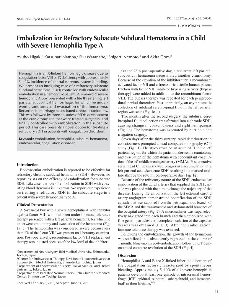
On the 28th post-operative day, a recurrent left parietal subcortical hematoma necessitated another craniotomy. Because of the elevation of the inhibitor titer, a recombinant activated factor VII and a freeze-dried sterile human plasma fraction with factor VIII inhibitor bypassing activity (bypass therapy) were added in addition to the recombinant factor VIII. The bypass therapy was repeated for each periproce-dural period thereafter. Post-operatively, an asymptomatic collection of subdural cerebrospinal fluid in the left parietal region was seen (Fig. 1c, d).
Two months after the second surgery, the subdural cere-brospinal fluid collection transformed into a chronic SDH, causing change in consciousness and right hemiparesis (Fig. 1e). The hematoma was evacuated by burr hole and irrigation surgery.
Seven days after the third surgery, rapid deterioration in consciousness prompted a head computed tomography (CT) study (Fig. 1f). The study revealed an acute SDH in the left parietal region, for which the patient underwent a craniotomy and evacuation of the hematoma with concomitant coagula-tion of the left middle meningeal artery (MMA). Post-operative serial head CT scans showed progressive accumulation of a left parietal acute/subacute SDH resulting in a marked mid-line shift by the seventh post-operative day (Fig. 1g).
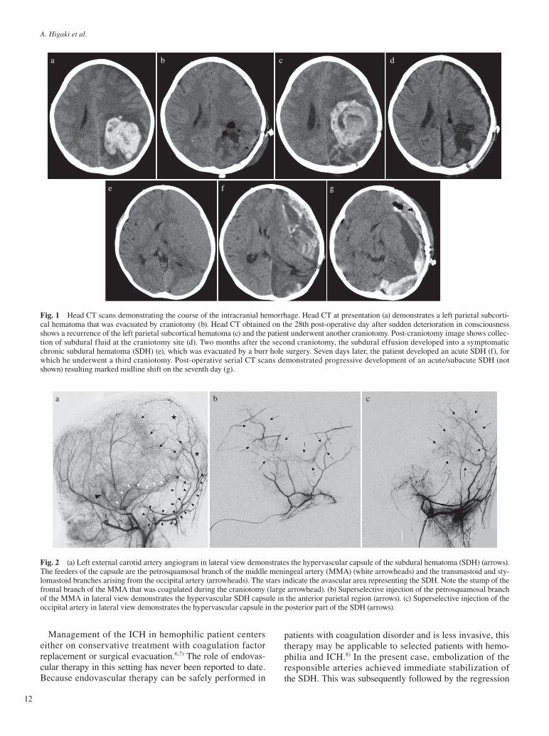
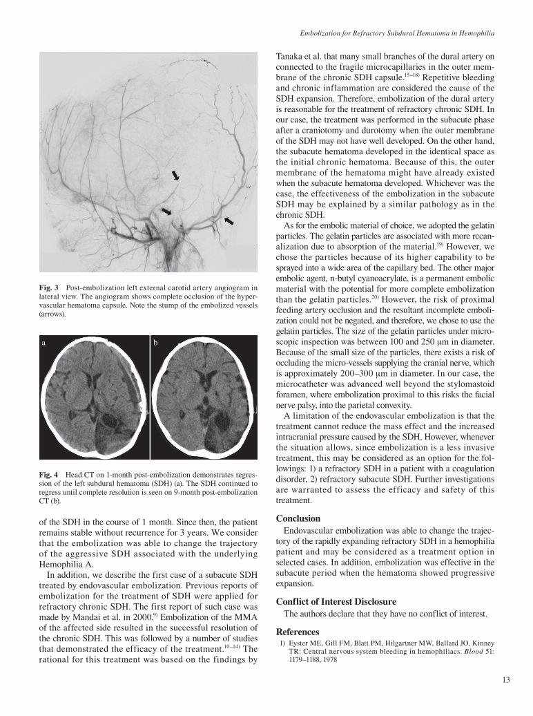
Because of the refractory nature of the SDH, endovascular embolization of the dural arteries that supplied the SDH cap-sule was planned with the aim to change the trajectory of the disease. During the embolization, the left external carotid artery angiogram demonstrated opacification of the SDH capsule that was supplied from the petrosquamous branch of the MMA and the transmastoid and stylomastoid branches of the occipital artery (Fig. 2). A microcatheter was superselec-tively navigated into each branch and then embolized with fine gelatin particles until complete occlusion of the capsular vessels was obtained (Fig. 3). After the embolization, immune tolerance therapy was resumed.
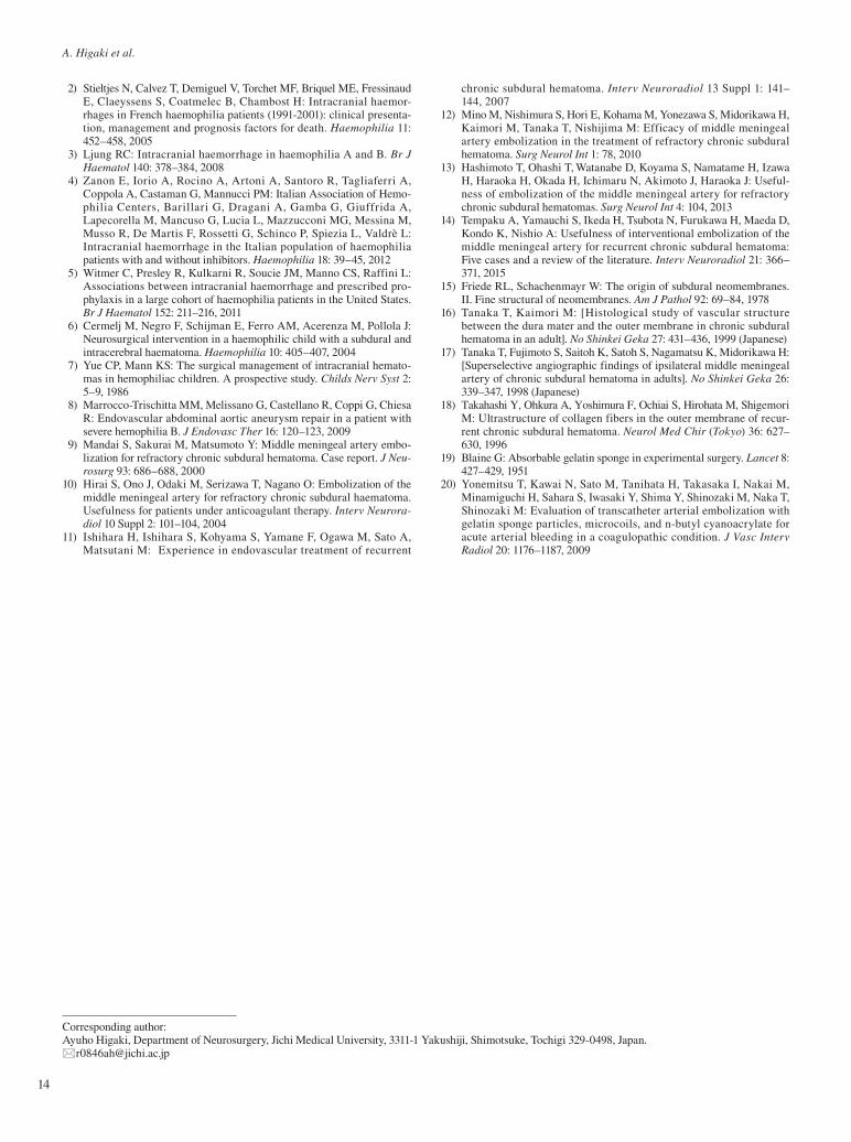
Following the embolization, the growth of the hematoma was stabilized and subsequently regressed in the course of 1 month. Nine-month post-embolization follow up CT dem-onstrated complete resolution of the SDH (Fig. 4).
DiscussionHemophilia A and B are X-linked inherited disorders of
the coagulation factors characterized by spontaneous bleeding. Approximately 5–10% of all severe hemophilic patients develop at least one episode of intracranial hemor-rhage (ICH: epidural, subdural, subarachnoid, and intracere-bral) in their lifetime.1–5)
Hemophilia is an X-linked hemorrhagic disease due to coagulation factor VIII or IX deficiency with approximately 5–10% incidence of central nervous system bleeding. We present an intriguing case of a refractory subacute subdural hematoma (SDH) controlled with endovascular embolization in a hemophilic patient. A 5-year-old severe hemophilic A boy presented with a life threatening left parietal subcortical hemorrhage, for which he under-went craniotomy and evacuation of the hematoma. Recurrent hemorrhage necessitated a repeat craniotomy. This was followed by three episodes of SDH development at the craniotomy site that were treated surgically, and finally controlled with embolization in the subacute period. This case presents a novel option for treating a refractory SDH in patients with coagulation disorders.
Keywords: embolization, hemophilia, subdural hematoma, endovascular, coagulation disorder
IntroductionEndovascular embolization is reported to be effective for
refractory chronic subdural hematoma (SDH). However, no report exists on the efficacy of embolization for subacute SDH. Likewise, the role of embolization in SDH with coex-isting blood dyscrasia is unknown. We report our experience on treating a refractory SDH in the subacute stage in a patient with severe hemophilia type A.
Clinical PresentationA 5-year-old boy with a severe hemophilia A with inhibitor
against factor VIII who had been under immune tolerance therapy presented with a left parietal hematoma, for which he underwent craniotomy and evacuation of the hematoma (Fig. 1a, b). The hemophilia was considered severe because less than 1% of the factor VIII was present on laboratory examina-tion. Post-operatively, recombinant factor VIII replacement therapy was initiated because of the low level of the inhibitor.
12
A. Higaki et al.
Fig. 1 Head CT scans demonstrating the course of the intracranial hemorrhage. Head CT at presentation (a) demonstrates a left parietal subcorti-cal hematoma that was evacuated by craniotomy (b). Head CT obtained on the 28th post-operative day after sudden deterioration in consciousness shows a recurrence of the left parietal subcortical hematoma (c) and the patient underwent another craniotomy. Post-craniotomy image shows collec-tion of subdural fluid at the craniotomy site (d). Two months after the second craniotomy, the subdural effusion developed into a symptomatic chronic subdural hematoma (SDH) (e), which was evacuated by a burr hole surgery. Seven days later, the patient developed an acute SDH (f), for which he underwent a third craniotomy. Post-operative serial CT scans demonstrated progressive development of an acute/subacute SDH (not shown) resulting marked midline shift on the seventh day (g).
b c d
e f g
a
Fig. 2 (a) Left external carotid artery angiogram in lateral view demonstrates the hypervascular capsule of the subdural hematoma (SDH) (arrows). The feeders of the capsule are the petrosquamosal branch of the middle meningeal artery (MMA) (white arrowheads) and the transmastoid and sty-lomastoid branches arising from the occipital artery (arrowheads). The stars indicate the avascular area representing the SDH. Note the stump of the frontal branch of the MMA that was coagulated during the craniotomy (large arrowhead). (b) Superselective injection of the petrosquamosal branch of the MMA in lateral view demonstrates the hypervascular SDH capsule in the anterior parietal region (arrows). (c) Superselective injection of the occipital artery in lateral view demonstrates the hypervascular capsule in the posterior part of the SDH (arrows).
ba c
Management of the ICH in hemophilic patient centers either on conservative treatment with coagulation factor replacement or surgical evacuation.6,7) The role of endovas-cular therapy in this setting has never been reported to date. Because endovascular therapy can be safely performed in
patients with coagulation disorder and is less invasive, this therapy may be applicable to selected patients with hemo-philia and ICH.8) In the present case, embolization of the responsible arteries achieved immediate stabilization of the SDH. This was subsequently followed by the regression
Embolization for Refractory Subdural Hematoma in Hemophilia
13
Fig. 3 Post-embolization left external carotid artery angiogram in lateral view. The angiogram shows complete occlusion of the hyper-vascular hematoma capsule. Note the stump of the embolized vessels (arrows).
of the SDH in the course of 1 month. Since then, the patient remains stable without recurrence for 3 years. We consider that the embolization was able to change the trajectory of the aggressive SDH associated with the underlying Hemophilia A.
In addition, we describe the first case of a subacute SDH treated by endovascular embolization. Previous reports of embolization for the treatment of SDH were applied for refractory chronic SDH. The first report of such case was made by Mandai et al. in 2000.9) Embolization of the MMA of the affected side resulted in the successful resolution of the chronic SDH. This was followed by a number of studies that demonstrated the efficacy of the treatment.10–14) The rational for this treatment was based on the findings by
Tanaka et al. that many small branches of the dural artery on connected to the fragile microcapillaries in the outer mem-brane of the chronic SDH capsule.15–18) Repetitive bleeding and chronic inflammation are considered the cause of the SDH expansion. Therefore, embolization of the dural artery is reasonable for the treatment of refractory chronic SDH. In our case, the treatment was performed in the subacute phase after a craniotomy and durotomy when the outer membrane of the SDH may not have well developed. On the other hand, the subacute hematoma developed in the identical space as the initial chronic hematoma. Because of this, the outer membrane of the hematoma might have already existed when the subacute hematoma developed. Whichever was the case, the effectiveness of the embolization in the subacute SDH may be explained by a similar pathology as in the chronic SDH.
As for the embolic material of choice, we adopted the gelatin particles. The gelatin particles are associated with more recan-alization due to absorption of the material.19) However, we chose the particles because of its higher capability to be sprayed into a wide area of the capillary bed. The other major embolic agent, n-butyl cyanoacrylate, is a permanent embolic material with the potential for more complete embolization than the gelatin particles.20) However, the risk of proximal feeding artery occlusion and the resultant incomplete emboli-zation could not be negated, and therefore, we chose to use the gelatin particles. The size of the gelatin particles under micro-scopic inspection was between 100 and 250 μm in diameter. Because of the small size of the particles, there exists a risk of occluding the micro-vessels supplying the cranial nerve, which is approximately 200–300 μm in diameter. In our case, the microcatheter was advanced well beyond the stylomastoid foramen, where embolization proximal to this risks the facial nerve palsy, into the parietal convexity.
A limitation of the endovascular embolization is that the treatment cannot reduce the mass effect and the increased intracranial pressure caused by the SDH. However, whenever the situation allows, since embolization is a less invasive treatment, this may be considered as an option for the fol-lowings: 1) a refractory SDH in a patient with a coagulation disorder, 2) refractory subacute SDH. Further investigations are warranted to assess the efficacy and safety of this treatment.
ConclusionEndovascular embolization was able to change the trajec-
tory of the rapidly expanding refractory SDH in a hemophilia patient and may be considered as a treatment option in selected cases. In addition, embolization was effective in the subacute period when the hematoma showed progressive expansion.
Conflict of Interest DisclosureThe authors declare that they have no conflict of interest.
References 1) Eyster ME, Gill FM, Blatt PM, Hilgartner MW, Ballard JO, Kinney
TR: Central nervous system bleeding in hemophiliacs. Blood 51: 1179–1188, 1978
Fig. 4 Head CT on 1-month post-embolization demonstrates regres-sion of the left subdural hematoma (SDH) (a). The SDH continued to regress until complete resolution is seen on 9-month post-embolization CT (b).
ba
14
A. Higaki et al.
2) Stieltjes N, Calvez T, Demiguel V, Torchet MF, Briquel ME, Fressinaud E, Claeyssens S, Coatmelec B, Chambost H: Intracranial haemor-rhages in French haemophilia patients (1991-2001): clinical presenta-tion, management and prognosis factors for death. Haemophilia 11: 452–458, 2005
3) Ljung RC: Intracranial haemorrhage in haemophilia A and B. Br J Haematol 140: 378–384, 2008
4) Zanon E, Iorio A, Rocino A, Artoni A, Santoro R, Tagliaferri A, Coppola A, Castaman G, Mannucci PM: Italian Association of Hemo-philia Centers, Barillari G, Dragani A, Gamba G, Giuffrida A, Lapecorella M, Mancuso G, Lucia L, Mazzucconi MG, Messina M, Musso R, De Martis F, Rossetti G, Schinco P, Spiezia L, Valdrè L: Intracranial haemorrhage in the Italian population of haemophilia patients with and without inhibitors. Haemophilia 18: 39–45, 2012
5) Witmer C, Presley R, Kulkarni R, Soucie JM, Manno CS, Raffini L: Associations between intracranial haemorrhage and prescribed pro-phylaxis in a large cohort of haemophilia patients in the United States. Br J Haematol 152: 211–216, 2011
6) Cermelj M, Negro F, Schijman E, Ferro AM, Acerenza M, Pollola J: Neurosurgical intervention in a haemophilic child with a subdural and intracerebral haematoma. Haemophilia 10: 405–407, 2004
7) Yue CP, Mann KS: The surgical management of intracranial hemato-mas in hemophiliac children. A prospective study. Childs Nerv Syst 2: 5–9, 1986
8) Marrocco-Trischitta MM, Melissano G, Castellano R, Coppi G, Chiesa R: Endovascular abdominal aortic aneurysm repair in a patient with severe hemophilia B. J Endovasc Ther 16: 120–123, 2009
9) Mandai S, Sakurai M, Matsumoto Y: Middle meningeal artery embo-lization for refractory chronic subdural hematoma. Case report. J Neu-rosurg 93: 686–688, 2000
10) Hirai S, Ono J, Odaki M, Serizawa T, Nagano O: Embolization of the middle meningeal artery for refractory chronic subdural haematoma. Usefulness for patients under anticoagulant therapy. Interv Neurora-diol 10 Suppl 2: 101–104, 2004
11) Ishihara H, Ishihara S, Kohyama S, Yamane F, Ogawa M, Sato A, Matsutani M: Experience in endovascular treatment of recurrent
chronic subdural hematoma. Interv Neuroradiol 13 Suppl 1: 141–144, 2007
12) Mino M, Nishimura S, Hori E, Kohama M, Yonezawa S, Midorikawa H, Kaimori M, Tanaka T, Nishijima M: Efficacy of middle meningeal artery embolization in the treatment of refractory chronic subdural hematoma. Surg Neurol Int 1: 78, 2010
13) Hashimoto T, Ohashi T, Watanabe D, Koyama S, Namatame H, Izawa H, Haraoka H, Okada H, Ichimaru N, Akimoto J, Haraoka J: Useful-ness of embolization of the middle meningeal artery for refractory chronic subdural hematomas. Surg Neurol Int 4: 104, 2013
14) Tempaku A, Yamauchi S, Ikeda H, Tsubota N, Furukawa H, Maeda D, Kondo K, Nishio A: Usefulness of interventional embolization of the middle meningeal artery for recurrent chronic subdural hematoma: Five cases and a review of the literature. Interv Neuroradiol 21: 366–371, 2015
15) Friede RL, Schachenmayr W: The origin of subdural neomembranes. II. Fine structural of neomembranes. Am J Pathol 92: 69–84, 1978
16) Tanaka T, Kaimori M: [Histological study of vascular structure between the dura mater and the outer membrane in chronic subdural hematoma in an adult]. No Shinkei Geka 27: 431–436, 1999 (Japanese)
17) Tanaka T, Fujimoto S, Saitoh K, Satoh S, Nagamatsu K, Midorikawa H: [Superselective angiographic findings of ipsilateral middle meningeal artery of chronic subdural hematoma in adults]. No Shinkei Geka 26: 339–347, 1998 (Japanese)
18) Takahashi Y, Ohkura A, Yoshimura F, Ochiai S, Hirohata M, Shigemori M: Ultrastructure of collagen fibers in the outer membrane of recur-rent chronic subdural hematoma. Neurol Med Chir (Tokyo) 36: 627–630, 1996
19) Blaine G: Absorbable gelatin sponge in experimental surgery. Lancet 8: 427–429, 1951
20) Yonemitsu T, Kawai N, Sato M, Tanihata H, Takasaka I, Nakai M, Minamiguchi H, Sahara S, Iwasaki Y, Shima Y, Shinozaki M, Naka T, Shinozaki M: Evaluation of transcatheter arterial embolization with gelatin sponge particles, microcoils, and n-butyl cyanoacrylate for acute arterial bleeding in a coagulopathic condition. J Vasc Interv Radiol 20: 1176–1187, 2009
Corresponding author: Ayuho Higaki, Department of Neurosurgery, Jichi Medical University, 3311-1 Yakushiji, Shimotsuke, Tochigi 329-0498, Japan.* [email protected]