Embed Size (px)
Citation preview
JYVÄSKYLÄ STUDIES IN BIOLOGICAL AND ENVIRONMENTAL SCIENCE
324
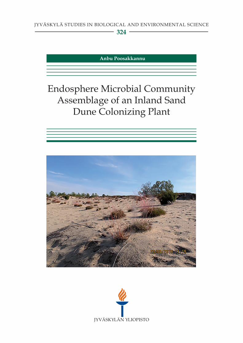
Endosphere Microbial Community Assemblage of an Inland Sand
Dune Colonizing Plant
Anbu Poosakkannu
JYVÄSKYLÄ STUDIES IN BIOLOGICAL AND ENVIRONMENTAL SCIENCE 324
Anbu Poosakkannu
Endosphere Microbial Community Assemblage of an Inland Sand Dune
Colonizing Plant
Esitetään Jyväskylän yliopiston matemaattis-luonnontieteellisen tiedekunnan suostumuksellajulkisesti tarkastettavaksi yliopiston Ylistönrinteen salissaYAA 303,
joulukuun 9. päivänä 2016 kello 12.
Academic dissertation to be publicly discussed, by permission ofthe Faculty of Mathematics and Science of the University of Jyväskylä,in Ylistönrinne, hall YAA 303, on December 9, 2016 at 12 o’clock noon.
UNIVERSITY OF JYVÄSKYLÄ
JYVÄSKYLÄ 2016
Endosphere Microbial Community Assemblage of an Inland Sand Dune
Colonizing Plant
JYVÄSKYLÄ STUDIES IN BIOLOGICAL AND ENVIRONMENTAL SCIENCE 324
Anbu Poosakkannu
Endosphere Microbial Community Assemblage of an Inland Sand Dune
Colonizing Plant
UNIVERSITY OF JYVÄSKYLÄ
JYVÄSKYLÄ 2016
EditorsJari HaimiDepartment of Biological and Environmental Science, University of JyväskyläPekka OlsboPublishing Unit, University Library of Jyväskylä
Jyväskylä Studies in Biological and Environmental ScienceEditorial Board
Jari Haimi, Anssi Lensu, Timo Marjomäki, Varpu MarjomäkiDepartment of Biological and Environmental Science, University of Jyväskylä
URN:ISBN:978-951-39-6848-9 ISBN 978-951-39-6848-9 (PDF)
ISBN 978-951-39-6847-2 (nid.)ISSN 1456-9701
Copyright © 2016, by University of Jyväskylä
Jyväskylä University Printing House, Jyväskylä 2016
ABSTRACT
Poosakkannu, Anbu Endosphere microbial community assemblage of an inland sand dune colonizing plant Jyväskylä: University of Jyväskylä, 2016, 38 p. (Jyväskylä Studies in Biological and Environmental Science ISSN 1456-9701; 324) ISBN 978-951-39-6847-2 (nid.) ISBN 978-951-39-6848-9 (PDF) Yhteenveto: Metsälauhan endofyyttisten mikrobien yhteisökoostumus lentohiekka-alueiden sukkessiossa Diss.
Plant-associated microbes could play a role in plant colonization of sand dune ecosystems, but microbes associated with plants colonizing those ecosystems in the arctic are poorly known. I characterized Deschampsia flexuosa-associated microbiomes in two successional stages (early and late) of arctic inland sand dune differ in their plant species richness and soil physiochemical properties. The work based on culturable microbes showed that different plant parts harbour generalist and specific groups of endosphere microbes and most of the endosphere bacteria were closely related to other cold habitat microbes. Also, most of the endosphere bacteria possessed an important plant growth promoting property of solubilizing organic phosphate. Next generation sequencing methods showed that endosphere microbial species richness was determined by soil characteristics (succession) and plant compartment. Successional stage strongly affected the microbial community composition. Further, reciprocal transplantation experiment showed that endosphere microbial species richness was determined by successional stage rather than transplantation type (self or reciprocal). Irrespective of successional stage, after reciprocal transplantation microbial community compositions in most of the leaf and root compartments differed from local non-transplanted control. In contrast, the microbial community composition only in few root compartments was affected by self-transplantation. Further, leaf endosphere bacterial community composition was significantly affected by arbuscular mycorrhizal fungal (AMF) inoculation under greenhouse conditions. Overall, my work provided data from poorly characterized arctic biota and novel insight into endosphere bacterial and fungal community assemblage in arctic inland sand dune ecosystem. These results could be utilized when restoring vegetation in sand dunes and similar extreme ecosystems.
Keywords: Succession; transplantation; endosphere microbes; arbuscular mycorrhiza; sand dunes; arctic.
Anbu Poosakkannu, University of Jyväskylä, Department of Biological and Environmental Science, P.O. Box 35, FI-40014 University of Jyväskylä, Finland
Author’s address Anbu Poosakkannu Department of Biological and Environmental Science P.O. Box 35 FI-40014 University of Jyväskylä Finland [email protected]
Supervisors Dr. Minna-Maarit Kytöviita Department of Biological and Environmental Science P.O. Box 35 FI-40014 University of Jyväskylä Finland
Dr. Riitta Nissinen Department of Biological and Environmental Science P.O. Box 35 FI-40014 University of Jyväskylä Finland
Reviewers Dr. Joana Falcao Salles Nijenborgh 7 9747AG Groningen The Netherlands
Dr. Sabine Ravsnskov Forsøgsvej 1 4200 Slagelse Denmark
Opponent Dr. LS (Leo) van Overbeek PO Box 16 6700AA Wageningen The Netherlands
CONTENTS
LIST OF ORIGINAL PUBLICATIONS
1 INTRODUCTION ................................................................................................. 71.1 Plant-associated microbes ............................................................................ 71.2 Endosphere microbes ..................................................................................... 71.3 Succession ........................................................................................................ 8
1.3.1 Ecological succession .......................................................................... 81.3.2 Sand dune primary successional habitats ........................................ 9
1.4 Deschampsia flexuosa ........................................................................................ 91.5 Reciprocal transplantation .......................................................................... 101.6 Arbuscular mycorrhiza fungi interaction with other microbes ............. 101.7 Aim of the study ........................................................................................... 11
2 MATERIALS AND METHODS ........................................................................ 132.1 The field experiments ................................................................................... 13
2.1.1 Study site ............................................................................................. 132.1.2 Experiments and the sampling scheme .......................................... 142.1.3 Study I ................................................................................................. 142.1.4 Study II ................................................................................................ 142.1.5 Study III ............................................................................................... 14
2.2 Greenhouse experiment (Study IV) ........................................................... 152.3 Plant tissue surface sterilization ................................................................. 152.4 Endophyte isolation, identification and characterization ....................... 152.5 Molecular analyses of microbes in plants and soil .................................. 16
2.5.1 DNA isolation, PCR and Ion torrent sequencing .......................... 162.5.2 Bioinformatics .................................................................................... 162.5.3 Statistical analyses ............................................................................. 17
3 RESULTS AND DISCUSSION .......................................................................... 183.1 Cold habitat specificity of culturable endophytic microbes (I) .............. 183.2 Seed endophytes able to mobilize organic phosphate (I) ....................... 183.3 Succession affects the microbial communities (I and II) ......................... 193.4 Transplantation affects endosphere microbial communities (III) ......... 203.5 Mycorrhiza affects leaf endosphere bacterial composition (IV) ............ 21
4 CONCLUSIONS .................................................................................................. 23
Acknowledgements ..................................................................................................... 26 YHTEENVETO ............................................................................................................. 28 REFERENCES ............................................................................................................... 30
LIST OF ORIGINAL PUBLICATIONS
The thesis is based on the following original papers, which will be referred to in the text by their roman numerals I-IV.
I Poosakkannu, A., Nissinen R. & Kytöviita, M.-M 2015. Culturable endophytic microbial communities in the circumpolar grass, Deschampsia flexuosa in a sub-Arctic inland primary succession are habitat and growth stage specific. Environmental Microbiology Reports 7: 111–122.
II Poosakkannu, A., Nissinen R. Männistö, M. & Kytöviita, M.-M 2016. Microbial community composition but not diversity changes along succession in arctic sand dunes. Environmental Microbiology, in press.
III Poosakkannu A., & Kytöviita M.-M 2016. Endosphere microbial legacy after plant transplantation in arctic sand dunes. Manuscript.
IV Poosakkannu, A., Nissinen R. & Kytöviita, M.-M 2016. Native arbuscular mycorrhizal symbiosis changes leaf endosphere bacterial community composition in Deschampsia flexuosa. Manuscript.
The table shows the contributions to the original papers.
I II III IVPlanning AP, RN, MMK AP, RN, MM, MMK AP, MMK AP, RN, MMK Data AP, RN, MMK AP, RN, MMK AP, MMK AP, MMK Analyses AP, RN, MMK AP, RN, MMK AP, MMK AP, MMK Writing AP, RN, MMK AP, RN, MM, MMK AP, MMK AP, RN, MMK
AP = Anbu Poosakkannu, RN = Riitta Nissinen, MM = Minna Männistö, and MMK = Minna-Maarit Kytöviita
1 INTRODUCTION
1.1 Plant-associated microbes
The microbes that live in close association with plants are known as plant-associated microbes. They are mainly divided into phyllosphere (leaf surface), rhizosphere (root surface) and endosphere (inside plant) microbes. The plant-associated microbes mainly include bacteria and fungi. Many of these microbes have neutral or positive impact on host plants such as improved growth and protection from different biotic and abiotic stresses (Haney et al. 2015, Ludwig-Müller 2015, Panke-Buisse et al. 2015, Hansen and Moran 2014, Karasov et al. 2014). Recent advances in next generation sequencing techniques enhanced understanding the plant-associated microbes at comprehensive community level (Gottel et al. 2011, Bulgarelli et al. 2012, Peiffer et al. 2013, Bulgarelli et al. 2015, Edwards et al. 2015, Coleman Derr et al. 2016). These studies mainly focused on tropical and temperate agricultural ecosystems and show that rhizosphere microbes are much more diverse than endosphere microbes. However, comprehensive microbial community level studies on arctic plant species are still in its infancy.
1.2 Endosphere microbes
Endosphere microbes are taxonomically diverse and mixture of bacterial and fungal communities lives inside the asymptomatic host plants (van Overbeek and Saikkonen 2016). The endosphere microbes interact with each other and also interact with their host (van Overbeek and Saikkonen 2016). Many of the endosphere microbes are known as commensals and their function in plants yet unknown (Hardoim et al. 2015). Some of the endosphere microbes are known to have beneficial effect on plants such as increasing the plant growth and development by biological nitrogen fixation, phosphate solubilisation and indole acetic acid production (Compant et al. 2010). They also protect the plants
8
from different biotic and abiotic stresses (Compant et al. 2010). A very few endosphere microbes are known to have negative effect on plants (Hardoim et al. 2015).
The major source of the rhizosphere and phyllosphere microbes is soil (Berg and Smalla 2009, Normander and Prosser 2000, Philippot et al. 2013, Singh et al. 2007, Zarraonaindia et al. 2015) and air (Maignien et al. 2014). This means that rhizosphere and phyllosphere act as major sources of endosphere microbes (Bulgarelli et al. 2012, Edwards et al. 2015, Long et al. 2008, Lundberg et al. 2012). Recent evidence suggests that invertebrates such as insects could act as vectors of microbial transmission in to the plants (Pažoutová et al. 2013). Vertical transmission via seeds is also one of the significant sources of the endosphere microbes (Johnston-Monje et al. 2014, Johnston-Monje and Raizada 2011, Hardoim et al. 2012, Puente et al. 2009). The soil, microbial and plant factors determine the endosphere microbe survival, colonization, compatibility, and competing ability within the plant (Gaiero et al. 2013).
1.3 Succession
1.3.1 Ecological succession
Ecological succession is the sequence of changes in structural and functional properties of species community composition over a period of time (Drury and Nisbet 1973). The change in species composition in a newly formed land such as lava ows, sand dunes, landslides, and glacial till is known as primary succession (Nemergut et al. 2007). In contrast, secondary succession includes the changes in already established ecosystem after disturbance (e.g., forest fire, and cyclone).
The conceptual understanding of the ecological succession was initiated from the study of vegetation succession (Clements 1916). It is a general assumption that the plant and animal species richness increase along the progressive ecosystem succession (Bazzaz 1975, Tews et al. 2004). Also, soil organic matter increases towards climax stage (Berendse et al. 1998, Walker and del Moral 2003, Chapin et al. 1994). Similar understanding of succession in microbial system is poor (Schmid et al. 2014). Recent evidence suggests that soil microbial species richness increases along the successional trajectory in primary successional sites (Brown and Jumpponen 2014, Brown and Jumpponen 2015). These studies are mainly focused on vegetated or non-vegetated soil microbial succession. However, the microbes have the ability to colonize different plant parts or different soil compartments for the nutritional (Belnap and Lange 2003) or non-nutritional (Diaz et al. 1993) purposes. Nevertheless, comparisons of plant-associated microbe successional dynamics are rare (Blaalid et al. 2012) and could give us an idea of whether general concept of plant successional trajectory is also applicable to microbial communities.
9
1.3.2 Sand dune primary successional habitats
Sand dunes are considered as severe environments which are characterized by low available nutrients, low water holding capacity and persistent strong winds. In arctic sand dune habitat, above factors combined with cold and highly fluctuating temperature make it even harder for plant survival and establishment. Sand dunes are dynamic ecosystems presenting a sequence of vegetation spanning from barren sand to climax vegetation and provide different successional stages with distinct plant species richness (Cowles 1899, Olson 1958, Lichter 1998, Maun 2009). It is possible to study the primary successional patterns using the spatial gradient as a substitute for long time observations.
Inland sand dunes are expanding in some parts of the world, which reduces the forest and arable land area. It could be possible to stabilize the sand dunes by successful establishment of vegetation. There is some evidence that microbial aggregates and especially mycorrhizal fungi play a role in establishment of plant species in sand dune areas (Sutton and Sheppard 1976, Koske and Poison 1984, Forster 1990, Miller and Jastrow 1992, Belnap and Lange 2003). However, there are no studies examining the possible role of endosphere microbes in plant establishment in sand dunes. A comprehensive community level study of endosphere microbes related to sand dune colonizing plant species along the succession could give us an idea of their potential role in sand dune primary successional sites.
1.4 Deschampsia flexuosa
Deschampsia flexuosa is commonly known as wavy hair grass, a cosmopolitan species of temperate and subarctic regions (Scurfield 1954). The optimum pH for D. flexuosa growth is 5.5 to 6. However, the plant has the ability to grow also in more acidic soils (Hackett 1965). Further, D. flexuosa is one of the early colonizers of sand dunes (Borgegård et al. 1990, Ujházy et al. 2011).
A few studies have characterized the plant-associated microbes in seeds and roots of D. flexuosa. Studies using the staining techniques show that seeds and roots of D. flexuosa harbour mutualistic and endophytic fungi (Saikkonen et al. 2000, Pietikäinen et al. 2005, Ruotsalainen et al. 2007). A study using culturable and clone library methods shows that D. flexuosa root endosphere mainly comprises dark septate endophytic fungi (Phialocephala fortinii), the abundance of which increases along the coastal sand dune succession (Tejesvi et al. 2010). Further, D. flexuosa is known to host root endosphere fungal communities dissimilar to those in neighbouring plant species (Tejesvi et al. 2013).
10
1.5 Reciprocal transplantation
Reciprocal transplantation is one of the oldest approaches in macro-ecology to study local adaptation of an organism in to the new environment (Ross et al. 2009, Scheepens and Stöcklin 2013). In reciprocal transplantation experiments, plants are reciprocally transplanted from local (home) environment to a novel (away) environment which can be compared with self-transplanted from local to local (Ågren and Schemske 2012).
Recently, soil microbiologists adopted the reciprocal transplantation strategy to tests for effects of both environment and microbial community composition (and their interactions) on the functioning of microorganism to multiple environments from their original environments (Reed and Martiny 2007, Rawls et al. 2006). After transplantation to new environment, microbial communities seem resistant to the new environmental conditions for long time (Bottomley et al. 2006, Liu et al. 2015, Zhao et al. 2014). In contrast, planting soil back to home system seems not to affect the soil microbiota (Lazzaro et al. 2011). It is unknown that whether endosphere microbes which depend on both soil and plant characteristics follow the same resistant behaviour as their soil microbial counterparts.
It could be possible to study phenotypic plasticity of a plant species in a new environment using reciprocal transplantation and correlate it with change or no change in endosphere microbial community status of the transplanted plants. It has been shown that a specific endosymbiotic microbial association is one of the factors affecting the phenotypic plasticity of the plants in new environment (Sultan 1995, Rodriguez et al., 2008, Rodriguez et al., 2010). However, it is not known how the endosphere microbial community shift is reflected in plant adaptation in new environment.
1.6 Arbuscular mycorrhiza fungi interaction with other microbes
Arbuscular mycorrhizal fungi (AMF) live inside the plant root as well as in the soil connecting the inside of the host plant to the outer soil (Miller et al. 1995). AMF increase the host plant nutrient and water uptake from soil (Finlay 2008) and utilize photosynthetic carbohydrates of host plants. The positive effect of AMF on plant growth and development is well documented.
It has been shown that AMF interact with other soil microbes (Frey-Klett et al. 2007, Bonfante and Anca 2009, Scervino et al. 2009). They are known to affect the physiology and functioning of AMF. Also, AMF could affect the soil bacterial and fungal populations (Andrade et al. 1997). AMF interaction with other plant-associated microbes are rarely studied and restricted to root-associated microbes, mainly to the rhizosphere. AMF could affect the soil and plant physio-chemical properties, for example, by altering the root exudate composition which in turn affects the root-associated microbes (Wamberg et al.
11
2003, Gryndler 2000, Jeffries et al. 2003, Scheffknecht et al. 2006, Gupta 2003, Vigo et al. 2000, Marschner et al. 2001). However, all these studies used culturable microbes or molecular finger-printing methods. There are no studies specifically targeted to endosphere microbes. Considering the role of AMF in assisting the plants in nutrient poor and harsh environments such as sand dunes (Sutton and Sheppard 1976), the study of AMF interaction with other endosphere microbes could give a new dimension in ecological relevance of their role in plant physiology.
1.7 Aim of the study
In this PhD project, I focused on the grass Deschampsia flexuosa that has circumpolar distribution and is a major component of the northern ecosystems. Deschampsia flexuosa has a wide ecological tolerance and is commonly found in early successional ecosystems to late, climax stage ecosystems. Especially D. flexuosa has an important role as reindeer fodder and as one of the first plant species colonizing eroded arctic soil and thus enabling ecosystem restoration. The long-term goal of this project is to identify the potential microbes that could be used in soil ecosystem restoration and in enhancing plant survival and well-being in high stress environments in general. It would be possible only if we know the different microbial species present and the factors responsible for their assemblage.
Considering that plant-associated microbes in arctic sand dune habitats are yet unknown, this thesis mainly aimed at exploring the sand dune colonizing grass plant, Deschampsia flexuosa associated microbes and their ecological relevance to possible extent. I characterized in-depth composition of endosphere microbes in two successional stages of arctic inland sand dune primary successional sites. In this thesis, I investigated the effect of different factors such as succession, transplantation and AMF inoculation on endosphere microbial assemblage. In the studies I, II and III, I investigated the changes in endosphere microbial community in two different successional stages located closely to each other in natural sand dune ecosystem. I adopted greenhouse condition for study IV. It helped me to control the AMF infection and study changes in endosphere microbial community.
In study I, microbes were isolated and grown in pure culture to identify the differences at species level and answer the following specific questions: (i) Does the culturable endophyte community composition reflect the cold climate of their host plants? (ii) Do different plant parts (leaf, root and seed) and plants in different successional stages have distinct culturable endophyte communities? and (iii) Do endophytic bacterial isolates solubilize organic phosphate rather than mineral phosphate?
In study II, I used high throughput sequencing techniques to compare the total microbial communities to answer the following specific questions: (i) Does late succession host more numerous microbial species than early succession? (ii)
12
Does successional stage of the ecosystem affect microbial community composition significantly? and (iii) Are co-occurring microbes succession stage specific?
In study III, I used reciprocal transplantation strategy to elucidate the following questions: i) will original endosphere community displaced by destination habitat microbes result in marked community shift or change in species richness? and (ii) is plant performance lower in the novel environment when compared to home environment?
In study IV, plants inoculated with AMF were used to answer the question whether AMF colonization affects microbial species richness and microbial community composition inside plant tissues.
2 MATERIALS AND METHODS
2.1 The field experiments
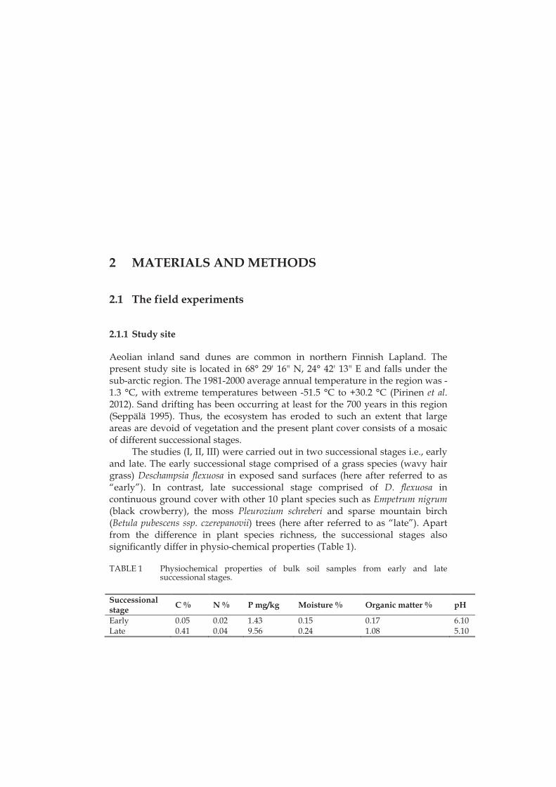
2.1.1 Study site
Aeolian inland sand dunes are common in northern Finnish Lapland. The present study site is located in 68° 29' 16" N, 24° 42' 13" E and falls under the sub-arctic region. The 1981-2000 average annual temperature in the region was -1.3 °C, with extreme temperatures between -51.5 °C to +30.2 °C (Pirinen et al. 2012). Sand drifting has been occurring at least for the 700 years in this region (Seppälä 1995). Thus, the ecosystem has eroded to such an extent that large areas are devoid of vegetation and the present plant cover consists of a mosaic of different successional stages.
The studies (I, II, III) were carried out in two successional stages i.e., early and late. The early successional stage comprised of a grass species (wavy hair grass) Deschampsia flexuosa in exposed sand surfaces (here after referred to as “early”). In contrast, late successional stage comprised of D. flexuosa in continuous ground cover with other 10 plant species such as Empetrum nigrum (black crowberry), the moss Pleurozium schreberi and sparse mountain birch (Betula pubescens ssp. czerepanovii) trees (here after referred to as “late”). Apart from the difference in plant species richness, the successional stages also significantly differ in physio-chemical properties (Table 1).
TABLE 1 Physiochemical properties of bulk soil samples from early and late successional stages.
Successional stage C % N % P mg/kg Moisture % Organic matter % pH
Early 0.05 0.02 1.43 0.15 0.17 6.10Late 0.41 0.04 9.56 0.24 1.08 5.10
14
2.1.2 Experiments and the sampling scheme
Plant and soil samples from two successional stages (early and late) were collected in four different areas between 150 and 2250 meters distance apart. These areas are referred to as ‘block’ hereafter. Within block, the distance between early and late succession stage was 10-20 meters.
2.1.3 Study I
This experiment included five different Deschampsia flexuosa plant parts (108 samples): seeds, seedlings of less than 3cm in height collected from the field (hereafter referred to as “field seedlings”), and seedlings germinated from surface sterilized seeds in green house (hereafter referred to as “experimental seedlings”), and matured plant leaves and roots. In field, a total of 84 samples of seeds (early -12 and late - 12), field seedlings (Only early - 12), and matured plant leaves (early -12 and late - 12) and roots (early -12 and late - 12) were collected. A total of 24 experimental seedlings were germinated in greenhouse using the seeds collected from two successional stages i.e., 12 seedlings per succession. Deschampsia flexuosa field seedlings and leaves and roots of matured plants were collected in 24th July 2011 and seeds were collected 29th August 2011.
2.1.4 Study II
This experiment included two soil categories and two plant parts: bulk soils, rhizosphere soils, and D. flexuosa leaves and roots (the same leaf and root samples were used in study I and II). These different sample types are collectively referred to as “compartment” hereafter. Each of these compartment samples comprised of 24 replicates, i.e. there were 12 replicates per each succession. Successional stage samples were collected in four blocks, i.e., there were 3 replicates per block, per succession and per compartment. The samples were collected in 24th July 2011.
2.1.5 Study III
Deschampsia flexuosa plants naturally growing in the two successional stages were transplanted in 27 and 28th August 2011 within successional stage (self-transplantation) and between successional stages (reciprocal transplantation) in four different blocks. The transplanted plants were allowed to grow for two years. The transplanted (both self and reciprocal) plant leaf and root samples and control (non-transplanted plant samples) were collected in 19th August 2013. The plant biomass before and after transplantation were measured.
15
2.2 Greenhouse experiment (Study IV)
The experiment was carried out in greenhouse because it is possible to control the AMF colonization in greenhouse. Deschampsia flexuosa associated AMF (Claroideoglomus etunicatum) isolated from our study site was used in this experiment. The AMF inoculation experiment was started in 1st May 2014. A total of six plants for each control (no AMF inoculation) and AMF inoculated treatments were maintained in greenhouse. The harvesting was done 15th October 2014. The start and end plant biomasses were measured.
2.3 Plant tissue surface sterilization
The plant samples from all the studies (I, II, III and IV) were surface sterilized using the following method. Pre-weighed tissues of seeds, seedlings, leaves and roots were soaked in 70% ethanol for 1 min, 3% sodium hypochlorite for 3 minutes (except seeds, 6 minutes), 1% sodium thiosulphate for 3 minutes, and washed three times with sterile deionized water for 3 minutes.
2.4 Endophyte isolation, identification and characterization
The bacteria were isolated using the sterilized and homogenized plant tissues. The seedlings, leaves and roots were homogenized in 50 mM potassium phosphate buffer (pH 6.5) and plated in serial dilutions on the R2A media. The seed sterilization was carried out as described by Nissinen et al. (2012). In brief, the seeds were homogenized in BSE buffer (50 mM Tris–HCl [pH7.5], 1% Triton X-100 and 2 mM 2-mercaptoethanol) and centrifuged at 300×g for 5 min (at15°C) to get the supernatant. The second centrifugation was done at 12 000 ×gfor 15 min (at 10°C) to get the pellets and they were suspended in 50 mMpotassium phosphate buffer (pH 6.5). Serial dilutions of suspended pellets wereprepared and plated on the R2A media (pH 6.5). Plates were incubated at roomtemperature for a week and single colonies of bacteria were transferred to newplates to obtain pure cultures.
The fungi were isolated using the sterilized tissues cut into 1 cm pieces and plated directly into malt extract fungal media (Zijlstra et al. 2005). Plates were incubated at room temperature for a month or more. Hyphal tips of the developing fungal colonies were transferred to fresh malt extract agar plates.
The 16S rRNA gene of bacterial isolates were sequenced using PCR products amplified with 27F (5´-AGAGTTTGATCCTGGCTCAG-3´) and 1492R (5 -GGYTACTTGTTACGACTT-3 ) primer pairs. The ITS region of fungal isolates was sequenced using PCR products amplified with ITS1 (5’-CTTGGTCATTTAGAGGAAGTAA-3’) and ITS4 (5’-
16
TCCTCCGCTTATTGATATGC-3’) primer pairs. These isolates sequence data have been submitted to the GenBank databases under accession number KJ528986-KJ529110. The close phylogenetic relatives of bacterial and fungal isolates were identified by NCBI BLAST analysis.
I characterized all bacterial strains for their ability to solubilize mineral as well as organic forms of phosphate using National Botanical Research Institute’s phosphate growth medium (NBRIPM; Nautiyal 1999) and phytase screening medium (PSM; Jorquera et al. 2011). The ability to utilize tri-calcium phosphate and phytate on NBRIPM and PSM agar was examined after incubation for 4 days at room temperature. The development of clearing zone around the colonies was used as an indicator of phosphate solubilization by the isolates.
2.5 Molecular analyses of microbes in plants and soil
2.5.1 DNA isolation, PCR and Ion torrent sequencing
Microbial DNA was extracted from bulk and rhizosphere soil using the PowerSoil DNA isolation kit (MoBio, Carlsbad, CA, USA) and from plant samples using the NucleoSpin Plant II kit (Macherey-Nagel, Düren, Germany) or Invisorb Spin Plant Mini Kit (Stratec Biomedical AG, Germany) following manufacturer’s instructions.
I used nested PCR approach for bacterial 16S rRNA region amplification, first round of 16S rRNA PCR was performed with 799f/ 1492R primer pairs to exclude plastid DNA amplification (Chelius and Triplett 2001). The second round of 16S rRNA PCR was performed with 1062F/1390R primer pairs. I used fITS7/ITS4 primer pairs for the amplification of fungal ITS regions (Ihrmark et al. 2012). I used M13 system for library preparation as described by Mäki et al., (2016). The sequencing was carried out using the Ion PGM Sequencing 400 Kit (Ion 314 chips; Life Technologies, Thermo Fisher Scientific, Waltham, Massachusetts, USA) following the manufacturer's instructions.
2.5.2 Bioinformatics
The bacterial 16S rRNA and fungal ITS sequences were reassigned to their respective samples and quality filtered using the Mothur v.1.35.0 (parameters: minlength = 200; maxambigs = 0; maxhomop = 8; qwindowaverage = 25; qwindowsize = 50; and bdiffs = 1). Further processing of bacterial 16S rRNA gene sequences in Mothur was performed following a standard procedure (Schloss et al. 2011). Fungal ITS sequences were processed as described in Tedersoo et al. (2014). The OTUs were clustered at 97% similarity level. The rare OTUs with five or less than five sequences across the samples were excluded from downstream analyses. The OTU abundance tables were rarefied to their minimum sequencing depth. The raw sequence data are available in National
17
Center for Biotechnology Information Sequence Read Archive under accession number SRP063711, SRP087752, SRP087758. The observed species richness, estimated species richness (Chao 1) and Shannon diversity indices were estimated using Mothur v.1.35.0.
2.5.3 Statistical analyses
To visualize shifts in the microbial community composition, principal coordinate analysis (PCoA) based on Bray-Curtis dissimilarities and Permutational Multivariate Analysis of Variance (PERMANOVA; Anderson, 2001) were used. The PCoAs and PERMANOVA were performed in PRIMER software v6 (http://www.primer-e.com, Clark and Warwick 2001).
Linear statistics were applied to find out significant differences in observed species richness, estimated species richness (Chao 1) and Shannon diversity indices between different samples. I performed Kruskal Wallis test with the log transformed (log [X+1]) relative abundance data to identify the taxa (phyla/class/OTUs) that are responsible for community separation between different samples. I carried out co-occurrence network analyses as described by Williams et al. (2014) to identify modules of co-occurring OTUs within communities. Linear statistics, co-occurrence analyses and Kruskal Wallis test were performed in R statistical software (version 3.3.0).
3 RESULTS AND DISCUSSION
3.1 Cold habitat specificity of culturable endophytic microbes (I)
In our study, 52% of the total endophytic bacterial isolate 16S rRNA sequences were highly similar to bacterial sequences from cold environments, including arctic, antarctic and high alpine soils, snow, and in glacier or arctic-alpine plants. In the study by Nissinen et al. (2012) focusing on three arctic plant species, 40% of the bacterial endophytic isolates were similar to bacteria from cold climates. Sheng et al. (2011) reported that 46% of the endophytic isolates from subnival plants were highly similar to bacteria from other cold climates. Furthermore, 58-100% of the isolates in the vegetative tissues in D. flexuosa were very closely related (99-100% sequence identity) to endophytes from arctic plants from fell tundra in Lapland (Nissinen et al. 2012) and a great portion of these were also closely related to soil bacteria in Lapland, with greatest relative abundance of close relatives of arctic soil bacteria (Männistö and Häggblom 2006) found in the root tissues, indicating horizontal, but selective acquisition of endophytes. Taken together, these results suggest that cold climate bacteria are to large extent habitat specific. In contrast, only 13% of the endophytic fungal isolates were similar to fungi isolated from other cold environments. This indicates that fungal communities in cold environments are poorly studied when compared to their bacterial counterpart, or that unlike fungi, bacteria have developed lineages endemic to cold climates.
3.2 Seed endophytes able to mobilize organic phosphate (I)
Phosphorus is one of the main nutrients limiting plant growth worldwide. Soil mineral phosphorus is often bound to phosphates inaccessible to plants and in plants phosphorus is stored as phytate. Conversion of phytate into inorganic phosphate by the phytase enzyme is one of the important steps in seed germination (Scott and Loewus 1986). I compared the ability of endophytic
19
bacterial isolates for the solubilisation of mineral (tricalcium phosphate) and organic phosphate (phytate). A greater portion of the endophytes were able to mobilize organic (74% of the isolates) than mineral (57%) phosphate. In particular, great majority of seed endophytes were able to solubilize organic phosphate (92%). The seed isolates in the genus Pseudomonas were able to solubilize organic phosphate (100%) and mineral phosphate (87%). It seems likely that seed endophytic bacteria have a significant role in resource mobilization from stored reserves in the plant. Abundance of phosphate solubilizing bacteria was higher in the early than late successional experimental seedlings, which may be linked to the difference in phosphorus availability in these two successions.
3.3 Succession affects the microbial communities (I and II)
Effect of plant succession on microbial species richness was compartment dependent. In our arctic sand dune ecosystem bulk soil bacterial richness increased across the succession. However, succession did not affect soil fungal richness. Similar observation is reported in the primary succession of glacier forefront (Brown and Jumpponen 2014, Brown and Jumpponen 2015). Apparently, more energy was available for microbes in late successional stage with more organic matter and higher carbon content in the soil than in early successional stage in our ecosystem. The results of this study violated the general assumptions that organic matter quantity and quality as well as amount of energy in ecosystems drive fungal species richness (Read 1989, Evans et al. 2005, Pennanen et al. 2001, van der Wal et al. 2013).
Increased plant species richness leads to higher root exudates and available nutrients (de Ridder-Duine et al. 2005, Compant et al. 2010). In our ecosystem, both bacterial and fungal species richness of rhizosphere compartment didn’t increase across the succession. In contrast, bacterial species richness in rhizosphere compartment has been shown to increase along the succession in salt marsh chronosequence (Wang et al. 2015). In the present study, rhizosphere microbial species richness was similar across the succession suggesting that host plant species specific selection rather than bulk soil characteristics determined microbial species richness in rhizosphere. In contrast to rhizosphere, I found that endosphere microbes in leaf (bacteria) and root (fungi) follow the traditional successional trajectory in the arctic inland sand dune ecosystem.
Succession had significant effect on microbial community composition (i.e., both bacteria and fungi) in all compartments. The relative abundance of different microbial taxa (at phyla/class/OTU level) in rhizosphere was the most affected by succession followed by bulk soil, root and leaf endosphere. Both culturable and non-culturable methods showed that Actinobacteria and Acidobacteria were dominant in the early and late successional stages, respectively. Actinobacteria are known to be oligotrophic and successful in
20
nutrient deficient and dry environments (Dion and Nautiyal 2008). Some of them are able to fix atmospheric nitrogen, which could explain their prevalence in the early successional stage with low plant cover. Late successional stage soils in our system had clearly higher organic matter content and lower soil pH in comparison to the early successional stage, which could explain increased relative abundance of Acidobacteria. It is known that Acidobacteria dominate acidic, high organic matter content soils in arctic tundra and to correlate negatively with pH (Jones et al. 2009, Männistö et al. 2013).
Ubiquitous and specific microbial groups were identified from different plant parts and two successional stages. Both root and leaf isolates of the fungus Phialocephala fortinii were confined to the late successional stage in line with the general notion that P. fortinii along with other dark septate endophytes favour habitats with high soil organic matter content (Caldwell et al. 2000; Tejesvi et al. 2010). Also members of the genus Hymenoscyphus isolated in this study in root samples of late successional stage are common endophytes in roots of ericaceous species (Zijlstra et al. 2005) and in other woody species such as pines (Villarreal Ruiz et al. 2004).
Among the different Pseudomonas sp. identified, a tightly clustering group of isolates closely related to P. fluorescens were isolated from the root, leaf and experimental seedlings in both successional stages. This suggests that this group of endophytes is tightly associated with D. flexuosa, and that these P. fluorescens strains may be vertically transmitted. Pseudomonas fluorescens is a well-known plant growth promoting bacterium that has been isolated, among others, in many grasses previously and has been shown to affect plant growth and development, but also reduce seedling disease incidence in rice (Adhikari et al. 2001, Mercado-Blanco and Bakker 2007). In contrast to P. fluorescens, the isolates closely related to P. graminis and P. chlororaphis were more abundant in D. flexuosa in the early succession. Pseudomonas graminis has been reported previously in temperate sand dune plants (Park et al. 2005). Colonization of plants by P. chlororaphis has been proven to be effective in increasing drought tolerance as well as directly inhibiting the growth of fungal pathogens (Cho et al. 2008). All the isolates of P. graminis and P. chlororaphis have the ability to solubilize organic phosphate suggests that they may have prominent role in establishment of D. flexuosa in early succession.
3.4 Transplantation affects endosphere microbial communities (III)
In the transplantation experiment, effect of transplantation (self and reciprocal) on microbial species richness and microbial community composition was successional stage dependent and independent, respectively. Transplantation affected marginally the microbial species richness in some of the early successional stage samples, mainly leaf, but it did not affect late successional
21
stage sample richness. It is often assumed that change in species richness due to disturbance is a “lottery effect” which creates random opportunities for the species living in a similar niche to be nearby the resources after disturbance (Chesson and Warner 1981). It could be possible that nutrient availability for different microbial species due to disturbance was higher in early successional stage which was characterized by low and uneven resource availability to start with.
In this study, after reciprocal transplantation microbial community compositions in most of the leaf and root compartments were different from local non-transplanted control. In contrast, self-transplantation affected the microbial community composition only in few root compartments. Further, most of the differentially expressed OTUs in self-transplantation were also differentially expressed in reciprocal transplantation, but reciprocal transplantation possessed more unique differentially expressed OTUs. This could be due to the marginal effect of self-transplantation in comparison to reciprocal transplantation. Also, pairwise comparison of reciprocal transplantation community compositions to their original successional stage (before transplantation) un-transplanted control community composition shows that two of the root endophytic community compositions still resembled their origin. In previous experiments, it has taken at least two years (Waldrop and Firestone 2006, Liang et al. 2015, Sun et al. 2014) for the soil microbial community composition in transplanted soil samples to resemble the destination soil. These results together with mine indicate that soil and endosphere microbial community compositional changes in a new environmental condition are a slow process.
3.5 Mycorrhiza affects leaf endosphere bacterial composition (IV)
Genetically modified plants that are able to avoid mycorrhizal colonization in field harbour similar root-associated fungal species richness and bacterial richness (except one transgenic line) to mycorrhiza-colonized plants (Groten et al. 2015). In our study, arbuscular mycorrhizal fungi (AMF) inoculated and non-inoculated plants harboured similar bacterial and fungal species richness in both leaf and root endosphere. The difference in bacterial richness of the one transgenic line in Groten et al. (2015) could be due to lack of CCaMK expression which in uences the bacterial colonization of roots (Sanchez et al. 2005). Also, I used clonally propagated initial plant materials which most likely contained same initial microbial community and my study exclusively targeted endosphere microbes.
In my study, AMF inoculation affected the leaf endosphere bacterial community composition. It is possible that changes in nutrient and carbon availability of AMF treated plants lead to change in bacterial populations (Snellgrove et al. 1982). In my study, AMF inoculation had positive effect on the abundance of the representative of the phylum Firmicutes and negative effect

22
on the representatives of the phyla Proteobacteria (Alpha, Beta and Gamma) and Bacteroidetes in leaf endosphere. The positive or negative impact of AMF on microbes could be due to simulation or repression of those bacteria (Wamberg et al. 2003). However, more investigations are needed to understand the interactions between AMF and leaf endosphere microbes.
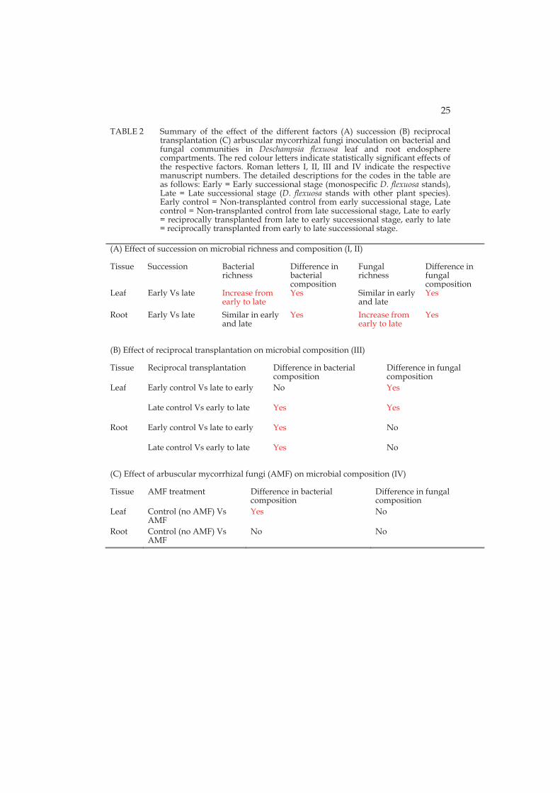
4 CONCLUSIONS
My results suggest that endosphere bacteria in Deschampsia flexuosa growing in an arctic habitat are to large extent cold habitat specific and most of them are able to solubilize organic and inorganic phosphate. Overall, my research shows that succession, transplantation and AMF colonization can affect the endosphere microbial assemblage in a sand dune colonizing plant species (Table 2). In my work, microbial diversity (Shannon) was not affected by succession, which runs counter the general conception that species diversity is increases along succession. Species diversity is expected to be linked with the amount of energy in the ecosystem. In my work, I could not link the microbial diversity with the amount of energy in the soil ecosystem. Moreover, my results show that plant succession affects endosphere microbial species richness in compartment dependent manner and community composition in compartment independent manner. Root but not leaf fungal endophyte richness increased across succession. In contrast, leaf but not root bacterial endophyte richness increased across succession. Based on this limited evidence, it seems that the aboveground endophytic richness responds to succession in terms of bacteria and belowground in terms of fungi. This pattern was also supported by the transplantation experiment. Furhermore, the transplantion experiment shows that endosphere microbial community changes after transplantation to novel conditions are a slow process. Further research is needed including different plant species in different ecosystems to fully understand the successional differences between prokaryotic and eukaryotic microbes.
In my work, I discovered microbes that are sensitive to disturbance. Changes in microbial communities due to disturbance may directly affect ecosystem processes and are therefore of specific interest. In previous studies related to endosphere microbial community assemblages the arbuscular mycorrhiza was not included as a potential factor that affects the other endosphere microbial community. My experiment showed that mycorrhizal inoculum may affect plant leaf bacterial endophyte community composition, but whether these changes are related to mycorrhizal effect on plant physiology need to be studied in targeted experiments.
24
All of the factors I studied mainly affected the microbial community composition. Bacteria and fungi responded differently either in terms of microbial species richness (in case of succession) or microbial community composition (in case of reciprocal transplantation and AMF inoculation). All through my thesis work, bacteria and fungi responded differently to manipulations (Table 2). Although commonly used, it may be more appropriate to use the word ‘bacteria’ when only bacteria have been studied in stead of ‘micro-organisms’. Similarly, it may be more correct to limit the generalization to fungi when only fungi have been studied as it seems that the two kingdoms do not respond similarly to environment.
25
TABLE 2 Summary of the effect of the different factors (A) succession (B) reciprocal transplantation (C) arbuscular mycorrhizal fungi inoculation on bacterial and fungal communities in Deschampsia flexuosa leaf and root endosphere compartments. The red colour letters indicate statistically significant effects of the respective factors. Roman letters I, II, III and IV indicate the respective manuscript numbers. The detailed descriptions for the codes in the table are as follows: Early = Early successional stage (monospecific D. flexuosa stands), Late = Late successional stage (D. flexuosa stands with other plant species). Early control = Non-transplanted control from early successional stage, Late control = Non-transplanted control from late successional stage, Late to early = reciprocally transplanted from late to early successional stage, early to late = reciprocally transplanted from early to late successional stage.
(A) Effect of succession on microbial richness and composition (I, II)
Tissue Succession Bacterialrichness
Difference in bacterial composition
Fungal richness
Difference in fungal composition
Leaf Early Vs late Increase from early to late
Yes Similar in early and late
Yes
Root Early Vs late Similar in early and late
Yes Increase from early to late
Yes
(B) Effect of reciprocal transplantation on microbial composition (III)
Tissue Reciprocal transplantation Difference in bacterial composition
Difference in fungal composition
Leaf Early control Vs late to early No Yes
Late control Vs early to late Yes Yes
Root Early control Vs late to early Yes No
Late control Vs early to late Yes No
(C) Effect of arbuscular mycorrhizal fungi (AMF) on microbial composition (IV)
Tissue AMF treatment Difference in bacterial composition
Difference in fungal composition
Leaf Control (no AMF) Vs AMF
Yes No
Root Control (no AMF) Vs AMF
No No
26
Acknowledgements
If I start to acknowledge my supervisor Dr. Minna-Maarit Kytöviita for every aspect starting from selecting me as a PhD student to selecting referees for my thesis, the whole page would not be enough. To keep it simple, I would like to convey my thankfulness to her for being a true inspirational, motivational and helpful supervisor for me. I would like to acknowledge that she is one of the best supervisors you could ever have for the PhD. I would like to extent gratitude to my second supervisor Dr. Riitta Nissinen for helping me in different aspects. A special thanks to Dr. Riitta Nissinen for giving an idea of for the weekly kalmakaltio project meetings for planning the experiments and problem discussions.
I have a big list of following funding agencies to thanks for their kindness in providing me personal grant or scholarship. It was all started with Maj and Tor Nessling foundation and definitely they deserve more credit in this thesis work. I am very thankful to the Centre for International Mobility (Finland) for the scholarship which confirmed my PhD student status in University of Jyväskylä. The personal grants from Oskar Öflunds Stiftelse foundation and Department of Biological and Environmental Science, University of Jyväskylä extended my stay here in Finland for my PhD thesis. Then, Academy of Finland and Finnish cultural foundation personal grants helped me to wind up my PhD. Thankful to the above mentioned foundation without which writing this acknowledgment section of this thesis is not at all possible. Last but not least, a special thanks to the Ella and George foundation for providing the sequencing cost for some of the experiment samples.
I am very lucky to have such a nice supporting group, thank you Dr. Emily Knott and Dr. Lutz Fromhage for your help to get funding from department and advices during the meetings. Also, thanks to the head of the department Dr. Mikko Mönkkönen for different types of help such as awarding me a personal grant, and accepting my project proposal for the funding application.
Dr. Gaia Francini and Dr. Roci Vega Frutis were helped in my first Finland arrival and thanks for their help. I would like to thank my fellow PhD students Tiina Savolainen, Cindy Given and Manoj Kumar for their kind co-ordination and help in the lab or field work. Also, my gratitude extent to Helena Jauhiainen, Dr. Minna Männistö and Sandra Savinen for their great help during the field work, Prof. Marja Tiirola, Elina Virtanen and Anita Mäki for their valuable help and guidance in ion torrent sequencing stream. Dr. Heli Juottonen helped me in different aspects and certainly I am very grateful to her. Thanks to Dr. Jelmer A. Elzinga, Dr. Joanneke H. Reudler Talsma and Dr. Sandra Varga for giving me a quiet and peaceful environment in office.

27
Thanks to my thesis reviewers (Dr. Joana Falcao Salles and Dr. Sabine Ravsnskov) for their suggestion and comments. Also, thanks to the Dr. LS (Leo) van Overbeek for accepting to be an opponent.
What a family I have! It can’t be explained in words how much supportive my father, mother, wife, son and in-laws (father, mother and brother) during my hard times. Simply I wish every student should have similar family and support for their PhD studies.
28
YHTEENVETO (RÉSUMÉ IN FINNISH) TRANSLATED BY MINNA-MAARIT KYTÖVIITA
Metsälauhan endofyyttisten mikrobien yhteisökoostumus lentohiekka-alueiden sukkessiossa
Kasvien kanssa elää suuri joukko mikrobeja enemmän tai vähemmän kiinteästi. Nämä liittolaiset ovat tärkeitä kasvien menestymiselle erilaisilla kasvupaikoilla. Erityisen tärkeitä kiinteästi kasveihin liittyneet mikrobit ovat kasvipeitteen muodostumisessa alkujaan kasvittomilla alueilla kuten hiekkadyyneillä. Tietoa kasvien kanssa liittoutuneiden mikrobien merkityksestä arktisilla alueilla ylei-sesti ja erityisesti lentohiekka-alueilla ja hiekkadyyneillä on hyvin vähän. Tässä väitöskirjatyössä selvitin metsälauhan (Deschampsia flexuosa) kanssa liittoutu-neiden mikrobien yhteisöjä kahdessa eri sukkession vaiheessa Enontekiön La-pissa. Ensimmäinen vaihe edusti sukkession alkuvaihetta, jossa kasvipeite oli hyvin niukka ja suurin osa maan pinnasta on liikkuvan lentohiekan peittämää. Vain muutama kasvilaji kasvaa arktisilla lentohiekka-alueilla ja tutkimukseeni valitsin alueita, joilla kasvoi metsälauhaa harvakseltaan puhtaina kasvustoina. Toinen tutkimuksieni sukkession vaihe edusti myöhäistä vaihetta, joka syntyy pitkän ajan kuluessa kasvillisuuden kehittyessä ilman suuria häiriöitä. Sukkes-sion päätevaihe tutkimusalueillani on harva tunturikoivikko, jonka aluskasvilli-suudessa kasvaa varpuja (Vaccinium uliginosum, Empetrum nigrum), ruohovarti-sia kasveja kuten metsälauhaa, kultapiiskua (Solidago virgaurea), lampaannataa (Festuca ovina) ja kissankäpälää (Antennaria dioica), pohjakerroksen muodostaa yhtenäinen seinäsammalkerros (Pleurozium schreberi). Sukkession myötä lento-hiekan pinnalle muodostuu loppuvaiheseen mennessä paksu kerros humusta, mikä muuttaa maan kemiallista koostumusta huomattavasti.
Ensimmäisessä työssäni eristin puhdasviljelmäkasvatuksiin metsälauhan lehtien ja juurten sisällä kasvavia sieniä ja bakteereita. Osa metsälauhan lehtien ja juurten sisältä eristetyistä mikrobeista oli sellaisia, joita tapaa myös maape-rässä. Osa eristetyistä mikrobeista oli kuitenkin aiemmin löydetty ainoastaan endofyyttisinä eli kasvin sisäisinä. Suurin osa eristetyistä mikrobeista oli arkti-sia, eli nykytietämyksen mukaan niiden elinalue rajoittuu maapallon kylmiin osiin. Suurin osa eristetyistä endofyyttisistä mikrobeista kykeni liuottamaan eloperäistä fosfaattia, mikä on kasvien kasvun kannalta erittäin tärkeä ominai-suus.
Kolmessa seuraavassa työssäni käytin nykyaikaisia sekvensointimenetel-miä, joiden avulla pystyin kuvaamaan mikrobiyhteisöt kattavasti. Yhdessä osa-työssä kasvatin metsälauhaa kasvihuoneessa keräsienen (Claroideoglomus etuni-catum) kanssa tai ilman sienijuurisientä. Kasvien inokulointi keräsienellä muutti lehtien endofyyttistä bakteeriyhteisöä, mutta ei vaikuttanut kasvin juuren sisäl-lä kasvavien sienten tai bakteerien yhteisöihin. Maastokokeiden tulosten mu-kaan sukkession vaihe ja kasvin osa (lehti, juuri) vaikutti merkitsevästi metsä-lauhan endofyyttisten mikrobien lajirunsauteen. Sukkession vaihe vaikutti myös erittäin voimakkaasti mikrobien yhteisökostumukseen. Tein siirtoistutus-
29
kokeen, jossa metsälauhaa siirtoistutettiin kasvupaikallaan tai sukkessiovai-heesta toiseen. Myös tämän tämän tutkimuksen perusteella kasvupaikan suk-kession vaihe vaikutti endofyyttisten mikrobien lajirunsauteen. Kasvupaikan sukkession vaiheesta riippumatta siirtositutettujen kasvien endofyyttisten mik-robien yhteisökoostumus erosi useimmissa tapauksissa (juuri, verso, sienet, bakteerit, alkuvaiheesta loppuvaiheeseen siirretyt kasvit, loppuvaiheesta alku-vaiheeseen siirretyt kasvit) kasvupaikalla kasvavien siirtämättömien kasvien mikrobiyhteisöistä. Sen sijaan kasvupaikallaan siirtoistutettujen kasvien koh-dalla vain endofyyttisten bakteerien yhteisöt erosivat muutamassa tapauksessa.
Yleisesti ottaen ekologisen teorian mukaan lajistollinen monimuotoisuus lisääntyy sukkession edetessä. Syyksi monimuotoisuuden kasvuun on esitetty ekosysteemin sisältämän energian määrää. Vastoin näitä yleisiä käsityksiä, suk-kession vaihe ei vaikuttanut mikrobien lajistolliseen monimuotoisuuteen omis-sa tutkimuksissani. Sen sijaan sukkessio vaikutti mikrobien lajirunsauteen riip-puen tutkitusta kasvin osasta ja mikrobiyhteisöjen koostumukseen tutkitusta kasvin osasta riippumatta. Juurten sisällä kasvavien sienten lajirunsaus lisään-tyi sukkession myötä, mutta bakteerien lajirunsaus ei. Sen sijaan lehden sisällä kasvavien bakteerien lajirunsaus lisääntyi sukkession myötä, mutta sienten ei. Tämä tulos saatiin myös kokeessa, jossa siirtoistutettiin kasveja sukkession myöhäisestä vaiheesta alkuvaiheeseen ja päinvastoin. Näiden rajallisten tutki-musten valossa näyttää siltä, että kasvin maanpäällisissä osissa sukkession ete-nemiseen reagoivat bakteerit, maanalaisissa osissa sienet. Lisäksi siirtoistutus-koe osoitti, että endofyyttisten mikrobien yhteisökoostumuksen sopeutuminen uuteen kasvupaikkaan on hidas prosessi.
Koska endofyyttisten sieni- ja bakteeriyhteisöjen koostumuksen, lajirun-sauden ja monimuotoisuuden suhdetta sukkession etenemiseen ei ole aiemmin tutkittu, omien tulosteni yleispätevyys jää tulevien tutkimusten selvitettäväksi.
Kaikenkaikkiaan työni lisäsi heikosti tunnettujen ekosysteemien ja eliöryhmien tuntemusta. Työni tuloksia voidaan käyttää hyväksi arktisten alu-eiden kasvillisuuden ennallistamisessa esimerkiksi kaivostoiminnan jälkeen.
30
REFERENCES
Adhikari T.B., Joseph C., Yang G., Phillips D.A. & Nelson L.M. 2001. Evaluation of bacteria isolated from rice for plant growth promotion and biological control of seedling disease of rice. Can. J. Microbiol. 47: 916–924.
Ågren J. & Schemske D.W. 2012. Reciprocal transplants demonstrate strong adaptive differentiation of the model organism Arabidopsis thaliana in its native range. New Phytol. 194: 1112–1122.
Allison S.D. & Martiny J.B. 2008. Colloquium paper: resistance, resilience, and redundancy in microbial communities. Proc. Natl. Acad. Sci. USA 105 Suppl 1: 11512–11519.
Anderson M.J. 2005. Permutational multivariate analysis of variance. Department of Statistics, University of Auckland, Auckland 26: 32–46.
Andrade G., Mihara K., Linderman R. & Bethlenfalvay G. 1997. Bacteria from rhizosphere and hyphosphere soils of different arbuscular–mycorrhizal fungi. Plant Soil 192: 71–79.
Bazzaz F. 1975. Plant species diversity in old–field successional ecosystems in southern Illinois. J. Ecol. 56: 485–488.
Belnap J. & Lange O. 2003. Biological Soil Crust: Structure, Function, and Management. Ecological Studies, Springer, Berlin, Heidelberg 150.
Berendse F., Lammerts E. & Olff H. 1998. Soil organic matter accumulation and its implications for nitrogen mineralization and plant species composition during succession in coastal dune slacks. Plant Ecol. 137: 71–78.
Berg G. & Smalla K. 2009. Plant species and soil type cooperatively shape the structure and function of microbial communities in the rhizosphere. FEMS Microbiol. Ecol. 68: 1–13.
Blaalid R., Carlsen T., Kumar S., Halvorsen R., Ugland K.I., Fontana G. & Kauserud H. 2012. Changes in the root associated fungal communities along a primary succession gradient analysed by 454 pyrosequencing. Mol. Ecol. 21: 1897–1908.
Bonfante P. & Anca I. 2009. Plants, mycorrhizal fungi, and bacteria: a network of interactions. Annu. Rev. Microbiol. 63: 363–383.
Borgegård S. 1990. Vegetation development in abandoned gravel pits: effects of surrounding vegetation, substrate and regionality. J.Veg. Sci. 1: 675–682.
Bottomley P., Yarwood R., Kageyama S., Waterstripe K., Williams M., Cromack Jr K. & Myrold D. 2006. Responses of soil bacterial and fungal communities to reciprocal transfers of soil between adjacent coniferous forest and meadow vegetation in the Cascade Mountains of Oregon. Plant Soil 289: 35–45.
Brown S.P. & Jumpponen A. 2015. Phylogenetic diversity analyses reveal disparity between fungal and bacterial communities during microbial primary succession. Soil Biol. Biochem. 89: 52–60.

31
Brown S.P. & Jumpponen A. 2014. Contrasting primary successional trajectories of fungi and bacteria in retreating glacier soils. Mol. Ecol. 23: 481–497.
Bulgarelli D., Garrido–Oter R., Münch P.C., Weiman A., Dröge J., Pan Y., McHardy A.C. & Schulze–Lefert P. 2015. Structure and Function of the Bacterial Root Microbiota in Wild and Domesticated Barley. Cell host & microbe 17: 392–403.
Bulgarelli D., Rott M., Schlaeppi K., van Themaat, Emiel Ver Loren, Ahmadinejad N., Assenza F., Rauf P., Huettel B., Reinhardt R. & Schmelzer E. 2012. Revealing structure and assembly cues for Arabidopsis root–inhabiting bacterial microbiota. Nature 488: 91–95.
Caldwell B.A., Jumpponen A. & Trappe J.M. 2000. Utilization of major detrital substrates by dark–septate, root endophytes. Mycologia : 230–232.
Chapin F.S., Walker L.R., Fastie C.L. & Sharman L.C. 1994. Mechanisms of primary succession following deglaciation at Glacier Bay, Alaska. Ecol. Monogr. 64: 149–175.
Chelius M. & Triplett E. 2001. The Diversity of Archaea and Bacteria in Association with the Roots of Zea mays L. Microb. Ecol. 41: 252–263.
Chesson P.L. & Warner R.R. 1981. Environmental variability promotes coexistence in lottery competitive systems. Am. Nat. 117: 923–943.
Cho S.M., Kang B.R., Han S.H., Anderson A.J., Park J., Lee Y., Cho B.H., Yang K., Ryu C. & Kim Y.C. 2008. 2R, 3R–butanediol, a bacterial volatile produced by Pseudomonas chlororaphis O6, is involved in induction of systemic tolerance to drought in Arabidopsis thaliana. Mol. Plant–Microbe Interact. 21: 1067–1075.
Clarke K. & Warwick R. 2001. Change in Marine Communities: An Approach to Statistical Analysis and Interpretation (PRIMER–E), 2nd edition. Plymouth Marine Laboratory, Plymouth, UK 172.
Clements F.E. 1916. Plant succession: an analysis of the development of vegetation. Carnegie Institution of Washington Pub 242.
Coleman Derr D., Desgarennes D., Fonseca Garcia C., Gross S., Clingenpeel S., Woyke T., North G., Visel A., Partida Martinez L.P. & Tringe S.G. 2016. Plant compartment and biogeography affect microbiome composition in cultivated and native Agave species. New Phytol. 209: 798–811.
Compant S., Clément C. & Sessitsch A. 2010. Plant growth–promoting bacteria in the rhizo–and endosphere of plants: their role, colonization, mechanisms involved and prospects for utilization. Soil Biol. Biochem. 42: 669–678.
Cowles H.C. 1899. The Ecological Relations of the Vegetation on the Sand Dunes of Lake Michigan. Part I.–Geographical Relations of the Dune Floras. Bot. Gaz. 27: 95–117.
de Ridder–Duine A.S., Kowalchuk G.A., Gunnewiek P.J.K., Smant W., van Veen J.A. & de Boer W. 2005. Rhizosphere bacterial community composition in natural stands of Carex arenaria (sand sedge) is determined by bulk soil community composition. Soil Biol. Biochem. 37: 349–357.
32
Diaz S., Grime J., Harris J. & McPherson E. 1993. Evidence of a feedback mechanism limiting plant response to elevated carbon dioxide. Nature 364: 616–617.
Dion P. & Nautiyal C. 2008. Extreme Views on Prokaryote Evolution. In: Dion P. & Nautiyal C. (eds.), – Microbiology of Extreme Soils. Microbiology of Extreme Soils (eds DionP, NautiyalCS) ed., – Springer, Berlin Heidelberg, 45–70.
Drury W.H. & Nisbet I.C. 1973. Succession. J. Arnold Arbor 54: 331–368. Edwards J., Johnson C., Santos–Medellin C., Lurie E., Podishetty N.K.,
Bhatnagar S., Eisen J.A. & Sundaresan V. 2015. Structure, variation, and assembly of the root–associated microbiomes of rice. Proc. Natl. Acad. Sci. USA 112: E911–20.
Evans K.L., Greenwood J.J. & Gaston K.J. 2005. Dissecting the species–energy relationship. Proc. Biol. Sci. 272: 2155–2163.
Finlay R.D. 2008. Ecological aspects of mycorrhizal symbiosis: with special emphasis on the functional diversity of interactions involving the extraradical mycelium. J. Exp. Bot. 59: 1115–1126.
Forster S.M. 1990. The role of microorganisms in aggregate formation and soil stabilization: Types of aggregation. Arid Soil Res. Rehabil. 4: 85–98.
Frey Klett P., Garbaye J.a. & Tarkka M. 2007. The mycorrhiza helper bacteria revisited. New Phytol. 176: 22–36.
Gottel N.R., Castro H.F., Kerley M., Yang Z., Pelletier D.A., Podar M., Karpinets T., Uberbacher E., Tuskan G.A., Vilgalys R., Doktycz M.J. & Schadt C.W. 2011. Distinct microbial communities within the endosphere and rhizosphere of Populus deltoides roots across contrasting soil types. Appl. Environ. Microbiol. 77: 5934–5944.
Groten K., Nawaz A., Nguyen N.H., Santhanam R. & Baldwin I.T. 2015. Silencing a key gene of the common symbiosis pathway in Nicotiana attenuata specifically impairs arbuscular mycorrhizal infection without influencing the root associated microbiome or plant growth. Plant, Cell Environ. 38: 2398–2416.
Gryndler M. 2000. Interactions of arbuscular mycorrhizal fungi with other soil organisms. In: Anonymous Arbuscular mycorrhizas: Physiology and function, Springer, 239–262.
Gupta Sood S. 2003. Chemotactic response of plant–growth–promoting bacteria towards roots of vesicular–arbuscular mycorrhizal tomato plants. FEMS Microbiol. Ecol. 45: 219–227.
Hackett C. 1965. Ecological Aspects of the Nutrition of Deschampsia Flexuosa (L.) Trin.: II. The Effects of A1, Ca, Fe, K. Mn, N, P and PH on the Growth of Seedlings an Established Plants. J.Ecol. 52: 315–333.
Haney C.H., Samuel B.S., Bush J. & Ausubel F.M. 2015. Associations with rhizosphere bacteria can confer an adaptive advantage to plants. Nature plants 1:15051.
Hansen A.K. & Moran N.A. 2014. The impact of microbial symbionts on host plant utilization by herbivorous insects. Mol. Ecol. 23: 1473–1496.
33
Hardoim P.R., Hardoim C.C., van Overbeek L.S. & van Elsas J.D. 2012. Dynamics of seed–borne rice endophytes on early plant growth stages. PLoS One 7: e30438.
Hardoim, P. R., van Overbeek, L. S., Berg, G., Pirttila, A. M., Compant, S., Campisano, A. et al. (2015) The Hidden World within Plants: Ecological and Evolutionary Considerations for Defining Functioning of Microbial Endophytes. Microbiol Mol Biol Rev 79: 293-320.
Ihrmark K., Bodeker I.T., Cruz–Martinez K., Friberg H., Kubartova A., Schenck J., Strid Y., Stenlid J., Brandstrom–Durling M., Clemmensen K.E. & Lindahl B.D. 2012. New primers to amplify the fungal ITS2 region evaluation by 454–sequencing of artificial and natural communities. FEMS Microbiol. Ecol. 82: 666–677.
Jeffries P., Gianinazzi S., Perotto S., Turnau K. & Barea J. 2003. The contribution of arbuscular mycorrhizal fungi in sustainable maintenance of plant health and soil fertility. Biol. Fertility Soils 37: 1–16.
Johnston–Monje D. & Raizada M.N. 2011. Conservation and diversity of seed associated endophytes in Zea across boundaries of evolution, ethnography and ecology. PLoS One 6: e20396.
Johnston–Monje D., Mousa W.K., Lazarovits G. & Raizada M.N. 2014. Impact of swapping soils on the endophytic bacterial communities of pre–domesticated, ancient and modern maize. BMC plant biol. 14: 1.
Jones R.T., Robeson M.S., Lauber C.L., Hamady M., Knight R. & Fierer N. 2009. A comprehensive survey of soil acidobacterial diversity using pyrosequencing and clone library analyses. ISME J. 3: 442–453.
Jorquera M.A., Crowley D.E., Marschner P., Greiner R., Fernández M.T., Romero D., Menezes Blackburn D. & De La Luz Mora, María. 2011. Identification of propeller phytase encoding genes in culturable Paenibacillus and Bacillus spp. from the rhizosphere of pasture plants on volcanic soils. FEMS Microbiol. Ecol. 75: 163–172.
Karasov T.L., Kniskern J.M., Gao L., DeYoung B.J., Ding J., Dubiella U., Lastra R.O., Nallu S., Roux F. & Innes R.W. 2014. The long–term maintenance ofa resistance polymorphism through diffuse interactions. Nature 512: 436–440.
Koske R. & Polson W. 1984. Are VA mycorrhizae required for sand dune stabilization? Bioscience 34: 420–424.
Lazzaro A., Gauer A. & Zeyer J. 2011. Field–scale transplantation experiment to investigate structures of soil bacterial communities at pioneering sites. Appl. Environ. Microbiol. 77: 8241–8248.
Liang Y., Jiang Y., Wang F., Wen C., Deng Y., Xue K., Qin Y., Yang Y., Wu L. & Zhou J. 2015. Long–term soil transplant simulating climate change with latitude significantly alters microbial temporal turnover. ISME J. 9: 2561–2572.
Lichter J. 1998. Primary succession and forest development on coastal Lake Michigan sand dunes. Ecol. Monogr. 68: 487–510.
Liu S., Wang F., Xue K., Sun B., Zhang Y., He Z., Van Nostrand J.D., Zhou J. & Yang Y. 2015. The interactive effects of soil transplant into colder regions
34
and cropping on soil microbiology and biogeochemistry. Environ. Microbiol. 17: 566–576.
Long H.H., Schmidt D.D. & Baldwin I.T. 2008. Native bacterial endophytes promote host growth in a species–specific manner; phytohormone manipulations do not result in common growth responses. PLoS One 3: e2702.
Ludwig–Müller J. 2015. Bacteria and fungi controlling plant growth by manipulating auxin: balance between development and defense. J. Plant Physiol. 172: 4–12.
Lundberg D.S., Lebeis S.L., Paredes S.H., Yourstone S., Gehring J., Malfatti S., Tremblay J., Engelbrektson A., Kunin V. & Del Rio T.G. 2012. Defining the core Arabidopsis thaliana root microbiome. Nature 488: 86–90.
Maignien L., DeForce E.A., Chafee M.E., Eren A.M. & Simmons S.L. 2014. Ecological succession and stochastic variation in the assembly of Arabidopsis thaliana phyllosphere communities. MBio 5: e00682–13.
Maki A., Rissanen A.J. & Tiirola M. 2016. A practical method for barcoding and size–trimming PCR templates for amplicon sequencing. BioTechniques 60: 88–90.
Männistö M.K. & Häggblom M.M. 2006. Characterization of psychrotolerant heterotrophic bacteria from Finnish Lapland. Syst. Appl. Microbiol. 29: 229–243.
Männistö M.K., Kurhela E., Tiirola M. & Haggblom M.M. 2013. Acidobacteria dominate the active bacterial communities of Arctic tundra with widely divergent winter–time snow accumulation and soil temperatures. FEMS Microbiol. Ecol. 84: 47–59.
Marschner P., Crowley D. & Lieberei R. 2001. Arbuscular mycorrhizal infection changes the bacterial 16 S rDNA community composition in the rhizosphere of maize. Mycorrhiza 11: 297–302.
Maun M.A. 2009. The biology of coastal sand dunes. Oxford University Press. Mercado–Blanco J. & Bakker P.A. 2007. Interactions between plants and
beneficial Pseudomonas spp.: exploiting bacterial traits for crop protection. Antonie Van Leeuwenhoek 92: 367–389.
Miller R. & Jastrow J. 1992. The application of VA mycorrhizae to ecosystem restoration and reclamation. Mycorrhizal functioning: an integrative plant–fungal process. Chapman and Hall, New York, 438–467.
Miller R., Jastrow J. & Reinhardt D. 1995. External hyphal production of vesicular–arbuscular mycorrhizal fungi in pasture and tallgrass prairie communities. Oecologia 103: 17–23.
Nautiyal C.S. 1999. An efficient microbiological growth medium for screening phosphate solubilizing microorganisms. FEMS Microbiol. Lett. 170: 265–270.
Nemergut D.R., Anderson S.P., Cleveland C.C., Martin A.P., Miller A.E., Seimon A. & Schmidt S.K. 2007. Microbial community succession in an unvegetated, recently deglaciated soil. Microb. Ecol. 53: 110–122.
35
Nissinen R.M., Männistö M.K. & Elsas J.D. 2012. Endophytic bacterial communities in three arctic plants from low arctic fell tundra are coldadapted and host plant specific. FEMS Microbiol. Ecol. 82: 510–522.
Normander B. & Prosser J.I. 2000. Bacterial origin and community composition in the barley phytosphere as a function of habitat and presowing conditions. Appl. Environ. Microbiol. 66: 4372–4377.
Olson J.S. 1958. Rates of succession and soil changes on southern Lake Michigan sand dunes. Bot. Gaz. 119: 125–170.
Panke–Buisse K., Poole A.C., Goodrich J.K., Ley R.E. & Kao–Kniffin J. 2015. Selection on soil microbiomes reveals reproducible impacts on plant function. ISME J. 9: 980–989.
Park M.S., Jung S.R., Lee M.S., Kim K.O., Do J.O., Lee K.H., Kim S.B. & Bae K.S. 2005. Isolation and characterization of bacteria associated with two sand dune plant species, Calystegia soldanella and Elymus mollis. J.microbial. 43: 219.
Pažoutová S., Follert S., Bitzer J., Keck M., Surup F., Šr tka P., Holuša J. & Stadler M. 2013. A new endophytic insect–associated Daldinia species, recognised from a comparison of secondary metabolite profiles and molecular phylogeny. Fungal Divers. 60: 107–123.
Peiffer J.A., Spor A., Koren O., Jin Z., Tringe S.G., Dangl J.L., Buckler E.S. & Ley R.E. 2013. Diversity and heritability of the maize rhizosphere microbiome under field conditions. Proc. Natl. Acad. Sci. USA 110: 6548–6553.
Pennanen T., Strömmer R., Markkola A. & Fritze H. 2001. Microbial and plant community structure across a primary succession gradient. Scand. J. For. Res. 16: 37–43.
Philippot L., Raaijmakers J.M., Lemanceau P. & van der Putten, Wim H. 2013. Going back to the roots: the microbial ecology of the rhizosphere. Nature Reviews Microbiology 11: 789–799.
Pietikäinen A., Kytöviita M. & Vuoti U. 2005. Mycorrhiza and seedling establishment in a subarctic meadow: effects of fertilization and defoliation. J. Veg. Sci. 16: 175–182.
Pirinen P., Simola H., Aalto J., Kaukoranta J., Karlsson P. & Ruuhela R. 2012. Climatological statistics of Finland 1981–2010. Finnish Meteorological Institute Reports 1: 1–96.
Puente M.E., Li C.Y. & Bashan Y. 2009. Endophytic bacteria in cacti seeds can improve the development of cactus seedlings. Environ. Exp. Bot. 66: 402–408.
Rawls J.F., Mahowald M.A., Ley R.E. & Gordon J.I. 2006. Reciprocal gut microbiota transplants from zebrafish and mice to germ–free recipients reveal host habitat selection. Cell 127: 423–433.
Read D. 1989. Mycorrhizas and nutrient cycling in sand dune ecosystems. Proc.Roy Soc EdinB B 96: 80–110.
Reed H.E. & Martiny J.B. 2007. Testing the functional significance of microbial composition in natural communities. FEMS Microbiol. Ecol. 62: 161–170.
36
Rodriguez R.J., Woodward C. & Redman R.S. 2010. Adaptation and survival of plants in high stress habitats via fungal endophyte conferred stress tolerance. In: Anonymous Symbioses and Stress, Springer, 461-476.
Rodriguez R.J., Henson J., Van Volkenburgh E., Hoy M., Wright L., Beckwith F., Kim Y. & Redman R.S. 2008. Stress tolerance in plants via habitat-adapted symbiosis. ISME J. 2: 404-416.
Ross C.A., Faust D. & Auge H. 2009. Mahonia invasions in different habitats: local adaptation or general–purpose genotypes? Biol. Invasions 11: 441–452.
Ruotsalainen A.L., Markkola A. & Kozlov M.V. 2007. Root fungal colonisation in Deschampsia flexuosa: Effects of pollution and neighbouring trees. Environmental pollution 147: 723–728.
Saikkonen K., Ahlholm J., Helander M., Lehtimäki S. & Niemeläinen O. 2000. Endophytic fungi in wild and cultivated grasses in Finland. Ecography 23: 360–366.
Sanchez L., Weidmann S., Arnould C., Bernard A.R., Gianinazzi S. & Gianinazzi–Pearson V. 2005. Pseudomonas fluorescens and Glomus mosseae trigger DMI3–dependent activation of genes related to a signal transduction pathway in roots of Medicago truncatula. Plant Physiol. 139: 1065–1077.
Scervino J., Gottlieb A., Silvani V., Pérgola M., Fernández L. & Godeas A. 2009. Exudates of dark septate endophyte (DSE) modulate the development of the arbuscular mycorrhizal fungus (AMF) Gigaspora rosea. Soil Biol. Biochem. 41: 1753–1756.
Scheepens J. & Stöcklin J. 2013. Flowering phenology and reproductive fitness along a mountain slope: maladaptive responses to transplantation to a warmer climate in Campanula thyrsoides. Oecologia 171: 679–691.
Scheffknecht S., Mammerler R., Steinkellner S. & Vierheilig H. 2006. Root exudates of mycorrhizal tomato plants exhibit a different effect on microconidia germination of Fusarium oxysporum f. sp. lycopersici than root exudates from non–mycorrhizal tomato plants. Mycorrhiza 16: 365–370.
Schloss P.D., Gevers D. & Westcott S.L. 2011. Reducing the effects of PCR amplification and sequencing artifacts on 16S rRNA–based studies. PLoS One 6: e27310.
Schmidt S., Nemergut D., Darcy J. & Lynch R. 2014. Do bacterial and fungal communities assemble differently during primary succession? Mol. Ecol. 23: 254–258.
Scott J.J. & Loewus F.A. 1986. A calcium-activated phytase from pollen of Lilium longiflorum. Plant physiol. 82: 333-335.
Scurfield G. 1954. Deschampsia flexuosa (L.) Trin. J. Ecol. 42: 225–233. Seppälä M. 1995. Deflation and redeposition of sand dunes in Finnish Lapland.
Quaternary Science Reviews 14: 799–809. Sheng H.M., Gao H.S., Xue L.G., Ding S., Song C.L., Feng H.Y. & An L.Z. 2011.
Analysis of the composition and characteristics of culturable endophytic
37
bacteria within subnival plants of the Tianshan Mountains, northwestern China. Curr. Microbiol. 62: 923–932.
Singh B.K., Munro S., Potts J.M. & Millard P. 2007. Influence of grass species and soil type on rhizosphere microbial community structure in grassland soils. Appl. Soil Ecol. 36: 147–155.
Snellgrove R., Splittstoesser W., Stribley D. & Tinker P. 1982. The distribution of carbon and the demand of the fungal symbiont in leek plants with vesicular arbuscular mycorrhizas. New Phytol. 92: 75–87.
Sultan S. 1995. Phenotypic plasticity and plant adaptation. Acta Bot. Neerl. 44: 363-383.
Sun B., Wang F., Jiang Y., Li Y., Dong Z., Li Z. & Zhang X. 2014. A long term field experiment of soil transplantation demonstrating the role of contemporary geographic separation in shaping soil microbial community structure. Ecol. evol. 4: 1073–1087.
Sutton J. & Sheppard B. 1976. Aggregation of sand–dune soil by endomycorrhizal fungi. Can. J. Bot. 54: 326–333.
Tedersoo L., Bahram M., Polme S., Koljalg U., Yorou N.S., Wijesundera R., Villarreal Ruiz L., Vasco–Palacios A.M., Thu P.Q., Suija A., Smith M.E., Sharp C., Saluveer E., Saitta A., Rosas M., Riit T., Ratkowsky D., Pritsch K., Poldmaa K., Piepenbring M., Phosri C., Peterson M., Parts K., Partel K., Otsing E., Nouhra E., Njouonkou A.L., Nilsson R.H., Morgado L.N., Mayor J., May T.W., Majuakim L., Lodge D.J., Lee S.S., Larsson K.H., Kohout P., Hosaka K., Hiiesalu I., Henkel T.W., Harend H., Guo L.D., Greslebin A., Grelet G., Geml J., Gates G., Dunstan W., Dunk C., Drenkhan R., Dearnaley J., De Kesel A., Dang T., Chen X., Buegger F., Brearley F.Q., Bonito G., Anslan S., Abell S. & Abarenkov K. 2014. Fungal biogeography. Global diversity and geography of soil fungi. Science 346: 1256688.
Tejesvi M., Ruotsalainen A., Markkola A. & Pirttilä A. 2010. Root endophytes along a primary succession gradient in northern Finland. Fungal Divers. 41: 125–134.
Tejesvi M.V., Sauvola T., Pirttilä A.M. & Ruotsalainen A.L. 2013. Neighboring Deschampsia flexuosa and Trientalis europaea harbor contrasting root fungal endophytic communities. Mycorrhiza 23: 1–10.
Tews J., Brose U., Grimm V., Tielbörger K., Wichmann M., Schwager M. & Jeltsch F. 2004. Animal species diversity driven by habitat heterogeneity/diversity: the importance of keystone structures. J. Biogeogr. 31: 79–92.
Ujházy K., Fanta J. & Prach K. 2011. Two centuries of vegetation succession in an inland sand dune area, central Netherlands. Appl. Veg. Sci. 14: 316–325.
van der Wal A., Geydan T.D., Kuyper T.W. & de Boer W. 2013. A thready affair: linking fungal diversity and community dynamics to terrestrial decomposition processes. FEMS Microbiol. Rev. 37: 477–494.
38
van Overbeek, L. S., & Saikkonen, K. (2016) Impact of Bacterial–Fungal Interactions on the Colonization of the Endosphere. Trends Plant Sci. 21: 230-242.
Vigo C., Norman J. & Hooker J. 2000. Biocontrol of the pathogen Phytophthora parasitica by arbuscular mycorrhizal fungi is a consequence of effects on infection loci. Plant Pathol. 49: 509–514.
Villarreal Ruiz L., Anderson I.C. & Alexander I.J. 2004. Interaction between an isolate from the Hymenoscyphus ericae aggregate and roots of Pinus and Vaccinium. New Phytol. 164: 183–192.
Waldrop M. & Firestone M. 2006. Response of microbial community composition and function to soil climate change. Microb. Ecol. 52: 716–724.
Walker L.R. & Del Moral R. 2003. Primary succession and ecosystem rehabilitation. Cambridge University Press.
Wamberg C., Christensen S., Jakobsen I., Müller A. & Sørensen S.J. 2003. The mycorrhizal fungus (Glomus intraradices) affects microbial activity in the rhizosphere of pea plants (Pisum sativum). Soil Biol. Biochem. 35: 1349–1357.
Wang M., Yang P. & Salles J.F. 2015. Distribution of root–associated bacterial communities along a salt–marsh primary succession. Front.plant sci. 6: 1–11.
Williams R.J., Howe A. & Hofmockel K.S. 2014. Demonstrating microbial co–occurrence pattern analyses within and between ecosystems. Front. microbial. 5: 1–10.
Zarraonaindia I., Owens S.M., Weisenhorn P., West K., Hampton–Marcell J., Lax S., Bokulich N.A., Mills D.A., Martin G., Taghavi S., van der Lelie D. & Gilbert J.A. 2015. The soil microbiome influences grapevine–associated microbiota. MBio 6: 10.1128/mBio.02527–14.
Zhao M., Xue K., Wang F., Liu S., Bai S., Sun B., Zhou J. & Yang Y. 2014. Microbial mediation of biogeochemical cycles revealed by simulation of global changes with soil transplant and cropping. ISME J. 8: 2045–2055.
Zijlstra J.D., Van't Hof P., Braakhekke W.G., Berendse F., Baar J., Paradi I., Verkley G.J. & Summerbell R.C. 2005. Diversity of symbiotic root endophytes of the Helotiales in ericaceous plants and the grass, Deschampsia flexuosa. Stud. Mycol. 53: 147–162.
ORIGINAL PAPERS
I
CULTURABLE ENDOPHYTIC MICROBIAL COMMUNITIES IN THE CIRCUMPOLAR GRASS, DESCHAMPSIA FLEXUOSA IN A
SUB-ARCTIC INLAND PRIMARY SUCCESSION ARE HABITAT AND GROWTH STAGE SPECIFIC
by
Anbu Poosakkannu, Riitta Nissinen, & Minna-Maarit Kytöviita 2016
Environmental microbiology reports 7: 111–122.
Reprinted with kind permission of © Society for Applied Microbiology and John Wiley & Sons Ltd
Culturable endophytic microbial communities in thecircumpolar grass, Deschampsia flexuosa in asub-Arctic inland primary succession are habitat andgrowth stage specific
Anbu Poosakkannu,* Riitta Nissinen andMinna-Maarit KytöviitaDepartment of Biological and Environmental Science,University of Jyväskylä, Jyväskylä, Finland.
Summary
Little is known about endophytic microbes in coldclimate plants and how their communities are formed.We compared culturable putative endophytic bacteriaand fungi in the ecologically important circumpolargrass, Deschampsia flexuosa growing in two succes-sional stages of subarctic sand dune (68°29′N).Sequence analyses of partial 16S rRNA and internaltranscribed spacer (ITS) sequences of culturableendophytes showed that diverse bacteria and fungiinhabit different tissues of D. flexuosa. A total of 178bacterial isolates representing seven taxonomicdivisions, Alpha, Beta and Gammaproteobacteria,Actinobacteria, Bacteroidetes, Firmicutes and Acido-bacteria, and 30 fungal isolates representing thephylum Ascomycota were identified. Severalendophytes were affiliated with specific plant tissuesor successional stages. This first report of bacterialendophytes in D. flexuosa revealed that the genusPseudomonas is tightly associated with D. flexuosa,and encompassed 39% of the bacterial isolates, and58% of seed isolates. Based on 16S rRNA and ITSsequence data, most of the D. flexuosa endophyteswere closely related to microbes from other cold envi-ronments. The majority of seed endophytic bacterialisolates were able to solubilize organic form of phos-phate suggesting that these endophytes could play arole in resource mobilization in germinating seeds innutrient-poor habitat.
Introduction
Ever since their emergence, plants have lived in constantinteraction with different bacteria, fungi and viruses
(Sessitsch et al., 2004; Aly et al., 2011; Reinhold-Hurekand Hurek, 2011). Microbial association with plants can beexternal as is the case of the rhizosphere or phyllospheremicrobes and internal as for the endophytic microbes.Endophytic microbes are ubiquitous in plants, and theycan provide plants with mineral nutrients or fixed nitrogenin exchange for carbon (Pillay and Nowak, 1997;Compant et al., 2010; Reinhold-Hurek and Hurek, 2011).Endophytes can also help their hosts to overcomeenvironmental stresses by producing or modifyingphytohormones or secondary metabolites (Sturz et al.,1998; Siciliano et al., 2001; Barac et al., 2004; Ma et al.,2011a,b; Ardanov et al., 2012). Most of endophytes arethought to be acquired horizontally, mainly from therhizosphere (Hardoim et al., 2008), but endophytes canalso be transmitted vertically from parent to offspring(Ferreira et al., 2008; Hardoim et al., 2012). Further, dif-ferent plant organs and tissues have been shown toharbour different microbial communities (Wearn et al.,2012; Bodenhausen et al., 2013).Bacterial and fungal endophytes are known to have
wide range of hosts (Cannon and Simmons, 2002; Shenget al., 2011) or show host specificity (Arnold et al., 2001;Long et al., 2008; Nissinen et al., 2012). Most of thereports on endophytic diversity concern tropical or tem-perate agriculture and forest systems, and there is verylittle data on endophytic microbial diversity in cold envi-ronments, especially information from the arctic is lacking.Recently, the first reports on arctic plant endophytic bac-teria (Nissinen et al., 2012) and endophytic fungi (Higginset al., 2007; Bjorbækmo et al., 2010; Walker et al., 2011)have emerged. These studies were conducted on differ-ent plant species, and currently, there is no knowledge ofthe culturable microbial community composed of bothbacteria and fungi in any arctic plant species.In this study, we focused on the microbial endophytes in
the grass Deschampsia flexuosa growing in subarcticAeolian sand dune area. Aeolian sand dunes were formedin the northern hemisphere during glacial retreat whenstrong winds resulted in the formation of large areascovered by sand dunes (Koster, 1988). These Aeoliansand dunes were later stabilized by vegetation, buterosion has created large deflation areas without
Received 18 March, 2014; accepted 14 July, 2014. *For correspond-ence. E-mail [email protected]; Tel. +358408053842,+358468826863; Fax +358-14-617 239.
bs_bs_banner
Environmental Microbiology Reports (2015) 7(2), 111–122 doi:10.1111/1758-2229.12195
© 2014 Society for Applied Microbiology and John Wiley & Sons Ltd
vegetation relatively recently (Seppälä, 1995). Plant colo-nization in the Aeolian sand dunes is very slow because ofextreme environmental conditions such as highly fluctuat-ing temperatures, short growing season, water and nutri-ent limitation and unstable soil structure (Hodkinson et al.,2003). Together these result in primary successional gra-dient (Seppälä, 1995) characterized by different succes-sional stages starting from no vegetation to change inplant species composition from pioneer plant species tolater successional plant species (Svoboda and Henry,1987; Hodkinson et al., 2003). The role of soil microbes inplant development in primary successional areas(Grootjans et al., 1997) and in arctic primary successionalareas (Borin et al., 2010) have been recognized, but theimportance of plant endophytic microbial communitiesremains unexplored.The vegetation in our experimental area has been
destroyed in several occasions by fire during the last 800years (Seppälä, 1995). The fires together with wind effectand intensive grazing by herbivores, particularly by domes-tic reindeer, have resulted in the reversion of succession insome areas. As a result, the current vegetation is patchyand large areas without any vegetation cover exist next torelatively undisturbed mountain birch forest stands andother successional stages. Deschampsia flexuosa is oneof the fewpioneer plants that are able to colonize bare sandunder arctic conditions (Polunin, 1938).We compared the putative endophytic microbial com-
munity in D. flexuosa growing in two successional stages,one that consists of the grass D. flexuosa as the only plantspecies (early successional) and one that represents thevegetation cover that will develop without extreme disturb-ance (late successional) in a subarctic inland sand dunearea. We focused on culturable endophytes because oftheir feasibility for future bioassays. We isolated both fungiand bacteria in leaves, roots, seeds and seedlings.Although usually explored separately, we analysed bothfungi and bacteria in order to tentatively explore theirinteractive roles in plant life in cold climate. We charac-terized the bacterial endophytes for their ability tosolubilize mineral phosphate which is considered animportant trait in plant growth promoting bacteria (deFreitas et al., 1997; Richardson, 2001). Phytate is themain storage form of phosphorus seeds in grass speciesin particular and up to 80% of the phosphorus is in theform of phytate in plants (Scott and Loewus, 1986). Con-version of phytate into inorganic phosphate by thephytase enzyme is one of the important steps in seedgermination (Scott and Loewus, 1986). Phytate degrada-tion by soil microbes and their role in plant growth inphosphate-poor environment has been reported (Idrisset al., 2002). However, the role of endophytes in phytatedegradation is poorly understood, and might be relevantin arctic habitats with low levels of soluble nutrients.
We asked the following specific questions: (i) Does theculturable endophyte community composition reflect thecold climate of their host plants? (ii) Do different plantparts (shoot, root and seed) and plants in different suc-cessional stages have distinct culturable endophytecommunities? and (iii) Do endophytic bacterial isolatessolubilize of organic phosphate rather than mineralphosphate?
Results and discussion
Culturable bacterial endophytes
A total of 178 putative endophytic bacterial strains wereisolated from D. flexuosa plants collected from the twosuccessional stages. Based on genomic fingerprinting(BOX PCR, for details, see Appendix S1), 101 uniqueisolates were detected and tentatively identified by 16SrRNA gene sequencing and comparison to referencesequences in public databases. Overall, the endophyticbacterial isolates from D. flexuosa represented seventaxonomic divisions: Alpha, Beta and Gammaproteo-bacteria (10%, 11% and 60% of total community respec-tively), Actinobacteria (8%), Bacteroidetes (8%),Firmicutes (2%) and Acidobacteria (1%). The relativeabundance of several phyla between samples collectedfrom the different successional stages differed.Actinobacteria and Alphaproteobacteria were more abun-dant in plants growing in sand, whereas BacteroidetesandGammaproteobacteria were relatively more abundantin forest (Fig. S1). The isolates representing Firmicutesand Acidobacteria (4 and 2 isolates respectively) were allisolated from forest samples. Actinobacteria are known tobe oligotrophic and successful in nutrient-deficient anddry environments (Dion, 2008), and this could explaintheir prevalence in the sand samples.Likewise, several phyla were unevenly detected in dif-
ferent tissues of D. flexuosa; Gammaproteobacteria werepredominant in seed and experimental seedling samples,with 95% and 80% of isolates respectively. In contrast,Actinobacteria, Beta and Alphaproteobacteria were foundin leaf, root and field seedling samples, but not in seeds orexperimental seedlings, suggesting their acquisition fromenvironment during plant development.The isolates represented in total 32 bacterial genera,
with the highest taxonomic diversity detected in root (16genera) and leaf (15) tissues, with 7, 6 and 8 genera infield seedlings, seeds and in experimental seedlingsrespectively. Pseudomonas was the most prevalentgenus in our isolate collection, accounting for 39% of all(total isolate collection) isolates, followed by Burkholderia(5%), Pedobacter (5%), Rahnella (5%) and Pantoea (4%).Pseudomonas was consistently present in all D. flexuosatissues and in both successional stages studied.
112 A. Poosakkannu, R. Nissinen and M.-M. Kytöviita
© 2014 Society for Applied Microbiology and John Wiley & Sons Ltd, Environmental Microbiology Reports, 7, 111–122

However, several other genera showed preferenceto specific tissue or successional stage (Figs 1 and 2).For example, Alphaproteobacterial genera Sphin-gomonas, Rhizobium and Mesorhizobium were almostexclusively detected in samples from the sand (Fig. 2).This could be related to their function in nitrogenacquisiton in a nutrient-poor sand habitat; Rhizobium andMesorhizobium are well-known nitrogen-fixing genera(Franche et al., 2009). Endophytic bacteria belonging tothe genera Pseudomonas, Erwinia, Paenibacillus,Flavobacterium and Stenotrophomonas were identified inseed material. These same genera have been discoveredpreviously in seed material in temperate (Ferreira et al.,2008; Hardoim et al., 2012) as well as arctic (Nissinenet al., 2012) plant species. Within endophytic genera,tissue or habitat specific groups were detected (Fig. 2 andTable 1). For example, isolates SL56, SR-A19, FL97-1and FR302, closely related to Burkholderia sordidicolatype strain SNU 020123, were present in leaf and rootsamples of both successional stages. In contrast, isolatesSL28-1 and SR306, closely related to Curtobacteriumflaccumfaciens, were present only in the leaf and rootsamples from the sand.
Pseudomonas is the dominant genus in D. flexuosaendophytic bacterial communities
Pseudomonas spp. isolates dominated the endophytecommunity of D. flexuosa in both sand and forestsamples, and were present in all the tissues and growthstages of D. flexuosa investigated in this study. Theseresults agree well with previous reports: temperate sanddune plant species as well as Pennisetum glaucum (agrass family plant) grown on low-nutrient soil have beenshown to be dominated by Gammaproteobacteria, in par-ticular Pseudomonas (Park et al., 2005; Gupta et al.,2013). Pseudomonas isolates were most abundant inseed tissues, with 58% relative abundance, followed by44% relative abundance in field seedlings, 35% in leaves,34% in experimental seedlings and 27% in root. In orderto gain better insight into this genus, we performed aseparate phylogenetic analysis of partial 16S rRNA genesequences of all Pseudomonas isolates in our collection(Fig. 3). This enabled detection of several distinct tightlyclustered groups within the genus Pseudomonas.For example, a group of isolates (SL10, SR310-1, FL97-2,FR129, SGS1 and FGS11), closely related toP.fluorescens type strain IAM12022 and glacier isolateKOPRI 25853, were present in all D. flexuosa tissues andboth successional stages (Fig. 3). This suggests that thisgroup of endophytes is tightly associated with D. flexuosaand might be vertically transmitted. Pseudomonasfluorescens is a well-known plant growth promoting bac-terium that has been isolated, among others, in many
grasses previously and has been shown to affect plantgrowth and development, but also reduce seedlingdisease incidence in rice (Adhikari et al., 2001;Mercado-Blanco and Bakker, 2007). In contrast, anothergroup of isolates (SL11, SR12, SS4, SS8 and SS12) wasmainly detected in the sand samples (seeds, leavesand roots). These isolates were closely related toPseudomonas graminis type strain DSM11363 and to agroup of strains isolated previously from seeds ofarctic plants (Fig. 3). Pseudomonas graminis has beenpreviously detected in temperate sand dune plants (Parket al., 2005), and also in seeds of Juncus trifidus andDiapensia lapponica collected in low-nutrient, high-stresshabitats in fell tundra in northwestern Finland (Nissinenet al., 2012).
Phosphate solubilization ability in endophytic bacteria
All the putative endophytic bacterial isolates were quali-tatively analysed for their in vitro mineral as well asorganic phosphate solubilizing ability in order to evaluatethe potential of the isolates for plant growth promotion (fordetails, see Appendix S1). In total, 57% of the endophyticbacterial isolates were able to solubilize the tricalcium(mineral) phosphate (TCP) and 74% were able to degradephytate, the organic form of phosphate, common phos-phate storage form in plant seeds. All TCP solubilizingisolates were also able to degrade organic phosphate.Proportionally most phosphate solubilizing isolates weredetected in forest root samples, with 73% (TCP) and 80%(phytate) in the forest and 39% (TCP) and 57% (phytate)in the sand respectively. In contrast, relatively higher pro-portion of phytate solubilizing isolates was detected inexperimental seedlings from sand samples, with 100% inthe sand samples and 70% in the forest samples respec-tively (Table S1).Soil mineral phosphorus is often bound to phosphates
inaccessible to plants. In plants, phosphorus is stored asphytate. Conversion of phytate into inorganic phosphateby the phytase enzyme is one of the important steps inseed germination (Scott and Loewus, 1986). We hypoth-esized that endophytic bacterial isolates solubilize theorganic phosphate rather than inorganic phosphate. Theresults supported our hypothesis and greater portion ofthe endophytes was able to mobilize organic (74%) thanmineral (57%) phosphate. In particular, the great majorityof seed endophytes was able to solubilize organicphosphate (92%). The seed isolates in the genusPseudomonas were able to solubilize organic phosphate(100%) and mineral phosphate (87%). It seems likely thatseed endophytic bacteria have a significant role inresource mobilization from stored reserves in the plant.Abundance of organic phosphate solubilizing bacteriawas relatively higher in the early than late successional
Culturable endophytic microbes in Deschampsia flexuosa 113
© 2014 Society for Applied Microbiology and John Wiley & Sons Ltd, Environmental Microbiology Reports, 7, 111–122
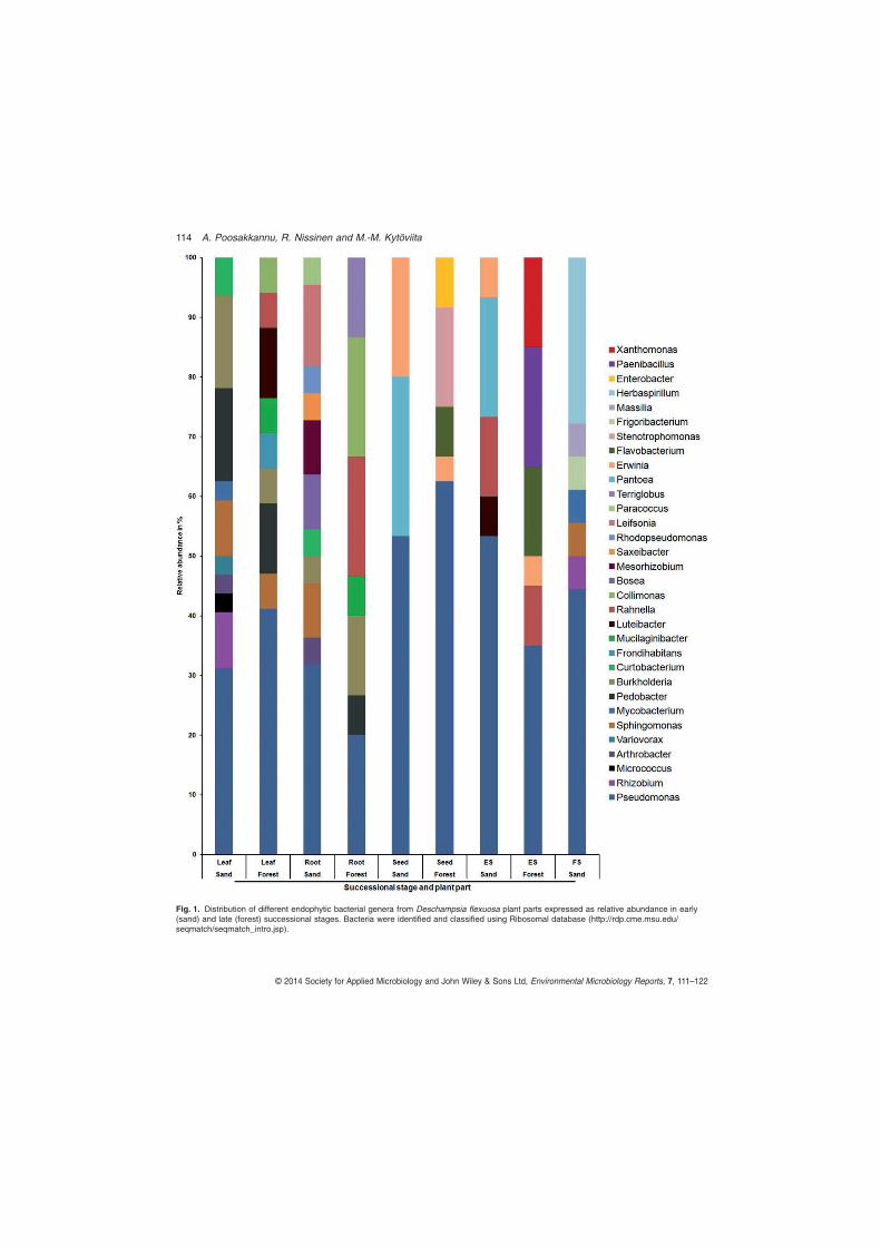
Fig. 1. Distribution of different endophytic bacterial genera from Deschampsia flexuosa plant parts expressed as relative abundance in early(sand) and late (forest) successional stages. Bacteria were identified and classified using Ribosomal database (http://rdp.cme.msu.edu/seqmatch/seqmatch_intro.jsp).
114 A. Poosakkannu, R. Nissinen and M.-M. Kytöviita
© 2014 Society for Applied Microbiology and John Wiley & Sons Ltd, Environmental Microbiology Reports, 7, 111–122
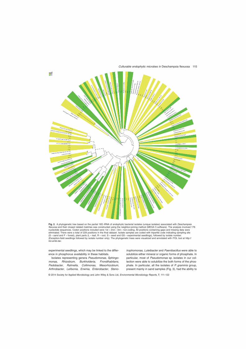
experimental seedlings, which may be linked to the differ-ence in phosphorus availability in these habitats.Isolates representing genera Pseudomonas, Sphingo-
monas, Rhizobium, Burkholderia, Frondihabitans,Pedobacter, Rahnella, Collimonas, Mesorhizobium,Arthrobacter, Leifsonia, Erwinia, Enterobacter, Steno-
trophomonas, Luteibacter and Paenibacillus were able tosolubilize either mineral or organic forms of phosphate. Inparticular, most of Pseudomonas sp. isolates in our col-lection were able to solubilize the both forms of the phos-phate. In particular, all the isolates of P. graminis group,present mainly in sand samples (Fig. 3), had the ability to
Fig. 2. A phylogenetic tree based on the partial 16S rRNA of endophytic bacterial isolates (unique isolates) associated with Deschampsiaflexuosa and their closest related matches was constructed using the neighbor-joining method (MEGA 5 software). The analysis involved 176nucleotide sequences. Codon positions included were 1st + 2nd + 3rd + non-coding. All positions containing gaps and missing data wereeliminated. There were a total of 229 positions in the final dataset. Isolate samples are coded with tripartite code indicating sampling site(S = sand and F = forest), plant parts (L = leaf, R = root, S = seed and GS = experimental seedlings), followed by isolate number.(Exception-field seedlings followed by isolate number only). The phylogenetic trees were visualized and annotated with iTOL tool at http://itol.embl.de/.
Culturable endophytic microbes in Deschampsia flexuosa 115
© 2014 Society for Applied Microbiology and John Wiley & Sons Ltd, Environmental Microbiology Reports, 7, 111–122
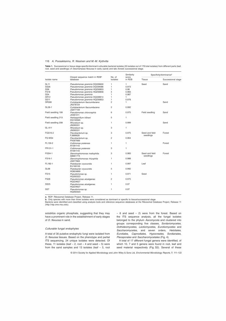
solubilize organic phosphate, suggesting that they mayhave a prominent role in the establishment of early stagesof D. flexuosa in sand.
Culturable fungal endophytes
A total of 30 putative endophytic fungi were isolated fromD. flexuosa tissues. Based on the phenotype and partialITS sequencing, 24 unique isolates were detected. Ofthese, 11 isolates (leaf – 2, root – 4 and seed – 5) werefrom the sand samples and 13 isolates (leaf – 5, root
– 6 and seed – 2) were from the forest. Based onthe ITS sequence analysis, all the fungal isolatesbelonged to the phylum Ascomycota and clustered intogroups corresponding five classes, Sordariomycetes,Dothideomycetes, Leotiomycetes, Eurotiomycetes andSaccharomycetes, and seven orders, Helotiales,Eurotiales, Capnodiales, Hypocreales, Sordiariales,Pleosporales and Saccharomycetales (Fig. 4).A total of 17 different fungal genera were identified, of
which 10, 7 and 5 genera were found in root, leaf andseed material respectively (Fig. S2). Several of these
Table 1. Successional or tissue stage specific/dominant culturable bacterial isolates (53 isolates out of 178 total isolates) from different parts (leaf,root, seed and seedlings) of Deschampsia flexuosa in early (sand) and late (forest) successional stage.
Isolate nameClosest sequence match in RDBa
databaseNo. ofisolates
Similarityscorein RDB
Specificity/dominanceb
Tissue Successional stage
SL11 Pseudomonas graminis DQ339600 1 1 Seed SandSS26 Pseudomonas graminis DQ339586 1 0.973SS8 Pseudomonas graminis HQ256853 1 0.98FS18 Pseudomonas graminis HQ256858 4 0.983SS4 Pseudomonas graminis 1 0.987SR12 Pseudomonas graminis DQ339614 1 1SS11 Pseudomonas graminis HQ256853 1 0.978SR306 Curtobacterium flaccumfaciens
JN3787242 1 Sand
SL28-1 Curtobacterium flaccumfaciensJQ977194
2 0.992
Field seedling 199 Pseudomonas chlororaphisJX081311
6 0.975 Field seedling Sand
Field seedling 213 Herbaspirillum hiltneriDQ150565
5 1
Field seedling 208 Rhizobium sp.JN590331
1 0.999 Sand
SL-A11 Rhizobium sp.JN590331
3 1
FGS16-2 Flavobacterium sp.FJ889628
3 0.975 Seed and fieldseedlings
Forest
FS-WS4 Flavobacterium sp.FN397666
2 0.964
FL139-2 Collimonas pratensisAY281143
1 1 Forest
FR131-1 Collimonas pratensisAY281143
3 1
FGS4-1 Stenotrophomonas maltophiliaAB661774
3 0.983 Seed and fieldseedlings
Forest
FS16-1 Stenotrophomonas rhizophilaJQ977663
1 0.988
FL182-1 Pedobacter cryoconitisEU169155
1 0.997 Leaf
SL68 Pedobacter cryoconitisHQ824869
5 0.992
FS15 Pseudomonas sp.HQ260323
1 0.971 Seed
FS28 Pseudomonas alcaligenesHQ224627
2 0.975
SS25 Pseudomonas alcaligenesHQ224627
1 0.97
SS7 Pseudomonas sp.HQ260323
1 0.97
a. RDP: Ribosomal Database Project, Release 11.b. Only species with more than three isolates were considered as dominant or specific to tissue/successional stage.Bacteria were identified and classified using analysis tools and reference sequence databases at the Ribosomal Database Project, Release 11(http://rdp.cme.msu.edu).
116 A. Poosakkannu, R. Nissinen and M.-M. Kytöviita
© 2014 Society for Applied Microbiology and John Wiley & Sons Ltd, Environmental Microbiology Reports, 7, 111–122

Fig. 3. Phylogenetic tree of partial 16S rRNA gene sequences of Pseudomonas spp. isolates from Deschampsia flexuosa (representativesequences from different plant parts), seed isolates from Nissinen and colleagues 2012 (Juncus trifidus and Diapensia lapponica) and theirclosest relatives from public databases was constructed using the neighbor-joining method (MEGA 5 software). The analysis involved 66nucleotide sequences. All positions containing gaps and missing data were eliminated, and there were a total of 424 positions in the finaldataset. Isolate sequences are coded with tripartite code indicating successional stage (S = sand and F = forest), plant tissue (L = leaf,R = root, S = seed and GS = experimental seedlings), followed by isolate number. (Exception: field seedlings are followed by isolate number.)Diapensia lapponica seed isolates = DS and J. trifidus seed isolates = JS, followed by isolate number only. The phylogenetic tree wasvisualized and annotated with iTOL tool at http://itol.embl.de/.
Culturable endophytic microbes in Deschampsia flexuosa 117
© 2014 Society for Applied Microbiology and John Wiley & Sons Ltd, Environmental Microbiology Reports, 7, 111–122
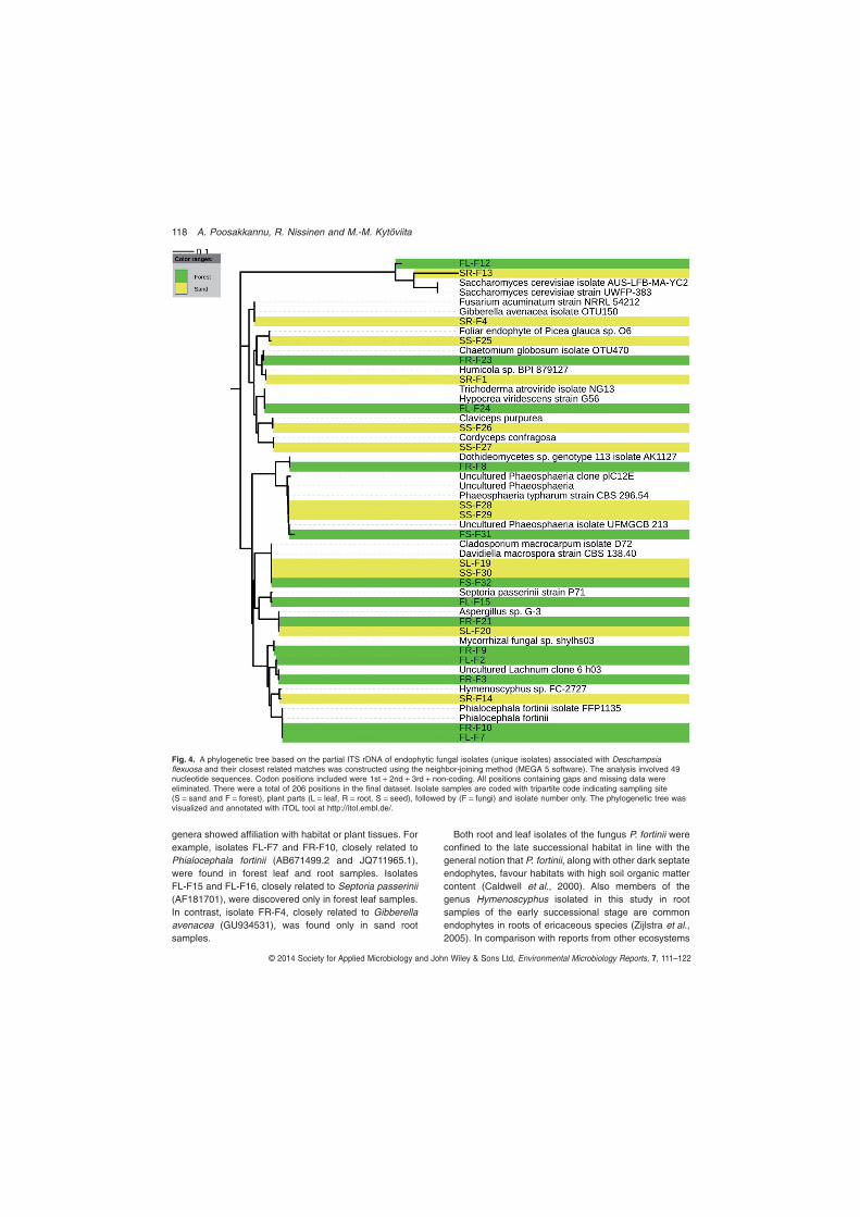
genera showed affiliation with habitat or plant tissues. Forexample, isolates FL-F7 and FR-F10, closely related toPhialocephala fortinii (AB671499.2 and JQ711965.1),were found in forest leaf and root samples. IsolatesFL-F15 and FL-F16, closely related to Septoria passerinii(AF181701), were discovered only in forest leaf samples.In contrast, isolate FR-F4, closely related to Gibberellaavenacea (GU934531), was found only in sand rootsamples.
Both root and leaf isolates of the fungus P. fortinii wereconfined to the late successional habitat in line with thegeneral notion that P. fortinii, along with other dark septateendophytes, favour habitats with high soil organic mattercontent (Caldwell et al., 2000). Also members of thegenus Hymenoscyphus isolated in this study in rootsamples of the early successional stage are commonendophytes in roots of ericaceous species (Zijlstra et al.,2005). In comparison with reports from other ecosystems
Fig. 4. A phylogenetic tree based on the partial ITS rDNA of endophytic fungal isolates (unique isolates) associated with Deschampsiaflexuosa and their closest related matches was constructed using the neighbor-joining method (MEGA 5 software). The analysis involved 49nucleotide sequences. Codon positions included were 1st + 2nd + 3rd + non-coding. All positions containing gaps and missing data wereeliminated. There were a total of 206 positions in the final dataset. Isolate samples are coded with tripartite code indicating sampling site(S = sand and F = forest), plant parts (L = leaf, R = root, S = seed), followed by (F = fungi) and isolate number only. The phylogenetic tree wasvisualized and annotated with iTOL tool at http://itol.embl.de/.
118 A. Poosakkannu, R. Nissinen and M.-M. Kytöviita
© 2014 Society for Applied Microbiology and John Wiley & Sons Ltd, Environmental Microbiology Reports, 7, 111–122
(Zijlstra et al., 2005; Tejesvi et al., 2010; 2013), it seemsthat so far, there is no common fungal endophyte speciesidentified in D. flexuosa except P. fortinii. Phialocephalafortinii is commonly isolated from roots of different plantspecies (Menkis et al., 2004; Addy et al., 2005). It hasbeen described as an important fungus in plant rootsforming the so-called ‘dark-septate endophyte’ coloniza-tion (Jumpponen and Trappe, 1998). This is the first reportof P. fortinii from the plant leaves, which likely reflects therelatively low number of studies on leaf fungal endophytesin comparison with root fungal endophytes.It is highly likely that endophytic bacteria and fungi may
also form functional associations with each other. Anendophytic fungus carrying Luteibacter sp. as endohyphalbacteria produced more of the plant hormone auxin thanfungus alone (Hoffman et al., 2013). In our study, thebacterial isolate FR131-1 (Collimonas sp.) and the fungalisolate FR-F23 (Chaetomium globosum) were specificto the late successional stage and plant tissue type,suggesting potential cohabitation. In earlier studies,Collimonas sp. (AJ496444) was previously isolated fromliving fungal hyphae of C. globosum, Fusarium culmorumand Mucor hiemalis from sand dune soils in the Nether-lands (de Boer et al., 2001; 2004). However, it will requiretargeted studies to reveal whether the bacteria and fungiisolated from the same tissues are functionally linked.
Plant association and cold habitat specificity ofendophytic isolates
Sequence alignment analysis revealed that 71% of theclosest matches in public data banks for our putativebacterial endophyte isolate 16S rRNA genes sequenceswere from plant-associated bacteria and 25% of the besthits originated from grass family plants (Fig. 2). Interest-ingly, the closest relatives of 52% of our endophytic bac-terial isolates had been isolated from cold environments,including arctic, antarctic and high alpine soils, snow,glacier or arcto-alpine plants. For example, endophyticbacterial isolates SL11, SL32 and SL28-1 were closelyrelated to P. graminis (DQ339600), Sphingomonas sp.(JQ977308) and C. flaccumfaciens (JQ977194) fromsubnival plants of high-elevation habitats in Tibet andChina (Sheng et al., 2011) respectively. Nissinen andcolleagues (2012) and Sheng and colleagues (2011),focusing on arctic and alpine plant species, reported that40–46% of the bacterial endophytic isolates were similarto bacteria from cold climates.We also performed a targeted screen of the isolate
sequences with a collection of endophyte sequences fromJ. trifidus, D. lapponica and Oxyria digyna isolated innorthwestern Lapland (Nissinen et al., 2012; 325 strains,NCBI accession HE814625–HE815460). Intriguingly,65% of the endophytes from D. flexuosa vegetative
tissues (roots, leaves, field seedlings) showed 99–100%sequence homology to the endophytic isolates reportedfrom Lapland (Nissinen et al., 2012), whereas only 10% ofthe seed or experimental seedling isolates had 99–100%match, and all of these were P. graminis isolates fromseeds of mainly J. trifidus, also a monocot plant species.To test whether our endophytes are related to coldclimate, we performed another targeted screen againstcollection of over 600 bacterial isolates from soils inLapland (Männistö and Häggblom, 2006). Of theD. flexuosa isolates, 40% from vegetative tissue (99% orhigher sequence homology), but none of the seed andseedling isolates, were closely related to the soil bacteriafrom subarctic Lapland (Table 2). The root tissues hadhighest relative abundance of close relatives of arctic soilbacteria. Taken together, these findings suggest a pres-ence of vertically transmitted (seed) core microbiota, andhorizontal, but selective acquisition of endophytes fromsoil in later growth stages.Like the bacterial endophytes, the great majority (83%
of closest relatives based on ITS sequence alignment) ofthe endophytic fungal isolates were plant-associated fungipreviously isolated from phyllo-, endo- or rhizospheres ofdifferent plant species. For example, the fungal isolatesSS-F25 is a close relative of the foliar endophyte(AY561215) of Picea glauca. A total of 17% of isolateswere closely related to fungi isolated from grass familyplants. For example, the fungal isolate FL-F15 is a closerelative of Septoria passerinii (AF181701) of barleyplants. However, unlike our bacterial isolates, only 13% ofclosest relatives of our endophytic fungal isolates weresimilar to fungi isolated from cold environments. Thiscould suggest that fungal communities from cold environ-ments are poorly studied when compared with their
Table 2. Percentage of bacterial isolate partial 16S rRNA sequencesfrom Deschampsia flexuosa tissues in this study with 99–100%sequence identity (16S rRNA gene, nucleotides 390–900) to 16SrRNA gene sequences from other endophytic (Nissinen et al., 2012)or soil bacterial isolates (Männistö and Häggblom, 2006) fromsubarctic–low arctic Finnish Lapland in NCBI nucleotide collection.
Tissue
Arcticendophyticbacteria (%)
Arctic soilbacteria(%)
Number ofD. flexuosaendophyte isolatesequencesanalysed
Seed 16 0 32Experimentalseedlings
0 0 16
Field seedlings 100 44 9Root 58 58 26Leaf 59 22 27Average/total 41 23 110
The first two columns in the table indicate the percentage of isolateswith 99–100% identity; the last column indicates the number of isolatesequences from different D. flexuosa tissues analysed.
Culturable endophytic microbes in Deschampsia flexuosa 119
© 2014 Society for Applied Microbiology and John Wiley & Sons Ltd, Environmental Microbiology Reports, 7, 111–122
bacterial counterpart, or that, unlike fungi, bacteria havedeveloped lineages endemic to cold climates.To the best of our knowledge, this is the first report on
diversity of culturable endophytes of both bacteria andfungi in an arctic plant species. The isolated putativeendophytes were tissue and habitat specific to variousdegrees. Overall, Actinobacteria and Alphaproteobacteriawere affiliated with sand whereas Bacteroidetes andGammaproteobacteria with forest habitat. Most of thebacterial isolates possessed capacity to solubilize organicphosphate. Also, many of our bacterial isolates had highlevel of homology to isolates from other cold environ-ments. They are potential candidates for plant growthpromoting bacteria in the cold habitat. Although the isola-tion method does not reveal the full microbial richness, itis necessary to work on arctic microbial isolates as theymay be used in future experiments and ultimately inrestoring vegetation in extreme environmental conditions.
Acknowledgements
We would like to thank Maj and Tor Nessling foundation andCentre for International Mobility, Finland for providing finan-cial support. A particular thank goes to Manoj Kumar, MinnaMännistö and Helena Jauhiainen for their invaluable helpduring the field work.
References
Addy, H., Piercey, M., and Currah, R. (2005) Microfungalendophytes in roots. Can J Bot 83: 1–13.
Adhikari, T.B., Joseph, C., Yang, G., Phillips, D.A., andNelson, L.M. (2001) Evaluation of bacteria isolated fromrice for plant growth promotion and biological control ofseedling disease of rice. Can J Microbiol 47: 916–924.
Aly, A.H., Debbab, A., and Proksch, P. (2011) Fungalendophytes: unique plant inhabitants with great promises.Appl Microbiol Biotechnol 90: 1829–1845.
Ardanov, P., Sessitsch, A., Haggman, H., Kozyrovska, N.,and Pirttila, A.M. (2012) Methylobacterium-inducedendophyte community changes correspond with protec-tion of plants against pathogen attack. PLoS ONE 7:e46802.
Arnold, A.E., Maynard, Z., and Gilbert, G.S. (2001) Fungalendophytes in dicotyledonous neotropical trees: patterns ofabundance and diversity. Mycol Res 105: 1502–1507.
Barac, T., Taghavi, S., Borremans, B., Provoost, A., Oeyen,L., Colpaert, J.V., et al. (2004) Engineered endophytic bac-teria improve phytoremediation of water-soluble, volatile,organic pollutants. Nat Biotechnol 22: 583–588.
Bjorbækmo, M., Carlsen, T., Brysting, A., Vrålstad, T.,Høiland, K., Ugland, K., et al. (2010) High diversity of rootassociated fungi in both alpine and arctic Dryas octopetala.BMC Plant Biol 10: 244.
Bodenhausen, N., Horton, M., and Bergelson, J. (2013) Bac-terial communities associated with the leaves and the rootsof Arabidopsis thaliana. PLoS ONE 8: e56329.
de Boer, W., Gunnewiek, P.J.K., Kowalchuk, G.A., andVan Veen, J.A. (2001) Growth of chitinolytic dune soilβ-subclass proteobacteria in response to invading fungalhyphae. Appl Environ Microbiol 67: 3358–3362.
de Boer, W., Leveau, J.H., Kowalchuk, G.A., Gunnewiek,P.J.K., Abeln, E.C., Figge, M.J., et al. (2004) Collimonasfungivorans gen. nov., sp. nov., a chitinolytic soil bacteriumwith the ability to grow on living fungal hyphae. Int J SystEvol Microbiol 54: 857–864.
Borin, S., Ventura, S., Tambone, F., Mapelli, F., Schubotz, F.,Brusetti, L., et al. (2010) Rock weathering creates oases oflife in a High Arctic desert. Environ Microbiol 12: 293–303.
Caldwell, B.A., Jumpponen, A., and Trappe, J.M. (2000) Uti-lization of major detrital substrates by dark-septate, rootendophytes. Mycologia 92: 230–232.
Cannon, P.F., and Simmons, C.M. (2002) Diversity and hostpreference of leaf endophytic fungi in the Iwokrama ForestReserve, Guyana. Mycologia 94: 210–220.
Compant, S., van der Heijden, M.G., and Sessitsch, A. (2010)Climate change effects on beneficial plant-microorganisminteractions. FEMS Microbiol Ecol 73: 197–214.
Dion, P. (2008) Extreme views on prokaryote evolution. InMicrobiology of Extreme Soils. Dion, P., and Nautiyal,C.S. (eds). Berlin Heidelberg, Germany: Springer,pp. 45–70.
Ferreira, A., Quecine, M.C., Lacava, P.T., Oda, S., Azevedo,J.L., and Araujo, W.L. (2008) Diversity of endophytic bac-teria from Eucalyptus species seeds and colonization ofseedlings by Pantoea agglomerans. FEMS Microbiol Lett287: 8–14.
Franche, C., Lindström, K., and Elmerich, C. (2009) Nitrogen-fixing bacteria associated with leguminous and non-leguminous plants. Plant Soil 321: 35–59.
de Freitas, J., Banerjee, M., and Germida, J. (1997)Phosphate-solubilizing rhizobacteria enhance the growthand yield but not phosphorus uptake of canola (Brassicanapus L.). Biol Fertil Soils 24: 358–364.
Grootjans, A., Van den Ende, F., and Walsweer, A. (1997)The role of microbial mats during primary succession incalcareous dune slacks: an experimental approach.J Coast Conserv 3: 95–102.
Gupta, G., Panwar, J., and Jha, P.N. (2013) Natural occur-rence of Pseudomonas aeruginosa, a dominant cultivablediazotrophic endophytic bacterium colonizing Pennisetumglaucum (L.) R. Br. Appl Soil Ecol 64: 252–261.
Hardoim, P.R., Hardoim, C.C., van Overbeek, L.S., and vanElsas, J.D. (2008) Properties of bacterial endophytes andtheir proposed role in plant growth. Trends Microbiol 16:463–471.
Hardoim, P.R., van Overbeek, L.S., and Elsas, J.D. (2012)Dynamics of seed-borne rice endophytes on early plantgrowth stages. PLoS ONE 7: e30438.
Higgins, K.L., Arnold, A.E., Miadlikowska, J., Sarvate, S.D.,and Lutzoni, F. (2007) Phylogenetic relationships, hostaffinity, and geographic structure of boreal and arcticendophytes from three major plant lineages. MolPhylogenet Evol 42: 543–555.
Hodkinson, I.D., Coulson, S.J., and Webb, N.R. (2003) Com-munity assembly along proglacial chronosequences in thehigh Arctic: vegetation and soil development in north-westSvalbard. J Ecol 91: 651–663.
120 A. Poosakkannu, R. Nissinen and M.-M. Kytöviita
© 2014 Society for Applied Microbiology and John Wiley & Sons Ltd, Environmental Microbiology Reports, 7, 111–122
Hoffman, M.T., Gunatilaka, M.K., Wijeratne, K., Gunatilaka,L., and Arnold, A.E. (2013) Endohyphal bacteriumenhances production of indole-3-acetic acid by a foliarfungal endophyte. PLoS ONE 8: e73132.
Idriss, E.E., Makarewicz, O., Farouk, A., Rosner, K., Greiner,R., Bochow, H., et al. (2002) Extracellular phytase activityof Bacillus amyloliquefaciens FZB45 contributes to itsplant-growth-promoting effect. Microbiology 148: 2097–2109.
Jumpponen, A., and Trappe, J.M. (1998) Dark septateendophytes: a review of facultative biotrophic root-colonizing fungi. New Phytol 140: 295–310.
Koster, E.A. (1988) Ancient and modern cold-climate aeoliansand deposition: a review. J Quaternary Sci 3: 69–83.
Long, H.H., Schmidt, D.D., and Baldwin, I.T. (2008) Nativebacterial endophytes promote host growth in a species-specific manner; phytohormone manipulations do notresult in common growth responses. PLoS ONE 3: e2702.
Ma, Y., Prasad, M., Rajkumar, M., and Freitas, H. (2011a)Plant growth promoting rhizobacteria and endophytesaccelerate phytoremediation of metalliferous soils.Biotechnol Adv 29: 248–258.
Ma, Y., Rajkumar, M., Luo, Y., and Freitas, H. (2011b) Inocu-lation of endophytic bacteria on host and non-host plants –effects on plant growth and Ni uptake. J Hazard Mater 195:230–237.
Männistö, M.K., and Häggblom, M.M. (2006) Characteriza-tion of psychrotolerant heterotrophic bacteria from FinnishLapland. Syst Appl Microbiol 29: 229–243.
Menkis, A., Allmer, J., Vasiliauskas, R., Lygis, V., Stenlid, J.,and Finlay, R. (2004) Ecology and molecular characteriza-tion of dark septate fungi from roots, living stems, coarseand fine woody debris. Mycol Res 108: 965–973.
Mercado-Blanco, J., and Bakker, P.A. (2007) Interactionsbetween plants and beneficial Pseudomonas spp.: exploit-ing bacterial traits for crop protection. Antonie Van Leeu-wenhoek 92: 367–389.
Nissinen, R.M., Mannisto, M.K., and van Elsas, J.D. (2012)Endophytic bacterial communities in three arctic plantsfrom low arctic fell tundra are cold-adapted and host-plantspecific. FEMS Microbiol Ecol 82: 510–522.
Park, M.S., Jung, S.R., Lee, M.S., Kim, K.O., Do, J.O., Lee,K.H., et al. (2005) Isolation and characterization of bac-teria associated with two sand dune plant species,Calystegia soldanella and Elymus mollis. J Microbiol 43:219–227.
Pillay, V., and Nowak, J. (1997) Inoculum density, tempera-ture, and genotype effects on in vitro growth promotionand epiphytic and endophytic colonization of tomato(Lycopersicon esculentum L.) seedlings inoculated with apseudomonad bacterium. Can J Microbiol 43: 354–361.
Polunin, N. (1938) Notes on a botanical journey in SWGreen-land, 1937. Bull Misc Inform Kew 3: 89–98.
Reinhold-Hurek, B., and Hurek, T. (2011) Living insideplants: bacterial endophytes. Curr Opin Plant Biol 14: 435–443.
Richardson, M.J. (2001) Coprophilous fungi from Brazil. BrazArch Biol Technol 44: 283–289.
Scott, J.J., and Loewus, F.A. (1986) Phytate metabolism inplants. In Phytic acid: Chemistry and Applications. Graf, E.(ed.). Minneapolis, MN, USA: Pilatus Press, pp. 23–42.
Seppälä, M. (1995) Deflation and redeposition of sand dunesin Finnish Lapland. Quat Sci Rev 14: 799–809.
Sessitsch, A., Reiter, B., and Berg, G. (2004) Endophyticbacterial communities of field-grown potato plants and theirplant-growth-promoting and antagonistic abilities. Can JMicrobiol 50: 239–249.
Sheng, H.M., Gao, H.S., Xue, L.G., Ding, S., Song, C.L.,Feng, H.Y., and An, L.Z. (2011) Analysis of the compositionand characteristics of culturable endophytic bacteria withinsubnival plants of the Tianshan Mountains, northwesternChina. Curr Microbiol 62: 923–932.
Siciliano, S.D., Fortin, N., Mihoc, A., Wisse, G., Labelle, S.,Beaumier, D., et al. (2001) Selection of specific endophyticbacterial genotypes by plants in response to soil contami-nation. Appl Environ Microbiol 67: 2469–2475.
Sturz, A., Christie, B., and Matheson, B. (1998) Associationsof bacterial endophyte populations from red clover andpotato crops with potential for beneficial allelopathy. Can JMicrobiol 44: 162–167.
Svoboda, J., and Henry, G. (1987) Succession in marginalarctic environments. Arctic Antarct Alp Res 19: 373–384.
Tejesvi, M.V., Sauvola, T., Pirttila, A.M., and Ruotsalainen,A.L. (2010) Root endophytes along a primary successiongradient in northern Finland. Fungal Divers 41: 125–134.
Tejesvi, M.V., Sauvola, T., Pirttila, A.M., and Ruotsalainen,A.L. (2013) Neighboring Deschampsia flexuosa andTrientalis europaea harbor contrasting root fungalendophytic communities. Mycorrhiza 23: 1–10.
Walker, J.F., Aldrich-Wolfe, L., Riffel, A., Barbare, H.,Simpson, N.B., Trowbridge, J., and Jumpponen, A. (2011)Diverse Helotiales associated with the roots of threespecies of Arctic Ericaceae provide no evidence for hostspecificity. New Phytol 191: 515–527.
Wearn, J.A., Sutton, B.C., Morley, N.J., and Gange, A.C.(2012) Species and organ specificity of fungal endophytesin herbaceous grassland plants. J Ecol 100: 1085–1092.
Zijlstra, J.D., Van’t Hof, P., Braakhekke, W.G., Berendse, F.,Baar, J., Paradi, I., et al. (2005) Diversity of symbiotic rootendophytes of the Helotiales in ericaceous plants andthe grass, Deschampsia flexuosa. Stud Mycol 53: 147–162.
Supporting information
Additional Supporting Information may be found in the onlineversion of this article at the publisher’s web-site:
Fig. S1. Phylum level distribution based on partial 16S rRNAsequence of endophytic bacteria (total isolates) associatedwith Deschampsia flexuosa expressed as relative abundancein early (sand) and late (forest) successional stages. Bacteriawere identified and classified using Ribosomal data base(http://rdp.cme.msu.edu/seqmatch/seqmatch_intro.jsp).Fig. S2. Distribution of different endophytic fungal genera(total isolates) from different Deschampsia flexuosa plantparts expressed as relative abundance in early (sand) andlate (forest) successional stages. Fungi were identifiedand classified using NCBI nucleotide database (http://
Culturable endophytic microbes in Deschampsia flexuosa 121
© 2014 Society for Applied Microbiology and John Wiley & Sons Ltd, Environmental Microbiology Reports, 7, 111–122
blast.ncbi.nlm.nih.gov/Blast.cgi?PROGRAM=blastn&BLAST_PROGRAMS=megaBlast&PAGE_TYPE=BlastSearch).Table S1. Percentage of phosphate (mineral or organicphosphate solubilizing assay) solubilizing bacterial isolates
(out of 178 total isolates) from different parts (leaf, root, seedand seedlings) of Deschampsia flexuosa in early (sand) andlate (forest) successional stage.Appendix S1. Materials and methods.
122 A. Poosakkannu, R. Nissinen and M.-M. Kytöviita
© 2014 Society for Applied Microbiology and John Wiley & Sons Ltd, Environmental Microbiology Reports, 7, 111–122
Experimental procedures Study site The study site is located in an Aeolian sand dune area in subarctic Northern Fennoscandia (68° 29' 16" N, 24° 42' 13" E). The region belongs to the belt of discontinuous permafrost. The sand dunes were formed after Ice Age and vegetated. Some parts of the dunes have lost the vegetation and wind has created blow-outs and drifting sands. Sand drifting has been occurring at least for the last 700 years (Seppälä 1995). Between the non-vegetated blow-outs different successional stages occur. The climax vegetation consists of mountain birch (Betula pubescens ssp. czerepanovii) forest, with the ground vegetation of dwarf shrubs such as Vaccinium uliginosum, V. vitis-idaea, Empetrum nigrum, Betula nana, and Pleurozium schreberii. In other successional stages Arctostaphylos uva-ursi, A. alpina, the grasses D. flexuosa and Festuca ovina, and the herbs Antennaria dioica and Solidago virgaurea occur. The 1981-2000 average annual temperature in the region is -1.3 °C (Pirinen et al., 2012). The area has a highly variable temperature with maximal temperatures between -51.5 °C (winter) to +30 °C (summer) (Pirinen et al., 2012), and large temperature fluctuations arecommon during the 100 days of growing period. The dune sands consist ofAeolian quartz particles with median grain size close to 0.2 mm (Seppälä 1995).Due to the porosity of the sand substrate and the low amount of annualprecipitation (400 mm), the vegetation is subjected to periods of drought. Somevegetation types have developed organic matter layer (e.g. 10 cm in mountainbirch forest), but some are without (e.g. D. flexuosa in deflated sand areas). Thetop soil pH varies between 4.7 (birch forest) and 5.8 (D. flexuosa in deflated sandareas) and total phosphorus between 1.91 mg/kg (D. flexuosa in deflated sandareas) and 14.12 mg/kg (birch forest), respectively.
Collection of Deschampsia flexuosa material Sampling was conducted during the growth season in 2011 in two successional stages. Early successional stage (hereafter referred to as ‘sand’) was characterized by the grass D. flexuosa growing as monoculture in the blow-out areas. The sparse D. flexuosa tussock biomass was 9 grams per m2 (Francini et al., unpublished). Late successional stage was mountain birch forest vegetation with continuous ground cover vegetation composed of abundant D. flexuosa together with E. nigrum and the moss P. schreberi under the cover of mountain birch trees (hereafter ‘forest’). Samples of D. flexuosa were collected from four different blow-out areas between 150 and 2250 meters apart. In each area, we established three plots 20 to 30 meters apart. In each plot, we harvested one plant in both the two successional stages situated within 10 meters from each other. Altogether, the plant material consists of (4x2x3) 24 plants.
Deschampsia flexuosa shoots and roots were collected in 24th July 2011. In addition, small seedlings less than 3 cm in height were collected from sand (called ‘field seedlings’ hereafter) at the same time. Deschampsia flexuosa seeds were collected 29th August 2011 from both sand and forest in all the four areas.
Endophytic microbe isolation Preweighed plant tissues of root, shoot, seedlings and seeds were surface sterilized by soaking in 70% ethanol for 1 min, 3% sodium hypochlorite for 3 minutes (except seeds, 6 minutes) , 1% sodium thiosulphate for 3 minutes, and washing three times with sterile deionized water for 3 minutes. Surface sterilized seeds were divided in to two portions. One portion was directly used for isolating microbes. Another portion was germinated on a sterile wet filter paper in Petri dishes for 10 days in greenhouse. The germinated seeds were used for microbe isolations (‘experimental seedlings’ hereafter).
In order to isolate bacteria, 1g of sterilized tissue (except seed) was homogenized in 3-4 ml of 50 mM potassium phosphate buffer at pH 6.5, and dilution plated as described below. Sterilized seeds were homogenized in 8 ml of BSE buffer (50 mM Tris–HCl [pH7.5], 1% Triton X-100 and 2 mM 2-mercaptoethanol) and centrifuged at 300×g for 5 min (at 15°C). Supernatant was transferred to new tubes and centrifuged at 12 000 ×g for 15 min (at 10°C). Pellets were suspended in 400 l of 50 mM potassium phosphate buffer set to pH 6.5. Serial dilutions were prepared and plated on the R2A media, pH 6.5. Plates were incubated at room temperature for a week whereafter they were moved to +4°C and monitored for new colonies. Single colonies of bacteria were transferred to new plates to obtain pure cultures. Pure strains were stored at -80 °C in R2A-glycerol solution.
In order to isolate fungi, sterilized tissues were cut into 1 cm pieces and plated directly into malt extract fungal media (Zijlstra et al., 2005). Plates were incubated in room temperature for a month or more. Hyphal tips of the developing fungal colonies were transferred to fresh malt extract agar plates.
16S rRNA and ITS rDNA amplification Single bacterial colonies were transferred to tubes containing 100 L of sterile deionized water and suspensions were heat lysed at 95 °C for 10 minutes and centrifuged at 13000x g for 5 minutes. Heat lysed suspensions were used as the DNA templates for PCR reactions. The 16S rRNA amplification was performed in a 50- l reaction mixture including 1- l DNA template, 0.25 M of primers 27F (5´-AGAGTTTGATCCTGGCTCAG-3´) and 1492R (5 -GGYTACTTGTTACGACTT-3 ), 0.25 M dNTPs, 1X of Taq buffer, and 1 U Taq DNA polymerase (Fermentas). The PCR amplification was performed as follows: one cycle of 5 minutes at 94 °C, followed by 30 cycles of 30 seconds at 94 °C, 45 seconds at 51 °C, and 1:30 minutes at 72 °C, followed by one cycle of 7 minutes at 72 °C.
Fungal genomic DNA was isolated from fresh mycelia scraped from plates using the Qiagen DNA isolation kit according to manufacturer’s protocol and used as template in PCR amplifications. The ITS region was amplified from genomic DNA using the forward primer ITS1 (5’-CTTGGTCATTTAGAGGAAGTAA-3’) and the reverse primer ITS4 (5’- TCCTCCGCTTATTGATATGC-3’). The PCR amplification was performed in a
50- l reaction mixture including 1- l DNA template, 0.25 M of primers, 0.25 M dNTPs, 1X of Taq buffer, and 1 U Taq DNA polymerase (Fermentas).The PCR program consisted of one initial denaturation step at 95 °C for 5 min followed by 30 cycles at 95 °C for 30 sec, 52 °C for 30 sec, 72 °C for 1 min, with a final extension at 72 °C for 7 min. Sequencing and sequence analysis The PCR products were purified by ethanol/EDTA precipitation and purified PCR products were sequenced with an automated multicapillary DNA sequencer, ABI Prism 3130xl genetic analyzer (Applied Biosystems, USA). Sequencing reactions were carried out using Big Dye Terminator v3.1 Cycle Sequencing kit (Applied Biosystems, Foster City, CA, USA) according to the manufacturer’s instructions. The RDB SEQMATCH (https://rdp.cme.msu.edu/seqmatch/seqmatch_intro.jsp) and NCBI BLAST (http://blast.ncbi.nlm.nih.gov/Blast.cgi?PROGRAM=blastn&BLAST_PROGRAMS=megaBlast&PAGE_TYPE=BlastSearch) were used to identify the closest phylogenetic relatives. The sequences were aligned, trimmed and analysed with MEGA 5 software (Tamura et al., 2011). The phylogenetic trees were visualized and annotated with iTOL tool at http://itol.embl.de/. These sequence data have been submitted to the GenBank databases under accession number KJ528986-KJ529110 Phosphate solubilization assays All bacterial strains were tested for their ability to solubilize mineral as well as organic forms of phosphate using National Botanical Research Institute’s phosphate growth medium (NBRIPM) supplemented with (g /L) 15 agar, 10 glucose, 5 Ca3(PO4)2, 5 MgCl2• 6H2O, 0.25 MgSO4• 7H2O, 0.2 KCl and 0.1 (NH4)2SO4 (Nautiyal 1999) and phytase screening medium (PSM) supplemented with (g /L) 15 agar, 10 D-glucose, 2 CaCl2, 5 NH4NO3, 0.5 KCl, 0.5 MgSO4 . 7H2O, 0.01 FeSO4 .7H2O, 0.01 MnSO4.H2O, and 3 phytate (phytic acid sodium salt hydrate) (Jorquera et al., 2011) . Seven strains per plate were stabbed in triplicate using inoculation loop. The ability to utilize tricalcium phosphate and phytate on NBRIPM and PSM agar was examined after incubation for 4 days at room temperature. The development of clearing zone around the colonies was used as an indicator of phosphate solubilization by the isolates.
Reference Hagerup, O. (1939) Studies on the significance of polyploidy III. Deschampsia
and Aira. Hereditas 25:185-192 Jorquera, M. A., Crowley, D. E., Marschner, P., Greiner, R., Fernández, M. T.,
Romero, D. et al. (2011) Identification of propeller phytase encoding genes in culturable Paenibacillus and Bacillus spp. from the rhizosphere of pasture plants on volcanic soils. FEMS Microbiol Ecol 75:163-172
Nautiyal, C. S. (1999) An efficient microbiological growth medium for screening phosphate solubilizing microorganisms. FEMS Microbiol Lett 170:265-270
Pirinen, P.,Simola, H., Aalto, J., Kaukoranta, J. P., Karlsson, P., & Ruuhela, R. (2012) Climatological statistics of Finland 1981–2010. Finnish Meteorological Institute Reports 2012:1
Tamura, K., Peterson, D., Peterson, N., Stecher, G., Nei, M., & Kumar, S. (2011) MEGA5: molecular evolutionary genetics analysis using maximum likelihood, evolutionary distance, and maximum parsimony methods. Mol Biol Evol 28: 2731-2739

Fig. S1. Phylum level distribution based on partial 16S rRNA sequence of endophytic bacteria (total isolates) associated with Deschampsia flexuosa expressed as relative abundance in early (sand) and late (forest) successional stages. Bacteria were identified and classified using Ribosomal data base (http://rdp.cme.msu.edu/seqmatch/seqmatch_intro.jsp).
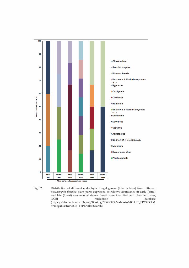
Fig. S2. Distribution of different endophytic fungal genera (total isolates) from different
Deschampsia flexuosa plant parts expressed as relative abundance in early (sand) and late (forest) successional stages. Fungi were identified and classified using NCBI nucleotide database (https://blast.ncbi.nlm.nih.gov/Blast.cgi?PROGRAM=blastn&BLAST_PROGRAMS=megaBlast&PAGE_TYPE=BlastSearch)
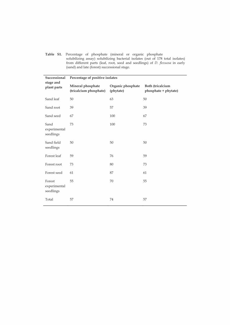
Table 1. Percentage of phosphate (mineral or organic phosphate solubilizing assay) solubilizing bacterial isolates (out of 178 total isolates) from different parts (leaf, root, seed and seedlings) of D. flexuosa in early (sand) and late (forest) successional stage.
Successional stage and plant parts
Percentage of positive isolates
Mineral phosphate (tricalcium phosphate)
Organic phosphate (phytate)
Both (tricalcium phosphate + phytate)
Sand leaf 50 63 50
Sand root 39 57 39
Sand seed 67 100 67
Sand experimental seedlings
73 100 73
Sand field seedlings
50 50 50
Forest leaf 59 76 59
Forest root 73 80 73
Forest seed 61 87 61
Forest experimental seedlings
55 70 55
Total 57 74 57
II
MICROBIAL COMMUNITY COMPOSITION BUT NOT DIVERSITY CHANGES ALONG SUCCESSION IN ARCTIC
SAND DUNES
by
Anbu Poosakkannu, Riitta Nissinen, Minna Männistö & Minna-Maarit Kytöviita 2016
Environmental microbiology, in press.
Reprinted with kind permission of © Society for Applied Microbiology and John Wiley & Sons Ltd
III
ENDOSPHERE MICROBIAL LEGACY AFTER PLANT TRANSPLANTATION IN ARCTIC SAND DUNES
by
Anbu Poosakkannu & Minna-Maarit Kytöviita 2016
Manuscript
IV
NATIVE ARBUSCULAR MYCORRHIZAL SYMBIOSIS CHANGES LEAF ENDOSPHERE BACTERIAL COMMUNITY
COMPOSITION IN DESCHAMPSIA FLEXUOSA
by
Anbu Poosakkannu, Riitta Nissinen & Minna-Maarit Kytöviita 2016
Manuscript