Embed Size (px)
Citation preview
Vol:.(1234567890)
Journal of Natural Medicines (2018) 72:32–42https://doi.org/10.1007/s11418-017-1144-z
1 3
REVIEW
Enzyme‑linked immunosorbent assay for the quantitative/qualitative analysis of plant secondary metabolites
Seiichi Sakamoto1 · Waraporn Putalun2 · Sornkanok Vimolmangkang3 · Waranyoo Phoolcharoen3 · Yukihiro Shoyama4 · Hiroyuki Tanaka1 · Satoshi Morimoto1
Received: 23 June 2017 / Accepted: 9 October 2017 / Published online: 21 November 2017 © The Author(s) 2017. This article is an open access publication
AbstractImmunoassays are antibody-based analytical methods for quantitative/qualitative analysis. Since the principle of immunoas-says is based on specific antigen–antibody reaction, the assays have been utilized worldwide for diagnosis, pharmacokinetic studies by drug monitoring, and the quality control of commercially available products. Berson and Yalow were the first to develop an immunoassay, known as radioimmunoassay (RIA), for detecting endogenous plasma insulin [1], a development for which Yalow was awarded the Nobel Prize in Physiology or Medicine in 1977. Even today, after half a century, immunoas-says are widely utilized with some modifications from the originally proposed system, e.g., radioisotopes have been replaced with enzymes because of safety concerns regarding the use of radioactivity, which is referred to as enzyme immunoassay/enzyme-linked immunosorbent assay (ELISA). In addition, progress has been made in ELISA with the recent advances in recombinant DNA technology, leading to increase in the range of antibodies, probes, and even systems. This review article describes ELISA and its applications for the detection of plant secondary metabolites.
Keywords Antibodies · Enzyme-linked immunosorbent assay (ELISA) · Hapten · Plant secondary metabolites
Introduction
Since the development of radioimmunoassay (RIA) in 1960, there has been a rapid increase in immunoassay techniques using radioactive labels [1]. However, radioactive labels have been gradually replaced with enzyme labels because of safety concerns associated with radioactivity since the study by Avrameas in 1969, who coupled antigens or antibodies and enzymes using glutaraldehyde [2]. Currently, ELISA has a higher number of immunoassays compared to RIA.
Plant secondary metabolites are plant-produced organic compounds that play an important role in the defense of plants against herbivores, pests, and pathogens, as well as in their adaptation to the environment, although they are not directly involved in the growth and development of organ-isms [3, 4]. Because of their diverse functions, there has been a dramatic increase in their demand in pharmaceu-ticals, cosmetics, and pesticides, as well as in food addi-tives [5]. Quality control of these commercial products containing secondary metabolites is crucial as the quality directly affects their potential activity. In addition, Cragg and Newman recently reported that 34% of the currently used drugs originate from natural products [6]. Meanwhile,
The original version of this article was revised due to a retrospective Open Access order.
* Seiichi Sakamoto [email protected]
* Hiroyuki Tanaka [email protected]
1 Department of Pharmacognosy, Graduate School of Pharmaceutical Sciences, Kyushu University, 3-1-1 Maidashi, Higashi-ku, Fukuoka 812-8582, Japan
2 Research Group for Pharmaceutical Activities of Natural Products using Pharmaceutical Biotechnology (PANPB), Faculty of Pharmaceutical Sciences, Khon Kaen University, Khon Kaen 40002, Thailand
3 Department of Pharmacognosy and Pharmaceutical Botany, Faculty of Pharmaceutical Sciences, Chulalongkorn University, 254 Phayathai Rd. Pathumwan, Bangkok 10330, Thailand
4 Department of Pharmacognosy, Faculty of Pharmaceutical Sciences, Nagasaki International University, 2825-7 Huis Ten Bosch, Sasebo, Nagasaki 859-3298, Japan
33Journal of Natural Medicines (2018) 72:32–42
1 3
simple, selective, and sensitive analytical techniques are also required in pharmacodynamic studies for monitoring effec-tive concentration, side effects, and metabolism, leading to a better quality of life for patients. Thus far, various analyti-cal methods have been developed for such purposes, mainly based on high-performance liquid chromatography (HPLC). However, ELISA exhibits several advantages over such tech-niques because of its simplicity, selectivity, and sensitivity.
The basic facts about ELISA and its practical use for measuring plant secondary metabolites are described in this review.
General principle of ELISA
ELISA is based on the concept of antigen–antibody reac-tions, representing the chemical interaction between anti-bodies produced by the B cells of leukocytes and antigens. This specific immune response plays an important role in protecting the body from invaders such as pathogens and toxins. Hence, by exploiting this reaction, ELISA permits the highly sensitive and selective quantitative/qualitative analysis of antigens, including proteins, peptides, nucleic acids, hormones, herbicides, and plant secondary metabo-lites. To detect these molecules, an antigen or antibody is labeled using enzymes, the so-called enzyme immunoas-say, in which alkaline phosphatase (ALP) [7], horseradish peroxidase (HRP) [8], and β-galactosidase [9–11] are com-monly used. The antigen in the fluid phase is immobilized on a solid phase, such as a microtiter plate constituting rigid polystyrene, polyvinyl chloride, and polypropylene. Subsequently, the antigen is allowed to react with a specific antibody, which is detected by an enzyme-labeled second-ary antibody. The development of color using a chromog-enic substrate corresponds to the presence of the antigen. For instance, ALP hydrolyzes p-nitrophenyl phosphate to
produce p-nitrophenol, which can be detected at 405 nm (yellow color), and HRP catalyzes the conversion of chro-mogenic substrates, e.g., 2,2′-azino-bis(3-ethylbenzothia-zoline-6-sulfonic acid) diammonium salt, 3,3′,5,5′-tetra-methylbenzidine, and o-phenylenediamine into colored products. By using chemiluminescent substrates such as chloro-5-substituted adamantyl-1,2-dioxetane phosphate and luminol for ALP and HRP, respectively, and fluoro-genic substrates such as 4-methylumbelliferyl galactoside and nitrophenyl galactoside for β-galactosidase, even more sensitive detection can be achieved. These enzyme–sub-strate reactions are typically completed within 30–60 min, and the reaction stops with the addition of an appropriate solution, e.g., sodium hydroxide, hydrochloric acid, sul-furic acid, sodium carbonate, and sodium azide, for indi-vidual reactions [12, 13]. Finally, colored or fluorescent products are detected using a microtiter plate reader.
Advantages and disadvantages of ELISA
Advantages and disadvantages of ELISA are summarized in Table 1. ELISA exhibits the following advantages: (i) Simple procedure. (ii) High specificity and sensitiv-ity, because of an antigen–antibody reaction. (iii) High efficiency, as simultaneous analyses can be performed without complicated sample pre-treatment. (iv) Gener-ally safe and eco-friendly, because radioactive substances and large amounts of organic solvents are not required. (v) Cost-effective assay, as low-cost reagents are used. However, ELISA exhibits the following disadvantages: (i) Labor-intensive and expensive to prepare antibody because it is a sophisticated technique, and expensive cul-ture cell media are required to obtain a specific antibody. (ii) High possibility of false positive or negative results
Table 1 Advantages and disadvantages of ELISA
Advantages Disadvantages
Simple procedure Labor-intensive and expensive to prepare antibody Easy to perform with simple procedure Sophisticated techniques and expensive culture media are required
High specificity and sensitivity High possibility of false positive/negative ELISA is based on antigen–antibody reaction Insufficient blocking of immobilized antigen results in false results
High efficiency Antibody instability Simultaneous analysis can be performed without complicated
sample pre-treatmentRefrigerated transport and storage are required as an antibody is a protein
Generally safe and eco-friendly Radioactive substances and large amounts of organic solvent are not
requiredCost-effective assay Reagents are relatively low cost
34 Journal of Natural Medicines (2018) 72:32–42
1 3
because of insufficient blocking of the surface of microtiter plate immobilized with antigen. (iii) Antibody instability because an antibody is a protein that requires refrigerated transport and storage.
Types of ELISA
Direct ELISA
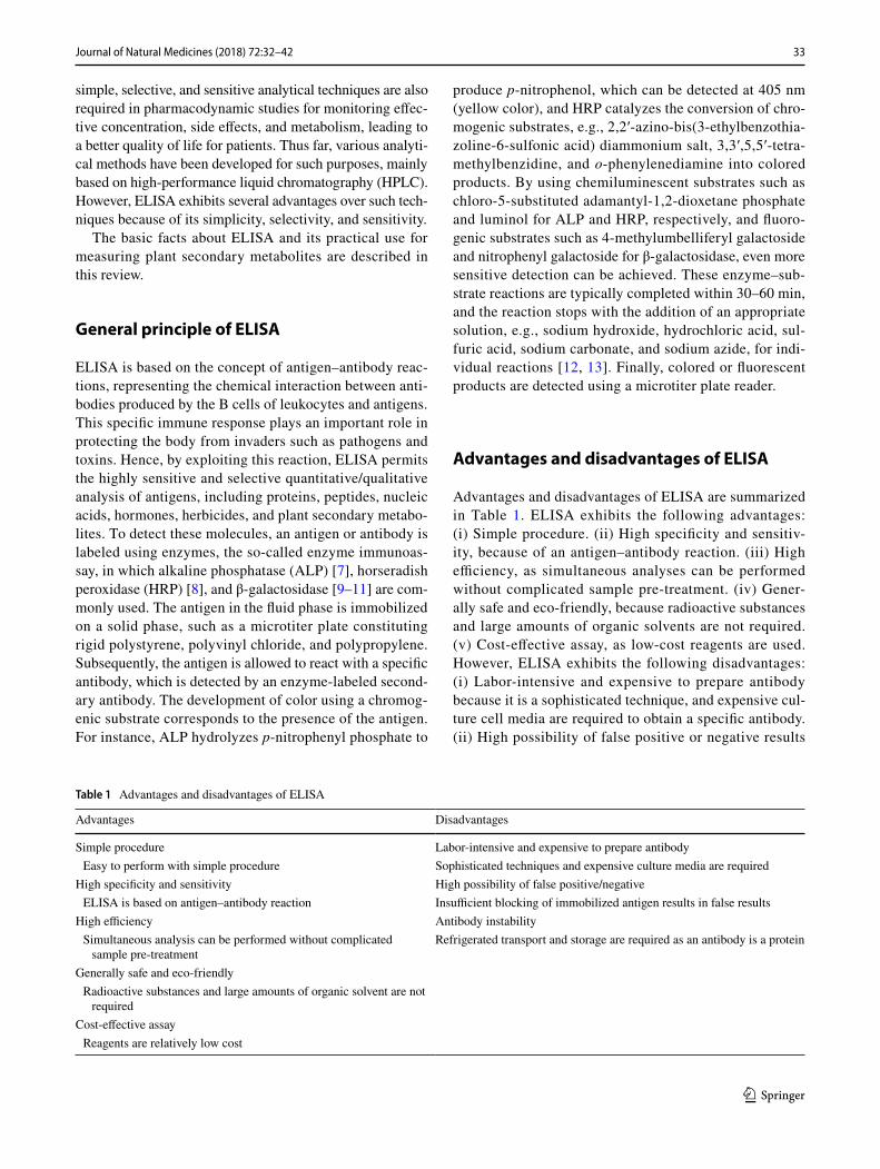
In 1971, Engvall and Perlmann [14] and Van Weemen and Schuurs [15] were the first to develop direct ELISA (Fig. 1), which was the base style for other types of ELISA. Primar-ily, an antigen or an antibody is immobilized on the surface of microtiter plate. After the surface is blocked with other proteins (e.g., albumin, gelatin, casein, and skimmed-milk [13]) to avoid the non-specific adsorption of other pro-teins, the corresponding enzyme-labeled antibody or anti-gen is allowed to react with the immobilized targets, fol-lowed by color development with appropriate substrates. With an increasing amount of targets, the signal increases. Direct ELISA is suitable for the qualitative analysis of macromolecules.
Competitive ELISA
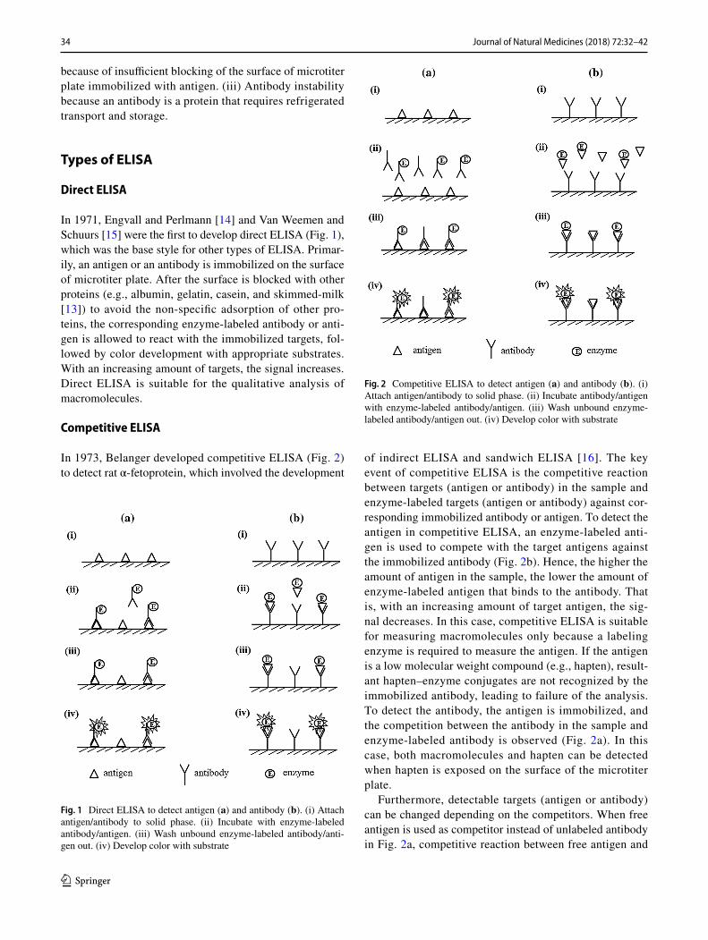
In 1973, Belanger developed competitive ELISA (Fig. 2) to detect rat α-fetoprotein, which involved the development
of indirect ELISA and sandwich ELISA [16]. The key event of competitive ELISA is the competitive reaction between targets (antigen or antibody) in the sample and enzyme-labeled targets (antigen or antibody) against cor-responding immobilized antibody or antigen. To detect the antigen in competitive ELISA, an enzyme-labeled anti-gen is used to compete with the target antigens against the immobilized antibody (Fig. 2b). Hence, the higher the amount of antigen in the sample, the lower the amount of enzyme-labeled antigen that binds to the antibody. That is, with an increasing amount of target antigen, the sig-nal decreases. In this case, competitive ELISA is suitable for measuring macromolecules only because a labeling enzyme is required to measure the antigen. If the antigen is a low molecular weight compound (e.g., hapten), result-ant hapten–enzyme conjugates are not recognized by the immobilized antibody, leading to failure of the analysis. To detect the antibody, the antigen is immobilized, and the competition between the antibody in the sample and enzyme-labeled antibody is observed (Fig. 2a). In this case, both macromolecules and hapten can be detected when hapten is exposed on the surface of the microtiter plate.
Furthermore, detectable targets (antigen or antibody) can be changed depending on the competitors. When free antigen is used as competitor instead of unlabeled antibody in Fig. 2a, competitive reaction between free antigen and
Fig. 1 Direct ELISA to detect antigen (a) and antibody (b). (i) Attach antigen/antibody to solid phase. (ii) Incubate with enzyme-labeled antibody/antigen. (iii) Wash unbound enzyme-labeled antibody/anti-gen out. (iv) Develop color with substrate
Fig. 2 Competitive ELISA to detect antigen (a) and antibody (b). (i) Attach antigen/antibody to solid phase. (ii) Incubate antibody/antigen with enzyme-labeled antibody/antigen. (iii) Wash unbound enzyme-labeled antibody/antigen out. (iv) Develop color with substrate
35Journal of Natural Medicines (2018) 72:32–42
1 3
immobilized antigen against enzyme-labeled antibody can be observed, enabling the detection of free antigen (mac-romolecules and hapten) in the sample in this competitive system, and vice versa when free antibody is used instead of unlabeled antigen in Fig. 2b.
Direct and competitive ELISA methods are simple because only one antibody is required. However, the labe-ling step is required for each of the ELISA methods, possibly leading to inactivation of the antibody (Table 2).
Indirect ELISA
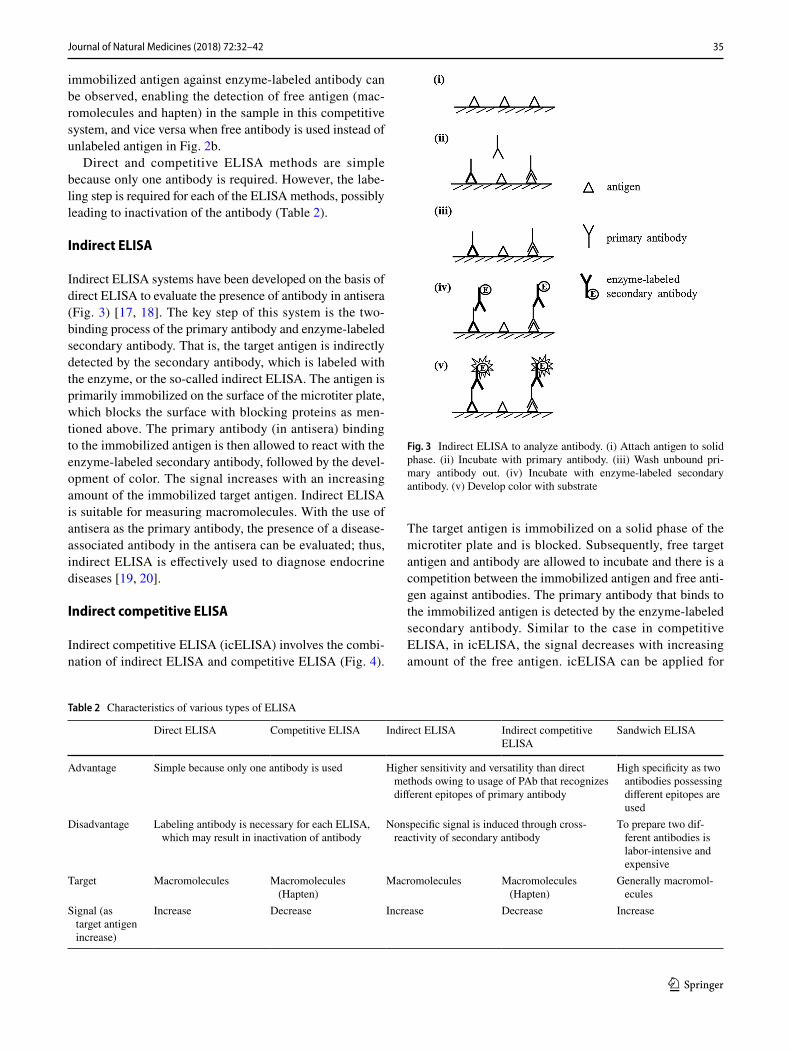
Indirect ELISA systems have been developed on the basis of direct ELISA to evaluate the presence of antibody in antisera (Fig. 3) [17, 18]. The key step of this system is the two-binding process of the primary antibody and enzyme-labeled secondary antibody. That is, the target antigen is indirectly detected by the secondary antibody, which is labeled with the enzyme, or the so-called indirect ELISA. The antigen is primarily immobilized on the surface of the microtiter plate, which blocks the surface with blocking proteins as men-tioned above. The primary antibody (in antisera) binding to the immobilized antigen is then allowed to react with the enzyme-labeled secondary antibody, followed by the devel-opment of color. The signal increases with an increasing amount of the immobilized target antigen. Indirect ELISA is suitable for measuring macromolecules. With the use of antisera as the primary antibody, the presence of a disease-associated antibody in the antisera can be evaluated; thus, indirect ELISA is effectively used to diagnose endocrine diseases [19, 20].
Indirect competitive ELISA
Indirect competitive ELISA (icELISA) involves the combi-nation of indirect ELISA and competitive ELISA (Fig. 4).
The target antigen is immobilized on a solid phase of the microtiter plate and is blocked. Subsequently, free target antigen and antibody are allowed to incubate and there is a competition between the immobilized antigen and free anti-gen against antibodies. The primary antibody that binds to the immobilized antigen is detected by the enzyme-labeled secondary antibody. Similar to the case in competitive ELISA, in icELISA, the signal decreases with increasing amount of the free antigen. icELISA can be applied for
Table 2 Characteristics of various types of ELISA
Direct ELISA Competitive ELISA Indirect ELISA Indirect competitive ELISA
Sandwich ELISA
Advantage Simple because only one antibody is used Higher sensitivity and versatility than direct methods owing to usage of PAb that recognizes different epitopes of primary antibody
High specificity as two antibodies possessing different epitopes are used
Disadvantage Labeling antibody is necessary for each ELISA, which may result in inactivation of antibody
Nonspecific signal is induced through cross-reactivity of secondary antibody
To prepare two dif-ferent antibodies is labor-intensive and expensive
Target Macromolecules Macromolecules (Hapten)
Macromolecules Macromolecules (Hapten)
Generally macromol-ecules
Signal (as target antigen increase)
Increase Decrease Increase Decrease Increase
Fig. 3 Indirect ELISA to analyze antibody. (i) Attach antigen to solid phase. (ii) Incubate with primary antibody. (iii) Wash unbound pri-mary antibody out. (iv) Incubate with enzyme-labeled secondary antibody. (v) Develop color with substrate
36 Journal of Natural Medicines (2018) 72:32–42
1 3
measuring both the macromolecules and hapten when hapten is exposed on the surface of the microtiter plate.
The use of enzyme-labeled secondary antibodies in indi-rect methods (e.g., indirect ELISA and icELISA) exhibit advantages over direct methods (direct and competitive ELISA) with respect to sensitivity and versatility [16]. Polyclonal antibody is a type of enzyme-labeled secondary antibody that recognizes different epitopes of the primary antibody, leading to increased sensitivity as compared to direct methods. In addition, a universal secondary antibody can be used if the original animal species of the primary antibody are unified. Thus, the secondary antibody is com-mercially available, leading to high versatility. Indirect ELISA clearly exhibits disadvantages with respect to the secondary antibody, i.e., the cross-reaction of the secondary antibody should be considered (Table 2).
Sandwich ELISA
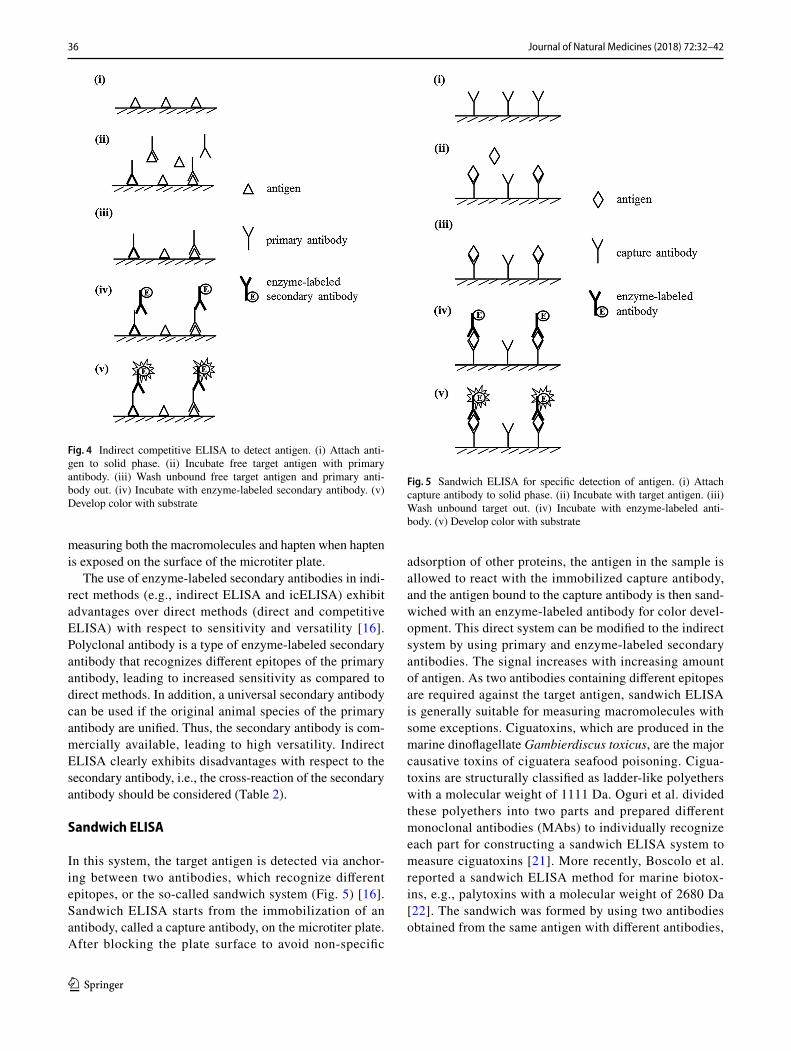
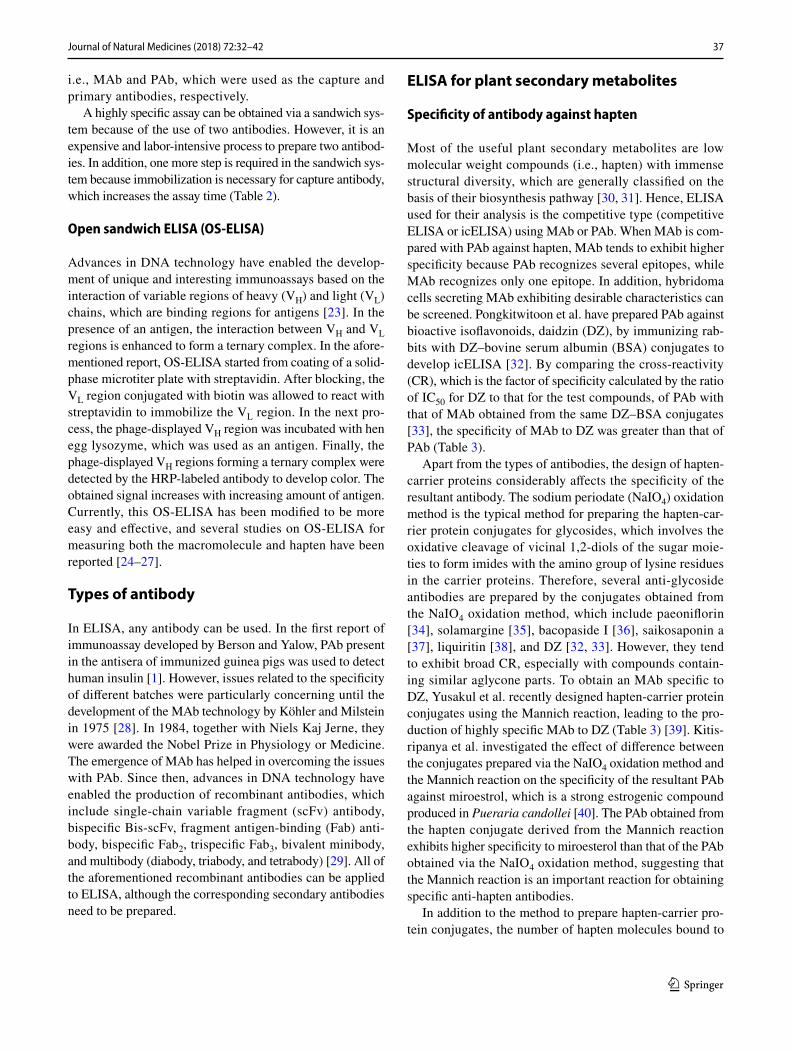
In this system, the target antigen is detected via anchor-ing between two antibodies, which recognize different epitopes, or the so-called sandwich system (Fig. 5) [16]. Sandwich ELISA starts from the immobilization of an antibody, called a capture antibody, on the microtiter plate. After blocking the plate surface to avoid non-specific
adsorption of other proteins, the antigen in the sample is allowed to react with the immobilized capture antibody, and the antigen bound to the capture antibody is then sand-wiched with an enzyme-labeled antibody for color devel-opment. This direct system can be modified to the indirect system by using primary and enzyme-labeled secondary antibodies. The signal increases with increasing amount of antigen. As two antibodies containing different epitopes are required against the target antigen, sandwich ELISA is generally suitable for measuring macromolecules with some exceptions. Ciguatoxins, which are produced in the marine dinoflagellate Gambierdiscus toxicus, are the major causative toxins of ciguatera seafood poisoning. Cigua-toxins are structurally classified as ladder-like polyethers with a molecular weight of 1111 Da. Oguri et al. divided these polyethers into two parts and prepared different monoclonal antibodies (MAbs) to individually recognize each part for constructing a sandwich ELISA system to measure ciguatoxins [21]. More recently, Boscolo et al. reported a sandwich ELISA method for marine biotox-ins, e.g., palytoxins with a molecular weight of 2680 Da [22]. The sandwich was formed by using two antibodies obtained from the same antigen with different antibodies,
Fig. 4 Indirect competitive ELISA to detect antigen. (i) Attach anti-gen to solid phase. (ii) Incubate free target antigen with primary antibody. (iii) Wash unbound free target antigen and primary anti-body out. (iv) Incubate with enzyme-labeled secondary antibody. (v) Develop color with substrate
Fig. 5 Sandwich ELISA for specific detection of antigen. (i) Attach capture antibody to solid phase. (ii) Incubate with target antigen. (iii) Wash unbound target out. (iv) Incubate with enzyme-labeled anti-body. (v) Develop color with substrate
37Journal of Natural Medicines (2018) 72:32–42
1 3
i.e., MAb and PAb, which were used as the capture and primary antibodies, respectively.
A highly specific assay can be obtained via a sandwich sys-tem because of the use of two antibodies. However, it is an expensive and labor-intensive process to prepare two antibod-ies. In addition, one more step is required in the sandwich sys-tem because immobilization is necessary for capture antibody, which increases the assay time (Table 2).
Open sandwich ELISA (OS‑ELISA)
Advances in DNA technology have enabled the develop-ment of unique and interesting immunoassays based on the interaction of variable regions of heavy (VH) and light (VL) chains, which are binding regions for antigens [23]. In the presence of an antigen, the interaction between VH and VL regions is enhanced to form a ternary complex. In the afore-mentioned report, OS-ELISA started from coating of a solid-phase microtiter plate with streptavidin. After blocking, the VL region conjugated with biotin was allowed to react with streptavidin to immobilize the VL region. In the next pro-cess, the phage-displayed VH region was incubated with hen egg lysozyme, which was used as an antigen. Finally, the phage-displayed VH regions forming a ternary complex were detected by the HRP-labeled antibody to develop color. The obtained signal increases with increasing amount of antigen. Currently, this OS-ELISA has been modified to be more easy and effective, and several studies on OS-ELISA for measuring both the macromolecule and hapten have been reported [24–27].
Types of antibody
In ELISA, any antibody can be used. In the first report of immunoassay developed by Berson and Yalow, PAb present in the antisera of immunized guinea pigs was used to detect human insulin [1]. However, issues related to the specificity of different batches were particularly concerning until the development of the MAb technology by Köhler and Milstein in 1975 [28]. In 1984, together with Niels Kaj Jerne, they were awarded the Nobel Prize in Physiology or Medicine. The emergence of MAb has helped in overcoming the issues with PAb. Since then, advances in DNA technology have enabled the production of recombinant antibodies, which include single-chain variable fragment (scFv) antibody, bispecific Bis-scFv, fragment antigen-binding (Fab) anti-body, bispecific Fab2, trispecific Fab3, bivalent minibody, and multibody (diabody, triabody, and tetrabody) [29]. All of the aforementioned recombinant antibodies can be applied to ELISA, although the corresponding secondary antibodies need to be prepared.
ELISA for plant secondary metabolites
Specificity of antibody against hapten
Most of the useful plant secondary metabolites are low molecular weight compounds (i.e., hapten) with immense structural diversity, which are generally classified on the basis of their biosynthesis pathway [30, 31]. Hence, ELISA used for their analysis is the competitive type (competitive ELISA or icELISA) using MAb or PAb. When MAb is com-pared with PAb against hapten, MAb tends to exhibit higher specificity because PAb recognizes several epitopes, while MAb recognizes only one epitope. In addition, hybridoma cells secreting MAb exhibiting desirable characteristics can be screened. Pongkitwitoon et al. have prepared PAb against bioactive isoflavonoids, daidzin (DZ), by immunizing rab-bits with DZ–bovine serum albumin (BSA) conjugates to develop icELISA [32]. By comparing the cross-reactivity (CR), which is the factor of specificity calculated by the ratio of IC50 for DZ to that for the test compounds, of PAb with that of MAb obtained from the same DZ–BSA conjugates [33], the specificity of MAb to DZ was greater than that of PAb (Table 3).
Apart from the types of antibodies, the design of hapten-carrier proteins considerably affects the specificity of the resultant antibody. The sodium periodate (NaIO4) oxidation method is the typical method for preparing the hapten-car-rier protein conjugates for glycosides, which involves the oxidative cleavage of vicinal 1,2-diols of the sugar moie-ties to form imides with the amino group of lysine residues in the carrier proteins. Therefore, several anti-glycoside antibodies are prepared by the conjugates obtained from the NaIO4 oxidation method, which include paeoniflorin [34], solamargine [35], bacopaside I [36], saikosaponin a [37], liquiritin [38], and DZ [32, 33]. However, they tend to exhibit broad CR, especially with compounds contain-ing similar aglycone parts. To obtain an MAb specific to DZ, Yusakul et al. recently designed hapten-carrier protein conjugates using the Mannich reaction, leading to the pro-duction of highly specific MAb to DZ (Table 3) [39]. Kitis-ripanya et al. investigated the effect of difference between the conjugates prepared via the NaIO4 oxidation method and the Mannich reaction on the specificity of the resultant PAb against miroestrol, which is a strong estrogenic compound produced in Pueraria candollei [40]. The PAb obtained from the hapten conjugate derived from the Mannich reaction exhibits higher specificity to miroesterol than that of the PAb obtained via the NaIO4 oxidation method, suggesting that the Mannich reaction is an important reaction for obtaining specific anti-hapten antibodies.
In addition to the method to prepare hapten-carrier pro-tein conjugates, the number of hapten molecules bound to

38 Journal of Natural Medicines (2018) 72:32–42
1 3
carrier proteins affects the specificity of antibodies. The hapten numbers are typically evaluated via matrix-assisted laser desorption/ionization time-of-flight mass spectrom-etry (MALDI-TOF–MS) using sinapinic acid as the matrix [41]. The relationship between the number of hapten mol-ecules and antibody specificity has been investigated using a mercaptopropionic acid derivative of atrazine: high anti-body titers with moderate antibody specificity are induced from 15–30 hapten molecules per carrier protein, while a lower number of hapten molecules exhibits a slower immune response with higher specificity [42]. This obser-vation was also supported by the PAb against miroesterol reported by Kitisripanya et al. [40]. Recently, MAbs against the Cephalotaxus alkaloid harringtonine and pyrrolizidine alkaloid monocrotaline have been independently produced from their BSA conjugates using the NaIO4- and N,N′-carbonyldiimidazole-mediated methods, respectively, for their determination in plants. Both of the resultant MAbs exhibit extremely high specificity to both targets with high sensitivity, although only two hapten molecules are bound to BSA [43, 44].
Utilization of antibody in icELISA depending on specificity
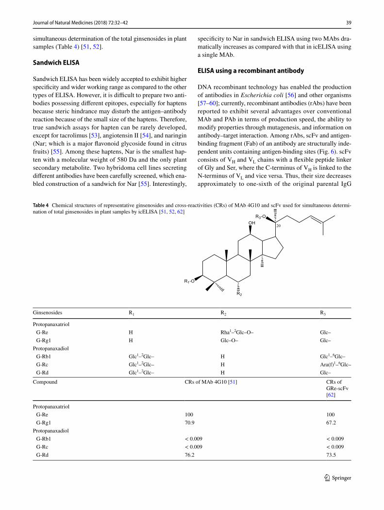
Antibodies exhibiting broad CR sometimes act as a useful and effective tool for recognizing a bioactive skeleton or a group of bioactive compounds because of the simultaneous determination by icELISA using the antibodies. Ginseno-sides are the major compounds produced in ginseng and are classified into two groups according to their structure: 20(S)-protopanaxadiol and 20(S)-protopanaxatriol [45]. As they are considered as active compounds that exert various pharmacological activities of ginseng, such as tonic, immu-nomodulatory, antimutagenic, and anti-aging activities, they are focused as a target for quantitative/qualitative analysis in ELISA. With respect to 20(S)-protopanaxadiol, MAbs against G-Rb1 [46] and Rg3 [47] have been produced, while those against G-Re [48], G-Rg1 [49], and Rh1 and Rg2 [50] have been produced as representatives of 20(S)-protopanaxa-triol for their specific determination. Interestingly, Morinaga et al. have produced MAb against G-Re exhibiting broad CR with G-Rd (76.2%) and G-Rg1 (70.9%) in addition to G-Re (100%) itself, enabling the development of icELISA for the
Table 3 Chemical structures of representative isoflavonoids and cross-reactivities (CRs) of PAb [32], MAb [33] produced from DZ–BSA conjugates prepared by NaIO4 oxidation method, and MAb [39] produced from DZ–cBSA conjugates obtained by Mannich reac-tion
Isoflavonoids R1 R2 R3
Daidzin (DZ) H Glc– HDaidzein H H HGenistin H Glc– OHGenistein H H OHPuerarin Glc– H H
Compound CRs of PAb [32] (NaIO4 oxidation) CRs of MAb [33] (NaIO4 oxidation) CRs of MAb [39] (Mannich reac-tion)
Diadzin (DZ) 100 100 100Daidzein 93.4 16.2 1.6Genistin 49.0 82.4 0.044Genistein 45.1 24.4 < 0.015Puerarin 0.1 3.4 < 0.015
39Journal of Natural Medicines (2018) 72:32–42
1 3
simultaneous determination of the total ginsenosides in plant samples (Table 4) [51, 52].
Sandwich ELISA
Sandwich ELISA has been widely accepted to exhibit higher specificity and wider working range as compared to the other types of ELISA. However, it is difficult to prepare two anti-bodies possessing different epitopes, especially for haptens because steric hindrance may disturb the antigen–antibody reaction because of the small size of the haptens. Therefore, true sandwich assays for hapten can be rarely developed, except for tacrolimus [53], angiotensin II [54], and naringin (Nar; which is a major flavonoid glycoside found in citrus fruits) [55]. Among these haptens, Nar is the smallest hap-ten with a molecular weight of 580 Da and the only plant secondary metabolite. Two hybridoma cell lines secreting different antibodies have been carefully screened, which ena-bled construction of a sandwich for Nar [55]. Interestingly,
specificity to Nar in sandwich ELISA using two MAbs dra-matically increases as compared with that in icELISA using a single MAb.
ELISA using a recombinant antibody
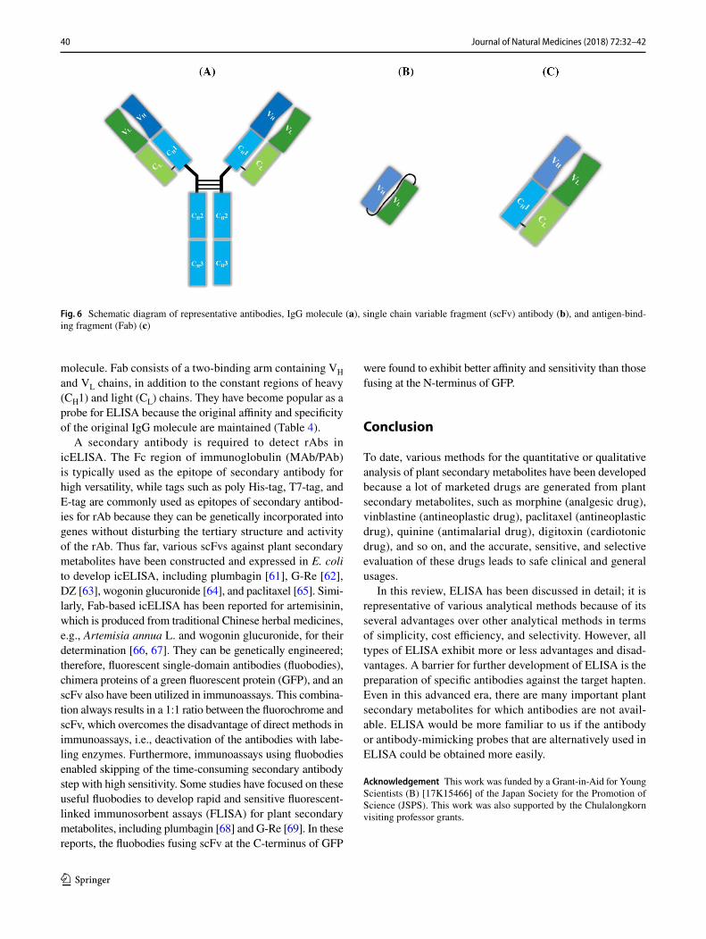
DNA recombinant technology has enabled the production of antibodies in Escherichia coli [56] and other organisms [57–60]; currently, recombinant antibodies (rAbs) have been reported to exhibit several advantages over conventional MAb and PAb in terms of production speed, the ability to modify properties through mutagenesis, and information on antibody–target interaction. Among rAbs, scFv and antigen-binding fragment (Fab) of an antibody are structurally inde-pendent units containing antigen-binding sites (Fig. 6). scFv consists of VH and VL chains with a flexible peptide linker of Gly and Ser, where the C-terminus of VH is linked to the N-terminus of VL and vice versa. Thus, their size decreases approximately to one-sixth of the original parental IgG
Table 4 Chemical structures of representative ginsenosides and cross-reactivities (CRs) of MAb 4G10 and scFv used for simultaneous determi-nation of total ginsenosides in plant samples by icELISA [51, 52, 62]
Ginsenosides R1 R2 R3
Protopanaxatriol G-Re H Rha1–2Glc–O– Glc– G-Rg1 H Glc–O– Glc–
Protopanaxadiol G-Rb1 Glc1–2Glc– H Glc1–6Glc– G-Rc Glc1–2Glc– H Ara(f)1–6Glc– G-Rd Glc1–2Glc– H Glc–
Compound CRs of MAb 4G10 [51] CRs of GRe-scFv [62]
Protopanaxatriol G-Re 100 100 G-Rg1 70.9 67.2
Protopanaxadiol G-Rb1 < 0.009 < 0.009 G-Rc < 0.009 < 0.009 G-Rd 76.2 73.5
40 Journal of Natural Medicines (2018) 72:32–42
1 3
molecule. Fab consists of a two-binding arm containing VH and VL chains, in addition to the constant regions of heavy (CH1) and light (CL) chains. They have become popular as a probe for ELISA because the original affinity and specificity of the original IgG molecule are maintained (Table 4).
A secondary antibody is required to detect rAbs in icELISA. The Fc region of immunoglobulin (MAb/PAb) is typically used as the epitope of secondary antibody for high versatility, while tags such as poly His-tag, T7-tag, and E-tag are commonly used as epitopes of secondary antibod-ies for rAb because they can be genetically incorporated into genes without disturbing the tertiary structure and activity of the rAb. Thus far, various scFvs against plant secondary metabolites have been constructed and expressed in E. coli to develop icELISA, including plumbagin [61], G-Re [62], DZ [63], wogonin glucuronide [64], and paclitaxel [65]. Simi-larly, Fab-based icELISA has been reported for artemisinin, which is produced from traditional Chinese herbal medicines, e.g., Artemisia annua L. and wogonin glucuronide, for their determination [66, 67]. They can be genetically engineered; therefore, fluorescent single-domain antibodies (fluobodies), chimera proteins of a green fluorescent protein (GFP), and an scFv also have been utilized in immunoassays. This combina-tion always results in a 1:1 ratio between the fluorochrome and scFv, which overcomes the disadvantage of direct methods in immunoassays, i.e., deactivation of the antibodies with labe-ling enzymes. Furthermore, immunoassays using fluobodies enabled skipping of the time-consuming secondary antibody step with high sensitivity. Some studies have focused on these useful fluobodies to develop rapid and sensitive fluorescent-linked immunosorbent assays (FLISA) for plant secondary metabolites, including plumbagin [68] and G-Re [69]. In these reports, the fluobodies fusing scFv at the C-terminus of GFP
were found to exhibit better affinity and sensitivity than those fusing at the N-terminus of GFP.
Conclusion
To date, various methods for the quantitative or qualitative analysis of plant secondary metabolites have been developed because a lot of marketed drugs are generated from plant secondary metabolites, such as morphine (analgesic drug), vinblastine (antineoplastic drug), paclitaxel (antineoplastic drug), quinine (antimalarial drug), digitoxin (cardiotonic drug), and so on, and the accurate, sensitive, and selective evaluation of these drugs leads to safe clinical and general usages.
In this review, ELISA has been discussed in detail; it is representative of various analytical methods because of its several advantages over other analytical methods in terms of simplicity, cost efficiency, and selectivity. However, all types of ELISA exhibit more or less advantages and disad-vantages. A barrier for further development of ELISA is the preparation of specific antibodies against the target hapten. Even in this advanced era, there are many important plant secondary metabolites for which antibodies are not avail-able. ELISA would be more familiar to us if the antibody or antibody-mimicking probes that are alternatively used in ELISA could be obtained more easily.
Acknowledgement This work was funded by a Grant-in-Aid for Young Scientists (B) [17K15466] of the Japan Society for the Promotion of Science (JSPS). This work was also supported by the Chulalongkorn visiting professor grants.
Fig. 6 Schematic diagram of representative antibodies, IgG molecule (a), single chain variable fragment (scFv) antibody (b), and antigen-bind-ing fragment (Fab) (c)
41Journal of Natural Medicines (2018) 72:32–42
1 3
Open Access This article is distributed under the terms of the Creative Commons Attribution 4.0 International License (http://creativecom-mons.org/licenses/by/4.0/), which permits use, duplication, adaptation, distribution and reproduction in any medium or format, as long as you give appropriate credit to the original author(s) and the source, provide a link to the Creative Commons license and indicate if changes were made.
References
1. Yalow RS, Berson SA (1960) Immunoassay of endogenous plasma insulin in man. J Clin Invest 39:1157–1175
2. Avrameas S (1969) Coupling of enzymes to proteins with glutar-aldehyde. Immunochemistry 6:43–52
3. Bennett RN, Wallsgrove RM (1999) Secondary metabolites in plant defence mechanisms. New Phytol 127:617–633
4. Fürstenberg-Hägg J, Zagrobelny M, Bak S (2013) Plant defense against insect herbivores. Int J Mol Sci 14:10242–10297
5. Bourgaud F, Gravot A, Milesi S, Gontier E (2001) Production of plant secondary metabolites: a historical perspective. Plant Sci 161:839–851
6. Cragg GM, Newman DJ (2013) Natural products: a con-tinuing source of novel drug leads. Biochim Biophys Acta 1830:3670–3695
7. Avrameas S, Ternynck T, Guesdon JL (1978) Coupling of enzymes to antibodies and antigens. Scand J Immunol 8:7–23
8. Nakane PK, Kawaoi A (1974) Peroxidase-labeled antibody. A new method of conjugation. J Histochem Cytochem 22:1084–1091
9. Craven GR, Steers E, Anfinsen CB (1965) Purification, composi-tion and molecular weight of the β-galactosidase of Escherichia coli K12. J Biol Chem 240:2468–2477
10. Dray F, Andrieu JM, Renaud F (1975) Enzyme immunoassay of progesterone at the pictogram level using β-galactosidase as label. Biochim Biophys Acta 403:131–138
11. Comoglio S, Celada F (1976) An immuno-enzymatic assay of cortisol using E. coli β-galactosidase as label. J Immunol Methods 10:161–170
12. Hitchcock CHS, Bailey FJ, Crimes AA, Dean DAG, Davis PJ (1981) Determination of soya proteins in food using an enzyme-linked immunosorbent assay procedure. J Sci Food Agric 32:157–165
13. Crowther JR (2009) Stages in ELISA. Methods Mol Biol 516:43–78
14. Engvall E, Perlmann P (1971) Enzyme-linked immunosorbent assay (ELISA). Quant Assay immunoglobul G Immunochem 8:871–874
15. Van Weemen BK, Schuurs AH (1971) Immunoassay using anti-gen-enzyme conjugates. FEBS Lett 15:232–236
16. Belanger L, Sylvestre C, Dufour D (1973) Enzyme-linked immu-noassay for alpha-fetoprotein by competitive and sandwich pro-cedures. Clin Chim Acta 48:15–18
17. Lindström P, Wager O (1978) IgG autoantibody to human serum albumin studied by the ELISA-technique. Scand J Immunol 7:419–425
18. Slightom EL (1978) The analysis of drugs in blood, bile, and tissue with an indirect homogeneous enzyme immunoassay. J Forensic Sci 23:292–303
19. Nierwenhuijzen Kruseman AC (1983) Application of ELISA for assessment of antiserum immunoreactivity in endocrine immuno-cytochemical studies. J Clin Pathol 36:406–410
20. Matyjaszek-Matuszek B, Pyzik A, Nowakowski A, Jarosz MJ (2013) Diagnostic methods of TSH in thyroid screening tests. Ann Agric Environ Med 20:731–735
21. Oguri H, Hirama M, Tsumuraya T, Fujii I, Maruyama M, Uehara H, Nagumo Y (2003) Synthesis-based approach toward direct sandwich immunoassay for ciguatoxin CTX3C. J Am Chem Soc 125:7608–7612
22. Boscolo S, Pelin M, De Bortoli M, Fontanive G, Barreras A, Berti F, Sosa S, Chaloin O, Bianco A, Yasumoto T, Prato M, Poli M, Tubaro A (2013) Sandwich ELISA assay for the quantitation of palytoxin and its analogs in natural samples. Environ Sci Technol 47:2034–2042
23. Ueda H, Tsumoto K, Kubota K, Suzuki E, Nagamune T, Nishimura H, Schueler PA, Winter G, Kumagai I, Mohoney WC (1996) Open sandwich ELISA: a novel immunoassay based on the interchain interaction of antibody variable region. Nat Biotechnol 14:1714–1718
24. Suzuki T, Munakata Y, Morita K, Shinoda T, Ueda H (2007) Sensitive detection of estrogenic mycotoxin zearalenone by open sandwich immunoassay. Anal Sci 23:65–70
25. Ihara M, Suzuki T, Kobayashi N, Goto J, Ueda H (2009) Open-sandwich enzyme immunoassay for one-step noncompeti-tive detection of corticosteroid 11-deoxycortisol. Anal Chem 81:8298–8304
26. Islam KN, Ihara M, Dong J, Kasagi N, Mori T, Ueda H (2011) Direct construction of an open-sandwich enzyme immunoassay for one-step noncompetitive detection of thyroid hormone T4. Anal Chem 83:1008–1014
27. Hara Y, Dong J, Ueda H (2013) Open-sandwich immunoassay for sensitive and broad-range detection of a shellfish toxin gonyau-toxin. Anal Chim Acta 793:107–113
28. Köhler G, Milstein C (1975) Continuous cultures of fused cells secreting antibody of predefined specificity. Nature 256:495–497
29. Holliger P, Hudson PJ (2005) Engineered antibody fragments and the rise of single domains. Nat Biotechnol 23:1126–1136
30. Hadacek F, Bachmann G (2015) Low-molecular-weight metabo-lite systems chemistry. Front Environ Sci 3:12
31. Harborne JB (1999) Classes and functions of secondary products. In: Walton NJ, Brown DE (eds) Chemicals from plants, perspec-tives secondary plant products. Imperial College Press, London, pp 1–25
32. Pongkitwitoon B, Sakamoto S, Tanaka H, Tsuchihashi R, Kinjo J, Morimoto S, Putalun W (2010) Enzyme-linked immunosorb-ent assay for total isoflavonoids in Pueraria candollei using anti-puerarin and anti-daidzin polyclonal antibodies. Planta Med 76:831–836
33. Sakamoto S, Yusakul G, Pongkitwitoon B, Paudel MK, Tanaka H, Morimoto S (2015) Simultaneous determination of soy isoflavone glycosides, daidzin and genistin by monoclonal antibody-based highly sensitive indirect competitive enzyme-linked immunosorb-ent assay. Food Chem 169:127–133
34. Lu Z, Morinaga O, Tanaka H, Shoyama Y (2003) A quantitative ELISA using monoclonal antibody to survey paeoniflorin and albiflorin in crude drugs and traditional Chinese herbal medicines. Biol Pharm Bull 26:862–866
35. Ishiyama M, Shoyama Y, Murakami H, Shinohara H (1995) Pro-duction of monoclonal antibodies and development of an ELISA for solamargine. Cytotechnology 18:153–158
36. Phrompittayarat W, Putalun W, Tanaka H, Jetiyanon K, Wittaya-Areekul S, Ingkaninan K (2007) Determination of pseudojujubo-genin glycosides from Brahmi based on immunoassay using a monoclonal antibody against bacopaside I. Phytochem Anal 18:411–418
37. Zhu S, Shimokawa S, Shoyama Y, Tanaka H (2006) A novel ana-lytical ELISA-based methodology for pharmacologically active saikosaponins. Fitoterapia 77:100–108
38. Fujii S, Morinaga O, Uto T, Nomura S, Shoyama Y (2014) Devel-opment of a monoclonal antibody-based immunochemical assay
42 Journal of Natural Medicines (2018) 72:32–42
1 3
for liquiritin and its application to the quality control of licorice products. J Agric Food Chem 62:3377–3383
39. Yusakul G, Sakamoto S, Juengwatanatrakul T, Putalun W, Tanaka H, Morimoto S (2016) Preparation and application of a mono-clonal antibody against the isoflavone glycoside daidzin using a Mannich reaction-derived hapten conjugate. Phytochem Anal 27:81–88
40. Kitisripanya T, Jutathis K, Inyai C, Komaikul J, Udomsin O, Yusakul G, Tanaka H, Putalun W (2016) Anti-miroestrol poly-clonal antibodies: a comparison of immunogen preparations used to obtain desired antibody properties. J Nat Med 70:296–299
41. Tanaka H, Xuan LJ, Morimoto S, Shoyama Y, Isobe R, Nojima K (2001) Direct determination of naturally occurring biologically active compound–serum albumin conjugate by matrix-assisted laser desorption/ionization mass spectrometry. J Spectrosc 15:1–18
42. Singh KV, Kaur J, Varshney GC, Raje M, Suri CR (2004) Synthe-sis and characterization of hapten-protein conjugates for antibody production against small molecules. Bioconjug Chem 15:168–173
43. Sakamoto S, Yusakul G, Tsuneura Y, Putalun W, Usui K, Miy-amoto T, Tanaka H, Morimoto S (2017) Sodium periodate-medi-ated conjugation of harringtonine enabling the production of a highly specific monoclonal antibody, and the development of a sensitive quantitative analysis method. Analyst 142:1140–1148
44. Sakamoto S, Nagamitsu R, Yusakul G, Miyamoto T, Tan-aka H, Morimoto S (2017) Ultrasensitive immunoassay for monocrotaline using monoclonal antibody produced by N,N′-carbonyldiimidazole mediated hapten-carrier protein conjugates. Talanta 168:67–72
45. Leung KW, Wong AST (2010) Pharmacology of ginsenosides: a literature review. Chin Med 5:20
46. Tanaka H, Fukuda N, Shoyama Y (1999) Formation of monoclo-nal antibody against a major ginseng component, ginsenoside Rb1 and its characterization. Cytotechnology 29:115–120
47. Joo EJ, Ha YW, Shin H, Son SH, Kim YS (2009) Generation and characterization of monoclonal antibody to ginsenoside Rg3. Biol Pharm Bull 32:548–552
48. Morinaga O, Tanaka H, Shoyama Y (2006) Detection and quantifi-cation of ginsenoside Re in ginseng samples by a chromatographic immunostaining method using monoclonal antibody against gin-senoside Re. J Chromatogr B 830:100–104
49. Fukuda N, Tanaka H, Shoyama Y (2000) Formation of mono-clonal antibody against a major ginseng component, ginsenoside Rg1 and its characterization. Monoclonal antibody for a ginseng saponin. Cytotechnology 34:197–204
50. Qu H, Wang Y, Shan W, Zhang Y, Feng H, Sai J, Wang Q, Zhao Y (2015) Development of ELISA for detection of Rh1 and Rg2 and potential method of immunoaffinity chromatography for separa-tion of epimers. J Chromatogr B 985:197–205
51. Morinaga O, Tanaka H, Shoyama Y (2006) Enzyme-linked immu-nosorbent assay for the determination of total ginsenosides in gin-seng. Anal Lett 39:287–296
52. Sritularak B, Morinaga O, Yuan CS, Shoyama Y, Tanaka H (2009) Quantitative analysis of ginsenosides Rb1, Rg1, and Re in Ameri-can ginseng berry and flower samples by ELISA using monoclo-nal antibodies. J Nat Med 63:360–363
53. Wei TQ, Zheng YF, Dubowy M, Sharma M (2014) Sandwich assay for tacrolimus using 2 antitacrolimus antibodies. Clin Chem 60:621–630
54. Towbin H, Motz J, Oroszlan P, Zingel O (1995) Sandwich immu-noassay for the hapten angiotensin II. A novel assay principle
based on antibodies against immune complexes. J Immunol Meth-ods 181:167–176
55. Qu H, Wang X, Qu B, Kong H, Zhang Y, Shan W, Cheng J, Wang Q, Zhao Y (2016) Sandwich enzyme-linked immunosorbent assay for naringin. Anal Chim Acta 903:149–155
56. Better M, Chang CP, Robinson RR, Horwitz AH (1988) Escheri-chia coli secretion of an active chimeric antibody fragment. Sci-ence 240:1041–1043
57. Jost CR, Kurucz I, Jacobus CM, Titus JA, George AJT, Segal DM (1994) Mammalian expression and secretion of functional single-chain Fv molecules. J Biol Chem 269:26267–26273
58. Bei R, Schlom J, Kashmiri SV (1995) Baculovirus expression of a functional single-chain immunoglobulin and its IL-2 fusion protein. J Immunol Methods 186:245–255
59. Davis GT, Bedzyk WD, Voss EW, Jacobs TW (1991) Single chain antibody (SCA) encoding genes: one-step construction and expression in eukaryotic cells. Biotechnology 9:165–169
60. Whitelam GC, Cockburn W, Owen MR (1994) Antibody produc-tion in transgenic plants. Biochem Soc Trans 22:940–945
61. Sakamoto S, Taura F, Putalun W, Pongkitwitoon B, Tsuchihashi R, Morimoto S, Kinjo J, Shoyama Y, Tanaka H (2009) Construc-tion and expression of specificity-improved single-chain variable fragments against the bioactive naphthoquinone, plumbagin. Biol Pharm Bull 32:434–439
62. Pongkitwitoon B, Sakamoto S, Morinaga O, Juengwatanatrakul T, Shoyama Y, Tanaka H, Morimoto S (2011) Single-chain variable fragment antibody against ginsenoside Re as an effective tool for the determination of ginsenosides in various ginsengs. J Nat Med 65:24–30
63. Yusakul G, Sakamoto S, Pongkitwitoon B, Tanaka H, Morimoto S (2016) Effect of linker length between variable domains of single chain variable fragment antibody against daidzin on its reactivity. Biosci Biotechnol Biochem 80:1306–1312
64. Paudel MK, Sakamoto S, Huy LV, Tanaka H, Miyamoto T, Mori-moto S (2017) The effect of varying the peptide linker length in a single chain variable fragment antibody against wogonin glucu-ronide. J Biotechnol 251:47–52
65. Yusakul G, Sakamoto S, Tanaka H, Morimoto S (2016) Efficient expression of single chain variable fragment antibody against paclitaxel using Bombyx mori nucleopolyhedrovirus bacmid DNA system and its characterizations. J Nat Med 70:592–601
66. Paudel MK, Takei A, Sakoda J, Juengwatanatrakul T, Sasaki-Tabata K, Putalun W, Shoyama Y, Tanaka H, Morimoto S (2012) Preparation of a single-chain variable fragment and a recombinant antigen-binding fragment against the anti-malarial drugs, arte-misinin and artesunate, and their application in an ELISA. Anal Chem 84:2002–2008
67. Paudel MK, Sakamoto S, Tanaka H, Morimoto S (2017) An over-view and comparison of a recombinant antigen-binding fragment and an antigen-binding fragment from a monoclonal antibody against wogonin glucuronide. J Nat Med 71:703–710
68. Sakamoto S, Taura F, Pongkitwitoon B, Putalun W, Tsuchihashi R, Kinjo J, Tanaka H, Morimoto S (2010) Development of sensi-tivity-improved fluorescence-linked immunosorbent assay using a fluorescent single-domain antibody against the bioactive naph-thoquinone, plumbagin. Anal Bioanal Chem 396:2955–2963
69. Sakamoto S, Tanizaki Y, Pongkitwitoon B, Tanaka H, Morimoto S (2011) A chimera of green fluorescent protein with single chain variable fragment antibody against ginsenosides for fluorescence-linked immunosorbent assay. Protein Expr Purif 77:124–130
![ENZYME-LINKED IMMUNOSORBENT ASSAY [ELISA]fac.ksu.edu.sa/sites/default/files/6_elisa_ppt.pdf · Enzyme-linked immunosorbent assay. Is a biochemical plate-based assay technique designed](https://img.pdfslide.tips/doc/110x75/6071be0f5a7c35211f622a5b/enzyme-linked-immunosorbent-assay-elisafacksuedusasitesdefaultfiles6elisapptpdf.jpg)


![R9901 Ractopamin 15-10-19 - food.r-biopharm.com · [1] Weilin L. Shelver, and David J. Smith: Application of a Monoclonal Antibody-Based Enzyme-Linked Immunosorbent Assay for the](https://img.pdfslide.tips/doc/110x75/5e0e6fe79a29ee2e8a15bc08/r9901-ractopamin-15-10-19-foodr-1-weilin-l-shelver-and-david-j-smith-application.jpg)