Embed Size (px)
Citation preview

EURURO-8276; No. of Pages 12
Guidelines
European Association of Urology Guidelines on Renal CellCarcinoma: The 2019 Update
Borje Ljungberg a,*, Laurance Albiges b, Yasmin Abu-Ghanem c, Karim Bensalah d,Saeed Dabestani e, Sergio Fernandez-Pello Montes f, Rachel H. Giles g, Fabian Hofmann h,Milan Hora i, Markus A. Kuczyk j, Teele Kuusk k, Thomas B. Lam l,m, Lorenzo Marconi n,Axel S. Merseburger o, Thomas Powles p, Michael Staehler q, Rana Tahbaz r,Alessandro Volpe s, Axel Bex t,u,v
aDepartment of Surgical and Perioperative Sciences, Urology and Andrology, Umeå University, Umeå, Sweden; bDepartment of Cancer Medicine, Gustave
Roussy, Université Paris-Saclay, Villejuif, France; cDepartment of Urology, Chaim Sheba Medical Center, Tel-Hashomer, Ramat-Gan, Israel; dDepartment of
Urology, University of Rennes, Rennes, France; eDepartment of Clinical Sciences Lund, Skåne University Hospital, Lund, Sweden; fDepartment of Urology,
Cabueñes University Hospital, Gijón, Spain; gDepartment of Nephrology and Hypertension, Patient Advocate International Kidney Cancer Coalition (IKCC),
University Medical Center Utrecht, Utrecht, The Netherlands; hDepartment of Urology, Sunderby Hospital, Sunderby, Sweden; iDepartment of Urology,
Faculty Hospital and Faculty of Medicine in Pilsen, Charles University in Prague, Prague, Czech Republic; jDepartment of Urology and Urologic Oncology,
Hannover Medical School, Hannover, Germany; kDepartment of Urology, Royal Free Hospital, Pond Street, London, UK; lAcademic Urology Unit, University of
Aberdeen, Aberdeen, UK; mDepartment of Urology, Aberdeen Royal Infirmary, Aberdeen, UK; nDepartment of Urology, Coimbra University Hospital, Coimbra,
Portugal; oDepartment of Urology, University Hospital Schleswig-Holstein, Lübeck, Germany; p The Royal Free NHS Trust and Barts Cancer Institute, Queen
Mary University of London, London, UK; qDepartment of Urology, Ludwig-Maximilians University, Munich, Germany; rDepartment of Urology, University
Hospital Hamburg Eppendorf, Hamburg, Germany; sDivision of Urology, Maggiore della Carità Hospital, University of Eastern Piedmont, Novara, Italy; t The
Royal Free London NHS Foundation Trust, London, UK; uUCL Division of Surgery and Interventional Science, London, UK; vDepartment of Urology,
The Netherlands Cancer Institute, Antoni van Leeuwenhoek Hospital, Amsterdam, The Netherlands
E U R O P E A N U R O L O G Y X X X ( 2 0 18 ) X X X – X X X
ava i lable at www.sc iencedirect .com
journa l homepage: www.europea nurology.com
Article info
Article history:Accepted February 7, 2019
Associate Editor:
James Catto
Keywords:
Renal cell cancerGuidelinesDiagnosisManagementSurgeryMedicalFollow-up
Abstract
Context: The European Association of Urology Renal Cell Carcinoma (RCC) GuidelinePanel has prepared evidence-based guidelines and recommendations for the manage-ment of RCC.Objective: To provide an updated RCC guideline based on standardised methodologyincluding systematic reviews, which is robust, transparent, reproducible, and reliable.Evidence acquisition: For the 2019 update, evidence synthesis was undertaken based ona comprehensive and structured literature assessment for new and relevant data. Wherenecessary, formal systematic reviews adhering to the Preferred Reporting Items forSystematic Reviews and Meta-analyses (PRISMA) guidelines were undertaken. Relevantdatabases (Medline, Cochrane Libraries, trial registries, conference proceedings) weresearched until June 2018, including randomised controlled trials (RCTs) and retrospec-tive or controlled studies with a comparator arm, systematic reviews, and meta-analyses. Where relevant, risk of bias (RoB) assessment, and qualitative and quantitativesyntheses of the evidence were performed. The remaining sections of the documentwere updated following a structured literature assessment. Clinical practice recom-mendations were developed and issued based on the modified GRADE framework.Evidence synthesis: All chapters of the RCC guidelines were updated based on a
ass
. De, Swng
European Association of Urology structured literature
* Corresponding authorUmea University, UmeaE-mail address: borje.lju
Please cite this article in press as: Ljungberg B, et al. European As2019 Update. Eur Urol (2019), https://doi.org/10.1016/j.eururo.201
https://doi.org/10.1016/j.eururo.2019.02.0110302-2838/© 2019 European Association of Urology. Published by Elsevier B
essment, for prioritised topics based on the availability of robust
partment of Surgical and Perioperative Sciences, Urology and Andrology,eden. Tel. +46 907 851 330; Fax: +46 901 253 [email protected] (B. Ljungberg).
sociation of Urology Guidelines on Renal Cell Carcinoma: The9.02.011
.V. All rights reserved.
http://guide.medlive.cn/
data. For RCTs, RoB was low across studies. For most non-RCTs, clinical and methodo-logical heterogeneity prevented pooling of data. The majority of included studies wereretrospective with matched or unmatched cohorts, based on single- or multi-institu-tional data or national registries. The exception was for the treatment of metastatic RCC,for which there were several large RCTs, resulting in recommendations based on higherlevels of evidence.Conclusions: The 2019 RCC guidelines have been updated by the multidisciplinary panelusing the highest methodological standards. These guidelines provide the most reliablecontemporary evidence base for the management of RCC in 2019.Patient summary: The European Association of Urology Renal Cell Carcinoma GuidelinePanel has thoroughly evaluated the available research data on kidney cancer to establishinternational standards for the care of kidney cancer patients.© 2019 European Association of Urology. Published by Elsevier B.V. All rights reserved.
E U R O P E A N U R O L O G Y X X X ( 2 0 18 ) X X X – X X X2
EURURO-8276; No. of Pages 12
1. Introduction
The European Association of Urology (EAU) renal cell cancer(RCC) guidelines provide clinicians with evidence-basedinformation and recommendations for the management ofpatients with RCC. The RCC Panel is an international groupconsisting of clinicians with expertise in this field. Themultidisciplinary panel includes urologists, medical oncol-ogists, a pathologist, a radiologist, a methodologist, and amember of a patient advocacy group. The EAU RCC guide-lines were first published in 2000 [1]. For the 2019 update,the entire guideline has been updated based on acomprehensive and structured literature assessment, withseveral sections requiring a formal systematic review (SR)based on the availability of data (Table 1). A detailed versionof the current guideline including full references, level ofevidence (LE), and grade of recommendations is available athttp://uroweb.org/guideline/renal-cell-carcinoma/ [2].
2. Evidence acquisition
All chapters of the 2019 RCC guideline publication wereupdated using a structured literature assessment [2]. Liter-ature searches were conducted in the following databases:MEDLINE, MEDLINE In-Process, Embase, and CochraneDatabase of Systematic Reviews and Controlled TrialsRegister. Additionally, a series of topics and questions wereprioritised a priori, on which formal, protocol-driven SRswere undertaken, for which the review methodology has
Table 1 – Description of guideline update and summary of review met
Chapter
1. Introduction Not applicable2. Methods Not applicable3. Epidemiology, aetiology, and pathology. This chapter was updated by a4. Staging and grading classification systems This chapter was updated by a5. Diagnostic evaluation Diagnostic imaging was revise
literature assessment.6. Prognosis This chapter was updated by a7. Treatment (disease management) Treatment of localised and loc
cancer was updated by the mliterature assessment. System
8. Surveillance following radical or partialnephrectomy or ablative therapies
This chapter was updated by a
SR = systematic review.
Please cite this article in press as: Ljungberg B, et al. European As2019 Update. Eur Urol (2019), https://doi.org/10.1016/j.eururo.20
been described elsewhere [3,4]. These were conducted inaccordance with the Preferred Reporting Items for System-atic Reviews and Meta-analyses (PRISMA) guidelines[2,5]. For each SR, elements for inclusion and exclusion,including patient population, intervention, comparison,outcomes (PICO); study design; and search terms andrestrictions were developed using an iterative processinvolving all members of the panel in a consensus model.Where relevant, confounding variables were identified foreach question to facilitate the assessment of nonrandomis-ed studies. The SR protocols containing details of the reviewprocess and the search strategies used, as well as, referencelists of all included studies are published on uroweb.org[2]. The search was conducted up to the end of June2018. Two independent reviewers screened abstracts andfull texts, carried out data abstraction, and assessed risk ofbias (RoB). Data were assessed according to their level ofscientific evidence (LE) based on the 2009 Oxford Centre forEvidence-based Medicine Levels of Evidence (http://www.cebm.net/index.aspx?o=1025 [accessed date January2019]). The majority of included studies were retrospectiveanalyses that included some larger multicentre or well-designed controlled comparative studies, except for thetopic of systemic treatment of metastatic RCC (mRCC), inwhich several practice-changing randomised controlledtrials (RCTs) have been performed, resulting in a higher LE.Once the LE for a particular topic or question had beendetermined, a guideline recommendation was made using atransparent, reproducible, and reliable process modified
hodology
Brief description of review methodology
traditional narrative review, based on a structured literature assessment. traditional narrative review, based on a structured literature assessment.d based on an SR. The remainder of the chapter was updated by a structured
traditional narrative review, based on a structured literature assessment.ally advanced disease was revised based on an updated SR. Non-clear-celleans of an SR. The remainder of the chapter was updated using a structuredic therapy for metastatic disease was updated by SRs.
traditional narrative review, based on a structured literature assessment.
sociation of Urology Guidelines on Renal Cell Carcinoma: The19.02.011 http://guide.medlive.cn/
Table 2 – Recommendations for the diagnostic assessment of renalcell carcinoma
Strength rating
Use multiphasic contrast-enhanced computedtomography (CT) of abdomen and chest for thediagnosis and staging of renal tumours.
Strong
Use magnetic resonance imaging to betterevaluate venous involvement, reduce radiation,or avoid intravenous CT contrast medium.
Weak
Use nonionising modalities, mainly contrast-enhanced ultrasound, for furthercharacterisation of small renal masses, tumourthrombus, and differentiation of unclear renalmasses.
Strong
Do not routinely use bone scan and/or positronemission tomography CT for staging of RCC.
Weak
Perform a renal tumour biopsy before ablativetherapy and systemic therapy without previouspathology.
Strong
Perform a percutaneous biopsy in select patientswho are considering active surveillance.
Weak
Use a coaxial technique when performing a renaltumour biopsy.
Strong
Do not perform a renal tumour biopsy of cysticrenal masses.
Strong
Use a core biopsy technique rather than fineneedle aspiration for histologicalcharacterisation for solid renal tumours.
Strong
RCC = renal cell carcinoma.
E U R O P E A N U R O L O G Y X X X ( 2 0 18 ) X X X – X X X 3
EURURO-8276; No. of Pages 12
from the GRADE framework [6]. This approach allows theintegration of the LE with other essential elements, includingcertainty of the evidence, magnitude of effects, balancebetween consequences, and patient values and preferences[7], in order to issue clinical practice recommendations[2,6]. Where there was heterogeneity of opinions amongstpanel members, formal consensus methods were used toarrive at the final recommendation [8].
3. Evidence synthesis
3.1. Epidemiology and aetiology
RCC represents approximately 3% of all cancers, with thehighest incidence occurring in Western countries [9]. Gen-erally, during the last 2 decades, there has been an annualincrease of 2% in incidence both worldwide and in Europe,leading to approximately 99 200 new RCC cases and39 100 kidney cancer-related deaths within the EuropeanUnion in 2018 [9]. In Europe, overall mortality rates forRCC increased until the early 1990s, with rates generallystabilising or declining thereafter [10]. There has been adecrease in mortality since the 1980s in Scandinaviancountries and since the early 1990s in France, Germany,Austria, the Netherlands, and Italy. However, in someEuropean countries (Croatia, Estonia, Greece, Ireland, andSlovakia), mortality rates still show an upward trend[9,10]. RCC is the most common solid lesion within thekidney and accounts for approximately 90% of all kidneymalignancies. There is a 1.5:1 predominance in men overwomen, with a peak incidence at 60–70 yr of age [11]. Itcomprises different RCC subtypes with specific histopath-ological and genetic characteristics [12]
Aetiology includes lifestyle factors such as smoking,obesity, and hypertension [13]. Having a first-degreerelative with RCC is also associated with an increased risk.A number of other factors have been suggested to beassociated with a higher or lower risk of RCC. These includespecific dietary habits and occupational exposure to specificcarcinogens, but the literature is inconclusive [13,14]. Mod-erate alcohol consumption appears to have a protectiveeffect for unknown reasons [15]. The most effectiveprophylaxis is to avoid cigarette smoking and reduceobesity [13].
3.2. Diagnosis and staging
3.2.1. Symptoms
Many patients with renal masses (RMs) remain asymptom-atic until the late stages. Today, >60% of RCCs are detectedincidentally with abdominal ultrasound (US)or computedtomography (CT) performed for other reasons (LE: 3). Theclassic triad of flank pain, visible haematuria, and palpableabdominal mass is rare today, and correlates with advanceddisease and subtypes associated with poor prognosis (LE: 3)[11]. Paraneoplastic syndromes are found in approximately30% of patients with symptomatic RCCs (LE: 4). A fewpatients present with symptoms caused by mRCC, such as
Please cite this article in press as: Ljungberg B, et al. European As2019 Update. Eur Urol (2019), https://doi.org/10.1016/j.eururo.201
bone pain, deterioration of performance status (PS), orpersistent cough (LE: 3) [16].
The EAU RCC guideline recommendations for diagnosticprocedures of RMs are shown in Table 2.
3.2.2. Imaging
CT, US, and magnetic resonance imaging (MRI) are theimaging modalities used to detect and characterise RMs[17]. RMs can be classified as solid or cystic on the basis ofthe imaging findings. With solid RMs, the most importantcriterion for differentiating malignant lesions is thepresence of contrast enhancement or restriction (LE: 3)[18]. Contrast-enhanced US (CEUS) can be helpful in specificcases (LE: 3) [17]). However, CT and MRI cannot reliablydistinguish oncocytoma and fat-free angiomyolipoma frommalignant renal neoplasms (LE: 3). Positron emissiontomography is not currently a standard investigation (LE:3) [19]. In patients with RCC, chest CT is the most accurateinvestigation to diagnose lung metastases or enlargedmediastinal lymph nodes (LNs) (LE: 3) [20]. Since mostbone and brain metastases are symptomatic at diagnosis,bone or brain imaging is performed on indication (LE: 3)[21]. In the case of a renal cystic mass, the Bosniakclassification distinguishes five categories based on CTpresentation, can predict the risk of malignancy (LE: 3), andprovides guidance for management [22]. MRI and CEUSshow higher sensitivity and specificity than CT, and both arerecommended to evaluate unclear cystic lesions, especiallyBosniak III cysts [23]. Bosniak I, II, IIF, III, and IV cysts aremalignant in around 0%, 0%, 10%, 50%, and 100% of surgically
sociation of Urology Guidelines on Renal Cell Carcinoma: The9.02.011 http://guide.medlive.cn/
E U R O P E A N U R O L O G Y X X X ( 2 0 18 ) X X X – X X X4
EURURO-8276; No. of Pages 12
treated cases, respectively [24]. Cautious surveillance ofBosniak III cysts is a reasonable option to the conventionalmanagement [24].
3.2.3. Renal tumour biopsy
Percutaneous renal tumour biopsies are increasinglyused for histological diagnosis in order to avoid unnec-essary surgery in the event of a benign lesion, to selectpatients for surveillance, and to obtain histology beforeablative treatment. Tumour biopsies are also used inmRCC for the selection of medical and surgical treatment(LE: 3) [25]. Core biopsies are preferable to fine needleaspiration for solid RMs (LE: 2b) and are not recom-mended for cystic RMs due to their low diagnostic yield,unless areas with a solid pattern are present (Bosniak IVcysts; LE: 2b) [25].
A core biopsy should be performed with 18G needle anda coaxial technique to minimise the risk of seeding (LE: 2b)[26]. At least two quality cores (nonfragmented, >10 mm inlength) should be obtained, and necrotic areas should beavoided in order to maximise diagnostic accuracy (LE: 4).Peripheral biopsies are preferable for larger tumours, toavoid central necrosis (LE: 2b). In experienced centres,percutaneous core biopsies have low morbidity, a highdiagnostic yield, and accuracy for the diagnosis ofmalignancy and RCC type (LE: 2b) [25]. However, they arenondiagnostic in 2.5–22% of cases (LE: 2b) [26]. If a biopsy isnondiagnostic, a second biopsy or surgical explorationshould be considered (LE: 4).
3.2.4. Histological diagnosis
Renal neoplasms comprise a broad spectrum of histo-pathological entities described in the 2016 World HealthOrganization (WHO) classification [12]. There are threemain RCC types: clear-cell RCC (ccRCC; 80–90%), papil-lary RCC (pRCC—types I and II; 10–15%, of which 60–70%are type I), and chromophobe RCC (4–5%). Differences intumour stage, grade, and cancer-specific survival (CSS)exist between RCC subtypes, and they have an impact onprognosis. Histological diagnosis includes, besides RCCtype, evaluation of nuclear grade, sarcomatoid features,vascular invasion, tumour necrosis, invasion of thecollecting system and perirenal fat, and LN status. Thefour-tiered WHO/ISUP (International Society of Urologi-cal Pathology) grading system has replaced the Fuhrmangrading system [27]. Sarcomatoid differentiation can befound in all RCC subtypes, and denotes high grade andvery aggressive tumours. Collecting duct carcinoma andother infrequent renal tumours are shown in Table 3.
3.3. Classification and prognostic factors
3.3.1. Tumour-node-metastasis stage classification
The 2017 version of the tumour-node-metastasis (TNM)classification should be used for clinical and scientificstaging. The prognostic value of the TNM classification hasbeen validated in both single- and multi-institutionalstudies [28].
Please cite this article in press as: Ljungberg B, et al. European As2019 Update. Eur Urol (2019), https://doi.org/10.1016/j.eururo.20
3.3.2. Prognostic factors
Anatomical, histological, clinical, and molecular factorsgive prognostic information. Anatomical factors arereflected in the TNM classification, providing the mostreliable information. In addition, complexity scores suchas the R.E.N.A.L. nephrometry score, amongst others, aimto standardise renal tumours and aid in the comparisonof treatment strategies [29]. Histological factors includenuclear grade, RCC subtype, sarcomatoid features,microvascular invasion, tumour necrosis, and invasionof the collecting system. Although affected by intra- andinterobserver variability, grade remains an independentprognostic factor [27]. Comparing RCC subtypes, pRCCtype 1 has a significantly reduced risk of death comparedwith ccRCC and pRCC type 2 in non-mRCC [30]. Postoper-ative prognostic nomograms have externally beenvalidated to predict survival, but none have been fullyvalidated in the contemporary patient population (LE: 3)[31].
Numerous molecular markers, including CAIX, PTEN,and CXCR4, as well as gene expression profiling, and deepand whole genome wide sequencing (GWAS) have beeninvestigated, but none of these techniques has yetyielded profiles that improve the current prognosticsystems [32].
he expression of the BAP1 and PBRM1 genes, situated onchromosome 3p in a region that is deleted in >90% ofccRCCs, have shown to be independent predictive factors fortumour recurrence [33]. Published reports suggest thatpatients with BAP1-mutant tumours have worse outcomesthan patients with PBRM1-mutant tumours. A 16-genesignature can predict relapse and was validated in adjuvanttrials [34]. This signature may be introduced in the clinicalsetting.
Prognostic information on cytokines and blockade ofimmune-inhibitory molecules such as PD-L1 have shownpromising therapeutic results, but their use in RCCtreatment remains to be explored [35].
Emerging data on chromosomal alterations, throughGWAS, miRNA, single nucleotide polymorphisms, and genemethylations, all contribute to improving diagnostic andprognostic information. A number of studies have con-firmed prognostic information based on the gain ofchromosomal regions 7q, 8q, and 20q, and chromosomallosses of regions 9p, 9q, and 14q, which are associated withpoor survival. CpG methylation-based assays also predictsurvival in ccRCC independently [36].
3.4. Other renal tumours
Besides the common RCC types, described in the 2016 WHOclassification [12,32], the remaining 10% include renal pelviscarcinoma; a variety of uncommon, sporadic, and familialcarcinomas, some of which have recently been described;and a group of unclassified carcinomas. Table 3 summarisesthe malignant potential of these rare renal tumours, withrecommendations for treatment. Additional details areprovided in the full guidelines [2].
sociation of Urology Guidelines on Renal Cell Carcinoma: The19.02.011 http://guide.medlive.cn/
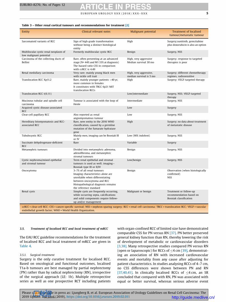
Table 3 – Other renal cortical tumours and recommendations for treatment [2]
Entity Clinical relevant notes Malignant potential Treatment of localisedtumour/metastatic tumour
Sarcomatoid variants of RCC Sign of high-grade transformationwithout being a distinct histologicalentity
High Surgery;sunitinib, gemcitabineplus doxorubicin is also an option
Multilocular cystic renal neoplasm oflow malignant potential
Formerly multilocular cystic RCC Benign Surgery, NSS
Carcinoma of the collecting ducts ofBellini
Rare, often presenting at an advancedstage (N+ 44% and M1 33% at diagnosis)The hazard ratio CSS in comparisonwith ccRCC is 4.49
High, very aggressiveMedian survival 30 mo
Surgery; response to targetedtherapies is poor
Renal medullary carcinoma Very rare; mainly young black menwith sickle cell trait
High, very aggressive,median survival is 5 mo
Surgery; different chemotherapyregimes, radiosensitive
Translocation RCC Xp11.2 Rare, mainly younger patients <40 yr,more common in femalesIt constitutes with TRCC 6p21 MiTtranslocation RCCs
High Surgery; VEGF-targeted therapy
Translocation RCC t(6;11) Low/intermediate Surgery, NSS; VEGF-targetedtherapy
Mucinous tubular and spindle cellcarcinoma
Tumour is associated with the loop ofHenle
Intermediate Surgery, NSS
Acquired cystic disease-associatedRCC
Low Surgery
Clear-cell papillary RCC Also reported as renalangiomyomatous tumour
Low Surgery, NSS
Hereditary leiomyomatosis and RCC-associated RCC
Rare, new entity in the 2016 WHOclassification, caused by a germlinemutation of the fumarate hydratasegene
High Surgery; no data about treatmentof metastatic disease
Tubulocystic RCC Mainly men, imaging can be Bosniak IIIor IV
Low (90% indolent) Surgery, NSS
Succinate dehydrogenase-deficientRCC
Rare Variable Surgery
Metanephric tumours Divided into metanephric adenoma,adenofibroma, and metanephricstromal tumours
Benign Surgery, NSS
Cystic nephroma/mixed epithelialand stromal tumour
Term renal epithelial and stromaltumours is used as well; imaging—Bosniak type III or II/IV
Low/benign Surgery, NSS
Oncocytoma 3–7% of all renal tumoursImaging characteristics alone areunreliable when differentiatingbetween oncocytoma and RCCHistopathological diagnosis remainsthe reference standard
Benign Observation (when histologicallyconfirmed)NSS
Renal cysts Simple cysts are frequently occurring,while occurring septa, calcificationsand solid components require follow-up and/or management
Malignant or benign Treatment or follow-uprecommendation based onBosniak classification
ccRCC = clear-cell RCC; CSS = cancer-specific survival; NSS = nephron-sparing surgery; RCC = renal cell carcinoma; TRCC = translocation RCC; VEGF = vascularendothelial growth factor; WHO = World Health Organization.
E U R O P E A N U R O L O G Y X X X ( 2 0 18 ) X X X – X X X 5
EURURO-8276; No. of Pages 12
3.5. Treatment of localised RCC and local treatment of mRCC
The EAU RCC guideline recommendations for the treatmentof localised RCC and local treatment of mRCC are given inTable 4.
3.5.1. Surgical treatment
Surgery is the only curative treatment for localised RCC.Based on oncological and functional outcomes, localisedT1a-b tumours are best managed by partial nephrectomy(PN) rather than by radical nephrectomy (RN), irrespectiveof the surgical approach (LE: 1b). Multiple retrospectiveseries as well as one prospective RCT including patients
Please cite this article in press as: Ljungberg B, et al. European As2019 Update. Eur Urol (2019), https://doi.org/10.1016/j.eururo.201
with organ-confined RCC of limited size have demonstratedcomparable CSS for PN versus RN [37]. PN better preservedgeneral kidney function than RN, thereby lowering the riskof development of metabolic or cardiovascular disorders[3,38]. Many retrospective studies compared PN versus RN(open or laparoscopic) for RCCs of �4 cm [39], demonstrat-ing an association of RN with increased cardiovascularevents and mortality from any cause after adjusting forpatient characteristics. In studies analysing RCCs of 4–7 cm,no CSS differences were shown between PN and RN[37,40,41]. In clinically localised RCCs of �4 cm, an SRconcluded that compared with RN, PN was associated withequal or better survival, whereas serious adverse event
sociation of Urology Guidelines on Renal Cell Carcinoma: The9.02.011 http://guide.medlive.cn/
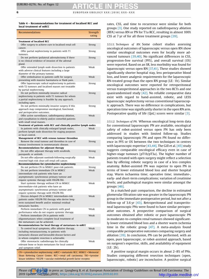
Table 4 – Recommendations for treatment of localised RCC andlocal treatment of mRCC
Recommendations Strengthrating
Treatment of localised RCCOffer surgery to achieve cure in localised renal cell
cancer.Strong
Offer partial nephrectomy to patients with T1tumours.
Strong
Do not perform ipsilateral adrenalectomy if thereis no clinical evidence of invasion of the adrenalgland.
Strong
Offer extended lymph node dissection to patientswith adverse clinical features including a largediameter of the primary tumour.
Weak
Offer embolisation to patients unfit for surgerypresenting with massive haematuria or flank pain.
Weak
Offer laparoscopic radical nephrectomy to patientswith T2 tumours and localised masses not treatableby partial nephrectomy.
Strong
Do not perform minimally invasive radicalnephrectomy in patients with T1 tumours for whoma partial nephrectomy is feasible by any approach,including open.
Strong
Do not perform minimally invasive surgery if thisapproach may compromise oncological, functional,and perioperative outcomes.
Strong
Offer active surveillance, radiofrequency ablation,and cryoablation to elderly and/or comorbid patientswith small renal masses.
Weak
Treatment of patients with RCC and clinically positive lymph nodesIn patients with clinically enlarged lymph nodes,
perform lymph node dissection for staging purposesor local control.
Weak
Management of RCC with venous tumour thrombusRemove the renal tumour and thrombus in case of
venous involvement in nonmetastatic disease.Strong
Recommendations for adjuvant therapyDo not offer adjuvant therapy with sorafenib,
pazopanib, or axitinib.Strong
Do not offer adjuvant sunitinib following surgicallyresected high-risk clear-cell renal cell cancer.
Weak
Recommendations for cytoreductive nephrectomyDo not perform CN in MSKCC poor-risk patients. StrongDo not perform immediate CN in MSKCC
intermediate-risk patients who have anasymptomatic synchronous primary tumour andrequire systemic therapy with VEGFR-TKI.
Weak
Start systemic therapy without CN in MSKCCintermediate-risk patients who have anasymptomatic synchronous primary tumour andrequire systemic therapy with VEGFR-TKI.
Weak
Discuss delayed CN in MSKCC intermediate-riskpatients under VEGFR-TKI therapy who derive long-term sustained benefit and/or minimal residualmetastatic burden.
Weak
Perform immediate CN in patients with goodperformance who do not require systemic therapy.
Weak
Perform immediate CN in patients witholigometastases when complete local treatment ofthe metastases can be achieved.
Weak
Recommendations for local therapy of metastases in mRCCTo control local symptoms, offer ablative therapy,
including metastasectomy, to patients withmetastatic disease and favourable disease factors andin whom complete resection is achievable.
Weak
Offer stereotactic radiotherapy for clinicallyrelevant bone or brain metastases for local controland symptom relief.
Weak
CN = cytoreductive nephrectomy; mRCC = metastatic RCC; MSKCC = MemorialSloan Kettering Cancer Center; RCC = renal cell carcinoma; TKI = tyrosinekinase inhibitor; VEGFR = vascular endothelial growth factor receptor.
E U R O P E A N U R O L O G Y X X X ( 2 0 18 ) X X X – X X X6
EURURO-8276; No. of Pages 12
Please cite this article in press as: Ljungberg B, et al. European As2019 Update. Eur Urol (2019), https://doi.org/10.1016/j.eururo.20
rates, CSS, and time to recurrence were similar for bothgroups [3]. One study reported on radiofrequency ablation(RFA) versus RN or PN for T1a RCC, resulting in almost 100%CSS at 7 yr of for all three treatment groups [39].
3.5.1.1. Techniques of RN. Some cohort studies assessingoncological outcomes of laparoscopic versus open RN showsimilar oncological outcomes even for locally more ad-vanced tumours [39,40]. No significant differences in CSS,progression-free survival (PFS), and overall survival (OS)were reported. Based on an SR, less morbidity was found forlaparoscopic versus open RN [37,41]. These studies showedsignificantly shorter hospital stay, less perioperative bloodloss, and lower analgesic requirements for the laparoscopicRN-treated group than the open RN group (LE: 1b). Similaroncological outcomes were reported for retroperitonealversus transperitoneal approaches in the two RCTs and onequasirandomised study [42]. No reliable comparative dataexist with regard to hand-assisted, robotic, and LESSlaparoscopic nephrectomy versus conventional laparoscop-ic approach. There was no difference in complications, butoperation time was significantly shorter in the open RN arm.Postoperative quality of life (QoL) scores were similar [3].
3.5.1.2. Techniques of PN. Whereas oncological long-term datafor conventional laparoscopic PN are available, oncologicalsafety of robot-assisted versus open PN has only beenaddressed in studies with limited follow-up. Studiescomparing laparoscopic PN and open PN found no differ-ence in PFS or OS between the two techniques in centreswith laparoscopic expertise [43,44]. The Gill et al. [45] studysuggests comparable oncological efficacy even in case ofhigher-stage tumours (pT1b/pT3a). The higher number ofpatients treated with open surgery might reflect a selectionbias by offering robotic surgery in case of a less complexanatomy. Robot-assisted PN was superior to open PN interms of lower estimated blood loss and shorter hospitalstay. Warm ischaemia time; operative time; immediate-,early- and short-term complications; variation of creatininelevels; and pathological margins were similar amongst thegroups [46].
In a matched-pair comparison, the decline in estimatedglomerular filtration rate was greater in the laparoscopic PNgroup in the immediate postoperative period, but not after afollow-up of 3.6 yr [43]. Retroperitoneal and transperito-neal laparoscopic PNs were found to have similar perioper-ative outcomes. A prospective comparison of surgicaloutcomes obtained after robotic or pure laparoscopic PNin moderate-to-complex renal tumours showed significant-ly lower estimated blood loss and a shorter warm ischemiatime in the robotic group [47]. A meta-analysis foundcomparable perioperative outcomes comparing surgery andablation [39]. In conclusion, PN can be performed, with anopen, pure laparoscopic, or robot-assisted approach, basedon surgeon's expertise, skills, and availability of equipment(LE: 2b).
A positive surgical margin occurs in about 2–8% of PNs.Studies comparing different resection techniques (open,laparoscopic, robotic) are inconclusive. A positive surgical
sociation of Urology Guidelines on Renal Cell Carcinoma: The19.02.011 http://guide.medlive.cn/
E U R O P E A N U R O L O G Y X X X ( 2 0 18 ) X X X – X X X 7
EURURO-8276; No. of Pages 12
margin status occurs more frequently in cases in whichsurgery is imperative (solitary kidney, bilateral disease) andin patients with adverse pathological features (pT2a, pT3a,grade III–IV). Local tumour recurrence was found in 16% inpositive surgical margins compared with 3% in negativemargins [48]. Patients with positive surgical margins are notindicated immediately to any reintervention but to a moreintense surveillance.
3.5.1.3. Adrenalectomy. One nonrandomised study on PN andtwo small studies on RN compared the outcomes with, orwithout, ipsilateral adrenalectomy [4,49]. Multivariateanalysis showed that upper pole location was not predictiveof adrenal involvement, but tumour size was. No differencein OS was seen with or without adrenalectomy. Adrenalec-tomy was justified using criteria based on radiographic andintraoperative findings. Only 48 of 2065 patients under-went concurrent ipsilateral adrenalectomy, of whom42 were for benign lesions [49].
3.5.1.4. LN dissection for clinically negative LNs (cN0). Clinicalassessment of LN status is based on the detection of LNenlargement either by CT/MRI or by intraoperative palpa-tion of enlarged nodes. Both CT and MRI are unsuitable fordetecting malignant disease in nodes of normal shape andsize [4]. For patients with clinically negative LNs (cN0), LNdissection (LND) was not associated with a reduced risk ofdistant metastases, or cancer-specific or all-cause mortality[50,51]. Neither did LND improve oncological outcomesamongst patients at a high risk of radiographic cN1 [51].
3.5.2. Management of RCC with venous tumour thrombus
An SR on the management of venous tumour thrombus(VTT), in non-mRCC, included only five studies with a highRoB across all studies [52]. Minimal-access techniquesresulted in significantly shorter operating time comparedwith sternotomy. Preoperative embolisation was associatedwith increased operating time, blood loss, hospital stay, andperioperative mortality. No significant differences inoncological and process outcomes were observed betweencardiopulmonary bypass with deep hypothermic circulato-ry arrest, or partial bypass under normothermia or singlecaval clamp without circulatory support. No surgicalmethod was shown to be superior for the excision of VTT.The surgical method was dependent on the upper level oftumour thrombus. The relative benefits and harms of otherstrategies and approaches regarding access to the inferiorvena cava (IVC), and the role of IVC filters and bypassprocedures remain uncertain with non-mRCC. Neverthe-less, the findings support that all patients with nonmeta-static disease and VTT should be considered for surgicalintervention, irrespective of the extent of tumour thrombusat presentation (LE: 3) [53]. PS can significantly improveafter removal; therefore, deterioration of PS due tothrombus should not be an exclusion for surgery,
3.5.3. Therapeutic approaches as alternative to surgery
3.5.3.1. Embolisation. Before a routine nephrectomy, there isno benefit in performing tumour embolisation. In patients
Please cite this article in press as: Ljungberg B, et al. European As2019 Update. Eur Urol (2019), https://doi.org/10.1016/j.eururo.201
unfit for surgery and suffering from massive haematuria orflank pain, embolisation can be a beneficial palliativeintervention (LE: 3).
3.5.3.2. Surveillance. Elderly and comorbid patients withincidentally detected small RMs have relatively low RCC-specific mortality and significant competing-cause mortal-ity [54]. Active surveillance (AS) can be offered to thiscategory of patients and is defined as the initial monitoringof tumour size by serial abdominal imaging (US, CT, or MRI),with delayed intervention reserved for those tumours thatshow clinical progression during follow-up. A renal biopsyis recommended prior to surveillance (LE: 3). In the largestreported prospective series of AS, the growth rate of the RMwas slow in most cases and progression to mRCC occurredin 1.1% of patients (LE: 3) [54]. Frequency of serial imaging inthis study consisted of CT, MRI, or US at months 3 and 6,every 6 mo until 3 yr, and annually thereafter (LE: 3). In alarge prospective nonrandomised study comparing AS orprimary active intervention for small RMs, OS and CSS werenot significantly different in the two treatment groups [55].
3.5.3.3. Ablative therapies. The most commonly performedablative therapies for renal tumours are percutaneousRFA, and laparoscopically assisted or percutaneous cryoa-blation (CA). Microwave ablation, stereotactic radiosurgery,laser ablation, and high-intensity focused US ablation areconsidered experimental. Indications for thermal ablationinclude elderly, comorbid patients with a small RM who areconsidered unfit for surgery; patients with a geneticpredisposition to develop multiple tumours; and patientswith bilateral tumours or with a solitary kidney, and a highrisk of complete loss of renal function following PN. Largertumours or those located at the hilum or near the proximalureter are not recommended for ablation. There are no RCTscomparing RFA or CA with PN [37]. Low-quality studiessuggest a higher local recurrence rate for thermal ablationcompared with PN (LE: 3). The quality of the available datadoes not allow any definitive conclusions regardingmorbidity and oncological outcomes for RFA and CA (LE:3) [56].
3.5.4. Adjuvant therapy
There is currently no evidence from randomised phase IIItrials that adjuvant therapy offers an OS benefit. Besidestumour vaccination, CAIX, and adjuvant Interferon therapy,recent evidence is based on adjuvant trials with targetedtherapies in high-risk patients. These included the ASSUREstudy comparing sunitinib versus sorafenib versus placebo,the PROTECT study comparing pazopanib and placebo, andthe S-TRAC study comparing sunitinib with placebo[57]. The results showed a benefit of sunitinib over placebofor disease-free survival (DFS) in the S-TRAC study(p = 0.03), but in 2018, data for OS remained immaturesince median OS was not reached in either arm. Grade 3/4toxicity in the study was 61% for patients receiving sunitiniband 21% for patients on placebo. A pooled analysis ofvascular endothelial growth factor receptor (VEGFR) tyro-sine kinase inhibitor (TKI) versus placebo demonstrated
sociation of Urology Guidelines on Renal Cell Carcinoma: The9.02.011 http://guide.medlive.cn/
E U R O P E A N U R O L O G Y X X X ( 2 0 18 ) X X X – X X X8
EURURO-8276; No. of Pages 12
that VEGFR-targeted therapy was not statistically signifi-cantly associated with improved DFS or OS compared withplacebo [58]. In addition, the ATLAS study comparingaxitinib with placebo did not meet its primary endpoint[59]. In summary, there is currently a lack of proven benefitof adjuvant therapy with VEGFR-TKIs for patients with high-risk RCC after nephrectomy, and their use is not recom-mended (LE: 1a).
3.5.5. Surgical treatment of mRCC
Most patients with mRCC require systemic therapy, and therole and sequence of cytoreductive nephrectomy (CN) hasbeen investigated in two RCTs. In the previous cytokine era,increased long-term survival was found in patients treatedwith CN + immunotherapy [60]. The SURTIME studyrevealed that the sequence of CN and sunitinib did notaffect PFS. The trial accrued poorly, and the results aremainly exploratory. In secondary endpoint analysis, a strongOS benefit was observed in favour of the deferred CNapproach in the intent-to-treat (ITT) population, withmedian OS of 32.4 mo in the deferred CN arm versus15.0 mo in the immediate CN arm. The CARMENA studyshowed that sunitinib alone was not inferior to immediateCN followed by sunitinib with regard to OS [8]. In an ITTanalysis, median OS was 13.9 mo with CN versus 18.4 mowith sunitinib alone. This noninferiority study did not reachthe full accrual of planned (450 out of 576) patients. Thirty-eight patients in the sunitinib-only arm (17%) requiredsecondary CN due to acute symptoms or for complete ornear-complete response.
In summary, immediate CN is not recommended inMemorial Sloan Kettering Cancer Center (MSKCC) interme-diate- and high-risk patients requiring sunitinib, or anequivalent VEGFR-TKI (LE: 1b). Those patients are recom-mended immediate sunitinib, and weak evidence from bothCARMENA and SURTIME supports performing a deferred CNat 3 mo, or later, in patients who do not progress on VEGFR-TKI therapy (LE: 2b).
Neither CARMENA nor SURTIME answered the questionof CN in patients with low-volume metastatic disease, goodPS, and a favourable and intermediate risk, who do notrequire immediate VEGFR-TKI treatment but may beobserved instead [8]. In these patients, immediate CNretains its role, since observation until progression requir-ing systemic treatment can result in substantial time toonset of VEGF-targeted therapy (LE: 2b) [61].
However, due to a paradigm change in first-linetreatment for intermediate- and poor-risk patients shownin the CheckMate 214 study [62], the role and sequence ofCN in the era of immunotherapy need to be reinvestigated.
3.5.6. Local therapy of metastases in RCC
An SR of comparative studies evaluated local treatment ofmetastases from RCC in any organ [63]. Interventionsincluded metastasectomy, various radiotherapy modalities,and no local treatment. The outcomes assessed weresurvival (OS, CSS, and PFS), local symptom control, andadverse events. All included studies were retrospective,nonrandomised, comparative studies showing a high RoB
Please cite this article in press as: Ljungberg B, et al. European As2019 Update. Eur Urol (2019), https://doi.org/10.1016/j.eururo.20
associated with nonrandomisation, attrition, and selectivereporting. With the exception of brain and possibly bonemetastases frequently treated by stereotactic radiotherapy,metastasectomy remained, by default, an appropriate localtreatment for most sites. Retrospective comparative studiesconsistently point towards a benefit of complete metasta-sectomy in mRCC patients in terms of OS, CSS, and delay ofsystemic therapy. Radiotherapy, especially stereotacticradiotherapy, to bone and brain metastases from RCC caninduce significant relief from local symptoms (all LE: 3) [63].
3.6. Systemic therapy for mRCC
3.6.1. Targeted therapies
Until targeted therapies were introduced in 2006, thetreatment of mRCC was generally based on immunothera-pies such as interferon-a (IFN-a) and interleukin-2. Withthe introduction of targeting agents, stabilisation of thedisease and prolonged survival was achieved. Severaltargeting drugs have been approved for the treatment ofmRCC: sunitinib, sorafenib, pazopanib, axitinib, tivozanib,cabozantinib, the mTOR inhibitors everolimus and temsir-olimus, as well as bevacizumab combined with IFN-a.Treatment recommendations on first-line treatment andsubsequent treatment lines are based on RCTs with a highLE. A detailed description of the targeting agents can befound in the RCC guideline text at www.uroweb.org[2]. Fortreatment recommendations, see Fig. 1 and SRs [2,64].
Most published trials have selected ccRCCs only; thus,the robust evidence-based recommendations are applicableonly for ccRCC.
The International Metastatic Renal Cancer DatabaseConsortium risk model has been established and validatedto aid in an accurate prognosis of patients treated withtargeted therapy. Neutrophilia and thrombocytosis havebeen added to the list of MSKCC risk factors, while serumlactate dehydrogenase has been removed [65].
3.6.2. Immunotherapy
Immunotherapy trials using immune checkpoint blockadewith monoclonal antibodies blocking the inhibitory T-cellreceptor PD-1 or cytotoxic T-lymphocyte-associated anti-gen 4 (CTLA-4) signalling to restore tumour-specific T-cellimmunity have been conducted. The CheckMate 214 studyreported superiority of nivolumab and ipilimumab oversunitinib in intermediate- and poor-risk patients, leading toa paradigm shift in the first-line management of mRCCpatients. OS with nivolumab plus ipilimumab in bothintermediate- and poor-risk patients is longer than onewould predict for PFS, suggesting significant activity ofsubsequent agents [62]. Results showed that a combinationof ipilimumab and nivolumab was associated with theachievement of durable remissions in a higher proportion ofpatients. These findings resulted in an updated recommen-dation of the systemic treatment of mRCC patients, asshown in Figure 1. The impact on subsequent therapies isunclear, since therapy for patients with disease refractory tonivolumab plus ipilimumab in a first-line setting has notbeen tested. A phase III trial of nivolumab versus everolimus
sociation of Urology Guidelines on Renal Cell Carcinoma: The19.02.011 http://guide.medlive.cn/
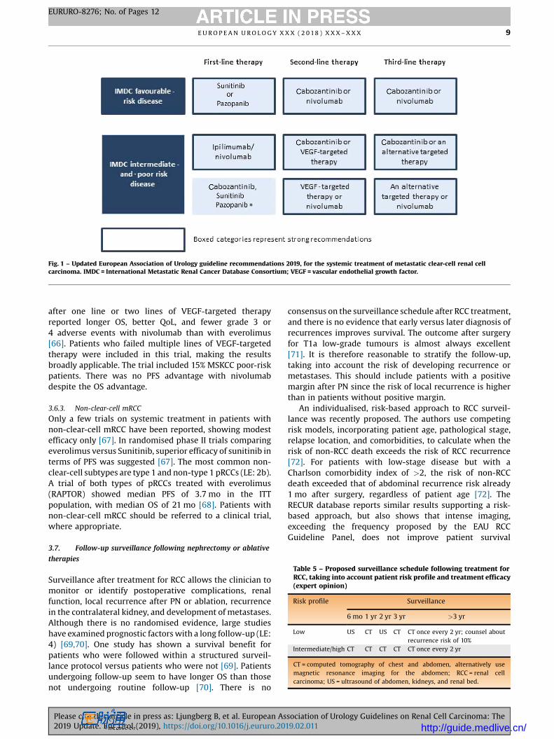
Fig. 1 – Updated European Association of Urology guideline recommendations 2019, for the systemic treatment of metastatic clear-cell renal cellcarcinoma. IMDC = International Metastatic Renal Cancer Database Consortium; VEGF = vascular endothelial growth factor.
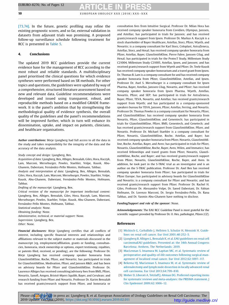
Table 5 – Proposed surveillance schedule following treatment forRCC, taking into account patient risk profile and treatment efficacy(expert opinion)
Risk profile Surveillance
6 mo 1 yr 2 yr 3 yr >3 yr
Low US CT US CT CT once every 2 yr; counsel aboutrecurrence risk of 10%
Intermediate/high CT CT CT CT CT once every 2 yr
CT = computed tomography of chest and abdomen, alternatively usemagnetic resonance imaging for the abdomen; RCC = renal cellcarcinoma; US = ultrasound of abdomen, kidneys, and renal bed.
E U R O P E A N U R O L O G Y X X X ( 2 0 18 ) X X X – X X X 9
EURURO-8276; No. of Pages 12
after one line or two lines of VEGF-targeted therapyreported longer OS, better QoL, and fewer grade 3 or4 adverse events with nivolumab than with everolimus[66]. Patients who failed multiple lines of VEGF-targetedtherapy were included in this trial, making the resultsbroadly applicable. The trial included 15% MSKCC poor-riskpatients. There was no PFS advantage with nivolumabdespite the OS advantage.
3.6.3. Non-clear-cell mRCC
Only a few trials on systemic treatment in patients withnon-clear-cell mRCC have been reported, showing modestefficacy only [67]. In randomised phase II trials comparingeverolimus versus Sunitinib, superior efficacy of sunitinib interms of PFS was suggested [67]. The most common non-clear-cell subtypes are type 1 and non-type 1 pRCCs (LE: 2b).A trial of both types of pRCCs treated with everolimus(RAPTOR) showed median PFS of 3.7 mo in the ITTpopulation, with median OS of 21 mo [68]. Patients withnon-clear-cell mRCC should be referred to a clinical trial,where appropriate.
3.7. Follow-up surveillance following nephrectomy or ablative
therapies
Surveillance after treatment for RCC allows the clinician tomonitor or identify postoperative complications, renalfunction, local recurrence after PN or ablation, recurrencein the contralateral kidney, and development of metastases.Although there is no randomised evidence, large studieshave examined prognostic factors with a long follow-up (LE:4) [69,70]. One study has shown a survival benefit forpatients who were followed within a structured surveil-lance protocol versus patients who were not [69]. Patientsundergoing follow-up seem to have longer OS than thosenot undergoing routine follow-up [70]. There is no
Please cite this article in press as: Ljungberg B, et al. European As2019 Update. Eur Urol (2019), https://doi.org/10.1016/j.eururo.201
consensus on the surveillance schedule after RCC treatment,and there is no evidence that early versus later diagnosis ofrecurrences improves survival. The outcome after surgeryfor T1a low-grade tumours is almost always excellent[71]. It is therefore reasonable to stratify the follow-up,taking into account the risk of developing recurrence ormetastases. This should include patients with a positivemargin after PN since the risk of local recurrence is higherthan in patients without positive margin.
An individualised, risk-based approach to RCC surveil-lance was recently proposed. The authors use competingrisk models, incorporating patient age, pathological stage,relapse location, and comorbidities, to calculate when therisk of non-RCC death exceeds the risk of RCC recurrence[72]. For patients with low-stage disease but with aCharlson comorbidity index of >2, the risk of non-RCCdeath exceeded that of abdominal recurrence risk already1 mo after surgery, regardless of patient age [72]. TheRECUR database reports similar results supporting a risk-based approach, but also shows that intense imaging,exceeding the frequency proposed by the EAU RCCGuideline Panel, does not improve patient survival
sociation of Urology Guidelines on Renal Cell Carcinoma: The9.02.011 http://guide.medlive.cn/
E U R O P E A N U R O L O G Y X X X ( 2 0 18 ) X X X – X X X10
EURURO-8276; No. of Pages 12
[73,74]. In the future, genetic profiling may refine theexisting prognostic scores, and so far, external validation indatasets from adjuvant trials was promising. A proposedfollow-up surveillance schedule following treatment forRCC is presented in Table 5.
4. Conclusions
The updated 2019 RCC guidelines provide the currentevidence base for the management of RCC according to themost robust and reliable standards. A multidisciplinarypanel prioritised the clinical questions for which evidencesyntheses were performed based on SR methods. For othertopics and questions, the guidelines were updated by way ofa comprehensive, structured literature assessment based onnew and relevant data. Guideline recommendations weredeveloped and issued using transparent, robust, andreproducible methods based on a modified GRADE frame-work. It is the panel's ambition that by strengthening themethodological quality of evidence synthesis, the overallquality of the guidelines and the panel's recommendationswill be improved further, which in turn will enhance itsdissemination, uptake, and impact on patients, clinicians,and healthcare organisations.
Author contributions: Börje Ljungberg had full access to all the data inthe study and takes responsibility for the integrity of the data and theaccuracy of the data analysis.
Study concept and design: Ljungberg, Bex.Acquisition of data: Ljungberg, Bex, Albiges, Bensalah, Giles, Hora, Kuczyk,Lam, Marconi, Merseburger, Powles, Staehler, Volpe, Kuusk, Abu-Ghanem, Dabestani, Fernández-Pello Montes, Hofmann, Tahbaz.Analysis and interpretation of data: Ljungberg, Bex, Albiges, Bensalah,Giles, Hora, Kuczyk, Lam, Marconi, Merseburger, Powles, Staehler, Volpe,Kuusk, Abu-Ghanem, Dabestani, Fernández-Pello Montes, Hofmann,Tahbaz.Drafting of the manuscript: Ljungberg, Bex.Critical revision of the manuscript for important intellectual content:
Ljungberg, Bex, Albiges, Bensalah, Giles, Hora, Kuczyk, Lam, Marconi,Merseburger, Powles, Staehler, Volpe, Kuusk, Abu-Ghanem, Dabestani,Fernández-Pello Montes, Hofmann, Tahbaz.Statistical analysis: None.Obtaining funding: None.Administrative, technical, or material support: None.Supervision: Ljungberg, Bex.Other: None.
Financial disclosures: Börje Ljungberg certifies that all conflicts ofinterest, including specific financial interests and relationships andaffiliations relevant to the subject matter or materials discussed in themanuscript (eg, employment/affiliation, grants or funding, consultan-cies, honoraria, stock ownership or options, expert testimony, royalties,or patents filed, received, or pending), are the following: Professor Dr.Börje Ljungberg has received company speaker honoraria fromGlaxoSmithKline, Roche, Pfizer, and Novartis; has participated in trialsfor GlaxoSmithKline, Medivation, Pfizer, and Janssen R&D; and has beenon advisory boards for Pfizer and GlaxoSmithKline. Professor Dr.Laurence Albiges has received consulting/advisory fees from BMS, Pfizer,Novartis, Sanofi, Amgen, Bristol-Myers Squibb, Bayer, and Cerulean; andresearch funding from Pfizer and Novartis. Professor Dr. Karim Bensalahhas received grants/research support from Pfizer, and honoraria or
Please cite this article in press as: Ljungberg B, et al. European As2019 Update. Eur Urol (2019), https://doi.org/10.1016/j.eururo.20
consultation fees from Intuitive Surgical. Professor Dr. Milan Hora hasreceived company speaker honoraria from Covidien, Olympus, Janssen,and Astellas; has participated in trials for Janssen; and has receivedgrants/research support from Ipsen. Professor Dr. Markus A. Kuczyk is astock shareholder of Bayer Healthcare, Astellas, Storz, Pfizer, Wyeth, andNovartis; is a company consultant for Karl Storz, Coloplast, AstraZeneca,Astellas, Storz, and Hexal; has received company speaker honoraria fromPfizer, Astellas, Bayer, GlaxoSmithKline, Pierre Fabre, Janssen Cilag, andHexal; has participated in trials for the ProtecT Study, Millenium StudyC21004, Millenium Study C21005, Astellas, Ipsen, and Janssen; and hasreceived grants/research support from Wyeth and Pfizer. Dr. Teele Kuuskhas received company speaker honorarium and a travel grant from Ipsen.Dr. Thomas B. Lam is a company consultant for and has received companyspeaker honoraria from Pfizer, GlaxoSmithKline, Astellas, and Ipsen.Professor Dr. Axel S. Merseburger is a company consultant for IpsenPharma, Bayer, Astellas, Janssen Cilag, Novartis, and Pfizer; has receivedcompany speaker honoraria from Ipsen Pharma, Wyeth, Astellas,Novartis, Pfizer, and SEP; has participated in trials for AstraZeneca,Bayer, Pfizer, TEVA, Novartis, and Astellas; has received grants/researchsupport from Wyeth; and has participated in a company-sponsoredspeakers bureau for TEVA, Janssen, Pfizer, Astellas, Ferring, and Novartis.Professor Dr. Thomas Powles is a company consultant for Novartis, Pfizer,and GlaxoSmithKline; has received company speaker honoraria fromNovartis, Pfizer, GlaxoSmithKline, and Genentech; has participated intrials for GlaxoSmithKline, Pfizer, BMS, Genentech, and Genetech; andhas received grants/research support from GlaxoSmithKline, Pfizer, andNovartis. Professor Dr. Michael Staehler is a company consultant forPfizer, Novartis, GlaxoSmithKline, Roche, Astellas, and Bayer; hasreceived company speaker honoraria from Pfizer, Novartis, GlaxoSmithK-line, Roche, Astellas, Bayer, and Aveo; has participated in trials for Pfizer,Novartis, GlaxoSmithKline, Roche, Bayer, Aveo, Wilex, and Immatics; hasreceived fellowships and travel grants from Pfizer, Novartis, GlaxoS-mithKline, Roche, and Bayer; and has received grants/research supportfrom Pfizer, Novartis, GlaxoSmithKline, Roche, Bayer, and Aveo. Inaddition, he took part in the S-TRAC trial as an investigator and is anauthor on the S-TRAC publication. Professor Dr. Axel Bex has receivedcompany speaker honoraria from Pfizer; has participated in trials forPfizer Europe; has participated in advisory boards for GlaxoSmithKlineand Novartis; is a company consultant for Pfizer and Novartis; and hasreceived grants/research support from Pfizer. Professor Dr. Rachel H.Giles, Professor Dr. Alessandro Volpe, Dr. Saeed Dabestani, Dr. FabianHofmann, Dr. Lorenzo Marconi, Dr. Sergio Fernández-Pello, Dr. RanaTahbaz, and Dr. Yasmin Abu-Ghanem have nothing to disclose.
Funding/Support and role of the sponsor: None.
Acknowledgements: The EAU RCC Guideline Panel is most grateful for thescientific support provided by Professor Dr. O. Hes, pathologist, Pilzen (CZ).
References
[1] Mickisch G, Carballido J, Hellsten S, Schulze H, Mensink H. Guide-lines on renal cell cancer. Eur Urol 2001;40:252–5.
[2] Ljungberg B, Albiges L, Bensalah K, et al. EAU guidelines on renal cellcarcinomaEAU guidelines. Presented at: the 34th Annual CongressBarcelona; Arnhem, The Netherlands; 2019.
[3] MacLennan S, Imamura M, Lapitan MC, et al. Systematic review ofperioperative and quality-of-life outcomes following surgical man-agement of localised renal cancer. Eur Urol 2012;62:1097–117.
[4] Bekema HJ, MacLennan S, Imamura M, et al. Systematic review ofadrenalectomy and lymph node dissection in locally advanced renalcell carcinoma. Eur Urol 2013;64:799–810.
[5] Moher D, Liberati A, Tetzlaff J, Altman DG. Preferred reporting itemsfor systematic reviews and meta-analyses: the PRISMA statement. JClin Epidemiol 2009;62:1006–12.
sociation of Urology Guidelines on Renal Cell Carcinoma: The19.02.011 http://guide.medlive.cn/
E U R O P E A N U R O L O G Y X X X ( 2 0 18 ) X X X – X X X 11
EURURO-8276; No. of Pages 12
[6] Guyatt GH, Oxman AD, Kunz R, et al. Going from evidence torecommendations. BMJ 2008;336:1049–51.
[7] Knoll T, Omar MI, Maclennan S, et al. Key steps in conductingsystematic reviews for underpinning clinical practice guidelines:methodology of the European Association of Urology. Eur Urol2018;73:290–300.
[8] Bex A, Albiges L, Ljungberg B, et al. Updated European Association ofUrology guidelines for cytoreductive nephrectomy in patients withsynchronous metastatic clear-cell renal cell carcinoma. Eur Urol2018;74:805–9.
[9] Ferlay J, Colombet M, Soerjomataram I, et al. Cancer incidence andmortality patterns in Europe: estimates for 40 countries and 25 ma-jor cancers in 2018. Eur J Cancer 2018;103:356–87.
[10] Levi F, Ferlay J, Galeone C, et al. The changing pattern of kidney cancerincidence and mortality in Europe. BJU Int 2008;101:949–58.
[11] Thorstenson A, Bergman M, Scherman-Plogell AH, et al. Tumourcharacteristics and surgical treatment of renal cell carcinoma inSweden 2005–2010: a population-based study from the nationalSwedish kidney cancer register. Scand J Urol 2014;48:231–8.
[12] Moch H, Cubilla AL, Humphrey PA, Reuter VE, Ulbright TM. The2016 WHO classification of tumours of the urinary system and malegenital organs—part a: renal, penile, and testicular tumours. EurUrol 2016;70:93–105.
[13] Tahbaz R, Schmid M, Merseburger AS. Prevention of kidney cancerincidence and recurrence: lifestyle, medication and nutrition. CurrOpin Urol 2018;28:62–79.
[14] Daniel CR, Cross AJ, Graubard BI, et al. Large prospective investiga-tion of meat intake, related mutagens, and risk of renal cell carci-noma. Am J Clin Nutr 2012;95:155–62.
[15] Bellocco R, Pasquali E, Rota M, et al. Alcohol drinking and risk ofrenal cell carcinoma: results of a meta-analysis. Ann Oncol2012;23:2235–44.
[16] Kim HL, Belldegrun AS, Freitas DG, et al. Paraneoplastic signs andsymptoms of renal cell carcinoma: implications for prognosis. J Urol2003;170:1742–6.
[17] Vogel C, Ziegelmüller B, Ljungberg B, et al. Imaging in suspectedrenal-cell carcinoma: systematic review. Clin Genitourin Cancer. Inpress. https://doi.org/10.1016/j.clgc.2018.07.024.
[18] Rossi SH, Prezzi D, Kelly-Morland C, Goh V. Imaging for the diagno-sis and response assessment of renal tumours. World J Urol2018;36:1927–42.
[19] Ma H, Shen G, Liu B, Yang Y, Ren P, Kuang A. Diagnostic performance of18F-FDG PETor PET/CT in restaging renal cell carcinoma: a systematicreview and meta-analysis. Nucl Med Commun 2017;38:156–63.
[20] Lim DJ, Carter MF. Computerized tomography in the preoperativestaging for pulmonary metastases in patients with renal cell carci-noma. J Urol 1993;150:1112–4.
[21] Koga S, Tsuda S, Nishikido M, et al. The diagnostic value of bone scanin patients with renal cell carcinoma. J Urol 2001;166:2126–8.
[22] Warren KS, McFarlane J. The Bosniak classification of renal cysticmasses. BJU Int 2005;95:939–42.
[23] Schoots IG, Zaccai K, Hunink MG, Verhagen P. Bosniak classificationfor complex renal cysts reevaluated: a systematic review. J Urol2017;198:12–21.
[24] Chandrasekar T, Ahmad AE, Fadaak K, et al. Natural history ofcomplex renal cysts: clinical evidence supporting active surveil-lance. J Urol 2018;199:633–40.
[25] Marconi L, Dabestani S, Lam TB, et al. Systematic review and meta-analysis of diagnostic accuracy of percutaneous renal tumour biop-sy. Eur Urol 2016;69:660–73.
[26] Leveridge MJ, Finelli A, Kachura JR, et al. Outcomes of small renalmass needle core biopsy, nondiagnostic percutaneous biopsy, andthe role of repeat biopsy. Eur Urol 2011;60:578–84.
Please cite this article in press as: Ljungberg B, et al. European As2019 Update. Eur Urol (2019), https://doi.org/10.1016/j.eururo.201
[27] Dagher J, Delahunt B, Rioux-Leclercq N, et al. Clear cell renal cellcarcinoma: validation of World Health Organization/InternationalSociety of Urological Pathology grading. Histopathology2017;71:918–25.
[28] Gospodarowicz MK, Brierley JD, Wittekind C, editors. TNM classifi-cation of malignant tumors. UICC International Union AgainstCancer. 8th ed. Wiley-Blackwell; 2017. p. 199.
[29] Kutikov A, Uzzo RG. The R.E.N.A.L. nephrometry score: a compre-hensive standardized system for quantitating renal tumor size,location and depth. J Urol 2009;182:844–53.
[30] Wagener N, Edelmann D, Benner A, et al. Outcome of papillaryversus clear cell renal cell carcinoma varies significantly in non-metastatic disease. PLoS One 2017;12:e1730184.
[31] Speed JM, Trinh QD, Choueiri TK, Sun M. Recurrence in localizedrenal cell carcinoma: a systematic review of contemporary data.Curr Urol Rep 2017;18:15.
[32] Inamura K. Renal cell tumors: understanding their molecular path-ological epidemiology and the 2016 WHO classification. Int J Mol Sci2017;18:2195.
[33] Joseph RW, Kapur P, Serie DJ, et al. Clear cell renal cell carcinomasubtypes identified by BAP1 and PBRM1 expression. J Urol2016;195:180–7.
[34] Rini BI, Escudier B, Martini JF, et al. Validation of the 16-generecurrence score in patients with locoregional, high-risk renal cellcarcinoma from a phase III trial of adjuvant sunitinib. Clin CancerRes 2018;24:4407–15.
[35] Lopez-Beltran A, Henriques V, Cimadamore A, et al. The identifica-tion of immunological biomarkers in kidney cancers. Front Oncol2018;8:456.
[36] Köhn L, Svenson U, Ljungberg B, Roos G. Specific genomic aberra-tions predict survival, but low mutation rate in cancer hot spots, inclear cell renal cell carcinoma. Appl Immunohistochem Mol Mor-phol 2015;23:334–42.
[37] MacLennan S, Imamura M, Lapitan MC, et al. Systematic review ofoncological outcomes following surgical management of localisedrenal cancer. Eur Urol 2012;61:972–93.
[38] Capitanio U, Terrone C, Antonelli A, et al. Nephron-sparing techni-ques independently decrease the risk of cardiovascular eventsrelative to radical nephrectomy in patients with a T1a-T1b renalmass and normal preoperative renal function. Eur Urol2015;67:683–9.
[39] Alam R, Patel HD, Osumah T, et al. Comparative effectiveness ofmanagement options for patients with small renal masses: a pro-spective cohort study. BJU Int 2019;123:42–50.
[40] Sprenkle PC, Power N, Ghoneim T, et al. Comparison of open andminimally invasive partial nephrectomy for renal tumors 4-7 cen-timeters. Eur Urol 2012;61:593–9.
[41] Badalato GM, Kates M, Wisnivesky JP, Choudhury AR, McKiernanJM. Survival after partial and radical nephrectomy for the treatmentof stage T1bN0M0 renal cell carcinoma (RCC) in the USA: a propen-sity scoring approach. BJU Int 2012;109:1457–62.
[42] Desai MM, Strzempkowski B, Matin SF, et al. Prospective random-ized comparison of transperitoneal versus retroperitoneal laparo-scopic radical nephrectomy. J Urol 2005;173:38–41.
[43] Marszalek M, Meixl H, Polajnar M, Rauchenwald M, Jeschke K,Madersbacher S. Laparoscopic and open partial nephrectomy: amatched-pair comparison of 200 patients. Eur Urol 2009;55:1171–1178.
[44] Lane BR, Gill IS. 7-Year oncological outcomes after laparoscopic andopen partial nephrectomy. J Urol 2010;183:473–9.
[45] Gill IS, Kavoussi LR, Lane BR, et al. Comparison of 1,800 laparoscopicand open partial nephrectomies for single renal tumors. J Urol2007;178:41–6.
sociation of Urology Guidelines on Renal Cell Carcinoma: The9.02.011 http://guide.medlive.cn/
E U R O P E A N U R O L O G Y X X X ( 2 0 18 ) X X X – X X X12
EURURO-8276; No. of Pages 12
[46] Garisto J, Bertolo R, Dagenais J, et al. Robotic versus open partial nephrec-tomy for highly complex renal masses: comparison of perioperative,functional, and oncological outcomes. Urol Oncol 2018;36:471.e1–e.
[47] Masson-Lecomte A, Bensalah K, Seringe E, et al. A prospectivecomparison of surgical and pathological outcomes obtained afterrobot-assisted or pure laparoscopic partial nephrectomy in moder-ate to complex renal tumours: results from a French multicentrecollaborative study. BJU Int 2013;111:256–63.
[48] Wood EL, Adibi M, Qiao W, et al. Local tumor bed recurrencefollowing partial nephrectomy in patients with small renal masses.J Urol 2018;199:393–400.
[49] Lane BR, Tiong HY, Campbell SC, et al. Management of the adrenalgland during partial nephrectomy. J Urol 2009;181:2430–6, [dis-cussion 2436–7].
[50] Blom JH, van Poppel H, Marechal JM, et al. Radical nephrectomywith and without lymph-node dissection: final results of EuropeanOrganization for Research and Treatment of Cancer (EORTC) ran-domized phase 3 trial 30881. Eur Urol 2009;55:28–34.
[51] GershmanB,ThompsonRH,BoorjianSA,etal.Radicalnephrectomywithor without lymph node dissection for high risk nonmetastatic renal cellcarcinoma: a multi-institutional analysis. J Urol 2018;199:1143–8.
[52] Lardas M, Stewart F, Scrimgeour D, et al. Systematic review ofsurgical management of nonmetastatic renal cell carcinoma withvena caval thrombus. Eur Urol 2016;70:265–80.
[53] Kirkali Z, Van Poppel H. A critical analysis of surgery for kidneycancer with vena cava invasion. Eur Urol 2007;52:658–62.
[54] Jewett MA, Mattar K, Basiuk J, et al. Active surveillance of smallrenal masses: progression patterns of early stage kidney cancer. EurUrol 2011;60:39–44.
[55] Pierorazio PM, Johnson MH, Ball MW, et al. Five-year analysis of amulti-institutional prospective clinical trial of delayed interventionand surveillance for small renal masses: the DISSRM registry. EurUrol 2015;68:408–15.
[56] Prins FM, Kerkmeijer LGW, Pronk AA, et al. Renal cell carcinoma:alternative nephron-sparing treatment options for small renalmasses, a systematic review. J Endourol 2017;31:963–75.
[57] Bex A, Albiges L, Ljungberg B, et al. Updated European Association ofUrology guidelines regarding adjuvant therapy for renal cell carci-noma. Eur Urol 2017;71:719–22.
[58] Sun M, Marconi L, Eisen T, et al. Adjuvant vascular endothelialgrowth factor-targeted therapy in renal cell carcinoma: a system-atic review and pooled analysis. Eur Urol 2018;74:611–20.
[59] Gross-Goupil M, Kwon TG, Eto M, et al. Axitinib versus placebo as anadjuvant treatment for renal cell carcinoma: results from the phaseIII, randomized ATLAS trial. Ann Oncol 2018;29:2371–8.
[60] Bhindi B, Abel EJ, Albiges L, et al. Systematic review of the role ofcytoreductive nephrectomy in the targeted therapy era and beyond:an individualized approach to metastatic renal cell carcinoma. EurUrol 2019;75:111–28.
[61] Rini BI, Dorff TB, Elson P, et al. Active surveillance in metastaticrenal-cell carcinoma: a prospective, phase 2 trial. Lancet Oncol2016;17:1317–24.
Please cite this article in press as: Ljungberg B, et al. European As2019 Update. Eur Urol (2019), https://doi.org/10.1016/j.eururo.20
[62] Motzer RJ, Tannir NM, McDermott DF, et al. Nivolumab plus ipili-mumab versus sunitinib in advanced renal-cell carcinoma. N Engl JMed 2018;378:1277–90.
[63] Dabestani S, Marconi L, Hofmann F, et al. Local treatments formetastases of renal cell carcinoma: a systematic review. LancetOncol 2014;15:e549–61.
[64] Powles T, Albiges L, Staehler M, et al. Updated European Associ-ation of Urology guidelines recommendations for the treatmentof first-line metastatic clear cell renal cancer. Eur Urol2018;73:311–5.
[65] Ko JJ, Xie W, Kroeger N, et al. The International Metastatic Renal CellCarcinoma Database Consortium model as a prognostic tool inpatients with metastatic renal cell carcinoma previously treatedwith first-line targeted therapy: a population-based study. LancetOncol 2015;16:293–300.
[66] Escudier B, Sharma P, McDermott DF, et al. CheckMate 025 ran-domized phase 3 study: outcomes by key baseline factors andprior therapy for nivolumab versus everolimus in advanced renalcell carcinoma. Eur Urol 2017;72:962–71, [Erratum in: Eur Urol2018 Jan 3].
[67] Fernández-Pello S, Hofmann F, Tahbaz R, et al. A systematic reviewand meta-analysis comparing the effectiveness and adverse effectsof different systemic treatments for non-clear cell renal cell carci-noma. Eur Urol 2017;71:426–36.
[68] Escudier B, Molinie V, Bracarda S, et al. Open-label phase 2 trial offirst-line everolimus monotherapy in patients with papillary met-astatic renal cell carcinoma: RAPTOR final analysis. Eur J Cancer2016;69:226–35.
[69] Rieken M, Kluth LA, Fajkovic H, et al. Predictors of cancer-specificsurvival after disease recurrence in patients with renal cell carci-noma: the effect of time to recurrence. Clin Genitourin Cancer2018;16:e903–8.
[70] Beisland C, Guethbrandsdottir G, Reisaeter LA, Bostad L, Hjelle KM.A prospective risk-stratified follow-up programme for radicallytreated renal cell carcinoma patients: evaluation after eight yearsof clinical use. World J Urol 2016;34:1087–99.
[71] Scoll BJ, Wong YN, Egleston BL, Kunkle DA, Saad IR, Uzzo RG. Age,tumor size and relative survival of patients with localized renal cellcarcinoma: a surveillance, epidemiology and end results analysis. JUrol 2009;181:506–11.
[72] Stewart-Merrill SB, Thompson RH, Boorjian SA, et al. Oncologicsurveillance after surgical resection for renal cell carcinoma: anovel risk-based approach. J Clin Oncol 2015;33:4151–7.
[73] Dabestani S, Beisland C, Stewart GD, et al. Long-term outcomes offollow-up for initially localised clear cell renal cell carcinoma:RECUR database analysis. Eur Urol Focus. In press. https://doi.org/10.1016/j.euf.2018.02.010.
[74] Dabestani S, Beisland C, Stewart GD, et al. Intensive imaging-basedfollow-up of surgically treated localised renal cell carcinoma doesnot improve post-recurrence survival: results from a Europeanmulticentre database (RECUR). Eur Urol 2019;75:261–4.
sociation of Urology Guidelines on Renal Cell Carcinoma: The19.02.011 http://guide.medlive.cn/