Embed Size (px)
Citation preview
Int J Anal Bio-Sci Vol. 4, No 3 (2016)
― 46 ―
1Faculty of Pharmaceutical Sciences, Toho University,
2-2-1 Miyama, Funabashi, Chiba 274-8510, Japan.2Hokkaido University, Kita 5 Nishi 8, Kita Ward,
Sapporo, Hokkaido 060-0808, Japan.3Okinawa Churashima Research Center, 888 Ishikawa,
Motobu-cho, Okinawa 905-0206, Japan.4School of Medicine, Toho University, 564-1 Shimoshizu,
Sakura-shi, Chiba 285-8741, Japan.
5Faculty of Science, Toho University, 2-2-1 Miyama,
Funabashi, Chiba 274-8510, Japan.
*To whom correspondence should be addresses.
Tel&Fax +81-47-472-1301, E-mail [email protected].
ac.jp.
Received for publication July 21, 2016
Accepted for publication August 1, 2016
〈Original article〉
Examination of the hearts and blood vascular system of Eptatretus okinoseanus using computed tomography images, diag-
nostic sonography, and histology
Yoshikazu Nishiguchi1*, Taketeru Tomita2, Keiichi Sato3, Makio Yanagisawa3, Kiyomi Murakumo3, Haruka Kamisako3, Atsushi Kaneko3, Nobuyuki Hiruta4,
Kensuke Terai5, Akira Takahara1 and Mitsumasa Okada5
Summary The heart is an organ that pumps blood by creating positive and negative pressures at a
regular rhythm. Vertebrates improved the pump design by creating cardiac chambers, while acces-
sory pumps that are present in many species, such as hagfish, remained redundant. However, the
process by which the blood circulatory system develops in vertebrates, including the development
of the heart, largely remains unknown. The Atlantic hagfish Myxine glutinosa has five accessory
hearts, the functions of which remain unclear. Despite these findings, it is still an open question as
to whether the blood circulatory system of M. glutinosa represents the ancestral condition of verte-
brates. In this study, we examined the hearts and blood circulatory system of the hagfish Eptatretus
okinoseanus.
We examined the structure of the hearts in E. okinoseanus using computed tomography images
and diagnostic sonography and identified the three features of those hearts. First, E. okinoseanus
has four hearts: two cardinal hearts, one branchial heart, and one portal heart. Second, we observed
vascular blood circulations, the sinus, and two types of blood vessels (the dorsal aorta, anterior
cardinal veins). Third, the hearts pump by creating positive and negative pressure at a regular
rhythm. Histological analyses revealed many empty spaces in the heart tissues of all types. Those
hearts pump blood in and out of the whole body in a similar manner as a sponge absorbs and
discharges water. The cardinal hearts showed primitive characteristics similar to the skeletal
muscle. The branchial heart was the main operator, while the cardinal and portal hearts were
assisting because the pumping ability of the branchial heart was much greater than that of the other
hearts, the cardinal and portal hearts.

Int J Anal Bio-Sci Vol. 4, No 3 (2016)
― 47 ―
1. Introduction
The heart is an organ that pumps blood by
creating positive and negative pressures at a regular
rhythm. One of the mysteries of the vertebrate heart
concerns its early evolution. A possible vertebrate
ancestral blood circulatory system can be found in
the amphioxus a stem taxon of extant vertebrates1.
Unlike most vertebrates, amphioxus do not have a
completely closed blood circulation system and no
distinct heart, and circulation is powered by smooth
muscle peristaltic vessels2. Vertebrates improved the
pump design by creating cardiac chambers and
maintained redundant accessory pumps that are
present in many species, including the hagfish
(Myxiniformes)3. Alternatively, they generate blood
flow by rhythmical contractions of blood vessels.
However, the process by which blood circulatory
system develops in vertebrates, including the devel-
opment of the heart, is still largely unsolved.
The blood circulatory system of the hagfish has
been extensively studied as a key group for clari-
fying the early evolution of the vertebrate heart. This
is because the hagfish together with the lamprey
(Petromyzoniformes) is phylogenetically located
between the amphioxus and all jawed vertebrates
(Gnathostomata)4. As in other vertebrates, the
Atlantic hagfish, Myxine glutinosa, has a closed
blood circulation system. However, one of its unique
features is the presence of five accessory hearts5.
These accessory hearts are distributed in several
places, including the cranial, trunk, and caudal
regions. These accessory hearts can be distinguished
from a “true” heart (which is occasionally called a
branchial heart in M. glutinosa) because they lack
the heart-specific cardiac muscle seen in other
vertebrates.
The function of these accessory hearts is
unclear, although it is likely that they have a role in
assisting the “true” heart. The arterial blood pressure
of M. glutinosa is extremely low, ranging from 3 to
8 mm Hg, the lowest among all vertebrates5. Such
low arterial blood pressure may cause problems for
efficient blood circulation. The accessory hearts of
M. glutinosa may be important to increase venous
blood pressure and help the blood returning to the
“true” heart.
Despite these findings, it is still an open ques-
tion as to whether the blood circulatory system of M.
glutinosa represents the ancestral condition of verte-
brates. This is mainly because our current knowledge
of the blood circulatory systems of lower vertebrates
is quite limited. The present study described the
blood circulatory system of the hagfish, Eptatretus
okinoseanus. The genus Eptatretus is the only
member of the Eptateretiformes, a sister taxon of the
Myxiniforms. Our data should be informative for
clarifying the phylogenetic distribution of the unique
blood circulatory system found in Myxine sp.
2. Materials and methods
2.1. Materials
Three E. okinoseanus specimens were exam-
ined, which were collected from the East China Sea
off Okinawa Island (26°28.946’N, 127°41.207’E), at
a depth between 550 and 600 m. All specimens were
female. The total lengths (TL) and weights of each
specimen were 73 cm TL and 990 g, 80 cm TL and
1100 g, and 89 cm TL and 1200 g, respectively.
2.2. Computed tomography (CT)
CT images were obtained at the Okinawa
Churaumi Aquarium using a SOMATOM Spirit CT
scanner (SIEMES Medical) at an X-ray setting of
In the heart evolution of E. okinoseanus, four hearts were derived from the amphioxus. The
two cardinal hearts in both sides of the brain may be primitive because their cell structure resem-
bled that of the skeletal muscle from which it may have evolved.
Key words: Eptatretus okinoseanus, hagfish, heart, blood vascular system

Int J Anal Bio-Sci Vol. 4, No 3 (2016)
― 48 ―
130 kV and 50–80 mA. X-ray intensities were
measured using an RLS detector with two channels
spaced at 5-mm intervals. The scanner gathered
projection images that were then reconstructed into
1.25-mm slices. The CT scan slices were processed
using OsiriX software (version 2. 7. 5, 32 bit; Osirix
Foundation, Geneva), which enabled a three-dimen-
sional reconstruction of the hearts. An Iopamiron
300 (Bayer Co., Ltd., Japan) was used as the contrast
medium of the CT image. During CT image capture,
the specimens were anesthetized with 0.2 mL
alfaxan (Meiji seika, Co., Ltd., Japan).
2.3. Histology
All excised tissue specimens were fixed with
formalin, embedded in paraffin, and sliced with a
microtome to a thickness of 3 μm. After stretching
and removing the paraffin, the specimens were
stained with hematoxylin and eosin (HE)6 or phos-
photungstic acid–hematoxylin (PTAH)7.
2.4. Diagnostic sonography
Ultrasonographic imaging was performed on a
living E. okinoseanus at the Okinawa Churaumi
Aquarium using the sonography diagnostic imaging
system FAZONE M (Fujifilm Co., Ltd., Japan). The
sonography transducer was placed on the body
surface, and the experiment was conducted in
seawater maintained at ca. 4°C.
2.5. Terminology
Anatomical terminology was described
according to Kardong8.
3. Results
One branchial heart and two accessory hearts
(cardinal and portal hearts) were observed (Figs.
1A–1D). The cardinal hearts were a paired organ
located in both sides of the brain (Fig. 1A). The
branchial heart was located between the gills and the
liver and consists of four chambers, the sinus
venosus, atrium, ventricle, and ventral aorta (Fig.
1B). The portal heart was a small, round-shaped
organ located on the surface of the intestine (Fig.
1C).
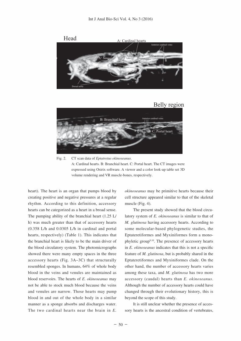
Figure 2 shows CT images of E. okinoseanus.
The upper figure shows the head area consisting of
the two cardinal hearts, the sinus, and two types of
blood vessels (the dorsal aorta and anterior-cardinal-
veins). Blood flow from the sinus to the two cardinal
hearts via the dorsal aorta was observed. The lower
figure shows the branchial and anterior body cavities
consisting of the branchial and portal hearts and
three blood vessels (anterior cardinal veins). A portal
heart in one of the three anterior cardinal veins was
observed.
Figure 3 shows a photomicrograph of heart
tissues stained with HE. The original objective
magnification was ×40. There were many empty
spaces (white spaces among the cells) in the three
types of heart tissues (Fig. 3A, cardinal hearts; Fig.
3B, branchial heart; Fig. 3C, portal heart). These
empty spaces suggested that the hearts push blood in
and out of the whole body in a similar manner as a
sponge absorbs and discharges water.
Figure 4 shows a photomicrograph of heart
tissues stained with HE or PTAH. The original
objective magnification was ×1000. Cross-striation
was found in three types of heart tissues. The
cardinal heart tissues were thick and straight, while
the branchial and portal heart tissues were thin and
branching. Cardinal hearts appeared similar skeletal
muscle tissues, suggesting that this heart evolved
from the skeletal muscle.
Figure 5 shows successive heart images using
diagnostic sonography. In the upper panel, the probe
of the sonograph was placed on three points (A, B,
or C) which corresponded with the three heart types.
The second (A), third (B), and fourth panels (C)
from the top showed the cardinal hearts, branchial
heart, and portal heart, respectively. These data
showed the time direction from left to right and
show three phases: the expression phase at the start,
the compression phase, and the expression after
compression. The heart rates of the three heart types
were measured from the time between the expansion
phase to the compression phase. The volume of the
three heart types was calculated as an ellipsoid
volume (4π × length × width × high/3) when E.

Int J Anal Bio-Sci Vol. 4, No 3 (2016)
― 49 ―
okinoseanus was anatomized. The flow rate was
calculated as the heart volume multiplied by the
heart rate.
Table 1 shows the ability (heart rate and flow
rate) and the heart volume of the three heart types.
The heart volumes were 0.0926, 0.289, and 0.0250
cm3 for the cardinal hearts, branchial heart, and
portal heart, respectively. The heart rates were 1.08,
1.20, and 0.339 beats/s for the cardinal hearts, bran-
chial heart, and portal heart, respectively.
4. Discussion
In addition to the “true” brachial heart in E.
okinoseanus, we also found three accessory hearts
(i.e., a pair of cardinal hearts and a single portal
Fig. 1. Anatomy of Eptatretus okinoseanus.
A: Cardinal hearts. B: Branchial heart. Tissue samples were fixed in 10% buffered formalin
and cut horizontally. C: Portal heart. D: Body overview. The presumed circulation is
showed by a white line, and the hearts are shown as white bulges. Scale bar: 1 cm.
Int J Anal Bio-Sci Vol. 4, No 3 (2016)
― 50 ―
heart). The heart is an organ that pumps blood by
creating positive and negative pressures at a regular
rhythm. According to this definition, accessory
hearts can be categorized as a heart in a broad sense.
The pumping ability of the branchial heart (1.25 L/
h) was much greater than that of accessory hearts
(0.358 L/h and 0.0305 L/h in cardinal and portal
hearts, respectively) (Table 1). This indicates that
the branchial heart is likely to be the main driver of
the blood circulatory system. The photomicrographs
showed there were many empty spaces in the three
accessory hearts (Fig. 3A–3C) that structurally
resembled sponges. In humans, 64% of whole body
blood in the veins and venules are maintained as
blood reservoirs. The hearts of E. okinoseanus may
not be able to stock much blood because the veins
and venules are narrow. Those hearts may pump
blood in and out of the whole body in a similar
manner as a sponge absorbs and discharges water.
The two cardinal hearts near the brain in E.
okinoseanus may be primitive hearts because their
cell structure appeared similar to that of the skeletal
muscle (Fig. 4).
The present study showed that the blood circu-
latory system of E. okinoseanus is similar to that of
M. glutinosa having accessory hearts. According to
some molecular-based phylogenetic studies, the
Eptateretiformes and Myxiniformes form a mono-
phyletic group9,10. The presence of accessory hearts
in E. okinoseanus indicates that this is not a specific
feature of M. glutinosa, but is probably shared in the
Eptateretiformes and Myxiniformes clade. On the
other hand, the number of accessory hearts varies
among these taxa, and M. glutinosa has two more
accessory (caudal) hearts than E. okinoseanus.
Although the number of accessory hearts could have
changed through their evolutionary history, this is
beyond the scope of this study.
It is still unclear whether the presence of acces-
sory hearts is the ancestral condition of vertebrates,
Fig. 2. CT scan data of Eptatretus okinoseanus.
A: Cardinal hearts. B: Branchial heart. C: Portal heart. The CT images were
expressed using Osirix software. A viewer and a color look-up table set 3D
volume rendering and VR muscle-bones, respectively.

Int J Anal Bio-Sci Vol. 4, No 3 (2016)
― 51 ―
but it is noteworthy that the muscular structure of E.
okinoseanus cardinal hearts possibly represents a
primitive condition of the vertebrate heart. Although
cardinal hearts have the ability to pump like a “true”
heart, their histology more closely resembles the
skeletal muscle than the “true” heart muscle. In
general, the vertebrate heart is composed of a cardiac
muscle characterized by highly branching muscle
fiber bundles. In contrast, accessory heart muscles
lack branching and more closely resemble skeletal
muscles that surround blood vessels in vertebrates. It
is widely accepted that the vertebrate heart is derived
from blood vessels2. We can hypothesize that the
heart of primitive vertebrates was composed of skel-
etal muscles, as seen in the cardinal hearts of E.
okinoseanus and M. glutinosa.
We have studied lactate dehydrogenase
(L-Lactate: NAD oxidoreductase, EC 1.1.1.27) A
and B subunits (LD-A, B) in hagfishes, including E.
okinoseanus, since 1997 11-19. Vertebrates, including
hagfishes, mainly have two subunits: LD-A rich
skeletal muscle and B rich heart muscle20, 21. In our
studies, two LD-Bs contained in six different hearts
were found (data not shown), and the LD-A and -B
Fig. 3. A photomicrograph showing heart tissues stained with hematoxylin and eosin.
The original objective magnification was ×40. Scale bar: 0.5 mm. Fig. 4 shows higher
magnification of the squares in Fig. 3.
A: cardinal hearts. B: branchial heart. C: portal heart

Int J Anal Bio-Sci Vol. 4, No 3 (2016)
― 52 ―
in vertebrates were of one type. Lampreys have only
one LD. In the evolution from lamprey to hagfish,
LD changed from LD to one LD-A and two LD-Bs.
In the future, we will examine the ratio of the two
LD-Bs in the three heart types, which may determine
clear relationships between vertebrate heart evolu-
tion and those of LDs.
We examined the heart structures in E. okinose-
anus using CT images and diagnostic sonography
and identified the three features of those hearts. First,
E. okinoseanus has four hearts, two cardinal hearts,
one branchial heart, and one portal heart. Second,
we observed vascular blood circulation, the sinus,
and two types of blood vessels (the dorsal aorta and
anterior cardinal veins). Third, the hearts pump by
creating positive and negative pressure at a regular
rhythm. We examined heart histology to determine
heart function. We observed many empty spaces in
all heart tissue types, and hearts pump blood in and
out of the whole body in a similar manner as a
sponge absorbs and discharges water. The cardinal
hearts showed primitive characteristics similar to the
skeletal muscle. The branchial heart was the main
operator, while the cardinal and portal hearts were
Fig. 4. A photomicrograph showing heart tissues stained with HE or PTAH.
The original objective magnification was ×1000. Scale bar: 20 μm. This
shows a higher magnification of squares in Fig. 3.
A: cardinal hearts. B: branchial heart. C: portal heart

Int J Anal Bio-Sci Vol. 4, No 3 (2016)
― 53 ―
assisting.
In heart evolution, the four hearts of E.
okinoseanus were derived from the amphioxus. The
two cardinal hearts in both sides of the brain may be
primitive hearts because their cell structure resem-
bled that of the skeletal muscle. Thus, the heart may
have evolved from the skeletal muscle.
Fig. 5. Successive heart images using transthoracic echocardiography.
A: cardinal hearts. B: branchial heart. C: portal heart
Table 1. The ability of three types of hearts in E.
okinoseanus

Int J Anal Bio-Sci Vol. 4, No 3 (2016)
― 54 ―
Conflicts of interest
The authors declare no conflict of interests.
Acknowledgements
We thank Drs. Teruaki Nishikawa (Toho
University), Souichirou Kubota (Toho University),
and Masumi Nozaki (Niigata University) for their
valuable suggestions and helpful discussion of the
manuscript.
References1. Moller PC and Philpott CW: The circulatory system
of amphioxus (Branchiostoma floridae) I. Morphology
of the major vessels of the pharyngeal area. J
Morphol, 139: 389-406, 1973.
2. Simões-Costa MS, Vasconcelos M, Sampaio AC,
Cravo RM, Linhares VL, Hochgreb T, Yan CY,
Davidson B, Xavier-Neto J: The evolutionary origin
of cardiac chambers. Dev Biol, 277: 1-15, 2005.
3. Jensen D: The hagfish. Sci Am, 214: 82-90, 1966.
4. Kuraku S and Kuratani S: Time scale for cyclostome
evolution inferred with a phylogenetic diagnosis of
hagfish and lamprey cDNA sequences. Zool Sci, 23:
1053-1064, 2006.
5. Johansen K: The cardiovascular system of Myxine
glutinosa L. The biology of Myxine: 289-316, 1963.
6. Fischer AH, Jacobson KA, Rose J and Zeller R:
Hematoxylin and eosin staining of tissue and cell
sections. CSH Protoc., 2008: pdb. prot4986, 2008.
7. Terner JY, Gurland J and Gaer F: Phosphotungstic
Acid-Hematoxylin; Spectropho-Tometry of the Lake
in Solution and in Stained Tissue. Stain Technol., 39:
141-153, 1964.
8. KV Kardong: Vertebrates: comparative anatomy,
function, evolution, McGraw-Hill Boston (2006)
9. Kuo C, Huang S and Lee S: Phylogeny of hagfish
based on the mitochondrial 16S rRNA gene. Mol
Phylogenet Evol, 28: 448-457, 2003.
10. Chen Y, Chang H and Mok H: Phylogenetic position
of Eptatretus chinensis (Myxinidae: Myxiniformes)
i n f e r r ed by 16S rRNA gene s equence and
morphology. Zool Stud, 44: 111-118, 2005.
11. Imai T, Mochizuki K, Nishiguchi Y, Naito S and
Yoshida M: Purification and amino acid sequence of
L-lactate dehydrogenase from the skeletal muscle of
the hagfish, Eptatretus okinoseanus. J Anal Bio-Sci
(Seibutsu Shiryo Bunseki), 20: 341-348, 1997.
12. Imai T, Nishiguchi Y, Naito S and Yoshida M:
Purification and some properties of skeletal muscle
lactate dehydrogenase from the Japanese hagfish,
Myxine garmani. J Anal Bio-Sci (Seibutsu Shiryo
Bunseki), 20: 307-314, 1997.
13. Nishiguchi Y, Naito S, Yoshida M and Imai T:
Purification and partial characterization of lactate
dehydrogenase from the skeletal muscle of hagfish,
Eptatretus burgeri. Medicine and biology, 135:
253-258, 1997.
14. Nishiguchi Y: Evolutionary implications of lactate
dehydrogenases (LDHs) of hagfishes compared to
lampreys: LDH cDNA sequences from Eptatretus
burgeri, Paramyxine atami and Eptatretus okinose-
anus. Zool Sci, 25: 475-479, 2008.
15. Nishiguchi Y, Miwa T and Abe F: Pressure-adaptive
differences in lactate dehydrogenases of three
hagfishes: Eptatretus burgeri, Paramyxine atami and
Eptatretus okinoseanus. Extremophiles, 12: 477-480,
2008.
16. Nishiguchi Y, Ito N and Okada M: Structure and
function of lactate dehydrogenase from hagfish.
Marine drugs, 8: 594-607, 2010.
17. Nishiguchi Y, Abe F and Okada M: Different pressure
resistance of lactate dehydrogenases from hagfish is
dependent on habitat depth and caused by tetrameric
structure dissociation. Marine Biotechnology, 13:
137-141, 2011.
18. Nishiguchi Y, Uchida A, Oshima N and Okada M:
Expression of the Lactate Dehydrogenase Gene from
Eptatretus okinoseanus in Escherichia coli. ISRN
Zoology, 2011.
19. Nishiguchi Y: Molecular evolutionary medicine based
on variations in hagfish lactate dehydrogenases. Int J
Anal Bio-Sci, 1: 50-54, 2013.
20. SHAW CR and BARTO E: Genetic Evidence for the
Subunit Structure of Lactate Dehydrogenase
Isozymes. Proc Natl Acad Sci U S A, 50: 211-214,
1963.
21. Markert CL and Moller F: Multiple Forms of
Enzymes: Tissue, Ontogenetic, and Species Specific
Patterns. Proc Natl Acad Sci U S A, 45: 753-763,
1959.