Embed Size (px)
Citation preview
RESEARCH ARTICLE
Exploring the microbiota of upper respiratory
tract during the development of pneumonia
in a mouse model
Yoshitomo MorinagaID1,2*, Yuki Take1, Daisuke Sasaki1, Kenji Ota1,2, Norihito KakuID
1,
Naoki Uno1,2, Kei Sakamoto1, Kosuke Kosai1,2, Taiga MiyazakiID2,3, Hiroo Hasegawa1,
Koichi Izumikawa2,3, Hiroshi Mukae2, Katsunori Yanagihara1
1 Department of Laboratory Medicine, Nagasaki University Graduate School of Biomedical Sciences,
Nagasaki, Nagasaki, Japan, 2 Department of Respiratory Medicine, Nagasaki University Graduate School of
Biomedical Sciences, Nagasaki, Nagasaki, Japan, 3 Department of Infectious Diseases, Nagasaki University
Graduate School of Biomedical Sciences, Nagasaki, Nagasaki, Japan
Abstract
The alteration of the microbial community in the upper respiratory tract (URT) can contribute
to the colonization and invasion of respiratory pathogens. However, there are no studies
regarding whether the characteristics of the URT microbiota can be affected by infections in
lower respiratory tract (LRT). To elucidate the microbial profiles of the URT during pneumo-
nia, the oral, nasal, and lung microbiota was evaluated at the early phase in a murine pneu-
monia model by direct intratracheal inoculation of Klebsiella pneumoniae. The meta 16S
rRNA sequencing of bronchoalveolar lavage fluid after K. pneumoniae inoculation pre-
sented alterations in the beta diversity of the microbes, but not in the alpha diversity. At this
point, a significant increase in microbial alpha diversity was observed in the oral cavity, but
not in the nasal cavity. The significant increase was observed in the family Carnobacteria-
ceae and family Enterococcaceae. These results suggest that characterizing the microbial
community of the respiratory tract may not just involve a simple downstream relationship
from the URT to the LRT. The health status of the LRT may influence the oral microbiota.
Thus, evaluation of the oral microbiota may contribute towards monitoring lung health; the
oral microbiota may act as a diagnostic marker of pneumonia.
Introduction
Microbial communities in the respiratory tract and their impact on health have been discussed
for a long time. Pathogens causing lower respiratory tract (LRT) infections have been consid-
ered to enter through the upper respiratory tract (URT) [1–3]. For most respiratory pathogens
such as Streptococcus pneumoniae, Staphylococcus aureus, and Klebsiella pneumoniae, coloni-
zation of the URT is the first step to cause respiratory infections [4, 5]. As the elderly display
increased oropharyngeal colonization by pathogens [4], there is a close relationship between
the pathogenesis of the upper and lower airways.
PLOS ONE | https://doi.org/10.1371/journal.pone.0222589 September 27, 2019 1 / 11
a1111111111
a1111111111
a1111111111
a1111111111
a1111111111
OPEN ACCESS
Citation: Morinaga Y, Take Y, Sasaki D, Ota K,
Kaku N, Uno N, et al. (2019) Exploring the
microbiota of upper respiratory tract during the
development of pneumonia in a mouse model.
PLoS ONE 14(9): e0222589. https://doi.org/
10.1371/journal.pone.0222589
Editor: Brenda A. Wilson, University of Illinois at
Urbana-Champaign, UNITED STATES
Received: February 9, 2019
Accepted: September 2, 2019
Published: September 27, 2019
Copyright: © 2019 Morinaga et al. This is an open
access article distributed under the terms of the
Creative Commons Attribution License, which
permits unrestricted use, distribution, and
reproduction in any medium, provided the original
author and source are credited.
Data Availability Statement: Nucleotide sequence
data reported are available in the NCBI Sequence
Read Archive under the accession numbers
DRX180226-DRX180255.
Funding: This study was supported by the Grant-
in-Aid for Scientific Research (C) from the Ministry
of Health, Labour, and Welfare in Japan
(15K09572).
Competing interests: The authors have declared
that no competing interests exist.
The components of the URT, such as the nasal and oral cavities, have their own unique
microbiota, and increasing evidences support the fact that the normal microbiota of the URT
basically has a protective role against pathogen invasion and colonization, with complex inter-
actions between microorganisms, such as competing for nutrients, producing bactericidal
molecules, and inducing metabolism shifting [1, 2, 6]. Indeed, alteration of the URT micro-
biota has been observed during pneumonia and viral infections [7, 8]. According to these
reports, it has been suggested that the altered microbial community in the URT can contribute
to colonization by respiratory pathogens and spreading of infection to the LRT.
The lung is classically thought to be sterile; however, recent molecular methods have
revealed that bacteria are also present in the lungs of healthy people at low levels, compared to
the upper respiratory tract (URT) [2, 9, 10]. Because the bacterial community of healthy lungs
has a composition similar to that in the mouth rather than the nose, microbial immigration
from the mouth can serve as the principal source of the lung microbiota in a healthy condition
[10]. In contrast, the LRT also has some mechanisms to eliminate bacteria, such as cough,
mucociliary clearance, and the innate and adaptive host defenses [2]. These microbial elimina-
tion systems can modify the microbial profile of the URT [2].
Thus, the microbial alteration of the URT may be possible to affect the microbial appear-
ance in the LRT; however, no studies have been examined whether the microbial characteris-
tics of the URT can be affected by LRT infections. To achieve the goal, we need to study the
microbial profiles using in vivo pneumonia model. Although it is often difficult to make pneu-
monia model due to the affinity between bacteria strains and mouse strains, we have a K. pneu-moniae strain which reproducibly cause pneumonia in mice [11]. Therefore, to determine
whether lung infections modify the URT microbiota, the nasal and oral microbial characteris-
tics of an animal model with K. pneumoniae pneumonia were studied.
Results
Microbial characteristics in the LRT
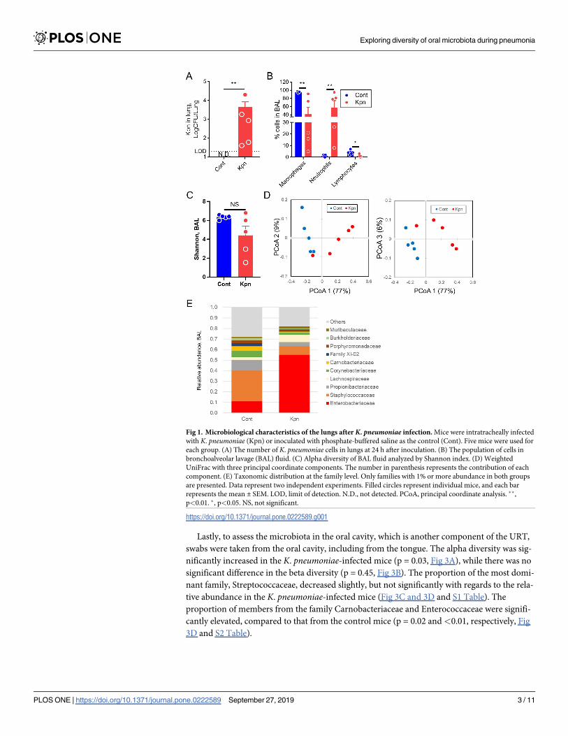
To study the microbial features in the respiratory tract during pneumonia, mice were directly
inoculated with K. pneumoniae into the LRT, and were evaluated at 24 h after inoculation
because the mice could loss the appetite due to the pneumonia. At this point, K. pneumoniaewas cultured in the lungs (Fig 1A), and a significant increase in the population of neutrophils
was observed in the bronchoalveolar lavage (BAL) fluid (Fig 1B). The alpha diversity in BAL
was at a level similar to that of the Shannon index in both the control and K. pneumoniae-infected mice (p = 0.22, Fig 1C). However, the beta diversity in the Weighted UniFrac of the
two groups was significantly different (p<0.01, Fig 1D). In control mice, Staphylococcaceae
and Propionibacteriaceae were predominant at the family level, whereas a decreased propor-
tion of these families of bacteria and an increased abundance of the members of the Enterobac-
teriaceae family were observed in the K. pneumoniae-infected mice (Fig 1E and S1 Table).
These results suggest that in the early phase of pneumonia caused by K. pneumoniae, alter-
ations in the lung microbiota were observed in case of the beta diversity, but not in the alpha
diversity.
Microbial characteristics in the URT
Next, to identify the nasal microbiota during pneumonia, the nasal airway lavage (NAL) fluid
was collected and analyzed by 16S metagenomic sequencing. No significant difference was
observed in the alpha (p = 0.69, Fig 2A) and beta diversity (p = 0.52, Fig 2B) between the con-
trol and K. pneumoniae-infected mice. The most abundant family was Staphylococcaceae in
the NAL, followed by Corynebacteriaceae (Fig 2C and S1 Table).
Exploring diversity of oral microbiota during pneumonia
PLOS ONE | https://doi.org/10.1371/journal.pone.0222589 September 27, 2019 2 / 11
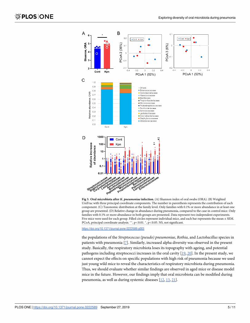
Lastly, to assess the microbiota in the oral cavity, which is another component of the URT,
swabs were taken from the oral cavity, including from the tongue. The alpha diversity was sig-
nificantly increased in the K. pneumoniae-infected mice (p = 0.03, Fig 3A), while there was no
significant difference in the beta diversity (p = 0.45, Fig 3B). The proportion of the most domi-
nant family, Streptococcaceae, decreased slightly, but not significantly with regards to the rela-
tive abundance in the K. pneumoniae-infected mice (Fig 3C and 3D and S1 Table). The
proportion of members from the family Carnobacteriaceae and Enterococcaceae were signifi-
cantly elevated, compared to that from the control mice (p = 0.02 and<0.01, respectively, Fig
3D and S2 Table).
Fig 1. Microbiological characteristics of the lungs after K. pneumoniae infection. Mice were intratracheally infected
with K. pneumoniae (Kpn) or inoculated with phosphate-buffered saline as the control (Cont). Five mice were used for
each group. (A) The number of K. pneumoniae cells in lungs at 24 h after inoculation. (B) The population of cells in
bronchoalveolar lavage (BAL) fluid. (C) Alpha diversity of BAL fluid analyzed by Shannon index. (D) Weighted
UniFrac with three principal coordinate components. The number in parenthesis represents the contribution of each
component. (E) Taxonomic distribution at the family level. Only families with 1% or more abundance in both groups
are presented. Data represent two independent experiments. Filled circles represent individual mice, and each bar
represents the mean ± SEM. LOD, limit of detection. N.D., not detected. PCoA, principal coordinate analysis. ��,
p<0.01. �, p<0.05. NS, not significant.
https://doi.org/10.1371/journal.pone.0222589.g001
Exploring diversity of oral microbiota during pneumonia
PLOS ONE | https://doi.org/10.1371/journal.pone.0222589 September 27, 2019 3 / 11
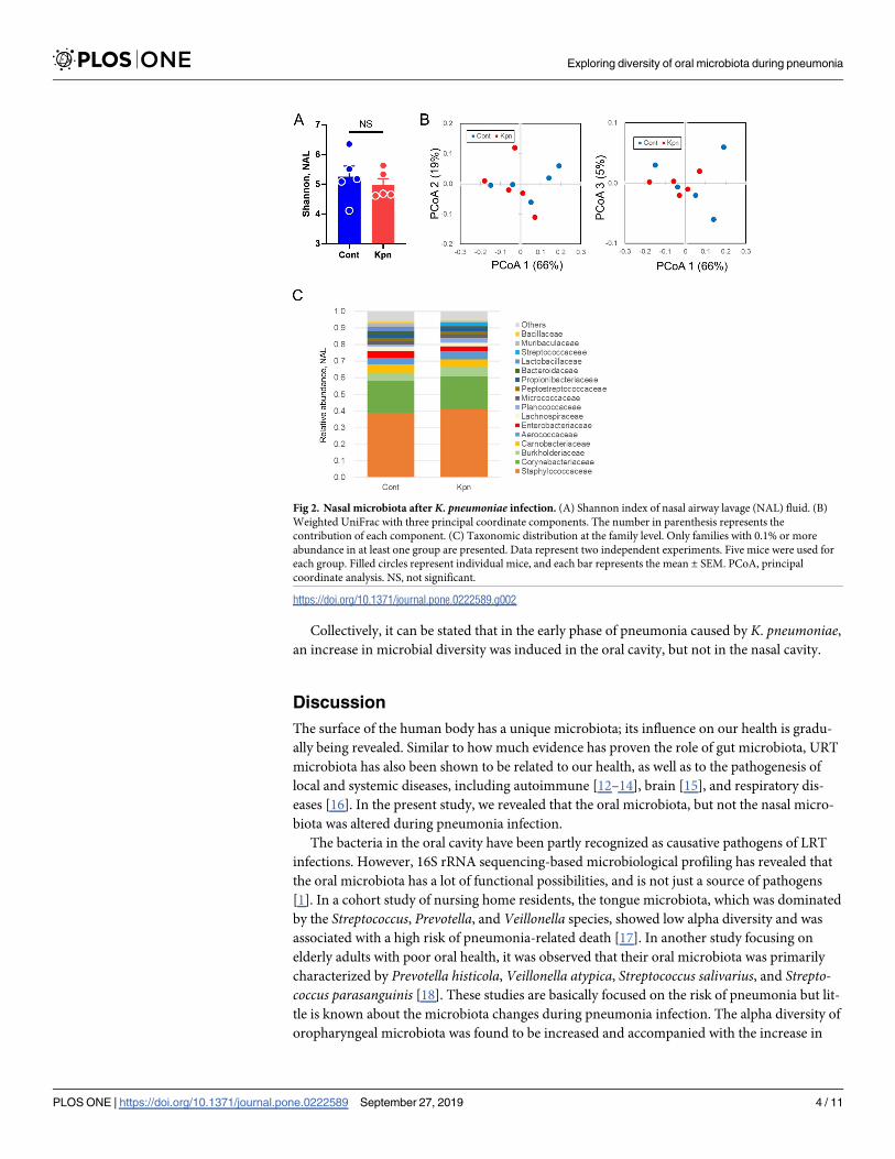
Collectively, it can be stated that in the early phase of pneumonia caused by K. pneumoniae,an increase in microbial diversity was induced in the oral cavity, but not in the nasal cavity.
Discussion
The surface of the human body has a unique microbiota; its influence on our health is gradu-
ally being revealed. Similar to how much evidence has proven the role of gut microbiota, URT
microbiota has also been shown to be related to our health, as well as to the pathogenesis of
local and systemic diseases, including autoimmune [12–14], brain [15], and respiratory dis-
eases [16]. In the present study, we revealed that the oral microbiota, but not the nasal micro-
biota was altered during pneumonia infection.
The bacteria in the oral cavity have been partly recognized as causative pathogens of LRT
infections. However, 16S rRNA sequencing-based microbiological profiling has revealed that
the oral microbiota has a lot of functional possibilities, and is not just a source of pathogens
[1]. In a cohort study of nursing home residents, the tongue microbiota, which was dominated
by the Streptococcus, Prevotella, and Veillonella species, showed low alpha diversity and was
associated with a high risk of pneumonia-related death [17]. In another study focusing on
elderly adults with poor oral health, it was observed that their oral microbiota was primarily
characterized by Prevotella histicola, Veillonella atypica, Streptococcus salivarius, and Strepto-coccus parasanguinis [18]. These studies are basically focused on the risk of pneumonia but lit-
tle is known about the microbiota changes during pneumonia infection. The alpha diversity of
oropharyngeal microbiota was found to be increased and accompanied with the increase in
Fig 2. Nasal microbiota after K. pneumoniae infection. (A) Shannon index of nasal airway lavage (NAL) fluid. (B)
Weighted UniFrac with three principal coordinate components. The number in parenthesis represents the
contribution of each component. (C) Taxonomic distribution at the family level. Only families with 0.1% or more
abundance in at least one group are presented. Data represent two independent experiments. Five mice were used for
each group. Filled circles represent individual mice, and each bar represents the mean ± SEM. PCoA, principal
coordinate analysis. NS, not significant.
https://doi.org/10.1371/journal.pone.0222589.g002
Exploring diversity of oral microbiota during pneumonia
PLOS ONE | https://doi.org/10.1371/journal.pone.0222589 September 27, 2019 4 / 11
the populations of the Streptococcus (pseudo) pneumoniae, Rothia, and Lactobacillus species in
patients with pneumonia [7]. Similarly, increased alpha diversity was observed in the present
study. Basically, the respiratory microbiota loses its topography with ageing, and potential
pathogens including streptococci increases in the oral cavity [19, 20]. In the present study, we
cannot expect the effects on specific populations with high risk of pneumonia because we used
just young wild mice to reveal the characteristics of respiratory microbiota during pneumonia.
Thus, we should evaluate whether similar findings are observed in aged mice or disease model
mice in the future. However, our findings imply that oral microbiota can be modified during
pneumonia, as well as during systemic diseases [12, 13, 21].
Fig 3. Oral microbiota after K. pneumoniae infection. (A) Shannon index of oral swabs (ORA). (B) Weighted
UniFrac with three principal coordinate components. The number in parenthesis represents the contribution of each
component. (C) Taxonomic distribution at the family level. Only families with 0.1% or more abundance in at least one
group are presented. (D) Relative change in abundance during pneumonia, compared to the case in control mice. Only
families with 0.1% or more abundance in both groups are presented. Data represent two independent experiments.
Five mice were used for each group. Filled circles represent individual mice, and each bar represents the mean ± SEM.
PCoA, principal coordinate analysis. ��, p<0.01. �, p<0.05. NS, not significant.
https://doi.org/10.1371/journal.pone.0222589.g003
Exploring diversity of oral microbiota during pneumonia
PLOS ONE | https://doi.org/10.1371/journal.pone.0222589 September 27, 2019 5 / 11
Little is known about the functional aspects of the LRT microbiota. Invading pathogens
and altered lung microbiota may influence circulating lymphocytes and contribute to immune
modulation, because the epithelial cells play the role of sentinels in the immune system, in
cooperation with associated lymphoid tissues [22]. For example, the inflammatory phenotype
of Th17 lymphocytes is associated with the aspiration-derived microbiota in the lungs [16].
The immunological crosstalk between LRT and URT may be one of a leader for the alteration
in microbiota in the mouth, but not in the nose. As another possibility, mechanical microbial
elimination systems such as mucociliary clearance and cough can affect the microbiota in
URT [2]. K. pneumoniae was not observed in both culture and PCR (data now shown) in
URT; however, the other bacterial transfer from LRT can modify the oral microbial commu-
nity. Although the mice did not show a loss in body weight and decrease in activity during a
short period of our study, our data could not exclude the possible influence of the loss of appe-
tite associated with pneumonia on the microbiota. This possibility can affect the oral micro-
biota directly as well as indirectly, because components and metabolites of the gut microbiota
influence the immune system [22].
In the present study, the oral microbiota was relatively altered compared to the nasal micro-
biota; however, the role of altered microbiota in the oral cavity during pneumonia is still
unknown. From a beneficial viewpoint, the slightly decreased abundance of Streptococcaceae
might reduce the additional involvement of oral bacteria on LRT infections because oral strep-
tococci is highly observed in patients with pneumonia [23]. Conversely, the alteration of oral
microbiota might lead to colonization by other pathogens [1]. The detected genus in Family
Carnobacteriaceae was mainly Atopostipes in the present study; however, the bacteria from the
genera Dolosigranulum and Granulicatella, which belong to the family Carnobacteriaceae,
have been known to be a part of the human oropharyngeal flora and also an important cause
of bacteremia and infective endocarditis [24–26]. As far as we searched, there are no reports
regarding the pathogenic roles of oral Enteroccocus in pneumonia patients. However, because
family Enterococcaceae is rarely observed in the oral cavity in healthy subjects [27] and known
to be present in root canal infections [28] and endodontic infections [29], the increase of
Enterococcus may imply the poor oral condition. Pathogenesis of infection caused by these
bacteria during pneumonia is still unknown, and further examination is required to under-
stand the role of the alteration of oral microbiota. However, the alteration of oral microbiota
in the early phase of pneumonia can be a possible diagnostic marker of pneumonia. In particu-
lar, it might be beneficial for the people who show poor signs or symptoms of pneumonia,
such as elderly and unconscious patients if inexpensive methods are developed for evaluation
of the oral microbiota.
There are some limitations in this study. Because we used just only one strain, it is still
uncertain whether similar findings are also observed in other K. pneumoniae strains or major
respiratory pathogens such as S. pneumoniae andH. influenzae. Future studies using these bac-
teria will support the universality and reinforce the weak points of our study such as small
sample sizes. Our study just presented the genomic microbial community using a single
method and the functional significance in the microbiome is still unknown. To reveal
unknown oral functions, studying bacterial transcriptomes using RNA-seq and metabolic pro-
filing will be helpful. The multi-omics study will contribute to the validation of our findings.
In conclusion, characterizing the microbial community of the respiratory tract may not
merely involve a simple relationship between the URT and the LRT. These possibilities should
be considered when the alteration of oral microbiota during LRT infections is studied. The
health status of the LRT may influence the oral microbiota, and the evaluation of oral micro-
biota may contribute to monitoring lung health.
Exploring diversity of oral microbiota during pneumonia
PLOS ONE | https://doi.org/10.1371/journal.pone.0222589 September 27, 2019 6 / 11
Materials and methods
Animals
Six- to eight-week-old female C57BL/6J mice were purchased from Charles River Laboratories
Japan, Inc. (Kanagawa, Japan). All animals were housed in a pathogen-free environment in the
Laboratory Animal Center for Biomedical Science at Nagasaki University and were provided
sterile food and water. Mice were co-housed for at least two weeks before experiments. The
Ethics Review Committee for Animal Experimentation (Institutional Animal Care and Use
Committee (IACUC) of Nagasaki University) approved all the experimental protocols used in
this study (Protocol Number: 1503101199).
Pneumonia model
A single colony of Klebsiella pneumoniae KEN-11 [11] which is a mouse-adaptive strain, was
sub-cultured in Luria-Bertani (LB) broth overnight. After 6–8 h of additional incubation in
fresh LB broth, the bacteria were adjusted to appropriate concentrations by turbidimetry.
After isoflurane anesthesia, KEN-11 (1 × 10⁴ CFUs/mouse) was directly inoculated into the
trachea as previously described [11]. Mice freely took sterile food and water in a sterile cage
(maximum 4 mice per cage). Mice were observed at the timing of bacterial inoculation and 24
h later, and sacrificed by isoflurane.
Bronchoalveolar lavage
After the pulmonary vasculature was flushed with 3 mL of normal saline via the right ventricle,
bronchoalveolar lavage (BAL) was performed by lavaging the LRT thrice with 0.8 mL of PBS,
as described previously [11]. Cytospin slides were prepared and stained with Diff-Quik (SYS-
MEX Co., Hyogo, Japan) for a differential cell count. The BAL fluids were stored at −20˚C
until further assays.
Nasal airway lavage
NAL on mice was performed using the trans-pharyngeal nasal lavaging technique [30]. For
this, 350 μL of PBS was flushed into the nasal airway, and the fluid was collected from the nos-
trils. The NAL fluids were stored at −20˚C until further assays.
Oral cavity swabbing
The oral cavity swabs were collected by placing a FLOQ swab (Copan Italia S.p.A., Brescia,
Italy) into the oral cavity [31]. Each swab was rotated 2–3 times before being withdrawn, and
placed in a tube containing 1 mL of PBS. The oral cavity swabs were stored at −20˚C until fur-
ther assays.
PCR amplification and preparation for 16S rRNA gene sequencing
DNA was extracted using a Quick-DNA Fecal/Soil Microbe Miniprep Kit (ZYMO Research,
Irvine, CA), according to the manufacturer’s instructions. The V1-V2 region of the bacterial
16S rRNA genes was amplified using the following primers: forward (50-AGAGTTTGATYMTGGCTCAG-30) with the Ion A adapter and sample-specific 13-base barcode sequences, and
reverse (50-TGCTGCCTCCCGTAGGAGT-30) with the Ion trP1 adapter sequence. The reaction
mixture contained 20 ng of the template DNA, 1 U Platinum SuperFi DNA Polymerase
(Thermo Fisher Scientific, Waltham, MA), 10 μL of 5X SuperFi buffer, 1 μL of 10 mM dNTP
mix (Thermo Fisher Scientific, Waltham, MA), and 25 pmol of each primer; DNase-RNase-free
Exploring diversity of oral microbiota during pneumonia
PLOS ONE | https://doi.org/10.1371/journal.pone.0222589 September 27, 2019 7 / 11
water was then added to achieve a final volume of 50 μL. The reaction conditions were: 94˚C for
5 min, 30 cycles of denaturation at 98˚C for 10 s, annealing at 59˚C for 10 s, extension at 72˚C
for 30 s, and a final extension at 72˚C for 5 min. The amplicons were purified using an AMPure
XP Kit (Beckman Coulter, Indianapolis, IN) and the concentration and fragment size of the
purified amplicons were measured using an Agilent 2100 Bioanalyzer (Agilent Technologies,
Santa Clara, CA). The purified amplicons were mixed in an equimolar manner. The final con-
centration of the samples was adjusted to 100 pM, for their use as template DNA in emulsion
PCR. Emulsion PCR and enrichment were performed using an Ion PGM HiQ View OT2 Kit
(Thermo Fisher Scientific, Waltham, MA) according to the manufacturer’s instructions. The
enriched samples were loaded onto an Ion 318 chip and sequencing was performed using the
Ion Torrent Personal Genome Analyzer with an Ion PGM HiQ View Sequencing Kit (Thermo
Fisher Scientific, Waltham, MA), according to the manufacturer’s instructions.
Sequence analysis
The sequencing reads were analyzed using CLC Genomics Workbench version 12.0.1 and
CLC Microbial Genomics Module version 3.6.11 (QIAGEN N. V., Venlo, Netherlands). After
removing the primer sequences and trimming the read length between 100 bp and 400bp
under 0.05% quality limit, samples with fewer than 100 reads and less than 50% from the
median were excluded from the further analyses. Chimeric reads were detected and filtered
using the chimera crossover detection algorithm with the default parameters (mismatch cost 1,
minimum score 40, gap cost 4, maximum unaligned end mismatches 5, chimera crossover
cost 3 and Kmer size 6). The reads were categorized into operational taxonomic units (OTUs)
with 97% similarity, and then assigned using SILVA release 132. The creation of new OTUs
was allowed when taxonomy similarity percentage was lower than 80% with minimum occur-
rence of two reads. OTUs were aligned using MUSCLE software implemented in CLC soft-
ware. Maximum likelihood phylogeny was created with Neighbor Joining as construction
method and Jukes Cantor as nucleotide substitution model. We excluded the OTUs with a low
abundance, or a combined abundance of less than 10 reads. The number of OTUs, Shannon
index (alpha diversity), and Weighted UniFrac distances were analyzed using the CLC Micro-
bial Genomics Module.
Statistical analysis
The groups were compared with Prism version 7 (GraphPad software, CA) using the Mann-
Whitney test; Fig 3D was analyzed by unpaired t-test because the result showed normal distri-
bution. The statistically significant alpha level was set as p�0.05. To compare the beta diversity,
the data were analyzed in PERMANOVA analysis using the CLC, and the statistically signifi-
cant alpha level was set as p�0.05, in false discovery rate (FDR).
Supporting information
S1 Table. Relative abundance of experiments in Family level. Relative abundance of ORA
(Sheet 1), NAL (Sheet 2), and BAL (Sheet 3) in Family level. The column names consist of
three components (the name of sample (ORA, NAL, or BAL) + group (C, control or K, infec-
tion with K. pneumoniae) + mouse number (1 to 5)).
(XLSX)
S2 Table. Individual values in Family level of oral microbiota. Scores indicate relative
increase of Family level from the average of control mice in oral microbiota.
(XLSX)
Exploring diversity of oral microbiota during pneumonia
PLOS ONE | https://doi.org/10.1371/journal.pone.0222589 September 27, 2019 8 / 11
Author Contributions
Conceptualization: Yoshitomo Morinaga, Katsunori Yanagihara.
Data curation: Yoshitomo Morinaga, Yuki Take, Daisuke Sasaki, Naoki Uno.
Formal analysis: Yoshitomo Morinaga, Yuki Take, Daisuke Sasaki.
Funding acquisition: Katsunori Yanagihara.
Investigation: Yoshitomo Morinaga, Yuki Take.
Methodology: Yoshitomo Morinaga, Yuki Take, Daisuke Sasaki, Kenji Ota.
Project administration: Yoshitomo Morinaga, Kei Sakamoto.
Resources: Kenji Ota, Norihito Kaku.
Supervision: Kei Sakamoto, Kosuke Kosai, Taiga Miyazaki, Hiroo Hasegawa, Koichi Izumi-
kawa, Hiroshi Mukae, Katsunori Yanagihara.
Validation: Kenji Ota, Norihito Kaku.
Writing – original draft: Yoshitomo Morinaga.
Writing – review & editing: Yoshitomo Morinaga, Taiga Miyazaki, Hiroo Hasegawa, Koichi
Izumikawa, Hiroshi Mukae, Katsunori Yanagihara.
References1. Man WH, de Steenhuijsen Piters WA, Bogaert D. The microbiota of the respiratory tract: gatekeeper to
respiratory health. Nature reviews Microbiology. 2017; 15(5):259–70. Epub 2017/03/21. doi: 10.1038/
nrmicro.2017.14. PMID: 28316330.
2. Dickson RP, Huffnagle GB. The Lung Microbiome: New Principles for Respiratory Bacteriology in
Health and Disease. PLoS pathogens. 2015; 11(7):e1004923. Epub 2015/07/15. doi: 10.1371/journal.
ppat.1004923. PMID: 26158874; PubMed Central PMCID: PMC4497592.
3. Cookson W, Cox MJ, Moffatt MF. New opportunities for managing acute and chronic lung infections.
Nature reviews Microbiology. 2018; 16(2):111–20. Epub 2017/10/25. doi: 10.1038/nrmicro.2017.122.
PMID: 29062070.
4. Marik PE, Kaplan D. Aspiration pneumonia and dysphagia in the elderly. Chest. 2003; 124(1):328–36.
Epub 2003/07/11. https://doi.org/10.1378/chest.124.1.328 PMID: 12853541.
5. Bogaert D, De Groot R, Hermans PW. Streptococcus pneumoniae colonisation: the key to pneumococ-
cal disease. The Lancet infectious diseases. 2004; 4(3):144–54. Epub 2004/03/05. doi: 10.1016/
S1473-3099(04)00938-7. PMID: 14998500.
6. Zipperer A, Konnerth MC, Laux C, Berscheid A, Janek D, Weidenmaier C, et al. Human commensals
producing a novel antibiotic impair pathogen colonization. Nature. 2016; 535(7613):511–6. Epub 2016/
07/29. doi: 10.1038/nature18634. PMID: 27466123.
7. de Steenhuijsen Piters WA, Huijskens EG, Wyllie AL, Biesbroek G, van den Bergh MR, Veenhoven RH,
et al. Dysbiosis of upper respiratory tract microbiota in elderly pneumonia patients. Isme j. 2016; 10
(1):97–108. Epub 2015/07/08. doi: 10.1038/ismej.2015.99. PMID: 26151645; PubMed Central PMCID:
PMC4681870.
8. Lu HF, Li A, Zhang T, Ren ZG, He KX, Zhang H, et al. Disordered oropharyngeal microbial communities
in H7N9 patients with or without secondary bacterial lung infection. Emerging microbes & infections.
2017; 6(12):e112. Epub 2017/12/21. doi: 10.1038/emi.2017.101. PMID: 29259328; PubMed Central
PMCID: PMC5750457.
9. Charlson ES, Bittinger K, Haas AR, Fitzgerald AS, Frank I, Yadav A, et al. Topographical continuity of
bacterial populations in the healthy human respiratory tract. American journal of respiratory and critical
care medicine. 2011; 184(8):957–63. Epub 2011/06/18. doi: 10.1164/rccm.201104-0655OC. PMID:
21680950; PubMed Central PMCID: PMC3208663.
10. Bassis CM, Erb-Downward JR, Dickson RP, Freeman CM, Schmidt TM, Young VB, et al. Analysis of
the upper respiratory tract microbiotas as the source of the lung and gastric microbiotas in healthy indi-
viduals. mBio. 2015; 6(2):e00037. Epub 2015/03/05. doi: 10.1128/mBio.00037-15. PMID: 25736890;
PubMed Central PMCID: PMC4358017.
Exploring diversity of oral microbiota during pneumonia
PLOS ONE | https://doi.org/10.1371/journal.pone.0222589 September 27, 2019 9 / 11
11. Harada Y, Morinaga Y, Kaku N, Nakamura S, Uno N, Hasegawa H, et al. In vitro and in vivo activities of
piperacillin-tazobactam and meropenem at different inoculum sizes of ESBL-producing Klebsiella pneu-
moniae. Clinical microbiology and infection: the official publication of the European Society of Clinical
Microbiology and Infectious Diseases. 2014. doi: 10.1111/1469-0691.12677. PMID: 24813594.
12. Abe K, Takahashi A, Fujita M, Imaizumi H, Hayashi M, Okai K, et al. Dysbiosis of oral microbiota and its
association with salivary immunological biomarkers in autoimmune liver disease. PloS one. 2018; 13
(7):e0198757. Epub 2018/07/04. doi: 10.1371/journal.pone.0198757. PMID: 29969462; PubMed Cen-
tral PMCID: PMC6029758.
13. Said HS, Suda W, Nakagome S, Chinen H, Oshima K, Kim S, et al. Dysbiosis of salivary microbiota in
inflammatory bowel disease and its association with oral immunological biomarkers. DNA research: an
international journal for rapid publication of reports on genes and genomes. 2014; 21(1):15–25. Epub
2013/09/10. doi: 10.1093/dnares/dst037. PMID: 24013298; PubMed Central PMCID: PMC3925391.
14. Zhang X, Zhang D, Jia H, Feng Q, Wang D, Liang D, et al. The oral and gut microbiomes are perturbed
in rheumatoid arthritis and partly normalized after treatment. Nature medicine. 2015; 21(8):895–905.
Epub 2015/07/28. doi: 10.1038/nm.3914. PMID: 26214836.
15. Boaden E, Lyons M, Singhrao SK, Dickinson H, Leathley M, Lightbody CE, et al. Oral flora in acute
stroke patients: A prospective exploratory observational study. Gerodontology. 2017; 34(3):343–56.
Epub 2017/05/26. doi: 10.1111/ger.12271. PMID: 28543778.
16. Segal LN, Clemente JC, Tsay JC, Koralov SB, Keller BC, Wu BG, et al. Enrichment of the lung micro-
biome with oral taxa is associated with lung inflammation of a Th17 phenotype. Nature microbiology.
2016; 1:16031. Epub 2016/08/31. doi: 10.1038/nmicrobiol.2016.31. PMID: 27572644; PubMed Central
PMCID: PMC5010013.
17. Kageyama S, Takeshita T, Furuta M, Tomioka M, Asakawa M, Suma S, et al. Relationships of Varia-
tions in the Tongue Microbiota and Pneumonia Mortality in Nursing Home Residents. The journals of
gerontology Series A, Biological sciences and medical sciences. 2018; 73(8):1097–102. Epub 2017/10/
21. doi: 10.1093/gerona/glx205. PMID: 29053769.
18. Asakawa M, Takeshita T, Furuta M, Kageyama S, Takeuchi K, Hata J, et al. Tongue Microbiota and
Oral Health Status in Community-Dwelling Elderly Adults. mSphere. 2018; 3(4). Epub 2018/08/17. doi:
10.1128/mSphere.00332-18. PMID: 30111628; PubMed Central PMCID: PMC6094060.
19. Dickson RP, Erb-Downward JR, Freeman CM, McCloskey L, Falkowski NR, Huffnagle GB, et al. Bacte-
rial Topography of the Healthy Human Lower Respiratory Tract. mBio. 2017; 8(1). Epub 2017/02/16.
doi: 10.1128/mBio.02287-16. PMID: 28196961; PubMed Central PMCID: PMC5312084.
20. Whelan FJ, Verschoor CP, Stearns JC, Rossi L, Luinstra K, Loeb M, et al. The loss of topography in the
microbial communities of the upper respiratory tract in the elderly. Annals of the American Thoracic
Society. 2014; 11(4):513–21. Epub 2014/03/08. doi: 10.1513/AnnalsATS.201310-351OC. PMID:
24601676.
21. Han YW, Wang X. Mobile microbiome: oral bacteria in extra-oral infections and inflammation. Journal of
dental research. 2013; 92(6):485–91. Epub 2013/04/30. doi: 10.1177/0022034513487559. PMID:
23625375; PubMed Central PMCID: PMC3654760.
22. Budden KF, Gellatly SL, Wood DL, Cooper MA, Morrison M, Hugenholtz P, et al. Emerging pathogenic
links between microbiota and the gut-lung axis. Nature reviews Microbiology. 2017; 15(1):55–63. Epub
2016/11/01. doi: 10.1038/nrmicro.2016.142. PMID: 27694885.
23. Akata K, Yatera K, Yamasaki K, Kawanami T, Naito K, Noguchi S, et al. The significance of oral strepto-
cocci in patients with pneumonia with risk factors for aspiration: the bacterial floral analysis of 16S ribo-
somal RNA gene using bronchoalveolar lavage fluid. BMC Pulm Med. 2016; 16(1):79. Epub 2016/05/
14. doi: 10.1186/s12890-016-0235-z. PMID: 27169775; PubMed Central PMCID: PMC4864928.
24. Ruoff KL. Nutritionally variant streptococci. Clinical microbiology reviews. 1991; 4(2):184–90. Epub
1991/04/01. https://doi.org/10.1128/cmr.4.2.184 PMID: 2070344; PubMed Central PMCID:
PMC358190.
25. Laclaire L, Facklam R. Antimicrobial susceptibility and clinical sources of Dolosigranulum pigrum cul-
tures. Antimicrobial agents and chemotherapy. 2000; 44(7):2001–3. Epub 2000/06/20. https://doi.org/
10.1128/aac.44.7.2001-2003.2000 PMID: 10858372; PubMed Central PMCID: PMC90003.
26. Alberti MO, Hindler JA, Humphries RM. Antimicrobial Susceptibilities of Abiotrophia defectiva, Granuli-
catella adiacens, and Granulicatella elegans. Antimicrobial agents and chemotherapy. 2015; 60
(3):1411–20. Epub 2015/12/17. doi: 10.1128/AAC.02645-15. PMID: 26666926; PubMed Central
PMCID: PMC4776019.
27. Sedgley CM, Lennan SL, Clewell DB. Prevalence, phenotype and genotype of oral enterococci. Oral
microbiology and immunology. 2004; 19(2):95–101. Epub 2004/02/12. PMID: 14871348.
28. Kato H, Yoshida A, Ansai T, Watari H, Notomi T, Takehara T. Loop-mediated isothermal amplification
method for the rapid detection of Enterococcus faecalis in infected root canals. Oral microbiology and
Exploring diversity of oral microbiota during pneumonia
PLOS ONE | https://doi.org/10.1371/journal.pone.0222589 September 27, 2019 10 / 11
immunology. 2007; 22(2):131–5. Epub 2007/02/22. doi: 10.1111/j.1399-302X.2007.00328.x. PMID:
17311637.
29. Rocas IN, Siqueira JF Jr., Santos KR. Association of Enterococcus faecalis with different forms of peri-
radicular diseases. Journal of endodontics. 2004; 30(5):315–20. Epub 2004/04/27. doi: 10.1097/
00004770-200405000-00004. PMID: 15107642.
30. Cho SH, Oh SY, Zhu Z, Lee J, Lane AP. Spontaneous eosinophilic nasal inflammation in a genetically-
mutant mouse: comparative study with an allergic inflammation model. PloS one. 2012; 7(4):e35114.
doi: 10.1371/journal.pone.0035114. PMID: 22509389; PubMed Central PMCID: PMC3324406.
31. Abusleme L, Hong BY, Hoare A, Konkel JE, Diaz PI, Moutsopoulos NM. Oral Microbiome Characteriza-
tion in Murine Models. Bio-protocol. 2017; 7(24). Epub 2018/01/16. doi: 10.21769/BioProtoc.2655.
PMID: 29333479; PubMed Central PMCID: PMC5760993.
Exploring diversity of oral microbiota during pneumonia
PLOS ONE | https://doi.org/10.1371/journal.pone.0222589 September 27, 2019 11 / 11