Embed Size (px)
Citation preview
FIP1L1-PDGFR� Imposes Eosinophil Lineage Commitment onHematopoietic Stem/Progenitor Cells*
Received for publication, September 26, 2008, and in revised form, December 22, 2008 Published, JBC Papers in Press, January 14, 2009, DOI 10.1074/jbc.M807489200
Kentaro Fukushima‡, Itaru Matsumura‡, Sachiko Ezoe‡§1, Masahiro Tokunaga‡, Masato Yasumi‡, Yusuke Satoh‡,Hirohiko Shibayama‡, Hirokazu Tanaka‡, Atsushi Iwama¶, and Yuzuru Kanakura‡
From the ‡Department of Hematology and Oncology, Osaka University Graduate School of Medicine, 2-2, Yamada-oka, Suita,Osaka 565-0871, Japan, the §Medical Center of Translational Research, Osaka University Hospital, Suita, Osaka 565-0871, Japan,and ¶Cellular and Molecular Medicine, Graduate School of Medicine, Chiba University, Chiba 260-8670, Japan
Although leukemogenic tyrosine kinases (LTKs) activate acommon set of downstreammolecules, the phenotypes of leu-kemia caused by LTKs are rather distinct. Here we report themolecular mechanism underlying the development ofhypereosinophilic syndrome/chronic eosinophilic leukemiaby FIP1L1-PDGFR�. When introduced into c-KithighSca-1�Lineage� cells, FIP1L1-PDGFR� conferred cytokine-inde-pendent growth on these cells and enhanced their self-renewal,whereas it did not immortalize commonmyeloid progenitors inin vitro replating assays and transplantation assays. Impor-tantly, FIP1L1-PDGFR� but not TEL-PDGFR� enhanced thedevelopment of Gr-1�IL-5R�� eosinophil progenitors fromc-KithighSca-1�Lineage� cells. FIP1L1-PDGFR� also promotedeosinophil development from common myeloid progenitors.Furthermore, when expressed in megakaryocyte/erythrocyteprogenitors and common lymphoid progenitors, FIP1L1-PDGFR� not only inhibited differentiation toward erythroidcells, megakaryocytes, and B-lymphocytes but aberrantly devel-oped eosinophil progenitors from megakaryocyte/erythrocyteprogenitors and common lymphoid progenitors. As for themechanism of FIP1L1-PDGFR�-induced eosinophil devel-opment, FIP1L1-PDGFR� was found to more intensely activateMEK1/2 and p38MAPK than TEL-PDGFR�. In addition, aMEK1/2 inhibitor and a p38MAPK inhibitor suppressed FIP1L1-PDGFR�-promoted eosinophil development. Also, reversetranscription-PCR analysis revealed that FIP1L1-PDGFR� aug-mented the expression of C/EBP�, GATA-1, and GATA-2,whereas it hardly affected PU.1 expression. In addition, shorthairpin RNAs against C/EBP� and GATA-2 and GATA-3KRR,which can act as a dominant-negative formover all GATAmem-bers, inhibited FIP1L1-PDGFR�-induced eosinophil develop-ment. Furthermore, FIP1L1-PDGFR� and its downstream Rasinhibited PU.1 activity in luciferase assays. Together, theseresults indicate that FIP1L1-PDGFR� enhances eosinophildevelopment by modifying the expression and activity of lin-eage-specific transcription factors through Ras/MEK andp38MAPK cascades.
During the last decade, it has become clear that hematopoi-etic growth factors regulate only growth and survival of hema-topoietic cells, whereas lineage-specific transcription factors,such as GATA-1, GATA-3, PU.1, Pax-5, C/EBP�, and C/EBP�,crucially control the lineage commitment and lineage-specificdifferentiation. For example, granulocyte colony-stimulatingfactor signaling induced megakaryopoiesis in granulocyte col-ony-stimulating factor receptor-transgenic mice (1). Also,erythropoietin (EPO)2 was found to promote terminal granulo-cytic differentiation in EPO receptor-transgenic mice. Fromthese data, we speculated that signal transduction moleculesactivated by hematopoietic growth factors would not influencethe lineage commitment of hematopoietic stem cells/progeni-tor cells (HSCs/HPCs) or subsequent lineage-specific differen-tiation (2). However, it has very recently been shown that theMEK/ERK pathway is involved inmyeloid lineage commitment(3). Also, PKB (c-Akt) was shown to be involved in lineage deci-sion during myelopoiesis (4). In addition, FLT3-activatingmutations were proved to inhibit C/EBP� activity throughERK1/2-mediated phosphorylation (3, 5). These results suggestthat signal transduction molecules activated by hematopoieticgrowth factors or their geneticmutationswould not only promotegrowth and survival but also influence lineage commitment andsubsequent differentiation of hematopoietic cells.Activating mutations of the tyrosine kinases (TKs), such as
c-Kit, platelet-derived growth factor receptor (PDGFR), FLT3,and c-ABL, are provoked by several mechanisms, includingchromosomal translocations and various mutations involvingtheir self-regulatory regions. These mutations are ofteninvolved in the pathogenesis of various types of hematologicmalignancies. BCR-ABL is known to cause chronic myeloge-nous leukemia and acute lymphoblastic leukemia. Mostpatients with PDGFR� rearrangement reveal common clinical
* This work was supported by grants from the Ministry of Education, Science,Sports, and Culture and Technology of Japan. The costs of publication ofthis article were defrayed in part by the payment of page charges. Thisarticle must therefore be hereby marked “advertisement” in accordancewith 18 U.S.C. Section 1734 solely to indicate this fact.
1 To whom correspondence should be addressed: Dept. of Hematology andOncology, Osaka University Graduate School of Medicine, 2-2, Yamada-oka, Suita, Osaka 565-0871, Japan. Tel.: 81-6-6879-3871; Fax: 81-6-6879-3879; E-mail: [email protected].
2 The abbreviations used are: EPO, erythropoietin; LTK, leukemogenic tyro-sine kinase; PDGFR�, platelet-derived factor receptor �; PDGFR�, platelet-derived factor receptor �; KSL, Lin�Sca�1hic-Kithi cell; CMP, common mye-loid progenitor; CLP, common lymphoid progenitor; GMP, commongranulocyte/monocyte progenitor; HSC, hematopoietic stem cell; HPC,hematopoietic progenitor cell; C/EBP, CCAAT/enhancer-binding protein;MPD, myeloproliferative disorder; TK, tyrosine kinase; AML, acute myeloidleukemia; MAPK, mitogen-activated protein kinase; STAT, signal transduc-ers and activators of transcription; HES, hypereosinophilic syndrome; CEL,chronic eosinophilic leukemia; ERK, extracellular signal-regulated kinase;LSC, leukemic stem cell; MEP, megakaryocyte/erythroid progenitor; FACS,fluorescence-activated cell sorter; shRNA, short hairpin RNA; RT, reversetranscription; GFP, green fluorescent protein; MEK, mitogen-activated pro-tein kinase/extracellular signal-regulated kinase kinase.
THE JOURNAL OF BIOLOGICAL CHEMISTRY VOL. 284, NO. 12, pp. 7719 –7732, March 20, 2009© 2009 by The American Society for Biochemistry and Molecular Biology, Inc. Printed in the U.S.A.
MARCH 20, 2009 • VOLUME 284 • NUMBER 12 JOURNAL OF BIOLOGICAL CHEMISTRY 7719
by guest on February 12, 2018http://w
ww
.jbc.org/D
ownloaded from
features resembling chronic myelogenous leukemia or cho-ronic myelomonocytic leukemia. In contrast, FLT3 mutations(ITD and point mutations in the TK domain) are primarilydetectable in acute myeloid leukemia (AML) or myelodysplas-tic syndrome (6–8). Also, c-KIT mutations in the TK domain(Asp816 3 Val, Tyr, Phe, or His) are found in patients withaggressive mastocytosis, myelodysplastic syndrome, and AML(9–15). Although these leukemogenic TKs (LTKs) activate acommon set of downstream signaling molecules, such as Ras/MAPK, PI3-K/Akt/mTOR, and STATs, the mechanisms bywhich LTKs cause different disease phenotypes remain to beclarified.FIP1L1-PDGFR� is a fusion gene, which was originally iden-
tified in the patients with hypereosinophilic syndrome (HES)/chronic eosinophilic leukemia (CEL) (16, 17). FIP1L1-PDGFR�fusion protein supports cytokine-independent growth and sur-vival of hematopoietic cells as a constitutively active TK (16,18–21). As for the downstream signaling molecules, FIP1L1-PDGFR� was shown to activate STAT5, phosphatidylinositol3-kinase, and Ras/ERK pathways like other LTKs, such as BCR-ABL, TEL-ABL, TEL-JAK2, and TEL-PDGFR� (18). In addi-tion, Buitenhuis et al. (22) recently reported that activation ofphosphatidylinositol 3-kinase, ERK1/2, and STAT5 is pivotalfor FIP1L1/PDGFR�-induced myeloproliferation.
The concept of “cancer stem cell” haswidely been recognizedand validated in various types of cancers, including breast can-cer, brain tumors, colon cancer, lung cancer, and malignantmelanoma. This concept was originally established in AML as a“leukemic stem cell (LSC)” (23, 24). In this concept, LSCs aredefined as specific leukemic cells that can cause leukemia whentransplanted into NOD/SCID mice. In AML, although leuke-mic blasts often display relatively homogenous features, theyare organized in a hierarchy. Among them, LSCs reveal themost immature CD34�CD38� phenotype similar to normalHSCs, whereas several antigen expressions are different. LSCs,which account for only 0.2–1.0% of AML cells in the bonemar-row (BM), have both abilities to self-renew and to producerestrictedly differentiated leukemia cells, thereby maintainingthemselves and yielding leukemia cells composing the majority(23, 25, 26). It is still unclear whether LSCs originate solely fromHSCs or are generated from nonstem immature cells that haveacquired de novo self-renewal ability. It has been shown that,although common myeloid progenitors (CMPs) and granulo-cyte/monocyte progenitors (GMPs) have very limited lifespans, several leukemogenic oncogenes, such as MLL-ENL,MOZ-TIF2, andMLL-AF9, have an ability to immortalize thesecells, thereby enabling them to act as LSCs (27, 28). On theother hand, although LSCs in a chronic phase of chronicmyelogenous leukemia are at an HSC level, chronic myeloge-nous leukemia cells at a CMP/GMP level can act as LSCs in anaccelerated phase, suggesting that additional gene mutationscan change the main LSC population during disease progres-sion. From these findings, it is now speculated that the leukemiaphenotype is determined by the biologic property of the mutatedgene and/or the lineage and the differentiation state of LSCs.In an attempt to analyze themolecularmechanisms bywhich
each LTK causes leukemia with the specific phenotype, weintroduced FIP1L1-PDGFR�, which plays a causal role in HES/
CEL, into murine HSCs and various types of HPCs. As a result,we found that FIP1L1-PDGFR� specifically enhanced eosino-phil development from HSCs/HPCs and imposed the lineageconversion to eosinophil lineage on megakaryocyte/erythroidprogenitors (MEPs) and common lymphoid progenitors (CLPs)through Ras/MEK and p38MAPK cascades by modifying theexpression and activity of lineage-specific transcription factors.
EXPERIMENTAL PROCEDURES
Reagents and Antibodies—Recombinant human TPO wasprovided by Kirin Brewery (Tokyo, Japan). Recombinanthuman FLT3L, human IL-6, murine SCF, murine IL-5, murineIL-7, murine granulocyte-macrophage colony-stimulating fac-tor, and human EPO were purchased from Peprotech (Ham-burg, Germany).Antibodies, Cell Staining, and Sorting—To isolate KSLs and
CLPs, murine BM cells were stained with phycoerythrin-con-jugated anti-IL-7R� chain (SB/199) (eBioscience, San Diego,CA), fluorescein isothiocyanate-conjugated anti-Sca-1 (E13-161-7), and APC-conjugated anti-c-Kit (2B8) monoclonal anti-bodies, and biotinylated rat antibodies specific for the lineagemarkers Ter119, CD3� (145-2C11), B220 (RA3–6B2), andGr-1(RB6-8C5), followed by staining with streptavidin-PerCP/Cy5.5 (BD Biosciences). Then KSLs and CLPs were sorted asIL-7R��Lin�Sca�1hic-Kithi and IL-7R��Lin�Sca-1loc-Kitlopopulations, respectively. For myeloid progenitor sorting,murine BM cells were stained with phycoerythrin-conju-gated anti-Fc�RII/III (2.4G2), fluorescein isothiocyanate-conjugated anti-CD34 (RAM34) (BD Biosciences), APC-conjugated anti-c-Kit, biotinylated anti-Sca-1, and anti-IL-7R�(SB/199) (Serotec, Raleigh, NC) monoclonal antibodies, andthe above described lineage mixture of monoclonal antibodies(BD Biosciences), followed by staining with avidin-APC/Cy7(BD Biosciences). After the staining, IL-7R��Lin�Sca-1�c-Kit�CD34�Fc�RII/IIIlo were sorted as CMPs, IL-7R��Lin�Sca-1�c-Kit�CD34�Fc�RII/IIIhi as GMPs, and IL-7R��Lin�Sca-1�c-Kit�CD34�Fc�RII/IIIlo as MEPs, as described previously(29). All of these HSCs and HPCs were isolated using a FACSAria (BD Bioscience, San Jose, CA). In all analyses and sorting,dead cells were excluded by stainingwith 7-amino-actinomycinD (Calbiochem). Cells were stained with phycoerythrin-conju-gated CD125 (IL-5 receptor �-subunit, T21) and APC-conju-gated Gr-1(RB6–8C5) (BD Biosciences) for detection of eosin-ophil lineage.Plasmids—Expression vectors for FIP1L1-PDGFR� and TEL-
PDGFR� were kindly provided by Dr. D. Gary Gilliland (HarvardMedical School, Boston, MA). Expression vectors for PDGFR�V561D and D842V were kindly provided by Dr. S. Hirota(HyogoMedical School, Hyogo, Japan). FIP1L1-PDGFR�,TEL-PDGFR�, PDGFR�V561D, and PDGFR�D842V were clonedinto the murine stem cell virus-internal ribosome entry site-EGFP (pMie) vector. Also, we constructed FIP1L1-PDGFR�and TEL-PDGFR� by the PCR method and subcloned theminto pMie.Short hairpin RNA (shRNA) interference oligonucleotides
against GATA-2 and C/EBP� described previously (30, 31)were cloned into an shRNA expression vector, pCS-RfA-CG,which was kindly provided by Dr. Miyoshi H (RIKEN Bio-
Enforced Eosinophil Development by FIP1L1-PDGFR�
7720 JOURNAL OF BIOLOGICAL CHEMISTRY VOLUME 284 • NUMBER 12 • MARCH 20, 2009
by guest on February 12, 2018http://w
ww
.jbc.org/D
ownloaded from
Resource center, Tsukuba, Japan). The retrovirus expressionvector for dominant negative GATA was constructed by clon-ing human GATA-3KRR cDNA that can inhibit GATA-1,GATA-2, and GATA-3 (32) into pMie.Cell Culture and Preparation—Murine BM cells were
obtained from 6–8-week-old C57BL/6J mice, which were pur-chased from CLEA (Tokyo, Japan). After sedimentation of thered blood cells with 6% hydroxyethyl starch, mononuclear cellswere separated by density gradient centrifugation, using HIS-TOPAQUE 1083 (Sigma). KSLs, CMPs, GMPs, andMEPs werepurified from mononuclear cells and cultured in RPMI1641medium (Nacalai Tesque, Kyoto, Japan) supplemented with10% fetal calf serum (EQUITECH-BIO, Kerrville, TX) in thepresence of murine SCF (50 ng/ml), human FLT3L (10 ng/ml),human IL-6 (50 ng/ml), and human TPO (50 ng/ml) for 48 h,and the cells were subjected to the retrovirus infection.Retrovirus Transduction—The conditioned media contain-
ing high titer retrovirus particles were prepared as describedpreviously (33). Briefly, an ecotropic packaging cell line, 293gp,kindly provided byDr.H.Miyoshi (RIKENBioResourceCenter,Tsukuba, Japan), was transfected with each retrovirus vector bythe calcium phosphate coprecipitation method. After 12 h, thecells were washed and cultured for 48 h. To produce lentivirus,293T cells were transfectedwith each shRNAexpression vectortogether with a packaging vector (pCAG-HIVgp) and a lentivi-rus envelope and Rev construct (pCMV-VSV-G-RSV-Rev),both of which were provided by Dr. Miyoshi. Then the super-natant containing virus particles was collected, centrifuged,and concentrated 50-fold in volume. The precultured murineBM cells were infected with each retrovirus in the RPMI1641medium supplemented with the same medium containingprotamine sulfate for 48 h in 6-well dishes coated with Ret-roNectin (Takara Bio Inc., Shiga, Japan).Colony Assays—Cells were seeded into methylcellulose
medium (MethoCult GFM3434; StemCell Technologies, Van-couver, Canada) at a density 2.5 � 102 cells/35-mm dish andwere cultured with 5% CO2 at 37 °C. All cultures were per-formed in triplicate, and the numbers of colonies were countedafter 10 days.In Vitro Immortalization Assays for HPCs—Immortalization
assays of HPCs in vitrowere performed as previously described(34). In brief, 104 cells were plated in 1.1 ml of methylcellulosemedium (Methocult M3434). After the 1 week of culture, col-ony numbers were counted, and single-cell suspensions of col-onies (104 cells) were subsequently replated under identicalconditions. Replating was repeated every week in the sameway.Luciferase Assays—Luciferase assays were performed with a
dual luciferase reporter system (Promega, Madison, WI), aspreviously described (35). Briefly, 293T or NIH3T3 cells (2 �105 cells) were seeded in a 60-mm dish and cultured for 24 h.Using the calcium phosphate coprecipitation method, cellswere transfected with 2 �g of reporter gene (pGL3-3�M�P-luciferase, 3�MHC-luciferase, or 1�MPO-luciferase) in com-bination with 2 �g of pcDNA3-GATA1, pcDNA3-PU.1 (36), orpcDNA3-C/EBP� together with 6 �g of an empty vector or aneffector vector for FIP1L1-PDGFR�, H-RasG12V, 1*6-STAT5A (37), or CAAX-p110 (38) and 10 ng of pRL-CMV, aRenilla luciferase expression vector. After 12 h, cells were
washed, serum-starved for 24 h, and subjected to luciferaseassays. After 36 h, the cells were lysed and subjected to a meas-urement for luciferase activity. The relative firefly luciferaseactivity was calculated by normalizing transfection efficiencyaccording to the Renilla luciferase activity.Semiquantitative RT-PCR Analysis—Total RNA was iso-
lated from 5 � 104 FACS-sorted GFP-positive cells usingTRIzol reagent (Invitrogen). RT-PCR was performed usingSuperScript II reverse transcriptase (Invitrogen) accordingto the manufacturer’s instructions. The cDNA product (1 �l)was resuspended in 20�l of the PCR buffer containing 0.5 unitsof TaqGold DNA polymerase (PerkinElmer Life Sciences), 2mM MgCl2, and 15 pmol of forward and reverse primers. Thesequences of forward/reverse primer sets were as follows:C/EBP�, 5�-GCC TGG CCT TGA CCA AGG AG-3� and5�-CAC AGG ACT AGA ACA CCT GC-3�; GATA-1, 5�-GGAATT CGG GCC CCT TGT GAG GCC AGA GAG-3� and5�-CGG GGT ACC TCA CGC TCC AGC CAG ATT CGACCC-3�; GATA-2, 5�-CGG AAT TCG ACA CAC CAC CCGATA CCC ACC TAT-3� and 5�-CGG AAT TCG CCT ACGCCA TGG CAG TCA CCA TGC T-3�; IL5-R�, 5�-GCC CTTTGA TCA GCT GTT CAG TCC AC-3� and 5�-CGG AACCGG TGG AAA CAA CCT GGT C-3�; MBP, 5�-ACC TGTCGC TAC CTC CTA-3� and 5�-GTG GTG GCA GAT GTGTGA-3�; PU.1, 5�-GATGGAGAAAGCCATAGCGA-3� and5�- TTG TGC TTG GAC GAG AAC TG-3�; HPRT, 5�-CACAGG ACT AGA ACA CCT GC-3� and 5�-GCT GGT GAAAAG GAC CTC T-3�.The PCR products were electrophoresed in agarose gels con-
taining ethidium bromide, and their amounts were analyzedwith a Fluor Imager595 and ImageQuant software (AmershamBiosciences).Transplantation Assays—Transplantation assays were per-
formed according to procedures described previously (39).Briefly, 8–12-week-old CD45.2 mice were lethally irradiated(900 rads) 24 h before the transplantation. BM cells isolatedfrom congenic C57BL/6 (B6-Ly5.1) mice were transduced withFIP1L1-PDGFR�, and 10,000 GFP-positive cells were injectedintravenously in combination with 2 � 105 normal BM cellswith CD45.2 phenotype. Chimeric analyses were performed at4 weeks and 8 weeks, and mice were sacrificed 16 weeks aftertransplantation. Animal care was performed according to insti-tutional guidelines.Measurement of Phosphorylation of Intracellular Signaling
Molecules—Phosphorylation of intracellular molecules wasassessed using Phosflow technology (BD Biosciences) accord-ing to the manufacturer’s recommendation. Briefly, cells werefixedwith PhosflowFix Buffer and incubated at 37 °C for 10–15min. After permeabilization at room temperature for 10 min,cells were washed twice with Phosflow Perm/Wash Buffer andincubated at room temperature for 10 min. After the bindingreaction to each antibody, cells were washed once with Phos-flow Perm/Wash buffer, resuspended in 500 �l of BD Pharm-ingen stain buffer (BD Bioscience), and then subjected to flowcytometric analysis. All experiments were repeated independ-ently at least three times, and reproducibility was confirmed.Statistical Analyses—Statistical analyses were performed
using Student’s t test.
Enforced Eosinophil Development by FIP1L1-PDGFR�
MARCH 20, 2009 • VOLUME 284 • NUMBER 12 JOURNAL OF BIOLOGICAL CHEMISTRY 7721
by guest on February 12, 2018http://w
ww
.jbc.org/D
ownloaded from
RESULTS
Effects of FIP1L1-PDGFR� on the Growth and Survival ofMurine KSL Cells—To investigate the effects of LTKs on thegrowth, differentiation, and survival of HSCs/HPCs, we con-structed bicistronic retrovirus vectors for FIP1L1-PDGFR� andTEL-PDGFR�, which express these cDNAs together with
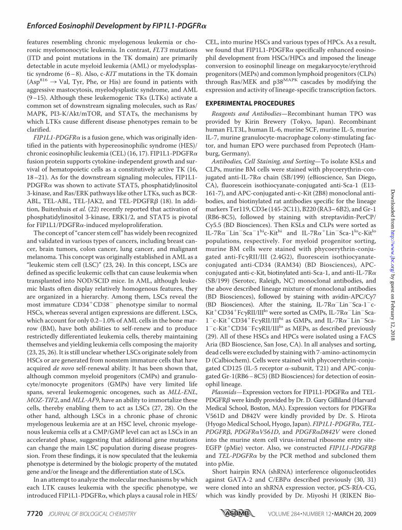
EGFP through the internal ribo-some entry site in the infected cells.At first, we introduced these retro-virus vectors into KSL cells. After a48-h infection, 55–65% of KSLswere found to be GFP-positive in allof transfectants by flow cytometricanalysis (data not shown). Next, weisolated retrovirus-infected cells asGFP-positive cells and cultured themin themediumwith or without SCF,TPO, FLT3L, and IL-6. As shown inFig. 1A (left), neither FIP1L1-PDGFR� nor TEL-PDGFR� furth-er augmented cytokine-dependentgrowth of KSLs. However, theseLTKs enabled KSLs to survive andproliferate under cytokine-deprivedconditions at least for 96 h, whereasmock (an empty retrovirus)-in-fected KSLs rapidly led to apoptosisin this condition (Fig. 1A, right).Next, we performed colony as-
says using these retrovirus-infectedKSLs. After 2-day retrovirus infec-tion, GFP-positive cells were sortedand plated into methylcellulose me-dium containing the cytokine mix-ture (EPO, TPO, SCF, granulocytecolony-stimulating factor, and IL-3),and numbers of colonies werecounted after 10 days. As shown inFig. 1B, the total number of colo-nies that developed from FIP1L1-PDGFR�- or TEL-PDGFR�-infectedKSLs was increased by 40–50% ascompared with that from mock-in-fected KSLs. Also, these colonieswere larger than those yielded frommock-infected KSLs (data notshown). However, the proportion ofCFU-GEMM, CFU-GM, CFU-G,CFU-M, and BFU-Ewas roughly thesame among three transfectants,indicating that these LTKs scarcelyinfluence the lineage commitmentand differentiation of KSLs in col-ony assays performed in this cyto-kine combination (Fig. 1B).We next performed an in vitro
immortalization assay using FIP1L1-PDGFR�-, TEL-PDGFR�-, or mock-
transduced KSLs and CMPs. After the first and second plating,both FIP1L1-PDGFR�- and TEL-PDGFR�-transduced KSLsyielded a slightly increased number of colonies relative tomock-transducedKSLs, whereas these differenceswere not sig-nificant (Fig. 1C, left). Also, in contrast to mock-transducedKSLs, FIP1L1-PDGFR�- or TEL-PDGFR�-transduced KSLs
FIGURE 1. The effects of leukemogenic tyrosine kinases on proliferation and survival of hematopoieticstem/progenitor cells. A, KSLs were isolated from murine bone marrow mononuclear cells. After the retrovi-rus (mock, FIP1L1-PDGFR�, or TEL-PDGFR�) infection, retrovirus-infected KSLs were sorted as GFP� cells andcultured with (left) or without (right) SCF, TPO, FLT3L, and IL-6 for 96 h. During these cultures, total viable cellnumbers were counted at the time indicated. B, KSLs infected with each retrovirus were sorted and seeded intothe methylcellulose medium containing EPO, TPO, SCF, granulocyte colony-stimulating factor, and IL-3. Colonynumbers were counted on day 12. C, immortalization assays for retrovirus-infected KSLs and CMPs. Retrovirus-infected KSLs and CMPs (103 cells) were plated into methylcellulose medium, and colony numbers werecounted after 1 week. Then single-cell suspensions of colonies (103 cells) were serially replated every week inthe same way. Bars, number of colonies obtained after each round of replating in methylcellulose as means �S.D. (n � 3). **, p � 0.01 compared with the value of mock-transduced cells.
Enforced Eosinophil Development by FIP1L1-PDGFR�
7722 JOURNAL OF BIOLOGICAL CHEMISTRY VOLUME 284 • NUMBER 12 • MARCH 20, 2009
by guest on February 12, 2018http://w
ww
.jbc.org/D
ownloaded from
still kept colony-forming activities even after the third andfourth plating (FIP1L1-PDGFR� versusmock at the third plat-ing, p � 0.01; at the fourth plating, p � 0.01), although theseactivities were rather reduced (Fig. 1C, left). On the other hand,even if FIP1L1-PDGFR� or TEL-PDGFR� was introduced,CMPs could not form any colony at the third plating, as was thecase withmock-infected CMPs (Fig. 1C, right). To evaluate leu-kemogenic potential of FIP1L1-PDGFR�-transduced KSLs invivo, we transplanted these cells into lethally irradiated mice incombination with freshly prepared competitor KSLs. As aresult, although none of the mice transplanted with mock-transduced KSLs developed leukemia or MPD, FIP1L1-PDGFR�-transduced KSLs developed MPD in three mice andacute leukemia in one mouse of five recipient mice within 15weeks after transplantation (Table 1). However, in agreementwith the previous report (16), none of the five recipient micedeveloped eosinophilic disorders. In addition, none of the 10mice transplanted with FIP1L1-PDGFR�-transduced CMPsdevelopedMPD or leukemia (data not shown). Together, theseresults indicate that FIP1L1-PDGFR� can confer the ability ofcytokine-independent growth/survival on KSLs and enhancetheir self-renewal, whereas it cannot immortalizeCMPs in vitroor in vivo.Effects of FIP1L1-PDGFR� and TEL-PDGFR� on Differenti-
ation from KSLs—We next investigated whether FIP1L1-PDGFR� or TEL-PDGFR� influences the lineage commitmentand subsequent differentiation of KSLs. For this purpose, weinfected retrovirus harboring FIP1L1-PDGFR� or TEL-PDGFR� into KSLs; cultured themwith SCF, TPO, FLT3L, andIL-6; and examined the expression of a granulocyte marker(Gr-1) and an eosinophil marker (IL-5 receptor �, CD125) inGFP-positive cells by flow cytometry. After 4-day cultures,there was not an apparent difference in the expression patternof these markers among FIP1L1-PDGFR�-, TEL-PDGFR�-,andmock-transduced KSLs (Fig. 2A, top). However, after 6-daycultures, TEL-PDGFR�- or FIP1L1-PDGFR�-transducedKSLsyielded significantly increased Gr-1� fraction (66.8 and 77.5%,respectively) compared with mock-transduced KSLs (49.6%).In addition, it was of particular interest that 51.8% of FIP1L1-PDGFR�-transduced KSLs grew to express CD125 and Gr-1simultaneously, whereas only 6.0% of mock-transduced and14.0% of TEL-PDGFR�-transduced KSLs revealed this pheno-type (FIP1L1-PDGFR� versusmock, p � 0.01; Fig. 2A, bottom).These results imply that FIP1L1-PDGFR� but not TEL-
PDGFR� preferentially imposes the commitment and differen-tiation to the eosinophilic lineage.To examine whether Gr-1�CD125� cells that developed
from FIP1L1-PDGFR�-transduced KSLs are actually eosino-phil precursors, we further cultured these KSLs with a cytokinemixture containing IL-5 for an additional 5 days. As a result,most of FIP1L1-PDGFR�-transduced but not mock- or TEL-PDGFR�-transduced KSLs came to possess large granule char-acteristics ofmature eosinophil in theMG staining, whichwerepositive for the eosinostain (Fig. 2B). Furthermore, after 10-daycultures, we examined the mRNA expression of eosinophil-re-lated genes,GATA-1, IL-5R�, andC/EBP�, by RT-PCR analysisusing sorted GFP-positive cells. As shown in Fig. 2C, IL-5R�and C/EBP� mRNAs were detected only in FIP1L1-PDGFR�-transduced KSLs. Also, GATA-1 mRNA was more intensivelyexpressed in FIP1L1-PDGFR�-transduced KSLs than in mock-or TEL-PDGFR�-transduced KSLs. These data indicate thatGr-1�CD125� cells that developed from FIP1L1-PDGFR�-transduced KSLs can indeed differentiate into matureeosinophils.Effects of FIP1L1-PDGFR� onDifferentiation of CMPs,MEPs,
and CLPs—It was previously shown that eosinophil precursorsstochastically develop from HSCs through MMP, CMP, andGMP (40, 41). Therefore, at first, we examined whetherFIP1L1-PDGFR� can enhance the development of eosinophilsfrom CMPs. For this purpose, we isolated CMPs from murineBMmononuclear cells by FACSusing severalmarkers (Fig. 3A).Then we introduced FIP1L1-PDGFR� into these cells and cul-tured them with SCF, IL-6, FLT3L, and TPO for 6 days. As wasthe case with KSLs, FIP1L1-PDGFR� remarkably enhanced thedevelopment of Gr-1�CD125� cells from CMPs comparedwith mock cultures (57% versus 6%, p � 0.01; Fig. 3B).Our next question was whether FIP1L1-PDGFR� could con-
vert the lineages of MEPs and CLPs, which were already com-mitted to the other lineages, into the eosinophil lineage. Toaddress this issue, we introduced FIP1L1-PDGFR� or TEL-PDGFR� into MEPs. When cocultured with a stroma cell lineOP-9 in the presence of SCF and EPO for 9 days, 58% of mock-infected and 41% of TEL-PDGFR�-infected MEPs came toreveal the Ter119�CD125� erythroid phenotype. In contrast,only 26% of FIP1L1-PDGFR�-infectedMEPs revealed this phe-notype (FIP1L1-PDGFR� versus mock, p � 0.05; Fig. 3C, top).Moreover, 50% of FIP1L1-PDGFR�-transduced MEPs differ-entiated into CD125�Gr-1� cells, whereas only 16% of mock-infected and 14% of TEL-PDGFR�-infectedMEPs revealed thisphenotype (FIP1L1-PDGFR� versus mock, p � 0.01; Fig. 3C,bottom). Similarly, after 9-day cultures in serum-free mediumsupplementedwithTPOand IL-11, althoughmock-transducedMEPs effectively gave rise to CD41�Gr-1� cells (17%), only 2%of FIP1L1-PDGFR�-infected MEPs revealed this phenotype(FIP1L1-PDGFR� versusmock, p � 0.01; Fig. 3D). Also, mock-transduced MEPs were found to become large polyploidmegakaryocytes inmorphological analysis, whereasmost of theFIP1L1-PDGFR�-transduced MEPs remained small andmononuclear (Fig. 3E). Together, these results indicate thatFIPIL1-PDGFR� inhibits erythroid and megakaryocytic differ-entiation from MEPs and imposes lineage conversion to theeosinophil lineage.
TABLE 1Peripheral blood examinations 16 weeks after transplantation
Mouse White blood cells � 109/liter Eosinophil%
Mock-1 88.3 1.2Mock-2 96.2 2.4Mock-3 83.5 2.6Mock-4 102.2 0.8Mock-5 88.2 2FIP1L1-PDGFR�-1 563.2 3.6FIP1L1-PDGFR�-2 121.1 1.2FIP1L1-PDGFR�-3 492.3 4.8FIP1L1-PDGFR�-4 140.1 3.6FIP1L1-PDGFR�-5 662.3a 0.2
a CD3(�)CD8(�) cells: 96%.
Enforced Eosinophil Development by FIP1L1-PDGFR�
MARCH 20, 2009 • VOLUME 284 • NUMBER 12 JOURNAL OF BIOLOGICAL CHEMISTRY 7723
by guest on February 12, 2018http://w
ww
.jbc.org/D
ownloaded from

FIGURE 2. Eosinophil development from KSLs. A, after retrovirus transduction, KSLs were cultured with SCF, TPO, IL-6, and FLT3L, and FACS analysis wasperformed after 4 days (top) and 6 days (bottom). GFP� cells were gated, and the expression of Gr-1 and CD125 was analyzed. **, p � 0.01 compared with thevalue of mock-transduced cells (n � 3). B, after 6-day cultures with SCF, TPO, IL-6, and FLT3L, retrovirus-infected KSLs were further cultured with a cytokinemixture containing IL-5 for 5 days. Transduced cells were subjected to May-Giemsa staining (top) and eosinostain (bottom). C, after 10-day cultures with TPO,IL-6, FLT3L, and SCF, GFP-positive cells were sorted, and the expression of eosinophil-related genes was analyzed by RT-PCR analysis.
Enforced Eosinophil Development by FIP1L1-PDGFR�
7724 JOURNAL OF BIOLOGICAL CHEMISTRY VOLUME 284 • NUMBER 12 • MARCH 20, 2009
by guest on February 12, 2018http://w
ww
.jbc.org/D
ownloaded from
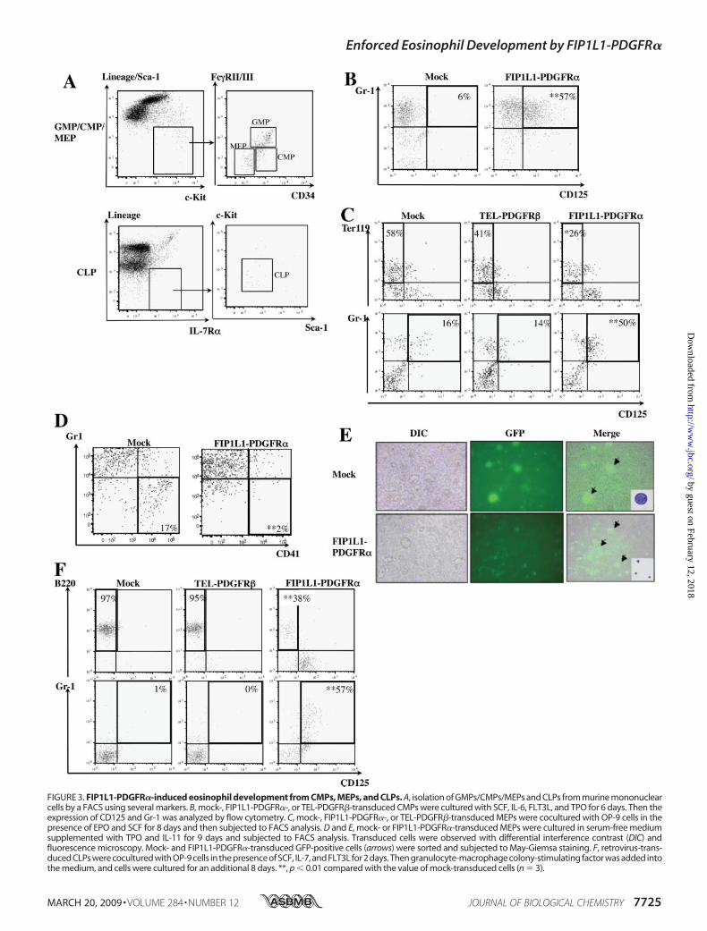
FIGURE 3. FIP1L1-PDGFR�-induced eosinophil development from CMPs, MEPs, and CLPs. A, isolation of GMPs/CMPs/MEPs and CLPs from murine mononuclearcells by a FACS using several markers. B, mock-, FIP1L1-PDGFR�-, or TEL-PDGFR�-transduced CMPs were cultured with SCF, IL-6, FLT3L, and TPO for 6 days. Then theexpression of CD125 and Gr-1 was analyzed by flow cytometry. C, mock-, FIP1L1-PDGFR�-, or TEL-PDGFR�-transduced MEPs were cocultured with OP-9 cells in thepresence of EPO and SCF for 8 days and then subjected to FACS analysis. D and E, mock- or FIP1L1-PDGFR�-transduced MEPs were cultured in serum-free mediumsupplemented with TPO and IL-11 for 9 days and subjected to FACS analysis. Transduced cells were observed with differential interference contrast (DIC) andfluorescence microscopy. Mock- and FIP1L1-PDGFR�-transduced GFP-positive cells (arrows) were sorted and subjected to May-Giemsa staining. F, retrovirus-trans-duced CLPs were cocultured with OP-9 cells in the presence of SCF, IL-7, and FLT3L for 2 days. Then granulocyte-macrophage colony-stimulating factor was added intothe medium, and cells were cultured for an additional 8 days. **, p � 0.01 compared with the value of mock-transduced cells (n � 3).
Enforced Eosinophil Development by FIP1L1-PDGFR�
MARCH 20, 2009 • VOLUME 284 • NUMBER 12 JOURNAL OF BIOLOGICAL CHEMISTRY 7725
by guest on February 12, 2018http://w
ww
.jbc.org/D
ownloaded from
Next, we introduced FIP1L1-PDGFR� into CLPs andcocultured them with OP-9 cells in the presence of SCF,IL-7, and FLT3L. After 10-day cultures, 97% of mock- and95% of TEL-PDGFR�-transduced CLPs came to have the
B220�CD125� B-lymphoid phenotype, whereas only 38% ofFIP1L1-PDGFR�-transduced CLPs had this phenotype(FIP1L1-PDGFR� versusmock, p � 0.01). Furthermore, a con-siderable proportion of FIP1L1-PDGFR�-transduced CLPs but
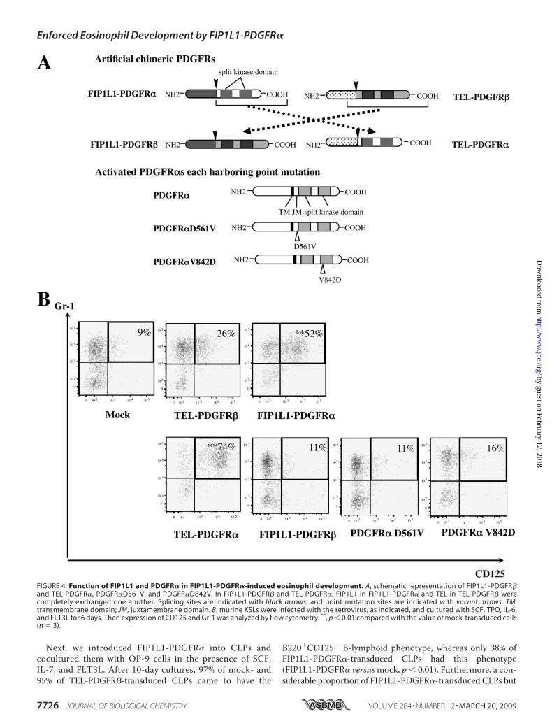
FIGURE 4. Function of FIP1L1 and PDGFR� in FIP1L1-PDGFR�-induced eosinophil development. A, schematic representation of FIP1L1-PDGFR�and TEL-PDGFR�, PDGFR�D561V, and PDGFR�D842V. In FIP1L1-PDGFR� and TEL-PDGFR�, FIP1L1 in FIP1L1-PDGFR� and TEL in TEL-PDGFR� werecompletely exchanged one another. Splicing sites are indicated with black arrows, and point mutation sites are indicated with vacant arrows. TM,transmembrane domain; JM, juxtamembrane domain. B, murine KSLs were infected with the retrovirus, as indicated, and cultured with SCF, TPO, IL-6,and FLT3L for 6 days. Then expression of CD125 and Gr-1 was analyzed by flow cytometry. **, p � 0.01 compared with the value of mock-transduced cells(n � 3).
Enforced Eosinophil Development by FIP1L1-PDGFR�
7726 JOURNAL OF BIOLOGICAL CHEMISTRY VOLUME 284 • NUMBER 12 • MARCH 20, 2009
by guest on February 12, 2018http://w
ww
.jbc.org/D
ownloaded from
not mock- or TEL-PDGFR�-transduced CLPs aberrantlydifferentiate into Gr-1�CD125� cells (percentage ofGr-1�CD125� cells as follows: FIP1L1-PDGFR�, 57% versusmock (1%) (p � 0.01); TEL-PDGFR�, 0% (Fig. 3F). We furthercultured Gr-1�CD125� cells that developed from FIP1L1-PDGFR�-transduced CLPs with a cytokine mixture containing
IL-5andconfirmedthat thesecellsbecamepositive foreosinostain(data not shown). These results indicate that FIP1L1-PDGFR�inhibits B-lymphoid differentiation fromCLPs and instructs themto differentiate into the eosinophil lineage.Function of FIP1L1 and PDGFR� in the Fusion Protein—It
was previously shown that the FIP1L1 moiety is dispensable
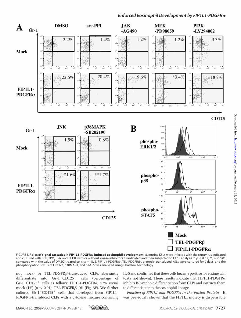
FIGURE 5. Roles of signal cascades in FIP1L1-PDGFR�-induced eosinophil development. A, murine KSLs were infected with the retrovirus indicatedand cultured with SCF, TPO, IL-6, and FLT3L with or without kinase inhibitors as indicated and then subjected to FACS analysis. *, p � 0.05; **, p � 0.01compared with the value of DMSO-treated cells (n � 4). B, FIP1L1-PDGFR�-, TEL-PDGFR�-, or mock- transduced KSLs were cultured for 2 days, and thephosphorylation status of ERK1/2, p38MAPK, and STAT5 was analyzed using Phosflow technology.
Enforced Eosinophil Development by FIP1L1-PDGFR�
MARCH 20, 2009 • VOLUME 284 • NUMBER 12 JOURNAL OF BIOLOGICAL CHEMISTRY 7727
by guest on February 12, 2018http://w
ww
.jbc.org/D
ownloaded from
for kinase activation and for transforming properties ofFIP1L1-PDGFR� (42). To determine the role of FIP1L1 inFIP1L1-PDGFR�-enhanced eosinophil development, we gen-
erated two artificial chimeric constructs, FIP1L1-PDGFR� andTEL-PDGFR�, inwhich FIP1L1 in FIP1L1-PDGFR� andTEL inTEL-PDGFR� were completely replaced (Fig. 4A). In addition,
Enforced Eosinophil Development by FIP1L1-PDGFR�
7728 JOURNAL OF BIOLOGICAL CHEMISTRY VOLUME 284 • NUMBER 12 • MARCH 20, 2009
by guest on February 12, 2018http://w
ww
.jbc.org/D
ownloaded from
we generated retrovirus vectors for constitutively active PDGFR�(PDGFR�V561D and PDGFR�D842V), which are considered tobe causative mutations of gastrointestinal stromal tumors (43)(Fig. 4A). When expressed in a murine IL-3-dependent cell line,Ba/F3, all of the fourPDGFRmutants conferred IL-3-independentgrowthon thesecells (datanot shown).Also,Westernblot analysisdemonstrated that these PDGFRmutants phosphorylated variouscellular proteins, including themselves (data not shown), indicat-ing that these proteins act as constitutively active tyrosine kinases.We transduced these retrovirus expression vectors into KSLs
and cultured themwith SCF, TPO, FLT3L, and IL-6 for 6 days. Asshown in Fig. 4B, only TEL-PDGFR� and not FIP1L1-PDGFR�,PDGFR�V561D, or PDGFR�D842V promoted eosinophil devel-opment from KSLs (percentage of Gr-1�CD125� fraction asfollows: TEL-PDGFR�, 74%; FIP1L1-PDGFR�, 11%;PDGFR�V561D, 11%; PDGFR�D842V, 16%) (TEL-PDGFR�versusmock, p� 0.01) (Fig. 4B), indicating that FIP1L1 is dispen-sable for FIP1L1-PDGFR�-mediated eosinophil development andthat PDGFR�-mediated signaling but not PDGFR�-mediated sig-naling is required for inducing eosinophil development. However,because neither PDGFR�V561D nor PDGFR�D842V promotedeosinophil development, specific kinase activity transmitted fromchimeric PDGFR� was supposed to be necessary to enhanceeosinophil development.Both a MEK1/2 Inhibitor and a p38MAPK Inhibitor Blocked
FIP1L1/PDGFR�-induced Eosinophil Development from KSLs—PDGFR� activates various downstream cascades, therebyexerting its biologic activity (44). To seek out the mechanismunderlying instructive eosinophil differentiation inducedby FIP1L1-PDGFR�, FIP1L1-PDGFR�- or mock-transducedKSLs were cultured with or without several kinase inhibitors asindicated (Fig. 5A).As shown in Fig. 5A (top), neither a c-Jun N-terminal kinase
inhibitor, a phosphatidylinositol 3-kinase inhibitor (LY294002),an Src inhibitor (PPI), nor a JAK2/STAT inhibitor (AG490)influenced FIP1L1-PDGFR�-enhanced eosinophil develop-ment, since about 20% of cells came to be CD125�Gr1� after5-day cultures as was seen after the culture without an inhibitor(Fig. 2A). In contrast, a MEK inhibitor (PD98059) and ap38MAPK inhibitor (SB202190) reduced the CD125�Gr1� frac-tion to 3.4% (p� 0.05) and 1.7% (p� 0.01), respectively (Fig. 5A,bottom). We also analyzed the phosphorylation states of ERK,STAT5, and p38MAPK in FIP1L1-PDGFR�- or TEL-PDGFR�-transduced KSLs by flow cytometry. As shown in Fig. 5B,ERK1/2 and p38MAPK but not STAT5 were phosphorylatedmore intensely in FIP1L1-PDGFR�-transduced KSLs than inmock- or TEL-PDGFR�-transduced KSLs. These data suggestthat FIP1L1-PDGFR� instructs HSCs/HPCs to differentiate
into eosinophil progenitors through the activation of MEK1/2-ERK1/2 and p38 pathways.Effects of FIP1L1-PDGFR� on the Expression and Activity of
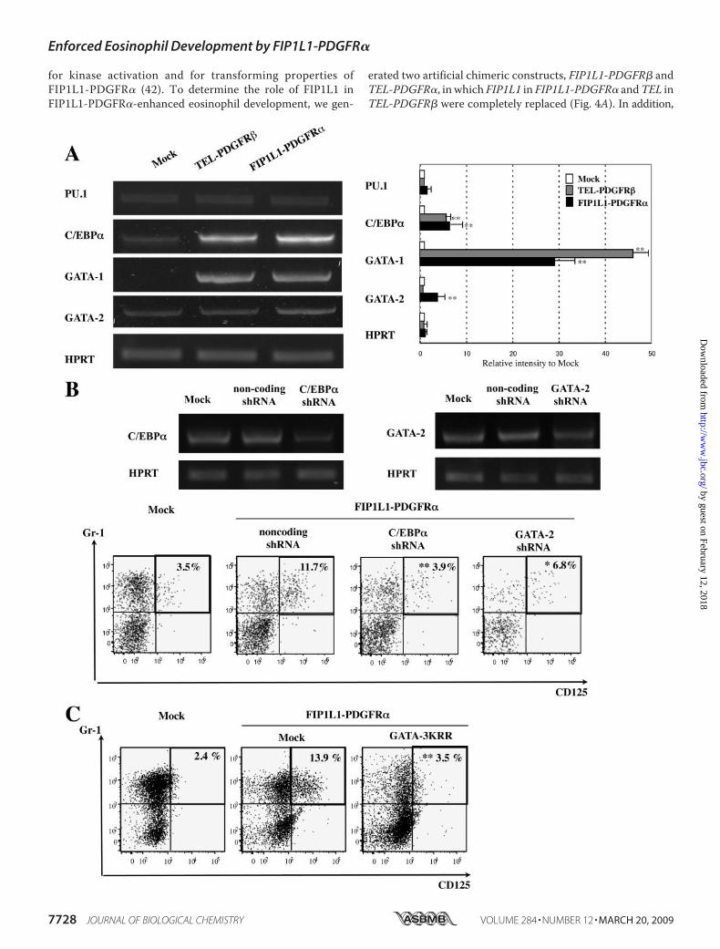
Lineage-specific Transcription Factors in KSLs—To furtherclarify the mechanism through which FIP1L1-PDGFR�enhanced eosinophil development, we analyzed the effects ofFIP1L1-PDGFR� on the expression of GATA-1, GATA-2,C/EBP�, and PU.1, all of which have been reported to be keytranscription factors for eosinophil development (45–47). Todetect the changes in the expression of these factors that pre-cede the phenotypic change, we isolated mRNA from sortedGFP-positive KSLs after 48-h retrovirus infection and per-formed semiquantitative RT-PCR analysis, since an apparentphenotypic change was not observed until 4 days (Fig. 2A, top).As shown in Fig. 6A, although the expression of PU.1was not sodifferent among three transfectants, FIP1L1-PDGFR� aug-mented the expression ofC/EBP� (p� 0.01) andGATA-1 (p�
FIGURE 6. Effects of FIP1L1-PDGFR� and its downstream molecules on the expressions of eosinophil-related transcription factors and effects ofinhibition of these molecules. A, the expressions of eosinophil-related transcription factors in KSLs were analyzed by RT-PCR analysis 48 h after retrovirustransfection. PCR products were electrophoresed and visualized by ethidium bromide staining (left), and their intensities were quantified using a FluorImager595 and ImageQuant software. Relative intensities to the products from mock-transduced cells are indicated (right). *, p � 0.05; **, p � 0.01 as comparedwith the value in mock-transduced cells. Data represent means � S.D. (n � 3). B, murine KSLs were infected with lentivirus-expressing noncoding or encodingshRNA against C/EBP� or GATA-2 to evaluate the suppression efficacy of each shRNA. After a 48-h culture, cells were subjected to RT-PCR analyses (top). Next,FIP1L1-PDGFR�-transduced murine KSLs were further infected with these shRNAs and cultured with SCF, TPO, IL-6, and FLT3L, which were subjected to FACSanalyses upon the expression of CD125 and Gr-1 (bottom). *, p � 0.05; **, p � 0.01 as compared with the value in the cells coexpressing FIP1L1-PDGFR� andnoncoding shRNA (n � 3). C, FIP1L1-PDGFR�-transduced murine KSLs were further infected with retrovirus encoding mock or a dominant negative form ofGATAs (GATA-3KRR). **, p � 0.01 as compared with the value in FIP1L1-PDGFR�- and mock-cotransduced cells (n � 3).
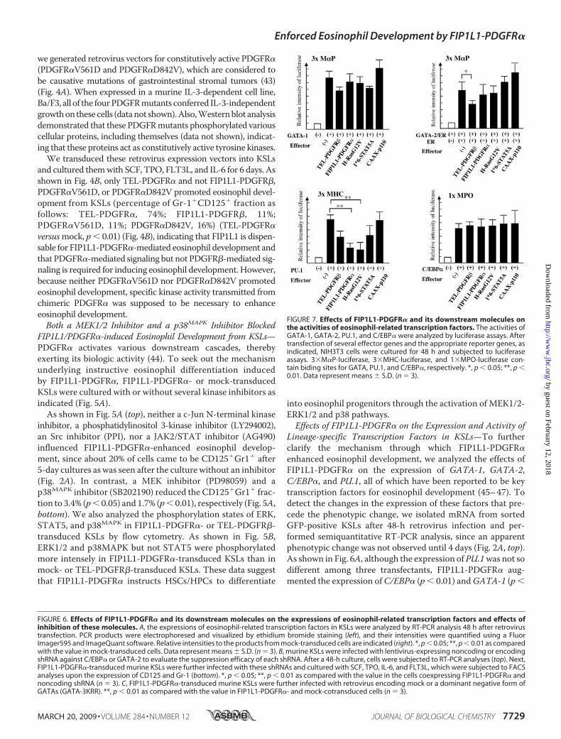
FIGURE 7. Effects of FIP1L1-PDGFR� and its downstream molecules onthe activities of eosinophil-related transcription factors. The activities ofGATA-1, GATA-2, PU.1, and C/EBP� were analyzed by luciferase assays. Aftertransfection of several effector genes and the appropriate reporter genes, asindicated, NIH3T3 cells were cultured for 48 h and subjected to luciferaseassays. 3�M�P-luciferase, 3�MHC-luciferase, and 1�MPO-luciferase con-tain biding sites for GATA, PU.1, and C/EBP�, respectively. *, p � 0.05; **, p �0.01. Data represent means � S.D. (n � 3).
Enforced Eosinophil Development by FIP1L1-PDGFR�
MARCH 20, 2009 • VOLUME 284 • NUMBER 12 JOURNAL OF BIOLOGICAL CHEMISTRY 7729
by guest on February 12, 2018http://w
ww
.jbc.org/D
ownloaded from
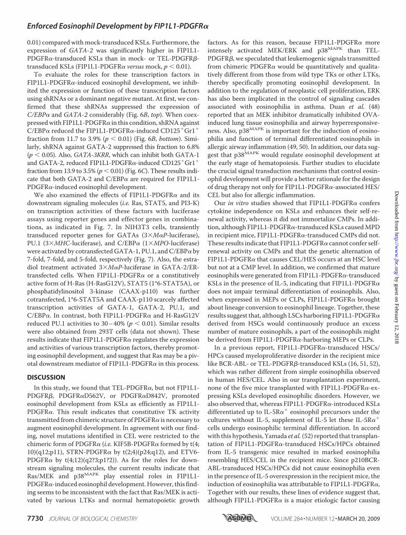
0.01) compared withmock-transduced KSLs. Furthermore, theexpression of GATA-2 was significantly higher in FIP1L1-PDGFR�-transduced KSLs than in mock- or TEL-PDGFR�-transduced KSLs (FIP1L1-PDGFR� versusmock, p � 0.01).To evaluate the roles for these transcription factors in
FIP1L1-PDGFR�-induced eosinophil development, we inhib-ited the expression or function of these transcription factorsusing shRNAs or a dominant negative mutant. At first, we con-firmed that these shRNAs suppressed the expression ofC/EBP� and GATA-2 considerably (Fig. 6B, top). When coex-pressedwith FIP1L1-PDGFR� in this condition, shRNAagainstC/EBP� reduced the FIP1L1-PDGFR�-induced CD125�Gr1�
fraction from 11.7 to 3.9% (p � 0.01) (Fig. 6B, bottom). Simi-larly, shRNA against GATA-2 suppressed this fraction to 6.8%(p � 0.05). Also, GATA-3KRR, which can inhibit both GATA-1and GATA-2, reduced FIP1L1-PDGFR�-induced CD125�Gr1�
fraction from 13.9 to 3.5% (p� 0.01) (Fig. 6C). These results indi-cate that both GATA-2 and C/EBP� are required for FIP1L1-PDGFR�-induced eosinophil development.
We also examined the effects of FIP1L1-PDGFR� and itsdownstream signaling molecules (i.e. Ras, STAT5, and PI3-K)on transcription activities of these factors with luciferaseassays using reporter genes and effector genes in combina-tions, as indicated in Fig. 7. In NIH3T3 cells, transientlytransduced reporter genes for GATAs (3�M�P-luciferase),PU.1 (3�MHC-luciferase), and C/EBP� (1�MPO-luciferase)were activated by cotransfectedGATA-1, PU.1, andC/EBP� by7-fold, 7-fold, and 5-fold, respectively (Fig. 7). Also, the estra-diol treatment activated 3�M�P-luciferase in GATA-2/ER-transfected cells. When FIP1L1-PDGFR� or a constitutivelyactive form of H-Ras (H-RasG12V), STAT5 (1*6-STAT5A), orphosphatidylinositol 3-kinase (CAAX-p110) was furthercotransfected, 1*6-STAT5A and CAAX-p110 scarcely affectedtranscription activities of GATA-1, GATA-2, PU.1, andC/EBP�. In contrast, both FIP1L1-PDGFR� and H-RasG12Vreduced PU.1 activities to 30–40% (p � 0.01). Similar resultswere also obtained from 293T cells (data not shown). Theseresults indicate that FIP1L1-PDGFR� regulates the expressionand activities of various transcription factors, thereby promot-ing eosinophil development, and suggest that Ras may be a piv-otal downstream mediator of FIP1L1-PDGFR� in this process.
DISCUSSION
In this study, we found that TEL-PDGFR�, but not FIP1L1-PDGFR�, PDGFR�D562V, or PDGFR�D842V, promotedeosinophil development from KSLs as efficiently as FIP1L1-PDGFR�. This result indicates that constitutive TK activitytransmitted fromchimeric structure of PDGFR� is necessary toaugment eosinophil development. In agreement with our find-ing, novel mutations identified in CEL were restricted to thechimeric form of PDGFR� (i.e. KIF5B-PDGFR� formed by t(4;10)(q12;p11), STRN-PDGFR� by t(2;4)(p24;q12), and ETV6-PDGFR� by t(4;12)(q2?3;p1?2)). As for the roles for down-stream signaling molecules, the current results indicate thatRas/MEK and p38MAPK play essential roles in FIP1L1-PDGFR�-induced eosinophil development.However, this find-ing seems to be inconsistent with the fact that Ras/MEK is acti-vated by various LTKs and normal hematopoietic growth
factors. As for this reason, because FIP1L1-PDGFR� moreintensely activated MEK/ERK and p38MAPK than TEL-PDGFR�, we speculated that leukemogenic signals transmittedfrom chimeric PDGFR� would be quantitatively and qualita-tively different from those from wild type TKs or other LTKs,thereby specifically promoting eosinophil development. Inaddition to the regulation of neoplastic cell proliferation, ERKhas also been implicated in the control of signaling cascadesassociated with eosinophilia in asthma. Duan et al. (48)reported that an MEK inhibitor dramatically inhibited OVA-induced lung tissue eosinophilia and airway hyperresponsive-ness. Also, p38MAPK is important for the induction of eosino-philia and function of terminal differentiated eosinophils inallergic airway inflammation (49, 50). In addition, our data sug-gest that p38MAPK would regulate eosinophil development atthe early stage of hematopoiesis. Further studies to elucidatethe crucial signal transduction mechanisms that control eosin-ophil development will provide a better rationale for the designof drug therapy not only for FIP1L1-PDGFR�-associated HES/CEL but also for allergic inflammation.Our in vitro studies showed that FIP1L1-PDGFR� confers
cytokine independence on KSLs and enhances their self-re-newal activity, whereas it did not immortalize CMPs. In addi-tion, although FIP1L1-PDGFR�-transducedKSLs causedMPDin recipient mice, FIP1L1-PDGFR�-transduced CMPs did not.These results indicate that FIP1L1-PDGFR� cannot confer self-renewal activity on CMPs and that the genetic alternation ofFIP1L1-PDGFR� that causes CEL/HES occurs at an HSC levelbut not at a CMP level. In addition, we confirmed that matureeosinophils were generated from FIP1L1-PDGFR�-transducedKSLs in the presence of IL-5, indicating that FIP1L1-PDGFR�does not impair terminal differentiation of eosinophils. Also,when expressed in MEPs or CLPs, FIP1L1-PDGFR� broughtabout lineage conversion to eosinophil lineage. Together, theseresults suggest that, although LSCs harboring FIP1L1-PDGFR�derived from HSCs would continuously produce an excessnumber of mature eosinophils, a part of the eosinophils mightbe derived from FIP1L1-PDGFR�-harboring MEPs or CLPs.
In a previous report, FIP1L1-PDGFR�-transduced HSCs/HPCs caused myeloproliferative disorder in the recipient micelike BCR-ABL- or TEL-PDGFR�-transduced KSLs (16, 51, 52),which was rather different from simple eosinophilia observedin human HES/CEL. Also in our transplantation experiment,none of the five mice transplanted with FIP1L1-PDGFR�-ex-pressing KSLs developed eosinophilic disorders. However, wealso observed that, whereas FIP1L1-PDGFR�-introduced KSLsdifferentiated up to IL-5R�� eosinophil precursors under thecultures without IL-5, supplement of IL-5 let these IL-5R��
cells undergo eosinophilic terminal differentiation. In accordwith this hypothesis, Yamada et al. (52) reported that transplan-tation of FIP1L1-PDGFR�-transduced HSCs/HPCs obtainedfrom IL-5 transgenic mice resulted in marked eosinophiliaresembling HES/CEL in the recipient mice. Since p210BCR-ABL-transduced HSCs/HPCs did not cause eosinophilia evenin the presence of IL-5 overexpression in the recipientmice, theinduction of eosinophilia was attributable to FIP1L1-PDGFR�,Together with our results, these lines of evidence suggest that,although FIP1L1-PDGFR� is a major etiologic factor causing
Enforced Eosinophil Development by FIP1L1-PDGFR�
7730 JOURNAL OF BIOLOGICAL CHEMISTRY VOLUME 284 • NUMBER 12 • MARCH 20, 2009
by guest on February 12, 2018http://w
ww
.jbc.org/D
ownloaded from
eosinophilia, it is not sufficient to induceHES/CEL but requiresadditional events, such as IL-5 overexpression. In fact, somepatients with FIP1L1-PDGFR�-associated HES were compli-catedwithT-cell lymphoma (53–55). The frequency of FIP1L1-PDGFR�-induced HES/CEL was not as high (about 10%) asinitially reported. However, similar LTK is supposed to beinvolved in the pathogenesis of HES/CEL, because imatinib iseffective in some patients who do not have a FIP1L1-PDGFR�mutation (56). Also, a significant proportion of patients withHES/CEL have abnormal T-lymphocyte populations, such asCD3�CD4�CD8� and CD3�CD8� T cells, which secret highlevels of IL-5 (57). Currently, HES is categorized into twogroups, “myeloproliferative variant” and “T-cell-mediatedHES,” and these groups are thought to be independent of eachother (58, 59). However, becauseT-cell differentiationmight beperturbed by FIP1L1-PDGFR�, it may be meaningful for thebetter understanding of the pathogenesis of HES/CEL to clarifythe relationship between these two groups.Iwasaki et al. (60) isolated eosinophil progenitors from
murine BM, and they concluded that eosinophil developmentalpathway would diverge from neutrophils and monocytes at theGMP stage. The lineage commitment of HSCs/HPCs and sub-sequent lineage-specific differentiation are crucially regulatedby lineage-specific transcription factors, such as GATA-1,GATA-3, PU.1, C/EBP�, and C/EBP�. Among them, GATA-1and PU.1 are known to antagonize each other and induce dif-ferentiation to erythroid/megakalyocyte or myeloid lineage,respectively (61–63). The CEBP family (CEBP� and CEBP�) isessential for the differentiation to myeloid lineage (64–66).FOG (Friend of GATA) and C/EBP� regulate the eosinophillineage induction antagonistically (67). Furthermore, enforcedexpression of C/EBP� converts MEPs to eosinophils (68), andexpression of PU.1 converts them to GMPs (61, 67). Also,forced expression of GATA-1 in myeloid cells induces the for-mation of either MEPs or eosinophils, depending on the con-centration of the factor (69). In addition, it was recentlyreported that C/EBP� expression followed by GATA-2 expres-sion inGMPs is critical for eosinophil lineage specification (46).However, it is plausible that themechanism of lineage commit-ment in leukemic cells is somewhat different from that in nor-mal hematopoietic cells. In this study, we found that FIP1L1-PDGFR� enhanced the expression of GATA-1, GATA-2, andC/EBP� and suppressed PU.1 expression. Also, FIP1L1-PDGFR� suppressed transcription activities of PU.1. Theseresults suggest that LTKs can influence the lineage commit-ment of HSCs/HPCs and subsequent differentiation by modi-fying the expression and activity of lineage-specific transcrip-tion factors.In conclusion, we here found that FIP1L1-PDGFR� can
enhance eosinophil development from HSCs/HPCs throughthe MEK/ERK and p38MAPK cascades by controlling theexpression and activity of lineage-specific transcription factors.Furthermore, as far as we explored, this is the first report pro-viding evidence that LTK has an ability to convert the lineage ofcommitted progenitor cells. Further studies based on thesefindings would undoubtedly provide more useful informationto understand the pathophysiology of various hematologicmalignancies caused by LTKs.
Acknowledgments—We thank Dr. Gary Gilliland for provision ofplasmids encoding FIP1L1-PDGFR� and TEL-PDGFR�, Dr. SeiichiHirota for provision of the plasmids encodingmutated PDGFR�s, andDr. Hiroyuki Miyoshi for provision of 293gp cells.
REFERENCES1. Semerad, C. L., Poursine-Laurent, J., Liu, F., and Link, D. C. (1999) Immu-
nity 11, 153–1612. Arcasoy, M. O., Maun, N. A., Perez, L., Forget, B. G., and Berliner, N.
(2001) Eur. J. Haematol. 67, 77–873. Hsu, C. L., Kikuchi, K., and Kondo, M. (2007) Blood 110, 1420–14284. Buitenhuis, M., Verhagen, L. P., van Deutekom, H. W., Castor, A., Verp-
loegen, S., Koenderman, L., Jacobsen, S. E., and Coffer, P. J. (2008) Blood111, 112–121
5. Radomska, H. S., Basseres, D. S., Zheng, R., Zhang, P., Dayaram, T.,Yamamoto, Y., Sternberg, D.W., Lokker, N., Giese, N. A., Bohlander, S. K.,Schnittger, S., Delmotte, M. H., Davis, R. J., Small, D., Hiddemann, W.,Gilliland, D. G., and Tenen, D. G. (2006) J. Exp. Med. 203, 371–381
6. Yamamoto, Y., Kiyoi, H., Nakano, Y., Suzuki, R., Kodera, Y., Miyawaki, S.,Asou, N., Kuriyama, K., Yagasaki, F., Shimazaki, C., Akiyama, H., Saito, K.,Nishimura, M., Motoji, T., Shinagawa, K., Takeshita, A., Saito, H., Ueda,R., Ohno, R., and Naoe, T. (2001) Blood 97, 2434–2439
7. Levis, M., Tse, K. F., Smith, B. D., Garrett, E., and Small, D. (2001) Blood98, 885–887
8. Griffith, J., Black, J., Faerman, C., Swenson, L.,Wynn,M., Lu, F., Lippke, J.,and Saxena, K. (2004)Mol. Cell. 13, 169–178
9. Bene, M. C., Bernier, M., Casasnovas, R. O., Castoldi, G., Knapp, W.,Lanza, F., Ludwig, W. D., Matutes, E., Orfao, A., Sperling, C., and van’tVeer, M. B. (1998) Blood 92, 596–599
10. Nagata, H.,Worobec, A. S., Oh, C. K., Chowdhury, B. A., Tannenbaum, S.,Suzuki, Y., and Metcalfe, D. D. (1995) Proc. Natl. Acad. Sci. U. S. A. 92,10560–10564
11. Longley, B. J., Tyrrell, L., Lu, S. Z.,Ma, Y. S., Langley, K., Ding, T. G., Duffy,T., Jacobs, P., Tang, L. H., and Modlin, I. (1996) Nat. Genet. 12, 312–314
12. Ikeda, H., Kanakura, Y., Tamaki, T., Kuriu, A., Kitayama, H., Ishikawa, J.,Kanayama, Y., Yonezawa, T., Tarui, S., and Griffin, J. D. (1991) Blood 78,2962–2968
13. Longley, B. J., Jr.,Metcalfe, D. D., Tharp,M.,Wang, X., Tyrrell, L., Lu, S. Z.,Heitjan, D., andMa, Y. (1999)Proc. Natl. Acad. Sci. U. S. A. 96, 1609–1614
14. Fritsche-Polanz, R., Jordan, J. H., Feix, A., Sperr,W. R., Sunder-Plassmann,G., Valent, P., and Fodinger, M. (2001) Br. J. Haematol. 113, 357–364
15. Furitsu, T., Tsujimura, T., Tono, T., Ikeda, H., Kitayama, H., Koshimizu,U., Sugahara, H., Butterfield, J. H., Ashman, L. K., Kanayama, Y., Matsu-zawa, Y., Kitamura, Y., and Kanakura, Y. (1993) J. Clin. Invest. 92,1736–1744
16. Cools, J., Stover, E. H., Boulton, C. L., Gotlib, J., Legare, R. D., Amaral,S.M., Curley, D. P., Duclos, N., Rowan, R., Kutok, J. L., Lee, B.H.,Williams,I. R., Coutre, S. E., Stone, R. M., DeAngelo, D. J., Marynen, P., Manley,P. W., Meyer, T., Fabbro, D., Neuberg, D., Weisberg, E., Griffin, J. D., andGilliland, D. G. (2003) Cancer Cell 3, 459–469
17. Klion, A. D., Noel, P., Akin, C., Law, M. A., Gilliland, D. G., Cools, J.,Metcalfe, D. D., and Nutman, T. B. (2003) Blood 101, 4660–4666
18. Stover, E. H., Chen, J., Lee, B. H., Cools, J., McDowell, E., Adelsperger, J.,Cullen, D., Coburn, A., Moore, S. A., Okabe, R., Fabbro, D., Manley, P.W.,Griffin, J. D., and Gilliland, D. G. (2005) Blood 106, 3206–3213
19. Cools, J., DeAngelo, D. J., Gotlib, J., Stover, E. H., Legare, R. D., Cortes, J.,Kutok, J., Clark, J., Galinsky, I., Griffin, J. D., Cross, N. C., Tefferi, A.,Malone, J., Alam, R., Schrier, S. L., Schmid, J., Rose, M., Vandenberghe, P.,Verhoef, G., Boogaerts, M., Wlodarska, I., Kantarjian, H., Marynen, P.,Coutre, S. E., Stone, R., and Gilliland, D. G. (2003) N. Engl. J. Med. 348,1201–1214
20. Griffin, J. H., Leung, J., Bruner, R. J., Caligiuri, M. A., and Briesewitz, R.(2003) Proc. Natl. Acad. Sci. U. S. A. 100, 7830–7835
21. La Starza, R., Specchia, G., Cuneo, A., Beacci, D., Nozzoli, C., Luciano, L.,Aventin, A., Sambani, C., Testoni, N., Foppoli, M., Invernizzi, R.,Marynen, P., Martelli, M. F., and Mecucci, C. (2005) Haematologica 90,
Enforced Eosinophil Development by FIP1L1-PDGFR�
MARCH 20, 2009 • VOLUME 284 • NUMBER 12 JOURNAL OF BIOLOGICAL CHEMISTRY 7731
by guest on February 12, 2018http://w
ww
.jbc.org/D
ownloaded from
596–60122. Buitenhuis, M., Verhagen, L. P., Cools, J., and Coffer, P. J. (2007) Cancer
Res. 67, 3759–376623. Bonnet, D., and Dick, J. E. (1997) Nat. Med. 3, 730–73724. Hope, K. J., Jin, L., and Dick, J. E. (2004) Nat. Immunol. 5, 738–74325. Sutherland, H. J., Blair, A., and Zapf, R. W. (1996) Blood 87, 4754–476126. Blair, A., Hogge, D. E., and Sutherland, H. J. (1998) Blood 92, 4325–433527. Cozzio, A., Passegue, E., Ayton, P. M., Karsunky, H., Cleary, M. L., and
Weissman, I. L. (2003) Genes Dev. 17, 3029–303528. Huntly, B. J., Shigematsu, H., Deguchi, K., Lee, B. H., Mizuno, S., Duclos,
N., Rowan, R., Amaral, S., Curley, D., Williams, I. R., Akashi, K., and Gilli-land, D. G. (2004) Cancer Cell 6, 587–596
29. Akashi, K., Traver, D., Miyamoto, T., and Weissman, I. L. (2000) Nature404, 193–197
30. Segawa, K.,Matsuda,M., Fukuhara, A.,Morita, K., Okuno, Y., Komuro, R.,and Shimomura, I. (2009) J. Endocrinology 200, 107–116
31. Okitsu, Y., Takahashi, S., Minegishi, N., Kameoka, J., Kaku, M.,Yamamoto, M., Sasaki, T., and Harigae, H. (2007) Biochem. Biophys. Res.Commun. 364, 383–387
32. Smith, V. M., Lee, P. P., Szychowski, S., andWinoto, A. (1995) The J. Biol.Chem. 270, 1515–1520
33. Ezoe, S., Matsumura, I., Nakata, S., Gale, K., Ishihara, K., Minegishi, N.,Machii, T., Kitamura, T., Yamamoto, M., Enver, T., and Kanakura, Y.(2002) Blood 100, 3512–3520
34. Ono, R., Ihara, M., Nakajima, H., Ozaki, K., Kataoka-Fujiwara, Y., Taki, T.,Nagata, K., Inagaki, M., Yoshida, N., Kitamura, T., Hayashi, Y., Kinoshita,M., and Nosaka, T. (2005)Mol. Cell. Biol. 25, 10965–10978
35. Ezoe, S., Matsumura, I., Gale, K., Satoh, Y., Ishikawa, J., Mizuki, M., Taka-hashi, S., Minegishi, N., Nakajima, K., Yamamoto, M., Enver, T., and Ka-nakura, Y. (2005) J. Biol. Chem. 280, 13163–13170
36. Matsumura, I., Kawasaki, A., Tanaka,H., Sonoyama, J., Ezoe, S.,Minegishi,N., Nakajima, K., Yamamoto, M., and Kanakura, Y. (2000) Blood 96,2440–2450
37. Matsumura, I., Kitamura, T., Wakao, H., Tanaka, H., Hashimoto, K., Al-banese, C., Downward, J., Pestell, R. G., and Kanakura, Y. (1999) EMBO J.18, 1367–1377
38. Doornbos, R. P., Theelen,M., van derHoeven, P. C., van Blitterswijk,W. J.,Verkleij, A. J., and van Bergen en Henegouwen, P. M. (1999) J. Biol. Chem.274, 8589–8596
39. Tanaka, H.,Matsumura, I., Itoh, K., Hatsuyama, A., Shikamura,M., Satoh,Y., Heike, T., Nakahata, T., and Kanakura, Y. (2006) Stem Cells 24,2592–2602
40. Abkowitz, J. L., Golinelli, D., Harrison, D. E., and Guttorp, P. (2000) Blood96, 3399–3405
41. Huang, S., Law, P., Francis, K., Palsson, B. O., and Ho, A. D. (1999) Blood94, 2595–2604
42. Stover, E. H., Chen, J., Folens, C., Lee, B. H., Mentens, N., Marynen, P.,Williams, I. R., Gilliland, D. G., and Cools, J. (2006) Proc. Natl. Acad. Sci.U. S. A. 103, 8078–8083
43. Heinrich, M. C., Corless, C. L., Duensing, A., McGreevey, L., Chen, C. J.,Joseph, N., Singer, S., Griffith, D. J., Haley, A., Town, A., Demetri, G. D.,Fletcher, C. D., and Fletcher, J. A. (2003) Science 299, 708–710
44. Heldin, C. H., and Westermark, B. (1999) Physiol. Rev. 79, 1283–131645. McNagny, K., and Graf, T. (2002) J. Exp. Med. 195, F43–F4746. Iwasaki, H., Mizuno, S., Arinobu, Y., Ozawa, H., Mori, Y., Shigematsu, H.,
Takatsu, K., Tenen, D. G., and Akashi, K. (2006) Genes Dev. 20,3010–3021
47. Du, J., Stankiewicz, M. J., Liu, Y., Xi, Q., Schmitz, J. E., Lekstrom-Himes,J. A., and Ackerman, S. J. (2002) J. Biol. Chem. 277, 43481–43494
48. Duan, W., Chan, J. H., Wong, C. H., Leung, B. P., andWong, W. S. (2004)J. Immunol. 172, 7053–7059
49. Kampen, G. T., Stafford, S., Adachi, T., Jinquan, T., Quan, S., Grant, J. A.,Skov, P. S., Poulsen, L. K., and Alam, R. (2000) Blood 95, 1911–1917
50. Wong, C. K., Zhang, J. P., Ip, W. K., and Lam, C. W. (2002) Clin. Exp.Immunol. 128, 483–489
51. Daley, G. Q., Van Etten, R. A., and Baltimore, D. (1990) Science 247,824–830
52. Yamada, Y., Rothenberg, M. E., Lee, A. W., Akei, H. S., Brandt, E. B.,Williams, D. A., and Cancelas, J. A. (2006) Blood 107, 4071–4079
53. McPherson, T., Cowen, E.W., McBurney, E., and Klion, A. D. (2006) Br. J.Dermatol. 155, 824–826
54. Robyn, J., Lemery, S., McCoy, J. P., Kubofcik, J., Kim, Y. J., Pack, S., Nut-man, T. B., Dunbar, C., and Klion, A. D. (2006) Br. J. Haematol. 132,286–292
55. Capovilla, M., Cayuela, J. M., Bilhou-Nabera, C., Gardin, C., Letestu, R.,Baran-Marzak, F., Fenaux, P., and Martin, A. (2008) Eur. J. Haematol. 80,81–86
56. Jovanovic, J. V., Score, J., Waghorn, K., Cilloni, D., Gottardi, E.,Metzgeroth, G., Erben, P., Popp, H., Walz, C., Hochhaus, A., Roche-Les-tienne, C., Preudhomme, C., Solomon, E., Apperley, J., Rondoni, M., Ot-taviani, E., Martinelli, G., Brito-Babapulle, F., Saglio, G., Hehlmann, R.,Cross, N. C., Reiter, A., and Grimwade, D. (2007) Blood 109, 4635–4640
57. Simon, H. U., Plotz, S. G., Dummer, R., and Blaser, K. (1999) N. Engl.J. Med. 341, 1112–1120
58. Roufosse, F., Cogan, E., and Goldman, M. (2003) Annu. Rev. Med. 54,169–184
59. Roufosse, F. E., Goldman, M., and Cogan, E. (2003) N. Engl. J. Med. 348,2687; Author Reply 2687
60. Iwasaki, H.,Mizuno, S.,Mayfield, R., Shigematsu, H., Arinobu, Y., Seed, B.,Gurish, M. F., Takatsu, K., and Akashi, K. (2005) J. Exp. Med. 201,1891–1897
61. Rekhtman, N., Radparvar, F., Evans, T., and Skoultchi, A. I. (1999) GenesDev. 13, 1398–1411
62. Zhang, P., Behre, G., Pan, J., Iwama, A., Wara-Aswapati, N., Radomska,H. S., Auron, P. E., Tenen, D. G., and Sun, Z. (1999) Proc. Natl. Acad. Sci.U. S. A. 96, 8705–8710
63. Nerlov, C., Querfurth, E., Kulessa, H., and Graf, T. (2000) Blood 95,2543–2551
64. Smith, L. T., Hohaus, S., Gonzalez, D. A., Dziennis, S. E., and Tenen, D. G.(1996) Blood 88, 1234–1247
65. Hohaus, S., Petrovick,M. S., Voso,M. T., Sun, Z., Zhang, D. E., and Tenen,D. G. (1995)Mol. Cell. Biol. 15, 5830–5845
66. Wang, N. D., Finegold, M. J., Bradley, A., Ou, C. N., Abdelsayed, S. V.,Wilde, M. D., Taylor, L. R., Wilson, D. R., and Darlington, G. J. (1995)Science 269, 1108–1112
67. Querfurth, E., Schuster, M., Kulessa, H., Crispino, J. D., Doderlein, G.,Orkin, S. H., Graf, T., and Nerlov, C. (2000) Genes Dev. 14, 2515–2525
68. McNagny, K. M., and Graf, T. (2003) Blood 101, 1103–111069. Kulessa, H., Frampton, J., and Graf, T. (1995) Genes Dev. 9, 1250–1262
Enforced Eosinophil Development by FIP1L1-PDGFR�
7732 JOURNAL OF BIOLOGICAL CHEMISTRY VOLUME 284 • NUMBER 12 • MARCH 20, 2009
by guest on February 12, 2018http://w
ww
.jbc.org/D
ownloaded from
Yuzuru KanakuraYasumi, Yusuke Satoh, Hirohiko Shibayama, Hirokazu Tanaka, Atsushi Iwama and Kentaro Fukushima, Itaru Matsumura, Sachiko Ezoe, Masahiro Tokunaga, Masato
Stem/Progenitor Cells Imposes Eosinophil Lineage Commitment on HematopoieticαFIP1L1-PDGFR
doi: 10.1074/jbc.M807489200 originally published online January 14, 20092009, 284:7719-7732.J. Biol. Chem.
10.1074/jbc.M807489200Access the most updated version of this article at doi:
Alerts:
When a correction for this article is posted•
When this article is cited•
to choose from all of JBC's e-mail alertsClick here
http://www.jbc.org/content/284/12/7719.full.html#ref-list-1
This article cites 69 references, 47 of which can be accessed free at
by guest on February 12, 2018http://w
ww
.jbc.org/D
ownloaded from
![홈피용) 조합원 혜택 정리 [호환 모드]emfc.gwd.go.kr/upload/board/BD000010/board_BD... · 7*CT . o o 1_ o O o — (MEH : + LYMPHOCYTE REIN(CAST) Ketosis, NEUTROPHIL EOSINOPHIL](https://img.pdfslide.tips/doc/110x75/5f587cc53cc98f3a1961172b/-oef-e-eeoeemfcgwdgokruploadboardbd000010boardbd.jpg)