Embed Size (px)
Citation preview
Molecular and Cellular Endocrinology, 74 (1990) 125-132
Elsevier Scientific Publishers Ireland, Ltd.
125
MOLCEL 02394
Fol~cl~-stimulating hormone-stimulated secretion of an immunorea~tive 29 kDa inhibin a-s&unit complex, but not of 32 kDa bioactive inhibin, from
cultured immature rat Sertoli cells
A.J. Grootenhuis, W.M.O. van Beurden, M.A. Timmerman and F.H. de Jong Department of Bj~~ernist~ (Dj~j~j#n of Chemical End~rjna~o~~, Erasmus Uniuersity Rotterda~ Rotte~da~ The Nether~a~~
(Received 13 June 1990; accepted 24 August 1990)
Key worcLF: Inhibin; Sertoli cell; Immunoactivity; Bioactivity; Follicle-stimulating hormone, stimulation
The medium of cultured Sertoli cells from immature rat testes contains 29 and 32 kDa proteins, which are recognized by an antiserum against the 22 N-terminal amino acids of the inhibin a-subunit. These proteins were detected by immunoprecipitation of labelled proteins after incubation of Sertoli cells with [~5S]met~o~ne, and by Western blotting. The amount of the 32 kDa protein was not affected by the
addition of follicle-stimulating hormone (FSH) to the culture medium of the Sertoli cells, whereas FSH induced a large increase of the amount of the 29 kDa protein. Finally, the 29 and 32 kDa proteins in the
medium from control and FSH-stimulated Sertoli cells were separated by sodium dodecyl sulfate-poly- acrylamide electrophoresis, and inhibin bio- and immunoactivity were determined in eluates of the slices of
the gel. Equal amounts of bioactivity were found in control and FSH-stimulated samples at 32 kDa, while the amount of i~unoacti~ty at 29 kDa was increased; no bioactivity was detected in the eluates of these
slices. It is concluded that FSH stimulates the secretion of a 29 kDa inhibin-like protein, which does not
contain inhibin bioactivity. This indicates that results of experiments, in which antibodies against N-terminal peptides of the inhibin a-subunit are used to detect inhibin, do not necessarily reflect the amount of bioactive i&bin produced.
Introduction
Inhibin is a gonadal glycoprotein which prefer- entially suppresses follicle-stimulating hormone (FSH) secretion from the pituitary gland. It is a dimer of an a-subunit and one of two highly homologous @subunits (P-A or P-B), joined by
disulphide bond(s) (for reviews see de Jong, 1988; Ying, 1988; de Kretser and Robertson, 1989). Recently, dimers of P-subunits were isolated from ovarian follicular fluid. Since these proteins stimulate FSH release from cultured pituitary cells, they were called activins (Ling et al., 1986; Vale et al., 1986). Finally the presence of loose inhibin a-subunits of 18 kDa and 26 kDa in follicular fluid was described (Knight et al., 1989; Robert-
Address for correspondence: F.H. de Jong, Department of
Biochemistry II, Erasmus University Rotterdam, P.O. Box
1738, 3000 DR Rotterdam, The Netherlands.
son et al., 1989; Sugino et al., 1989); the biological significance of the presence of these subunits is not clear.
0303-7207/90,/$03.50 0 1990 Elsevier Scientific Publishers Ireland, Ltd.

126
Detection of inhibin was initially dependent on bioassays, but with purification and elucidation of
the molecular structure of inhibin sensitive radio- immunoassays have been developed using antisera against the native protein (McLachlan et al., 1986;
Robertson et al., 1988) or against the synthetic peptides with the amino acid sequence of the
N-terminal part of the a-chain of 32 kDa inhibin of porcine (Bicsak et al., 1987; Ying et al., 1987)
bovine (Grootenhuis et al., 1989) or human (Knight et al., 1989) origin. The evaluation of the
specificity of these immunoassays has been of
particular concern, because several molecular weight forms of inhibin showed large differences in the ratio between bioactivity and immunoactiv- ity determined using antibodies against native in- hibin (McLachlan et al., 1986) or against an in- hibin derived synthetic peptide (Grootenhuis et al., 1989).
Sertoli cells are the source of bioactive inhibin in the testis (Le Gac and de Kretser, 1982; Ultee-
van Gessel et al., 1986; Grootenhuis et al., 1989). The secretion of inhibin by Sertoli cells can be
stimulated by FSH, although the magnitude of the effect depends on the conditions of the Sertoli cell
culture and the method used for the determination of inhibin. Toebosch et al. (1988) found that after
FSH stimulation of highly purified Sertoli cells the bioactivity/immunoactivity ratio (B/I ratio) of secreted inhibin decreased.
In this paper we describe that Sertoli cells from immature rat testes secrete bioactive inhibin with an apparent molecular weight of 32 kDa and an additional cY-subunit immunoreactive band with a molecular weight of 29 kDa. Under influence of FSH the inhibin 29 kDa a-subunit immunoreac-
tivity is increased, while the amount of inhibin bioactivity does not change. Results of im- munoprecipitation of 35S-labelled proteins, fol- lowed by autoradiography, indicate that the FSH- induced secretion of the 29 kDa protein can al- ready be detected at 2 h after the end of the labelling period.
Materials and methods
Isolation and culture of Sertoii cells Sertoli cells were isolated from testes of 21-day-
old Wistar rats and cultured in 150 cm2 plastic
flasks (Costar, Cambridge, MA, U.S.A.) in minimal essential medium (MEM, Gibco, Grand Island, NY, U.S.A.) containing 1% (v/v) fetal calf
serum (FCS, Gibco) in a humidified incubator in an atmosphere of 5% CO, in air at 37” C (Groo- tenhuis et al., 1989). After 24 h the Sertoli cells were washed and cultured further in 20 ml MEM
without FCS, with or without 500 ng ovine FSH/ml (NIH S16; a gift from NIH, U.S.A.).
Media were renewed twice weekly for 4 weeks.
Rat Sertoli cell-conditioned media (rSCCM) from
control cultures (CrSCCM) or FSH-stimulated cultures (FrSCCM) collected during the first 2 weeks and the second period of 2 weeks were pooled. Subsequently, the pools were concentrated 25-fold and exchanged with a Tris-HCl buffer (20 mmol/l; pH 7.9) using YMlO membranes (molec- ular weight cut-off at 10 kDa; Amicon, Lexington, MA, U.S.A.).
In the pulse-chase labelling experiments, Sertoli cells were cultured in 2 ml MEM containing 1% FCS, in 6 cm Petri dishes (Greiner, Solingen,
F.R.G.) for 72 h. Subsequently, a hypotonic shock was applied, in .order to remove spermatogenic cells (Toebosch et al., 1989). During a subsequent
period of 24 h, the cells were cultured in the presence or absence of 500 ng ovine FSH/ml, followed by the labelling with [ 35S]methionine.
Labelling of secreted Sertoli cell proteins with
[-“S/methionine Media were replaced with methionine-free
MEM for 30 min. Subsequently, cells were cul- tured for a period of 60 min in the presence of 100 PCi [35S]methionine (‘pulse’ medium; spec. act. 37 TBq/mmol, Amersham, Amersham, U.K.), fol-
lowed by replacement of the medium by MEM containing 100 PM methionine (‘chase’ medium) and further culture of the cells for various periods. FSH was added to all media of cells, which had also been pre-incubated in the presence of the hormone. After the chase period media were col- lected and centrifuged at 8000 X g for 5 min. Supernatants were stored at -20°C until im- munoprecipitation.
Proteins were immunoprecipitated by addition of a rabbit antiserum against the 22 N-terminal amino acids of the a-subunit of 32 kDa bovine inhibin (Grootenhuis et al., 1989) or against a

127
preparation of partially purified bovine, follicular fluid inhibin (de Jong et al., 1987). After overnight incubation at 4°C 50 ~1 of protein A-Sepharose suspension (Pha~acia, Uppsala, Sweden) was add-ed, followed by 30 min of continuous rotation of the tubes at 4°C. Tubes were subsequently
centrifuged for 20 s at 8000 x g, supernatants were discarded and pellets were washed 3 times with 1 ml phosphate-buffered saline (PBS; 10 mmol/l, pH 7.0, containing 150 mmol/l NaCl),
containing 1 mM EDTA, 0.05% sodium dodecyl sulphate (SDS) and 1% Triton X-100 and twice
with 1 ml PBS, lo-fold diluted with distilled water. Pellets were then taken up in 20 ~1 sample buffer
for SDS-polyac~la~de gel electrophoresis (SDS- PAGE), boiled for 3 min and centrifuged for 5 min at 8000 x g. Supernatants were loaded on SDS-gels.
SDS-PAGE
SDS-PAGE was performed on 5-15% gradient or 15% linear, 1.5 mm thick gels as described by Laemmli (1970), with or without prior reduction (5 ruin at lOO”C, 1% (v/v) P-mercaptoethanol). The gels were used for autoradiography, for West- em blotting, or were cut in 2 mm slices. Proteins were eluted from the slices overnight at 4°C in Tris-HCl(20 mmol/l; pH 7.5) containing 1% SDS,
and methanol precipitated (Grootenhuis et al., 1989). In parallel lanes the non-radioactive molec-
ular weight markers (Pharmacia) or radioactive ‘rainbow’ markers (Bio-Rad, Richmond, CA, U.S.A.) were separated. Apparent molecular weights of inhibin-related proteins were de-
termined by interpolation of R, values in a plot of R, versus Log (molecular weight) of the non-
radioactive marker proteins.
Western blotting After separation of the samples on 15% SDS-
PAGE, proteins were electroblotted onto nitrocel- lulose using a Tris (16.5 mmol/l)-glycine (pH 8.3) buffer containing 20% (v/v) methanol overnight at a constant voltage of 50 V. The nitrocellulose was incubated in PBS with 3% bovine serum al- bumin (BSA; Sigma, St. Louis, MO, U.S.A.) for 1 h, followed by a 1 h incubation with a 1 : 200
dilution of a rabbit antiserum raised against the 22 N-terminal amino acids of the cu-subunit of bovine
32 kDa inhibin as described previously (Groo- tenhuis et al., 1989). The antiserum was diluted in PBS containing 0.1% Tween 20 (PBS/Tween). The blots were subsequently washed 4 times for 10 min with PBS/Tween, incubated for 1 h with goat anti-rabbit horseradish peroxidase (Bio-Rad, 1 : 2000 diluted in PBS/Tween), washed 3 times for 10 min with PBS/Tween and developed. The development reagent used was Tris-HCl buffer (20 mmol/l; pH 7.5) containing 500 mmol/l NaCl,
hydrogen peroxide (0.015% v/v) and 0.05% (w/v) 4-chloro-l-naphthol.
The specificity of the Western blot procedure
was confirmed by the absence of bands after in- cubation with pre-immune rabbit serum (results not shown).
Bioassay of inhibin activity
Inhibin bioactivity was determined using an in vitro bioassay detecting the suppression of sponta-
neous FSH release from cultured rat pituitary cells. A bovine follicular fluid (bFF) preparation
with an arbitrary potency of 1 U/pg protein was used as a standard (Grootenhuis et al., 1989). The
international Research Standard of Inhibin (86,’ 690) has a relative specific activity of 60 rf: 10 U/i.tg (mean t- SEM, n = 5) when expressed in units of this bFF standard.
lnhibin radioimmunoassay
Inhibin immunoactivity was determined with the same antiserum as used for the Western blots,
and the bFF inhibin standard as in the bioassay (Grootenhuis et al., 1989).
Autoradiography
After SDS-PAGE of 3sS-labelled proteins, gels were fixed in water/acetic acid/methanol (3 : 2 : 5, v/v/v), stained with Coomassie brilliant blue, de- stained, incubated for 30 min in Amplify (Amersham) and dried on a Bio-Rad gel dryer. Subsequently, the gel was autoradiographed at - 70°C using Hyperfilm MP (Amersham) for periods up to 3 months.
Results
Immunoprecipitation of 3sS-labelled proteins in FrSCCM with the antiserum against the N-termi- nal 22 amino acids of the ff-subunit of bovine 32

128
A Fig. 1. Autoradiogram of 5-15% gradient SDS-polyacrylamide
gel, after electrophoresis of [ 35S]methione-containing proteins,
secreted by FSH-stimulated Sertoli cells, which were im-
munoprecipitated using an antiserum against native bovine
inhibin (lane A) or against the N-terminal 22 aminoacids of the
a-subunit of bovine 32 kDa inhibin (lane B).
kDa inhibin and with the antiserum against native bovine inhibin after 24 h of chasing yielded essen- tially similar results: FSH-stimulated Sertoli cells secrete a newly synthesized inhibin-related protein
with an apparent molecular weight between 20 and 30 kDa. The quantitative difference between
the results obtained with the two antisera may have been caused by differences in the titer or affinity of the antisera. Furthermore, the media contained a precipitated band at 90 kDa, and
some higher molecular weight material (Fig. 1). The 90 kDa band was also found if the im- munoprecipitation was performed- with pre-im- mune serum, and is probably due to the presence of transferrin, one of the most abundant proteins present in Sertoli cell secretions.
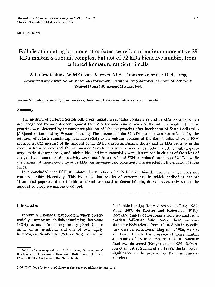
Because the antipeptide antiserum caused a more effective immunoprecipitation, this anti- serum was used in further experiments. Results of a time-course experiment in the absence or pres- ence of FSH are shown in Fig. 2. Immunoprecipi- tation after 2 and 4 h of chase showed the ap-
pearance of the 29 kDa protein in the medium of FSH-stimulated cells, which was stable between 5 and 24 h. In contrast, the small amount of im- munoprecipitated 32 kDa protein, presumably in-
hibin, was not affected by FSH. Reduction of the immunoprecipitated proteins yielded a 18 kDa
protein, presumably the inhibin a-subunit, and a low molecular weight radioactive protein in the
immunoprecipitate of the FSH-stimulated cells (Fig. 2, last two lanes). Finally, a 46 kDa radioac- tive protein was detected in the immunoprecipi- tates; the amount of this protein was increased after FSH stimulation. This protein disappeared with increasing chase period; its position did not change after reduction.
In order to obtain sufficient material for de- termination of immuno- and bioactivity, experi-
ments were performed with pooled CrSCCM and FrSCCM, collected over a 2-week period. In the concentrated CrSCCM both bioactive and im- munoactive inhibin were determined, resulting in
a B/I ratio of 3.4 k 0.2 (mean + SEM of four determinations). For the determination of the molecular weight of bioactive and immunoactive inhibin concentrated media from the first 2-week
pool of CrSCCM and FrSCCM were separated by SDS-PAGE. After slicing of the gel, a bioactive inhibin with an apparent molecular weight of 32
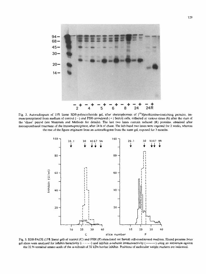
kDa was found in CrSCCM (Fig. 3). Inhibin (Y- subunit immunoreactivity was found between 25 and 35 kDa (total amount was 48 U/ml SCCM) and around 32 kDa 303 U/ml SCCM bioactive inhibin was detected. After the same procedure, 300 U/ml SCCM of 32 kDa bioactive inhibin was found in FrSCCM. The inhibin cY-subunit im- munoreactivity was increased in FrSCCM up to 294 U/ml SCCM, with a peak at 28-29 kDa (Fig. 3). Estimation of immunoreactive FSH in the eluates of the gel slices indicated that it was pres- ent at an apparent molecular weight of 38 kDa, but not in the slices in which inhibin bioactivity was found, indicating that the added FSH cannot interfere in the inhibin bioassay. Essentially the same results were found with the second pool of CrSCCM and FrSCCM and with a pool of SCCM stimulated with both FSH and testosterone (re- sults not shown). The proteins of the slices con- taining 28-29 kDa inhibin a-subunit immunoreac- tivity did not affect basal and inhibin-reduced
129
-*+ -4+ - + 5 -c3+ -t3+ - + 24
Fig. 2. Autaradio~~ of 15% linear S~S-~lyac~~a~de gel, after e~e~~ophoresis of [3sS]met~o~n~co~t~~ng proteins, im- munoprecipitated from medium of control (-) and FSH-stimulated ( + ) Sertoli cells, collected at various times (h) after the start of the ‘chase’ period (see Materials and Methods for details). The last two lanes contain reduced (R) proteins, obtained after mercaptoethanol treatment of the i~unopr~ipitate, after 24 h of chase. The left-hand two lanes were exposed for 2 weeks, whereas
the rest of the figure o~~nates from an autoradio~am from the same gel, exposed for 3 months.
80 80
60
I I I
20
I 1 I I
10 20 30 40
C slice number F
Fig. 3. SDS-PAGE (15% linear gel) of control (Cc) and FSN (F) stimulated rat Sertoli ceil-conditioned medium. Fluted proteins from gel slices were analyzed for inhibin bioactivity (- I - - - -) and inhibin a-subunit irnmunoactivity ( -) using an antiserum against
the 22 N-terminal amino acids of the a-subunit of 32 kDa bovine inbibin. Positions of molecular weight markers are indicated.

130
93 -
67 -
43 -
30 -
20.1 -
14.4-
MWM C
-_L
- 93
- 67
- 43
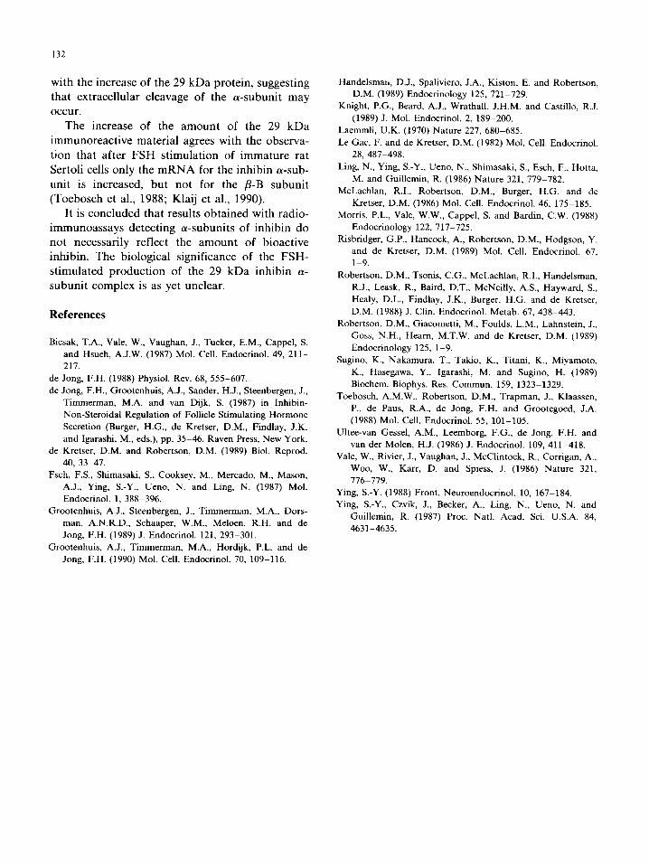
Fig. 4. Detection of inhibin o-subunits on Western blots with (+) or without (-) prior reduction with P-mercaptoethanol after
separation of control (C) and FSH (F) stimulated rat Sertoli cell-conditioned medium by 15% SDS-PAGE using an antiserum against
the 22 N-terminal amino acids of the o-subunit of 32 kDa bovine inhibin. Positions of molecular weight markers (MWM) are
indicated.
FSH release from cultured pituitary cells (data not shown).
Results of Western blots of the concentrated media are shown in Fig. 4. Two dominant im- munoreactive bands of 33.6 f 1.4 kDa (mean + SEM, n = 3 independent blots) and 28 * 1.7 kDa (mean k SEM, n = 3) were found in CrSCCM. In FrSCCM the 33.6 kDa band had the same inten- sity as in CrSCCM but the two smaller bands (29.6 k 0.700 kDa (mean k SEM, n = 2) and 28.4 + 0.9 kDa (mean _t SEM, n = 2) were much more prominent than in CrSCCM. After reduction of both CrSCCM and FrSCCM two prominent bands at 20.5 k 0.7 kDa (mean k SEM, n = 2) and 19.5 + 0.8 kDa (mean * SEM, n = 3) and a faint 46 kDa band were found.
Discussion
Two proteins with inhibin immunoreactivity were detected in the conditioned medium of cul- tured rat Sertoli cells, using each of three different
methods: immunoprecipitation, Western blotting and direct immunoassay in the eluates of slices of a gel, on which the conditioned medium was elec- trophorized. It is likely that the proteins, detected using the three methods, are identical: molecular weights, both before and after reduction, were the same and the amount of the lower molecular weight entity increased appreciably after FSH stimulation of the Sertoli cells, as detected using immunoprecipitation, Western blotting and direct radioimmunoassay.
One of these proteins is found at an apparent molecular weight of 32 kDa, and has both inhibin bioactivity and immunoactivity. This confirms earlier data of Grootenhuis et al. (1989, 1990) on the characterization of the molecular weight of inhibin in rat testis homogenates and CrSCCM, and of Risbridger et al. (1989), who showed the presence of immunoactive 30 kDa inhibin in a high performance liquid chromatography (HPLC) fraction of culture medium of seminiferous tu- bules, which also contained inhibin bioactivity.

131
The other protein is located at 29 kDa, interacts with antisera against the l-22 amino acid of the a-subunit of 32 kDa bovine inhibin and against a preparation of native bovine follicular fluid in- hibin, and has no inhibin bioactivity after elution from SDS-PAGE. This immunoreactivity was also
observed recently by Risbridger et al. (1989), who did not estimate bioactivity of inhibin in the
eluates obtained after SDS-PAGE. However, these authors found inhibin bioactivity in the HPLC
fractions which contained the 29 kDa protein, and ascribed the immune- and bioactivity to the same
molecule. Addition of FSH to the culture medium of
Sertoli cells caused a large increase of the im-
munoactivity in the medium, collected after 24 h and in pooled media collected during a 2-week period. This observation confirms earlier data of Ying et al. (1987), Bicsak et al. (1987) and Morris et al. (1988), who also used anti-peptide antisera, and of Toebosch et al. (1989) and Risbridger et al. (1989), who used antisera against native bovine inhibin. After SDS-PAGE fractionation of the proteins secreted by the stimulated Sertoli cells, increased secretion of a protein with inhibin im-
munoreactivity and an apparent molecular weight of 29 kDa was observed, whereas no clear increase
of the 32 kDa protein was found (Figs. 3 and 4). Comparable results were obtained after 24 h (data
not shown) and in the medium collected at 2- to 3-day intervals during 2 weeks, whereas the pulse- chase experiment also provided similar results. Estimation of the bioactivity and immunoactivity was only performed in the SDS-PAGE fractions of the media collected during 2 weeks, because only in these media sufficient material was present to yield reliable results. Again, the peak of the bioactivity was detected in the 32 kDa slice of the gel, and the total amount of inhibin bioactivity was not different from that in the slices of the gel on which CrSCCM was electrophorized. This ob- servation confirms earlier data of Ultee-van Ges- se1 et al. (1986), who did not observe increased inhibin bioactivity in the medium of FSH-stimu-
lated Sertoli cells cultured at 37 ‘C, but contrasts with the results of Le Gac and de Kretser (1982), Toebosch et al. (1988) and Handelsman et al. (1989), who did find increased inhibin bioactivity after culturing Sertoli cells with FSH at 37°C.
Risbridger et al. (1989) also observed increased release of inhibin bioactivity after addition of FSH to seminiferous tubule fractions in culture at 34OC. All of these authors observed decreasing bio- over immunoactivity ratios after addition of FSH, indicating that immunoactivity increased to
a larger extent than bioactivity. The reason for the discrepancy between the
present results - no increase in bioactivity after FSH - and the above-mentioned data remains
unclear. Differences in culture temperature and
cell purity may have played a role. Another possi-
bility is that the inhibin bioassay, based on the suppression of intracellular FSH, as used by all of the above-mentioned authors who observed an increase in i&bin bioactivity after addition of FSH, leads to results which differ from those based on suppression of the release of FSH, as used in this study and that reported by Ultee-van
Gessel et al. (1986). Finally, the discrepancy may have been caused by loss of presumed inhibin bioactivity of the 29 kDa protein during the pro-
cedure of electrophoresis, elution from the gel slices and methanol precipitation. The latter point can only be resolved after isolation of this entity using different procedures.
The molecular composition of the FSH-induced
29 kDa a-subunit immunoreactivity is not yet known. The presence of a 20 kDa inhibin a-sub-
unit immunoreactive band on Western blots after reduction indicates that it may consist of the
inhibin a-subunit linked to another peptide with disulphide bond(s). The observation of a 35S- labelled low molecular weight protein, detected after SDS-PAGE of the reduced immunoprecipi-
tated proteins, makes it likely that the FSH-in- duced 29 kDa inhibin a-subunit is the same mole-
cule as described by Sugino et al. (1989), who purified a 26 kDa inhibin a-subunit from porcine follicular fluid, which is composed of fragment 18-60 of the inhibin a-subunit precursor linked by disulphide bond(s) to fragment 227-360; frag- ment 18-60 contains one methionine residue, whereas four methionine residues are present in fragment 227-260 of rat inhibin (Esch et al., 1987). Finally, the 45 kDa immunoprecipitated protein, of which the secretion is also stimulated by FSH, might represent secretion of the in@bin a-subunit. The amount of this material decreases in parallel
132
with the increase of the 29 kDa protein, suggesting that extracellular cleavage of the a-subunit may occur.
The increase of the amount of the 29 kDa
immunoreactive material agrees with the observa- tion that after FSH stimulation of immature rat
Sertoli cells only the mRNA for the inhibin a-sub-
unit is increased, but not for the P-B subunit (Toebosch et al., 1988; Klaij et al., 1990).
It is concluded that results obtained with radio- immunoassays detecting a-subunits of inhibin do not necessarily reflect the amount of bioactive inhibin. The biological significance of the FSH- stimulated production of the 29 kDa inhibin LX- subunit complex is as yet unclear.
References
Bicsak, T.A., Vale, W., Vaughan, J., Tucker, E.M., Cappel, S.
and Hsueh, A.J.W. (1987) Mol. Cell. Endocrinol. 49, 211-
217.
de Jong, F.H. (1988) Physiol. Rev. 68, 555-607.
de Jong, F.H., Grootenhuis, A.J., Sander, H.J., Steenbergen, J.,
Timmerman, M.A. and van Dijk, S. (1987) in Inhibin-
Non-Steroidal Regulation of Follicle Stimulating Hormone
Secretion (Burger, H.G., de Kretser, D.M., Findlay, J.K.
and Igarashi, M., eds.), pp. 35-46, Raven Press, New York.
de Kretser, D.M. and Robertson, D.M. (1989) Biol. Reprod.
40, 33-47.
Esch, F.S., Shimasaki, S., Cooksey, M., Mercado, M., Mason,
A.J., Ying, S.-Y., Ueno, N. and Ling, N. (1987) Mol.
Endocrinol. 1, 388-396.
Grootenhuis, A.J., Steenbergen, J., Timmerman, M.A., Dors-
man, A.N.R.D., Schaaper, W.M., Meloen, R.H. and de
Jong, F.H. (1989) J. Endocrinol. 121, 293-301.
Grootenhuis, A.J., Timmerman, M.A., Hordijk, P.L. and de
Jong, F.H. (1990) Mol. Cell. Endocrinol. 70, 1099116.
Handelsmau, D.J., Spaliviero, J.A., Kiston, E. and Robertson,
D.M. (1989) Endocrinology 125, 721-729.
Knight, P.G., Beard, A.J., Wrathall, J.H.M. and Castillo, R.J.
(1989) J. Mol. Endocrinol. 2, 189-200.
Laemmli, U.K. (1970) Nature 227, 680-685.
Le Gac, F. and de Kretser, D.M. (1982) Mol. Cell. Endocrinol.
28,487-498.
Ling, N., Ying, S.-Y., Ueno, N., Shimasaki, S., Esch, F., Hotta,
M. and Guillemin, R. (1986) Nature 321, 779-782.
McLachlan, RI., Robertson, D.M., Burger, H.G. and de
Kretser, D.M. (1986) Mol. Cell. Endocrinol. 46, 175-185.
Morris, P.L., Vale, W.W., Cappel, S. and Bardin, C.W. (1988)
Endocrinology 122, 717-725.
Risbridger, G.P., Hancock, A., Robertson, D.M., Hodgson, Y.
and de Kretser, D.M. (1989) Mol. Cell. Endocrinol. 67,
l-9.
Robertson, D.M., Tsonis, C.G., McLachlan, RI., Handelsman,
R.J., Leask, R., Baird, D.T., McNeilly, A.S., Hayward, S.,
Healy, D.L., Findlay, J.K., Burger, H.G. and de Kretser,
D.M. (1988) J. Clin. Endocrinol. Metab. 67, 438-443.
Robertson, D.M., Giacometti, M., Foulds, L.M., Lahnstein, J.,
Goss, N.H., Heam, M.T.W. and de Kretser, D.M. (1989)
Endocrinology 125, 1-9.
Sugino, K., Nakamura, T., Takio, K., Titani, K., Miyamoto,
K., Hasegawa, Y., Igarashi, M. and Sugino, H. (1989)
Biochem. Biophys. Res. Commun. 159, 1323-1329.
Toebosch, A.M.W., Robertson, D.M., Trapman, J., Klaassen,
P., de Paus, R.A., de Jong, F.H. and Grootegoed, J.A.
(1988) Mol. Cell. Endocrinol. 55, 101-105.
Ultee-van Gessel, A.M., Leemborg, F.G., de Jong, F.H. and
van der Molen, H.J. (1986) J. Endocrinol. 109, 411-418.
Vale, W., Rivier, J., Vaughan, J., McClintock, R., Corrigan, A.,
Woo, W., Karr, D. and Spiess, J. (1986) Nature 321,
776-779.
Ying, S.-Y. (1988) Front. Neuroendocrinol. 10, 167-184.
Ying, S.-Y., Czvik, J., Becker, A., Ling, N., Ueno, N. and
Guillemin, R. (1987) Proc. Natl. Acad. Sci. U.S.A. 84,
4631-4635.