Embed Size (px)
Citation preview
Review
G protein-coupled receptors — recent advancesDorota Latek1#, Anna Modzelewska2#, Bartosz Trzaskowski2#, Krzysztof Palczewski3* and Sławomir Filipek2*
1Biomodeling Laboratory, International Institute of Molecular and Cell Biology, Warsaw, Poland; 2Biomodeling Laboratory, Faculty of Chemistry, University of Warsaw, Warsaw, Poland; 3Department of Pharmacology, School of Medicine, Case Western Reserve University, Cleveland, Ohio USA
The years 2000 and 2007 witnessed milestones in cur-rent understanding of G protein-coupled receptor (GPCR) structural biology. In 2000 the first GPCR, bovine rhodopsin, was crystallized and the structure was solved, while in 2007 the structure of β2-adrenergic receptor, the first GPCR with diffusible ligands, was determined ow-ing to advances in microcrystallization and an insertion of the fast-folding lysozyme into the receptor. In parallel with those crystallographic studies, the biological and biochemical characterization of GPCRs has advanced considerably because those receptors are molecular tar-gets for many of currently used drugs. Therefore, the mechanisms of activation and signal transduction to the cell interior deduced from known GPCRs structures are of the highest importance for drug discovery. These proteins are the most diversified membrane receptors encoded by hundreds of genes in our genome. They participate in processes responsible for vision, smell, taste and neuronal transmission in response to photons or binding of ions, hormones, peptides, chemokines and other factors. Although the GPCRs share a common seven-transmembrane α-helical bundle structure their binding sites can accommodate thousands of different ligands. The ligands, including agonists, antagonists or inverse agonists change the structure of the receptor. With bound agonists they can form a complex with a suitable G protein, be phosphorylated by kinases or bind arrestin. The discovered signaling cascades invoked by arrestin independently of G proteins makes the GPCR activating scheme more complex such that a ligand act-ing as an antagonist for G protein signaling can also act as an agonist in arrestin-dependent signaling. Addition-ally, the existence of multiple ligand-dependent partial activation states as well as dimerization of GPCRs result in a ‘microprocessor-like’ action of these receptors rather than an ‘on-off’ switch as was commonly believed only a decade ago.
Key words: G protein-coupled receptors, rhodopsin, β-adrenergic receptors, chemokine receptors, G protein, arrestin.
Received: 20 November, 2012; revised: 05 December, 2012; accepted: 10 December, 2012; available on-line: 18 December, 2012
InTRoduCTIon
In 2012 the Nobel Prize in chemistry was awarded jointly to two American scientists — Robert J. Lefkow-itz of Duke University in Durham, North Carolina and Brian K. Kobilka of Stanford University School of Med-icine in Palo Alto, California “for studies of G protein-coupled receptors (GPCRs)”. The scientific achievements
of Lefkowitz and Kobilka have allowed the pharmaceu-tical industry to develop more selective drugs with the hope of fewer side effects through improved under-standing of the signaling mechanisms initiated by en-dogenous substances such as adrenalin, serotonin, hista-mine, dopamine and many other hormones and neuro-transmitters. Lefkowitz started his work on GPCRs by identifying adrenergic receptors (α and β) affected by adrenaline in the 1980s (Shorr et al., 1981; Dixon et al., 1986). With the help of his student, Kobilka, he noticed that the genes for β-adrenergic receptors shows remark-able similarities to the one for rhodopsin, known then as a light-sensing receptor of the eye (Filipek et al., 2003; Palczewski, 2006), and concluded that there likely exists an entire protein family of such receptors with similar structure and function (Lefkowitz, 2000). Another key advance in Lefkowitz’s research was identification of the β-adrenergic receptor kinase (Benovic et al., 1986) fol-lowed by studies on β-arrestins (Luttrell & Lefkowitz, 2002; Lefkowitz & Shenoy, 2005). Continuing his work at Stanford, Kobilka together with Stevens from the Scripps Research Institute in California solved the crystal structure of β2-adrenergic receptor stabilized by lysozyme (Cherezov et al., 2007) and together with Schertler from MRC (Cambridge, UK) the structure of the same recep-tor stabilized by an antibody in the same year (Rasmus-sen et al., 2007). The structure of the second GPCR (af-ter vertebrate rhodopsin (Palczewski et al., 2000)) con-firmed the hypothesis about the common folding of all GPCRs comprising a seven-transmembrane helical bun-dle (Fig. 1). The structure of β2-adrenergic receptor was followed by several crystal structures of other GPCRs solved by e.g. the Stevens’ group (Jaakola et al., 2008). Those investigators employed lysozyme, apocytochrome BRIL, and nanobody molecules (Cherezov, et al., 2007) to stabilize not only the inactive state of these highly flexible receptors but also their extremely unstable active state in complex with trimeric G protein (Rasmussen et al., 2011). Other investigators successfully employed
*e-mail: Sławomir Filipek e-mail: [email protected]; Krzysz-tof Palczewski e-mail: [email protected]#These authors contributed equally to this workAbbreviations: A2AR, adenosine A2A receptor; BRIL, apocy-tochrome b(562)RIL; CRs, chemokine receptors; CXCR4, chemokine receptor CXCR4; D3R, dopamine D3 receptor; EC, extracellular; ECL, extracellular loop; GPCRs, G protein-coupled receptors; GRK, G protein-coupled receptor kinase; H1R, histamine H1 receptor; HBD, hormone-binding domain; IC, intracellular; ICL, intracellular loop; M2R, M3R, muscarinic M2 and M3 receptors; MCH, melanin-con-centrating hormone; NOP, nociceptin opioid receptor; OR, opioid receptor; PDB, protein data bank; PDE, phosphodiesterase; PKA, protein kinase A; SOG, somatostatin, opioid and galanin receptors; TM, transmembrane; 7TMH, seven transmembrane helices
Vol. 59, No 4/2012515–529
on-line at: www.actabp.pl
516 2012D. Latek and others
thermo-stabilizing mutations and truncations (Tate & Schertler, 2009; Lebon et al., 2012).
Seven-transmembrane spanning GPCRs are critically involved in transmitting extracellular stimuli including light, hormones and neurotransmitters into specific cellu-lar responses (Muller et al., 2008). GPCRs are also phar-macologically important because they are the targets of about 30% of commercially available drugs (Salon et al., 2011). The structure of vertebrate rhodopsin determined in 2000 marked the first three-dimensional atomic struc-ture of any native GPCR. Significant advances in the field include the structural determination of truncated invertebrate rhodopsin, adrenergic, histamine H1, adeno-sine A2A, opioid, muscarinic acetylcholine, CXCR4, do-pamine D3 and other receptors as well. In many cases GPCRs were modified with fusion proteins such as lysozyme. Whereas non-rhodopsin GPCRs require het-erologous expression and extensive protein engineering to enable their stabilization and crystallization, bovine rhodopsin remains the only native, intact GPCR with a determined structure (Palczewski, 2012). GPCRs contain the seven-transmembrane helical bundle that provides a binding site for their ligands. The ligand binding triggers a slight change in GPCR conformation that is propagat-ed through the whole protein, ultimately causing altera-tions at the receptor’s cytoplasmic surface that permit binding to its cognate G protein. Today, crystal struc-tures of all photoactivated intermediates of rhodopsin and several agonist- and antagonist-bound GPCRs have been determined (Palczewski, 2012). Moreover, the criti-cally important GPCR-G protein complex structure also has become better characterized by low- and high-res-olution experimental methods (Jastrzebska et al., 2011b; Rasmussen, et al., 2011; Orban et al., 2012). Research on GPCRs still remains of a high priority. Hopefully, know-ing better the connection between structure and function of GPCRs will improve our understanding of these mo-lecular machines as well as will promote their potential pharmacological applications.
GPCRs comprise seven transmembrane α-helices (7TMH) connected by three extracellular loops (ECL1-3) and three intracellular loops (ICL1-3) (Fig. 1). The ex-tracellular (EC) region, which is responsible for ligand binding, also includes the N-terminus that can range from relatively short sequences in rhodopsin-like recep-tors to large extracellular domains in other classes of GPCRs, e.g. the hormone-binding domain (HBD) in ad-hesion receptors. The intracellular (IC) region interacts with G proteins, arrestins and other downstream effec-tors. It includes cytoplasmic helix H8 and a C-terminus that may provide sites for palmitoylation. The 7TM heli-cal bundle contains a number of kinks (Fig. 1), mostly induced by Pro residues, that roughly divide the receptor into the EC and IC regions. The EC module respon-sible for ligand binding features a high structural diver-sity but small movement during activation. In contrast, the IC region, which is involved in binding downstream proteins including G proteins and arrestins, is more con-served in the GPCR family but is subjected to much larger conformational changes upon receptor activation than EC (Katritch et al., 2012).
The structures of GPCRs exhibit only limited differ-ences despite the fact that the receptors were crystallized differently: in different crystal packing orientations, with different antagonists and inverse agonists bound and also in two different activation states, either activated (agonist bound) or inactivated (antagonist or inverse-ag-onist bound) (Rasmussen et al., 2011). Even the confor-mations of extracellular loops (the most divergent part
of GPCR structures) can also be similar as is the case of crystal structures of the following receptors: dopamine receptor D3R (Chien et al., 2010), muscarinic receptors M2R (Haga et al., 2012) and M3R (Kruse et al., 2012), CXCR4 (Wu et al., 2010), µOR (Manglik et al., 2012), δOR (Granier et al., 2012), κOR (Wu et al., 2012), and nociceptin receptor (NOP) (Thompson et al., 2012). In the more distant (by sequence homology) CXCR4 and opioid receptors, ECL2 forms a β-hairpin which is ori-ented nearly vertically to the membrane surface which is much different than that of rhodopsin, which is ori-ented horizontally and entirely covers the retinal. How-ever, the ECL2 in rhodopsin is responsible for keeping the ligand in place, whereas in CXCR4 it is crucial for binding of either the small molecule IT1t, or the pep-tide antagonist CVX15 (they are both ligands in crystal structures of that receptor) that mimics the V3 loop of the HIV envelope glycoprotein gp120. In CXCR4 and opioid receptors, the ligand pocket is much larger than in other solved GPCR structures, and binding of peptide antagonists involves extensive interactions with ECL2. The N-terminus of GPCRs probably also participates in the binding of larger ligands, however, to date its struc-ture has only been resolved in rhodopsin and partially in CXCR4 and also in crystallized peptide neurotensin receptor (White et al., 2012) which is the first receptor structure in the β subfamily of rhodopsin-like GPCRs. Another important region in a GPCR structure, the cy-toplasmic helix H8, is anchored to the membrane by palmitoylation and this feature is essential for receptor stabilization and its proper functioning (Maeda et al., 2010). In a group of known GPCR structures, only in the case of CXCR4 and neurotensin receptor the helix H8 shows a disordered behavior. Nevertheless, it does not preclude the possibility of its formation in the cell membrane because this helix is present in all crystallized opioid receptor structures from the same g subfamily of rhodopsin-like GPCRs as CXCR4.
Our current understanding of the GPCR functioning has changed from a simple hypothesis of ‘on-off’ switch-es to the microprocessor-like action (Kenakin & Miller, 2010). Especially the phenomenon called ‘functional selectivity’, whereby certain ligands initiate only por-tions of the signaling mechanisms mediated by a given receptor, has opened new horizons for drug discovery. What should be discovered in the nearest future is a new receptor-ligand behavior with quantification of the drug effect in such complex systems. For example, some ago-nists selectively activate cellular pathways associated with a specific cell type and some antagonists actively induce receptor internalization without its activation. There are
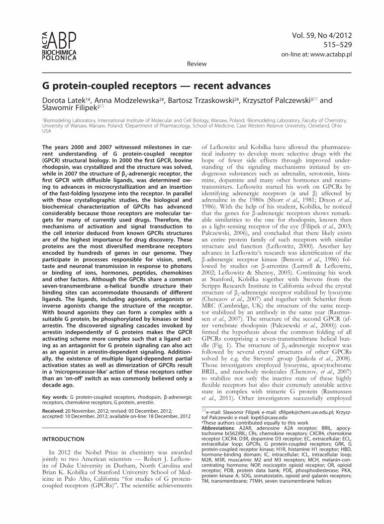
Figure 1. A scheme of shapes and tilts of transmembrane heli-ces of GPCRs based on the representative crystal structure of β2-adrenergic receptor (PdB id: 2RH1). Location of a ligand is marked by a red sphere whereas the loca-tion of protein G by a blue sphere.
Vol. 59 517G protein-coupled receptors
also allosteric modulators which can be linked to the co-binding ligands. Agonists are now known to have multi-ple efficacies that are associated with selected signaling pathways coupled to the receptor. So called ‘functional selectivity’, defined as biased agonism and biased antago-nism, is especially interesting in terms of its mechanism and potential therapeutic applications (see an extensive review by Park and colleagues (2008)).
ClAssIFICATIon oF GPCRs
Human GPCRs form a large family of about 800 membrane receptors with sequence lengths between 289 (Mas-related GPCR — uniprot ref. no. Q86SM5) and 3312 (EGF-like protein 1 — uniprot ref. no. Q9NYQ7) residues (Nov. 2012, reviewed Uniprot entries only), with most GPCRs consisting of 300-500 amino acid resi-dues (Mirzadegan et al., 2003). The disparity of sequence lengths is mainly due to the extracellular portion of GP-CRs involved in ligand recognition and/or cell signal-ing (Fig. 1) such as the N-terminus, extracellular loop 2 (ECL2) and ECL3 (Mirzadegan et al., 2003). The intra-cellular region of GPCRs, less varied in length than the extracellular part, is involved in signal transduction by in-teractions with G protein and arrestin. Despite the high degree of sequence variability (Trzaskowski et al., 2012) GPCRs share a common 7TMH core of a size exactly fitted to the cell membrane thickness. Interestingly, the transmembrane helices of GPCRs are frequently tilted with varied tilt and rotation angles that depend not only on the receptor type but also on its activation state. Un-fortunately, precise prediction of kinks in the TMHs of GPCRs is still limited as these helical deformations can-not be explained only by the presence of the well-known helix-breakers such as Pro, Ser, Thr or Gly residues. Most likely these deformations are introduced by tertiary interactions which are difficult to capture without a 3D structure of the receptor (Yohannan et al., 2004; Meruelo et al., 2011). In the most commonly used GRAFS classi-fication system (Schioth & Fredriksson, 2005) the GPCR superfamily is divided into five main families: glutamate (former class C), rhodopsin (former class A), adhesion (part of former class B), frizzled/taste2 (former class F), and secretin (part of former class B). Other former class-es D (fungal mating pheromone receptors) and E (cyclic AMP receptors) do not contain human receptors and they are not included in the GRAFS classification. So far, only members of the rhodopsin-like receptor fam-ily have been crystallized. As of November 2012 there are 15 unique GPCRs deposited through nearly 60 en-tries in the Protein Data Bank (PDB) involving a variety of ligands, activation states and point mutations. There also have been a few attempts to study GPCR structures by NMR in the ‘membrane-mimicking’ environment of phospholipid bilayers, e.g. CXCR1 by Park et al. (2012), but the resolution of such structures might be still too low to use them for drug discovery.
The rhodopsin family can be further divided into four subfamilies: α, β, γ and δ according to the clas-sification of Fredriksson et al. (2003). The α subfamily has five main branches: prostaglandin, amine, opsin, me-latonin and adenosine receptors. Currently in the PDB there are crystal structures of amine receptors (hista-mine H1R, dopamine D3R, muscarinic M2R and M3R, β1- and β2-adrenergic receptors), opsins (rhodopsin), adenosine A2AR and lipid S1P1R receptors). The one-branch β subfamily includes hypocretin receptors, neuro-peptide FF, tachykinin, cholecystokinin, neuropeptide Y,
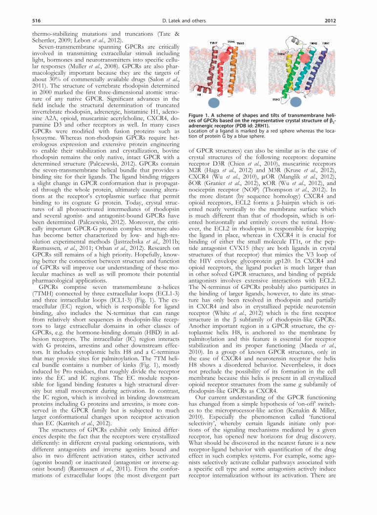
endothelin-related, gastrin-releasing peptide, neuromedin B, uterinbombesin, neurotensin, growth hormone secre-tagogue, neuromedin, thyrotropin releasing hormone, ghrelin, arginine vasopressin, gonadotropin-releasing hormone, oxytocin and orphan receptors. In the β sub-family only neurotensin receptor has been crystallized so far. The γ subfamily consists of three main branches: SOG receptors (including crystallized µOR, δOR, κOR and nociceptin opioid receptors), MCH receptors, and chemokine receptors (including crystallized CXCR4). The last δ subfamily of rhodopsin-like GPCRs has four main branches: Mas-related (oncogene) receptors, glycoprotein receptors, nucleotide receptors and olfactory receptors. However, the δ subfamily has no representative in the PDB so far and only the P2Y12 nucleotide receptor has been selected for crystallization in the near future by the Stevens' group (see http://gpcr.scripps.edu/tracking_sta-tus.htm). The above classification of the rhodopsin fam-ily is still under discussion as other methods have pro-vided different shapes for its phylogenetic tree (Surgand et al., 2006; Deville et al., 2009). For example, Pele et al. (2011) split the rhodopsin family into only four subfami-lies: G0 — peptide receptors, opsin and melatonin re-ceptors; G1 — somatostatin, opioid, chemokine and nu-cleotide receptors; G2 — amine and adenosine receptors; and G3 — including melanocortin, S1P and cannabinoid receptors, leucine-rich repeat (LRR)-containing receptors, prostaglandin and Mas-related receptors. The Pele’s clas-sification is not fully consistent with the previous one from Frederiksson et al. as members of G0 are included in both α and β subfamilies, G1 is split between δ and γ, G2 — only α, and finally G3 corresponds to mem-bers of both α and δ subfamilies. Even if two GPCRs are classified as members of the same subfamily, they can significantly differ in their amino acid composition (see Fig. 2). A notable exception is the highly populated group of olfactory receptors belonging to the δ subfam-ily in which most sequences are similar to each other (the two highest peaks in the δ subfamily sequence iden-tity histogram in Fig. 2). In general, sequence diversity is the highest within the extra- and intracellular loop re-gions, whereas the 7TMH core contains well conserved fragments (motifs) characteristic of GPCRs, for exam-ple: ‘D/ERY’ (TMH3), ‘CwxP’ (TMH6) and ‘nPxxy’ (TMH7). The high sequence diversity inside the rhodop-sin family corresponds to the high diversity of kinks and bulges in the TM helices and distinct conformations of
Figure 2. Histograms of sequence identity between members of four branches of rhodopsin-like family of GPCRs.
518 2012D. Latek and others
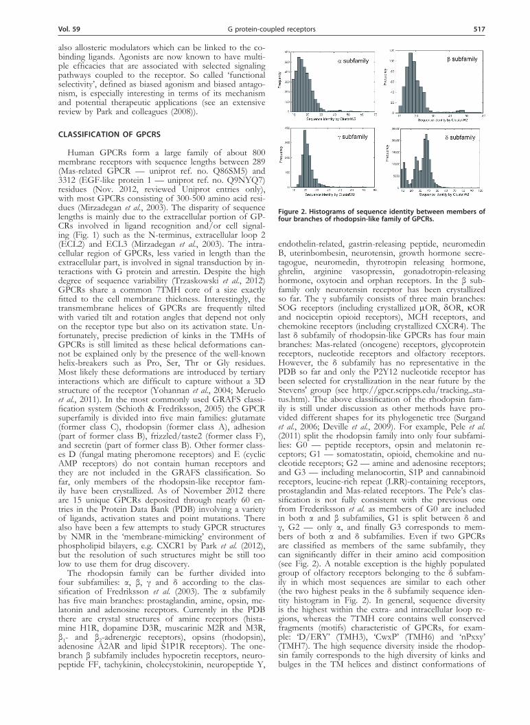
loops. Even for members of the same subfamily (such as rhodopsin and β2-adrenergic receptor (subfamily α) presented in Fig. 3), their structural diversity still makes homology modeling challenging.
CRysTAllIzATIon oF THe FIRsT GPCRs — RHodoPsIn And β2-AdReneRGIC ReCePToR
The first X-ray crystal structure of any GPCR was that of ground-state rhodopsin (Palczewski, et al., 2000). Refinement of crystallization conditions yielded higher resolution data, extending the model to 2.2 Å, the highest resolution of all rhodopsin structures de-termined to date (Okada et al., 2004). The first struc-ture of a diffusible ligand-responsive GPCR resulted from work of Kobilka and Stevens, who reported the crystal structure of a human β2-adrenergic receptor-T4 lysozyme fusion protein bound to the partial inverse agonist carazolol at 2.4 Å resolution (Cherezov et al., 2007; Rosenbaum et al., 2007). Several structures of mutant and fusion GPCRs followed (Mustafi & Pal-czewski, 2009) culminating with a 1.8 Å resolution structure of an engineered human A2A adenosine re-ceptor with its third intracellular loop replaced with apocytochrome b(562)RIL (Liu et al., 2012). This is the highest resolution structure of any GPCR with well-defined Na+ ions and water molecules. Wa-ter molecules play critical roles in the GPCR activa-tion process by stabilizing intramolecular interactions (Wikstrom et al., 2003; Osyczka et al., 2005; Garczarek & Gerwert, 2006). Observed in key structurally sen-sitive areas of many GPCRs, water molecules along with amino acid side chains can form a signal trans-mission network extending from the ligand-binding site to the cytoplasmic surface (Angel et al., 2009a; 2009b; Orban et al., 2010). Despite their low sequence similarities, the overall folds of structurally deter-mined GPCRs are remarkably similar. For all rhodop-sin structures, RMSDs for transmembrane regions are within 1.8 Å, and when adrenergic receptors are com-pared with rhodopsin, these deviations are 3.3–3.5 Å for the β2-adrenergic receptor and 4.3–4.7 Å for the
β1-adrenergic receptor (Jaakola, et al., 2008; Lodowski et al., 2009). Remarkably, preservation of only a few essential regions of a GPCR is required for activation of its cognate G protein (Mirzadegan et al., 2003).
Rhodopsin in its inactive, ground state undergoes a series of photointermediate steps upon absorption of a photon and isomerization of its chromophore 11-cis-retinylidene. These photointermediate states ex-hibit unique absorption maxima and can be isolated by trapping alone or with a G protein-derived peptide analog. Ultimately, the photoisomerized chromophore is hydrolyzed and released from the binding pocket yielding opsin and free all-trans-retinal (Jastrzebska et al., 2011a). Our laboratory expanded upon this work with the first photoactivated rhodopsin structure which exhibits spectral qualities of Meta II, the acti-vated state (Salom et al., 2006; Lodowski et al., 2007). For rhodopsin, only the Meta II intermediate is ca-pable of activating Gt and it differs chemically from other photo-intermediates only by deprotonation of the Schiff base and uptake of a proton from bulk sol-vent (Salom et al., 2006). By now, the structures of most photo-intermediates have been solved by X-ray and electron crystallographic methods (Breitman et al., 1989; Park et al., 2008; Lodowski et al., 2009). In-terestingly, the structures of those rhodopsin photo-intermediates did not exhibit large-scale movements of entire helices. Rather they showed that photoacti-vation was accomplished with just small-scale, local changes propagated to the cytoplasmic loops, especial-ly the ends of helices V and VI. The 2–8 Å structural shift observed for GPCRs upon activation suggests that such subtle changes directly lead to different re-ceptor activities or indirectly affect the key residues, e.g. the D(E)RY motif on the cytoplasmic surface re-sponsible for the efficacy of G protein coupling along with further conformational changes. This observation is consistent with activation of other GPCRs (summa-rized by Sprang (2011)).
Structures of opsin revealed marked similarities to photoactivated rhodopsin (Topiol & Sabio, 2009). An additional structure of a photoactivated rhodopsin, ob-tained by regenerating opsin crystals with all-trans-retinal, superposed well with opsin structures and is spectrally indistinguishable from either our photoactivated rhodop-sin structure or Meta II in solution (Choe et al., 2011) (summarized in (Breitman et al., 1989)). Moreover, struc-tures of constitutively active mutants of rhodopsin have also been reported (Standfuss et al., 2007; Standfuss et al., 2011; Xie et al., 2011; Deupi et al., 2012), revealing changes around helices V and VI as compared with rho-dopsin that are consistent with proposed models. When one considers the agonist-bound GPCR structures, it becomes readily apparent that the dynamics of the mol-ecule (which make it both a difficult structural target and play a key role in its activation) are recapitulated. The inherent flexibility of GPCRs permits dynamic and conformational changes triggered by only a fraction of the energy derived from ligand binding. Because various agonists can bind to a GPCR leading to varying levels of activity, the small changes induced by agonist bind-ing must account for the differences in the efficacy of such ligands in activating a given G protein (Rosenbaum et al., 2009), suggesting that interaction with signaling proteins may induce further changes on the cytoplasmic surface of these receptors (Sprang 2011). NMR studies of GPCRs, particularly rhodopsin, further advanced and refined those activation models (Smith 2010; 2012; Struts et al., 2011; Eilers et al., 2012).
Figure 3. Crystal structures of rhodopsin (PdB id: 1F88) and β2-adrenergic receptor (PdB id: 2RH1). Top, view along the membrane plane, bottom, from the extracel-lular side.
Vol. 59 519G protein-coupled receptors
InTeRACTIons WITH G PRoTeIn And ARResTIn — PAssInG on THe sIGnAl
Activation of a GPCR triggers binding of the asso-ciated heterotrimeric G protein and nucleotide (GDP) release from its α-subunit. This G protein activation is required for subsequent activation of the cascade of re-actions, processes required to advance stepwise signal transduction. The G protein is released from the GPCR by GTP and both its α- and βγ-subunits can activate the effector molecules as adenylyl cyclases and cation chan-nels. Rather than to photon (as in rhodopsin), most GP-CRs respond to molecules called ligands that upon bind-ing to a particular GPCR cause a ligand-specific cellular response. The signal is attenuated by receptor phospho-rylation and binding of a capping protein arrestin.
G protein
Heterotrimeric G proteins are composed of a nucle-otide-binding α-subunit (Gα) and a dimer consisting of the β- and γ-subunits (Gβγ). In their inactive form, Gα-subunits are bound to GDP and tightly associated with Gβγ. Interactions of β2-adrenergic receptor and Gs (the stimulatory G protein that activates adenylyl cyclase) formed the foundation of the ternary complex model of GPCR activation (Ross et al., 1977; De Lean et al., 1980). In the ternary complex consisting of agonist, re-ceptor and G protein, the affinity of the receptor for the agonist is enhanced and the specificity of the G protein for guanine nucleotides changes in favor of GTP over GDP. Agonist binding to the receptor promotes interac-tions with the GDP-bound Gsαβγ heterotrimer, leading to the exchange of GDP for GTP and the functional dissociation of G protein into Gα-GTP and Gβγ sub-units. These separate subunits can modulate the activity of different cellular effectors (channels, kinases or other enzymes). The intrinsic GTPase activity of Gα leads to hydrolysis of GTP to GDP and the re-association of Gα-GDP and Gβγ subunits with termination of signal-ing. The active state of a GPCR can be defined as that conformation that couples to and stabilizes a nucleotide-free G protein. In the agonist-β2-adrenergic receptor–Gs ternary complex, Gs has a higher affinity for GTP than for GDP, and the β2-adrenergic receptor has a rough-ly 100-fold higher affinity for agonists than does β2-adrenergic receptor alone (Rasmussen et al., 2011).
The α-subunit (Gα) of heterotrimeric G proteins mediates signal transduction in a variety of cell signal-ing pathways. These α-subunits can be divided into four families: Gαs, Gαi/Gαo, Gαq/Gα11, and Gα12/Gα13. Each family comprises various members that often show spe-cific expression patterns. The βγ-complex of mamma-lian G proteins is assembled from a repertoire of five G protein β-subunits and twelve γ-subunits (Wettschu-reck & Offermanns, 2005). Most GPCRs are able to activate more than one G protein subtype. Therefore, the activation of a GPCR usually results in the activa-tion of several signal transduction cascades via G protein α-subunits as well as through the freed βγ-complex. G proteins of the Gi/Go family are widely expressed and have been shown to mediate receptor-dependent inhibi-tion of various types of adenylyl cyclases (Sunahara et al., 1996). Because the expression levels of Gi and Go are relatively high, their receptor-dependent activation results in the release of relatively high amounts of βγ-complexes. Activation of Gi/Go is therefore believed to be the major coupling mechanism that results in the acti-vation of βγ-mediated signaling (Clapham & Neer, 1997;
Robishaw 2004). The Gq/G11 family of G proteins cou-ples receptors to β-isoforms of phospholipase C (Exton 1996; Rhee 2001). The G proteins G12 and G13 are often activated by receptors coupling to Gq/G11 (Strathmann & Simon, 1990; Dhanasekaran & Dermott, 1996). Analy-sis of cellular signaling processes regulated through G12 and G13 has been difficult because specific inhibitors of these G proteins are not available. In addition, G12/G13-coupled receptors usually activate other G proteins as well. The ubiquitously expressed G protein Gs couples many receptors to adenylyl cyclase and mediates activa-tion increasing intracellular cAMP concentration (Beavo & Brunton, 2002; Chin et al., 2002). β-adrenergic recep-tors couple primarily to Gs. The cAMP produced in re-sponse to Gs activation directly modulates the gating of hyperpolarization-activated, cyclic nucleotide-gated chan-nels and activates protein kinase A (PKA). PKA in turn phosphorylates many proteins involved in excitation-con-traction coupling including L-type Ca2+ channels, phos-pholamban, and/or troponin I (Bers 2002). Rhodopsin is coupled to rod-transducin (Gt, a homolog of Go), a member of the Gαi/Gαo family. Transducin couples the receptor in a stimulatory manner to cGMP-phosphodies-terase (PDE) by binding and sequestering the inhibitory γ-subunit of the retinal type 6 PDE (PDE6). Activation of PDE lowers cytosolic cGMP levels leading to a de-creased probability of cGMP-regulated cation channels in the plasma membrane being open, which eventu-ally causes hyperpolarization of photoreceptor cells (Ar-shavsky et al., 2002).
The signal captured by photoactivated rhodopsin or an agonist-occupied GPCR propagates along a cell’s plasma membrane to activate a G protein located 40 Å or more away to cause a cellular response. Perhaps our most advanced understanding of this activation process is derived from rhodopsin and the visual system. Ab-sorption of a photon triggers a change in the confor-mation of rhodopsin’s bound retinal chromophore which is propagated through this receptor, ultimately causing alterations at the cytoplasmic surface that permits bind-ing of G protein transducin, its cognate G protein. This triggers nucleotide release from G protein and its sub-sequent activation, processes required to advance visual signal transduction. Most GPCRs respond to molecular signals in the form of ligands which upon binding elicit a ligand-specific cellular response. For most GPCRs, the ligand-binding site coincides with the retinylidene-bind-ing pocket in rhodopsin. Thus, ligand binding causes similar conformational changes as those triggered by chromophore photoisomerization in rhodopsin, and the remaining molecular mechanisms for signal transduction are similar for all GPCRs. A model for the photoactivat-ed rhodopsin-G protein complex was described as a 22 Å low-resolution structure from single particle analysis (Jastrzebska et al., 2011b). Its molecular envelope is con-sistent with dimeric rhodopsin molecules together with one G protein heterotrimer, yielding a 2:1 molar ratio of photoactivated rhodopsin to G protein (Jastrzebska et al., 2011b). The heteropentameric structure for this complex was obtained from native proteins, both rhodopsin and G protein (see also (Jastrzebska et al., 2006)).
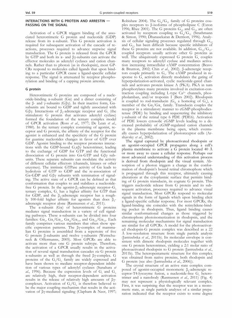
The crystal structure of an active state complex com-posed of agonist-occupied monomeric β2-adrenergic re-ceptor-T4-lysozyme fusion, a nucleotide-free Gs hetero-trimer and a nanobody (Rasmussen et al., 2011) (Fig. 4) may not represent a physiologically relevant complex. First, it was surprising that the receptor was in a mono-meric state, as single particle analyses of a similar prepa-ration indicated that the receptor exists to some degree
520 2012D. Latek and others
in a dimeric form in solution (Westfield et al., 2011). Second, it was unexpectedly observed that the entire α-helical domain (AH) of Gα was so largely displaced relative to the Ras-like GTPase domain (Rasmussen et al., 2011). This displacement could result from the nano-body stabilization of the β2-adrenergic receptor-Gs com-plex in the crystal or from the crystallization conditions used. But these discrepancies cannot be explained by dif-ferences in the inherent structure of either this GPCR (as mentioned earlier) or its G protein. Both the visual G protein and Gs display a relatively small root-mean-square deviation (~0.5–1.0 Å) between structures for the all three subunits. Future research should elucidate this discrepancy.
A wide range of different types of studies (Tesmer, 2010) have provided details of the conformational states of Gα, but the mechanism by which the GPCR interac-tion leads to release of the bound GDP from the Gα subunit and the structure of the resulting empty complex remain a major target for research in the field. The Gα subunit has two structural domains, namely a nucleo-tide binding or ras-like domain (Ras) and an α-helical domain (AH) that partially occludes the bound nucleo-tide. Numerous studies indicate that the C-terminus of Gα is bound tightly to the receptor in the nucleotide-free complex (Oldham et al., 2006). In addition, the N-terminal helix of Gα is associated with Gβγ and with the membrane via N-terminal myristoylation (Resh, 1999). Together, these constraints fix the position of the nucleotide domain with respect to the membrane. The helical domain is connected to the ras-like domain through two flexible linkers. The receptor-catalyzed nu-cleotide exchange in G proteins suggested a large-scale reorientation of domains in the α-subunit (Van Eps et al., 2011; Westfield, et al., 2011). As part of that, bind-ing to a GPCR requires a movement of two helices in Gα, the so called αN and α5 helices at the Gα N- and C-terminus, respectively. A possible sequence of interac-tions during formation of the nucleotide-free complex has been proposed for the β2-adrenergic receptor-Gs structure (Rasmussen et al., 2011). The first interaction of the β2-adrenergic receptor with the Gs heterotrimer would require a movement of the α5-helix to permit in-teractions with the β2-adrenergic receptor. The formation of more extensive interactions between the receptor and the amino terminus of Gαs requires a rotation of GαsRas relative to the receptor. This is associated with further
conformational changes in both the β2-adrenergic recep-tor and GαsRas. One cannot say when GDP is released during the formation of the complex. However, it is sug-gested that the GDP release precedes the uncoupling of the two Gαs domains.
Although much progress has been made in under-standing how Gα subunits interact with and regulate the activity of their downstream targets, it is less clear how activated GPCRs initiate this process by catalyzing nucleotide exchange on Gαβγ. The question is of great importance because it represents an essential, pharma-cologically relevant interaction that can regulate nearly all aspects of eukaryotic cell physiology. Moreover, an atomic-resolution understanding will explain the GPCR functional selectivity, namely the ability of different ago-nists to elicit distinct downstream effects from a single GPCR.
Arrestin
Additional knowledge about GPCRs and their interac-tions with desensitizing proteins has emanated from vis-ual research (Kuhn & Dreyer, 1972; Kuhn, 1974; Wey-and & Kuhn, 1990). Photoactivated rhodopsin is spe-cifically phosphorylated by G protein-coupled receptor kinases (GRKs) (Maeda et al., 2003; Singh et al., 2008) and preferential binding of arrestin to activated rhodop-sin blocks further G protein activation (Palczewski et al., 1991; Freedman & Lefkowitz, 1996). Lefkowitz, as many other investigators, was interested in the mecha-nism of signal termination by GPCRs. He demonstrated that other GPCRs are phosphorylated in an agonist-spe-cific manner and that the phosphorylated receptors bind β-arrestin (Lohse et al., 1990), revealing a mechanism of receptor desensitization shared among rhodopsin and most GPCRs.
Mammals have four arrestin subtypes that demon-strate over 50% amino acid conservation and similar structures in their basal state. Arrestin-1 (also known as visual or rod arrestin) and arrestin-4 (cone arrestin) are predominantly expressed in photoreceptors, whereas ar-restin-2 and -3 (also known as β-arrestin-1 and -2) are present in virtually every cell in the body with the great-est expression in mature neurons (Palczewski, 1994). Non-visual arrestins bind the great majority of GPCRs found in different mammalian species (Xiao et al., 2007). Visual arrestin shows high selectivity for its cognate re-ceptor rhodopsin, even though several other proteins have been identified to be bound by this arrestin subtype (Gurevich et al., 2011).
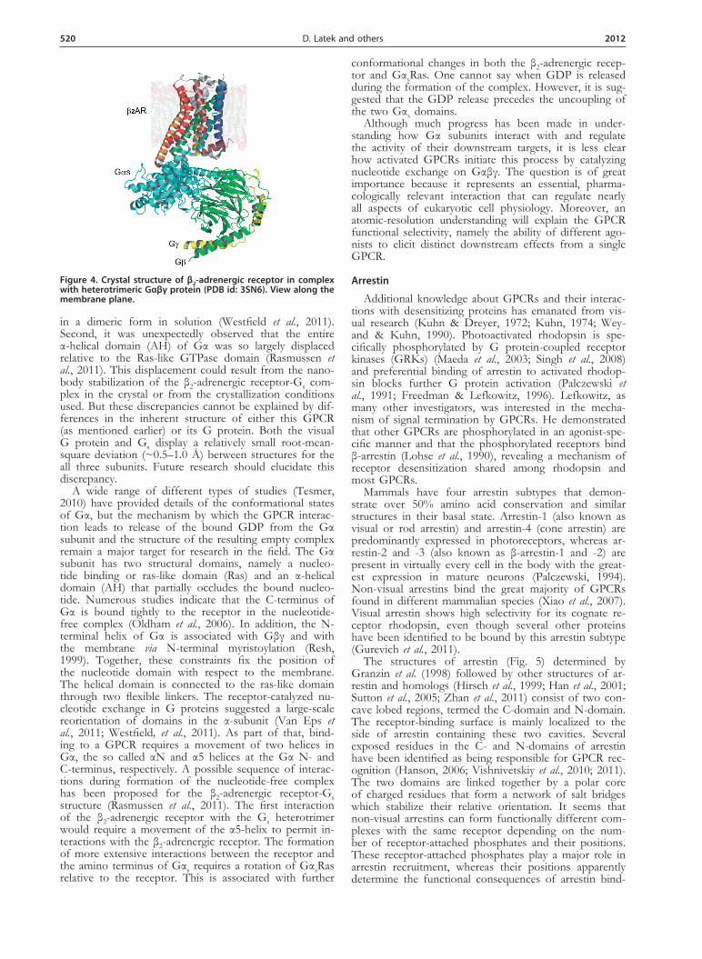
The structures of arrestin (Fig. 5) determined by Granzin et al. (1998) followed by other structures of ar-restin and homologs (Hirsch et al., 1999; Han et al., 2001; Sutton et al., 2005; Zhan et al., 2011) consist of two con-cave lobed regions, termed the C-domain and N-domain. The receptor-binding surface is mainly localized to the side of arrestin containing these two cavities. Several exposed residues in the C- and N-domains of arrestin have been identified as being responsible for GPCR rec-ognition (Hanson, 2006; Vishnivetskiy et al., 2010; 2011). The two domains are linked together by a polar core of charged residues that form a network of salt bridges which stabilize their relative orientation. It seems that non-visual arrestins can form functionally different com-plexes with the same receptor depending on the num-ber of receptor-attached phosphates and their positions. These receptor-attached phosphates play a major role in arrestin recruitment, whereas their positions apparently determine the functional consequences of arrestin bind-
Figure 4. Crystal structure of β2-adrenergic receptor in complex with heterotrimeric Gαβγ protein (PdB id: 3sn6). View along the membrane plane.
Vol. 59 521G protein-coupled receptors
ing to the phosphoreceptor (Tobin et al., 2008; Gimenez et al., 2012). Phosphorylation sites on rhodopsin were well defined a decade ago in vitro and in vivo (Ohguro et al., 1993; 1994; Ohguro & Palczewski, 1995; Ohguro et al., 1995; Kennedy et al., 2001). Lefkowitz’s group has correlated the phosphorylation sites of β2-adrenergic re-ceptor with β-arrestin functions (Nobles et al., 2011).
Arrestin-1 preferentially binds to activated and phos-phorylated rhodopsin. It also specifically binds inactive phosphorylated and active non-phosphorylated forms, but with much lower affinition. This observation was the first indication that arrestin-1 can recognize activa-tion and phosphorylation of rhodopsin independently of each other. A sequential multi-site binding model (Gurevich & Gurevich, 2004) posits that arrestin-1 first binds rhodopsin either via its structural elements that specifically interact with the light-activated rhodopsin conformation, or via residues that directly bind to the rhodopsin-attached phosphates. If the ‘activated state’ or the phosphates represent the only “attraction factor”, then arrestin-1 binds with low affinity. However, when arrestin-1 encounters phosphorylated photoactivated rho-dopsin, the engagement of both primary sites allows ar-restin to switch into the high-affinity rhodopsin-binding state, bringing additional arrestin elements into contact with rhodopsin. The new contact surface provides ex-tra energy to encourage the interaction, which accounts for arrestin-1’s much greater affinity for phosphorylated photoactivated rhodopsin.
The “hinge” region of all arrestins (residues 179–191) is flexible, allowing movement of the N- and C-domains relative to each other. However, there is no large relative motion of arrestin domains during complex formation, but a concerted movement of multiple flexible loops most probably helps arrestin mold itself onto the recep-tor (Kim et al., 2012) (Fig. 5). Lefkowitz’s studies on the conformation of non-visual arrestins in their active state revealed that although the overall activation mechanism is the same for all arrestin types, the final conformations might differ (Nobles et al., 2007). Moreover, β-arrestins might adopt multiple “active” conformations and, de-pending on their conformation, accomplish different functions (Shukla et al., 2008).
The stoichiometry of rhodopsin complexes with its cognate proteins is a matter of debate. Historically, one-
to-one binding was usually assumed (Hanson et al., 2007; Gurevich & Gurevich, 2008). However, it has also been proposed by Palczewski that a single arrestin molecule could accommodate two receptors (Liang et al., 2003; Modzelewska et al., 2006). Monomeric activated and phosphorylated rhodopsin in nanodiscs can bind arres-tin (Tsukamoto et al., 2010; Bayburt et al., 2011). At the same time, it has been reported that although arrestin requires at least a single phosphorylated photoactivated rhodopsin to bind to the membrane, a single arrestin can actually interact with a pair of receptors composed of two different photo-intermediate states. The bind-ing stoichiometry depends on the percentage of active receptors (Sommer et al., 2011). From a physiological standpoint, the different binding modes of arrestin cor-respond well to the functional needs of the cell at differ-ent light intensities. In the single-photon range, arrestin binds monomeric phosphorylated photoactivated rho-dopsin to quench signaling. But as the lighting level in-creases and photoactivates more rhodopsin, arrestin also binds dimeric photoactivated rhodopsin with only one phosphorylated protomer. Further studies have revealed that differentiated binding preferences of the two do-mains of arrestin allow it to accommodate the different functional forms of phosphorylated rhodopsin (Sommer et al., 2012). A general hypothesis is formed that the N-domain of arrestin mediates binding to agonist-activated receptor, whereas the less specific C-domain may serve various functions depending on the requirements of the biological system. The asymmetric ability of arrestin to stimulate ligand binding within receptor dimers is in line with studies on β2-adrenergic receptors (Gurevich et al., 1997) and N-formyl-peptide receptor (Key et al., 2001).
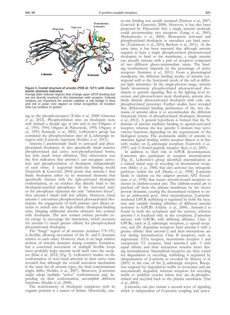
In addition to their role in GPCR desensitization, β-arrestins also participate in receptor internalization (Fig. 6). Lefkowitz’s group identified internalization as a critical initial step in recycling of desensitized recep-tors (Sibley et al., 1986) that also activates key mitogenic pathways within the cell (Daaka et al., 1998). β-arrestin binds to clathrin via the adaptor protein AP2 (Good-man et al., 1996) that causes arrestin-bound receptors to cluster in clathrin-coated pits. The clathrin-coated pit is pinched off from the plasma membrane by the motor protein dynamin, causing the desensitized receptor to en-ter an endosomal pool. After internalization, β-arrestin-mediated GPCR trafficking is regulated by both the loca-tion and variable binding affinities of different arrestin isoforms to GPCRs (Oakley et al., 2000). Arrestin-2 is found in both the cytoplasm and the nucleus, whereas arrestin-3 is localized only in the cytoplasm. β-arrestins interact with GPCRs with differing affinities. Class A GPCRs, such as β1-adrenergic receptors, μ opioid recep-tors, and D1 dopamine receptors bind arrestin-3 with a greater affinity than arrestin-2 and their interactions are lost during internalization. Class B receptors, such as angiotensin AT1a receptor, neurotensin receptor 1 and vasopressin V2 receptor, bind arrestin-2 and -3 with equal affinity and their interaction remains intact dur-ing internalization. Internalized receptors are then sorted for degradation or recycling; trafficking is regulated by ubiquitination of β-arrestin as revealed by Shenoy et al. (2001) in the case of the β2-adrenergic receptor. Recep-tors targeted for degradation traffic to lysosomes and are enzymatically degraded, whereas receptors for recycling traffic to acidified vesicles where they are de-phospho-rylated and recycled back to the plasma membrane (Tan et al., 2004).
β-arrestins can also initiate a second wave of signaling which is independent of G-protein coupling and activa-
Figure 5. Crystal structure of arrestin (PdB id: 1CF1) with charac-teristic elements indicated. Orange balls indicate regions that change upon GPCR binding but are not directly involved in the interaction with receptor. Colored residues are important for arrestin stability (a salt bridge in blue and red in polar core region) or initial recognition of receptor (two Lys residues in green).
522 2012D. Latek and others
tion (Fig. 6). Here they serve as adaptor or scaffold mol-ecules that bring crucial molecular components of specif-ic signaling pathways in close proximity to an activated GPCR. Interestingly, depending on the type of activated GPCR, either both β-arrestin isoforms are required to activate the second wave of signaling (termed co-de-pendent), or only one isoform is required, whereas the other serves to inhibit the pathway (termed reciprocal regulation) (DeWire et al., 2007). The group of Lefkowitz first found that arrestin-2 is complexed with the tyrosine kinase c-Src. This associates c-Src with the β2-adrenergic receptor, resulting in activation of c-Src which initiates a tyrosine phosphorylation signaling cascade leading to stimulation of the Ras-ERK1/2 pathway (Luttrell et al., 1999). Contributions of the Lefkowitz’s group to un-derstanding β-arrestin-mediated signaling have remained strong since their initiation.
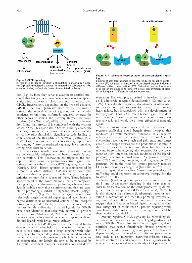
In many cases, signals transmitted by arrestin binding are demonstrably independent of heterotrimeric G pro-tein activation. This observation has triggered the con-cept of biased agonists, pathway-selective ligands that activate only a subset of the GPCR signaling repertoire (Kenakin, 2005). Biased agonism is best understood by a model in which different GPCR’s active conforma-tions are either competent for the full range of receptor activities or only for a subset of them. Thus, balanced ligands stabilize the conformations that are competent for signaling to all downstream pathways, whereas biased ligands stabilize only those conformations that are capa-ble of promoting a subset of signaling effects (Rajago-pal et al., 2010) (Fig. 7). The latter ligands could more selectively target beneficial signaling and even block or negate detrimental or unwanted actions of full receptor activation (e.g. side effects, toxicity or tolerance). Over the last decade a diversity of biased ligands for GPCRs have been identified that selectively activate G proteins or β-arrestins (Whalen et al., 2011), and several of these seem to have distinct functions when compared with tra-ditional ligands with broad range efficacy.
Treatment with GPCR agonists can be limited by the development of tachyphylaxis, a decrease in responsive-ness to the same dose of a drug, together with toler-ance, whereby higher drug doses are required to obtain the same effect. Both processes, which limit the utility of therapeutics, are largely thought to be regulated by β-arrestin-dependent receptor desensitization and down-
regulation. For example, arrestin-2 is involved in cardi-ac β2-adrenergic receptor desensitization (Conner et al., 1997). Clinically the β-agonist, dobutamine is often used to provide inotropic support for patients with severe heart failure, but is associated with the development of tachyphylaxis. Thus a G-protein-biased ligand that does not promote β-arrestin recruitment would cause less tachyphylaxis and would be a more effective therapeutic agent.
Several disease states associated with alterations in receptor trafficking could benefit from therapies that modulate β-arrestin-mediated functions. HIV requires cell-surface co-receptors, either the CCR5 or CXCR4 chemokine receptor, to attach and gain entry into target cells. CCR5-tropic viruses are the predominant species in the early stages of infection and there has been a sig-nificant interest in targeting this receptor for the treat-ment of HIV infection. Indeed, modified CCR5 ligands promote receptor internalization. As β-arrestins regu-late CCR5 trafficking, recycling and degradation (Op-permann, 2004), the modified ligands probably regulate CCR5 trafficking via changes in β-arrestin activity. Thus, use of a ligand that modifies β-arrestin-regulated CCR5 trafficking could represent an attractive therapy for the treatment of HIV.
Cardiac β1-adrenergic receptors can stimulate arres-tin-2- and -3-dependent signaling in the heart that re-sults in transactivation of the cardioprotective epidermal growth factor receptor (EGFR) (Noma et al., 2007). It is also thought that chronic β2-adrenergic receptor acti-vation is cardiotoxic and that this primarily involves Gs signaling (Xiao, 2001). These combined observations suggest that a β-arrestin-biased ligand acting as a clas-sical antagonist of cardiotoxic G protein signaling while stimulating cardioprotective β-arrestin signaling could be therapeutically beneficial.
Arrestins regulate GPCR signaling by controlling de-sensitization, endocytosis and recycling/degradation of most GPCRs. They also function as ligand-regulated scaffolds that recruit functionally diverse proteins to GPCRs to confer novel signaling properties. Arrestin-dependent signals are involved in different processes in vivo, such as cell migration, neurotransmission, cardiac muscle contraction, and apoptosis. These signals can be initiated or antagonized independently of G protein acti-
Figure 7. A schematic representation of arrestin-biased signal-ing. Binding of standard agonist to receptor induces an active confor-mation (R*) whereas binding of arrestin-biased agonist induces a different active conformation (R**). Distinct active conformations of receptor are coupled to different active conformation of arres-tin which govern different functional outcomes.
Figure 6. GPCR signaling. In response to ligand binding a stimulation signaling can occur via G-protein-mediated pathway terminated by subsequent GRK/arrestin binding, or/and via β-arrestin-mediated pathway.
Vol. 59 523G protein-coupled receptors
vation. Although arrestins have been extensively studied by different groups, the molecular mechanisms of recep-tor-arrestin complex formation/function remain unclear and much additional work will be required for advances in this field.
ImPoRTAnCe oF oTHeR GPCRs — CAse oF CHemoKIne ReCePToRs
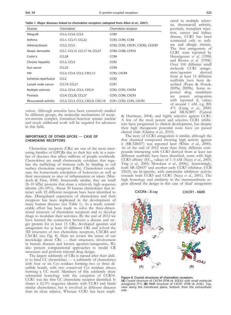
Chemokine receptors (CRs) are one of the most inter-esting families of GPCRs due to their key role in a num-ber of diseases that affect millions of people worldwide. Chemokines are small chemotactic cytokines that regu-late the trafficking of immune cells by binding to cell surface chemokine receptors (CRs). Chemokines coordi-nate the homeostatic circulation of leukocytes as well as their movement to sites of inflammation or injury (Mur-doch & Finn, 2000). Structurally similar, they are small (8–10 kDa) proteins that share a relatively high sequence identity (20–50%). About 50 human chemokines that in-teract with 22 different receptors have been identified to date. Disregulated expression of chemokines and their receptors has been implicated in the development of many human diseases (see Table 1). As a result, consid-erable effort has been made to solve the three-dimen-sional structure of chemokine receptors and to develop drugs to modulate their activities. By the end of 2012 we have learned the connection between a disease and tar-get protein for at least 15 CRs, developed agonists and antagonists for at least 10 different CRs and solved the 3D structures of two chemokine receptors, CXCR4 and CXCR1 (see Fig. 8). Here we review the status of our knowledge about CRs — their structures, involvement in human diseases and known agonists/antagonists. We also present computational approaches to model CR structures and perform rational drug design.
The largest subfamily of CRs is named after their abili-ty to bind CC chemokines — a subfamily of chemokines with four or six Cys residues forming two or three di-sulfide bonds, with two conserved Cys residues always forming a CC motif. Members of this subfamily share substantial homology with the exception of CCR10. CCR1 was the first CC chemokine receptor identified. It shares a 62.3% sequence identity with CCR3 and binds similar chemokines, but is involved in different diseases than its close relative. Whereas CCR1 has been impli-
cated in multiple sclero-sis, rheumatoid arthritis, psoriasis, transplant rejec-tion, cancer and kidney disease, CCR3 has been connected only to asth-ma and allergic rhinitis. The first antagonists of CCR1 were reported by Hesselgesser et al. (1998) and Brown et al. (1998). Over 100 different small molecule CCR1 antago-nists/agonists derived from at least 15 different scaffolds have been de-scribed (Pease & Horuk, 2009a; 2009b). Some re-ported drug candidates are potent antagonists with reported Ki values of around 1 nM, e.g. BX 471 (Liang et al., 2000) and MLN3897 (Carson
& Harriman, 2004), and highly selective against CCR1. A few of the most potent and selective CCR1 inhibi-tors have progressed to clinical development, but despite their high therapeutic potential none have yet passed clinical trials (Gladue et al., 2010).
The story of CCR3 antagonists is similar, although the first chemical compound showing high activity against it (SB-328437) was reported later (White et al., 2000). As of the end of 2012 more than thirty different com-pounds interacting with CCR3 derived from at least ten different scaffolds have been identified, some with high CCR3 affinity (EC50 values of 1–5 nM (Naya et al., 2003; Ting et al., 2005; Morokata et al., 2006)). Interestingly, both SB-328437 and another early CCR3 inhibitor, UCB 35625, are bi-specific, with nanomolar inhibitory activity towards both CCR3 and CCR1 (Naya et al., 2001). The high homology and similarity in the transmembrane re-gion allowed the design in this case of ‘dual’ antagonists
Table 1. major diseases linked to chemokine receptors (adopted from Allen et al., 2007).
Disease Chemokine Chemokine receptor
Allograft CCL3, CCL4, CCL5 CCR5
Asthma CCL1, CCL17, CCL22 CCR3, CCR4, CCR8
Atherosclerosis CCL2, CCL5 CCR2, CCR5, CXCR1, CXCR2, CX3CR1
Atopic dermatitis CCL1, CCL13, CCL17-18, CCL27 CCR4, CCR8, CCR10
Crohn’s CCL28 CCR9
Chronic hepatitis CCL3, CCL4 CCR5
Gut cancer CCL25 CCR9
HIV CCL3, CCL4, CCL5, CXCL12 CCR5, CXCR4
Ischemia-reperfusion CCL2 CCR2
Lymph node cancer CCL19, CCL21 CCR7
Multiple sclerosis CCL2, CCL4, CCL5, CXCL9 CCR2, CCR5, CXCR3
Psoriasis CCL4, CCL20, CCL27 CCR5, CCR6, CXCR3
Rheumatoid arthritis CCL2, CCL3, CCL5, CXCL9, CXCL10 CCR1, CCR2, CCR5, CXCR3
Figure 8. Crystal structures of chemokine receptors. (A) Crystal structure of CXCR4 (PDB id: 3ODU) with small molecule antagonist IT1t. (B) NMR structure of CXCR1 (PDB id: 2LNL). Top, view along the membrane plane, bottom, from the extracellular side.
524 2012D. Latek and others
affecting both receptors. More dual inhibitors have been reported (Dhanak et al., 2001a), some with subnanomolar activities against both CCR1 and CCR3 (Dhanak et al., 2001b). Though some of those inhibitors were tested in patients with various diseases, none have yet succeeded.
CCR2 and CCR5 constitute another interesting pair of similar chemokine receptors, sharing 63.4% sequence identity. They also interact with some of the same chemokines and both have been linked to the same im-munologic and cardiovascular diseases (Zhao, 2010). Of this pair, however, only CCR5 has achieved fame for serving as an entry factor for macrophage-tropic strains of HIV-1 (Moore et al., 1997), a role that has instigated a search for small molecular antagonists of this receptor that could block viral entry.
The first CCR2 antagonists were described in the lit-erature in 2000, six years after the successful cloning of this receptor (Forbes et al., 2000; Mirzadegan et al., 2000). By the end of 2012 several pharmaceutical com-panies have disclosed more than fifty CCR2 antagonists with different molecular scaffolds including piperidine, spiropiperidine, aminopyrrolidine, compounds with bisubstituted cyclohexane groups, and others. Some of the optimized ligands evidenced CCR2 inhibition in the nanomolar range, with the lowest IC50 (subnanomolar) value reported by Teijin company for one of their op-timized homopiperazine derivatives (Moree et al., 2008). But as in the CCR1/CCR3 case, no drug has yet been approved for use against this receptor despite consider-able effort.
Most CCR5 inhibitors were developed to prevent the cellular invasion of HIV. The first CCR5 inhibitor re-ported was TAK-779 synthesized by Takeda Chemical Industries in 1999 (Baba et al., 1999). This compound bound to the receptor at nanomolar concentrations and inhibited HIV cellular entry in vitro. Unfortunately, it also exhibited a poor oral bioavailability, but still was used as a CCR5 inhibitor model by other pharmaceutical compa-nies. In the last ten years at least forty different CCR5 antagonists have been identified and, due to the impor-tance of AIDS, many were optimized to display inhibi-tory efficacy at nanomolar or subnanomolar concentra-tions. Based on favorable HIV inhibition data, a rela-tively large number of these compounds entered clinical trials for treatment of AIDS. Unfortunately, most CCR5 inhibitors experienced multiple problems in stage II or III clinical trials (Wilkin & Gulick, 2012) and only one (maraviroc) was cleared and approved as an anti-HIV drug targeting CCR5 (Hitchcock, 2005).
The second largest subfamily of CRs was named for their ability to bind CXC chemokines that possess the conserved CXC motif. CXCR4 is one of the two chemokine receptors (together with CCR5) used by HIV to enter human cells, a finding that has greatly acceler-ated structural research aimed at this protein (Ober-lin et al., 1996). The first small molecular antagonist of CXCR4 was described in 1992 (De Clercq et al., 1992) and since then a number of potent antagonists (with binding in the nanomolar or even subnanomolar range) have been described (Ichiyama et al., 2003; Zhan et al., 2007). Moreover, the relatively large amount of data for this receptor provided insights into the binding modes of a number of antagonists (Vabeno et al., 2006).
A breakthrough in CXCR4-based anti-HIV research occurred with the experimental solving of crystal struc-tures of CXCR4 bound to either a small-molecular ligand or a cyclic peptide antagonist (Wu et al., 2010) (Fig. 8A). Earlier it was suggested that both those ligands block the interaction between CXCR4 and its natural ligand CXC4,
and also inhibit CXCL12 interactions with the HIV-1 glycoprotein gp120. Indeed, both ligands found in those crystal structures interacted with the receptor’s extracel-lular loops and N-terminus and most likely altered their conformation, making the interaction with CXCL12 and gp120 energetically unfavorable.
The structure of CXCR4 was rather surprising, espe-cially because it revealed a number of relatively large dif-ferences from the crystal structures of the β2-adrenergic receptor and other GPCRs obtained earlier. The most striking differences were the positions and rotations of helices 1, 2 and 6 that resulted in a much looser pack-ing of all helices, and also the lengths of helices 5 and 7. Surprisingly, the binding cavity of CXCR4 also was larg-er, more open and located much closer to the extracellu-lar surface than that of other known GPCRs. This inves-tigation not only provides invaluable information about the structure of CXCR4 and possibly other CR-family members, but it also serves as a platform for rational drug design, contributing to understanding of the HIV-1 entry process along with the elucidated structures of na-tive HIV-1 gp120 trimers (Liu et al., 2008). This progress should lead to a series of new scaffolds and compounds targeting CXCR4 as well as dual inhibitors able to bind to both CXCR4 and CCR5 (Murray et al., 2010). Inter-estingly, despite the known structure of CXCR4, no anti-HIV drug targeting this receptor has yet been approved for use. However, one of the first CXCR4 inhibitors found has been approved to be used to mobilize hemat-opoietic stem cells in cancer patients (AnorMED, 2007).
CXCR1 and CXCR2 form a pair of chemokine recep-tors which are most closely related to each other, with 76.6% sequence identity. The number of publications on the role of both receptors has increased exponentially within the last 10 years, in part due to the discovery of potent and selective CXCR1/CXCR2 dual antagonists. The first dual antagonist for this pair of proteins was shown to inhibit acute and chronic models of arthritis in the rabbit (Podolin et al., 2002). There is also an interest in developing new CXCR1/CXCR2 antagonists as thera-peutic agents for chronic obstructive pulmonary disease (COPD), asthma and various forms of cancer. Two such dual antagonists have passed all clinical trials and are now marketed — reparixin which attenuates inflamma-tory responses and promotes recovery of function after spinal cord injury (Gorio et al., 2007) and navarixin, an anti-COPD drug.
CXCR1 is also the second chemokine receptor with a known three-dimensional structure. In 2012 its struc-ture was obtained by using rotationally aligned solid-state NMR (Park et al., 2012) (Fig. 8B). That novel method involved the use of NMR for the first time in GPCR structural studies and provided important information about this receptor in its natural phospholipid bilayer environment. A comparison of its structure with that of CXCR4 (32.9% sequence identity) shows a high ho-mology and overall similarity. Four charged residues in the helical region of CXCR1 form a polar cluster in the ligand-binding site of this receptor. They most likely are important for ligand binding, similar to the three polar residues found in CXCR4. There also are some poten-tially important differences in positions and rotations of helices 1, 2 and 6, which alter the overall structure of the receptor’s core and could contribute to the different biological activities exhibited by CXCR1 and CXCR4. As with CXCR4, solution of the CXCR1 structure hope-fully will lead to the rational design of new CXCR1 and CXCR2 antagonists with improved properties.
Vol. 59 525G protein-coupled receptors
Chemokine receptors are a family of GPCRs that have always attracted much attention from researchers and health professionals. Immediately after their rec-ognition as potential targets in various diseases, these proteins became the focus of numerous programs run by pharmaceutical companies. Development of novel experimental and theoretical techniques has been cru-cial in finding and/or designing new CR antagonists. Of these recent innovations, the possibility of obtaining three-dimensional structures by using crystallography or NMR was one of the most important. This has led to the crystal structure of CXCR4 and the NMR structure of CXCR1 described above (Wu et al., 2010; Park et al., 2012). There are still many questions about the structure of other CRs, their probable oligomerization and dynam-ics in human cells, but our knowledge of this family of proteins is now increasing exponentially.
FuTuRe dIReCTIons
The GPCR research will further shape the field of pharmacology and medicine in the decades ahead. Thus it will be challenging but necessary to determine high resolution structures of native GPCRs alone and in com-plexes with their G protein, receptor kinase and arres-tin (Jastrzebska et al., 2010). Moreover, GPCRs and G proteins are critically altered by several post-translational modifications which should be taken into account in structural studies. In addition to these modifications, wa-ter molecules have been shown to play a key role in all proteins, including transmembrane ones. It will be essen-tial to determine the exact location of water molecules in these receptors and their re-arrangements during GPCR activation. Understanding the energy landscape of GP-CRs corresponding to their folding pathway and activa-tion is highly anticipated. Identification of key residues responsible for folding and membrane insertion is need-ed to explain the etiology of many human diseases as-sociated with receptor mutations. Moreover, GPCRs are not monomeric — they have a propensity to interact not only with each other but also with other transmembrane proteins. In recent years, more than 50% of published papers in the field have explored the oligomerization of GPCRs. But we are still far from understanding “the logic” of oligomerization at both structural and function-al levels. Understanding the structural complementarities of GPCR homo- and hetero-oligomerization offers sev-eral novel pharmacological opportunities to explore the high specificity of these interactions. A large number of GPCRs communicate with only a limited number of G proteins and even a smaller number of effectors such as enzymes and channels. Although the subcellular localiza-tion of specific sets of receptors and interacting proteins is clear, diffusible ligands such as cAMP or Ca2+ allow a cross-talk between many specialized pathways. It will be important to determine the intersections of different rel-evant GPCR signaling pathways in native tissues of in-terest. Modern 3D structural electron microscopy (cryo-electron microscopy and tomography) and hybrid micro-scopic techniques will facilitate obtaining high resolution structures of signaling complexes between GPCRs, their cognate G proteins and effector molecules in native tis-sues. Such studies can determine how the GPCR sign-aling complexes are compartmentalized within cells to evoke their local effects. We are confident that the dis-covery of mutations responsible for genetic diseases will be dramatically accelerated due to the DNA and RNA sequencing of whole personal genomes as well as tissue
transcriptomes. This new methodology will enable evalu-ation of the impact of mutations and polymorphisms af-fecting GPCR expression levels on human conditions. Hence, GPCRs will certainly play a central role in drug research in the foreseeable future.
Acknowledgements
This work was supported by funding from the Na-tional Institutes of Health, USA (EY008061) and the National Center of Science, Poland (DEC2011/03/B/NZ1/03204). K.P. is John H. Hord Professor of Phar-macology.
ReFeRenCes
Allen SJ, Crown SE, Handel TM (2007) Chemokine: receptor structure, interactions, and antagonism. Annu Rev Immunol 25: 787–820.
Angel TE, Chance MR, Palczewski K (2009a) Conserved waters medi-ate structural and functional activation of family A (rhodopsin-like) G protein-coupled receptors. Proc Natl Acad Sci USA 106: 8555–8560.
Angel TE, Gupta S, Jastrzebska B, Palczewski K, Chance MR (2009b) Structural waters define a functional channel mediating activation of the GPCR, rhodopsin. Proc Natl Acad Sci USA 106: 14367–14372.
AnorMED (2007) Plerixafor: AMD 3100, AMD3100, JM 3100, SDZ SID 791. Drugs R D 8: 113–119.
Arshavsky VY, Lamb TD, Pugh EN, Jr. (2002) G proteins and pho-totransduction. Annu Rev Physiol 64: 153–187.
Baba M, Nishimura O, Kanzaki N, Okamoto M, Sawada H, Iizawa Y, Shiraishi M, Aramaki Y, Okonogi K, Ogawa Y, Meguro K, Fu-jino M (1999) A small-molecule, nonpeptide CCR5 antagonist with highly potent and selective anti-HIV-1 activity. Proc Natl Acad Sci USA 96: 5698–5703.
Bayburt TH, Vishnivetskiy SA, McLean MA, Morizumi T, Huang CC, Tesmer JJ, Ernst OP, Sligar SG, Gurevich VV (2011) Monomeric rhodopsin is sufficient for normal rhodopsin kinase (GRK1) phos-phorylation and arrestin-1 binding. J Biol Chem. 286: 1420–1428.
Beavo JA, Brunton LL (2002) Cyclic nucleotide research — still ex-panding after half a century. Nat Rev Mol Cell Biol 3: 710–718.
Benovic JL, Strasser RH, Caron MG, Lefkowitz RJ (1986) Beta-adren-ergic receptor kinase: identification of a novel protein kinase that phosphorylates the agonist-occupied form of the receptor. Proc Natl Acad Sci USA 83: 2797–2801.
Bers DM (2002) Cardiac excitation-contraction coupling. Nature 415: 198–205.
Breitman ML, Bryce DM, Giddens E, Clapoff S, Goring D, Tsui LC, Klintworth GK, Bernstein A (1989) Analysis of lens cell fate and eye morphogenesis in transgenic mice ablated for cells of the lens lineage. Development 106: 457–463.
Cherezov V, Rosenbaum DM, Hanson MA, Rasmussen SG, Thian FS, Kobilka TS, Choi HJ, Kuhn P, Weis WI, Kobilka BK, Stevens RC (2007) High-resolution crystal structure of an engineered human beta2-adrenergic G protein-coupled receptor. Science 318: 1258–1265.
Chien EYT, Liu W, Zhao Q, Katritch V, Won Han G, Hanson MA, Shi L, Newman AH, Javitch JA, Cherezov V, Stevens RC (2010) Structure of the human dopamine D3 receptor in complex with a D2/D3 selective antagonist. Science 330: 1091–1095.
Chin K-V, Yang W-L, Ravatn R, Kita T, Reitman E, Vettori D, Cvi-jic ME, Shin M, Iacono L (2002) Reinventing the wheel of cyclic AMP: novel mechanisms of cAMP signaling. Ann N Y Acad Sci 968: 49–64.
Choe HW, Kim YJ, Park JH, Morizumi T, Pai EF, Krauss N, Hof-mann KP, Scheerer P, Ernst OP (2011) Crystal structure of metar-hodopsin II. Nature 471: 651–655.
Clapham DE, Neer EJ (1997) G protein beta gamma subunits. Annu Rev Pharmacol Toxicol 37: 167–203.
Conner DA, Mathier MA, Mortensen RM, Christe M, Vatner SF, Sei-dman CE, Seidman JG (1997) beta-Arrestin1 knockout mice appear normal but demonstrate altered cardiac responses to beta-adrenergic stimulation. Circ Res 81: 1021–1026.
Daaka Y, Luttrell LM, Ahn S, Della Rocca GJ, Ferguson SS, Caron MG, Lefkovitz RJ (1998) Essential role for G protein-coupled re-ceptor endocytosis in the activation of mitogen-activated protein kinase. J Biol Chem 273: 685–688.
De Clercq E, Yamamoto N, Pauwels R, Baba M, Schols D, Nakashima H, Balzarini J, Debyser Z, Murrer BA, Schwartz D, et al. (1992) Potent and selective inhibition of human immunodeficiency virus (HIV)-1 and HIV-2 replication by a class of bicyclams interacting with a viral uncoating event. Proc Natl Acad Sci USA 89: 5286–5290.
526 2012D. Latek and others
De Lean A, Stadel JM, Lefkowitz RJ (1980) A ternary complex model explains the agonist-specific binding properties of the adenylate cy-clase-coupled beta-adrenergic receptor. J Biol Chem. 255: 7108–7117.
Deupi X, Edwards P, Singhal A, Nickle B, Oprian D, Schertler G, Standfuss J (2012) Stabilized G protein binding site in the structure of constitutively active metarhodopsin-II. Proc Natl Acad Sci USA 109: 119–124.
Deville J, Rey J, Chabbert M (2009) An indel in transmembrane helix 2 helps to trace the molecular evolution of class A G-protein-coupled receptors. J Mol Evol 68: 475–489.
DeWire SM, Ahn S, Lefkowitz RJ, Shenoy SK (2007) beta-arrestins and cell signaling. Annu Rev Physiol 69: 483–510.
Dhanak D, Christmann LT, Darcy MG, Jurewicz AJ, Keenan RM, Lee J, Sarau HM, Widdowson KL, White JR (2001a) Discovery of po-tent and selective phenylalanine derived CCR3 antagonists. Part 1. Bioorg Med Chem Lett 11: 1441–1444.
Dhanak D, Christmann LT, Darcy MG, Keenan RM, Knight SD, Lee J, Ridgers LH, Sarau HM, Shah DH, White JR, Zhang L (2001b) Discovery of potent and selective phenylalanine derived CCR3 re-ceptor antagonists. Part 2. Bioorg Med Chem Lett 11: 1445–1450.
Dhanasekaran N, Dermott JM (1996) Signaling by the G12 class of G proteins. Cell Signal 8: 235–245.
Dixon RA, Kobilka BK, Strader DJ, Benovic JL, Dohlman HG, Frielle T, Bolanowski MA, Bennett CD, Rands E, Diehl RE, Mumford RA, Slater EE, Sigal IS, Caron MG, Lefkowitz RJ, Strader CD (1986) Cloning of the gene and cDNA for mammalian beta-adren-ergic receptor and homology with rhodopsin. Nature 321: 75–79.
Eilers M, Goncalves JA, Ahuja S, Kirkup C, Hirshfeld A, Simmerling C, Reeves PJ, Sheves M, Smith SO (2012) Structural transitions of transmembrane helix 6 in the formation of metarhodopsin I. J Phys Chem B.?
Exton JH (1996) Regulation of phosphoinositide phospholipases by hormones, neurotransmitters, and other agonists linked to G pro-teins. Annu Rev Pharmacol Toxicol 36: 481–509.
Filipek S, Stenkamp RE, Teller DC, Palczewski K (2003) G protein-coupled receptor rhodopsin: A prospectus. Annu Rev Physiol 65: 851–879.
Forbes IT, Cooper DG, Dodds EK, Hickey DM, Ife RJ, Meeson M, Stockley M, Berkhout TA, Gohil J, Groot PH, Moores K (2000) CCR2B receptor antagonists: conversion of a weak HTS hit to a potent lead compound. Bioorg Med Chem Lett 10: 1803–1806.
Fredriksson R, Lagerstrom MC, Lundin LG, Schioth HB (2003) The G-protein-coupled receptors in the human genome form five main families. Phylogenetic analysis, paralogon groups, and fingerprints. Mol Pharmacol 63: 1256–1272.
Freedman NJ, Lefkowitz RJ (1996) Desensitization of G protein-cou-pled receptors. Recent Prog Horm Res 51: 319–351.
Garczarek F, Gerwert K (2006) Functional waters in intraprotein pro-ton transfer monitored by FTIR difference spectroscopy. Nature 439: 109–112.
Gimenez LE, Kook S, Vishnivetskiy SA, Ahmed MR, Gurevich EV, Gurevich VV (2012) Role of receptor-attached phosphates in bind-ing of visual and non-visual arrestins to G protein-coupled recep-tors. J Biol Chem 287: 9028–9040.
Gladue PR, Brown FM, Zwillich HS (2010) CCR1 Antagonists: what have we learned from clinical trials. Curr Top Med Chem 10: 1268–1277.
Goodman OBJ, Krupnick JG, Santini F, Gurevich VV, Penn RB, Gag-non AW, Keen JH, Benovic JL (1996) Beta-arrestin acts as a clath-rin adaptor in endocytosis of the beta2-adrenergic receptor. Nature 383: 477–450.
Gorio A, Madaschi L, Zadra G, Marfia G, Cavalieri B, Bertini R, Di Giulio AM (2007) Reparixin, an inhibitor of CXCR2 function, at-tenuates inflammatory responses and promotes recovery of function after traumatic lesion to the spinal cord. J Pharmacol Exp Ther 322: 973–981.
Granier S, Manglik A, Kruse AC, Kobilka TS, Thian FS, Weis WI, Kobilka BK (2012) Structure of the delta-opioid receptor bound to naltrindole. Nature 485: 400–404.
Granzin J, Wilden U, Choe HW, Labahn J, Krafft B, Buldt G (1998) X-ray crystal structure of arrestin from bovine rod outer segments. Nature 391: 918–921.
Gurevich VV, Pals-Rylaarsdam R, Benovic JL, Hosey MM, Onorato JJ (1997) Agonist-receptor-arrestin, an alternative ternary complex with high agonist affinity. 272: 28849–28852.
Gurevich VV, Gurevich EV (2004) The molecular acrobatics of arres-tin activation. Trends Pharmacol Sci 25: 105–111.
Gurevich VV, Gurevich EV (2008) GPCR monomers and oligomers: it takes all kinds. Trends Neurosci 31: 74–81.
Gurevich VV, Hanson SM, Song X, Vishnivetskiy SA, Gurevich EV (2011) The functional cycle of visual arrestins in photoreceptor cells. Prog Retin Eye Res. 30: 405–430.
Haga K, Kruse AC, Asada H, Yurugi-Kobayashi T, Shiroishi M, Zhang C, Weis WI, Okada T, Kobilka BK, Haga T, Kobayashi T (2012) Structure of the human M2 muscarinic acetylcholine receptor bound to an antagonist. Nature 482: 547–551.
Han M, Gurevich VV, Vishnivetskiy SA, Sigler PB, Schubert C (2001) Crystal structure of beta-arrestin at 1.9 A: possible mechanism of receptor binding and membrane Translocation. Structure. 9: 869–880.
Hanson SM, Gurevich EV, Vishnivetskiy SA, Ahmed MR, Song X, Gurevich VV (2007) Each rhodopsin molecule binds its own ar-restin. Proc Natl Acad Sci USA 104: 3125–3128.
Hanson SM FD, Vishnivetskiy SA, Kolobova EA, Hubbell WL, Klug CS, Gurevich VV. (2006) Differential interaction of spin-labeled ar-restin with inactive and active phosphorhodopsin. Proc Natl Acad Sci USA 103: 4900–4905.
Hesselgesser J, Ng HP, Liang M, Zheng W, May K, Bauman JG, Mo-nahan S, Islam I, Wei GP, Ghannam A, Taub DD, Rosser M, Snid-er RM, Morrissey MM, Perez HD, Horuk R (1998) Identification and characterization of small molecule functional antagonists of the CCR1 chemokine receptor. J Biol Chem 273: 15687–15692.
Hirsch JA, Schubert C, Gurevich VV, Sigler PB (1999) The 2.8 A crys-tal structure of visual arrestin: a model for arrestin’s regulation. Cell. 97: 257–269.
Hitchcock CA (2005) The Discovery and Exploratory Development of Maraviroc (UK-427,857): A Novel CCR5 antagonist for the treat-ment of HIV. Retrovirology 2: S11.
Ichiyama K, Yokoyama-Kumakura S, Tanaka Y, Tanaka R, Hirose K, Bannai K, Edamatsu T, Yanaka M, Niitani Y, Miyano-Kurosaki N, Takaku H, Koyanagi Y, Yamamoto N (2003) A duodenally absorb-able CXC chemokine receptor 4 antagonist, KRH-1636, exhibits a potent and selective anti-HIV-1 activity. Proc Natl Acad Sci USA 100: 4185–4190.
Jaakola VP, Griffith MT, Hanson MA, Cherezov V, Chien EY, Lane JR, Ijzerman AP, Stevens RC (2008) The 2.6 angstrom crystal struc-ture of a human A2A adenosine receptor bound to an antagonist. Science 322: 1211–1217.
Jastrzebska B, Fotiadis D, Jang GF, Stenkamp RE, Engel A, Palczews-ki K (2006) Functional and structural characterization of rhodopsin oligomers. J Biol Chem 281: 11917–11922.
Jastrzebska B, Tsybovsky Y, Palczewski K (2010) Complexes between photoactivated rhodopsin and transducin: progress and questions. Biochem J 428: 1–10.
Jastrzebska B, Palczewski K, Golczak M (2011a) Role of bulk water in hydrolysis of the rhodopsin chromophore. J Biol Chem 286: 18930–18937.
Jastrzebska B, Ringler P, Lodowski DT, Moiseenkova-Bell V, Golczak M, Muller SA, Palczewski K, Engel A (2011b) Rhodopsin-trans-ducin heteropentamer: three-dimensional structure and biochemical characterization. J Struct Biol 176: 387–394.
Katritch V, Cherezov V, Stevens RC (2012) Diversity and modular-ity of G protein-coupled receptor structures. Trends Pharmacol Sci 33: 17–27.
Kenakin T (2005) New concepts in drug discovery: collateral efficacy and permissive antagonism. Nat Rev Drug Discov 4: 919–927.
Kenakin T, Miller LJ (2010) Seven transmembrane receptors as shapeshifting proteins: the impact of allosteric modulation and func-tional selectivity on new drug discovery. Pharmacol Rev 62: 265–304.
Kennedy MJ, Lee KA, Niemi GA, Craven KB, Garwin GG, Saari JC, Hurley JB (2001) Multiple phosphorylation of rhodopsin and the in vivo chemistry underlying rod photoreceptor dark adaptation. Neu-ron 31: 87–101.
Key TA, Bennett TA, Foutz TD, Gurevich VV, Sklar LA, Prossnitz ER (2001) Regulation of formyl peptide receptor agonist affinity by reconstitution with arrestins and heterotrimeric G proteins. J Biol Chem 276: 49204–49212.
Kim M, Vishnivetskiy SA, Van Eps N, Alexander NS, Cleghorn WM, Zhan X, Hanson SM, Morizumi T, Ernst OP, Meiler J, Gurevich VV, Hubbell WL (2012) Conformation of receptor-bound visual ar-restin. Proc Natl Acad Sci USA 109: 18407–18412.
Kruse AC, Hu J, Pan AC, Arlow DH, Rosenbaum DM, Rosemond E, Green HF, Liu T, Chae PS, Dror RO, Shaw DE, Weis WI, Wess J, Kobilka BK (2012) Structure and dynamics of the M3 muscarinic acetylcholine receptor. Nature 482: 552–556.
Kuhn H, Dreyer WJ (1972) Light dependent phosphorylation of rho-dopsin by ATP. FEBS Lett 20: 1–6.
Kuhn H (1974) Light-dependent phosphorylation of rhodopsin in liv-ing frogs. Nature 250: 588–590.
Lebon G, Warne T, Tate CG (2012) Agonist-bound structures of G protein-coupled receptors. Curr Opin Struct Biol 22: 482–490.
Lefkowitz RJ (2000) The superfamily of heptahelical receptors. Nat Cell Biol 2: E133–136.
Lefkowitz RJ, Shenoy SK (2005) Transduction of receptor signals by beta-arrestins. Science 308: 512–517.
Liang M, Mallari C, Rosser M, Ng HP, May K, Monahan S, Bauman JG, Islam I, Ghannam A, Buckman B, Shaw K, Wei GP, Xu W, Zhao Z, Ho E, Shen J, Oanh H, Subramanyam B, Vergona R, Taub D, Dunning L, Harvey S, Snider RM, Hesselgesser J, Morris-sey MM, Perez HD (2000) Identification and characterization of a potent, selective, and orally active antagonist of the CC chemokine receptor-1. J Biol Chem 275: 19000–19008.
Vol. 59 527G protein-coupled receptors
Liang Y, Fotiadis D, Filipek S, Saperstein DA, Palczewski K, Engel A (2003) Organization of the G protein-coupled receptors rhodopsin and opsin in native membranes. J Biol Chem 278: 21655–21662.
Liu J, Bartesaghi A, Borgnia MJ, Sapiro G, Subramaniam S (2008) Molecular architecture of native HIV-1 gp120 trimers. Nature 455: 109–113.
Liu W, Chun E, Thompson AA, Chubukov P, Xu F, Katritch V, Han GW, Roth CB, Heitman LH, Ijzerman AP, Cherezov V, Stevens RC (2012) Structural basis for allosteric regulation of GPCRs by so-dium ions. Science 337: 232–236.
Lodowski DT, Salom D, Le Trong I, Teller DC, Ballesteros JA, Palc-zewski K, Stenkamp RE (2007) Crystal packing analysis of Rhodop-sin crystals. J Struct Biol 158: 455–462.
Lodowski DT, Angel TE, Palczewski K (2009) Comparative analysis of GPCR crystal structures. Photochem Photobiol 85: 425–430.
Lohse MJ, Benovic JL, Codina J, Caron MG, Lefkowitz RJ (1990) beta-Arrestin: a protein that regulates beta-adrenergic receptor func-tion. Science 248: 1547–1550.
Luttrell LM, Ferguson SS, Daaka Y, Miller WE, Maudsley S, Della Rocca GJ, Lin F, Kawakatsu H, Owada K, Luttrell DK, Caron MG, Lefkowitz RJ (1999) Beta-arrestin-dependent formation of beta2 adrenergic receptor-Src protein kinase complexes. Science 283: 655–661.
Luttrell LM, Lefkowitz RJ (2002) The role of beta-arrestins in the ter-mination and transduction of G-protein-coupled receptor signals. J Cell Sci 115: 455–465.
Maeda A, Okano K, Park PS, Lem J, Crouch RK, Maeda T, Palcze-wski K (2010) Palmitoylation stabilizes unliganded rod opsin. Proc Natl Acad Sci USA 107: 8428–8433.
Maeda T, Imanishi Y, Palczewski K (2003) Rhodopsin phosphoryla-tion: 30 years later. Prog Retin Eye Res 22: 417–434.
Manglik A, Kruse AC, Kobilka TS, Thian FS, Mathiesen JM, Suna-hara RK, Pardo L, Weis WI, Kobilka BK, Granier S (2012) Crystal structure of the micro-opioid receptor bound to a morphinan an-tagonist. Nature 485: 321–326.
Meruelo AD, Samish I, Bowie JU (2011) TMKink: a method to predict transmembrane helix kinks. Protein science: a publication of the Protein Society 20: 1256–1264.
Mirzadegan T, Diehl F, Ebi B, Bhakta S, Polsky I, McCarley D, Mul-kins M, Weatherhead GS, Lapierre JM, Dankwardt J, Morgans D, Jr., Wilhelm R, Jarnagin K (2000) Identification of the binding site for a novel class of CCR2b chemokine receptor antagonists: binding to a common chemokine receptor motif within the helical bundle. J Biol Chem 275: 25562–25571.
Mirzadegan T, Benko G, Filipek S, Palczewski K (2003) Sequence analyses of G-protein-coupled receptors: similarities to rhodopsin. Biochemistry 42: 2759–2767.
Modzelewska A, Fillipek S, Palczewski K, Park PS. (2006) Arrestin in-teraction with rhodopsin: conceptual models. Cell Biochem Biophys. 46: 1–15.
Moore JP, Trkola A, Dragic T (1997) Co-receptors for HIV-1 entry. Curr Opin Immunol 9: 551–562.
Moree WJ, Kataoka K-i, Ramirez-Weinhouse MM, Shiota T, Imai M, Tsutsumi T, Sudo M, Endo N, Muroga Y, Hada T, Fanning D, Saunders J, Kato Y, Myers PL, Tarby CM (2008) Potent antagonists of the CCR2b receptor. Part 3: SAR of the (R)-3-aminopyrrolidine series. Bioorg Med Chem Lett 18: 1869–1873.
Morokata T, Suzuki K, Masunaga Y, Taguchi K, Morihira K, Sato I, Fujii M, Takizawa S, Torii Y, Yamamoto N, Kaneko M, Yamada T, Takahashi K, Shimizu Y (2006) A novel, selective, and orally avail-able antagonist for CC chemokine receptor 3. J Pharmacol Exp Ther 317: 244–250.
Muller DJ, Wu N, Palczewski K (2008) Vertebrate membrane proteins: structure, function, and insights from biophysical approaches. Phar-macol Rev 60: 43–78.
Murdoch C, Finn A (2000) Chemokine receptors and their role in in-flammation and infectious diseases. Blood 95: 3032–3043.
Murray EJ, Leaman DP, Pawa N, Perkins H, Pickford C, Perros M, Zwick MB, Butler SL (2010) A low-molecular-weight entry inhibi-tor of both CCR5- and CXCR4-tropic strains of human immu-nodeficiency virus type 1 targets a novel site on gp41. J Virol 84: 7288–7299.
Mustafi D, Palczewski K (2009) Topology of class A G protein-cou-pled receptors: insights gained from crystal structures of rhodopsins, adrenergic and adenosine receptors. Mol Pharmacol 75: 1–12.
Naya A, Kobayashi K, Ishikawa M, Ohwaki K, Saeki T, Noguchi K, Ohtake N (2001) Discovery of a novel CCR3 selective antagonist. Bioorg Med Chem Lett 11: 1219–1223.
Naya A, Kobayashi K, Ishikawa M, Ohwaki K, Saeki T, Noguchi K, Ohtake N (2003) Structure-activity relationships of 2-(benzothia-zolylthio)acetamide class of CCR3 selective antagonist. Chem Pharm Bull 51: 697–701.
Nobles KN, Guan Z, Xiao K, Oas TG, Lefkowitz RJ (2007) The ac-tive conformation of beta-arrestin1: direct evidence for the phos-phate sensor in the N-domain and conformational differences in
the active states of beta-arrestins1 and -2. J Biol Chem 282: 21370–21381.
Nobles KN, Xiao K, Ahn S, Shukla AK, Lam CM, Rajagopal S, Stra-chan RT, Huang TY, Bressler EA, Hara MR, Shenoy SK, Gygi SP, Lefkowitz RJ (2011) Distinct phosphorylation sites on the β(2)-adrenergic receptor establish a barcode that encodes differential functions of β-arrestin. Sci Signal 4: ra51.
Noma T, Lemaire A, Naga Prasad SV, Barki-Harrington L, Tilley DG, Chen J, Le Corvoisier P, Violin JD, Wei H, Lefkowitz RJ, Rockman HA (2007) Beta-arrestin-mediated beta1-adrenergic receptor trans-activation of the EGFR confers cardioprotection. J Clin Invest 117: 2445–2458.
Oakley RH, Laporte SA, Holt JA, Caron MG, Barak LS (2000) Differ-ential affinities of visual arrestin, beta arrestin1, and beta arrestin2 for G protein-coupled receptors delineate two major classes of re-ceptors. J Biol Chem 275: 17201–17210.
Oberlin E, Amara A, Bachelerie F, Bessia C, Virelizier J-L, Arenzana-Seisdedos F, Schwartz O, Heard J-M, Clark-Lewis I, Legler DF, Loetscher M, Baggiolini M, Moser B (1996) The CXC chemokine SDF-1 is the ligand for LESTR/fusin and prevents infection by T-cell-line-adapted HIV-1. Nature 382: 833–835.
Ohguro H, Palczewski K, Ericsson LH, Walsh KA, Johnson RS (1993) Sequential phosphorylation of rhodopsin at multiple sites. Biochemis-try 32: 5718–5724.
Ohguro H, Johnson RS, Ericsson LH, Walsh KA, Palczewski K (1994) Control of rhodopsin multiple phosphorylation. Biochemistry 33: 1023–1028.
Ohguro H, Palczewski K (1995) Separation of phospho- and non-phosphopeptides using reverse phase column chromatography. FEBS Lett 368: 452–454.
Ohguro H, Van Hooser JP, Milam AH, Palczewski K (1995) Rhodop-sin phosphorylation and dephosphorylation in vivo. J Biol Chem 270: 14259–14262.
Okada T, Sugihara M, Bondar AN, Elstner M, Entel P, Buss V (2004) The retinal conformation and its environment in rhodopsin in light of a new 2.2 A crystal structure. J Mol Biol 342: 571–583.
Oldham WM, Van Eps N, Preininger AM, Hubbell WL, Hamm HE (2006) Mechanism of the receptor-catalyzed activation of heterotri-meric G proteins. Nat Struct Mol Biol. 13: 772–777.
Oppermann M (2004) Chemokine receptor CCR5: insights into struc-ture, function, and regulation. Cell Signal. 16: 1201–1210.
Orban T, Gupta S, Palczewski K, Chance MR (2010) Visualizing water molecules in transmembrane proteins using radiolytic labeling meth-ods. Biochemistry 49: 827–834.
Orban T, Jastrzebska B, Gupta S, Wang B, Miyagi M, Chance MR, Palczewski K (2012) Conformational dynamics of activation for the pentameric complex of dimeric G protein-coupled receptor and het-erotrimeric G protein. Structure 20: 826–840.
Osyczka A, Moser CC, Dutton PL (2005) Fixing the Q cycle. Trends Biochem Sci 30: 176–182.
Palczewski K, McDowell JH, Jakes S, Ingebritsen TS, Hargrave PA (1991) Regulation of rhodopsin dephosphorylation by arrestin. J Biol Chem 264: 15770–15773.
Palczewski K (1994) Structure and functions of arrestins. Protein Sci 3: 1355–1361.
Palczewski K, Kumasaka T, Hori T, Behnke CA, Motoshima H, Fox BA, Le Trong I, Teller DC, Okada T, Stenkamp RE, Yamamoto M, Miyano M (2000) Crystal structure of rhodopsin: A G protein-coupled receptor. Science 289: 739–745.
Palczewski K (2006) G protein-coupled receptor rhodopsin. Annu Rev Biochem 75: 743–767.
Palczewski K (2012) Chemistry and biology of vision. J Biol Chem 287: 1612–1619.
Park PS, Lodowski DT, Palczewski K (2008) Activation of G protein-coupled receptors: beyond two-state models and tertiary conforma-tional changes. Annu Rev Pharmacol Toxicol 48: 107–141.
Park SH, Das BB, Casagrande F, Tian Y, Nothnagel HJ, Chu M, Kiefer H, Maier K, De Angelis AA, Marassi FM, Opella SJ (2012) Structure of the chemokine receptor CXCR1 in phospholipid bilay-ers. Nature.
Pease JE, Horuk R (2009a) Chemokine receptor antagonists: Part 1. Expert Opin Ther Pat 19: 39–58.
Pease JE, Horuk R (2009b) Chemokine receptor antagonists: Part 2. Expert Opin Ther Pat 19: 199–221.
Pele J, Abdi H, Moreau M, Thybert D, Chabbert M (2011) Multidi-mensional scaling reveals the main evolutionary pathways of class A G-protein-coupled receptors. PLoS One 6: e19094.
Podolin PL, Bolognese BJ, Foley JJ, Schmidt DB, Buckley PT, Wid-dowson KL, Jin Q, White JR, Lee JM, Goodman RB, Hagen TR, Kajikawa O, Marshall LA, Hay DW, Sarau HM (2002) A potent and selective nonpeptide antagonist of CXCR2 inhibits acute and chronic models of arthritis in the rabbit. J Immunol 169: 6435–6444.
Rajagopal S, Rajagopal K, Lefkowitz RJ (2010) Teaching old receptors new tricks: biasing seven-transmembrane receptors. Nat Rev Drug Discov 9: 373–386.
528 2012D. Latek and others
Rasmussen SG, DeVree BT, Zou Y, Kruse AC, Chung KY, Kobilka TS, Thian FS, Chae PS, Pardon E, Calinski D, Mathiesen JM, Shah ST, Lyons JA, Caffrey M, Gellman SH, Steyaert J, Skiniotis G, Weis WI, Sunahara RK, Kobilka BK (2011) Crystal structure of the beta2 adrenergic receptor-Gs protein complex. Nature 477: 549–555.
Rasmussen SGF, Choi HJ, Rosenbaum DM, Kobilka TS, Thian FS, Edwards PC, Burghammer M, Ratnala VRP, Sanishvili R, Fischetti RF, Schertler GFX, Weis WI, Kobilka BK (2007) Crystal structure of the human beta(2) adrenergic G-protein-coupled receptor. Nature 450: 383–387.
Resh MD (1999) Fatty acylation of proteins: new insights into mem-brane targeting of myristoylated and palmitoylated proteins. Biochim Biophys Acta 145: 1–16.
Rhee SG (2001) Regulation of phosphoinositide-specific phospholipase C. Annu Rev Biochem 70: 281–312.
Robishaw JD (2004) Specificity of G protein beta-gamma dimer signal-ing. In Handbook of Cell Signaling Bradshaw RA , Dennis EA, eds, pp 623–629, Academic, Boston, MA.
Rosenbaum DM, Cherezov V, Hanson MA, Rasmussen SG, Thian FS, Kobilka TS, Choi HJ, Yao XJ, Weis WI, Stevens RC, Kobilka BK (2007) GPCR engineering yields high-resolution structural insights into beta2-adrenergic receptor function. Science 318: 1266–1273.
Rosenbaum DM, Rasmussen SG, Kobilka BK (2009) The structure and function of G-protein-coupled receptors. Nature 459: 356–363.
Ross EM, Maguire ME, Sturgill TW, Biltonen RL, Gilman AG (1977) Relationship between the beta-adrenergic receptor and adenylate cy-clase. J Biol Chem 252: 5761–5775.
Salom D, Lodowski DT, Stenkamp RE, Le Trong I, Golczak M, Jas-trzebska B, Harris T, Ballesteros JA, Palczewski K (2006) Crystal structure of a photoactivated deprotonated intermediate of rhodop-sin. Proc Natl Acad Sci USA 103: 16123–16128.
Salon JA, Lodowski DT, Palczewski K (2011) The significance of G protein-coupled receptor crystallography for drug discovery. Pharma-col Rev 63: 901–937.
Schioth HB, Fredriksson R (2005) The GRAFS classification system of G-protein coupled receptors in comparative perspective. Gen Comp Endocrinol 142: 94–101.
Shenoy SK, McDonald PH, Kohout TA, Lefkowitz RJ (2001) Regula-tion of receptor fate by ubiquitination of activated beta 2-adrenergic receptor and beta-arrestin. Science 294: 1307–1313.
Shorr RG, Lefkowitz RJ, Caron MG (1981) Purification of the beta-adrenergic receptor. Identification of the hormone binding subunit. J Biol Chem 256: 5820–5826.
Shukla AK, Violin JD, Whalen EJ, Gesty-Palmer D, Shenoy SK, Lefkowitz RJ (2008) Distinct conformational changes in beta-arres-tin report biased agonism at seven-transmembrane receptors. Proc Natl Acad Sci USA 105: 9988–9993.
Sibley DR, Strasser RH, Benovic JL, Daniel K, Lefkowitz RJ (1986) Phosphorylation/dephosphorylation of the beta-adrenergic receptor regulates its functional coupling to adenylate cyclase and subcellular distribution. Proc Natl Acad Sci USA 83: 9408–9412.
Singh P, Wang B, Maeda T, Palczewski K, Tesmer JJG (2008) Struc-tures of rhodopsin kinase in different ligand states reveal key el-ements involved in G protein-coupled receptor kinase activation. J Biol Chem 283: 14053–14062.
Smith SO (2010) Structure and activation of the visual pigment rho-dopsin. Annu Rev Biophys 39: 309–328.
Smith SO (2012) Insights into the activation mechanism of the visual receptor rhodopsin. Biochem Soc Trans 40: 389–393.
Sommer ES, Hofmann KP, Heck M (2011) Arrestin-rhodopsin binding stoichiometry in isolated rod outer segment membranes depends on the percentage of activated receptors. J Biol Chem 286: 7359–7369.
Sommer ME, Hofmann KP, Heck M (2012) Distinct loops in arrestin differentially regulate ligand binding within the GPCR opsin. Nat Commun 3: 995.
Sprang SR (2011) Cell signalling: Binding the receptor at both ends. Nature 469: 172–173.
Standfuss J, Xie G, Edwards PC, Burghammer M, Oprian DD, Schertler GF (2007) Crystal structure of a thermally stable rhodop-sin mutant. J Mol Biol 372: 1179–1188.
Standfuss J, Edwards PC, D’Antona A, Fransen M, Xie G, Oprian DD, Schertler GF (2011) The structural basis of agonist-induced activation in constitutively active rhodopsin. Nature 471: 656–660.
Strathmann M, Simon MI (1990) G protein diversity: a distinct class of alpha subunits is present in vertebrates and invertebrates. Proc Natl Acad Sci USA 87: 9113–9117.
Struts AV, Salgado GF, Martinez-Mayorga K, Brown MF (2011) Reti-nal dynamics underlie its switch from inverse agonist to agonist during rhodopsin activation. Nat Struct Mol Biol 18: 392–394.
Sunahara RK, Dessauer CW, Gilman AG (1996) Complexity and di-versity of mammalian adenylyl cyclases. Annu Rev Pharmacol Toxicol 36: 461–480.
Surgand JS, Rodrigo J, Kellenberger E, Rognan D (2006) A chemog-enomic analysis of the transmembrane binding cavity of human G-protein-coupled receptors. Proteins 62: 509–538.
Sutton RB, Vishnivetskiy SA, Robert J, Hanson SM, Raman D, Knox BE, Kono M, Navarro J, Gurevich VV (2005) Crystal structure of cone arrestin at 2.3A: evolution of receptor specificity. J Mol Biol 354: 1069–1080.
Tan CM, Brady AE, Nickols HH, Wang Q, Limbird LE (2004) Mem-brane trafficking of G protein-coupled receptors. Annu Rev Pharma-col Toxicol 44: 559–609.
Tate CG, Schertler GF (2009) Engineering G protein-coupled recep-tors to facilitate their structure determination. Curr Opin Struct Biol 19: 386–395.
Tesmer JJ (2010) The quest to understand heterotrimeric G protein signaling. Nat Struct Mol Biol 17: 650–652.
Thompson AA, Liu W, Chun E, Katritch V, Wu H, Vardy E, Huang XP, Trapella C, Guerrini R, Calo G, Roth BL, Cherezov V, Ste-vens RC (2012) Structure of the nociceptin/orphanin FQ receptor in complex with a peptide mimetic. Nature 485: 395–399.
Ting PC, Umland SP, Aslanian R, Cao J, Garlisi CG, Huang Y, Jakway J, Liu Z, Shah H, Tian F, Wan Y, Shih N-Y (2005) The synthesis of substituted bipiperidine amide compounds as CCR3 ligands: An-tagonists versus agonists. Bioorg Med Chem Lett 15: 3020–3023.
Tobin AB, Butcher AJ, Kong KC (2008) Location, location, location...site-specific GPCR phosphorylation offers a mechanism for cell-type-specific signalling. Trends Pharmacol Sci 29: 413–420.
Topiol S, Sabio M (2009) X-ray structure breakthroughs in the GPCR transmembrane region. Biochem Pharmacol 78: 11–20.
Trzaskowski B, Latek D, Yuan S, Ghoshdastider U, Debinski A, Fili-pek S (2012) Action of molecular switches in GPCRs — theoretical and experimental studies. Curr Med Chem 19: 1090–1109.
Tsukamoto H, Sinha A, DeWitt M, Farrens DL (2010) Monomeric rhodopsin is the minimal functional unit required for arrestin bind-ing. J Mol Biol 399: 501–511.
Vabeno J, Nikiforovich GV, Marshall GR (2006) Insight into the bind-ing mode for cyclopentapeptide antagonists of the CXCR4 receptor. Chem Biol Drug Des 67: 346–354.
Van Eps N, Preininger AM, Alexander N, Kaya AI, Meier S, Meiler J, Hamm HE, Hubbell WL (2011) Interaction of a G protein with an activated receptor opens the interdomain interface in the alpha subunit. Proc Natl Acad Sci USA 108: 9420–9424.
Vishnivetskiy SA, Francis D, Van Eps N, Kim M, Hanson SM, Klug CS, Hubbell WL, Gurevich VV (2010) The role of arrestin alpha-helix I in receptor binding. J Mol Biol 395: 42–54.
Vishnivetskiy SA, Gimenez LE, Francis DJ, Hanson SM, Hubbell WL, Klug CS, Gurevich VV (2011) Few residues within an extensive binding interface drive receptor interaction and determine the speci-ficity of arrestin proteins. J Biol Chem 286: 24288–24299.
Westfield GH, Rasmussen SG, Su M, Dutta S, DeVree BT, Chung KY, Calinski D, Velez-Ruiz G, Oleskie AN, Pardon E, Chae PS, Liu T, Li S, Woods VL, Jr., Steyaert J, Kobilka BK, Sunahara RK, Skiniotis G (2011) Structural flexibility of the G alpha s alpha-hel-ical domain in the beta2-adrenoceptor Gs complex. Proc Natl Acad Sci USA 108: 16086–16091.
Wettschureck N, Offermanns S (2005) Mammalian G proteins and their cell type specific functions. Physiol Rev 85: 1154–1209.
Weyand I, Kuhn H (1990) Subspecies of arrestin from bovine retina. Equal functional binding to photoexcited rhodopsin but various isoelectric focusing phenotypes in individuals. Eur J Biochem 193: 459–467.
Whalen EJ, Rajagopal S, Lefkowitz RJ (2011) Therapeutic potential of beta-arrestin- and G protein-biased agonists. Trends Mol Med 17: 126–139.
White JF, Noinaj N, Shibata Y, Love J, Kloss B, Xu F, Gvozdenovic-Jeremic J, Shah P, Shiloach J, Tate CG, Grisshammer R (2012) Structure of the agonist-bound neurotensin receptor. Nature 490: 508–513.
White JR, Lee JM, Dede K, Imburgia CS, Jurewicz AJ, Chan G, Forn-wald JA, Dhanak D, Christmann LT, Darcy MG, Widdowson KL, Foley JJ, Schmidt DB, Sarau HM (2000) Identification of potent, selective non-peptide CC chemokine receptor-3 antagonist that in-hibits eotaxin-, eotaxin-2-, and monocyte chemotactic protein-4-in-duced eosinophil migration. J Biol Chem 275: 36626–36631.
Wikstrom M, Verkhovsky MI, Hummer G (2003) Water-gated mecha-nism of proton translocation by cytochrome c oxidase. Biochim Bio-phys Acta 1604: 61–65.
Wilkin TJ, Gulick RM (2012) CCR5 Antagonism in HIV infection: cur-rent concepts and future opportunities. Annu Rev Med 63: 81–93.
Wu B, Chien EY, Mol CD, Fenalti G, Liu W, Katritch V, Abagyan R, Brooun A, Wells P, Bi FC, Hamel DJ, Kuhn P, Handel TM, Cher-ezov V, Stevens RC (2010) Structures of the CXCR4 chemokine GPCR with small-molecule and cyclic peptide antagonists. Science 330: 1066–1071.
Wu H, Wacker D, Mileni M, Katritch V, Han GW, Vardy E, Liu W, Thompson AA, Huang XP, Carroll FI, Mascarella SW, Westkaem-per RB, Mosier PD, Roth BL, Cherezov V, Stevens RC (2012) Structure of the human kappa-opioid receptor in complex with JDTic. Nature 485: 327–332.
Vol. 59 529G protein-coupled receptors
Xiao K, McClatchy DB, Shukla AK, Zhao Y, Chen M, Shenoy SK, Yates JRr, Lefkowitz RJ (2007) Functional specialization of beta-arrestin interactions revealed by proteomic analysis. Proc Natl Acad Sci USA 104: 12011–12016.
Xiao RP (2001) Beta-adrenergic signaling in the heart: dual coupling of the beta2-adrenergic receptor to G(s) and G(i) proteins. Sci STKE 104: re15.
Xie G, D’Antona AM, Edwards PC, Fransen M, Standfuss J, Schertler GF, Oprian DD (2011) Preparation of an activated rhodopsin/transducin complex using a constitutively active mutant of rhodop-sin. Biochemistry 50: 10399–10407.
Yohannan S, Faham S, Yang D, Whitelegge JP, Bowie JU (2004) The evolution of transmembrane helix kinks and the structural diver-
sity of G protein-coupled receptors. Proc Natl Acad Sci USA 101: 959–963.
Zhan W, Liang Z, Zhu A, Kurtkaya S, Shim H, Snyder JP, Liotta DC (2007) Discovery of small molecule CXCR4 antagonists. J Med Chem 50: 5655–5664.
Zhan X, Gimenez LE, Gurevich VV, Spiller BW (2011) Crystal struc-ture of arrestin-3 reveals the basis of the difference in receptor binding between two non-visual subtypes. J Mol Biol 406: 467–478.
Zhao Q (2010) Dual targeting of CCR2 and CCR5: therapeutic poten-tial for immunologic and cardiovascular diseases. J Leukoc Biol 88: 41–55.