Embed Size (px)
Citation preview
General History
Sex : femaleBirth Date : 68 / 02 /10Date of Admission : 91 /08 / 04
Chief Complain
Epigastric pain with bloody vomitus for 1 day

Present Illness
This 22 year-old girl is a case of tuberous sclerosis who was diagnosed at Chang-GengMemory Hospital when she was 4 years old. In recent years, she was on regual F/U at our OPD. Besides, she has IDA under FerrumHausman tablet supplement.
She came to our ER for help tonight due to epigastric pain with bloody vomitus noted for 1 day at home. Dizziness, palpitation are complained too. Tracing back her recent course, she has tarry stool passage in recent one week. Besides, cough with yellowish sputum, fever, dyspnea are noted in recent 3 days. She denied dysuria, headache, chills, nor constipation.
ER :Leucocytosis (WBC 13130 ,Neut 88.0% )
CXR : infiltration at RLL and LLL.Hb :4.7, HCT :17.2 , MCH :21.3.So under the impression of anemia ,GI bleeding and pneumonia ,she was admitted.

Family history
Father , grandfather has Neurofibromatosis or lipoma history表姐:brain tumor
Personal history
Smoking : deniedAlcohol drinking : deniedAllergies : NKA
Past history
DM : deniedHTN :deniedRenal tumor (Angiomyolipoma) :87/6 ;87/7Neurocutaneous syndrome : 87/7EPS,seizure :87/12Esophageal ulcer :90/4

Physical examination
BP : 130/70 ,TPR : 37.7,104,22Conjunitiva :paleChest : breathing sound :right side crackleAbdomen : Bilateral flank tenderness(+) with palpable massExtremities : Ash-leaflet hypopigmented spots on right armSkin :Angiofibromata (Sebaceum adenomas) on face, head, back
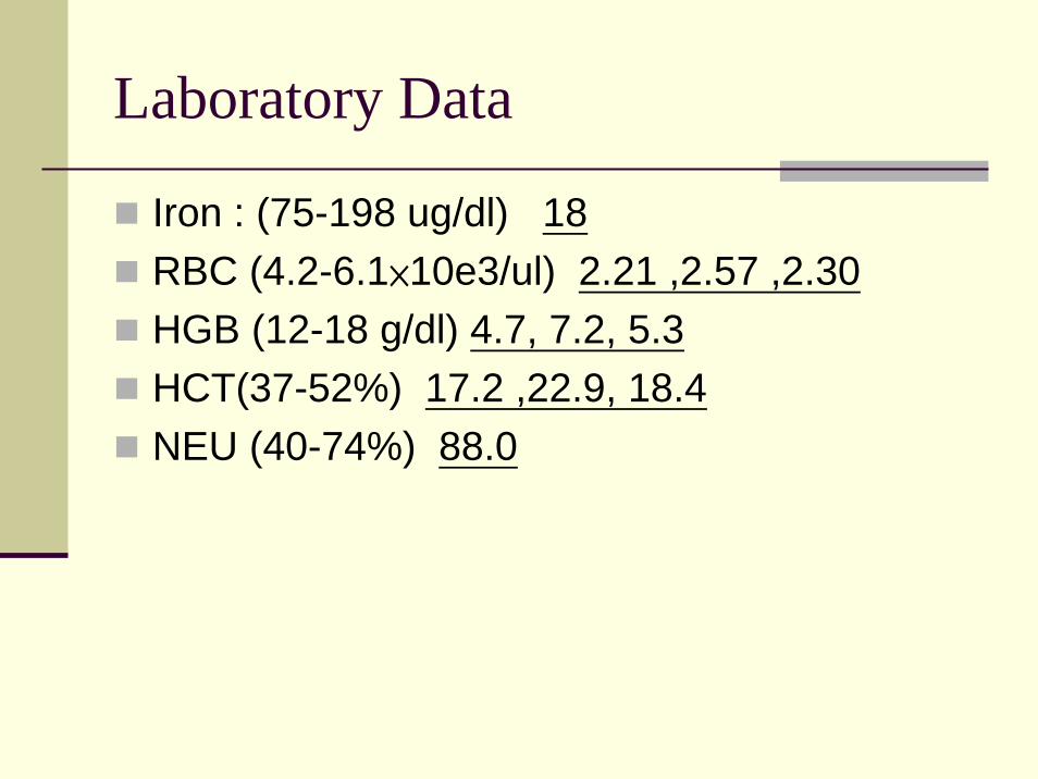
Laboratory Data
Iron : (75-198 ug/dl) 18RBC (4.2-6.1×10e3/ul) 2.21 ,2.57 ,2.30HGB (12-18 g/dl) 4.7, 7.2, 5.3HCT(37-52%) 17.2 ,22.9, 18.4NEU (40-74%) 88.0
Impression
Anemia ,R/O UGI bleedingPneumoniaTuberous sclerosis
Plan
Check iron profileCheck reticulocyte countNPO with NG decompressionCorrect dehydration note I/OArrange Abd CTBlood transfusionBlood culture ,sputum culture and smear
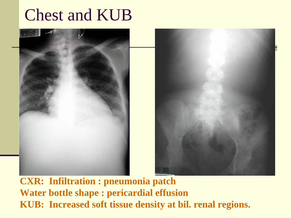
Chest and KUB
CXR: Infiltration : pneumonia patchWater bottle shape : pericardial effusionKUB: Increased soft tissue density at bil. renal regions.
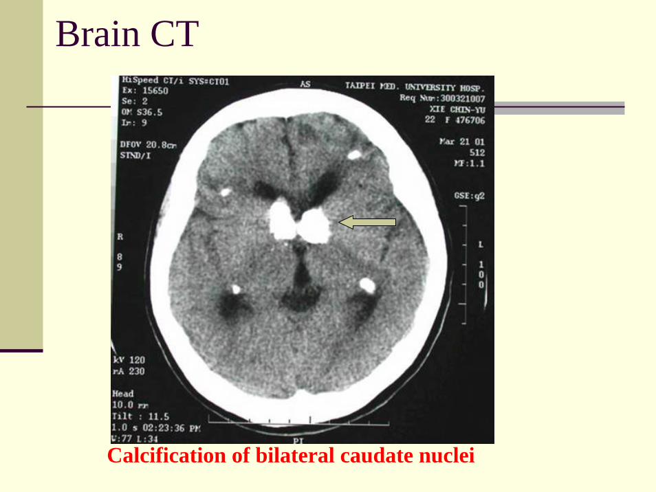
Brain CT
Calcification of bilateral caudate nuclei
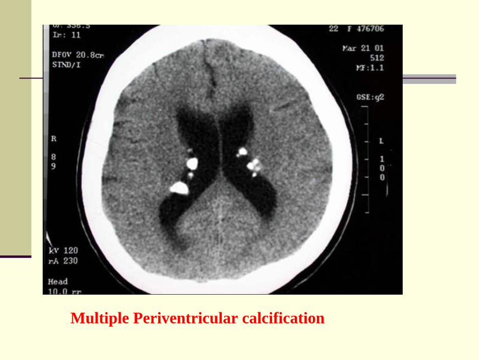
Multiple Periventricular calcification

Abdomen CT scan
Pre-contrast studyHuge mixed-density masses at both renal regions, with superior displacement of the liver and spleen
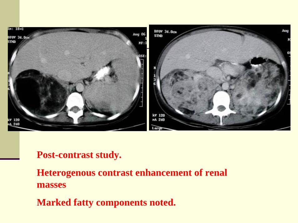
Post-contrast study.
Heterogenous contrast enhancement of renal masses
Marked fatty components noted.
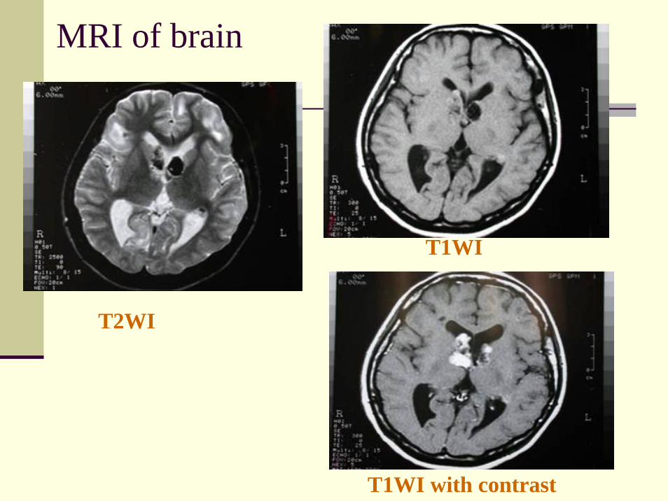
MRI of brain
T2WI
T1WI
T1WI with contrast
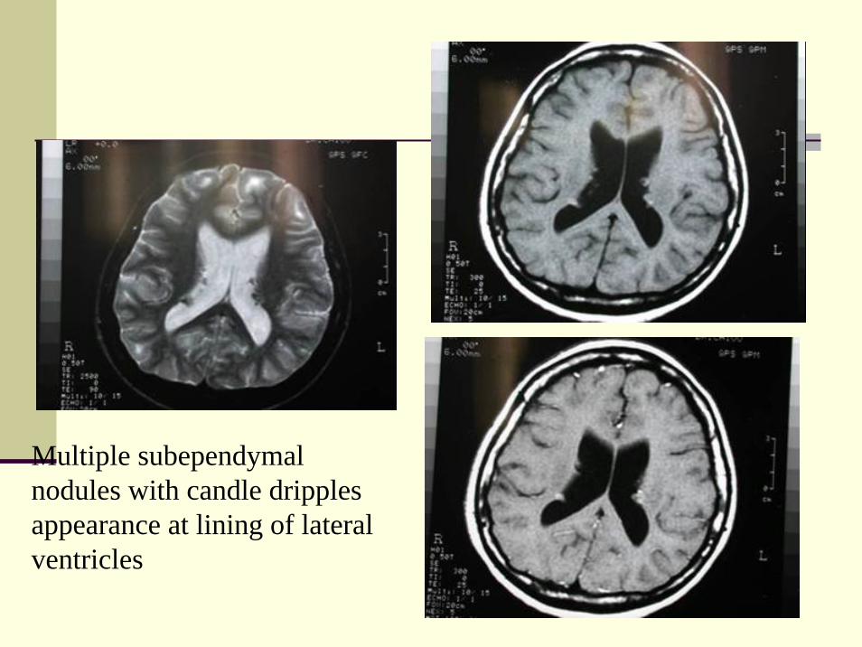
Multiple subependymalnodules with candle dripplesappearance at lining of lateral ventricles
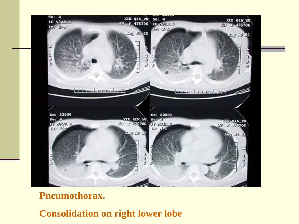
Pneumothorax.
Consolidation on right lower lobe
Imaging finding-1
Brain CT – calcification of bilateral caudate nuclei and multiple periventricular calcifications.MRI of brain – Low signal to iso-signal masses at periventricular region on T1WI and T2WI, with contrast enhancement, consistent with subenpendymal hamartomas.
Imaging finding-2Abdomen CT – Bilateral fatty renal masses, angiomyolipomas are most likely.Lung – Pneumothorax

Differentiation of periventricularcalcifications.
1. Tuberous sclerosis.2. Congenital infection:3. CMV4. Toxoplasmosis
Differential diagnosis of bilateral renal masses-1
1. Malignant tumor- Malignant lymphoma/Hodgkindisease
- Metastases- Renal cell carcinoma- Wilms tumor
Differential diagnosis of bilateral renal masses-2
2. Benign tumor- Angiomyolipoma- Nephroblastomosis
3. Cysts- Polycystic kidney disease(adult or acquired)

Diagnosis:
TUBEROUS SCLEROSIS
Tuberous sclerosis
Neuroectodermal disorder: genetic alternation of ectodermal and mesodermal cells with hyperplasia, with a disturbance in cellular differentiationClinical triad:1. Skin manifestations (96%)2. Seizures (86%)3. Mental retardation (49%)
Etiology
1/3 of cases are inherited as an autosomaldominent trait, others sporatic mutationsTSC1 on chromosome9q34 – hamartinTSC2 on chromosome16q13.3 – tuberinMajority relate to TSC2
Clinical manifestation-CNS
1. Subependymal hamartomas2. Giant cell astrocytoma3. Tubers (cortical/subcortical hamartomas)4. Heterotopic gray matter islands in white matter
Hypomelanotic macules:usually more than 3, 3 to 4 cmlance ovate or ash-leaflet spotsConfetti macules:multiple, discrete, small (1~2 mm) hypopigmented macules. These lesions are pathognomonic.
Clinical manifestation-Skin
Clinical manifestation-Skin
Angiofibroma:occur in the center of face. They are confirm and disseminated but may coalescence.
Ocular – Phakoma (whitish disk-shaped retinal hamartoma =astreocyte proliferation in/near optic disc)
Small calcification in region of optic nerve head
Optic nerve glioma
Renal – Angiomyolipoma (38%) multiple + bilateral risk of spontaneous hemorrhage
Multiple cysts of varying size in cortex + medulla mimicking adult polycystic kidney disease
Renal cell carcinoma(3%)bilateral 40%
Lung – interstitial fibrosis in lower lung fields and miliarynodules pattern (lymphangiomyomatosis)- Cystic change of lung parenchyma- Spontaneous pneumothorax (50 %)- chylothorax- cor pulmonale
Heart- Congenital cardiomyopathy- Rhabdomyloma (5%)- Aortic aneurysm
Other visceral involvement:1. Adenomas + lipomyomas of liver2. Adenomas of pancreas3. Tumors of spleen.

Treatment
Symptomatic – anticonvulsion therapy for control of seizures

Reference
Diagnostic radiology, Grainger and Allison’s,4th editionAdams and Victor’s Neurology, 7th editionColor atlas and synopsis of clinical dermatology, 4th edition