Embed Size (px)
Citation preview
DMD #11502
1
GENETIC POLYMORPHISM OF ALDEHYDE OXIDASE IN DONRYU RATS
Kunio Itoh, Akiko Masubuchi, Takamitsu Sasaki, Mayuko Adachi, Nobuaki Watanabe,
Kiyoshi Nagata, Yasushi Yamazoe, Masahiro Hiratsuka, Michinao Mizugaki, and
Yorihisa Tanaka
Department of Drug Metabolism and Pharmacokinetics, Tohoku Pharmaceutical
University, Sendai, Japan (K.I., A.M., T.S., M.A., Y.T.); Department of Clinical
Pharmaceutics, Tohoku Pharmaceutical University, Sendai, Japan (M.H., M.M.);
Department of Drug Metabolism and Molecular Toxicology, Graduate School of
Pharmaceutical Sciences, Tohoku University, Sendai, Japan (K.N., Y.Y.); Drug
Metabolism and Pharmacokinetics Research Laboratories, Sankyo Co. Ltd., Tokyo,
Japan (N.W.)
DMD Fast Forward. Published on February 9, 2007 as doi:10.1124/dmd.106.011502
Copyright 2007 by the American Society for Pharmacology and Experimental Therapeutics.
This article has not been copyedited and formatted. The final version may differ from this version.DMD Fast Forward. Published on February 9, 2007 as DOI: 10.1124/dmd.106.011502
at ASPE
T Journals on July 19, 2018
dmd.aspetjournals.org
Dow
nloaded from
DMD #11502
2
Running title: Genetic polymorphism of aldehyde oxidase in Donryu rats
Corresponding author: Dr. Yorihisa Tanaka; Department of Drug Metabolism and
Pharmacokinetics; Tohoku Pharmaceutical University; 4-4-1 Komatsushima; Aoba-ku,
Sendai 981-8558; Japan. E-mail: [email protected]
Number of text pages: 35
Number of figures: 4
Number of references: 24
Number of words in the Abstract: 249
Number of words in the Introduction: 454
Number of words in the Discussion: 1261
ABBREVIATIONS: MAO, monoamine oxidase; SD, Sprague-Dawley; PM, poor
metabolizer; EM, extensive metabolizer; UM, ultrarapid metabolizer; PCR, polymerase
chain reaction; SDS-PAGE, sodium dodecyl sulfate polyacrylamide gel electrophoresis;
This article has not been copyedited and formatted. The final version may differ from this version.DMD Fast Forward. Published on February 9, 2007 as DOI: 10.1124/dmd.106.011502
at ASPE
T Journals on July 19, 2018
dmd.aspetjournals.org
Dow
nloaded from
DMD #11502
3
pI, isoelectric point; SNP, single nucleotide polymorphism
This article has not been copyedited and formatted. The final version may differ from this version.DMD Fast Forward. Published on February 9, 2007 as DOI: 10.1124/dmd.106.011502
at ASPE
T Journals on July 19, 2018
dmd.aspetjournals.org
Dow
nloaded from
DMD #11502
4
ABSTRACT:
One of major metabolic pathways of RS-8359, a selective and reversible MAO-A
inhibitor, is the aldehyde oxidase catalyzed 2-hydroxylation at the pyrimidine ring.
Donryu rats showed a dimorphic pattern for the 2-oxidation activity with about
20~40-fold variations in the Vmax/Km values between a low and a high activity group.
The rats were classified as extensive metabolizers (EM) and poor metabolizers (PM) of
RS-8359, of which ratios were approximately 1:1. One rat among the EM rats of each sex
showed extremely high activity and were referred to as ultrarapid metabolizers (UM).
There was no significant difference in the expression levels of mRNA of aldehyde
oxidase between the EM and PM rats. Analysis of nucleotide sequences demonstrated
four substitutions, of which the substitutions at 377G>A and 2604C>T caused
110Gly-Ser and 852Ala-Val amino changes, respectively. Amino acid residue 110 is
located very near the second Fe-S center of aldehyde oxidase. Its change from non-chiral
Gly to chiral Ser might result in a conformational change of aldehyde oxidase protein
with the shift of pI value from 5.0 in the EM rats to 6.2 in the PM rats. The 110Gly-Ser
amino acid substitution (377G>A) might be primarily responsible for the variations of
This article has not been copyedited and formatted. The final version may differ from this version.DMD Fast Forward. Published on February 9, 2007 as DOI: 10.1124/dmd.106.011502
at ASPE
T Journals on July 19, 2018
dmd.aspetjournals.org
Dow
nloaded from
DMD #11502
5
aldehyde oxidase activity observed in Donryu rats in addition to the difference of
expression levels of aldehyde oxidase protein. If a new drug candidate is primarily
metabolized by aldehyde oxidase, attention should be given to using a rat strain with high
aldehyde oxidase and small individual variation.
This article has not been copyedited and formatted. The final version may differ from this version.DMD Fast Forward. Published on February 9, 2007 as DOI: 10.1124/dmd.106.011502
at ASPE
T Journals on July 19, 2018
dmd.aspetjournals.org
Dow
nloaded from
DMD #11502
6
RS-8359, [(±)-4-(4-cyanoanilino)-5,6-dihydro-7-hydroxy-7H-cyclopenta[d]-pyrimidine],
is a reversible and selective MAO-A inhibitor (Yokoyama et al., 1989; Miura et al., 1993),
which has been developed as an antidepressant (Puchler et al., 1997; Plenker et al., 1997).
One of the major metabolic pathways of RS-8359 is aldehyde oxidase-catalyzed
2-oxidation on the pyrimidine ring to give the 2-keto metabolite, which is preferential in
rats, monkeys, and humans (Itoh et al., 2005). Other pathways were hydroxylation on the
cyclopentanol ring to cis-diol and trans-diol catalyzed by cytochrome P450, which is
preferential in mice (Itoh et al., 2006), and glucuronidation catalyzed by
UDP-glucuronosyl transferase, which is preferential in dogs (Iwabuchi et al., 1998). All
of these major metabolic events proceed with high enantioselectivity for the
(S)-enantiomer that leads to more rapid disappearance of the (S)-enantiomer from plasma
in every animal species. In particular, monkeys and humans have an extremely high
aldehyde oxidase activity that results in an AUC(R)/AUC(S) ratio of 238 in monkeys and
is nearly negligible (S)-enantiomer in human plasma (Takasaki et al., 2005). There were
no large variations in the in vitro 2-oxidation activity of RS-8359 using five human liver
cytosol samples. The pharmacokinetic profiles of RS-8359 in thirty-six volunteers
This article has not been copyedited and formatted. The final version may differ from this version.DMD Fast Forward. Published on February 9, 2007 as DOI: 10.1124/dmd.106.011502
at ASPE
T Journals on July 19, 2018
dmd.aspetjournals.org
Dow
nloaded from
DMD #11502
7
conducted in clinical trials of RS-8359 (Puchler et al., 1997) showed reasonable
coefficients of variation for Cmax, AUC, and half-life. These results suggest that there is
no appreciable polymorphism in the aldehyde oxidase-catalyzed 2-oxidation of RS-8359
although the sample number was small. Indeed, genetic polymorphism of aldehyde
oxidase has not been reported in humans (Beedham et al., 2003) whereas a large variation
of in vitro activity has been well known (Kitamura et al., 1999; Al-Salmy, 2001).
Aldehyde oxidase (EC 1.2.3.1) is a major member of a relatively small family of
molybdenum hydroxylases that include xanthine oxidase (Beedham, 1985, 1987a, 1997,
2002; Kitamura et al., 2006). Aldehyde oxidase catalyzes the oxidation of a wide range of
endogenous and exogenous aldehydes and N-heterocyclic aromatic compounds.
N-heterocyclic drugs such as methotrexate, 6-mercaptopurine, cinchona alkaloids, and
famciclovir are oxidized by this enzyme (Beedham et al., 2002; Kitamura et al., 2006).
Further, aldehyde oxidase can catalyze the reduction of a variety of functional groups
including sulfoxides, N-oxides, azo dyes, and N-hydroxycarbamoyl substituents in the
presence of an appropriate donor (Kitamura et al., 2006).
Marked species differences have been well documented for the aldehyde oxidase-
This article has not been copyedited and formatted. The final version may differ from this version.DMD Fast Forward. Published on February 9, 2007 as DOI: 10.1124/dmd.106.011502
at ASPE
T Journals on July 19, 2018
dmd.aspetjournals.org
Dow
nloaded from
DMD #11502
8
catalyzed metabolism of drugs including methotrexate (Kitamura et al., 1999a; Jordan et
al., 1999) and famciclovir (Rashidi et al., 1997). A large rat strain variation was
demonstrated in the oxidation activity of benzaldehyde (Sugihara et al., 1995) and
methotrexate (Kitamura et al., 1999b). The genetic variation in aldehyde oxidase was also
reported by the electrophoresis method (Kunieda et al., 1999). Similar to those reports,
we observed remarkable species differences and rat strain differences in the metabolism
of the (S)-enantiomer of RS-8359 (Itoh et al., 2006; Sasaki et al., 2006). During the study
of rat strain differences in the 2-oxidation activity of RS-8359, we were aware of the
individual variations in Donryu rats, as demonstrated in Wistar rats (Gluecksohn-Waelsch
et al., 1967) and SD rats (Beedham et al., 1998). In the current study, we examined the
mechanism of individual variations of aldehyde oxidase in Donryu rats using the
technology of molecular biology.
This article has not been copyedited and formatted. The final version may differ from this version.DMD Fast Forward. Published on February 9, 2007 as DOI: 10.1124/dmd.106.011502
at ASPE
T Journals on July 19, 2018
dmd.aspetjournals.org
Dow
nloaded from
DMD #11502
9
Materials and Methods
Chemicals and Reagents. RS-8359, its (S)-enantiomer, and the 2-keto metabolite
were supplied by Ube Kosan Co. Ltd. (Yamaguchi, Japan). Hydrocortisone, an internal
standard of HPLC analysis, was purchased from Sigma Chemical Co. (St. Louis, MO).
All other reagents were of reagent grade.
Preparation of Liver Cytosolic Fractions. Eight-week-old male rats of Crj:Donryu
and Iar:Wistar (Wistar-Imamichi) strains were purchased from Charles River Japan
(Yokohama, Japan) and Imamichi Institute for Animal Reproductions (Saitama, Japan),
respectively. The animals were housed according to the Guidelines for Animal
Experimentation (Tohoku Pharmaceutical University) in cages in rooms with a
unidirectional airflow at a controlled temperature (22±2°C), relative humidity (50±10%),
and 12-h light/dark cycles (07.00-19.00 hours). Tap water was available ad libitum and
CE2 food (Clea Japan, Tokyo, Japan) was available ad libitum except for overnight
fasting before use. The animals were sacrificed by bleeding from the carotid artery under
anesthesia and their livers were immediately extracted. The livers were homogenized in
This article has not been copyedited and formatted. The final version may differ from this version.DMD Fast Forward. Published on February 9, 2007 as DOI: 10.1124/dmd.106.011502
at ASPE
T Journals on July 19, 2018
dmd.aspetjournals.org
Dow
nloaded from
DMD #11502
10
three volumes of 10 mM phosphate buffer (pH 7.4) containing 1.15% KCl and 100 µM
phenylmethanesulfonyl fluoride by a Potter-Elvehjem Teflon homogenizer. The cytosolic
fractions were prepared by successive centrifugation at 9000 g for 20 min and then at
105000 g for 60 min. The protein concentration was determined using BCA Protein
Assay Reagent (Pierce Biotech, Rockford, IL) with bovine serum albumin as the
standard.
Enzyme Activity Assay. The (S)-enantiomer of RS-8359 (3.1 µM~0.2 mM) was
incubated at 37°C for 10 min in a reaction mixture (0.25 ml) consisting of 80 mM
phosphate buffer (pH 7.4), 1.0 mM K3Fe(CN)6, 0.13 mM EDTA, and prepared cytosol
(20 mg/ml, 0.10 ml). The reaction was stopped by the addition of acetonitrile (0.50 ml)
containing 0.2 mg/ml of hydrocortisone as an internal standard after which the mixture
was centrifuged at 5000 g for 3 min. Aliquots (25 µl) of the supernatant were analyzed
for quantification of the oxidation product by reverse-phase HPLC on a YMC ODS
A-312 column (6.0 mm i.d. x 150 mm, YMC Co. Ltd., Kyoto, Japan). A mobile phase
was composed of acetonitrile/0.5% ammonium acetate (14:86); the flow rate was 1.0
ml/min. The HPLC instrument was a Shimadzu model 6A High Performance Liquid
This article has not been copyedited and formatted. The final version may differ from this version.DMD Fast Forward. Published on February 9, 2007 as DOI: 10.1124/dmd.106.011502
at ASPE
T Journals on July 19, 2018
dmd.aspetjournals.org
Dow
nloaded from
DMD #11502
11
Chromatograph System (Shimadzu Seisakusho Co. Ltd., Kyoto, Japan). The peaks were
monitored for absorbance at 315 nm and the peak area was calculated on a Chromatopak
C-R4A (Shimadzu Seisakusho).
Purification of Aldehyde Oxidase. Before purification, aldehyde oxidase activity
was measured in the liver cytosolic fractions from Crj:Donryu rats. The rats were divided
into low and high aldehyde oxidase activity groups and then the enzyme was purified
from each group according to the method described previously (Itoh et al., 2005). Briefly,
the cytosolic fraction was kept at 60°C for 10 min and then the precipitated proteins were
separated by centrifugation for 10 min at 9000 g. Ammonium sulfate was added to the
supernatant to 50% saturation. The protein precipitates were collected by centrifugation,
dissolved in 10 mM phosphate buffer (pH 7.4), and filtered through a 0.45 µm disk filter.
The filtrate was applied to a Benzamidine Sepharose 6B column (3 x 22 cm) followed by
Mono Q HR5/5 column (0.5 x 5.0 cm) chromatography. The purified enzymes showed a
single band on sodium dodecyl sulfate-polyacrylamide gel electrophoresis (SDS-PAGE)
performed on PhastGel Gradient 4-15 (Amersham Bioscience, Uppsala, Sweden).
Isoelectric Electrophoresis. Electrophoresis of the purified aldehyde oxidase (1 µg)
This article has not been copyedited and formatted. The final version may differ from this version.DMD Fast Forward. Published on February 9, 2007 as DOI: 10.1124/dmd.106.011502
at ASPE
T Journals on July 19, 2018
dmd.aspetjournals.org
Dow
nloaded from
DMD #11502
12
was performed on PhastGel IEF 3-9 (Amersham Biosciences). The gels were stained for
protein with Coomassie Tablet PhastGel Blue R-350 (Amersham Biosciences). A pI
Calibration Kit 3-10 (Amersham Biosciences) was used for isoelectric point markers.
Preparation of Anti-rat Liver Aldehyde Oxidase Antisera. Aldehyde oxidase was
isolated and purified from liver cytosolic fractions of Wistar-Imamichi rats according to
the method described previously (Itoh et al., 2005). The antibody preparation was
conducted at Trans Genic Inc. (Kumamoto, Japan). The purified enzyme (1.0 mg
protein/ml) was emulsified with an equal volume of Freund’s complete adjuvant (Sigma,
St. Louis, MO). Each of two rabbits was immunized with 1 ml of immunogen by
intradermal injections every 4 weeks. Four months after the first immunization, blood
was taken by cardiac puncture and antisera were prepared by a conventional method.
Pooled antisera were stored at -80°C until use for Western blot analysis.
cDNA Synthesis and Real-time PCR. The PCR reaction and subsequent cloning
was performed to generate the standard for quantitative PCR of aldehyde oxidase and
glyceraldehyde-3-phosphate dehydrogenase (GAPDH) as follows. Total RNA was
isolated from rat liver with TRIzol (Invitrogen, Carlsbad, CA) according to the
This article has not been copyedited and formatted. The final version may differ from this version.DMD Fast Forward. Published on February 9, 2007 as DOI: 10.1124/dmd.106.011502
at ASPE
T Journals on July 19, 2018
dmd.aspetjournals.org
Dow
nloaded from
DMD #11502
13
manufacturer's instructions. An aliquot of 1 µg of total RNA was used to synthesize the
first-strand cDNA with SuperScript II (Invitrogen). PCR amplification was conducted
with cDNA (1 µg), Ex-Taq polymerase, and respective oligonucleotide primers for
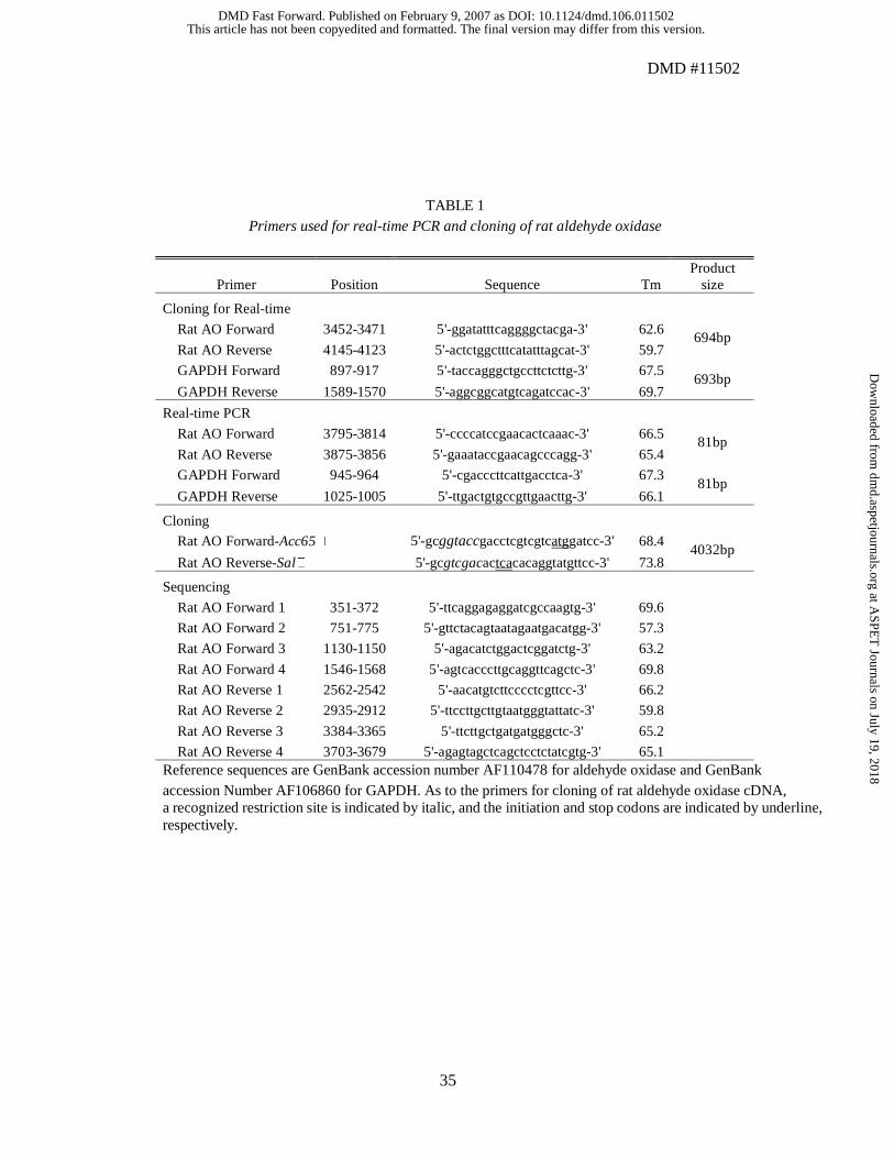
aldehyde oxidase and GAPDH, as shown in Table 1. The primers for aldehyde oxidase
were designed with reference to the nucleotide sequences of male SD rat aldehyde
oxidase (Wright et al., 1999). The PCR conditions were as follows: denaturation at 95°C
for 1 min, 25 amplification cycles of 94°C for 30 s, 62°C for 30 s, and 72°C for 1 min, and
a final extension of 72°C for 5 min. The amplified DNA fragment (1 µg) was subcloned in
the pCRII-TOPO vector using a TOPO TA Cloning Kit (Invitrogen). The resulting
plasmids were purified using a Wizard Plus Minipreps DNA Purification System
(Promega Co., Madison, WI). The DNA sequences of the products were determined by a
CEQ 8000 Analysis System (Beckman-Coulter Inc., Fullerton, CA) with a DTCS Quick
Start Kit (Beckman-Coulter) according to the recommended protocol. The respective
standard curves for aldehyde oxidase and GAPDH were constructed using serial dilutions
of plasmid DNA to determine the amount of template in each reaction. Plasmid DNAs
were linearized and quantified by spectrophotometry for amplification.
This article has not been copyedited and formatted. The final version may differ from this version.DMD Fast Forward. Published on February 9, 2007 as DOI: 10.1124/dmd.106.011502
at ASPE
T Journals on July 19, 2018
dmd.aspetjournals.org
Dow
nloaded from
DMD #11502
14
Quantitative real-time PCR analyses were performed using the PE ABI 7700 PRISM
Sequence Detection System (Perkin-Elmer Life Science, Boston, MA, USA) with SYBR
Green PCR Master Mix (Applied Biosystems, Warrington, UK), reverse transcribed
cDNA (1 µg), and gene-specific primers, shown in Table 1, which were designed using
Primer Express Software (PE Applied Biosystems). The PCR reactions were performed
at 95°C for 10 min, followed by 35 cycles of 95°C for 30 s, 60°C for 30 s, and 72°C for 30
s. At Ct 22~25, the RT-PCR products were confirmed by visualization on 1% TAE
agarose gel with ethidium bromide staining. All reactions were performed in triplicate to
confirm reproducibility and included a negative control without template to verify that no
primer-dimers were being generated.
Western Blot Analysis. Cytosolic proteins were separated by SDS-PAGE, which was
performed on PhastGel gradient 8-25 in PhastGel SDS buffer strips (Amersham
Bioscience). The proteins were transferred electrophoretically to a polyvinylidene
difluoride (PVDF) membrane (Daiichi Pure Chemicals Co. Ltd., Ibaraki, Japan) in
transfer buffer (15% methanol containing Tris 25 mM and glycine 192 mM, pH 8.3).
Detection of aldehyde oxidase was performed using an ECF Western Blotting Kit
This article has not been copyedited and formatted. The final version may differ from this version.DMD Fast Forward. Published on February 9, 2007 as DOI: 10.1124/dmd.106.011502
at ASPE
T Journals on July 19, 2018
dmd.aspetjournals.org
Dow
nloaded from
DMD #11502
15
(Amersham Bioscience). The membrane was blocked with 5% nonfat dry milk in
phosphate-buffered saline (PBS), and then incubated successively with a primary rabbit
anti-rat aldehyde oxidase antibody at 0.5 µg/ml, a secondary antibody (anti-rabbit
fluorescein-linked whole antibody) at a dilution of 1:600, and a tertiary antibody
(anti-fluorescein alkaline phosphatase conjugate) at a dilution of 1:2500. The blocking
and incubation at each immunoreaction step were performed at room temperature for 1 hr,
and the membrane was washed two or three times with PBS containing 0.1% Tween-20
(PBST). The target proteins on the membrane were detected by the enhanced
chemifluorescence (ECF) detection system (Amersham Bioscience). Relative densities
were measured by a Fluoro-image Analyzer FLA-3000G (Fuji Photo Film Co. Ltd.,
Kanagawa, Japan). An HMW Calibration Kit (Amersham Bioscience) was used for the
molecular weight standards.
Nucleotide Sequences of Aldehyde Oxidase cDNA. PCR fragments corresponding to
4032 bp of aldehyde oxidase were prepared using the cDNA synthesized above as the
template. The primers used are listed in Table 1. The reaction was performed in a 50 µl
solution containing 1×Ex-Taq buffer, 200 µM dNTP solution, 2.5 U Ex-Taq DNA
This article has not been copyedited and formatted. The final version may differ from this version.DMD Fast Forward. Published on February 9, 2007 as DOI: 10.1124/dmd.106.011502
at ASPE
T Journals on July 19, 2018
dmd.aspetjournals.org
Dow
nloaded from
DMD #11502
16
polymerase (TaKaRa Bio Inc., Shiga, Japan), 2 mM MgCl2, 1 µM of each primer, and 1 µl
of the cDNA template. PCR amplification was performed using a PCR thermal cycler MP
(TaKaRa) under the following conditions: initial denaturation at 94°C for 5 min, 35
cycles of 94°C for 30 s, 55°C for 30 s, 72°C for 4 min, and a final extension of 72°C for 7
min. The amplified DNA fragments were cloned with the TOPO TA Cloning Kit
(Invitrogen). The resulting plasmids were purified using a Wizard Plus Minipreps DNA
Purification System (Promega). The insertion of aldehyde oxidase cDNA was confirmed
by agarose gel electrophoresis after digestion with Sac I. The DNA sequences were
determined by a CEQ 8000 Analysis System with a DTCS Quick Start Kit. In addition to
the M13 sequence primer, the primers listed in Table 1 were used for sequencing.
Statistical Analysis. The results are expressed as the mean±SE for the number of
experiments. Statistical significance was compared between low and high activity groups
by a Student's t-test. Values with p < 0.05 were considered statistically significant.
TABLE 1
This article has not been copyedited and formatted. The final version may differ from this version.DMD Fast Forward. Published on February 9, 2007 as DOI: 10.1124/dmd.106.011502
at ASPE
T Journals on July 19, 2018
dmd.aspetjournals.org
Dow
nloaded from
DMD #11502
17
Results
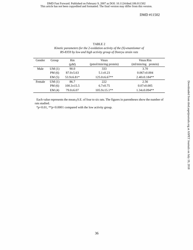
Individual Variations of Aldehyde Oxidase Activity in Donryu Rats. Aldehyde
oxidase activity was determined in the liver cytosolic fractions from 12 male and 11
female Donryu rats using the (S)-enantiomer of RS-8359 as a substrate. The enzyme
activity (pmol/min/mg protein) of individual rats together with their average data is
shown in Fig. 1. The results clearly indicate the presence of two groups with low and high
activity at a frequency ratio of about 1:1. The two groups were classified as poor
metabolizers (PM) and extensive metabolizers (EM) of RS-8359. The kinetic parameters
calculated from Michaelis-Menten plots are summarized in Table 2. The Vmax/Km
values of the EM group were approximately 20~40-fold greater than were those of the
PM group. Significantly larger Vmax values (p <0.0001) were observed in the EM group
than in the PM group. The Km values were significantly smaller (p <0.05) in the EM rats
than in the PM rats in males whereas there were no significant differences in females.
Thus, the differences in the intrinsic clearance were essentially due to the significant
differences in the Vmax values. One rat of each sex among the EM rats showed an
This article has not been copyedited and formatted. The final version may differ from this version.DMD Fast Forward. Published on February 9, 2007 as DOI: 10.1124/dmd.106.011502
at ASPE
T Journals on July 19, 2018
dmd.aspetjournals.org
Dow
nloaded from
DMD #11502
18
extremely high Vmax value of 220~330 pmol/min/mg protein whereas their Km values
(about 90 µM) were on an order similar to those of the other rats. The two individuals are
referred to as ultrarapid metabolizers (UM).
FIG. 1
TABLE 2
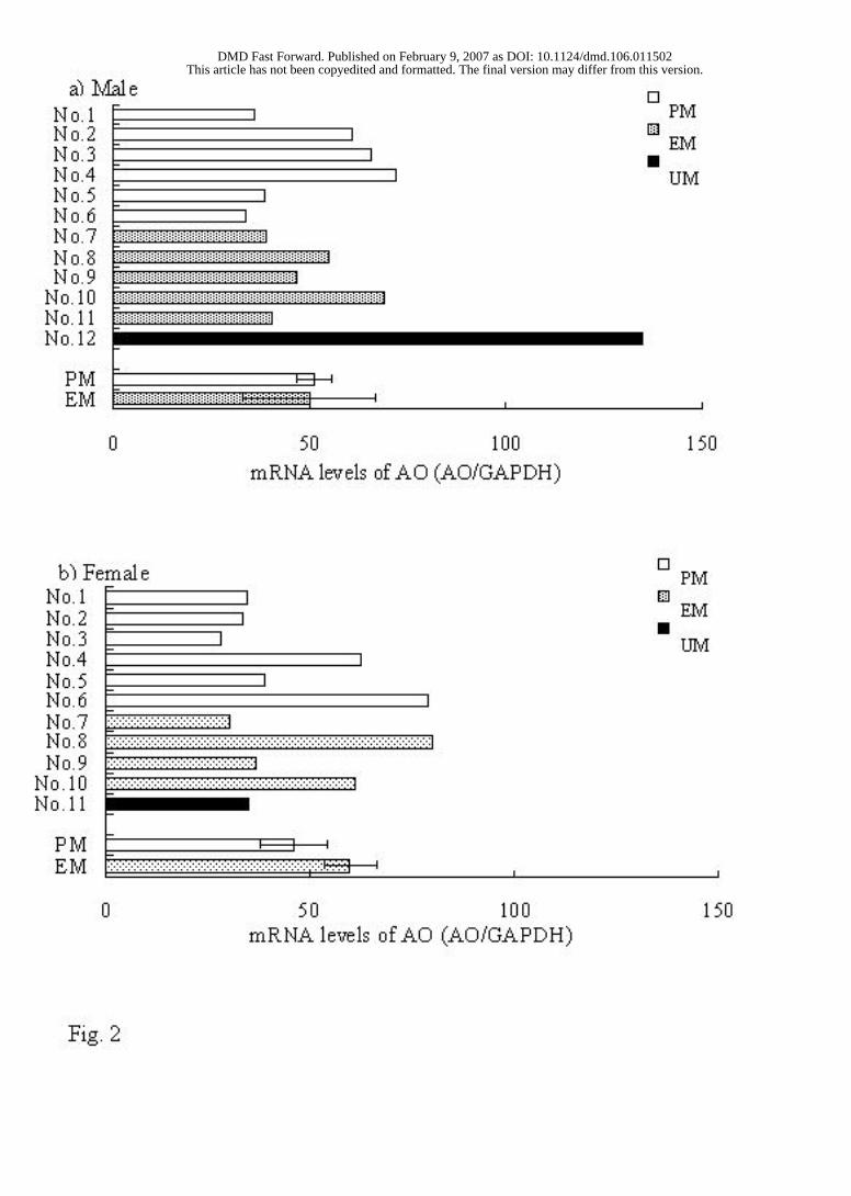
Real-time PCR Analysis. Fig. 2 shows the individual data of the mRNA expression
levels of aldehyde oxidase normalized by that of GAPDH and the average data. No
significant correlation was observed in the mRNA expression levels between the PM and
EM rats.
FIG. 2
This article has not been copyedited and formatted. The final version may differ from this version.DMD Fast Forward. Published on February 9, 2007 as DOI: 10.1124/dmd.106.011502
at ASPE
T Journals on July 19, 2018
dmd.aspetjournals.org
Dow
nloaded from
DMD #11502
19
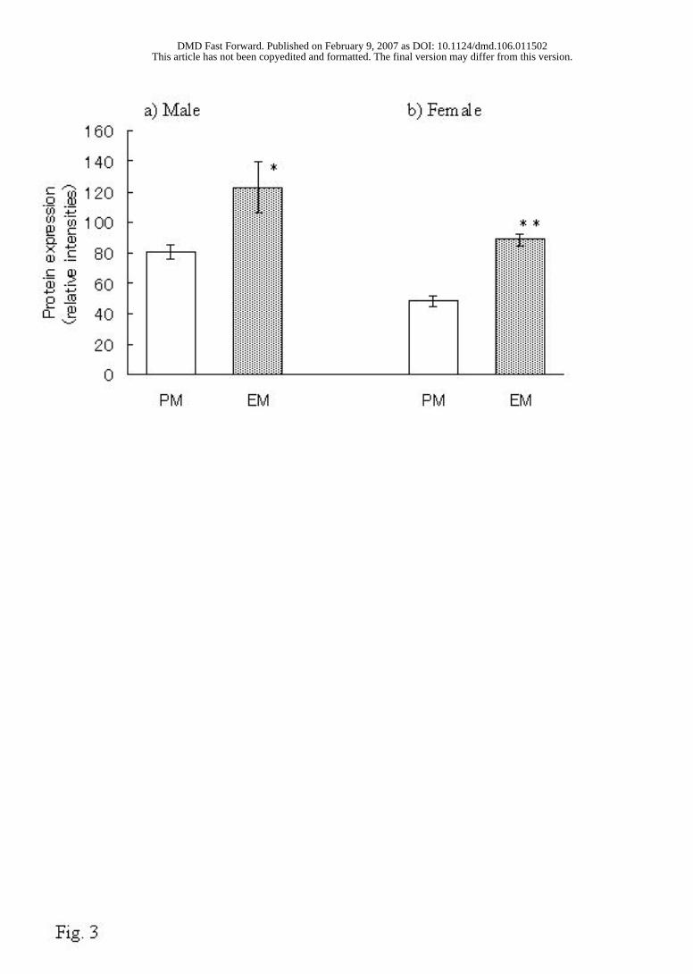
Western Blot Analysis. The immunoreactive protein levels of aldehyde oxidase were
measured by Western blot. After detection of immunoreactive bands by the enhanced
chemifluorescence (ECF) system, their relative densities were measured by FLA-3000G
(Fuji Photo Film, Tokyo, Japan). The relative density of the EM rats was stronger than
that of the PM rats (p <0.05 in males and p <0.01 in females), as shown in Fig. 3.
FIG. 3
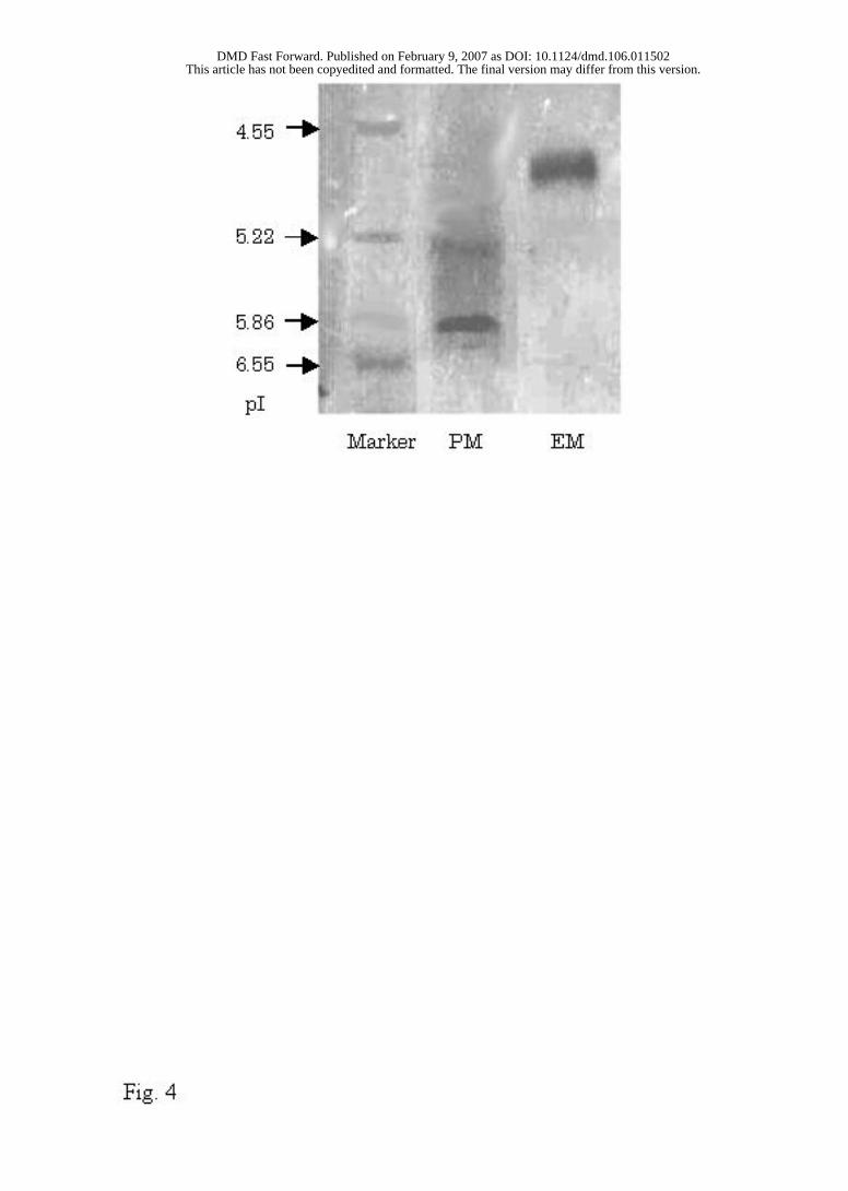
Isoelectric Electrophoresis. Aldehyde oxidase purified from the respective liver
cytosolic fractions of the PM and EM Donryu rats exhibited one band on SDS-PAGE.
The isoelecric point (pI) of the purified aldehyde oxidase was determined by isoelectric
electrophoresis (Fig. 4). Different pIs were observed for the EM rats (5.0) and the PM rats
(6.2).
FIG. 4
This article has not been copyedited and formatted. The final version may differ from this version.DMD Fast Forward. Published on February 9, 2007 as DOI: 10.1124/dmd.106.011502
at ASPE
T Journals on July 19, 2018
dmd.aspetjournals.org
Dow
nloaded from
DMD #11502
20
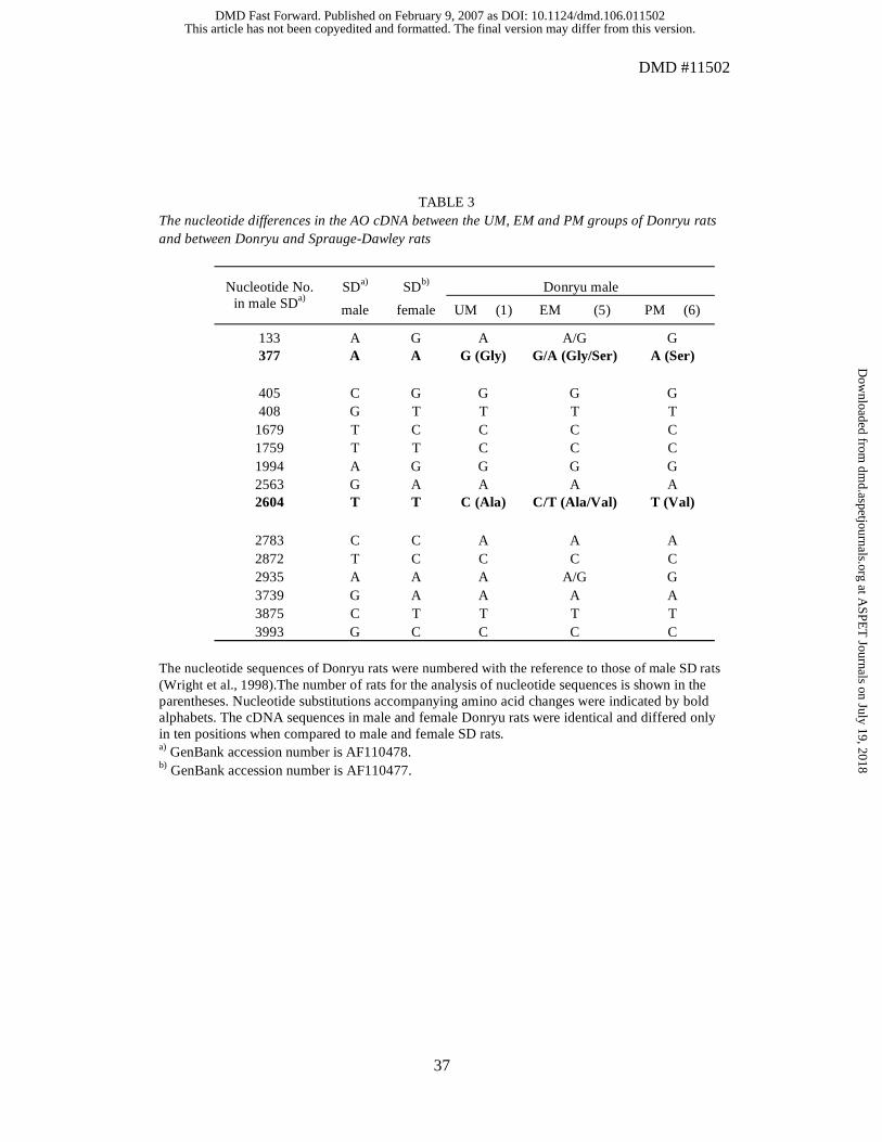
Nucleotide Sequences of Aldehyde Oxidase from PM and EM Rats. With reference to
the reported nucleotide sequences of aldehyde oxidase of the SD rats (AOX1 cDNA;
GenBank accession number: AF110478), full-length cDNA was cloned and sequenced
from the livers of 12 PM rats (6 males and 6 females), 9 EM rats (5 males and 4 females),
and 2 UM rats (1 male and 1 female) from the Donryu strain. Four nucleotide
substitutions were observed among the three phenotypes. PMs, EMs, and UMs had SNP
patterns as listed in Table 3. The SNPs at positions 377 and 2604 accompanied amino acid
changes of 110Gly-Ser and 852Ala-Val, respectively. All rats used in this study were
confirmed to show the respective SNPs according to their aldehyde oxidase activity. The
UM and PM rats were homozygous at all four nucleotide positions whereas the EM rats
were heterozygous. As demonstrated in this paper, the SNPs at positions 377 and 2604
were in accordance with the aldehyde oxidase activity. Both the male and female rats
showed the same nucleotide substitutions and amino acid changes. Concomitantly, three
SNPs, 1759T(SD)>C(Donryu), 2783C(SD)>A(Donryu), and 2935A(SD)>G(Donryu)
were detected between the female SD rats and the Donryu PM rats.
This article has not been copyedited and formatted. The final version may differ from this version.DMD Fast Forward. Published on February 9, 2007 as DOI: 10.1124/dmd.106.011502
at ASPE
T Journals on July 19, 2018
dmd.aspetjournals.org
Dow
nloaded from
DMD #11502
21
TABLE 3
This article has not been copyedited and formatted. The final version may differ from this version.DMD Fast Forward. Published on February 9, 2007 as DOI: 10.1124/dmd.106.011502
at ASPE
T Journals on July 19, 2018
dmd.aspetjournals.org
Dow
nloaded from
DMD #11502
22
Discussion
One of the major metabolic pathways of RS-8359 is the 2-oxidation catalyzed by
aldehyde oxidase (Iwabuchi et al., 1998; Takasaki et al., 2005). During the study of rat
strain differences in the activity, we were aware of the individual variations in Donryu
rats and carefully investigated the phenomenon from the perspective of molecular
biology.
The 2-oxidation activity was clearly divided into low and high activity groups with a
frequency ratio of approximately 1:1. This indicated that Donryu rats showed a dimorphic
pattern for the 2-oxidation activity, and so they were classified as poor metabolizers (PM),
and extensive metabolizers (EM) of RS-8359. The EM rats had Km/Vmax values about
20~40-fold greater than did those of the PM rats that were primarily due to the difference
in Vmax values. One rat among the EM rats of each sex showed an extremely high
2-oxidation activity that was referred to as an ultrarapid metabolizer (UM). The oxidation
activity of the UM rat was not included in the summary of the kinetic parameters of the
EM rats. As to the apparent polymorphism of aldehyde oxidase, there are at least two
This article has not been copyedited and formatted. The final version may differ from this version.DMD Fast Forward. Published on February 9, 2007 as DOI: 10.1124/dmd.106.011502
at ASPE
T Journals on July 19, 2018
dmd.aspetjournals.org
Dow
nloaded from
DMD #11502
23
reports. Using N1-methylnicotinamide as a substrate in Wistar rats, Gluecksohn-Waelsch
et al. (1967) reported that the activity was appreciable in only 36 out of 76 rats. Beedham
et al. (1998) also showed that 60% of SD rats were deficient of the functional activity of
aldehyde oxidase. The occurrence frequency of PM in Donryu rats was approximately
60% that were roughly comparable to those already reported in Wistar and SD rats. In
addition to a marked inter-strain difference in aldehyde oxidase in rats, a large intra-strain
difference is obvious in SD, Wistar, and Donryu rats. Generally, a proper selection of
animal models is needed for the pharmacological, toxicological, and pharmacokinetic
studies during development of a new drug. If a new drug candidate is primarily
metabolized by aldehyde oxidase, attention should be given to using a rat strain that has
high aldehyde oxidase activity as human and small individual variation.
The levels of mRNA and protein were analyzed by real-time PCR and Western blot,
respectively, to look for the reasons of polymorphism of aldehyde oxidase in Donryu rats.
The mRNA levels showed no significant differences between the EM and PM rats (p =
0.452 in males and p = 0.339 in females). On the other hand, the expression levels of
immunoreactive protein were significantly higher in the EM rats than were those in the
This article has not been copyedited and formatted. The final version may differ from this version.DMD Fast Forward. Published on February 9, 2007 as DOI: 10.1124/dmd.106.011502
at ASPE
T Journals on July 19, 2018
dmd.aspetjournals.org
Dow
nloaded from
DMD #11502
24
PM rats (p <0.05 in male and p <0.01 in female). However, the PM rats still maintained
approximately 60% of aldehyde oxidase protein level of the EM rats. This suggests that
the low expression levels of aldehyde oxidase protein only partially explain the extremely
low aldehyde oxidase activity in the PM rats. Analysis of the full-length nucleotide
sequences of the open reading frame of aldehyde oxidase revealed the four nucleotide
substitutions between the UM, EM, and PM Donryu rats regardless of sex. Among those,
the SNPs at 377 and 2604 accompanied the respective amino acid changes of 110Gly-Ser
and 852Ala-Val, as shown in Table 3. All rats used in this study showed the respective
SNPs according to their aldehyde oxidase activity. Thus, the UM and PM rats were
homozygous at all four nucleotide positions whereas the EM rats were heterozygous. In
the amino acid sequences of aldehyde oxidase of the Donryu PM rats, only one among
1333 amino acids was changed from His to Asn compared to those of female SD rats
reported by Wright et al (1998). However, the cDNA sequences in male and female
Donryu rats were identical and differed only in ten positions when compared to male and
female SD rats (Wright et al., 1998).
Aldehyde oxidase is a homodimer with a monomeric molecular weight of about
This article has not been copyedited and formatted. The final version may differ from this version.DMD Fast Forward. Published on February 9, 2007 as DOI: 10.1124/dmd.106.011502
at ASPE
T Journals on July 19, 2018
dmd.aspetjournals.org
Dow
nloaded from
DMD #11502
25
150 kDa; each monomer contains a molybdenum cofactor, a FAD, and two different
2Fe-2S redox centers (Beedham, 1998, 2002). The first and second Fe-S centers have
been reported as located between amino acid residues 43-74 and 112-155, respectively.
Five sites within the large molybdenum-pterin cofactor (MoCo) binding domain have
been identified as follows: MoCo 1 between amino acid residues 799-806, MoCo 2
between 914-923, MoCo 3 between 1043-1045, MoCo 4 between 1079-1084, and MoCo
5 between 1263-1268 (Wright et al., 1998). Because the two amino acid substitutions
confirmed in the present study were not included in these MoCo binding regions, it does
not seem that the marked decrease of catalytic activity might be caused by direct steric
change of the enzyme's active centers. Amino acid residue 852 is located between MoCo
1 and MoCo 2. The change from Ala to Val at that point does not seem to largely affect the
structure of aldehyde oxidase, because the two amino acids have very similar
physicochemical properties. On the other hand, amino acid residue 110 is located very
near the second Fe-S center and is substituted from non-chiral Gly in the UM rats to chiral
Ser in the PM rats. The isoelectric point of aldehyde oxidase purified from the liver of the
EM rats was at pH 5.0 compared to that from the PM rats at pH 6.2. This finding suggests
This article has not been copyedited and formatted. The final version may differ from this version.DMD Fast Forward. Published on February 9, 2007 as DOI: 10.1124/dmd.106.011502
at ASPE
T Journals on July 19, 2018
dmd.aspetjournals.org
Dow
nloaded from
DMD #11502
26
that the amino acid changes from Gly to Ser near the second Fe-S center may result in the
conformational change of aldehyde oxidase. The conformational change might affect the
surface electric charge of that protein and cause the shift of isoelectric points. As a result,
the severe decrease in enzyme activity could be produced in the PM rats. Indeed, amino
acid 110 is conserved in human, monkey, bovine, rabbit, rat, and mouse, and seems to be
important for function of aldehyde oxidase through possibly maintaining its
conformational structures.
The liver cytosol from the PM rats was treated with dithiothreitol for reduction of
disulfide bond and 4,4'-dithiodipyridine for oxidation of thiol groups in order to
investigate whether the activity will be changed. The low activity did not increase at all
by either treatment (data not shown). The results suggest that the variations in the
2-oxidation of (S)-RS-8359 might be caused by the SNPs, but not by the redox status of
aldehyde oxidase reported by Wright et al (1999) to distinguish male and female forms of
aldehyde oxidase in SD rat. Different mechanisms were suggested between the individual
variations of aldehyde oxidase in Donryu rats and the sex differences of aldehyde oxidase
in SD rats.
This article has not been copyedited and formatted. The final version may differ from this version.DMD Fast Forward. Published on February 9, 2007 as DOI: 10.1124/dmd.106.011502
at ASPE
T Journals on July 19, 2018
dmd.aspetjournals.org
Dow
nloaded from
DMD #11502
27
A recent study by Sasaki et al., (2006) revealed the low 2-oxidation activity of the
(S)-enantiomer in the SD strain. The amino acid sequences of aldehyde oxidase of female
SD rats have 1332/1333 identities to those of the Donryu PM rats, and both have 110Ser
and 852Val sequences. This finding suggests the importance of amino acid residues 110
and 852, especially 110, for the functional expression of aldehyde oxidase activity. We
are interested in whether the SNP at 377 is responsible as well as rat strain differences and
species differences in aldehyde oxidase, and large individual variation in human.
Aldehyde oxidase and its variants are being further characterized by in vitro expression
experiments.
The conclusion is that the 110Gly-Ser amino acid substitution (377G>A) might be
importantly responsible for the variations of aldehyde oxidase activity observed in
Donryu rats in addition to the difference of expression levels of aldehyde oxidase protein.
If a new drug candidate is primarily metabolized by aldehyde oxidase, attention should be
given to using a rat strain with high aldehyde oxidase and small individual variation.
This article has not been copyedited and formatted. The final version may differ from this version.DMD Fast Forward. Published on February 9, 2007 as DOI: 10.1124/dmd.106.011502
at ASPE
T Journals on July 19, 2018
dmd.aspetjournals.org
Dow
nloaded from
DMD #11502
28
Acknowledgments. The authors wish to thank Dr. T. Ikeda, Director of the Drug
Metabolism and Pharmacokinetics Research Laboratories, Sankyo Co. Ltd., and Drs. K.
Nishimura and Y. Kawahara, Ex-directors of the Research Laboratories, for their kind
encouragement.
This article has not been copyedited and formatted. The final version may differ from this version.DMD Fast Forward. Published on February 9, 2007 as DOI: 10.1124/dmd.106.011502
at ASPE
T Journals on July 19, 2018
dmd.aspetjournals.org
Dow
nloaded from
DMD #11502
29
References
Al-Salmy HS (2001) Individual variation in hepatic aldehyde oxidase activity. IUBMB
Life 51: 249-253.
Beedham C (1985) Molybdenum hydroxylases as drug-metabolizing enzymes. Drug
Metab Rev 16: 119-156.
Beedham C (1987) Molybdenum hydroxylases: Biological distribution and substrate-
inhibitor specificity. Prog Med Chem 24: 85-121.
Beedham C (1997) The role of non-P450 enzymes in drug oxidation. Pharm World Sci
19: 255-263.
Beedham C (1998) Molybdenum hydroxylases. In Metabolism of Xenobiotics. Gorrod
JW, Oeschlager H and Caldwell J (eds), Taylor & Francis, London and New York,
pp. 51-58.
Beedham C (2002) Molybdenum hydroxylase, in Enzyme systems that metabolise drug
and other xenobiotics (Ioannides C, ed) pp 147-187, John Wiley, Chichester, UK.
Beedham C, Miceli JJ, and Obach RS (2003) Ziprasidone metabolism, aldehyde
This article has not been copyedited and formatted. The final version may differ from this version.DMD Fast Forward. Published on February 9, 2007 as DOI: 10.1124/dmd.106.011502
at ASPE
T Journals on July 19, 2018
dmd.aspetjournals.org
Dow
nloaded from
DMD #11502
30
oxidase, and clinical implications. J Psychopharmacol 23: 229-232.
Glueksohn-Waelsch S, Greengard P, Quinn GP, and Teicher LS (1967) Genetic
variations of an oxidase in mammals. J Biol Chem 242: 1271-1273.
Itoh K, Yamamura M, Muramatsu S, Hoshino K, Masubuchi A, Sasaki T, and Tanaka
Y. (2005) Stereospecific oxidation of (S)-enantiomer of RS-8359, a selective and
reversible MAO-A inhibitor, by aldehyde oxidase. Xenobiotica 35:561-573.
Itoh K, Yamamura M, Takasaki W, Sasaki T, Masubuchi A, and Tanaka Y (2006)
Species differences in enantioselective 2-oxidation of RS-8359, a selective and
reversible MAO-A inhibitor, and cinchona alkaloids by aldehyde oxidase. Biopharm
Drug Dispos 27: 1133-139.
Iwabuchi H, Takasaki W, Kuwano H, Kobayashi T, and Tanaka Y (1998) Structural
determination of rat and dog urinary metabolites of a new antidepressant RS-8359.
Xenobio Metab Dispos 13:351-361.
Jordan CGM, Rashidi MR, Laljee H, Clarke SE, Brown JE, and Beedham C (1999)
Aldehyde oxidase-catalyzed oxidation of methotrexate in the liver of guinea pig,
rabbit and man. J Pharm Pharmacol 51:411-418.
This article has not been copyedited and formatted. The final version may differ from this version.DMD Fast Forward. Published on February 9, 2007 as DOI: 10.1124/dmd.106.011502
at ASPE
T Journals on July 19, 2018
dmd.aspetjournals.org
Dow
nloaded from
DMD #11502
31
Kitamura S, Sugihara K, Nakatani K, Ohta S, O'Hara T, Nimomiya S, Green CE, and
Tyson CA (1999a) Variation of hepatic methotrexate 7-hydroxylase activity in
animals and humans. UBMB Life 48:607-611.
Kitamura S, Nakatani K, Sugihara K, and Ohta S (1999b) Strain differences of the
ability to hydroxylate methotrexate in rats. Com Biochem Phys Part C 122:331-336.
Kitamura S, Sugihara K, and Ohta S (2006) Drug-metabolizing ability of molybdenum
hydroxylases. Drug Metab Pharmacokinet 21: 83-98.
Kunieda T, Kobayashi E, Tachibana M, and Ikada H (1999) A genetic linkage map of
rat chromosome 9 with a new locus for variant activity of liver aldehyde oxidase. Exp
Anim 48:43-45.
Miura H, Naoi M, Nakahara D, Ohta T, and Nagatsu T (1993) Changes in monoamine
levels in mouse brain elicited by forced-swimming stress, and the protective effect of
a new monoamine oxidase inhibitor, RS-8359. J Neural Transm 94:175-187.
Plenker A, Puchler K, and Volz HP. The effects of RS-8359 on cardiovascular function
in healthy subjects and depressed patients (1997) Int Clin Psychopharm 12:S25-S29.
Puchler K, Schaffler K, and Plenker A (1997) The comparative effects of single and
This article has not been copyedited and formatted. The final version may differ from this version.DMD Fast Forward. Published on February 9, 2007 as DOI: 10.1124/dmd.106.011502
at ASPE
T Journals on July 19, 2018
dmd.aspetjournals.org
Dow
nloaded from
DMD #11502
32
multiple doses of RS-8359, moclobemide and placebo on psychomotor function in
healthy subjects. Int Clin Psychopharm 12:S17-S23.
Rashidi MR, Smith JA, Clarke SE, and Beedham C (1997) In vitro oxidation of
famciclovir and 6-deoxypenciclovir by aldehyde oxidase from human, guinea pig,
rabbit and rat liver. Drug Metab Dispos 25:805-813.
Sasaki T, Masubuchi A, Yamamura M, Watanabe N, Hiratsuka M, Mizugaki M, Itoh K,
and Tanaka Y (2006) Rat strain differences in Stereospecific 2-oxidation of RS-8359,
a reversible and selective MAO-A inhibitor, by aldehyde oxidase. Biopharm Drug
Dispos 27: 247-255.
Stanlovic M and Chaykin S (1971) Aldehyde oxidase: Catalysis of the oxidation of N-
methylnicotinamide and pyridoxal. Arch Biochem Biophys 145:27-34.
Sugihara K, Kitamura S, and Tatsumi K (1995) Strain differences of liver aldehyde
oxidase in rats. Biochem Mol Biol Int 37:861-869.
Takasaki W, Yamamura M, Nozaki A, Nitanai T, Sasahara K, Itoh K, and Tanaka Y
(2005) Stereoselective pharmacokinetics of RS-8359, a selective and reversible
MAO-A inhibitor, by species-dependent drug metabolizing enzymes. Chirality
This article has not been copyedited and formatted. The final version may differ from this version.DMD Fast Forward. Published on February 9, 2007 as DOI: 10.1124/dmd.106.011502
at ASPE
T Journals on July 19, 2018
dmd.aspetjournals.org
Dow
nloaded from
DMD #11502
33
17:135-141.
Wright RM, Clayton DA, Riley MG, McManaman JL, and Repine JE (1999) cDNA
cloning, sequencing, and characterization of male and female rat liver aldehyde
oxidase (rAOX1). J Biol Chem 274: 3878-3886.
Yokoyama T, Karube T, and Iwata N (1989) Comparative studies of the effects of RS-
8359 and safrazine on monoamine oxidase in vitro and in vivo in mouse brain. J
Pharm Pharmacol 41:32-36.
This article has not been copyedited and formatted. The final version may differ from this version.DMD Fast Forward. Published on February 9, 2007 as DOI: 10.1124/dmd.106.011502
at ASPE
T Journals on July 19, 2018
dmd.aspetjournals.org
Dow
nloaded from
DMD #11502
34
Figure captions
FIG. 1. Individual and mean data of the 2-oxidation activity of the (S)-enantiomer of
RS-8359 by aldehyde oxidase in 12 male Donryu rats.
FIG. 2. Real-time PCR analysis of aldehyde oxidase in 12 male and 11 female Donryu
rats. The mRNA expression level of each rat was normalized by that of GAPDH.
Statistically significant difference between the EM and PM rats: *p <0.05, **p <0.01.
FIG. 3. Western blot analysis of aldehyde oxidase in 12 male and 11 female Donryu rats.
FIG. 4. Isoelectric electrophoresis of aldehyde oxidase purified from (a) PM rats and (b)
EM rats in the Donryu strain.
This article has not been copyedited and formatted. The final version may differ from this version.DMD Fast Forward. Published on February 9, 2007 as DOI: 10.1124/dmd.106.011502
at ASPE
T Journals on July 19, 2018
dmd.aspetjournals.org
Dow
nloaded from
DMD #11502
35
TABLE 1 Primers used for real-time PCR and cloning of rat aldehyde oxidase
Primer Position Sequence Tm Product
size
Cloning for Real-time Rat AO Forward 3452-3471 5'-ggatatttcaggggctacga-3' 62.6 Rat AO Reverse 4145-4123 5'-actctggctttcatatttagcat-3' 59.7
694bp
GAPDH Forward 897-917 5'-taccagggctgccttctcttg-3' 67.5
GAPDH Reverse 1589-1570 5'-aggcggcatgtcagatccac-3' 69.7 693bp
Real-time PCR Rat AO Forward 3795-3814 5'-ccccatccgaacactcaaac-3' 66.5
Rat AO Reverse 3875-3856 5'-gaaataccgaacagcccagg-3' 65.4 81bp
GAPDH Forward 945-964 5'-cgacccttcattgacctca-3' 67.3
GAPDH Reverse 1025-1005 5'-ttgactgtgccgttgaacttg-3' 66.1 81bp
Cloning Rat AO Forward-Acc65� 5'-gcggtaccgacctcgtcgtcatggatcc-3' 68.4
Rat AO Reverse-Sal� 5'-gcgtcgacactcacacaggtatgttcc-3' 73.8 4032bp
Sequencing
Rat AO Forward 1 351-372 5'-ttcaggagaggatcgccaagtg-3' 69.6 Rat AO Forward 2 751-775 5'-gttctacagtaatagaatgacatgg-3' 57.3 Rat AO Forward 3 1130-1150 5'-agacatctggactcggatctg-3' 63.2
Rat AO Forward 4 1546-1568 5'-agtcacccttgcaggttcagctc-3' 69.8 Rat AO Reverse 1 2562-2542 5'-aacatgtcttcccctcgttcc-3' 66.2 Rat AO Reverse 2 2935-2912 5'-ttccttgcttgtaatgggtattatc-3' 59.8
Rat AO Reverse 3 3384-3365 5'-ttcttgctgatgatgggctc-3' 65.2 Rat AO Reverse 4 3703-3679 5'-agagtagctcagctcctctatcgtg-3' 65.1 Reference sequences are GenBank accession number AF110478 for aldehyde oxidase and GenBank accession Number AF106860 for GAPDH. As to the primers for cloning of rat aldehyde oxidase cDNA, a recognized restriction site is indicated by italic, and the initiation and stop codons are indicated by underline, respectively.
This article has not been copyedited and formatted. The final version may differ from this version.DMD Fast Forward. Published on February 9, 2007 as DOI: 10.1124/dmd.106.011502
at ASPE
T Journals on July 19, 2018
dmd.aspetjournals.org
Dow
nloaded from
DMD #11502
36
TABLE 2 Kinetic parameters for the 2-oxidation activity of the (S)-enantiomer of
RS-8359 by low and high activity group of Donryu strain rats
Gender Group Km Vmax Vmax/Km
(µM) (pmol/min/mg protein) (ml/min/mg protein)
Male UM (1) 90.0 333 3.70
PM (6) 87.0±5.63 5.1±0.23 0.067±0.004
EM (5) 53.9±6.81* 125.0±6.67** 2.40±0.184**
Female UM (1) 86.7 222 2.56
PM (6) 100.3±15.5 6.7±0.75 0.07±0.005
EM (4) 79.0±6.07 105.9±15.1** 1.34±0.094**
Each value represents the mean±S.E. of four to six rats. The figures in parentheses show the number of rats studied. *p<0.01, **p<0.0001 compared with the low activity group.
This article has not been copyedited and formatted. The final version may differ from this version.DMD Fast Forward. Published on February 9, 2007 as DOI: 10.1124/dmd.106.011502
at ASPE
T Journals on July 19, 2018
dmd.aspetjournals.org
Dow
nloaded from
DMD #11502
37
TABLE 3 The nucleotide differences in the AO cDNA between the UM, EM and PM groups of Donryu rats and between Donryu and Sprauge-Dawley rats
SDa) SDb) Donryu male Nucleotide No. in male SDa) male female UM (1) EM (5) PM (6)
133 A G A A/G G 377 A A G (Gly) G/A (Gly/Ser) A (Ser)
405 C G G G G
408 G T T T T 1679 T C C C C 1759 T T C C C 1994 A G G G G 2563 G A A A A 2604 T T C (Ala) C/T (Ala/Val) T (Val)
2783 C C A A A
2872 T C C C C 2935 A A A A/G G 3739 G A A A A 3875 C T T T T 3993 G C C C C
The nucleotide sequences of Donryu rats were numbered with the reference to those of male SD rats (Wright et al., 1998).The number of rats for the analysis of nucleotide sequences is shown in the parentheses. Nucleotide substitutions accompanying amino acid changes were indicated by bold alphabets. The cDNA sequences in male and female Donryu rats were identical and differed only in ten positions when compared to male and female SD rats. a) GenBank accession number is AF110478. b) GenBank accession number is AF110477.
This article has not been copyedited and formatted. The final version may differ from this version.DMD Fast Forward. Published on February 9, 2007 as DOI: 10.1124/dmd.106.011502
at ASPE
T Journals on July 19, 2018
dmd.aspetjournals.org
Dow
nloaded from
This article has not been copyedited and formatted. The final version may differ from this version.DMD Fast Forward. Published on February 9, 2007 as DOI: 10.1124/dmd.106.011502
at ASPE
T Journals on July 19, 2018
dmd.aspetjournals.org
Dow
nloaded from
This article has not been copyedited and formatted. The final version may differ from this version.DMD Fast Forward. Published on February 9, 2007 as DOI: 10.1124/dmd.106.011502
at ASPE
T Journals on July 19, 2018
dmd.aspetjournals.org
Dow
nloaded from
This article has not been copyedited and formatted. The final version may differ from this version.DMD Fast Forward. Published on February 9, 2007 as DOI: 10.1124/dmd.106.011502
at ASPE
T Journals on July 19, 2018
dmd.aspetjournals.org
Dow
nloaded from
This article has not been copyedited and formatted. The final version may differ from this version.DMD Fast Forward. Published on February 9, 2007 as DOI: 10.1124/dmd.106.011502
at ASPE
T Journals on July 19, 2018
dmd.aspetjournals.org
Dow
nloaded from
![java1-lecture6.ppt [호환 모드]dis.dankook.ac.kr/lectures/java20/wp-content/... · Polymorphism 다형성(Polymorphism) 다형성(polymorphism)이란객체들의타입이다르면똑같은](https://img.pdfslide.tips/doc/110x75/5fcfbaad9d9260016a636609/java1-eeoedisdankookackrlecturesjava20wp-content-polymorphism.jpg)