Embed Size (px)
Citation preview
Med & Health 2008; 3(2): 263-274 ORIGINAL ARTICLE
Ginger Extract (Zingiber officinale Roscoe) Triggers Apoptosis in Hepatocarcinogenesis Induced Rats Yasmin Anum MY, Shahriza ZA, Looi ML, Shafina Hanim MH, Harlianshah H, Noor Aini AH, Suzana M, Wan Zurinah WN
Department of Biochemisty, Faculty of Medicine, Universiti Kebangsaan Malaysia, Jalan Raja Muda Abdul Aziz, 50300 Kuala Lumpur ABSTRAK Ekstrak halia telah dilaporkan oleh kumpulan kami sebelum ini yang ianya mempunyai kesan antikanser dan antioksidan dengan menurunkan beban tumor dan peroksidasi lipid pada tikus teraruh kanser. Kajian ini melihat pada pengekspresan protein pro-apoptosis kaspase-8 dan protein anti-apoptosis Bcl-2 pada tikus teraruh kanser. Tiga puluh ekor tikus Wistar dibahagi kepada lima kumpulan berdasarkan pada diet yang diberi, iaitu i) kawalan (makanan asas), ii) minyak zaitun iii) ekstrak halia (100 mg/kg berat badan) iv) diet kurang kolin + etionin dalam minuman, CDE (untuk mengaruh kanser) dan v) tikus teraruh kanser, CDE + ekstrak halia. Tikus dibunuh pada minggu ke-lapan dan tisu hepar dikeluarkan untuk pengesanan protein pro-apoptosis dan anti-apoptosis, kaspase-8 dan Bcl-2 melalui kaedah imunohistokimia. Pengamatan H&E menujukkan terdapat banyak sel oval yang mengesahkan terdapat kanser hepar pada kumpulan yang menerima diet CDE. Terdapat 91.6% (11/12) sampel dari kumpulan CDE yang menunjukkan kehadiran sel oval dan pengeksresan Bcl-2. Tetapi apabila kumpulan CDE dirawat dengan ekstrak halia, sel oval dan peng-ekspresan Bcl-2 me-nurun kepada 8.4% (1/12). Bagi kaspase-8 pula, 41.7% (5/12) sampel dari kumpulan CDE menunjukkan pewarnaan positif dan meningkat ke 100% (12/12) apabila dirawat dengan ekstrak halia. Penemuan dari kajian ini menunjukkan ekstrak halia mempunyai kesan antikanser dengan mengaruh apoptosis melalui peningkatan protein pro apop-tosis, kaspase-8, dan penurunan protein anti-apoptosis Bcl-2. Kata kunci: tikus teraruh kanser, ekstrak halia, apoptosis, kaspase-8, Bcl-2 ABSTRACT Ginger extract has been reported previously by our group to exhibit anticancer and an-tioxidant effects by reducing tumour burden and lipid peroxidation respectively in he-patocarcinogenesis induced rats. The current study examined the expression of pro-apoptotic protein caspase-8 and anti-apoptotic protein Bcl-2 in hepatocarcinogenesis treated rats. Thirty normal male Wistar rats were divided into 5 groups based on the diet given: i) control (normal rat chow), ii) olive oil, iii) ginger extract (100mg/kg body weight), iv) choline deficient diet + ethionine, CDE (to induce liver cancer) and v) CDE+
Address for correspondence and reprint requests: Professor Dr. Yasmin Anum Mohd Yusof, Department of Biochemistry, Universiti Kebangsaan Malaysia Medical Centre (UKMMC), Jalan Raja Muda Abdul Aziz,50300 Kuala Lumpur. Tel: 603-9289 5297; Fax: 603-2693 8037. Email: [email protected]
263
Med & Health 2008; 3(2): 263-274 Yasmin Anum M.Y. et al. ginger extract. Rats were killed at week 8, and liver tissues were excised for immuno-histochemical study to identify pro-apoptotic and anti-apoptotic proteins, caspase-8 and Bcl-2. The observation on H&E staining confirmed the CDE diet induced liver can-cer as indicated by the presence of numerous oval cells. Identification of Bcl-2 expres-sion showed that 91.6% (11/12) of the samples from the CDE group revealed positive staining while treatment with ginger extract however inhibited the expression with only 8.4% (1/12) samples showing positive staining for Bcl-2. As for caspase-8 protein, 41.7% (5/12) of the samples from CDE group showed positive staining, which in-creased to 100% (12/12) with ginger extract treatment. Our findings suggest that gin-ger extract has an anticancer effect by inducing apoptosis in liver cancer cells via up-regulation of the expression of pro-apoptotic protein, caspase-8 and down-regulation of the expression of anti-apoptotic protein Bcl-2. Key words: hepatocarcinogenesis-induced rats, ginger extract, apoptosis, caspase-8,
Bcl-2 INTRODUCTION Chemoprevention is a promising strategy to prevent cancer by using natural or synthetic substance. Many studies have successfully shown the effectiveness of diets having chemopreventive effects against the growth of cancer cells. White and green tea ameliorated colon cancer by inducing apoptosis and modulation of certain genes (Santana-Rios et al. 2002, Orner et al. 2003). Curcumin in tumeric induced apoptosis by activating caspases in malignant glioma cells (Gao et al. 2005). Zingiber officinale or commonly called ginger is widely used for medicinal purposes since thousands of years ago in Chinese, Arabs, Romans and Indian medicine (Ross 2002). In our previous study, ginger extract was shown to re-duce lipid peroxidation and tumour bur-den in hepatocarcinogenesis-induced rats. Other studies had reported that gin-ger exerted its anti-tumour properties by inhibiting proliferation and inducing apoptosis in HL-60 leukemia cells (Surh & Lee 1998) and Jurkat human T cell leukemia cells through down-regulation of anti-apoptotic protein Bcl-2 and en-hancement of pro-apoptotic protein Bax
(Miyoshi et al. 2003). Cancer is caused by an imbalance in
the rate of proliferation and apoptosis or cell death. Apoptosis is a form of programmed cell death characterized by morphological changes in cells executed by cysteine-aspartate proteases (cas-pases) and regulated by the Bcl-2 family of proteins involved in the signal trans-duction pathways (Coultas & Strasser 2003, Hanson et al. 2008). It is the preferential way of targetting and removing cancer cells. Dietary com-pounds that can trigger apoptosis would be a potential use in cancer chemo-prevention.
Hepatocellular carcinoma (HCC) is the fifth most common cancer worldwide with nearly half a million of new cases an-nually (Wei 2006). In Malaysia, HCC is the eleventh most common cancer among all malignant diseases (Gerard & Halimah 2003). The attributable risk fac-tors of HCC are chronic hepatitis B and C infections, cirrhosis, exposure to aflatoxin and certain chemicals such as polyamine hydrocarbon (PAH), diethylnitrosamine (DEN) and 2-acetylaminofluoren (AAF) (Schafer & Sorrell 1999). Currently, the treatments of HCC include surgical re-
264
Zingiber officinale Roscoe and apoptosis Med & Health 2008; 3(2): 263-274
section, liver transplantation, drug and radiation therapy (Forner et al 2006). All these procedures posed a lot of risk and side effects. The search for an alternative and safe compound or chemopreventive agent would benefit patients and reduce the unwanted side effects and risk fac-tors. Ginger is an indispensable ingre-dient of Asian foods and this report con-cerns the chemopreventive efficacy of ginger in an ethionine induced liver can-cer carcinogenesis model by examining the expression of pro- and anti-apoptotic proteins, caspase-8 and Bcl-2 respec-tively.
MATERIALS AND METHODS
Animals, Chemicals and Treatment Thirty male Wistar albino rats aged be-tween 3-4 months and weighing 200-250 g were supplied by the Animal Care Unit of Universiti Kebangsaan Malaysia (UKM) Medical Center. The study was approved by the Animal Ethics of the Fa-culty of Medicine, UKM. Animals were kept in a polycarbonate cage provided with food and water ad libitum. They were maintained under standard condi-tions of temperature and humidity with an alternating light and dark cycle. Rats were randomized into 5 groups of 6 ani-mals each. The first group and the second group served as the control group and were fed with normal rat chow (Gold Coin, Malaysia) and rat chow plus olive oil respectively. The latter served as a control for gavage method and the deli-very of ginger. Rats in group 3 received ginger extract at 100 mg/kg body weight by gavage method. Ginger extract was prepared by ethanol extraction and kept at 40C. It was dissolved in olive oil and force-fed to the rats. Rats in group 4 were fed with choline deficient diet (ICN Biochemicals, USA) plus 0.1% ethionine (Sigma Chemical Co., USA) in drinking water, known as CDE. This is the model
to induce the production of oval cells, which are the precursor cells of liver can-cer (Akhurst et al. 2001). Rats in group 5 received ginger as in group 3 plus CDE diet. All rats were killed at 8 weeks and the liver tissues were excised after perfu-sion and embedded in paraffin blocks for immunohistochemical staining. Liver perfusion and preparation of paraf-fin blocks The rats were sacrificed using ether. All equipments for dissection were sterilized using 70% alcohol before use. The rats were anasthesized intraperitoneally with Zoletil 50 (0.1 ml/100g body weight), fol-lowed by heparin (25, 000 U/ml) injection to the inferior vena cava. The portal vein was then canulated using an intravenous catheter, size 16G (2.25 inches) for the perfusion procedure. The liver was then perfused in PBS, pH 7.4 for 1 minute at a 10ml/min flow rate at room temperature, followed by 1:1 ratio of 4% para-formal-dehyde and 0.1% glutaraldehyde for 3 minutes. Then the liver was perfused back in PBS for another 2 minutes. A portion of the perfused liver was then immersed in 10% formalin for fixation before embedding in paraffin. Preparation of tissue sections The paraffin-embedded tissues were cut at 3 μm thick with a rotatory microtome (Leica, Germany). The tissue sections were placed on poly-L-Lysine (Sigma-Aldrich Co. USA) treated slides with 1:10 dilution. The slides were then dried overnight and stored at room tempe-rature until used for staining. Hematoxylin & eosin (H&E) staining The sections were deparaffinized and hydrated with sequential washes in xy-lene and alcohol. Nuclei were stained by immersing in Mayer’s hematoxylin solu-
265
Med & Health 2008; 3(2): 263-274 Yasmin Anum M.Y. et al.
266
tion (Lab Vision Corp., UK) for 8 minutes and rinsed under running tap water. The slides were then dipped in 1% acid alcohol to remove excess hematoxylin followed by immersion in 2% sodium acetate. Slides were rinsed in running water followed by eosin staining for 5 min to stain the cytoplasm. Finally, slides were dehydrated through a series of graded alcohols and mounted with dibutylpthalate xylene (DPX). Immunohistochemistry for detection of Bcl-2 and caspase-8 Paraffin sections (3 μm thick) were cut from liver specimens. Sections were de-paraffinized and rehydrated by sequential immersion in xylene, a series of alcohol concentrations (100, 95, 80 and 70%), and in running water. Slides were pre-incubated in 3% hydrogen peroxide for 10 minutes to block endogenous perox-idase activity. Slides were washed in Tris-HCl-buffered saline (TBS) before incubating with bovine serum albumin and biotin as a blocking step to reduce nonspecific staining. For detection of Bcl protein, slides were then immersed in target retrieval solution (pH9.9) (DAKO, U.S.A.) at 98oC in a water bath for 20 minutes. For caspase-8, the target re-trieval solution (pH9.9) (DAKO, U.S.A.) was heated in microwave oven for 20 minutes at 98oC. Slides were left at room temperature for 20 minutes before washing in Tris-buffered saline (TBS) for 3 changes at 3 minutes each. Tissues were then incubated with the primary an-tibody. For evaluation of Bcl-2 expres-sion, tissues were incubated with mono-clonal mouse anti-human Bcl-2 (DAKO, Denmark) at 1:50 dilution for 30 minutes while for the evaluation of caspase-8, tissues were incubated with rabbit poly-clonal to human Caspase-8 (Abcam, UK) at 1:100 dilution for 1 hour. After several washings, slides were then incubated with secondary antibody conjugated with
biotin and streptavidin labeled with horse-radish peroxidase (LSAB kit, DAKO, Denmark) for 30 minutes. The slides were then treated with diamino-benzi-dine, DAB (DAKO, U.S.A.) for 20 minutes before counterstaining with haematoxylin for visualization of antigen. TBS was used in place of the primary antibody for the negative control. Human tonsil and gastric tissues were used as positive controls for Bcl-2 and caspase-8 respec-tively. Immunostaining analysis Immunoreactivity evaluation was based on staining intensity and percentage of positive staining of Bcl-2 and caspase-8. Staining intensity was divided into 3 cate-gories: 3+ indicates strongly positive, 2+ indicates moderately positive, and 1+ indicates slightly positive. The percen-tage of positive staining was determined from 1% to 100% of cells stained posi-tively at 10 different fields observed un-der 400X magnification of light micro-scope. Twelve slides (2 slides from each rat) were prepared from a total of six rats to represent each group. Statistical analysis Descriptive analysis was used to com-pare the expression of apoptotic protein in different sample groups.
RESULTS
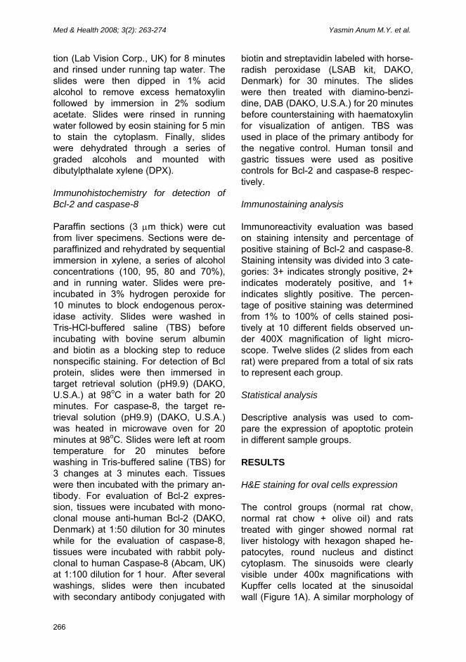
H&E staining for oval cells expression The control groups (normal rat chow, normal rat chow + olive oil) and rats treated with ginger showed normal rat liver histology with hexagon shaped he-patocytes, round nucleus and distinct cytoplasm. The sinusoids were clearly visible under 400x magnifications with Kupffer cells located at the sinusoidal wall (Figure 1A). A similar morphology of
Zingiber officinale Roscoe and apoptosis Med & Health 2008; 3(2): 263-274
Figure 1: Effects of ginger extract on expression of oval cells. (A) H&E staining of control, rat chow alone, (B) olive oil + rat chow, (C) ginger extract, 100mg/kg body weight, (D) liver cancer induced group, CDE (arrows indicate the presence of oval cells in CDE group) and (E) CDE group treated with ginger extract. H: Hepatocyte, K: Kupffer cell, PV: Portal vein, S: Sinusoid O: Oval cell (Magnification x400).
267
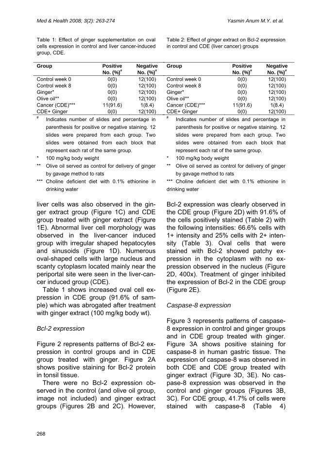
Med & Health 2008; 3(2): 263-274 Yasmin Anum M.Y. et al. Table 1: Effect of ginger supplementation on oval cells expression in control and liver cancer-induced group, CDE. Group Positive
No. (%)# Negative No. (%)#
Control week 0 Control week 8 Ginger* Olive oil** Cancer (CDE)*** CDE+ Ginger
0(0) 0(0) 0(0) 0(0)
11(91.6) 0(0)
12(100) 12(100) 12(100) 12(100) 1(8.4)
12(100) # Indicates number of slides and percentage in
parenthesis for positive or negative staining. 12 slides were prepared from each group. Two slides were obtained from each block that represent each rat of the same group.
* 100 mg/kg body weight ** Olive oil served as control for delivery of ginger
by gavage method to rats *** Choline deficient diet with 0.1% ethionine in
drinking water
Table 2: Effect of ginger extract on Bcl-2 expression in control and CDE (liver cancer) groups Group Positive
No. (%)# Negative No. (%)#
Control week 0 Control week 8 Ginger* Olive oil** Cancer (CDE)*** CDE+ Ginger
0(0) 0(0) 0(0) 0(0)
11(91.6) 0(0)
12(100) 12(100) 12(100) 12(100) 1(8.4)
12(100) # Indicates number of slides and percentage in
parenthesis for positive or negative staining. 12 slides were prepared from each group. Two slides were obtained from each block that represent each rat of the same group.
* 100 mg/kg body weight ** Olive oil served as control for delivery of ginger
by gavage method to rats *** Choline deficient diet with 0.1% ethionine in drinking water
liver cells was also observed in the gin-ger extract group (Figure 1C) and CDE group treated with ginger extract (Figure 1E). Abnormal liver cell morphology was observed in the liver-cancer induced group with irregular shaped hepatocytes and sinusoids (Figure 1D). Numerous oval-shaped cells with large nucleus and scanty cytoplasm located mainly near the periportal site were seen in the liver-can-cer induced group (CDE).
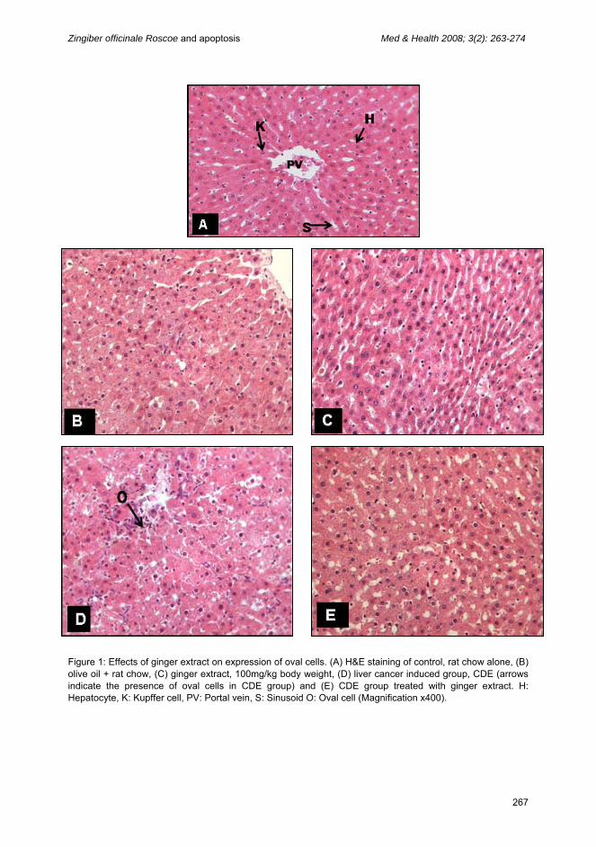
Table 1 shows increased oval cell ex-pression in CDE group (91.6% of sam-ple) which was abrogated after treatment with ginger extract (100 mg/kg body wt). Bcl-2 expression Figure 2 represents patterns of Bcl-2 ex-pression in control groups and in CDE group treated with ginger. Figure 2A shows positive staining for Bcl-2 protein in tonsil tissue.
There were no Bcl-2 expression ob-served in the control (and olive oil group, image not included) and ginger extract groups (Figures 2B and 2C). However,
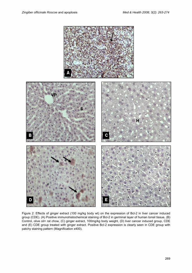
Bcl-2 expression was clearly observed in the CDE group (Figure 2D) with 91.6% of the cells positively stained (Table 2) with the following intensities: 66.6% cells with 1+ intensity and 25% cells with 2+ inten-sity (Table 3). Oval cells that were stained with Bcl-2 showed patchy ex-pression in the cytoplasm with no ex-pression observed in the nucleus (Figure 2D, 400x). Treatment of ginger inhibited the expression of Bcl-2 in the CDE group (Figure 2E).
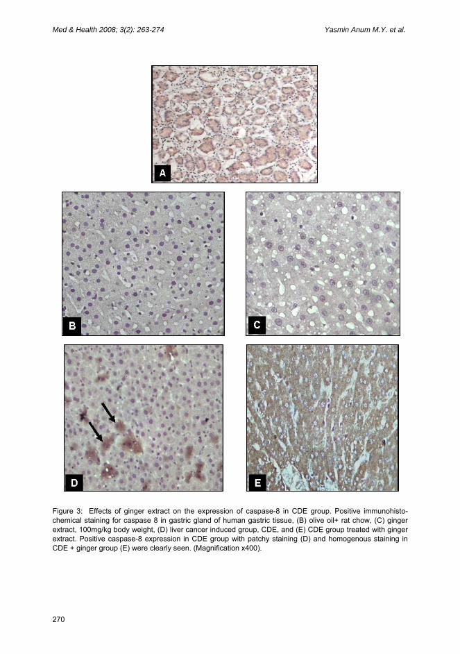
Caspase-8 expression Figure 3 represents patterns of caspase-8 expression in control and ginger groups and in CDE group treated with ginger. Figure 3A shows positive staining for caspase-8 in human gastric tissue. The expression of caspase-8 was observed in both CDE and CDE group treated with ginger extract (Figure 3D, 3E). No cas-pase-8 expression was observed in the control and ginger groups (Figures 3B, 3C). For CDE group, 41.7% of cells were stained with caspase-8 (Table 4)
268
Zingiber officinale Roscoe and apoptosis Med & Health 2008; 3(2): 263-274
Figure 2: Effects of ginger extract (100 mg/kg body wt) on the expression of Bcl-2 in liver cancer induced group (CDE). (A) Positive immunohistochemical staining of Bcl-2 in germinal layer of human tonsil tissue, (B) Control, olive oil+ rat chow, (C) ginger extract, 100mg/kg body weight, (D) liver cancer induced group, CDE and (E) CDE group treated with ginger extract. Positive Bcl-2 expression is clearly seen in CDE group with patchy staining pattern (Magnification x400).
269
Med & Health 2008; 3(2): 263-274 Yasmin Anum M.Y. et al.
270
Figure 3: Effects of ginger extract on the expression of caspase-8 in CDE group. Positive immunohisto-chemical staining for caspase 8 in gastric gland of human gastric tissue, (B) olive oil+ rat chow, (C) ginger extract, 100mg/kg body weight, (D) liver cancer induced group, CDE, and (E) CDE group treated with ginger extract. Positive caspase-8 expression in CDE group with patchy staining (D) and homogenous staining in CDE + ginger group (E) were clearly seen. (Magnification x400).
Zingiber officinale Roscoe and apoptosis Med & Health 2008; 3(2): 263-274
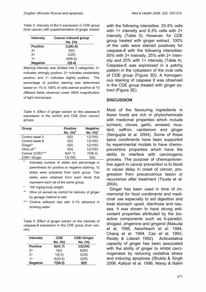
Table 3: Intensity of Bcl-2 expression in CDE group (liver cancer) with supplementation of ginger extract
Intensity Cancer induced group No. (%)
Positive 3+ 2+ 1+ Negative
11(91.6) 0(0)
3(25) 8(66.6) 1(8.4)
Staining intensity was divided into 3 categories: 3+ indicates strongly positive, 2+ indicates moderately positive, and 1+ indicates slightly positive. The percentage of positive staining was determined based on 1% to 100% of cells stained positive at 10 different fields observed under 400X magnification of light microscope. Table 4: Effect of ginger extract on the caspase-8 expression in the control and CDE (liver cancer) groups
Group Positive No. (%)#
Negative No. (%)#
Control week 0 Control week 8 Ginger* Olive oil** Cancer (CDE)*** CDE+ Ginger
0(0) 0(0) 0(0) 0(0)
5(41.7) 12(100)
12(100) 12(100) 12(100) 12(100) 7(58.3)
0(0) # Indicates number of slides and percentage in
parenthesis for positive or negative staining. 12 slides were prepared from each group. Two slides were obtained from each block that represent each rat of the same group.
* 100 mg/kg body weight ** Olive oil served as control for delivery of ginger
by gavage method to rats *** Choline deficient diet with 0.1% ethionine in
drinking water Table 5: Effect of ginger extract on the intensity of caspase-8 expression in the CDE group (liver can-cer).
Intensity CDE No. (%)
CDE+Ginger No. (%)
Positive 3+ 2+ 1+ Negative
5(41.7) 0(0)
1(8.3) 4(33.4) 7(58.3)
12(100) 6(50) 3(25) 3(25) 0(0)
with the following intensities: 33.4% cells with 1+ intensity and 8.3% cells with 2+ intensity (Table 5). However, for CDE group treated with ginger extract, 100% of the cells were stained positively for caspase-8 with the following intensities: 50% with 3+ intensity, 25% with 2+ inten-sity and 25% with 1+ intensity (Table 4). Caspase-8 was expressed in a patchy pattern in the cytoplasm of the oval cells of CDE group (Figure 3D). A homogen-ous staining of caspase 8 was observed in the CDE group treated with ginger ex-tract (Figure 3E). DISCUSSION
Most of the flavouring ingredients in Asian foods are rich in phytochemicals with medicinal properties which include turmeric, cloves, garlic, aniseed, mus-tard, saffron, cardamom and ginger (Sengupta et al. 2004). Some of these spice condiments have been supported by experimental models to have chemo-preventive properties which have the ability to interfere with carcinogenic process. The purpose of chemopreven-tive agent in cancer prevention is to block or cause delay in onset of cancer, pro-gression from precancerous lesion or recurrence after treatment (Tsuda et al. 2004).
Ginger has been used in time of im-memorial for food condiments and medi-cinal use especially to aid digestion and treat stomach upset, diarrhoea and nau-sea. It was shown to have strong anti-oxidant properties attributed by the bio-active components such as 6-paradol, shogaol, zingerone and gingerol (Masuda et al. 1995, Aeschbach et al. 1994, Chang et al. 1994, Cao et al. 1993, Reddy & Lokesh 1992). Antioxidative capacity of ginger has been associated with the ability of ginger to inhibit carci-nogenesis by reducing oxidative stress and inducing apoptosis (Shukla & Singh 2006, Katiyar et al. 1996, Manju & Nalini
271
Med & Health 2008; 3(2): 263-274 Yasmin Anum M.Y. et al. 2005, Rhode et al. 2007).
Our study clearly showed the efficacy of ginger having antitumour properties by inhibiting the proliferation of oval cells as evidenced by the reduced number of oval cells and the perpetuation of normal his-tological structure of liver tissues in liver cancer induced rats treated with ginger extract. Oval cell proliferation precedes neoplasia in many rodent models of he-patocellular carcinoma and in chronic liver disease of human studies (Ackhurst et al. 2001, Lowes et al. 1999). Preven-tion of this proliferative response can re-duce the risk of subsequent carcinoma. This was supported by our previous findings which showed that ginger re-duced oval cell proliferation and liver tu-mour formation in hepatocarcinogenesis induced rats (Mohd Habib et al. 2008). This property of ginger could be attri-buted to the presence of high phenolic compounds such as [6]-gingerol, and [6]-paradol. [6]-gingerol has been shown to suppress experimental metastases in tumour-bearing mice skin carcinogenesis probably via its anti-angiogenic activity (Kim et al. 2005) and inhibition of COX-2 expression along with suppressed NF-kB DNA binding activity (Kim et al. 2004). Since tumour promotion is closely linked to oxidative stress, a compound that ex-hibits antioxidant properties could act as an anticarcinogenic agent (Shukla & Singh 2006).
Some compounds present in ginger may exert cancer preventive effects by inducing apoptosis in cancerous or transformed cells. (Shukla & Singh 2006). Several studies have reported that compounds in ginger suppress prolifera-tion of human cancer cells through in-duction of apoptosis accompanied by downregulation of anti-apoptotic protein Bcl-2 and enhancement of pro-apoptotic protein Bax expression (Lee & Surh 1998, Miyoshi et al. 2003). The dimi-nished expression of anti-apoptotic Bcl-2 protein observed in CDE group treated
with ginger correlated with inhibition of oval cell expression. This confirmed that ginger must have bioactive compounds to produce such chemopreventive ef-fects. Our findings are supported by Reed et al. (1994), who have shown that Bcl-2 prevented the death of neoplastic cells via apoptosis. Vaux et al. (1988) reported that expression of Bcl-2 induced the tumour formation in mice. Our study showed patchy staining of Bcl-2 in oval cells and the results are supported by Frommel et al. (2000) who reported that Bcl-2 was only expressed in proliferative cells near the periportal site of liver (oval cells) but not in normal hepatocytes, Kupffer cells and bile duct epithelium.
Caspase-8 is an executor zymogen protein involved in apoptosis signaling pathway. Ishiguro et al (2007) proved that [6]-gingerol facilitated TRAIL-induced apoptosis by activation of caspase-3/7 activity in human gasric cancer cells. We observed caspase-8 staining in both CDE and CDE group treated with ginger, with enhanced staining observed in the latter. This observation confirmed that in cancer cells, apoptosis is a natural way of inhi-biting its growth, and chemopreventive agents, having the ability of inducing apoptosis, enhance further the effect of apoptosis as seen with the effect of gin-ger extract.
In conclusion, our study agrees with other studies which provide substantial evidence that ginger extract are effective inhibitors of carcinogenic process exhi-bited by reduction of oval cells, Bcl-2 protein and induction of caspase-8 ex-pression. REFERENCES
Aeschbach, R., Loliger, J., Scott, B.C., Murcia, A.,
Butler, B., Halliwell, B. and Aruoma, O.I. 1994. Antioxidant actions of thymol, carbacrol, 6-gingerol, zingerone and hydroxytyrosol. Food and Chem. Toxicol. 32:31-36.
Ahmad, N., Sulaiman, S., Mukti, N.A., Murad, N.A., Abdul Hamid, N.A., Mohd Yusof, Y.A.
272
Zingiber officinale Roscoe and apoptosis Med & Health 2008; 3(2): 263-274
2006. The Effects of Ginger extract (Zingiber officinale Roscoe) on Antioxidant Status of Hepatocellular carcinoma Induced Rats. Malaysian J of Biochem. and Molec. Biol, 14:7-12.
Akhurst, B., Croager, E.J., Farley-Roche, C.A., Ong, J.K., Dumble, M.L., Knight, B. & Yeoh, G.C. 2001. A modified Choline-Deficient, Ethionine Supplemented Diet protocol effectively induces oval cells in mouse liver. Hepatol. 34:519-522.
Cao, Z.F., Chen, Z.G., Guo, P., Zhang, S.M., Lian, L.X., Luo L. and Hu W.M. 1993. Scavenging effects of ginger on superoxide anion and hydroxyl radical. Chung-Kuo Chung Yao Tsa Chih 18:750-764.
Chang, W-S., Chang, Y-H., Lu, F-J. and Chiang, H-C. 1994. Inhibitory effects of phenolics on xanthine oxidase. Anticancer Res. 14:501-506.
Coultas, L. & Strasser, A. 2003. The role of the Bcl-2 protein family in cancer. Seminars in Cancer Biology. 13:115-123.
Forner, A., Amelia, J.H., Isabel, R., Jordi, B. 2006. Treatment of Hepatocellular Carcinoma. Crit.Rev.in Oncol/Hematol. 60:89–98.
Frommel, T, Yong, O.S., Edwin, J.Z. 2000. Immunohistochemical Evaluation of Bcl-2 Gene Family Expression in Liver of Hepatitis C and Cirrhotic Patients: A Novel Mechanism to Explain the High Incidence of Hepatocarcinoma in Cirrhotics. Current Opinion in Gastroenterol.. 16(3):B101-B164.
Gerard, L.C., Halimah, Y. 2003. Second Report Of The National Cancer Registry: Cancer Incidence In Malaysia. National Cancer Registry Ministry of Health Malaysia. ISSN 1675-8870.
Hanson, C.J., Bootman, M.D., Distelhorst, C.W., Maraldi, T., Roderick, H.L. 2008. The cellular concentration of Bcl-2 determines its pro- or anti-apoptotic effect. Cell Calcium. 44(3):243-258.
Ishiguro, K., Ando, T., Maeda, O., Ohmiya, N., Niwa, Y., Kadomatsu, K., Goto, H. 2007. Ginger ingredients reduce viability of gastric cancer cells via distinct mechansims. Biochem. Biophys. Res. Comm. 362:218-223.
Mohd Habib, S.H., Makpol, S., Abdul Hamid, N.A., Das, S., Wan Ngah, W.Z., Mohd Yusof, Y.A. 2008. Ginger extract (Zingiber officinale) has anti-cancer and anti-inflammatory effects on ethionine-induced hepatoma rats. Clinics (accepted for publication)
Katiyar, S.K., Agarwal, R. and Mukhtar, H. 1996. Inhibition of tumor promotion in SENCAR mouse skin by ethanol extract of Zingiber officinale rhizome. Cancer Res. 56(5):1023-
1030. Kim, S.O., Chun, K.S., Kundu, J.K., Surh, Y.J.
2004. Inhibitory effects of [6]-gingerol on PMA-induced COX-2 expression and activation of NF-kB and p38 MAPK in mouse skin. Biofactors 21:27-31
Kim, E.C., Min, J.K., Kim, T.Y., Lee, S.J., Yang, H.O., Han, S., Kim, Y.M., Kwon, Y.G. 2005. [6]-Gingerol, a pungent ingredient of ginger inhibits angiogenesis in vitro and in vivo. Biochem. and Biophy. Res. Comm. 335:300-308
Lee, E., Surh, Y.J. 1998. Induction of apoptosis in HL-60 cells by pungent vanilloids, (6)-gingerol and (6)-paradol. Cancer Lett. 134:163-168.
Lowes, K.S., Brennan, B.A., Yeoh, G.C., & Olynyk, J.K. 1999. Oval Cell Numbers in Human Chronic Liver Disease are Directly Related to Disease Severity. Am. J. Pathol. 154:537-541.
Manju, V. and Nalini, N. 2005. Chemopreventive efficacy of ginger, a naturally occurring anticarcinogen during the initiation, post-initiation stages of 1,2-dimethylhydrazine-induced colon cancer. Clin. Chim. Acta. 358:60-67
Masuda, T., Jitoe, A., Mabry, T.J. 1995. Isolation and structure determination of cassumunarins A, B, C : new anti-inflammatory antioxidants f rom a troical ginger, Zingible cassumunar. J Am. Oil Chem. Soc 72:1053-1057
Miyoshi, N., Yoshimasa, N., Uedaa, Y., Masako, A., Yoshio, O., Koji, U. & Osawa, T. 2003. Dietary ginger constituents, galanals A and B, are potent apoptosis inducers in Human T lymphoma Jurkat cells. Cancer Lett. 199:113–119.
Orner, G.A., Dashwood, M.W., Blum, C.A., Diaz, G.D., Li, Q., Dashwood, R.H. 2003. Supression of tumorigenesis in the Apcmin mouse:Down-regulation of β-catenin signaling by a combination of tea plus sulindac. Carcinogenesis 24:263-267
Reddy, A.A. & Lokesh, B.R. 1992. Studies on spice principles as antioxidants in the inhibition of lipid peroxidation of rat liver microsomes. Mol. Cell Biochem. 111:117-124.
Reed, C., Nikolaos, G., & Nikitakis, K. 1994. Immunohistochemical evaluation of cell proliferation antigen Ki-67 and apoptosis-related proteins Bcl-2 and caspase-3 in oral granular cell tumor. Oral Radiol Endod. 96:566-572.
Rhode, J., Fogoros, S., Zick, S., Wahl, H., Griffith, K.A., Huang, J., and Liu, J.R. 2007. Ginger inhibits cell growth and modulates angiogenic factors in ovarian cancer cells. BMC Complementary and Alternative Medicine 7:44
Ross, I.A. 2002. Medicinal Plants of the World.
273
Med & Health 2008; 3(2): 263-274 Yasmin Anum M.Y. et al.
274
Chemical Constituents, Traditional and Modern Medicinal Uses. New York: Humana Press. ISBN: 1-58829-129-4, pp 509-559.
Santana-Rios, G., Orner, G.A., Xu, M., Izquierdo-Pulido, M., and Dashwood, R.H. 2001. Inhibition by white tea of 2-amino-1-methyl-6-phenylimidazo [4,5-b]pyridine-induced colonic aberrant crypts in the F344 rats. Nutr.& Cancer 41:98-103
Schafer, D.F., Sorrell, M.F. 1999. Hepatocellular carcinoma. Lancet. 353:1253-1257.
Sengupta, A., Ghosh, S., Bhattachargee, S., Das, S. 2004. Indian food ingredients and cancer prevention-an experimental evaluation of anticarcinogenic effects of garlic in rat colon.
Asian Pac. J. Cancer Prev. 5:126-132. Shukla, Y., Singh, M. 2007. Cancer preventive
properties of ginger: A brief review. Food Chem. Toxicol. 45 (5):683-690
Tsuda, H., Ohshima, Y., Nomoto, H., Fujita, K., Matsuda, E., Iigo, M., Takasuka, N., & Moore, M.A. 2004. Cancer Prevention by Natural Compounds. Drug Metab. Pharmacokin. 19(4): 245-263.
Vaux, F., Samuel, L., Marion, K., & Patricia, J. 1988. Effect of bcl-2 overexpression in mice on ovotoxicity caused by 4-vinylcyclohexene. Toxicol. and Appl. Pharmacol. 215:51–56.
Wei, S.J. 2004. Cancer: The Basics. University of Pennsylvania. (5 November 2006).