Embed Size (px)
Citation preview
GlomerulonephritisGlomerulonephritis: Practical Approach: Practical Approachรศ.นพ. เถลิงศักดิ์ กาญจนบุษย
สาขาวิชาโรคไต ภาควิชาอายุรศาสตรหัวหนาศนูยวิจัยโรคไตและความผิดปกติทางเมตาบอลิซึม
จุฬาลงกรณมหาวิทยาลัย
JCMS 2009
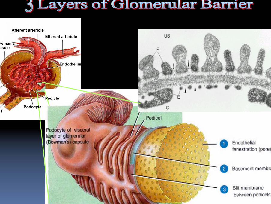
Afferent arterioleEfferent arteriole
Endothelium
Podocyte
Pedicle
Bowman’s capsule
PCT
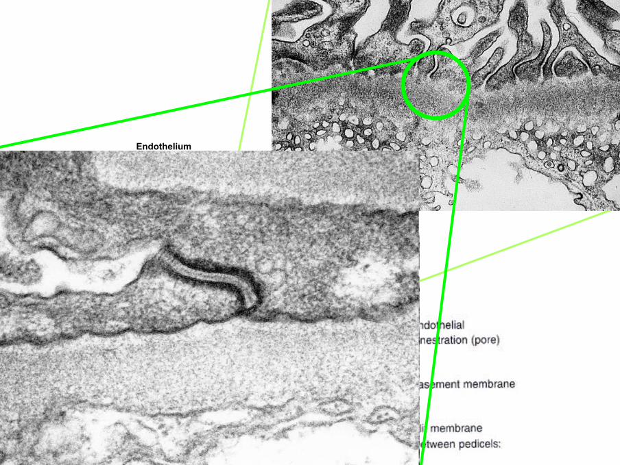
Endothelium
ก ข
ง
ก
ขค ง
ข
จ
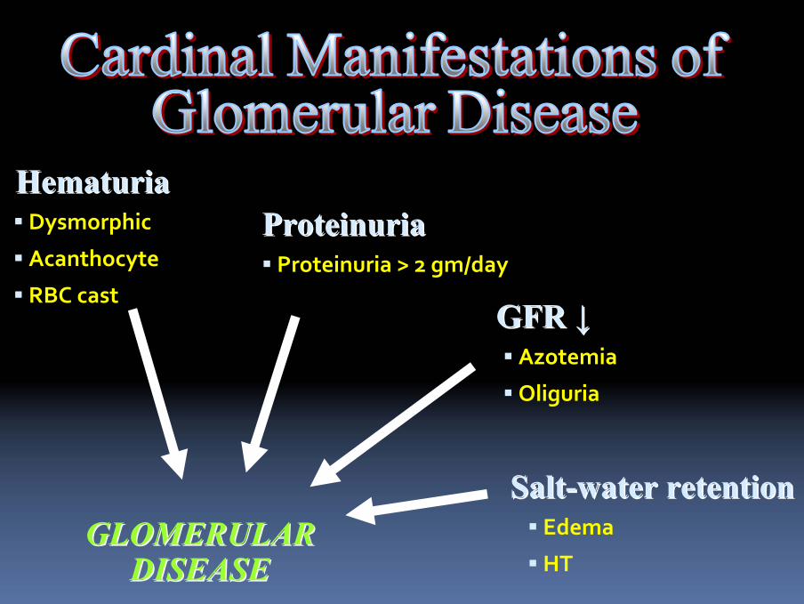
GLOMERULAR GLOMERULAR DISEASEDISEASE
HematuriaHematuriaDysmorphic
Acanthocyte
RBC cast
ProteinuriaProteinuriaProteinuria > 2 gm/day
SaltSalt--water retentionwater retentionEdema
HT
GFR GFR ↓↓Azotemia
Oliguria
Helen Liapis, 2002
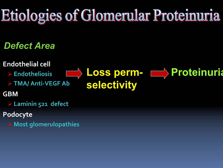
Endothelial cellEndotheliosisTMA/ Anti‐VEGF Ab
GBMLaminin 521 defect
PodocyteMost glomerulopathies
Loss perm-selectivity
Proteinuria
Defect Area
ก ก
ข
ค
ง
จ
ก
ฉ
จ
ง
ค
ข
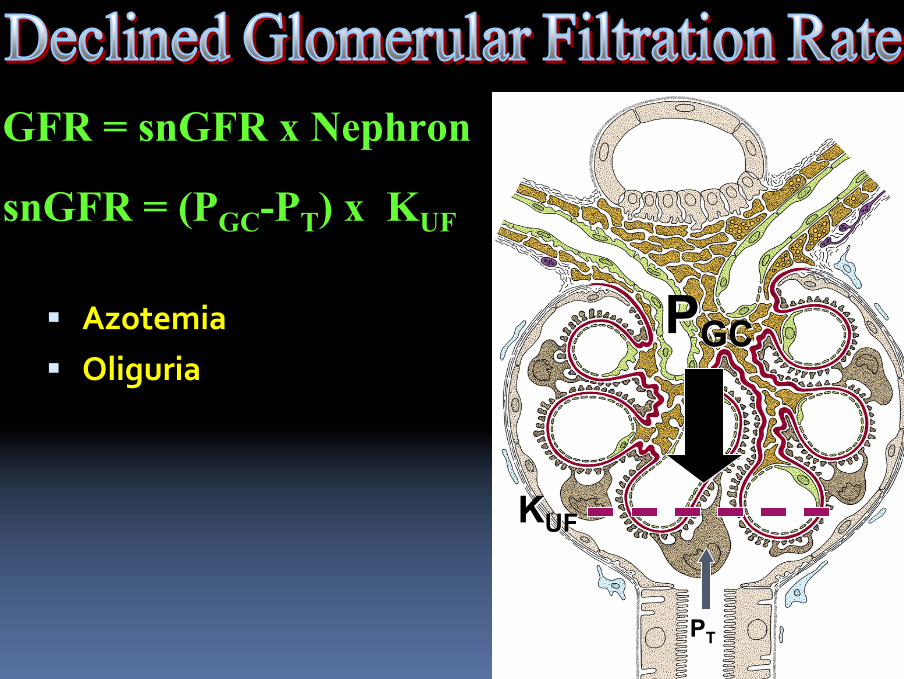
AzotemiaOliguria
PPGCGC
PPTT
KKUFUF
GFR = snGFR x Nephron
snGFR = (PGC-PT) x KUF
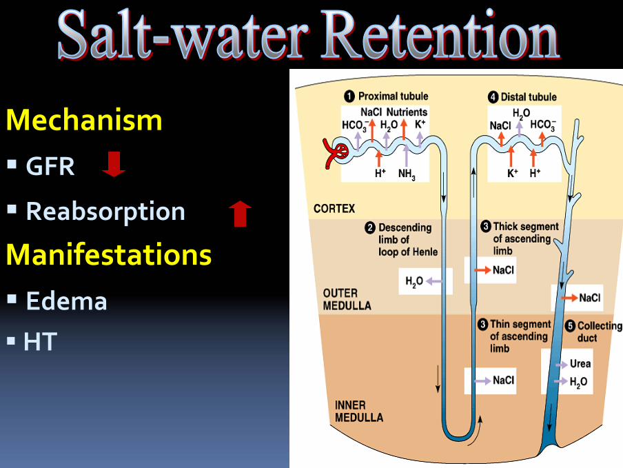
MechanismGFR
Reabsorption
ManifestationsEdemaHT
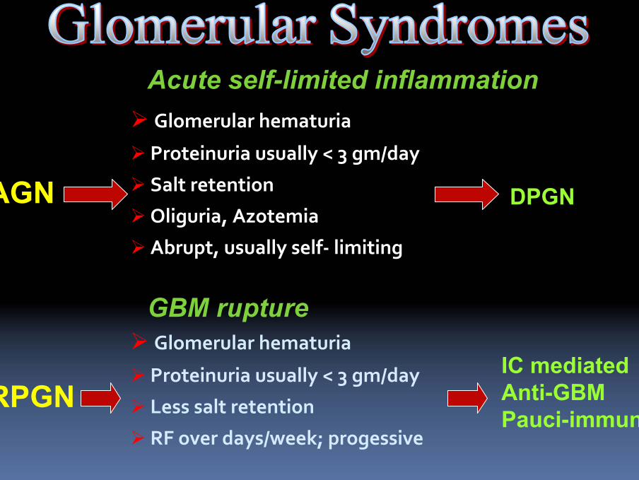
Glomerular hematuria
Proteinuria usually < 3 gm/day
Less salt retention
RF over days/week; progessive
Glomerular hematuria
Proteinuria usually < 3 gm/day
Salt retention
Oliguria, Azotemia
Abrupt, usually self‐ limiting
AGN
RPGN
DPGN
IC mediatedAnti-GBMPauci-immune
GBM rupture
Acute self-limited inflammation
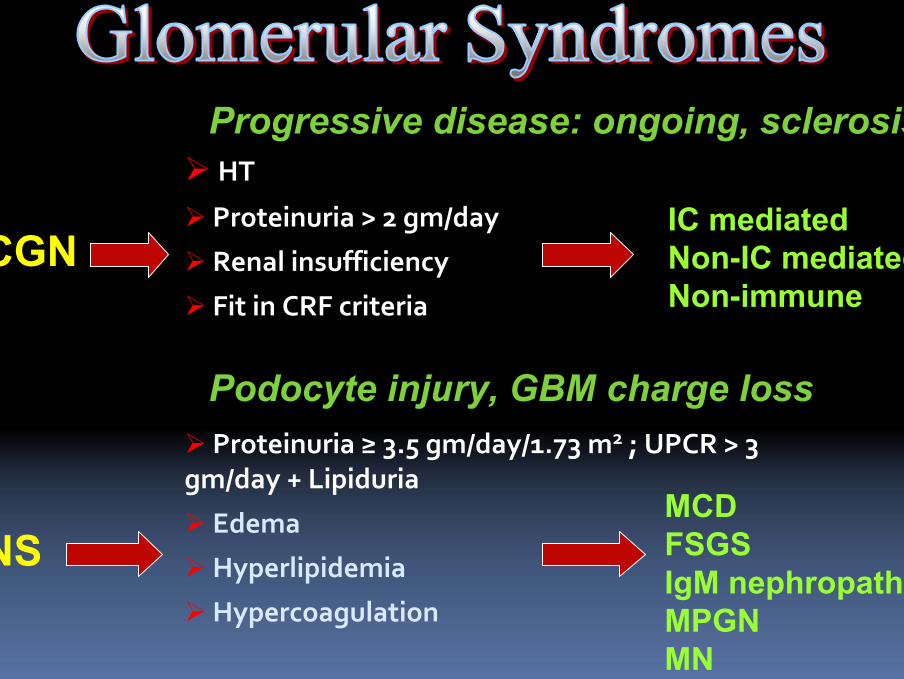
Proteinuria ≥ 3.5 gm/day/1.73 m2 ; UPCR > 3 gm/day + Lipiduria
Edema
Hyperlipidemia
Hypercoagulation
HT
Proteinuria > 2 gm/day
Renal insufficiency
Fit in CRF criteria
CGN
NS
IC mediatedNon-IC mediatedNon-immune
MCDFSGS IgM nephropathyMPGNMN
Progressive disease: ongoing, sclerosis
Podocyte injury, GBM charge loss
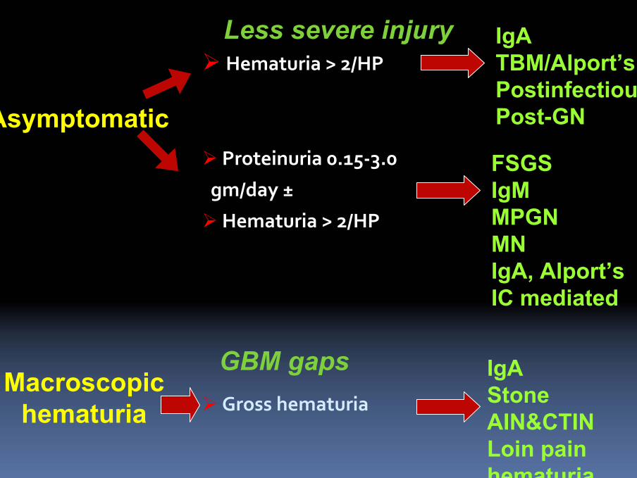
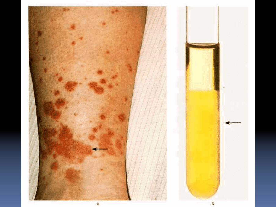
Gross hematuria
Hematuria > 2/HP
Proteinuria 0.15‐3.0
gm/day ±
Hematuria > 2/HP
Asymptomatic
Macroscopic hematuria
IgAStoneAIN&CTINLoin painhematuria
FSGS IgMMPGNMNIgA, Alport’sIC mediated
IgATBM/Alport’sPostinfectiousPost-GN
Less severe injury
GBM gaps

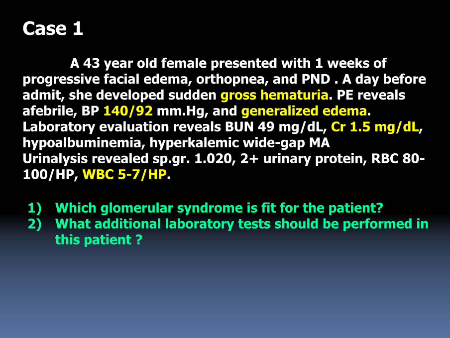
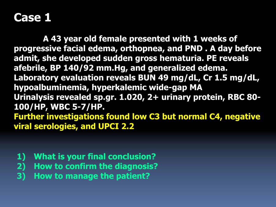
Case 1
A 43 year old female presented with 1 weeks of progressive facial edema, orthopnea, and PND . A day before admit, she developed sudden gross hematuria. PE reveals afebrile, BP 140/92 mm.Hg, and generalized edema. Laboratory evaluation reveals BUN 49 mg/dL, Cr 1.5 mg/dL, hypoalbuminemia, hyperkalemic wide-gap MAUrinalysis revealed sp.gr. 1.020, 2+ urinary protein, RBC 80-100/HP, WBC 5-7/HP.
1) Which glomerular syndrome is fit for the patient? 2) What additional laboratory tests should be performed in
this patient ?
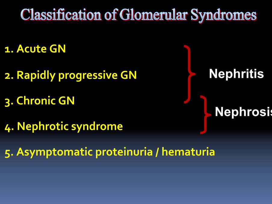
5. Asymptomatic proteinuria / hematuria
4. Nephrotic syndrome
3. Chronic GN
2. Rapidly progressive GN
1. Acute GN
Nephritis
Nephrosis
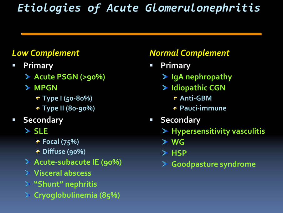
Etiologies of Acute Glomerulonephritis
Low ComplementPrimary
Acute PSGN (>90%)MPGN
Type I (50‐80%)Type II (80‐90%)
SecondarySLE
Focal (75%)Diffuse (90%)
Acute‐subacute IE (90%)Visceral abscess“Shunt” nephritisCryoglobulinemia (85%)
Normal ComplementPrimary
IgA nephropathyIdiopathic CGN
Anti‐GBMPauci‐immune
SecondaryHypersensitivity vasculitisWGHSPGoodpasture syndrome
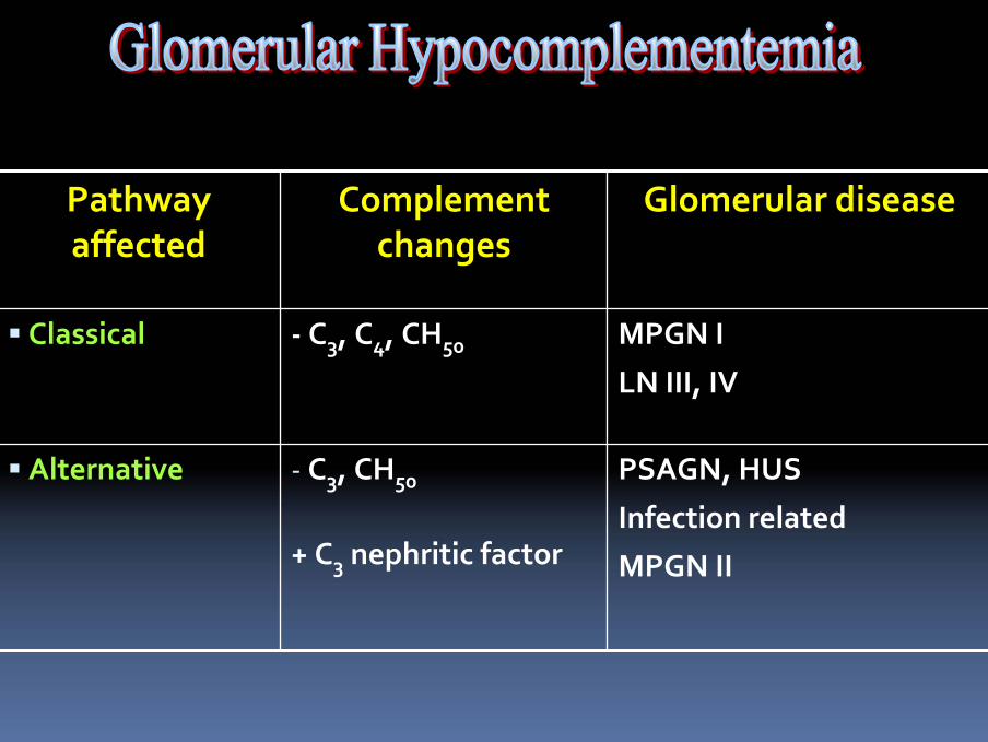
PSAGN, HUS
Infection related
MPGN II
‐ C3, CH50
+ C3 nephritic factor
Alternative
MPGN I
LN III, IV
‐ C3, C4, CH50Classical
Glomerular diseaseComplement changes
Pathway affected
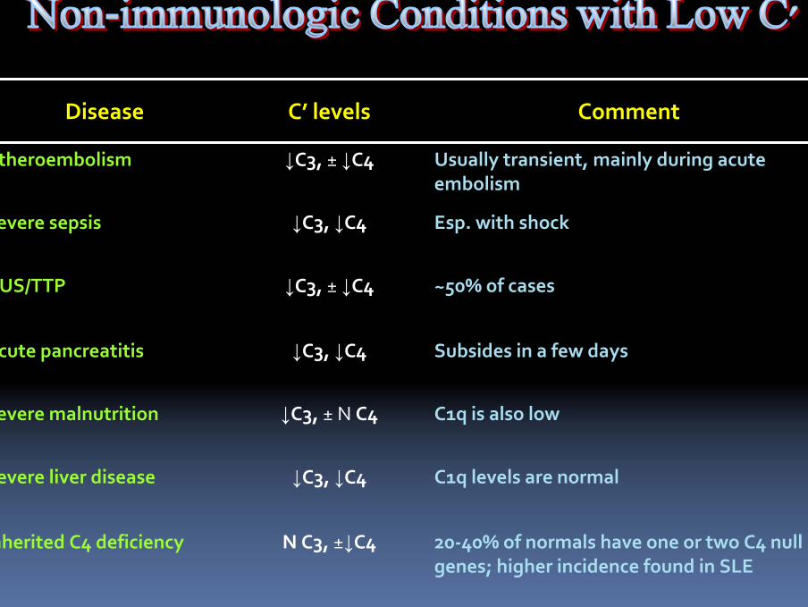
C1q is also low↓C3, ± N C4Severe malnutrition
C1q levels are normal↓C3, ↓C4Severe liver disease
20‐40% of normals have one or two C4 null genes; higher incidence found in SLE
N C3, ±↓C4Inherited C4 deficiency
Subsides in a few days↓C3, ↓C4Acute pancreatitis
~50% of cases↓C3, ± ↓C4HUS/TTP
Esp. with shock↓C3, ↓C4Severe sepsis
Usually transient, mainly during acute embolism
↓C3, ± ↓C4Atheroembolism
CommentC’ levelsDisease
Case 1
A 43 year old female presented with 1 weeks of progressive facial edema, orthopnea, and PND . A day before admit, she developed sudden gross hematuria. PE reveals afebrile, BP 140/92 mm.Hg, and generalized edema. Laboratory evaluation reveals BUN 49 mg/dL, Cr 1.5 mg/dL, hypoalbuminemia, hyperkalemic wide-gap MAUrinalysis revealed sp.gr. 1.020, 2+ urinary protein, RBC 80-100/HP, WBC 5-7/HP.Further investigations found low C3 but normal C4, negative viral serologies, and UPCI 2.2
1) What is your final conclusion?2) How to confirm the diagnosis?3) How to manage the patient?
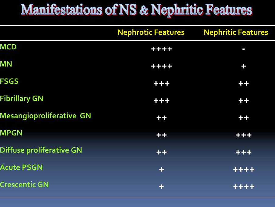
+
+++++Diffuse proliferative GN
+++++Crescentic GN
++++Acute PSGN
+++++MPGN
++++Mesangioproliferative GN
+++++Fibrillary GN
+++++FSGS
+++++MN
‐++++MCD
Nephritic FeaturesNephrotic Features
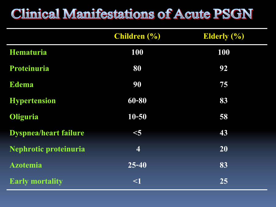
25<1Early mortality8325-40Azotemia204Nephrotic proteinuria43<5Dyspnea/heart failure5810-50Oliguria8360-80Hypertension7590Edema9280Proteinuria100100Hematuria
Elderly (%)Children (%)
Case 1
A 43 year old female presented with 1 weeks of progressive facial edema, orthopnea, and PND . A day before admit, she developed sudden gross hematuria. PE reveals afebrile, BP 140/92 mm.Hg, and generalized edema. Laboratory evaluation reveals BUN 49 mg/dL, Cr 1.5 mg/dL, hypoalbuminemia, hyperkalemic wide-gap MAUrinalysis revealed sp.gr. 1.020, 2+ urinary protein, RBC 80-100/HP, WBC 5-7/HP.Further investigations found low C3 but normal C4, negative viral serologies, and UPCI 2.2
1) What is your final conclusion?2) How to confirm the diagnosis?3) How to manage the patient?
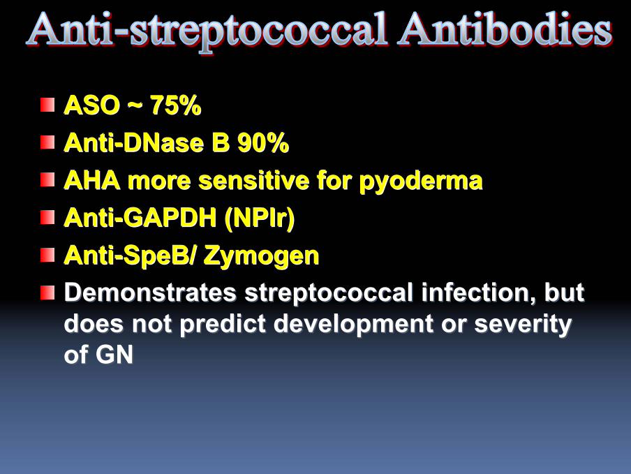
ASO ~ 75% ASO ~ 75% AntiAnti--DNaseDNase B 90%B 90%AHA more sensitive for AHA more sensitive for pyodermapyodermaAntiAnti--GAPDH (GAPDH (NPlrNPlr))AntiAnti--SpeBSpeB/ / ZymogenZymogenDemonstrates streptococcal infection, but Demonstrates streptococcal infection, but does not predict development or severity does not predict development or severity of GNof GN
Case 1
A 43 year old female presented with 1 weeks of progressive facial edema, orthopnea, and PND . A day before admit, she developed sudden gross hematuria. PE reveals afebrile, BP 140/92 mm.Hg, and generalized edema. Laboratory evaluation reveals BUN 49 mg/dL, Cr 1.5 mg/dL, hypoalbuminemia, hyperkalemic wide-gap MAUrinalysis revealed sp.gr. 1.020, 2+ urinary protein, RBC 80-100/HP, WBC 5-7/HP.Further investigations found low C3 but normal C4, negative viral serologies, and UPCI 2.2
1) What is your final conclusion?2) How to confirm the diagnosis?3) How to manage the patient?
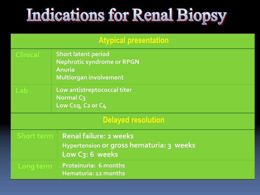
Proteinuria: 6 monthsHematuria: 12 months
Long term
Renal failure: 2 weeksHypertension or gross hematuria: 3 weeksLow C3: 6 weeks
Short term
Delayed resolution
Low antistreptococcal titerNormal C3Low C1q, C2 or C4
Lab
Short latent period Nephrotic syndrome or RPGNAnuriaMultiorgan involvement
Clinical
Atypical presentation
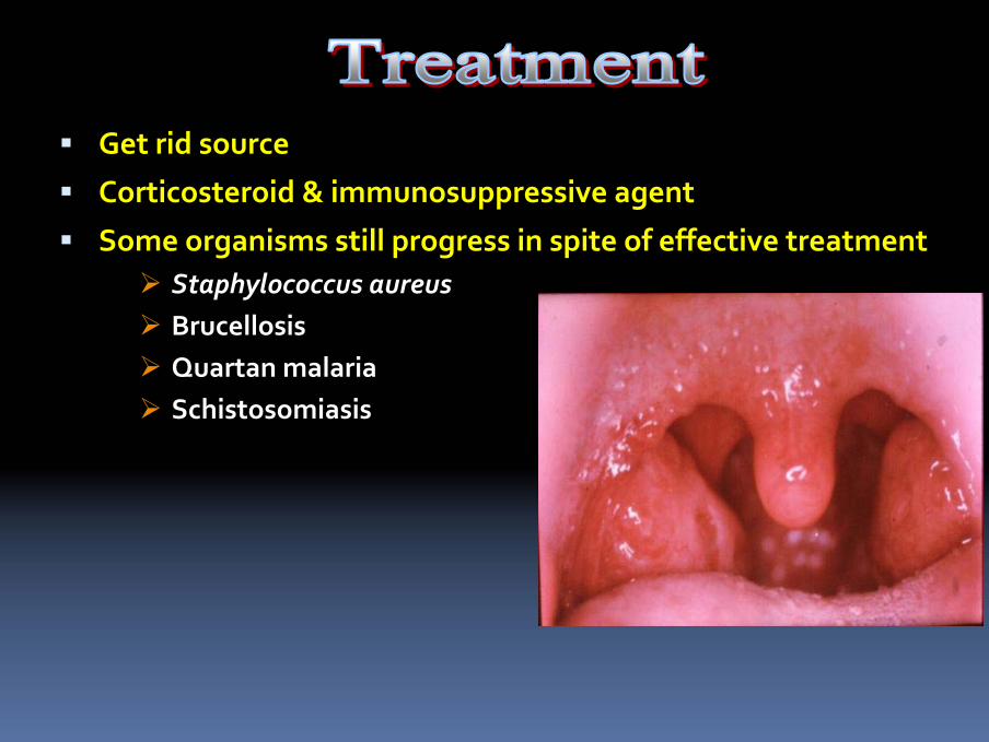
Get rid source
Corticosteroid & immunosuppressive agent
Some organisms still progress in spite of effective treatmentStaphylococcus aureusBrucellosisQuartan malariaSchistosomiasis

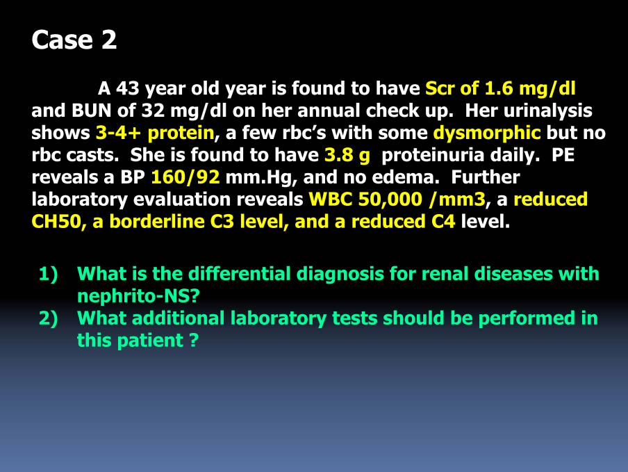
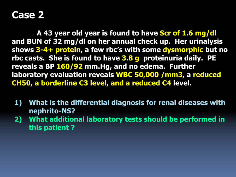
Case 2
A 43 year old year is found to have Scr of 1.6 mg/dland BUN of 32 mg/dl on her annual check up. Her urinalysis shows 3-4+ protein, a few rbc’s with some dysmorphic but no rbc casts. She is found to have 3.8 g proteinuria daily. PE reveals a BP 160/92 mm.Hg, and no edema. Further laboratory evaluation reveals WBC 50,000 /mm3, a reduced CH50, a borderline C3 level, and a reduced C4 level.
1) What is the differential diagnosis for renal diseases with nephrito-NS?
2) What additional laboratory tests should be performed in this patient ?
Classification of Glomerular Diseases
1o Glomerular Disease(idiopathic; renal‐limited)
MCDFSGSMesangial GN (IgA, IgM)MN MPGNCrescentic GNSclerosing GNUnclassified GN
2o Glomerular Disease(associated with multi‐system
disease)Systemic diseasesSystemic InfectionsVascular diseasesMetabolic & Deposition diseasesHereditary nephropathiesMiscellaneous
Mesangiocapillary GNDeposition diseases
Amyloidosis
Light‐chain deposition diseaseFibrillary GN and Immunotactoid GN
Hereditary disease: Alport’s syndromeSome cases of mesangial, focal and diffused proliferative GN
IgA nephropathy
Post‐infectious GNLupus nephritis
Diabetic nephropathy
Case 2
A 43 year old year is found to have Scr of 1.6 mg/dland BUN of 32 mg/dl on her annual check up. Her urinalysis shows 3-4+ protein, a few rbc’s with some dysmorphic but no rbc casts. She is found to have 3.8 g proteinuria daily. PE reveals a BP 160/92 mm.Hg, and no edema. Further laboratory evaluation reveals WBC 50,000 /mm3, a reduced CH50, a borderline C3 level, and a reduced C4 level.
1) What is the differential diagnosis for renal diseases with nephrito-NS?
2) What additional laboratory tests should be performed in this patient ?
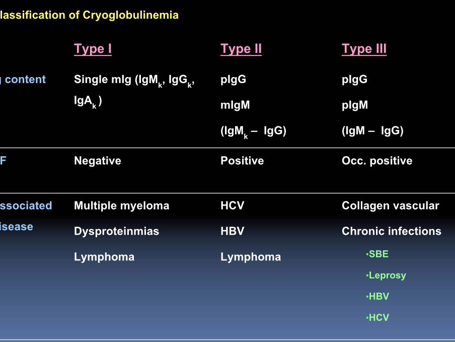
Classification of Cryoglobulinemia
Collagen vascularChronic infections
•SBE•Leprosy•HBV•HCV
HCVHBVLymphoma
Multiple myelomaDysproteinmiasLymphoma
Associated disease
Occ. positivePositiveNegativeRF
pIgGpIgM(IgM – IgG)
pIgGmIgM(IgMk – IgG)
Single mIg (IgMk, IgGk, IgAk )
Ig content
Type IIIType IIType I
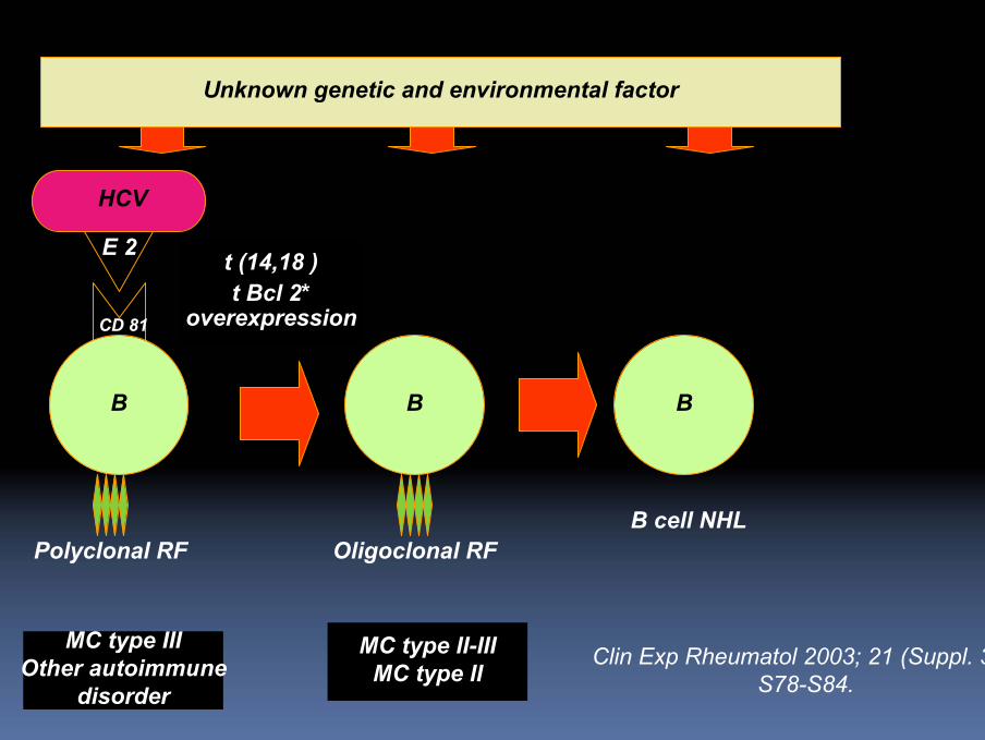
HCV
E 2
B B B
Polyclonal RF
t (14,18 )t Bcl 2*
overexpression
Oligoclonal RF
MC type IIIOther autoimmune
disorder
MC type II-IIIMC type II
B cell NHL
Unknown genetic and environmental factor
Clin Exp Rheumatol 2003; 21 (Suppl. 31)S78-S84.
CD 81
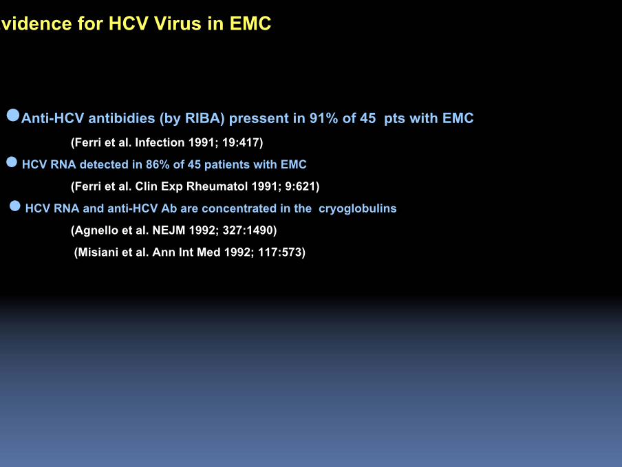
Evidence for HCV Virus in EMC
Anti-HCV antibidies (by RIBA) pressent in 91% of 45 pts with EMC(Ferri et al. Infection 1991; 19:417)
HCV RNA detected in 86% of 45 patients with EMC (Ferri et al. Clin Exp Rheumatol 1991; 9:621)
HCV RNA and anti-HCV Ab are concentrated in the cryoglobulins(Agnello et al. NEJM 1992; 327:1490)(Misiani et al. Ann Int Med 1992; 117:573)
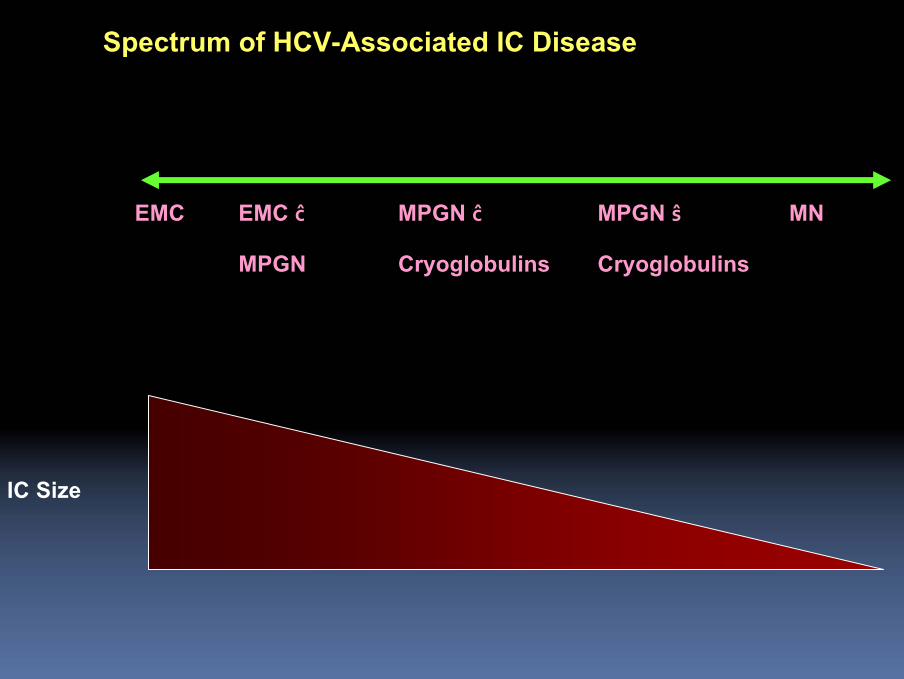
Spectrum of HCV-Associated IC Disease
IC Size
MNMPGN ŝCryoglobulins
MPGN ĉCryoglobulins
EMC ĉMPGN
EMC

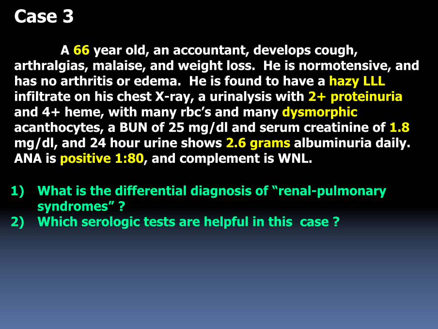
Case 3
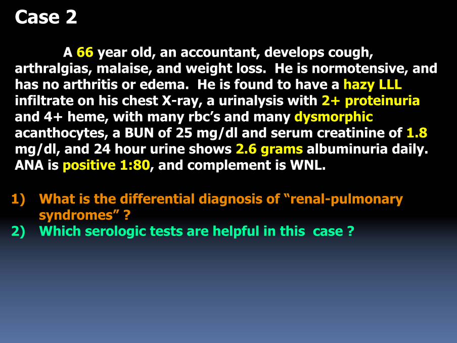
A 66 year old, an accountant, develops cough, arthralgias, malaise, and weight loss. He is normotensive, and has no arthritis or edema. He is found to have a hazy LLLinfiltrate on his chest X-ray, a urinalysis with 2+ proteinuriaand 4+ heme, with many rbc’s and many dysmorphicacanthocytes, a BUN of 25 mg/dl and serum creatinine of 1.8mg/dl, and 24 hour urine shows 2.6 grams albuminuria daily. ANA is positive 1:80, and complement is WNL.
1) What is the differential diagnosis of “renal-pulmonary syndromes” ?
2) Which serologic tests are helpful in this case ?
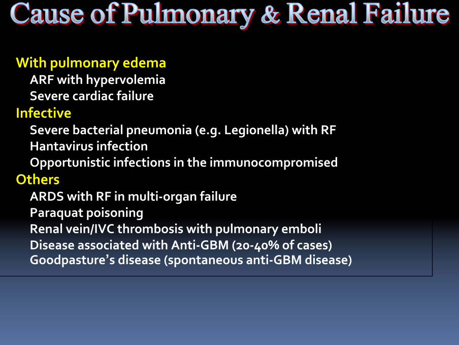
With pulmonary edemaARF with hypervolemiaSevere cardiac failure
InfectiveSevere bacterial pneumonia (e.g. Legionella) with RFHantavirus infectionOpportunistic infections in the immunocompromised
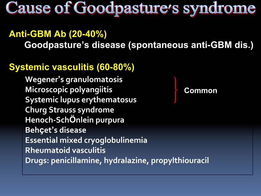
OthersARDS with RF in multi‐organ failureParaquat poisoningRenal vein/IVC thrombosis with pulmonary emboliDisease associated with Anti‐GBM (20‐40% of cases) Goodpasture’s disease (spontaneous anti‐GBM disease)
Common
Anti-GBM Ab (20-40%)Goodpasture’s disease (spontaneous anti-GBM dis.)
Systemic vasculitis (60-80%)Wegener’s granulomatosisMicroscopic polyangiitisSystemic lupus erythematosusChurg Strauss syndromeHenoch‐SchÖnlein purpuraBehçet’s diseaseEssential mixed cryoglobulinemiaRheumatoid vasculitisDrugs: penicillamine, hydralazine, propylthiouracil
Case 2
A 66 year old, an accountant, develops cough, arthralgias, malaise, and weight loss. He is normotensive, and has no arthritis or edema. He is found to have a hazy LLLinfiltrate on his chest X-ray, a urinalysis with 2+ proteinuriaand 4+ heme, with many rbc’s and many dysmorphicacanthocytes, a BUN of 25 mg/dl and serum creatinine of 1.8mg/dl, and 24 hour urine shows 2.6 grams albuminuria daily. ANA is positive 1:80, and complement is WNL.
1) What is the differential diagnosis of “renal-pulmonary syndromes” ?
2) Which serologic tests are helpful in this case ?
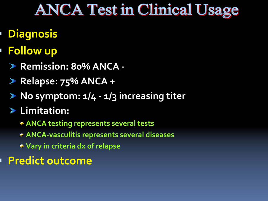
DiagnosisFollow up
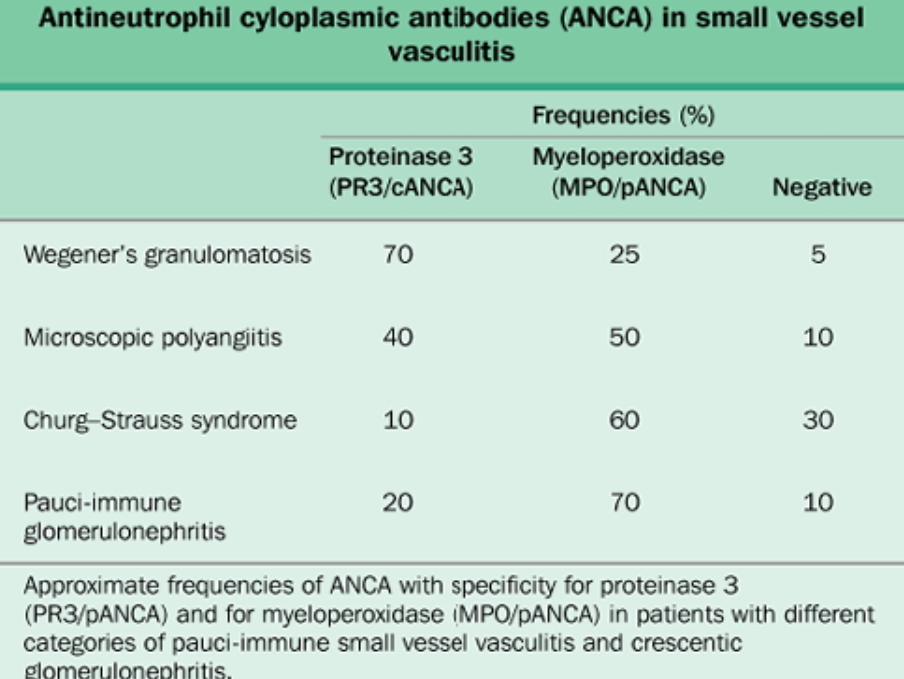
Remission: 80% ANCA ‐Relapse: 75% ANCA +No symptom: 1/4 ‐ 1/3 increasing titerLimitation:
ANCA testing represents several testsANCA‐vasculitis represents several diseasesVary in criteria dx of relapse
Predict outcome
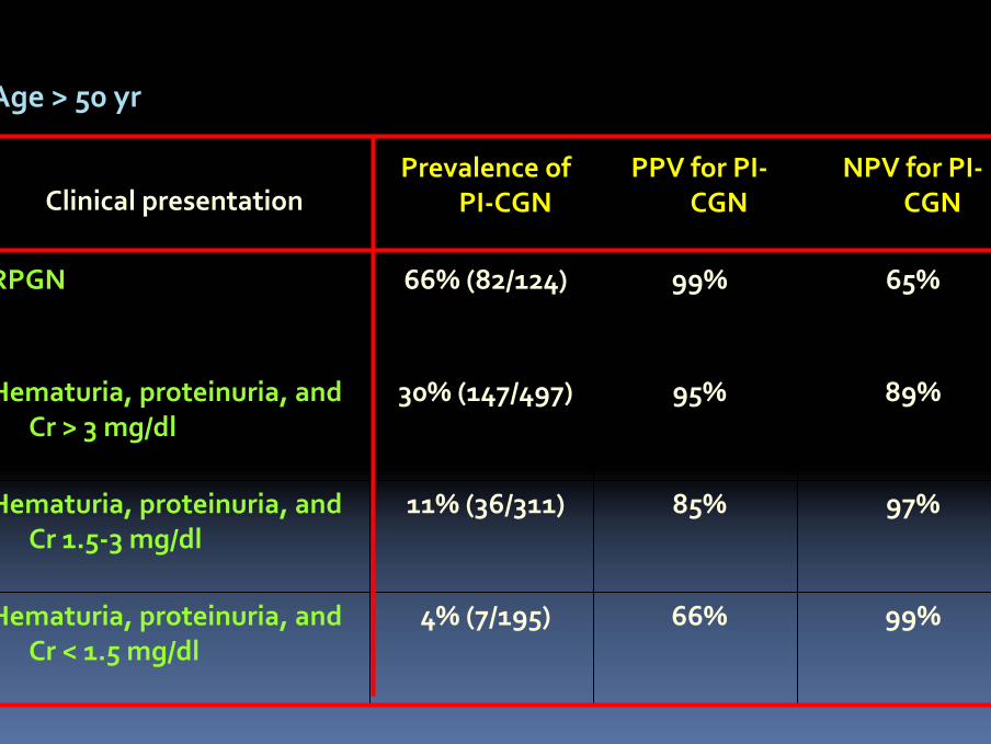
99%66%4% (7/195)Hematuria, proteinuria, and Cr < 1.5 mg/dl
97%85%11% (36/311)Hematuria, proteinuria, and Cr 1.5‐3 mg/dl
89%95%30% (147/497)Hematuria, proteinuria, and Cr > 3 mg/dl
65%99%66% (82/124)RPGN
NPV for PI‐CGN
PPV for PI‐CGN
Prevalence of PI‐CGNClinical presentation
Age > 50 yr

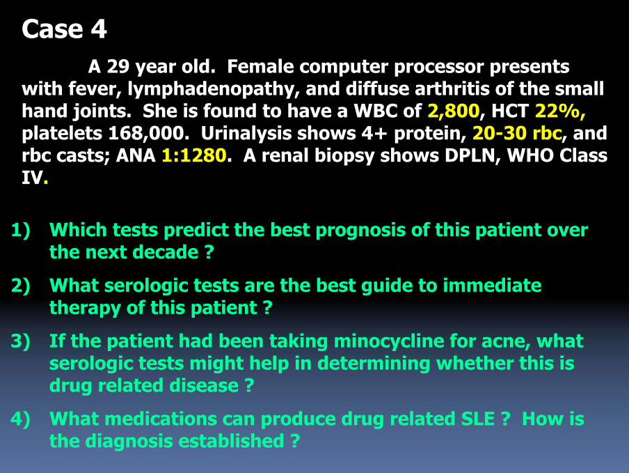
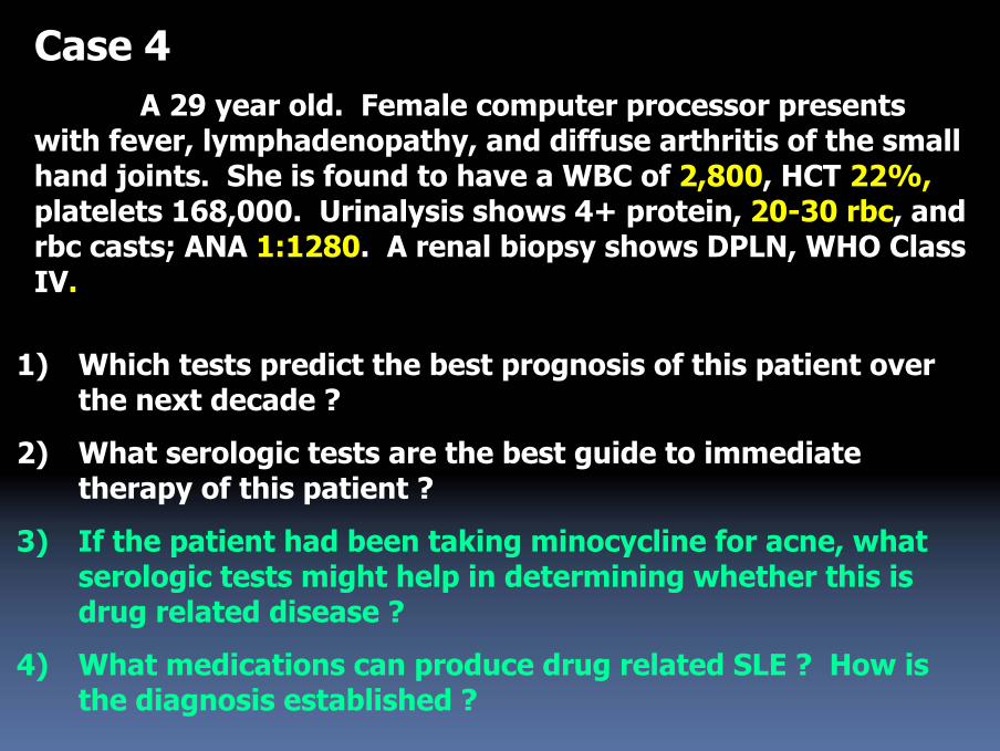
Case 4A 29 year old. Female computer processor presents
with fever, lymphadenopathy, and diffuse arthritis of the small hand joints. She is found to have a WBC of 2,800, HCT 22%,platelets 168,000. Urinalysis shows 4+ protein, 20-30 rbc, and rbc casts; ANA 1:1280. A renal biopsy shows DPLN, WHO Class IV.
1) Which tests predict the best prognosis of this patient over the next decade ?
2) What serologic tests are the best guide to immediate therapy of this patient ?
3) If the patient had been taking minocycline for acne, what serologic tests might help in determining whether this is drug related disease ?
4) What medications can produce drug related SLE ? How is the diagnosis established ?
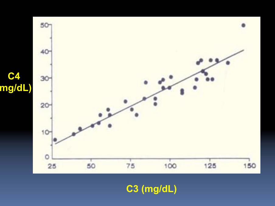
C4(mg/dL)
C3 (mg/dL)
Case 4A 29 year old. Female computer processor presents
with fever, lymphadenopathy, and diffuse arthritis of the small hand joints. She is found to have a WBC of 2,800, HCT 22%,platelets 168,000. Urinalysis shows 4+ protein, 20-30 rbc, and rbc casts; ANA 1:1280. A renal biopsy shows DPLN, WHO Class IV.
1) Which tests predict the best prognosis of this patient over the next decade ?
2) What serologic tests are the best guide to immediate therapy of this patient ?
3) If the patient had been taking minocycline for acne, what serologic tests might help in determining whether this is drug related disease ?
4) What medications can produce drug related SLE ? How is the diagnosis established ?
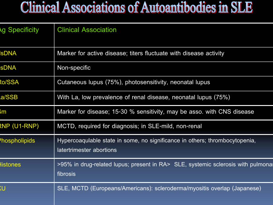
Clinical AssociationAg Specificity
dsDNA Marker for active disease; titers fluctuate with disease activity
ssDNA Non-specific
Ro/SSA Cutaneous lupus (75%), photosensitivity, neonatal lupus
La/SSB With La, low prevalence of renal disease, neonatal lupus (75%)
Sm Marker for disease; 15-30 % sensitivity, may be asso. with CNS disease
RNP (U1-RNP) MCTD, required for diagnosis; in SLE-mild, non-renal
Phospholipids Hypercoaqulable state in some, no significance in others; thrombocytopenia, latertrimester abortions
Histones >95% in drug-related lupus; present in RA> SLE, systemic sclerosis with pulmonary fibrosis
KU SLE, MCTD (Europeans/Americans): scleroderma/myositis overlap (Japanese)
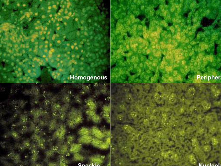
Homogenous Peripheral
Speckle Nucleolar
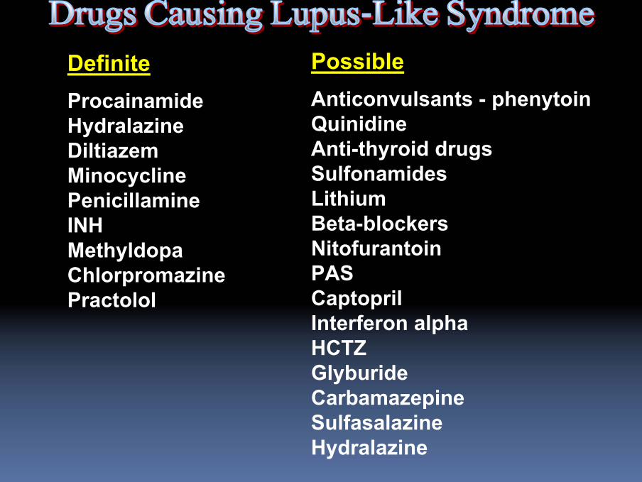
DefiniteProcainamideHydralazineDiltiazemMinocyclinePenicillamineINHMethyldopaChlorpromazinePractolol
PossibleAnticonvulsants - phenytoinQuinidineAnti-thyroid drugsSulfonamidesLithiumBeta-blockersNitofurantoinPASCaptoprilInterferon alphaHCTZGlyburideCarbamazepineSulfasalazineHydralazine

Fever, Fever, myalgiasmyalgias, rash, , rash, arthralgiasarthralgias--arthritis, arthritis, serositisserositis, , hemotologichemotologic abnomalitiesabnomalitiesKidney disease and CNS uncommonKidney disease and CNS uncommonAntiAnti--DNA DNA AbAb and low C` uncommonand low C` uncommonSome with renal disease are ANCA + (antiSome with renal disease are ANCA + (anti--MPO + MPO + lactoferinlactoferin))
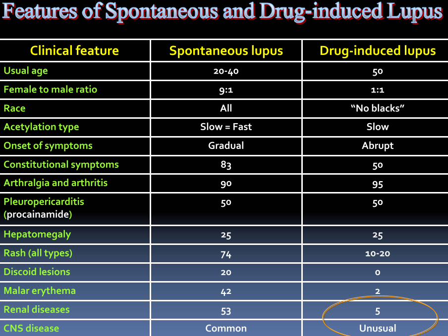
Clinical feature Spontaneous lupus Drug‐induced lupusUsual age 20‐40 50
Female to male ratio 9:1 1:1
Race All “No blacks”
Acetylation type Slow = Fast Slow
Onset of symptoms Gradual Abrupt
Constitutional symptoms 83 50
Arthralgia and arthritis 90 95
Pleuropericarditis(procainamide)
50 50
Hepatomegaly 25 25
Rash (all types) 74 10‐20
Discoid lesions 20 0
Malar erythema 42 2
Renal diseases 53 5
CNS disease Common Unusual
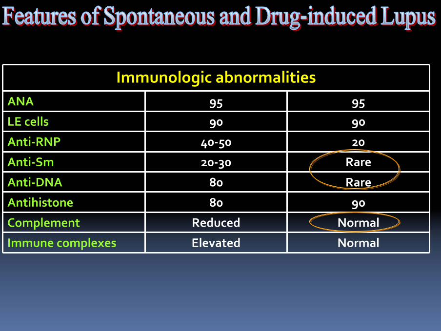
Immunologic abnormalitiesANA 95 95
LE cells 90 90
Anti‐RNP 40‐50 20
Anti‐Sm 20‐30 Rare
Anti‐DNA 80 Rare
Antihistone 80 90
Complement Reduced Normal
Immune complexes Elevated Normal
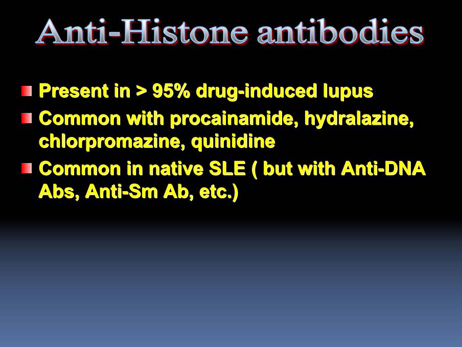
Present in > 95% drugPresent in > 95% drug--induced lupusinduced lupusCommon with Common with procainamideprocainamide, , hydralazinehydralazine, , chlorpromazine, chlorpromazine, quinidinequinidineCommon in native SLE ( but with AntiCommon in native SLE ( but with Anti--DNA DNA Abs, AntiAbs, Anti--SmSm AbAb, etc.), etc.)
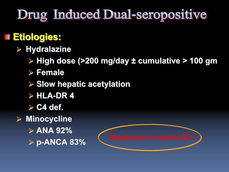
Etiologies:Etiologies:HydralazineHydralazine
High dose (>200 mg/day High dose (>200 mg/day ±± cumulative > 100 gmcumulative > 100 gmFemaleFemaleSlow hepatic Slow hepatic acetylationacetylationHLAHLA--DR 4DR 4C4 def.C4 def.
MinocyclineMinocyclineANA 92%ANA 92%pp--ANCA 83%ANCA 83% Should not be used in SLE
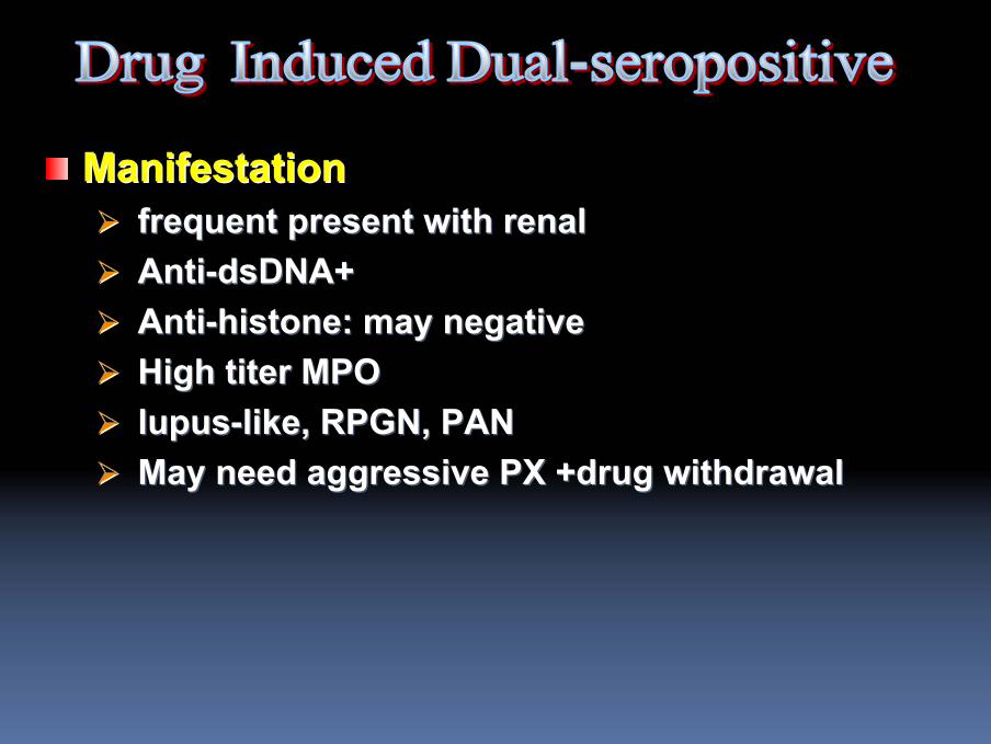
ManifestationManifestationfrequent present with renalfrequent present with renalAntiAnti--dsDNAdsDNA++AntiAnti--histonehistone: may negative: may negativeHigh titer MPOHigh titer MPOlupuslupus--like, RPGN, PANlike, RPGN, PANMay need aggressive PX +drug withdrawalMay need aggressive PX +drug withdrawal

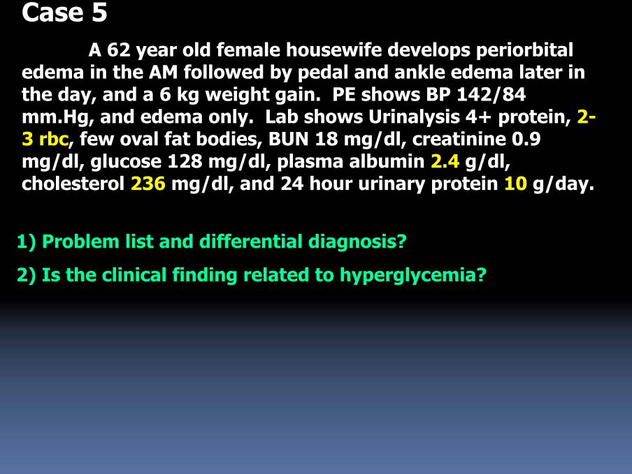
Case 5A 62 year old female housewife develops periorbital
edema in the AM followed by pedal and ankle edema later in the day, and a 6 kg weight gain. PE shows BP 142/84 mm.Hg, and edema only. Lab shows Urinalysis 4+ protein, 2-3 rbc, few oval fat bodies, BUN 18 mg/dl, creatinine 0.9 mg/dl, glucose 128 mg/dl, plasma albumin 2.4 g/dl, cholesterol 236 mg/dl, and 24 hour urinary protein 10 g/day.
1) Problem list and differential diagnosis?
2) Is the clinical finding related to hyperglycemia?
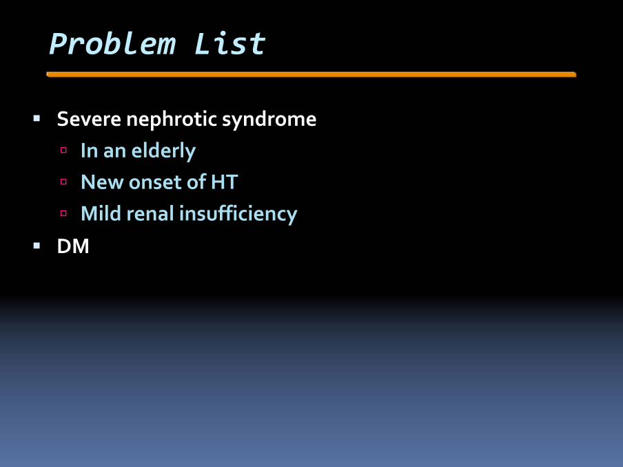
Problem List
Severe nephrotic syndromeIn an elderlyNew onset of HTMild renal insufficiency
DM
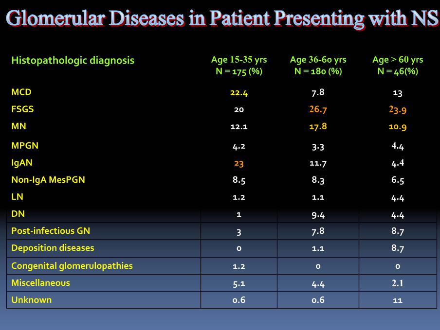
110.60.6Unknown
2.14.45.1Miscellaneous
001.2Congenital glomerulopathies
8.71.10Deposition diseases
8.77.83Post‐infectious GN
4.49.41DN
4.41.11.2LN
6.58.38.5Non‐IgA MesPGN
4.411.723IgAN
4.43.34.2MPGN
10.917.812.1MN
23.926.720FSGS
137.822.4MCD
Age > 60 yrsN = 46(%)
Age 36-60 yrsN = 180 (%)
Age 15-35 yrsN = 175 (%)
Histopathologic diagnosis
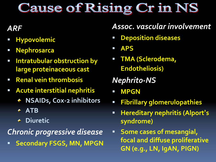
ARFHypovolemic
Nephrosarca
Intratubular obstruction by large proteinaceous cast
Renal vein thrombosis
Acute interstitial nephritisNSAIDs, Cox‐2 inhibitorsATBDiuretic
Chronic progressive diseaseSecondary FSGS, MN, MPGN
Assoc. vascular involvementDeposition diseases
APS
TMA (Sclerodema, Endotheliosis)
Nephrito‐NSMPGN
Fibrillary glomerulopathies
Hereditary nephritis (Alport’ssyndrome)
Some cases of mesangial, focal and diffuse proliferativeGN (e.g., LN, IgAN, PIGN)
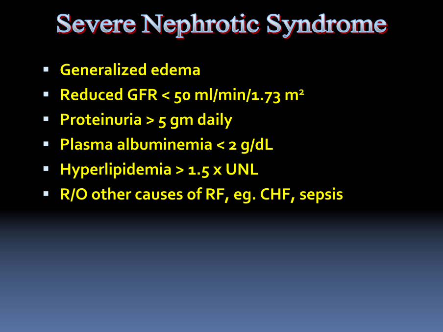
Generalized edema Reduced GFR < 50 ml/min/1.73 m2
Proteinuria > 5 gm dailyPlasma albuminemia < 2 g/dLHyperlipidemia > 1.5 x UNLR/O other causes of RF, eg. CHF, sepsis
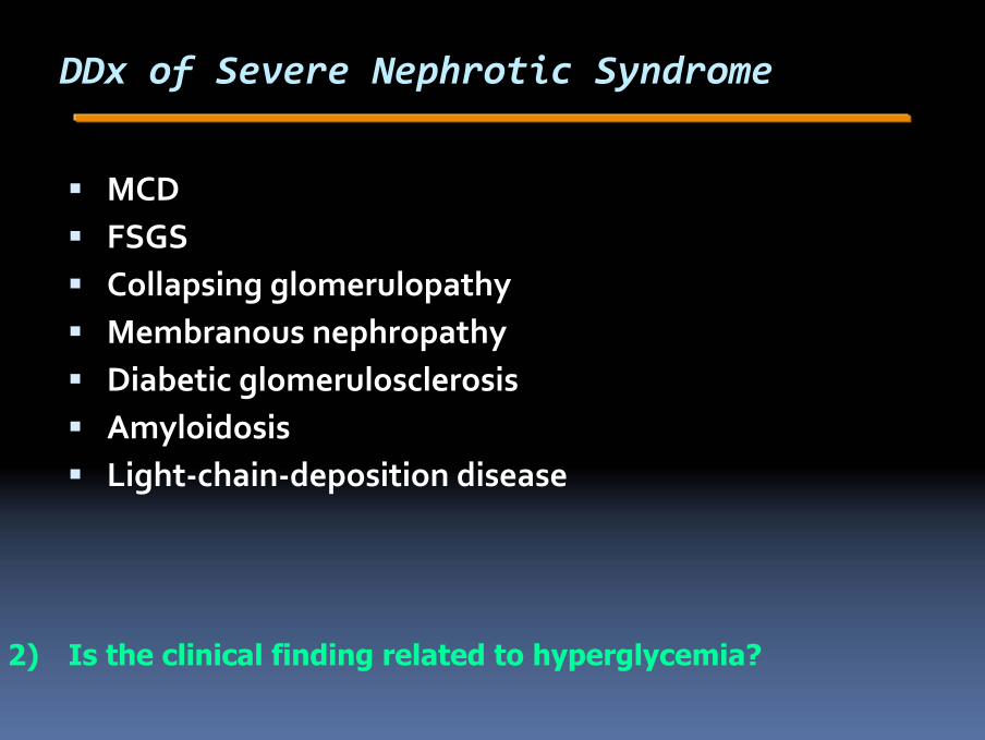
DDx of Severe Nephrotic Syndrome
MCDFSGSCollapsing glomerulopathyMembranous nephropathyDiabetic glomerulosclerosisAmyloidosisLight‐chain‐deposition disease
2) Is the clinical finding related to hyperglycemia?
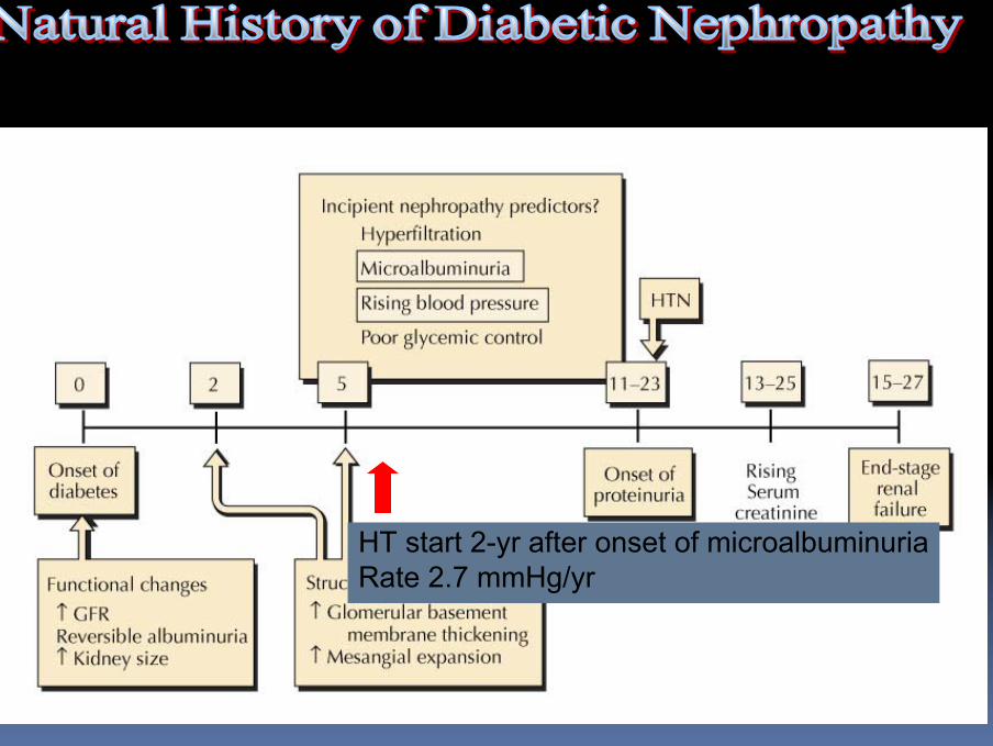
HT start 2-yr after onset of microalbuminuriaRate 2.7 mmHg/yr
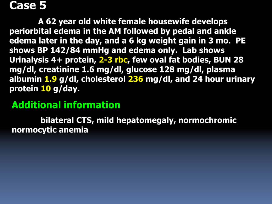
Case 5A 62 year old white female housewife develops
periorbital edema in the AM followed by pedal and ankle edema later in the day, and a 6 kg weight gain in 3 mo. PE shows BP 142/84 mmHg and edema only. Lab shows Urinalysis 4+ protein, 2-3 rbc, few oval fat bodies, BUN 28 mg/dl, creatinine 1.6 mg/dl, glucose 128 mg/dl, plasma albumin 1.9 g/dl, cholesterol 236 mg/dl, and 24 hour urinary protein 10 g/day.
Additional informationbilateral CTS, mild hepatomegaly, normochromic
normocytic anemia
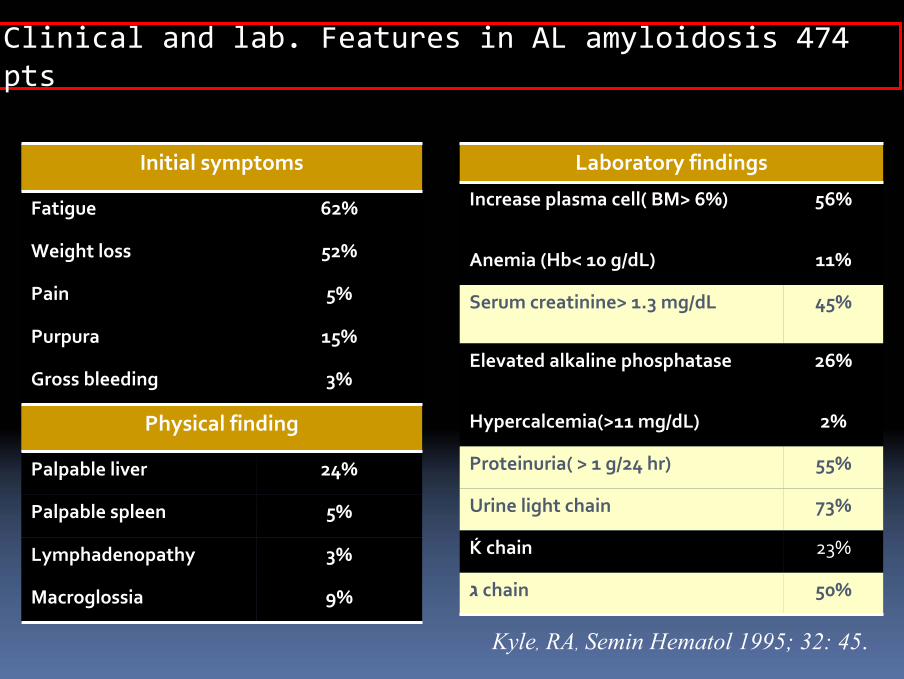
Clinical and lab. Features in AL amyloidosis 474 pts
Initial symptoms
Fatigue 62%
Weight loss 52%
Pain 5%
Purpura 15%
Gross bleeding 3%
Physical finding
Palpable liver 24%
Palpable spleen 5%
Lymphadenopathy 3%
Macroglossia 9%
Kyle, RA, Semin Hematol 1995; 32: 45.
Laboratory findings
Increase plasma cell( BM> 6%) 56%
Anemia (Hb< 10 g/dL) 11%
Serum creatinine> 1.3 mg/dL 45%
Elevated alkaline phosphatase 26%
Hypercalcemia(>11 mg/dL) 2%
Proteinuria( > 1 g/24 hr) 55%
Urine light chain 73%
Ќ chain 23%
ג chain 50%
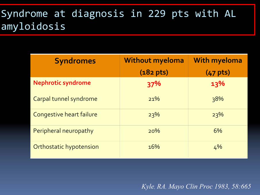
Syndrome at diagnosis in 229 pts with AL amyloidosis
Syndromes Without myeloma
(182 pts)
With myeloma
(47 pts)Nephrotic syndrome 37% 13%
Carpal tunnel syndrome 21% 38%
Congestive heart failure 23% 23%
Peripheral neuropathy 20% 6%
Orthostatic hypotension 16% 4%
Kyle, RA, Mayo Clin Proc 1983, 58:665
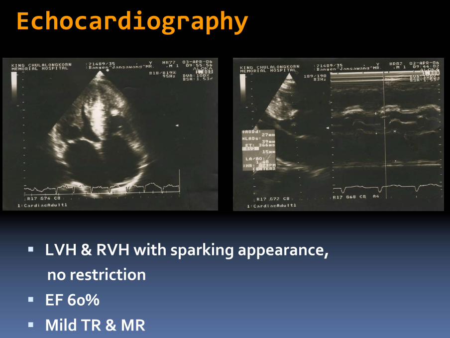
LVH & RVH with sparking appearance, no restrictionEF 60%Mild TR & MR
Echocardiography
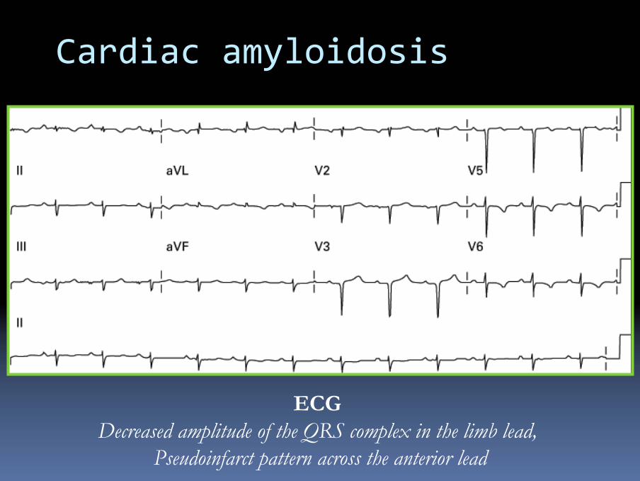
Cardiac amyloidosis
ECG Decreased amplitude of the QRS complex in the limb lead,
Pseudoinfarct pattern across the anterior lead
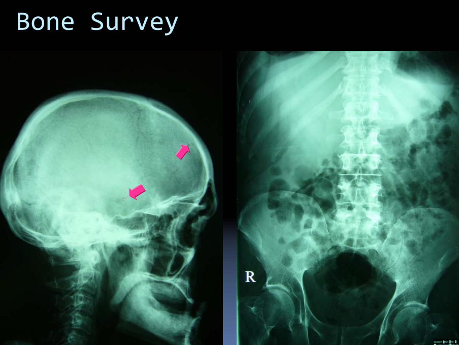
Bone Survey

Case 6
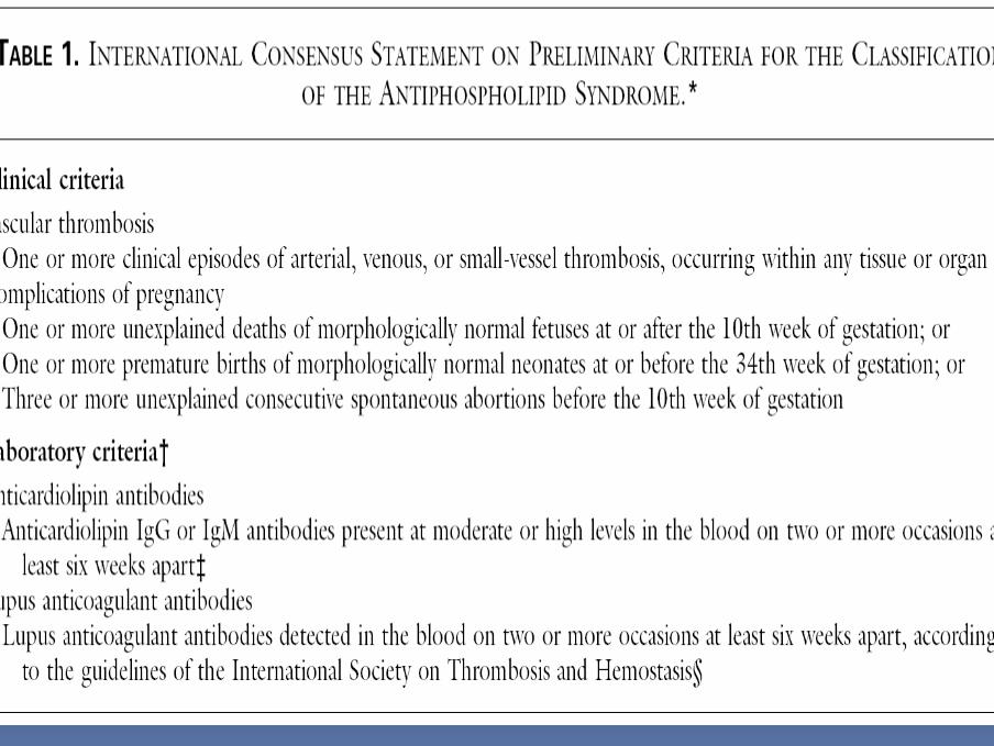
A 48 year old female pharmacist has a history of DVT 4 yrs ago, and a PE 2 years ago. She is found to have an elevated Scr of 1.8 mg/dl, urinalysis 3+ protein 5-10 rbc’s 0 casts. Physical exam shows BP 150/92 mm.Hg, 1+ pedal edema, prominent P2, and livedo reticularis of the legs. 24 hr urinary protein excretion is 1.9 g/day, ANA+ 1:160, anti DNA antibody negative, serum complement WNL; wbc 6,200, HCT 36%, platelets 100,000.
1) What serologic tests might confirm the diagnosis here ?
2) What features in the history might help in the diagnosis ?
3) What other routine laboratory features are useful diagnostically while the definitive serologic tests are pending ?

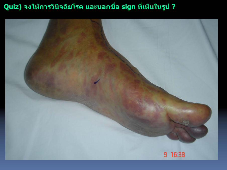
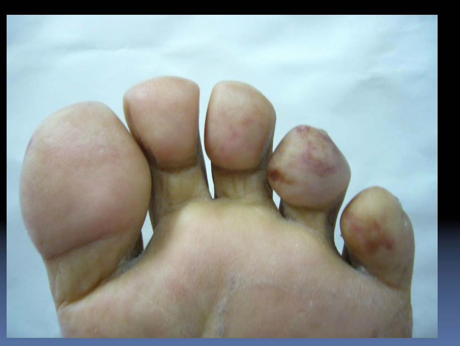
Quiz) จงใหการวินิจฉัยโรค และบอกชื่อ sign ที่เห็นในรูป ?

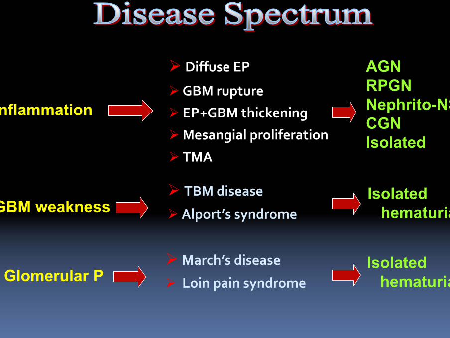
TBM disease
Alport’s syndrome
Diffuse EP
GBM rupture
EP+GBM thickening
Mesangial proliferation
TMA
GBM weakness
AGNRPGNNephrito-NS CGNIsolated
Isolated hematuria
Inflammation
March’s disease
Loin pain syndrome ↑ Glomerular PIsolated
hematuria
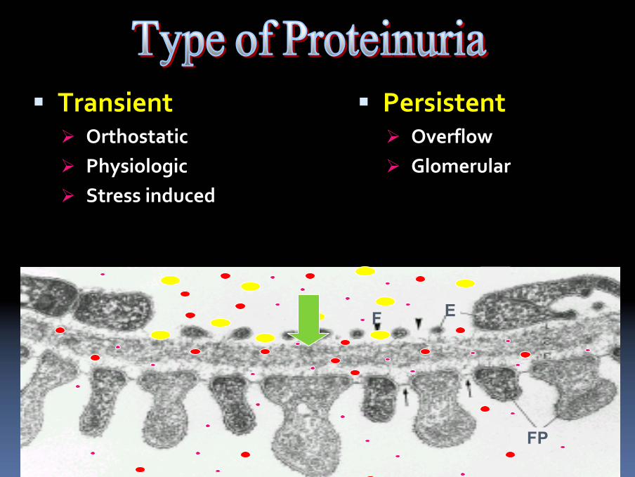
TransientOrthostaticPhysiologicStress induced
PersistentOverflow Glomerular
FP
EF
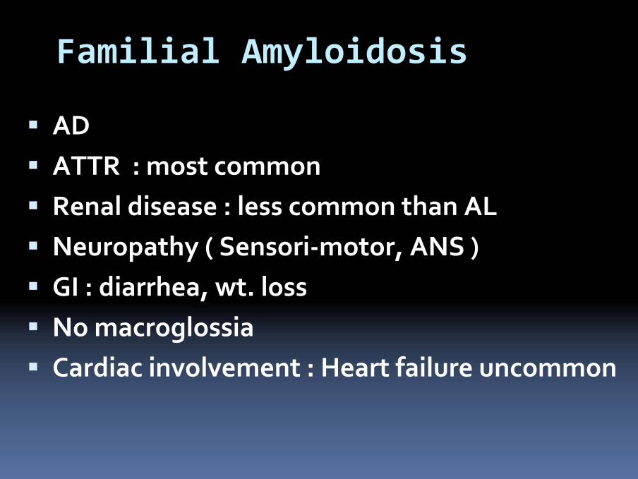
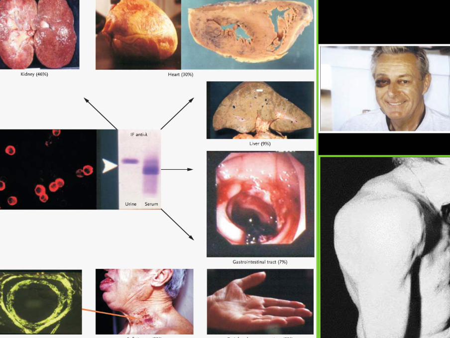
Familial Amyloidosis
ADATTR : most commonRenal disease : less common than ALNeuropathy ( Sensori‐motor, ANS )GI : diarrhea, wt. lossNo macroglossiaCardiac involvement : Heart failure uncommon