Embed Size (px)
Citation preview
Health Professional Handbook
UK Newborn Screening Programme Centre www.newbornbloodspot.screening.nhs.uk/
A guide to newborn blood spot screening for healthcare professionals December 2012
2
This is an update of the UK Newborn Screening Programme Centre’s Health Professional Handbook 2005, December 2012
Review date: December 2015
This publication has been produced by the UK Newborn Screening Programme Centre, which is funded by the Department of Health for the
whole of the UK.
© 2012 UK Newborn Screening Programme Centre
3
Contents
1. Introduction ......................................................................................................... 4
2. What is screening? ............................................................................................. 5
2.1 A definition ....................................................................................................... 5
2.2 The UK National Screening Committee ............................................................ 5
2.3 Limitations of screening ................................................................................... 5
3. Newborn blood spot screening .......................................................................... 6
3.1 What is newborn blood spot screening? ........................................................... 6
3.2 Phenylketonuria (PKU) .................................................................................... 6
3.3 Congenital hypothyroidism (CHT) .................................................................... 9
3.4 Sickle cell disease (SCD) ............................................................................... 13
3.5 Cystic fibrosis (CF)......................................................................................... 16
3.6 Medium-chain acyl-CoA dehydrogenase deficiency (MCADD) ....................... 19
4. The screening process ..................................................................................... 22
4.1 Informed choice and communication with parents .......................................... 23
4.2 Consent ......................................................................................................... 24
4.3 Residual blood spots ...................................................................................... 25
4.4 Use of identifiable data .................................................................................. 26
4.5 Communication during the screening process ................................................ 27
4.6 Repeat samples ............................................................................................. 32
4.7 Screening results ........................................................................................... 34
5. Taking the newborn blood spot sample .......................................................... 38
6. What happens next? ......................................................................................... 41
7. Performance monitoring ................................................................................... 46
7.1 Key performance indicators ........................................................................... 46
7.2 Standards ...................................................................................................... 46
8. Serious incidents .............................................................................................. 47
9. Abbreviations .................................................................................................... 48
10. Appendix 1 - An illustration of autosomal recessive inheritance ................ 49
11. Appendix 2 - Sources of information and support ........................................ 50
12. References ....................................................................................................... 51

4
1. Introduction The UK National Screening Committee (UK NSC) recommends that all babies in the UK are screened for phenylketonuria (PKU), congenital hypothyroidism (CHT), sickle cell disease (SCD), cystic fibrosis (CF) and medium‐chain acyl‐CoA dehydrogenase deficiency (MCADD). The UK Newborn Screening Programme Centre (UKNSPC) was established in 2002, with the overall objective of assuring high quality screening services for babies and their parents through the development of a quality assurance and performance management framework for the newborn blood spot screening programme. Over the past ten years, the antenatal and newborn screening programmes in England have evolved and been implemented with the support of regional teams who act as a link between national, regional and local organisations. As the UK NSC moves into Public Health England in 2013, the regional teams will develop into quality assurance teams across England. These teams will cover four clusters: North of England, Midlands and East, South of England and London. More information and contact details are available at www.screening.nhs.uk/regionalteams. Newborn blood spot screening is offered to all newborn babies up to one year of age. The programme aims to identify babies who are at high risk of having certain serious but rare conditions before they develop symptoms. By detecting these conditions early it is possible to treat them and reduce their severity. It is a complex programme delivered by a range of different organisations working together. It is also essential to minimise the adverse effects of screening, including anxiety, inaccurate information and unnecessary investigation, and to provide reassurance to the majority of parents whose babies are thought not to be affected. This handbook was first published in 2005, and it is designed to be a reference guide for all health professionals involved in the newborn blood spot screening programme. In particular, it was written to support midwives in communicating with parents about newborn blood spot screening and in collecting high quality blood spots, and to support health visitors (HVs) in talking to parents about results and supporting families of affected babies. This 2012 edition of the handbook reflects changes that have occurred in the programme.
5
2. What is screening? 2.1 A definition Screening is a process of identifying apparently healthy people who may be at increased risk of a disease or condition. They can then be offered information, further tests and appropriate treatment to reduce their risk and/or any complications arising from the disease or condition [1]. 2.2 The UK National Screening Committee The UK NSC advises Ministers and the NHS in the four UK countries about all aspects of screening and supports implementation of screening programmes. Using research evidence, pilot programmes and economic evaluation, it assesses the evidence for programmes against a set of internationally recognised criteria covering the condition, the test, the treatment options and the effectiveness and acceptability of the screening programme. Assessing programmes in this way is intended to ensure that they do more good than harm at a reasonable cost. The UK NSC also sets up practical mechanisms to oversee the introduction of new programmes in the English NHS and monitors the effectiveness and quality of these programmes. The UK NSC regularly reviews policy on screening for different conditions in the light of new research evidence becoming available. 2.3 Limitations of screening Screening has important ethical differences from usual clinical practice, as the health service is targeting apparently healthy people, offering to help individuals to make better informed choices about their health. However, there are risks involved and it is important that people have realistic expectations of what a screening programme can deliver. Whilst screening has the potential to save lives or to improve the quality of lives through early diagnosis of serious conditions, it is not a fool-proof process. Screening can reduce the risk of developing a condition or its complications but it cannot offer a guarantee of protection. In any screening programme, there is a minimum of false positive results (wrongly identified as having the condition) and false negative results (wrongly reported as not having the condition). The UK NSC is increasingly presenting screening as risk reduction to emphasise this point.
6
3. Newborn blood spot screening This section provides background information about newborn blood spot screening, and the conditions screened for as part of the national blood spot screening programmes in the UK. It is designed to inform health professionals and parents. 3.1 What is newborn blood spot screening? Newborn blood spot screening aims to identify babies who are at high risk of having certain serious but rare conditions before they develop symptoms. Screening is not the same as diagnosis; instead it identifies which babies need to go on to have diagnostic tests to determine whether or not they do have the condition. By detecting these conditions early it is possible to treat them and reduce their severity. Newborn blood spot screening is offered to all babies in the UK. The blood spot sample is taken on day 5, and in exceptional circumstances between day 5 and day 8 (day of birth is day 0). A health professional pricks the baby’s heel and collects a small amount of blood onto the newborn blood spot card (a special filter paper, formerly known as the Guthrie card). The card is sent to the regional newborn screening laboratory and analysed for five conditions*. All parents in the UK are offered screening for their babies for PKU, CHT, SCD, CF and MCADD. If PKU, CHT or MCADD are not picked up early, they almost always cause severe developmental problems, including serious mental disability or even, in the case of MCADD, death. For CF and SCD, although screening cannot prevent periods of illness, early detection of both of these conditions, before children become ill, can improve their health. Each of these five conditions and the reasons for screening are described below. *Please note that a study is taking place in five areas of England to explore the potential for an extended newborn screening programme. Five additional rare disorders are being included in the study, which will last for one year. For more information please visit www.expandedscreening.org/. 3.2 Phenylketonuria (PKU) Babies are screened for PKU as part of the newborn blood spot programme. The following information is based on the research evidence presented in a comprehensive systematic review about screening for PKU to the UK NSC in 1997 [2], and a further review of the literature in 2012 by the UKNSPC. What is PKU? People with PKU are unable to break down phenylalanine, an amino acid present in many foods. This is because the liver does not have enough of an enzyme called phenylalanine hydroxylase (PAH). This enzyme is needed to convert phenylalanine into tyrosine, which is essential for normal brain development after birth. People with
7
PKU accumulate too much phenylalanine in the body and not enough tyrosine. The level of phenylalanine in the blood is higher than normal so a blood test can show whether or not someone is likely to have PKU. The most serious form of PKU is sometimes called ‘classical’ PKU. If classical PKU is left untreated it almost always leads to severe mental disability as well as seizures [2-6]. People with untreated classical PKU are usually unable to talk, read or write and need help to look after themselves throughout their life [2]. A baby with untreated PKU doesn’t usually show any signs of the condition for the first few months. By the time they are 6-12 months they show delayed mental development [7]. By the time they are two years old they show serious and permanent mental disability [2]. Around a fifth of babies with PKU detected by newborn screening in the UK have the milder form of the condition. Milder forms of PKU are called hyperphenylalaninaemia (HPA), previously referred to as ‘benign’ PKU. Someone with untreated mild PKU might have ‘slight to moderate’ mental disability, depending partly on how much phenylalanine they have in their blood [5, 6]. A very small number of people who are found to have raised levels of phenylalanine do not have a primary defect in the enzyme PAH, but in its cofactor, biopterin [6]. Biopterin is also needed in order for the liver to break down phenylalanine. People with these rare biopterin defects have problems additional to PKU and are treated differently. An increased blood phenylalanine is not specific for PKU. In addition to disorders of PAH deficiency, increased phenylalanine detected via newborn screening at 5-8 days may occur in several other situations; in some cases the phenylalanine increase may also be associated with an increase in tyrosine. In these latter cases PKU is not suspected (although not impossible if there are two co-existing disorders) and these babies with an associated increase in tyrosine require different investigation and management, and urgent referral to an appropriate specialist clinician. It has been well established that an elevated phenylalanine and tyrosine on newborn screening is a feature of galactosaemia and for this reason it is important to consider galactosaemia as part of the differential diagnosis of a raised phenylalanine [8]. The incidence of galactosaemia in the UK is approximately 1 in 40,000 births. How common is PKU? In the UK and Europe there is an overall incidence of 1 in 10,000 live births [2, 4, 9, 10]. PKU is highest amongst white Caucasians, and is less prevalent in populations with Sub-Saharan African and South Asian ancestry [4, 10]. There are around 800,000 babies born in the UK each year, and in 2010-11 100 babies had a positive result for PKU following newborn blood spot screening [11]. The likelihood of having PKU is related to your ethnic origin. PKU is twice as common in Ireland, where there is around 1 baby born with PKU in every 4,500 births [2], compared to 1 in every 200,000 in Finland [4]. By contrast, PKU is very
8
uncommon amongst certain groups, including African-Americans and Ashkenazi Jews [2]. What treatment is available for PKU? If started early, treatment for PKU is very effective [2, 4-7, 9]. It involves a special diet. Patients cannot eat foods that contain a lot of phenylalanine, which means they have to be very careful about how much protein they eat. Because people need protein to stay healthy and children need it to grow, they need a special dietary supplement, containing all the other essential amino acids except phenylalanine, and extra minerals and vitamins. They need, and can manage, a very small amount of phenylalanine in low protein foods. Their blood is tested regularly to check their levels of phenylalanine. For the best treatment, a person with PKU needs help from a team of experienced health professionals, including doctors, nurses and dieticians [9]. There are several reports demonstrating the effectiveness of treatment for PKU. Babies who start on treatment when they are only a few weeks old have intellectual quotient (IQ) levels that are ‘approaching normal’. Children who are first treated a bit later in life have lower IQ levels [3, 5, 6, 9]. One research study shows that when treatment is started after two months the outcome is ‘poor’ [6]. Better results are seen when treatment starts as soon as possible after confirmation of diagnosis [3, 5, 9]. One research study found that almost all babies with PKU who are treated this early have IQs in the normal range. In the UK the aim is to start dietary treatment before 14 days to minimize the risk to the baby’s development. In the past, patients with PKU stopped taking their special diet as teenagers but, increasingly, research points to the fact that the adult brain remains sensitive to high levels of phenylalanine. Research has shown that some adult patients have developed severe neurological and mental problems [3, 5, 6]. This has usually occurred in adults that were originally treated late, or that were poorly managed; this may have made them more vulnerable to developing these problems later in life. The recommendation is now to maintain the diet for life [3, 5, 6, 9, 12]. This has major social and financial implications. It is possible for mothers to breast feed their babies with PKU so long as they balance the amount of breast milk with the baby’s special dietary supplement. This may require mothers to express and discard their breast milk initially, until their baby’s phenylalanine levels are within target levels. Women who have PKU and stopped the special diet when they became adults are at risk of having babies with physical and mental disabilities [4, 13]. This is due to the mother’s high phenylalanine levels affecting the baby in utero and not because the baby has PKU. The evidence shows this can be prevented through strict control of the mother’s phenylalanine levels; the mother must maintain the special diet before conception and throughout her pregnancy [4, 6]. For this reason it is especially
9
important for women with PKU and HPA to remain in contact with PKU services to ensure the diet is undertaken prior to conception and throughout pregnancy. Is there any way to prevent someone getting PKU? PKU is an autosomal recessive genetic condition (see Appendix 1 for illustration). If a person has only one copy of the altered gene that causes PKU, they are a healthy carrier of the condition. Either parent can be a carrier of the condition without knowing. When both parents are carriers they may both pass on the altered gene that causes PKU to their children. If both parents are carriers, they have a one in four chance, with each and every pregnancy, of having a baby with PKU. Once a baby with two copies of the altered gene is conceived there is no way to stop the baby having PKU. Pre-natal screening is not available on the NHS. However, the treatment for PKU can stop the baby from becoming mentally disabled. What are the benefits of screening for PKU? The aim of screening is to identify which newborns are more likely to have PKU so that treatment can begin as soon as possible. Newborn screening means early treatment for babies with PKU. If started early, treatment is highly effective at preventing the development of serious mental disability [4-6, 9]. Early diagnosis of a baby with PKU through screening can also alert the parents to their risk of having other affected children. When an infant is diagnosed with PKU, it is recommended that any future baby should be tested 48-72 hours after birth for PKU [8], rather than waiting until they are five days old. What difficulties does screening raise? Like all screening, the test for PKU is not 100% reliable (although it very nearly is). It is extremely rare for a baby to be thought to have PKU when they don’t (false positive), or more seriously, to be thought not to have PKU when they do (false negative) [2]. Very occasionally the blood spot sample is not taken or is mislaid. This is extremely rare. Is screening all babies for PKU cost effective? Whilst the advantages of newborn screening for babies affected by PKU and for their families are clearly enormous, in order to introduce a national screening programme it is important to justify the expense of screening. There are only a few well-conducted economic evaluations but the evidence suggests that screening all babies for PKU makes sense financially as well as for health and social reasons [2, 14]. 3.3 Congenital hypothyroidism (CHT) Babies are also screened for CHT as part of the newborn blood spot programme. The following information is based on the research evidence presented in a comprehensive systematic review about screening for CHT to the UK NSC in 1997 [2], and a further review of the literature in 2012 by the UKNSPC.
10
What is CHT? CHT is a condition where, for one of several reasons, the thyroid gland doesn’t work when a baby is born and fails to make the thyroid hormone called thyroxine. Problems with the gland itself are called primary CHT. This can be caused by the complete absence of the thyroid gland (called agenesis) or the lack of normal development of the gland, which means it is situated in the wrong place (called ectopic thyroid). Another cause of primary hypothyroidism is when the gland is of normal size and position but there is a problem with production of thyroxine (called dyshormonogenesis). Alternatively, hypothyroidism can be caused by defects earlier in the chemical pathway that regulates the production of thyroxine. The thyroid gland doesn’t work in people who have very low levels of thyroid-stimulating hormone (TSH) (also called thyrotropin), produced by the pituitary gland. This is called secondary hypothyroidism. Cases of secondary hypothyroidism are not detected by newborn screening. The thyroid gland usually starts working in the unborn fetus from about 20 weeks gestation. The mother’s own thyroid doesn’t provide enough thyroxine to maintain sufficiently high levels in the fetus. In very severe cases of CHT babies are born with, or quickly develop, the following symptoms: a very low hair-line, a protruding tongue, cold skin, an umbilical hernia, jaundice, feeding difficulties, constipation, and generally sluggish behaviour [2]. However, only very few babies will have all of these symptoms and primary CHT is rarely diagnosed by clinical means in the newborn period [15-18]. Common symptoms of CHT include decreased activity and increased sleep, feeding difficulty, constipation, and prolonged jaundice. On examination, common signs include a myxoedematous face, large fontanelles, macroglossia (a large tongue), a distended abdomen with umbilical hernia, and hypotonia [15]. If babies with CHT are not treated, they fail to grow properly and will have mild to severe mental disability. In general, patients with complete absence of the thyroid gland (thyroid agenesis) are the most severely affected [15, 19]. Babies with secondary hypothyroidism often have additional health problems. This means that it is easier to diagnose them because of other signs (a clinical diagnosis). These can include low blood sugar, jaundice, small genitalia and abnormal facial features. Some children with secondary hypothyroidism don’t develop the condition until they are older and present in later childhood with reduced physical growth from associated pituitary insufficiency. Children with secondary hypothyroidism will not be identified by newborn screening. Some babies are initially hypothyroid but it resolves on its own later in childhood [15, 17, 20-22]. This is called transient hypothyroidism and may be caused by exposure to iodine in antiseptics soon after birth or by maternal antibodies. It is very rare in full term babies. There may also be other reasons why a baby initially has insufficient thyroxine after birth but these are not yet well understood.
11
Preterm infants are at risk of hypothyroxinaemia (having a low level of thyroxine) due to several factors, including immaturity of thyroid function, the effects of acute illness and/or the use of iodine-containing compounds in imaging and surgery. There is no evidence that preterm infants are at increased risk of CHT when compared with term infants. The current TSH-based screening test may not detect those preterm infants who do have CHT, especially those born between 23 and 27 weeks gestation, because they show a delayed rise in TSH levels after birth, mainly due to immaturity of the hypothalamic-pituitary axis. To ensure preterm infants are appropriately screened for CHT, from 1st April 2012, all babies born at less than 32 weeks (less than or equal to 31 weeks + 6 days) should be offered a preterm repeat test at 28 days of age or discharge home, whichever is the sooner. This policy means that babies eligible for a CHT preterm repeat test should complete their newborn blood spot tests before they are discharged home from hospital. How common is CHT? The UKNSPC is currently funding a British Paediatric Surveillance Unit study on CHT, one of the aims of which is to determine the incidence of the condition in the UK [23]. Several papers in the literature quote an incidence of about 1 in 3,000 babies [24, 25]. There are around 800,000 babies born in the UK each year, and in 2010-11 over 600 babies had a positive result for CHT following newborn blood spot screening [11]. Similar rates of CHT are reported in the US and Europe [17, 20, 22, 26]. Several US programs have reported a higher incidence in the Asian, Native American and Hispanic populations and a lower incidence in the American Black population [15]. CHT is also reported to be higher in infants born to older women and in infants born preterm [17]. Nearly all screening programmes report an increased female prevalence, approaching a 2:1 female to male ratio [15, 16, 27]. What treatment is available for CHT? The aim of treatment is to normalise thyroid function as rapidly as possible to improve IQ [28]. Babies with primary hypothyroidism are treated with thyroxine, given as either a suspension or in the form of crushed tablets, mixed in a small quantity (less than 5 ml) of milk / water or not, by mouth [28]. If babies are treated early they will grow normally and reach normal height. The delay in mental development can also be largely corrected. The later treatment is started, the more the baby’s development is affected by the condition [15, 29]. Long term follow-up studies have documented excellent outcomes among children with CHT for whom an appropriate dosage of thyroid hormones is established soon after birth [29]. Children who had very low levels of thyroxine before treatment (possibly because they had more severe CHT, or because treatment was started later) had an IQ at age ten that was 10 points lower than normal. Children who had slightly higher thyroxine levels before starting treatment had normal IQs [28]. Whilst it
12
is known that newborn screening can prevent severe disability in babies with CHT, it is not known whether treatment for babies with CHT can reverse any effects of hypothyroidism caused before the baby was born. It is thought these effects will only be small. Is there any way to prevent someone getting CHT? CHT is not usually an inherited condition (with the possible exception of dyshormonogenesis). In 90% of cases CHT seems to happen completely by chance. One in ten cases is thought to be inherited. There is no way to prevent babies being born with CHT [2]. What are the benefits of screening for CHT? The aim of screening is to identify, as soon as possible, which newborns are more likely to have CHT, so that treatment can be started. Treatment is highly effective at preventing physical and mental disability. Only one in ten cases of CHT is thought to be inherited. Early diagnosis of a baby with CHT through screening does not therefore necessarily provide parents with information about their risk of having other affected children. What difficulties does screening raise? Occasionally the screening result for CHT may be borderline or unclear. This requires a second sample to be taken from the baby, at least seven days after the previous sample, to confirm the result (either positive or negative). Whilst it is understood that screening for CHT is highly effective in preventing severe mental disability in children, it is difficult to calculate exactly how accurate screening for CHT is. A positive screening result does not mean that the baby definitely has the condition and a negative result does not necessarily mean that they will never have the condition. There are a number of reasons for this:
• Transient hypothyroidism: Some children do have hypothyroidism, but it is the transient form. Some research suggests that up to 50% of children who have CHT get better on their own later in childhood [21, 27]. These children are usually sick preterm babies. Transient hypothyroidism is very rare in full term babies [15, 20, 30]
• Later onset of hypothyroidism: Other babies don’t develop hypothyroidism until they are a few weeks old. This means that they are not picked up by the newborn blood spot test (sample taken on day 5)
For these reasons, although screening for CHT is known to be highly effective, it is difficult to judge exactly the accuracy of the screening test, and therefore difficult to quantify the accuracy of newborn screening for CHT [2]. As with all the conditions screened for, very occasionally the blood spot sample is not taken or is mislaid. This can mean a baby with CHT is ‘missed’, although this is rare.
13
Is screening all babies for CHT cost effective? The American Office of Technology Assessment has reported CHT screening to be one of the few programmes of preventative medicine in public health to have a positive cost-benefit ratio (of 10:1) [16]. In the UK, the evidence suggests that screening all babies for CHT makes sense financially as well as for health and social reasons. A published systematic review shows that good evidence exists to support the cost-effectiveness of a national CHT screening programme [2]. 3.4 Sickle cell disease (SCD) Babies in England are also screened for SCD as part of the newborn blood spot programme. During pregnancy, women are offered screening for SCD (as well as thalassaemia, another abnormality of the production of haemoglobin). The information below is based on two Health Technology Assessment (HTA) reviews [31, 32]. In addition, another HTA report has been published on reporting newborn carrier results [33]. We have also drawn on the work of the NHS Sickle Cell and Thalassaemia Screening Programme [34]. What is SCD? SCD is the name given to a group of inherited genetic blood conditions that affect the haemoglobin in red blood cells. They are autosomal recessive conditions, so inheritance of an affected gene from both parents results in a disorder whilst inheritance of one abnormal gene results in a healthy carrier. See Appendix 1 for an illustration of autosomal recessive inheritance. SCD is a condition that affects the normal oxygen-carrying capacity of red blood cells. When the cells are de-oxygenated and under stress, in sickle cell conditions, they can change from round flexible disc-like cells to elongated sickle or crescent-moon shapes. The effect of these changes is that the cells do not pass freely through small capillaries and form clusters, which block the blood vessels. This blockage prevents oxygenation of the tissues in the affected areas resulting in tissue hypoxia and consequent pain (known as sickle cell crisis pain). Other symptoms of SCD can include severe anaemia, susceptibility to infections, early cerebrovascular accidents and damage to major organs. SCD is a lifelong condition that can be diagnosed from a simple blood test, before the baby becomes ill. The most serious type of SCD is sickle cell anaemia (Hb SS); other forms of SCD include Hb SC and Hb S beta thalassaemia. There are also some rarer forms. Babies with SCD who are not identified through screening usually begin to show symptoms in their first year. The first time they become ill can be fatal [31]. Babies with Hb SC, rather than Hb SS, are usually less severely affected [35, 36]. The severity and pattern of disease within each condition, as well as between individual patients, varies in an unpredictable way [37]. Despite improvements in life expectancy, SCD remains a cause of significant morbidity – even in developed countries. Boulet et al found that compared with other American black children, those with SCD were more likely to: show intellectual
14
disability, probably as a result of cerebrovascular accident; have higher incidence of severe headaches; have more frequent use of prescription medication and have a health status classed as ‘fair’ or ‘poor’ [38]. The objective of the newborn screening programme is to detect babies at risk of SCD so they can enter the healthcare system as early as possible. Screening aims to identify the following specified conditions: Hb SS, Hb S beta thalassaemia (beta+, beta0 and Lepore), Hb S/HPFH, Hb SC, Hb SDPunjab, Hb SE and Hb SOArab. Other clinically significant haemoglobinopathies likely to be detected as ‘by-products’ of newborn screening include beta thalassaemia major, Hb E beta thalassaemia, beta thalassaemia intermedia and Hb H disease. How common is SCD? Haemoglobinopathies are common in people whose family origins are in malarial parts of the world. In the UK, haemoglobinopathies are seen particularly among minority ethnic groups from Africa, the Caribbean, the Mediterranean, South East Asia, the Middle East, and the Far East [39], but can be found (less frequently) in all ethnic groups. Approximately 1000 haemoglobin gene variants have been identified worldwide. SCD is estimated to affect 1 in every 2,000 births in England [40]. 240,000 people are estimated to be carriers and more than 12,500 people have the disease. The highest prevalence is among Black Africans and Black Caribbeans [40]. Laboratory data submitted to the NHS Sickle cell and Thalassaemia Screening Programme show that just over 684,000 newborns were screened for SCD in 2009-10. Approximately 360 babies were identified with screen positive results for significant conditions, and over 9,700 babies were identified as a carrier of a haemoglobin variant [41]. What treatment is available for SCD? Treatment for SCD includes taking penicillin and receiving effective vaccines to reduce the risk from serious infections, especially while the child is under five when the risk is highest [42]. Educating patients and parent/carers on how to identify and treat symptoms as early as possible has been shown to be important in ensuring quick treatment for serious complications caused by SCD [43, 44]. Some adults and older children with SCD can also be treated with hydroxyurea, which reduces the frequency and severity of sickle cell crises [45, 46]. Regular transcranial doppler ultrasonography and appropriate treatment reduces the risk of stroke [47]. Bone marrow transplantation offers the only chance of a cure for children with SCD depending on the type of disorder, but this relies on there being a compatible donor [48]. The procedure itself is very risky as it involves suppressing the immune system and can cause death. The need for support counselling for parents of children with SCD is widely recognised and is available [33, 49-55].
15
Is there any way to prevent someone getting SCD? Screening for SCD early in pregnancy means that parents can be given an informed choice. The NHS Sickle cell and Thalassaemia Screening Programme offers parents screening for sickle cell and thalassaemia, identifies women/couples at risk of a pregnancy with sickle cell or thalassaemia and provides appropriate referral and care for prenatal diagnosis, so that parents can make an informed decision about continuing the pregnancy or deciding to have a termination. Antenatal screening identifies about 22,000 carriers of sickle cell and thalassaemia every year [56]. What are the benefits of newborn screening for SCD? The main aim of newborn screening is the early detection of babies with SCD, so that penicillin and other forms of treatment, as well as parent or carer education, can be started as soon as possible, preferably by the time the baby is 8 weeks old [49, 57-59]. It has been shown that early treatment improves the health of babies with SCD and can prevent death [43, 44, 51, 60]. Early diagnosis of a baby with SCD, or identification as a carrier of SCD, can also alert the parents to their risk of having other affected children. What difficulties does screening raise? As well as identifying babies with SCD the screening procedure also identifies babies who are healthy carriers of the sickle gene variants [31]. If a baby is found to be a healthy carrier this result is reported to the parents (often by a specially trained health visitor) and counselling is offered. Sometimes another blood test is requested to confirm the result. This carrier result needs to be carefully explained so that parents understand what it means and recorded in the personal child health record (PCHR) (also known as ‘the red book’) in the specific screening box. Parents have a right to know the results of their baby’s tests. It has been recommended that carrier status information should be provided to GPs and stored in a secure and accessible format [61]. Newborn screening for SCD can also identify babies with combinations of gene variants that do not cause illness [62]. This may have psychological effects on families, who may not fully understand the result and worry that their child may become ill. This has resource implications, because the health service needs to provide education and counselling for families with these results. It may also be useful to liaise with the local screening midwife if a child is identified as being a carrier, as information given may be useful if the parents have subsequent pregnancies. An area of concern in newborn screening for SCD relates to the possibility of SCD babies being missed because of a blood transfusion prior to a blood spot sample being taken [63]. These babies do not have a valid sickle cell screen result. The 2012 Guidelines for Newborn Blood Spot Sampling recommend taking one blood spot prior to transfusion [64]. For guidance and current policy from the NHS Sickle Cell and Thalassaemia Screening Programme, including information on failsafe processes for
16
DNA testing of transfused babies without a pre-transfusion sample, please see www.sct.screening.nhs.uk/cms.php?folder=2512. In 2009 the NHS Sickle cell and Thalassaemia Screening Programme commissioned a pilot failsafe service of DNA testing for babies who had not had a pre-transfusion sample taken. A recent report has indicated that this pilot is likely to continue [65]. This service is not intended to replace the pre-transfusion sample but to act as a safety net for babies that have not had a sample taken prior to transfusion. As with the other conditions screened for, there is a very small risk that a blood spot sample is not taken, or is mislaid, and as a result a baby with SCD is ‘missed’. Is screening all babies for SCD cost effective? Deciding whether screening all newborn babies for SCD is cost effective is complex [31, 32, 66]. The primary aim is to detect infants with SCD before they become symptomatic. Most countries offering newborn screening offer universal screening rather than a targeted approach. This is based on (i) equity and (ii) the increased effectiveness of mass throughput of newborn screening laboratories [67]. Good evidence exists to support the cost-effectiveness of a national sickle cell and thalassaemia screening programme [31]. 3.5 Cystic fibrosis (CF) Screening for CF, as part of the newborn blood spot programme, became universal across the UK from October 2007. The following information is based on the research evidence presented in a comprehensive systematic review about screening for CF [2], a Child Health Sub-Group review against the UK NSC criteria for neonatal CF screening (updated May 2005) [68], and a further review of the literature in 2012 by the UKNSPC. What is CF? In CF there is a problem transporting chloride across cell membranes. This affects certain organs in the body, particularly the pancreas and lungs; the thick secretions in these organs cause digestive problems and chest infections. The abnormal transport of chloride in sweat glands leads to an increased level of chloride in the sweat of children with CF. This is the basis of the ‘sweat test’, often used to investigate suspected cases. There are now other ways of diagnosing CF, including looking for alterations (mutations) in the CF gene. This gene is called the cystic fibrosis transmembrane conductance regulator (CFTR) gene. Alterations to this gene can cause CF. While some alterations usually cause severe symptoms, others are more often associated with milder forms of CF. CF can affect the baby before birth. Fifteen per cent of affected babies are born with blocked intestines, a condition called meconium ileus. About 70% of screen positive babies with CF show some symptoms by the time they have a diagnostic sweat test [2]. These can include problems absorbing food, as well as breathing difficulties. One study found that all babies with CF examined during their first year had inflamed
17
airways and in some it was present by four weeks [69, 70]. Eventually all patients with CF develop long-term chest infections. Now most people with CF can expect to live into early adulthood (25 years) [71]. How common is CF? About 1 in 2,500 babies born in the UK has CF [72, 73]. There are around 800,000 babies born in the UK each year, and in 2010-11 over 300 babies had a positive result for CF following newborn blood spot screening [11]. CF is caused by a large number of different alterations in the CFTR gene (over 1,200 are known). The most common in the UK is called Delta F508. Whilst most of the common alterations in the CFTR gene are known to cause CF (referred to from now on as ‘CF-causing alterations’), other alterations do not cause CF, and others are not yet well understood. What treatment is available for CF? Babies with CF are treated vigorously as soon as they are first diagnosed. Treatment of children with CF aims to do two things: improve nutrition by providing supplements containing enzymes to help digestion; and reduce chest infections with frequent physiotherapy and either occasional or continuous antibiotics. Treatment can slow down the effects of the disease, but cannot stop it progressing. With treatment, half of those with CF now live to be over 30 years old. Is there any way to prevent someone getting CF? CF is an autosomal recessive genetic condition. If a person has only one copy of the gene with a CF-causing alteration, they are a healthy carrier of the condition. Either parent can be a carrier of the condition without knowing. When both parents are carriers of a CF-causing alteration they may both pass the alteration to their child and they have a one in four chance with each and every pregnancy of having a baby with CF. Once a baby with two CF-causing alterations is conceived the baby will have CF. What are the benefits of screening for CF? The research evidence is inconclusive but several studies suggest that children who are diagnosed following newborn screening might be healthier than those diagnosed later [71, 72, 74, 75]. Newborn screening for CF may also reduce any delays in diagnosis, reducing anxiety and uncertainty about why the child is ill [69, 74]. Having said that, a confirmed diagnosis following newborn screening can also take time and cause parents anxiety [69, 73]. Early diagnosis of a baby with CF through screening can also alert the parents to their risk of having other affected children. Longer-term benefits of screening are hard to assess, as improved treatments make living with CF better than in the past (whether or not a person is diagnosed through screening).

18
What difficulties does screening raise? Screening does not identify all of the 15% of babies with CF who have meconium ileus. These babies will need treatment at birth for this condition and will therefore be diagnosed. An initial analysis of the blood spots is used to identify babies at higher risk of having CF; this test is called an immunoreactive trypsinogen (IRT) test. These babies will then have DNA testing on the same sample to look for CF-causing alterations. Some babies will require a second blood spot sample to be taken between day 21 and day 28 (day of birth is day 0) in order to confirm a CF screening result. Parents should be told of the reasons for the repeat sample (that their baby’s CF result is ‘borderline’). This need for a second sample may cause anxiety whilst they await the results. The UKNSPC Cystic Fibrosis Screening Advisory Board recommends that this second sample is taken ideally as near to day 21 as possible so that parents get a conclusive screening result as soon as possible to reduce their anxiety. The introduction of DNA analysis as part of the newborn CF screening protocol has meant that fewer second blood samples are required. Using DNA analysis as part of CF screening inadvertently identifies a small number of CF carriers, babies who carry one identified alteration on the CFTR gene and who are healthy. It can be difficult to distinguish between babies who are healthy carriers (i.e. those with only one alteration), and babies who have CF (i.e. those who have a second, unidentified alteration). Not all alterations can be tested for as there are over 1,200 identified alterations. Furthermore, not all these alterations affect a baby’s health. This uncertainty means some babies will need further tests and others may need to be followed up. Currently there is some variation in how these babies are managed across the UK. When carriers of CF are identified, specially trained health visitors or specialist genetic counsellors or CF nurses will inform parents of the results [76]. This is important because there are implications for siblings and other family members who might also carry the altered gene for CF. Other family members, including the parents, will be offered genetic counselling and testing. If both parents are carriers of a CF-causing alteration, for each subsequent baby, there is a one in four chance that the baby will have CF. As with other conditions screened for on the newborn blood spot, there is a very small risk that a blood sample is not taken, or is mislaid, and as a result a baby with CF is ‘missed’. Is screening all babies for CF cost effective? Newborn screening for CF is a small part of what it costs to diagnose and treat a person with CF [77]. The systematic review concluded that some evidence exists to support a CF newborn screening programme on the basis of cost and a more recent study confirmed this [70, 71].
19
3.6 Medium-chain acyl-CoA dehydrogenase deficiency (MCADD) Following a pilot, newborn screening for MCADD was introduced in England in 2009 and has subsequently been rolled out over the whole of the UK. The information below is based on two reviews of evidence about newborn screening for MCADD [78, 79], and a further review of the literature in 2012 by the UKNSPC. What is MCADD? MCADD is a rare inherited disorder where the body cannot metabolise (break down) fat properly [78, 79]. Most infants are asymptomatic at birth [80], and most cases present before 2 years of age (mean age 13 months) [7, 78, 79], but in rare cases they may not appear until adulthood [7]. Current estimates of mortality after the first few days are about 5 to 7% over the first 6 years, unless both early diagnosis and appropriate management are in place [81]. MCADD can cause drowsiness, lack of energy and diarrhoea, and there is a risk of complications such as seizures, breathing difficulties and even coma or sudden death [79, 82, 83]. The episodes are usually precipitated by metabolic stress such as inter-current infections, particularly gastroenteritis which may appear to be quite mild [78]. Among those who experience acute episodes the risk of death is estimated to be 25% [78, 80, 84, 85], and up to 30% of survivors have significant long term morbidity including fits, learning and behavioural difficulties and other neurological deficits [78, 83]. During long periods between eating, the body breaks down its fat stores to produce energy. Fats are broken down into fatty acids, which are then broken down into shorter and shorter lengths. At each step, energy is released. People with MCADD cannot break down fatty acids properly, because they lack one of the enzymes needed. This leads to a build-up of medium-chain fats, which can become toxic. Because they cannot get energy from fat, they must rely on glucose for their energy. Screening aims to identify infants with MCADD and prevent onset of symptoms by early dietary management to reduce adverse outcomes of intellectual disability and death [80, 84, 86]. How common is MCADD? MCADD is the most common inherited disorder of mitochondrial fatty acids oxidation in people [79, 82]. Around one in 10,000 babies born in the UK has MCADD [79, 83, 84]. There are around 800,000 babies born in the UK each year, and in 2010-11 74 babies had a positive result for MCADD following newborn blood spot screening [11]. The incidence is highest in the northern European population [2, 7, 78, 79, 82-84]. A review of the ethnicity of 1.1 million newborns in the UK did not find a single case of MCADD among babies of Black/Asian ethnic origin who were screened at birth [84].
20
Around one in 80 healthy people is a carrier of MCADD [7, 79, 82], and will not have any symptoms. However, if both parents are MCADD carriers, there is a one in four chance of their child being born with MCADD [78]. What treatment is available for MCADD? Once diagnosed, MCADD is usually quite straightforward to manage and children can lead healthy, normal lives. It is managed by eating regular meals and having frequent glucose drinks if the child falls ill, so that their blood glucose level does not fall [7, 78, 83]. Whenever patients are at risk, the emergency (illness or crisis) regimen should be started early [78]. When started early, treatment appears to be remarkably effective, reducing mortality and morbidity at specialist centres to near zero [78]. However, if MCADD is not identified and treated early, children may fall into repeated comas with complications including breathing problems, seizures, brain damage or sudden death. In the published literature, reported deaths after the diagnosis of MCADD have been rare and have mainly occurred in children diagnosed late or in whom early signs were unrecognised and / or intervention was delayed [80]. MCADD is a lifelong condition that will always need careful dietary management during periods of fasting or fever. Is there any way to prevent someone getting MCADD? MCADD is an autosomal recessive genetic condition. If a person has only one copy of the gene with an MCADD-causing alteration, they are a healthy carrier of the condition. Either parent can be a carrier of the condition without knowing. When both parents are carriers of an MCADD-causing alteration they may both pass the alteration to their child and they have a one in four chance with each and every pregnancy of having a baby with MCADD. Once a baby with two MCADD-causing alterations is conceived the baby will have MCADD. What are the benefits of screening for MCADD? The aim of screening is to identify which newborns are more likely to have MCADD so that management can begin as soon as possible. Newborn screening means early treatment for babies with MCADD. If started early, management is highly effective at preventing the development of serious mental disability. Early diagnosis of a baby with MCADD through screening can also alert the parents to their risk of having other affected children. When an infant is diagnosed with MCADD, it is recommended to screen any siblings for the condition as it is possible to find undiagnosed siblings [78]. It is also recommended that any future baby should be tested 24 to 48 hours after birth for MCADD [78, 87] (a routine blood spot sample should still be taken on day 5). What difficulties does screening raise? One concern is that a proportion of screen positive babies will never experience any ill-effects and there is no way of predicting which babies these will be. Thus, all babies identified as having MCADD by newborn screening must be treated as at risk. Fortunately, the treatment is not onerous or expensive, but the need to carefully
21
monitor inter-current illnesses for the first few years of life is bound to provoke parental anxiety [2]. Like all screening, the test for MCADD is not 100% reliable (although it very nearly is). It is extremely rare for a baby to be thought to have MCADD when they don’t (false positive), or more seriously, to be thought not to have MCADD when they do (false negative) [88]. A pilot screening service was introduced in England in March 2004 and ran for four years. In this cohort, no false negative diagnoses were reported (approximately 1.5 million infants) over a two year follow-up [88]. As with other conditions screened for on the newborn blood spot, there is a very small risk that a blood sample is not taken, or is mislaid, and as a result a baby with MCADD is ‘missed’. Is screening all babies for MCADD cost effective? MCADD deficiency shows a high frequency in the general population, especially in Caucasians of North European origin. The disorder is associated with increased mortality and morbidity and is relatively easily and cheaply treated with simple dietary manipulation [7, 89, 90].
22
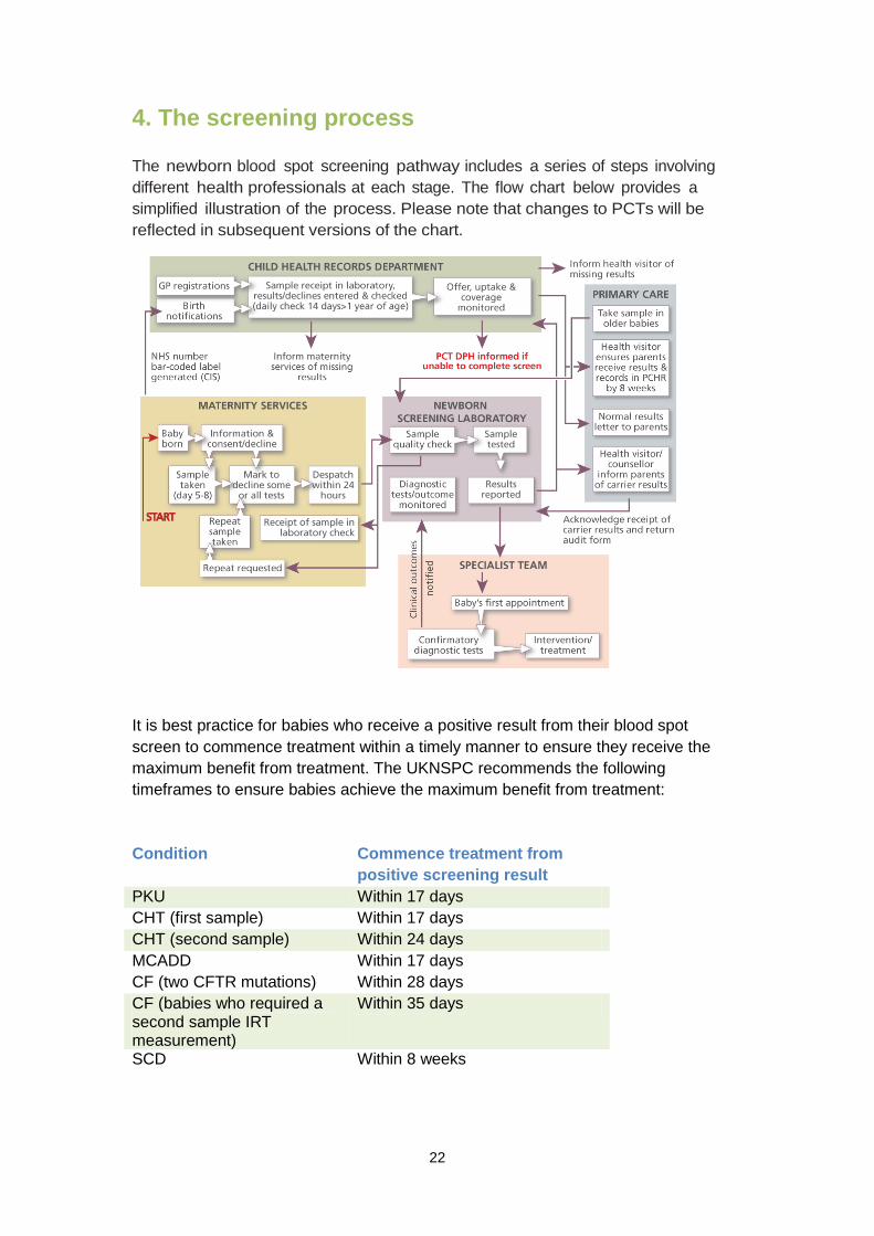
4. The screening process The newborn blood spot screening pathway includes a series of steps involving different health professionals at each stage. The flow chart below provides a simplified illustration of the process. Please note that changes to PCTs will be reflected in subsequent versions of the chart.
It is best practice for babies who receive a positive result from their blood spot screen to commence treatment within a timely manner to ensure they receive the maximum benefit from treatment. The UKNSPC recommends the following timeframes to ensure babies achieve the maximum benefit from treatment: Condition Commence treatment from
positive screening result PKU Within 17 days CHT (first sample) Within 17 days CHT (second sample) Within 24 days MCADD Within 17 days CF (two CFTR mutations) Within 28 days CF (babies who required a second sample IRT measurement)
Within 35 days
SCD Within 8 weeks
23
This means that parents must be offered screening, given time to make an informed decision, the blood spots collected and analysed and the results made available within the first weeks of a baby’s life. 4.1 Informed choice and communication with parents This section provides guidance for health professionals in offering parents an informed choice about newborn blood spot screening for their babies. The UKNSPC has developed a series of leaflets for parents to support communication about newborn blood spot screening. These include:
• A national pre-screening leaflet for all parents, ‘Blood spot screening for your newborn baby’, available in different languages: www.newbornbloodspot.screening.nhs.uk/languages
• Results leaflets for parents whose babies are suspected of having PKU, CHT, MCADD or CF, or are identified as a carrier of CF: www.newbornbloodspot.screening.nhs.uk/public
(Results and carrier leaflets for SCD are available from the NHS Sickle Cell and Thalassaemia Screening Programme’s website: www.sct.screening.nhs.uk/leaflets / www,sct.screening.nhs.uk/adultcarrierleaflets). The communication guidelines presented in this chapter are designed to ensure good communication with parents, and cover the following:
• Informed choice about newborn blood spot screening • Responding to parents who decline screening • Communication if a repeat sample is requested • Giving results
The approach towards informed choice presented in this chapter has been developed through consultation with parents and health professionals, taking into account legal and ethical advice. Why is communication important? Recent developments in newborn screening have highlighted the importance of communication between parents and health professionals about newborn blood spot screening [91-93]. Newborn blood spot screening began with conditions where single test results left little doubt about the health of the baby. Technological advances mean that more conditions can be identified from the newborn blood spots, but results may be less clear, or may identify conditions that have less impact on the baby’s health; so more parents need explanations. National newborn screening programmes based on the newborn blood spots now exist for five conditions (see section 3). Newborn screening can identify

24
babies who are carriers of (but not affected by) SCD and CF. Health professionals need up-to-date knowledge and skills in order to communicate with parents about these conditions [33]. Informed choice for newborn blood spot screening also has implications for health professionals’ communication with parents. There is a need for evidence-based information for parents and health professionals, and training for health professionals in order to support an informed choice about screening. The UKNSPC has developed guidelines on informed choice and communication and published a series of parent leaflets to support health professionals in their communication. These are available at www.newbornbloodspot.screening.nhs.uk/public. In addition, the UK NSC has developed the online resource ‘Continuing Professional Development for Screening’. This resource is available to all health professionals and has been designed as a 'one stop site' where health professionals can access all the cross-cutting education and training resources developed by the UK NSC in relation to the antenatal and newborn screening programmes, including communication. These are available at www.cpd.screening.nhs.uk/onlinelearning. Informed choice for newborn blood spot screening All parents should be offered newborn blood spot screening for their baby. Please be aware that local policies regarding informed choice and recording the parent’s decision may vary. The national recommendations are outlined below. Newborn blood spot screening should be strongly recommended to parents. This advice is based on high-quality research evidence which shows that newborn blood spot screening is effective in improving the health of babies affected by PKU, CHT, SCD and MCADD, preventing severe disability and even death [2, 6, 15, 78].
Newborn screening for CF improves families’ experience of diagnosis and improves the health of children with CF [2]. Whilst screening is strongly recommended, parents still retain the right to decline screening for their baby for one or all of the conditions [93]. 4.2 Consent When obtaining consent for newborn blood spot screening, you must ensure that parents understand they are consenting to the following:
• The sample being taken • The sample being booked in and analysed in the newborn screening
laboratory and used for quality assurance • The laboratory sending the results to the child health records
department • The results being stored on the child health information system
25
• The potential identification of their baby as a ‘carrier’ of SCD or CF • A referral to specialist services if a result is positive • The blood spot card being stored for a minimum period by the
laboratory, as detailed in the Code of Practice for the Retention and Storage of Residual Spots [94] (see 4.3; please note that this document is currently under review)
• Their baby’s anonymised data being used for research studies that help to improve the health of babies and their families in the UK, for example population studies (see 4.3)
• In rare circumstances, receipt of invitations from researchers who would like to use their baby’s blood spot card for research (this is optional – see 4.3)
• The use of identifiable data on babies, or children under age 5, with SCD or thalassaemia by the NHS Sickle Cell and Thalassaemia Screening Programme (see 4.4)
The following sections expand on the uses of residual spots and patient identifiable data. 4.3 Residual blood spots After the blood spot cards are analysed they are stored for a minimum period by the laboratory. This is detailed in the Code of Practice for the Retention and Storage of Residual Spots [94] (please note that this document is currently under review). Where there is sufficient residual blood they may then be used in several different ways. These different secondary uses for the spots are explained below. Parents should be made aware of these possible uses. To check the screening result, or for other tests recommended by the baby’s doctor If a baby becomes sick or dies unexpectedly, the laboratory and the baby’s doctor may wish to retest the baby’s newborn blood spots. This testing may help to answer parents’ questions about the baby’s illness or death. To improve the screening programme In order to check the quality of current laboratory analyses, or to explore the possibility of introducing new analyses, the stored blood samples may be re-analysed. This can involve re-analysing batches of blood spots of babies known to have particular conditions, as well as the blood spots of healthy babies. This use of the blood spots is crucial to the continuation of the newborn screening programme.
26
For research to help improve the health of babies and their families in the UK Residual newborn blood spots may also be used for research where the samples have been anonymised and the research project meets high ethical standards, as determined by the NHS Central Office for Research Ethics Committees (www.corec.org.uk). For example, such research could involve investigating possible links between sudden infant death syndrome and certain metabolic disorders. Access to the blood spot cards is controlled within each screening laboratory’s Trust by a system of Caldicott guardians whose role it is to ensure high standards of confidentiality and security within the NHS (more information is available on the Department of Health website: www.dh.gov.uk). Parents are not asked to consent to the use of their babies’ blood spots for this type of research - individual babies are not identified in publications of this research and parents will not be contacted. For research that involves contacting parents asking them if they would like to take part There is a very small chance that in the future researchers may wish to contact parents or their children inviting them to take part in research through this screening programme. In these circumstances, parents and/or their children will be informed about this research and allowed time to decide whether or not to accept such an invitation. Some parents may wish to indicate when the blood spots are collected that they do not want to receive any such invitations. If this is the case the blood spot card should be clearly marked ‘No research contact’. All research projects will have been approved by an ethics committee and be subject to peer review to ensure that the research is of high quality. More information is available at www.newbornbloodspot.screening.nhs.uk/FAQs#3c. 4.4 Use of identifiable data Since September 2010, the NHS Sickle Cell and Thalassaemia Screening Programme has been collecting identifiable data on babies, or children under age 5, with SCD or thalassaemia. The programme will assess:
• The health of babies or children affected with SCD or thalassaemia • Timely entry to care and start of treatment of affected babies or children • A look back at the mother’s antenatal screening history
Collecting named patient data is a sensitive issue. The programme needs data that includes the names of children with SCD or thalassaemia so that they can look back at the screening results of the baby and make sure that screening and care worked well. The use of this data is governed by the National Information Governance Board [95]. Parents can decline to have their baby’s screening data used in this way. If a parent
27
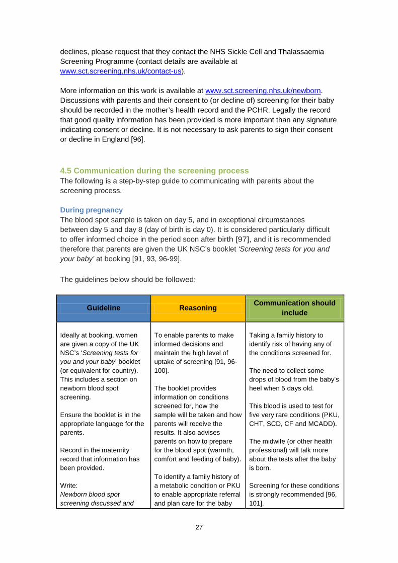
declines, please request that they contact the NHS Sickle Cell and Thalassaemia Screening Programme (contact details are available at www.sct.screening.nhs.uk/contact-us). More information on this work is available at www.sct.screening.nhs.uk/newborn. Discussions with parents and their consent to (or decline of) screening for their baby should be recorded in the mother’s health record and the PCHR. Legally the record that good quality information has been provided is more important than any signature indicating consent or decline. It is not necessary to ask parents to sign their consent or decline in England [96]. 4.5 Communication during the screening process The following is a step-by-step guide to communicating with parents about the screening process. During pregnancy The blood spot sample is taken on day 5, and in exceptional circumstances between day 5 and day 8 (day of birth is day 0). It is considered particularly difficult to offer informed choice in the period soon after birth [97], and it is recommended therefore that parents are given the UK NSC’s booklet ‘Screening tests for you and your baby’ at booking [91, 93, 96-99]. The guidelines below should be followed:
Guideline Reasoning Communication should include
Ideally at booking, women are given a copy of the UK NSC’s ‘Screening tests for you and your baby’ booklet (or equivalent for country). This includes a section on newborn blood spot screening. Ensure the booklet is in the appropriate language for the parents. Record in the maternity record that information has been provided. Write: Newborn blood spot screening discussed and
To enable parents to make informed decisions and maintain the high level of uptake of screening [91, 96-100]. The booklet provides information on conditions screened for, how the sample will be taken and how parents will receive the results. It also advises parents on how to prepare for the blood spot (warmth, comfort and feeding of baby). To identify a family history of a metabolic condition or PKU to enable appropriate referral and plan care for the baby
Taking a family history to identify risk of having any of the conditions screened for. The need to collect some drops of blood from the baby’s heel when 5 days old. This blood is used to test for five very rare conditions (PKU, CHT, SCD, CF and MCADD). The midwife (or other health professional) will talk more about the tests after the baby is born. Screening for these conditions is strongly recommended [96, 101].
28
recommended. Leaflet given. following birth. To help facilitate informed choice, allowing parents time to read the booklet and to ask questions.
Screening is important so that if a baby has one of the conditions, it can be diagnosed and treated quickly to prevent potentially serious health problems [96, 101]. If they have any questions they should speak to their midwife/health professional.
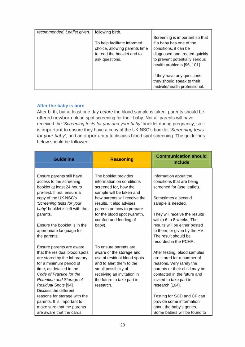
After the baby is born After birth, but at least one day before the blood sample is taken, parents should be offered newborn blood spot screening for their baby. Not all parents will have received the ‘Screening tests for you and your baby’ booklet during pregnancy, so it is important to ensure they have a copy of the UK NSC’s booklet ‘Screening tests for your baby’, and an opportunity to discuss blood spot screening. The guidelines below should be followed:
Guideline Reasoning Communication should include
Ensure parents still have access to the screening booklet at least 24 hours pre-test. If not, ensure a copy of the UK NSC’s ‘Screening tests for your baby’ booklet is left with the parents. Ensure the booklet is in the appropriate language for the parents. Ensure parents are aware that the residual blood spots are stored by the laboratory for a minimum period of time, as detailed in the Code of Practice for the Retention and Storage of Residual Spots [94]. Discuss the different reasons for storage with the parents. It is important to make sure that the parents are aware that the cards
The booklet provides information on conditions screened for, how the sample will be taken and how parents will receive the results. It also advises parents on how to prepare for the blood spot (warmth, comfort and feeding of baby). To ensure parents are aware of the storage and use of residual blood spots and to alert them to the small possibility of receiving an invitation in the future to take part in research.
Information about the conditions that are being screened for (use leaflet). Sometimes a second sample is needed. They will receive the results within 6 to 8 weeks. The results will be either posted to them, or given by the HV. The result should be recorded in the PCHR. After testing, blood samples are stored for a number of reasons. Very rarely the parents or their child may be contacted in the future and invited to take part in research [104]. Testing for SCD and CF can provide some information about the baby’s genes. Some babies will be found to
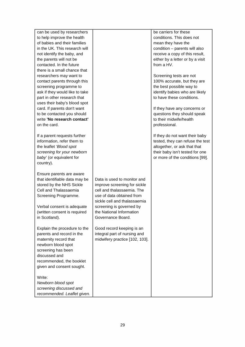
29
can be used by researchers to help improve the health of babies and their families in the UK. This research will not identify the baby, and the parents will not be contacted. In the future there is a small chance that researchers may want to contact parents through this screening programme to ask if they would like to take part in other research that uses their baby’s blood spot card. If parents don't want to be contacted you should write 'No research contact' on the card. If a parent requests further information, refer them to the leaflet ‘Blood spot screening for your newborn baby’ (or equivalent for country). Ensure parents are aware that identifiable data may be stored by the NHS Sickle Cell and Thalassaemia Screening Programme. Verbal consent is adequate (written consent is required in Scotland). Explain the procedure to the parents and record in the maternity record that newborn blood spot screening has been discussed and recommended, the booklet given and consent sought. Write: Newborn blood spot screening discussed and recommended. Leaflet given.
Data is used to monitor and improve screening for sickle cell and thalassaemia. The use of data obtained from sickle cell and thalassaemia screening is governed by the National Information Governance Board. Good record keeping is an integral part of nursing and midwifery practice [102, 103].
be carriers for these conditions. This does not mean they have the condition – parents will also receive a copy of this result, either by a letter or by a visit from a HV. Screening tests are not 100% accurate, but they are the best possible way to identify babies who are likely to have these conditions. If they have any concerns or questions they should speak to their midwife/health professional. If they do not want their baby tested, they can refuse the test altogether, or ask that that their baby isn’t tested for one or more of the conditions [99].
30
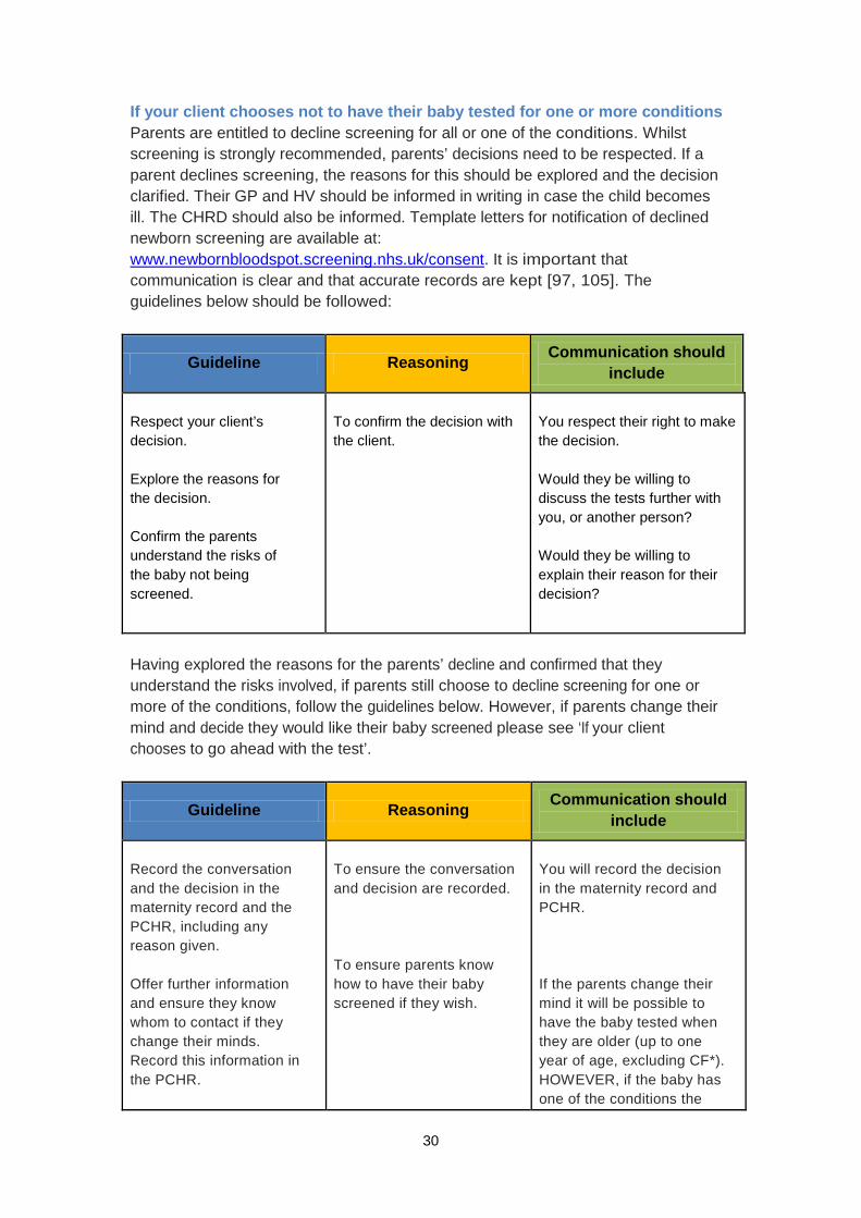
If your client chooses not to have their baby tested for one or more conditions Parents are entitled to decline screening for all or one of the conditions. Whilst screening is strongly recommended, parents’ decisions need to be respected. If a parent declines screening, the reasons for this should be explored and the decision clarified. Their GP and HV should be informed in writing in case the child becomes ill. The CHRD should also be informed. Template letters for notification of declined newborn screening are available at: www.newbornbloodspot.screening.nhs.uk/consent. It is important that communication is clear and that accurate records are kept [97, 105]. The guidelines below should be followed:
Guideline Reasoning Communication should include
Respect your client’s decision. Explore the reasons for the decision. Confirm the parents understand the risks of the baby not being screened.
To confirm the decision with the client.
You respect their right to make the decision. Would they be willing to discuss the tests further with you, or another person? Would they be willing to explain their reason for their decision?
Having explored the reasons for the parents’ decline and confirmed that they understand the risks involved, if parents still choose to decline screening for one or more of the conditions, follow the guidelines below. However, if parents change their mind and decide they would like their baby screened please see ‘If your client chooses to go ahead with the test’.
Guideline Reasoning Communication should include
Record the conversation and the decision in the maternity record and the PCHR, including any reason given. Offer further information and ensure they know whom to contact if they change their minds. Record this information in the PCHR.
To ensure the conversation and decision are recorded. To ensure parents know how to have their baby screened if they wish.
You will record the decision in the maternity record and PCHR. If the parents change their mind it will be possible to have the baby tested when they are older (up to one year of age, excluding CF*). HOWEVER, if the baby has one of the conditions the
31
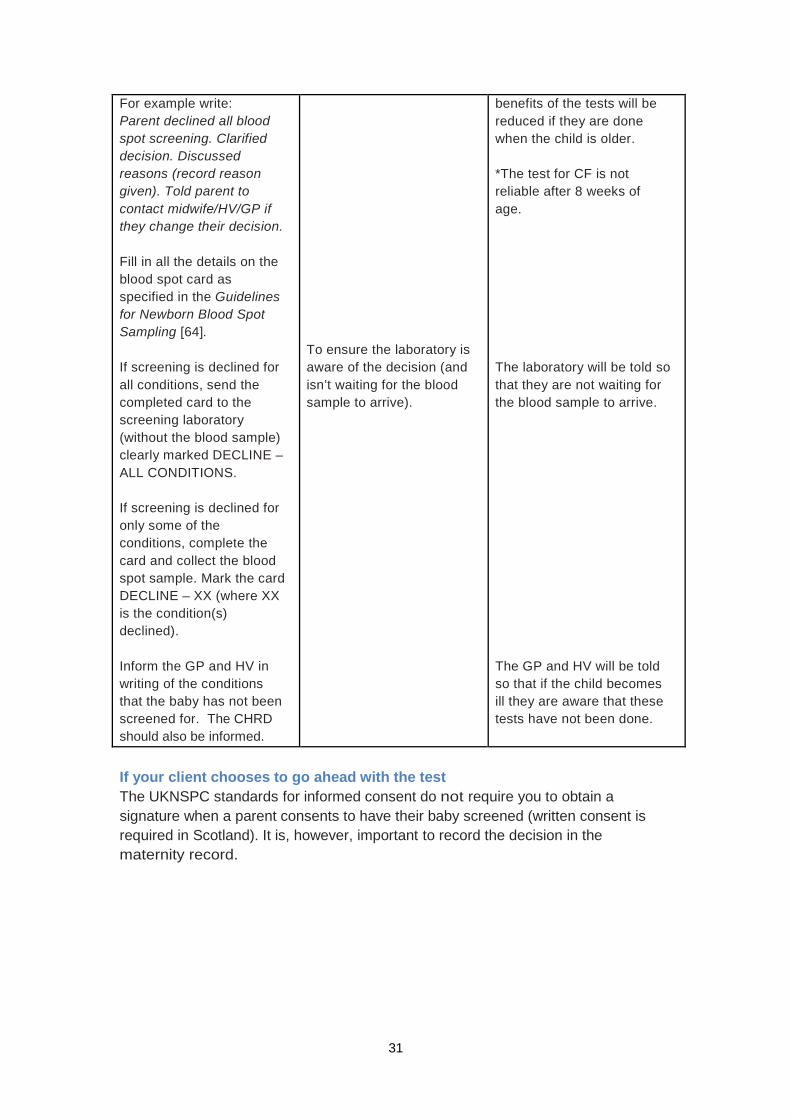
For example write: Parent declined all blood spot screening. Clarified decision. Discussed reasons (record reason given). Told parent to contact midwife/HV/GP if they change their decision. Fill in all the details on the blood spot card as specified in the Guidelines for Newborn Blood Spot Sampling [64]. If screening is declined for all conditions, send the completed card to the screening laboratory (without the blood sample) clearly marked DECLINE – ALL CONDITIONS. If screening is declined for only some of the conditions, complete the card and collect the blood spot sample. Mark the card DECLINE – XX (where XX is the condition(s) declined). Inform the GP and HV in writing of the conditions that the baby has not been screened for. The CHRD should also be informed.
To ensure the laboratory is aware of the decision (and isn’t waiting for the blood sample to arrive).
benefits of the tests will be reduced if they are done when the child is older. *The test for CF is not reliable after 8 weeks of age. The laboratory will be told so that they are not waiting for the blood sample to arrive. The GP and HV will be told so that if the child becomes ill they are aware that these tests have not been done.
If your client chooses to go ahead with the test The UKNSPC standards for informed consent do not require you to obtain a signature when a parent consents to have their baby screened (written consent is required in Scotland). It is, however, important to record the decision in the maternity record.
32
Guideline Reasoning Communication should include
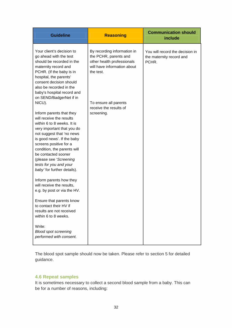
Your client’s decision to go ahead with the test should be recorded in the maternity record and PCHR. (If the baby is in hospital, the parents’ consent decision should also be recorded in the baby’s hospital record and on SEND/BadgerNet if in NICU). Inform parents that they will receive the results within 6 to 8 weeks. It is very important that you do not suggest that ‘no news is good news’. If the baby screens positive for a condition, the parents will be contacted sooner (please see ‘Screening tests for you and your baby’ for further details). Inform parents how they will receive the results, e.g. by post or via the HV. Ensure that parents know to contact their HV if results are not received within 6 to 8 weeks. Write: Blood spot screening performed with consent.
By recording information in the PCHR, parents and other health professionals will have information about the test. To ensure all parents receive the results of screening.
You will record the decision in the maternity record and PCHR.
The blood spot sample should now be taken. Please refer to section 5 for detailed guidance. 4.6 Repeat samples It is sometimes necessary to collect a second blood sample from a baby. This can be for a number of reasons, including:
33
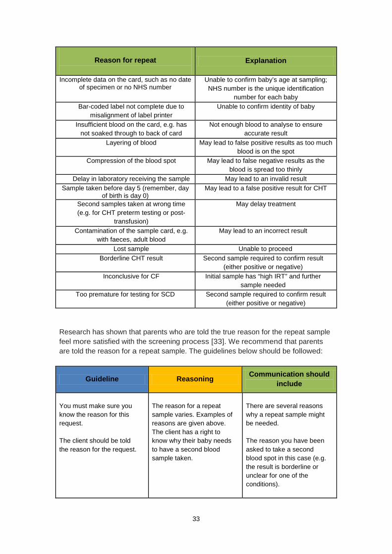
Reason for repeat
Explanation
Incomplete data on the card, such as no date of specimen or no NHS number
Unable to confirm baby’s age at sampling; NHS number is the unique identification
number for each baby Bar-coded label not complete due to
misalignment of label printer Unable to confirm identity of baby
Insufficient blood on the card, e.g. has
not soaked through to back of card Not enough blood to analyse to ensure
accurate result Layering of blood May lead to false positive results as too much
blood is on the spot Compression of the blood spot May lead to false negative results as the
blood is spread too thinly Delay in laboratory receiving the sample May lead to an invalid result
Sample taken before day 5 (remember, day of birth is day 0)
May lead to a false positive result for CHT
Second samples taken at wrong time (e.g. for CHT preterm testing or post-
transfusion)
May delay treatment
Contamination of the sample card, e.g. with faeces, adult blood
May lead to an incorrect result
Lost sample Unable to proceed Borderline CHT result Second sample required to confirm result
(either positive or negative) Inconclusive for CF
Initial sample has “high IRT” and further
sample needed Too premature for testing for SCD Second sample required to confirm result
(either positive or negative)
Research has shown that parents who are told the true reason for the repeat sample feel more satisfied with the screening process [33]. We recommend that parents are told the reason for a repeat sample. The guidelines below should be followed:
Guideline Reasoning Communication should include
You must make sure you know the reason for this request. The client should be told the reason for the request.
The reason for a repeat sample varies. Examples of reasons are given above. The client has a right to know why their baby needs to have a second blood sample taken.
There are several reasons why a repeat sample might be needed. The reason you have been asked to take a second blood spot in this case (e.g. the result is borderline or unclear for one of the conditions).

34
If there is thought to be any problem with their baby they will be contacted as soon as possible. At the latest, they will receive the result by 8 weeks of age. The benefit of these tests is that babies with the condition can be identified quickly, so they can be treated early (refer to the information on the conditions in the leaflet).
4.7 Screening results Laboratories are responsible for sending all screening results to child health records departments or their equivalent in a timely manner using appropriate status codes. Further information on laboratory reporting can be found in the condition-specific laboratory handbooks [8, 62, 87, 106]. Giving results – if there is no cause for concern (five ‘not suspected’ results) In 2008, the UKNSPC launched the ‘All results belong to parents’ campaign. The main aim of this project was to improve the communication of normal newborn screening results to parents, for recording in the PCHR. ‘Not suspected’ results should be given to parents as soon as possible, particularly if there has been any cause for concern earlier in the screening pathway, such as the need for a repeat blood sample. All parents should receive their baby’s results by the time of the 6 to 8 week check. The result must be recorded/filed in the PCHR. The agreed method for reporting ‘not suspected’ screening results (when results for all five conditions are normal) is to send a letter to parents. A template for a normal results letter was developed as part of the 2008 campaign (available at www.newbornbloodspot.screening.nhs.uk/getdata.php?id=11006).
Guideline Reasoning Communication should include
It is the responsibility of the HV to ensure that parents have been told the results by the 6 to 8 week check. Results should be recorded/filed in the PCHR.
Parents have a right to know their baby’s results.
That their baby’s screening result showed no cause for concern. That screening is not 100% accurate and if they have any concerns about their baby’s health they should see their GP or HV.
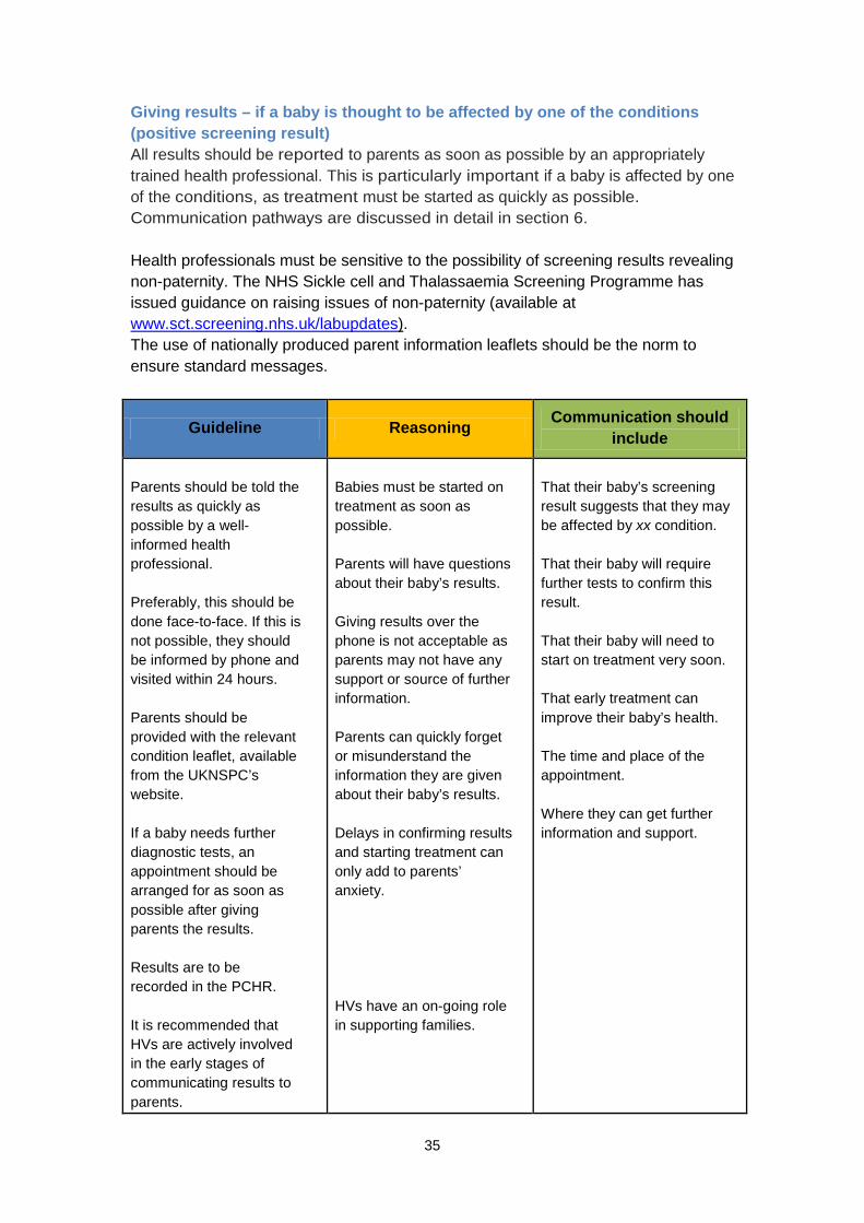
35
Giving results – if a baby is thought to be affected by one of the conditions (positive screening result) All results should be reported to parents as soon as possible by an appropriately trained health professional. This is particularly important if a baby is affected by one of the conditions, as treatment must be started as quickly as possible. Communication pathways are discussed in detail in section 6. Health professionals must be sensitive to the possibility of screening results revealing non-paternity. The NHS Sickle cell and Thalassaemia Screening Programme has issued guidance on raising issues of non-paternity (available at www.sct.screening.nhs.uk/labupdates). The use of nationally produced parent information leaflets should be the norm to ensure standard messages.
Guideline Reasoning Communication should include
Parents should be told the results as quickly as possible by a well-informed health professional. Preferably, this should be done face-to-face. If this is not possible, they should be informed by phone and visited within 24 hours. Parents should be provided with the relevant condition leaflet, available from the UKNSPC’s website. If a baby needs further diagnostic tests, an appointment should be arranged for as soon as possible after giving parents the results. Results are to be recorded in the PCHR. It is recommended that HVs are actively involved in the early stages of communicating results to parents.
Babies must be started on treatment as soon as possible. Parents will have questions about their baby’s results. Giving results over the phone is not acceptable as parents may not have any support or source of further information. Parents can quickly forget or misunderstand the information they are given about their baby’s results. Delays in confirming results and starting treatment can only add to parents’ anxiety. HVs have an on-going role in supporting families.
That their baby’s screening result suggests that they may be affected by xx condition. That their baby will require further tests to confirm this result. That their baby will need to start on treatment very soon. That early treatment can improve their baby’s health. The time and place of the appointment. Where they can get further information and support.
36
Giving results – if a baby is identified as a carrier of CF or SCD Parents should always be informed if their baby is identified as a carrier of one of these conditions, and the implications fully explained. The aim of carrier communication is to ensure that parents understand the benign health but important reproductive implications for their child, without concern [33]. In 2009, the HTA published ‘Communication of carrier status information following universal newborn screening for sickle cell disorders and cystic fibrosis: qualitative study of experience and practice’ [33]. The report identifies that some parents prefer a face-to-face conversation whereas parents who know they are carriers are happy to receive a letter followed by an appointment to discuss the result. The report also found that people prefer to be informed about results by someone who is well informed and can answer questions about the condition, rather than someone they know but who is not well informed about the condition. Carrier result for SCD Reporting carrier results has significant resource implications, with different challenges in high prevalence and low prevalence areas. The sickle cell and thalassaemia screening and newborn blood spot screening programmes recommend reporting carrier results through a face-to-face conversation; however, given the resource implications a letter or telephone call with a leaflet (translated if appropriate) and a follow up appointment could work as an interim solution. A template letter is available in the SCD newborn carrier protocol (available at www.sct.screening.nhs.uk/guidance). In areas with a specialist (or trained) HV, or equivalent, direct contact should be made to report carrier results to parents. When informing parents of newborn carrier results it should be made clear that the baby is well and is not thought to have the condition. Parents should also be informed of the screening results for the other four conditions. Health professionals must be sensitive to the possibility of screening results revealing non-paternity. The NHS Sickle cell and Thalassaemia Screening Programme has issued guidance on raising issues of non-paternity (available at www.sct.screening.nhs.uk/labupdates). Carrier result for CF In contrast to SCD, there is a two-stage screening protocol for CF. Following the initial heel prick test, a second sample will be needed from some babies. As the initial positive screen may indicate CF, the first communication to these parents following the initial heel prick may include that their child is at an elevated risk of having CF. As such, it has been suggested that this communication should be routinely carried out in person. The incidence of CF carriers following newborn screening is low when compared with the incidence of SCD carriers; therefore, face-to-face contact for these parents is a more realistic option. In England, CF carrier results are shared with parents by an
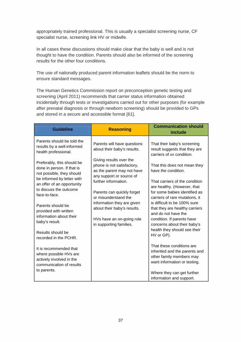
37
appropriately trained professional. This is usually a specialist screening nurse, CF specialist nurse, screening link HV or midwife. In all cases these discussions should make clear that the baby is well and is not thought to have the condition. Parents should also be informed of the screening results for the other four conditions. The use of nationally produced parent information leaflets should be the norm to ensure standard messages. The Human Genetics Commission report on preconception genetic testing and screening (April 2011) recommends that carrier status information obtained incidentally through tests or investigations carried out for other purposes (for example after prenatal diagnosis or through newborn screening) should be provided to GPs and stored in a secure and accessible format [61].
Guideline Reasoning Communication should include
Parents should be told the results by a well-informed health professional. Preferably, this should be done in person. If that is not possible, they should be informed by letter with an offer of an opportunity to discuss the outcome face-to-face. Parents should be provided with written information about their baby’s result. Results should be recorded in the PCHR. It is recommended that where possible HVs are actively involved in the communication of results to parents.
Parents will have questions about their baby’s results. Giving results over the phone is not satisfactory, as the parent may not have any support or source of further information. Parents can quickly forget or misunderstand the information they are given about their baby’s results. HVs have an on-going role in supporting families.
That their baby’s screening result suggests that they are carriers of xx condition. That this does not mean they have the condition. That carriers of the condition are healthy. (However, that for some babies identified as carriers of rare mutations, it is difficult to be 100% sure that they are healthy carriers and do not have the condition. If parents have concerns about their baby’s health they should see their HV or GP). That these conditions are inherited and the parents and other family members may want information or testing. Where they can get further information and support.
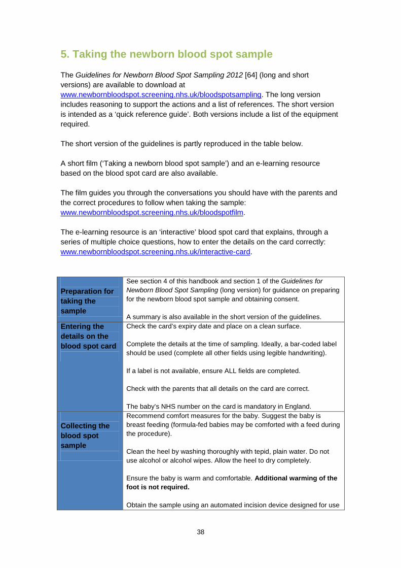
38
5. Taking the newborn blood spot sample The Guidelines for Newborn Blood Spot Sampling 2012 [64] (long and short versions) are available to download at www.newbornbloodspot.screening.nhs.uk/bloodspotsampling. The long version includes reasoning to support the actions and a list of references. The short version is intended as a ‘quick reference guide’. Both versions include a list of the equipment required. The short version of the guidelines is partly reproduced in the table below. A short film (‘Taking a newborn blood spot sample’) and an e-learning resource based on the blood spot card are also available. The film guides you through the conversations you should have with the parents and the correct procedures to follow when taking the sample: www.newbornbloodspot.screening.nhs.uk/bloodspotfilm. The e-learning resource is an ‘interactive’ blood spot card that explains, through a series of multiple choice questions, how to enter the details on the card correctly: www.newbornbloodspot.screening.nhs.uk/interactive-card. Preparation for taking the sample
See section 4 of this handbook and section 1 of the Guidelines for Newborn Blood Spot Sampling (long version) for guidance on preparing for the newborn blood spot sample and obtaining consent. A summary is also available in the short version of the guidelines.
Entering the details on the blood spot card
Check the card’s expiry date and place on a clean surface. Complete the details at the time of sampling. Ideally, a bar-coded label should be used (complete all other fields using legible handwriting). If a label is not available, ensure ALL fields are completed. Check with the parents that all details on the card are correct. The baby’s NHS number on the card is mandatory in England.
Collecting the blood spot sample
Recommend comfort measures for the baby. Suggest the baby is breast feeding (formula-fed babies may be comforted with a feed during the procedure). Clean the heel by washing thoroughly with tepid, plain water. Do not use alcohol or alcohol wipes. Allow the heel to dry completely. Ensure the baby is warm and comfortable. Additional warming of the foot is not required. Obtain the sample using an automated incision device designed for use
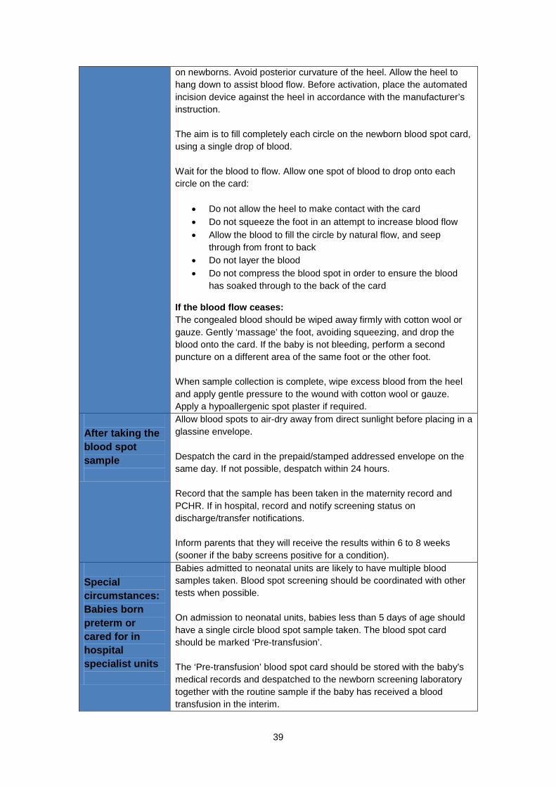
39
on newborns. Avoid posterior curvature of the heel. Allow the heel to hang down to assist blood flow. Before activation, place the automated incision device against the heel in accordance with the manufacturer’s instruction. The aim is to fill completely each circle on the newborn blood spot card, using a single drop of blood. Wait for the blood to flow. Allow one spot of blood to drop onto each circle on the card:
• Do not allow the heel to make contact with the card • Do not squeeze the foot in an attempt to increase blood flow • Allow the blood to fill the circle by natural flow, and seep
through from front to back • Do not layer the blood • Do not compress the blood spot in order to ensure the blood
has soaked through to the back of the card
If the blood flow ceases: The congealed blood should be wiped away firmly with cotton wool or gauze. Gently ‘massage’ the foot, avoiding squeezing, and drop the blood onto the card. If the baby is not bleeding, perform a second puncture on a different area of the same foot or the other foot. When sample collection is complete, wipe excess blood from the heel and apply gentle pressure to the wound with cotton wool or gauze. Apply a hypoallergenic spot plaster if required.
After taking the blood spot sample
Allow blood spots to air-dry away from direct sunlight before placing in a glassine envelope. Despatch the card in the prepaid/stamped addressed envelope on the same day. If not possible, despatch within 24 hours. Record that the sample has been taken in the maternity record and PCHR. If in hospital, record and notify screening status on discharge/transfer notifications. Inform parents that they will receive the results within 6 to 8 weeks (sooner if the baby screens positive for a condition).
Special circumstances: Babies born preterm or cared for in hospital specialist units
Babies admitted to neonatal units are likely to have multiple blood samples taken. Blood spot screening should be coordinated with other tests when possible. On admission to neonatal units, babies less than 5 days of age should have a single circle blood spot sample taken. The blood spot card should be marked ‘Pre-transfusion’. The ‘Pre-transfusion’ blood spot card should be stored with the baby’s medical records and despatched to the newborn screening laboratory together with the routine sample if the baby has received a blood transfusion in the interim.
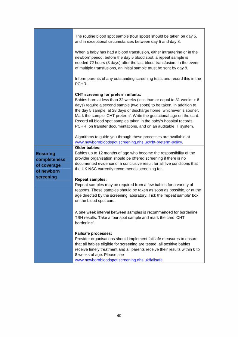
40
The routine blood spot sample (four spots) should be taken on day 5, and in exceptional circumstances between day 5 and day 8. When a baby has had a blood transfusion, either intrauterine or in the newborn period, before the day 5 blood spot, a repeat sample is needed 72 hours (3 days) after the last blood transfusion. In the event of multiple transfusions, an initial sample must be sent by day 8. Inform parents of any outstanding screening tests and record this in the PCHR. CHT screening for preterm infants: Babies born at less than 32 weeks (less than or equal to 31 weeks + 6 days) require a second sample (two spots) to be taken, in addition to the day 5 sample, at 28 days or discharge home, whichever is sooner. Mark the sample ‘CHT preterm’. Write the gestational age on the card. Record all blood spot samples taken in the baby’s hospital records, PCHR, on transfer documentations, and on an auditable IT system. Algorithms to guide you through these processes are available at www.newbornbloodspot.screening.nhs.uk/cht-preterm-policy.
Ensuring completeness of coverage of newborn screening
Older babies: Babies up to 12 months of age who become the responsibility of the provider organisation should be offered screening if there is no documented evidence of a conclusive result for all five conditions that the UK NSC currently recommends screening for. Repeat samples: Repeat samples may be required from a few babies for a variety of reasons. These samples should be taken as soon as possible, or at the age directed by the screening laboratory. Tick the ‘repeat sample’ box on the blood spot card. A one week interval between samples is recommended for borderline TSH results. Take a four spot sample and mark the card ‘CHT borderline’. Failsafe processes: Provider organisations should implement failsafe measures to ensure that all babies eligible for screening are tested, all positive babies receive timely treatment and all parents receive their results within 6 to 8 weeks of age. Please see www.newbornbloodspot.screening.nhs.uk/failsafe.
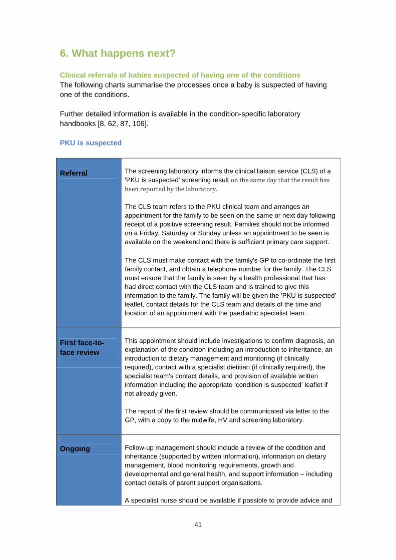
41
6. What happens next? Clinical referrals of babies suspected of having one of the conditions The following charts summarise the processes once a baby is suspected of having one of the conditions. Further detailed information is available in the condition-specific laboratory handbooks [8, 62, 87, 106]. PKU is suspected Referral
The screening laboratory informs the clinical liaison service (CLS) of a 'PKU is suspected’ screening result on the same day that the result has been reported by the laboratory. The CLS team refers to the PKU clinical team and arranges an appointment for the family to be seen on the same or next day following receipt of a positive screening result. Families should not be informed on a Friday, Saturday or Sunday unless an appointment to be seen is available on the weekend and there is sufficient primary care support. The CLS must make contact with the family’s GP to co-ordinate the first family contact, and obtain a telephone number for the family. The CLS must ensure that the family is seen by a health professional that has had direct contact with the CLS team and is trained to give this information to the family. The family will be given the 'PKU is suspected’ leaflet, contact details for the CLS team and details of the time and location of an appointment with the paediatric specialist team.
First face-to-face review
This appointment should include investigations to confirm diagnosis, an explanation of the condition including an introduction to inheritance, an introduction to dietary management and monitoring (if clinically required), contact with a specialist dietitian (if clinically required), the specialist team's contact details, and provision of available written information including the appropriate ‘condition is suspected’ leaflet if not already given. The report of the first review should be communicated via letter to the GP, with a copy to the midwife, HV and screening laboratory.
Ongoing
Follow-up management should include a review of the condition and inheritance (supported by written information), information on dietary management, blood monitoring requirements, growth and developmental and general health, and support information – including contact details of parent support organisations. A specialist nurse should be available if possible to provide advice and
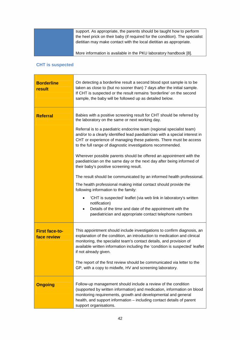
42
support. As appropriate, the parents should be taught how to perform the heel prick on their baby (if required for the condition). The specialist dietitian may make contact with the local dietitian as appropriate. More information is available in the PKU laboratory handbook [8].
CHT is suspected Borderline result
On detecting a borderline result a second blood spot sample is to be taken as close to (but no sooner than) 7 days after the initial sample. If CHT is suspected or the result remains ‘borderline’ on the second sample, the baby will be followed up as detailed below.
Referral
Babies with a positive screening result for CHT should be referred by the laboratory on the same or next working day. Referral is to a paediatric endocrine team (regional specialist team) and/or to a clearly identified lead paediatrician with a special interest in CHT or experience of managing these patients. There must be access to the full range of diagnostic investigations recommended. Wherever possible parents should be offered an appointment with the paediatrician on the same day or the next day after being informed of their baby’s positive screening result. The result should be communicated by an informed health professional.
The health professional making initial contact should provide the following information to the family:
• ‘CHT is suspected’ leaflet (via web link in laboratory’s written notification)
• Details of the time and date of the appointment with the paediatrician and appropriate contact telephone numbers
First face-to-face review
This appointment should include investigations to confirm diagnosis, an explanation of the condition, an introduction to medication and clinical monitoring, the specialist team's contact details, and provision of available written information including the ‘condition is suspected’ leaflet if not already given. The report of the first review should be communicated via letter to the GP, with a copy to midwife, HV and screening laboratory.
Ongoing
Follow-up management should include a review of the condition (supported by written information) and medication, information on blood monitoring requirements, growth and developmental and general health, and support information – including contact details of parent support organisations.
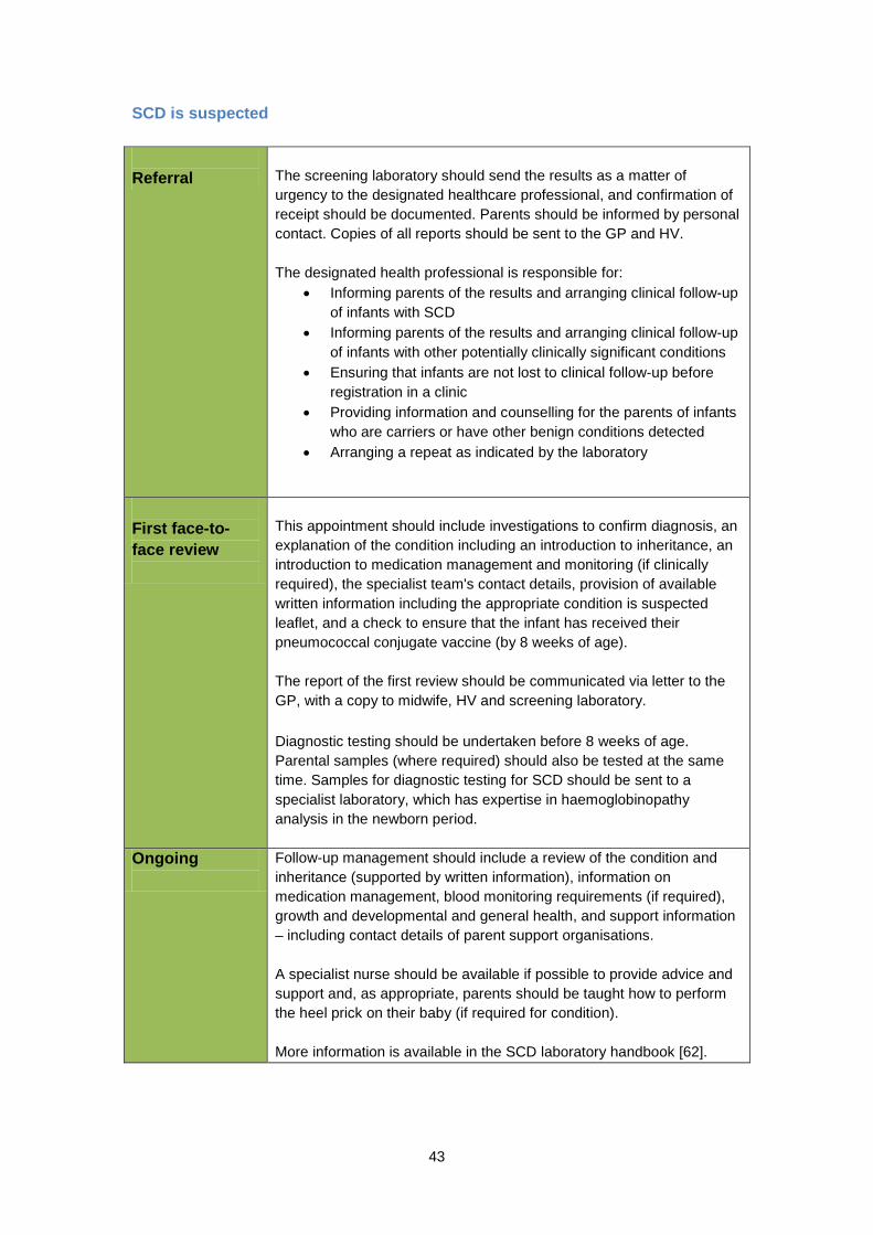
43
SCD is suspected Referral
The screening laboratory should send the results as a matter of urgency to the designated healthcare professional, and confirmation of receipt should be documented. Parents should be informed by personal contact. Copies of all reports should be sent to the GP and HV. The designated health professional is responsible for:
• Informing parents of the results and arranging clinical follow-up of infants with SCD
• Informing parents of the results and arranging clinical follow-up of infants with other potentially clinically significant conditions
• Ensuring that infants are not lost to clinical follow-up before registration in a clinic
• Providing information and counselling for the parents of infants who are carriers or have other benign conditions detected
• Arranging a repeat as indicated by the laboratory
First face-to-face review
This appointment should include investigations to confirm diagnosis, an explanation of the condition including an introduction to inheritance, an introduction to medication management and monitoring (if clinically required), the specialist team's contact details, provision of available written information including the appropriate condition is suspected leaflet, and a check to ensure that the infant has received their pneumococcal conjugate vaccine (by 8 weeks of age). The report of the first review should be communicated via letter to the GP, with a copy to midwife, HV and screening laboratory. Diagnostic testing should be undertaken before 8 weeks of age. Parental samples (where required) should also be tested at the same time. Samples for diagnostic testing for SCD should be sent to a specialist laboratory, which has expertise in haemoglobinopathy analysis in the newborn period.
Ongoing
Follow-up management should include a review of the condition and inheritance (supported by written information), information on medication management, blood monitoring requirements (if required), growth and developmental and general health, and support information – including contact details of parent support organisations. A specialist nurse should be available if possible to provide advice and support and, as appropriate, parents should be taught how to perform the heel prick on their baby (if required for condition). More information is available in the SCD laboratory handbook [62].
44
CF is suspected Repeat sample
A second blood spot sample may be required if the laboratory detects either one mutation on DNA analysis or a raised IRT on the first sample. The sample should be collected between day 21 and day 28. (Please note that the UKNSPC Cystic Fibrosis Board recommends that the second IRT sample is taken nearer to 21 days than 28 days so that parents get a conclusive screening result as soon as possible to reduce their anxiety and to achieve the 35 day standard [107]). If CF is suspected on the second sample, the baby will be followed up as detailed below.
Referral
The screening laboratory informs the CLS of a 'CF is suspected’ screening result in any of the following situations:
• Two detected CFTR mutations • One mutation and a second sample high IRT • High IRT concentration in two blood samples taken
approximately 3 weeks apart
The CLS team refers to the designated clinician for CF and arranges an appointment for the family to be seen within five days following receipt of a positive screening result. Families should not be informed on a Friday, Saturday or Sunday unless there is sufficient primary care support. The CLS must make contact with the family’s GP to co-ordinate first family contact, and obtain a telephone number for the family. The CLS must ensure that the family is seen by a health professional that has had direct contact with the CLS team and is trained to give this information to the family. The family will be given the 'CF is suspected' leaflet, contact details for the CLS team and details of the time and location of an appointment with the paediatric specialist team. More information is available in the CF laboratory handbook [106].
MCADD is suspected Referral
The screening laboratory informs the CLS of an 'MCADD is suspected’ screening result on the same day as the result has been reported by the laboratory. The CLS team refers to the MCADD clinical team and arranges an appointment for the family to be seen on the same or next day following receipt of a positive screening result. Families should not be informed
45
on a Friday, Saturday or Sunday unless an appointment to be seen is available on the weekend and there is sufficient primary care support. The CLS must make contact with the family’s GP to co-ordinate the first family contact, and obtain a telephone number for the family. The CLS must ensure that the family is seen by a health professional that has had direct contact with the CLS team and is trained to give this information to the family. The family will be given the 'MCADD is suspected’ leaflet, contact details for the CLS team and details of the time and location of an appointment with the paediatric specialist team.
First face-to-face review
This appointment should include investigations to confirm diagnosis, an explanation of the condition including an introduction to inheritance, an introduction to management and monitoring (if clinically required), contact with a specialist dietitian (if clinically required), the specialist team's contact details, and provision of available written information including the appropriate ‘condition is suspected’ leaflet if not already given. The report of the first review should be communicated via letter to the GP, with a copy to the midwife, HV and screening laboratory.
Ongoing
Follow-up management should include a review of the condition and inheritance (supported by written information), information on dietary management, growth and developmental and general health, and support information – including contact details of parent support organisations. A specialist nurse should be available if possible to provide advice and support. The specialist dietitian may make contact with the local dietitian as appropriate. More information is available in the MCADD laboratory handbook [87].
46
7. Performance monitoring 7.1 Key performance indicators The UK NSC has introduced key performance indicators (KPIs) to ensure quality monitoring, management and improvement within the screening pathway. These simple tools are required to ensure that commissioners have access to good, current management information. The purpose of these KPIs is to define consistent performance measures for a selection of public health priorities, using terminology that is clear and common across all the non-cancer screening programmes, so that performance can be understood, assessed and compared. Further information is available at www.screening.nhs.uk/kpi. 7.2 Standards In order to support health professionals in delivering a high quality and timely screening service, the UKNSPC has developed a set of standards to ensure smooth running of the screening pathway. The standards are measurable at two levels: acceptable and achievable. The acceptable standards set out the expected level of performance to deliver an acceptable level of quality. Whilst the achievable standards are unlikely to be met initially, they present measurable targets for enhanced quality. A summary of the standards is available at www.newbornbloodspot.screening.nhs.uk/standards.
47
8. Serious incidents A serious incident requiring investigation is defined as an incident that occurred in relation to NHS-funded services and care resulting in one of the following:
• Unexpected or avoidable death of one or more patients, staff, visitors or members of the public
• Serious harm to one or more patients, staff, visitors or members of the public or where the outcome requires life-saving intervention, major surgical/medical intervention, permanent harm or will shorten life expectancy or result in prolonged pain or psychological harm (this includes incidents graded under the National Patient Safety Agency definition of severe harm)
• A scenario that prevents or threatens to prevent a provider organisation's ability to continue to deliver healthcare services, for example, actual or potential loss of personal/organisational information, damage to property, reputation or the environment, or IT failure
• Allegations of abuse • Adverse media coverage or public concern about the organisation or the
wider NHS • One of the core set of "Never Events" as updated on an annual basis
For screening programmes this definition can be clarified further as follows: • An actual or possible failure at any stage in the pathway of the screening
service, which exposes the programme to unknown levels of risk that screening, assessment or treatment have been inadequate, and hence there are possible serious consequences for the clinical management of patients
• The level of risk to an individual may be low, but because of the large numbers involved the corporate risk may be very high
• Complex screening pathways often involve multidisciplinary teams working across several NHS organisations in both primary and secondary care, and inappropriate actions within one area, or communication failures between providers, can result in serious incidents.
All providers of NHS-funded care should have clear processes and policies for the identification, reporting and management of incidents and serious incidents. The UK NSC has developed a toolkit to highlight the application of the principles of incident management as they can be interpreted within the context of the English national screening programmes. This is available at www.screening.nhs.uk/si-toolkit. Please also see www.newbornbloodspot.screening.nhs.uk/serious-incidents.
48
9. Abbreviations CF cystic fibrosis CFTR cystic fibrosis transmembrane conductance regulator CHT congenital hypothyroidism CLS clinical liaison service DNA deoxyribonucleic acid GP general practitioner HPA hyperphenylalaninaemia HTA health technology assessment HV health visitor IQ intellectual quotient IRT immunoreactive trypsin IT information technology KPI key performance indicator MCADD medium-chain acyl-CoA dehydrogenase deficiency NHS National Health Service PAH phenylalanine hydroxylase PCHR personal child health record PKU phenylketonuria SCD sickle cell disease TSH thyroid-stimulating hormone UK NSC UK National Screening Committee UKNSPC UK Newborn Screening Programme Centre
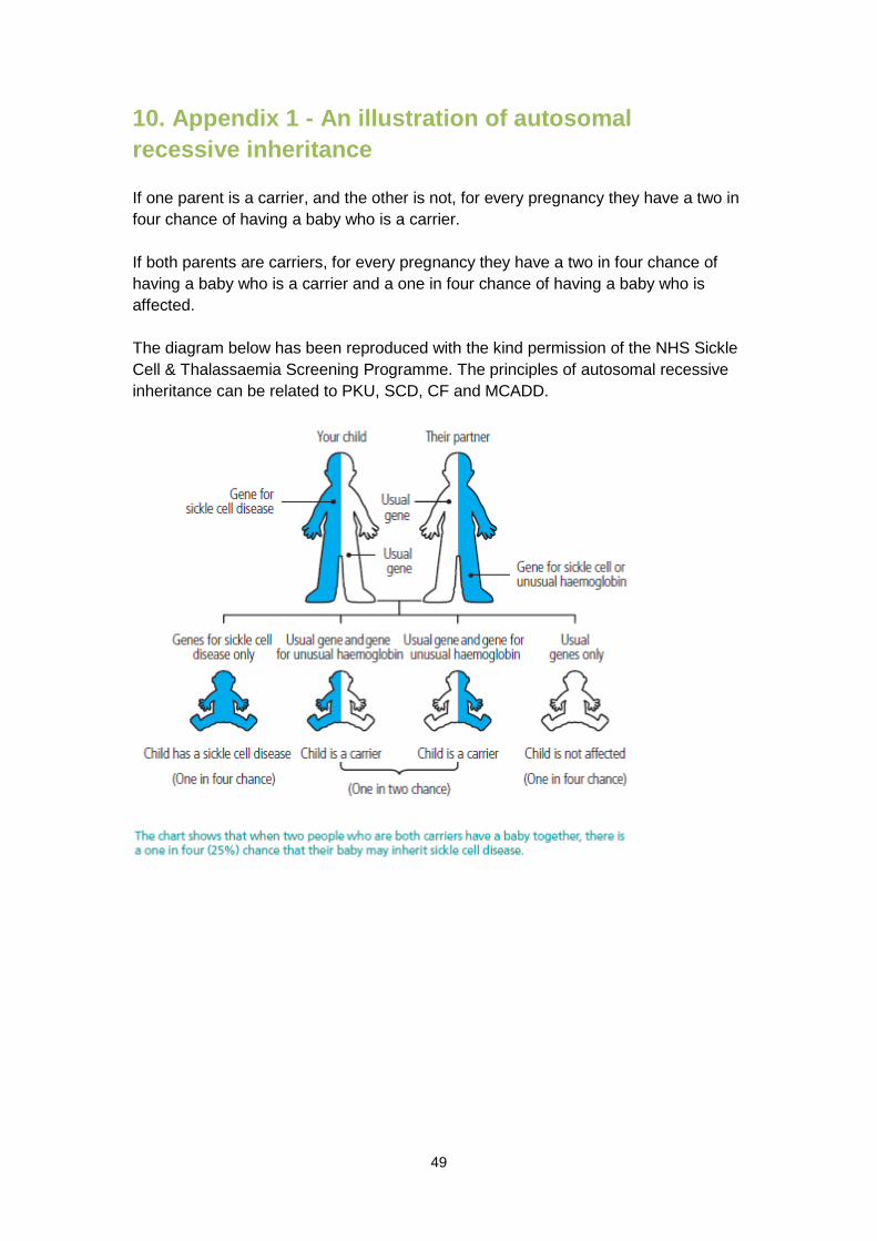
49
10. Appendix 1 - An illustration of autosomal recessive inheritance If one parent is a carrier, and the other is not, for every pregnancy they have a two in four chance of having a baby who is a carrier. If both parents are carriers, for every pregnancy they have a two in four chance of having a baby who is a carrier and a one in four chance of having a baby who is affected. The diagram below has been reproduced with the kind permission of the NHS Sickle Cell & Thalassaemia Screening Programme. The principles of autosomal recessive inheritance can be related to PKU, SCD, CF and MCADD.
50
11. Appendix 2 - Sources of information and support British Thyroid Foundation www.btf-thyroid.org
Child Growth Foundation www.childgrowthfoundation.org
Climb (Children Living with Inherited Metabolic Diseases) www.climb.org.uk
Contact a Family www.cafamily.org.uk
Cystic Fibrosis Trust www.cftrust.org.uk
Expanded Newborn Screening www.expandedscreening.org
Newlife Foundation for Disabled Children www.newlifecharity.co.uk
NHS Sickle Cell and Thalassaemia Screening Programme www.sct.screening.nhs.uk
Sickle Cell Society www.sicklecellsociety.org
Surveillance of congenital hypothyroidism (UKCS-CHT) project
www.ucl.ac.uk/ich/research-ich/mrc-cech/research/studies/ukcs-cht
The National Childbirth Trust www.nct.org.uk
The National Society for Phenylketonuria (NSPKU) www.nspku.org
UK National Screening Committee www.screening.nhs.uk
UK Thalassaemia Society www.ukts.org
51
12. References 1. UK National Screening Committee. What is screening? 2012 [cited 2012 16
May]; Available from: http://www.screening.nhs.uk/screening. 2. Pollitt, R.J., et al., Neonatal screening for inborn errors of metabolism: cost,
yield and outcome. Health Technol Assess, 1997. 1(7): p. i-iv, 1-202. 3. Enns, G.M., et al., Suboptimal outcomes in patients with PKU treated early
with diet alone: revisiting the evidence. Mol Genet Metab, 2010. 101(2-3): p. 99-109.
4. Williams, R.A., C.D. Mamotte, and J.R. Burnett, Phenylketonuria: an inborn error of phenylalanine metabolism. Clin Biochem Rev, 2008. 29(1): p. 31-41.
5. Poustie, V.J. and J. Wildgoose, Dietary interventions for phenylketonuria. Cochrane Database Syst Rev, 2010(1): p. CD001304.
6. Feillet, F., et al., Challenges and pitfalls in the management of phenylketonuria. Pediatrics, 2010. 126(2): p. 333-41.
7. Seymour, C.A., et al., Newborn screening for inborn errors of metabolism: a systematic review. Health Technol Assess, 1997. 1(11): p. i-iv, 1-95.
8. UK Newborn Screening Programme Centre, A laboratory guide to newborn screening in the UK for phenylketonuria 2011. 2011.
9. Blau, N., et al., Management of phenylketonuria in Europe: survey results from 19 countries. Mol Genet Metab, 2010. 99(2): p. 109-15.
10. Hardelid, P., et al., The birth prevalence of PKU in populations of European, South Asian and sub-Saharan African ancestry living in South East England. Ann Hum Genet, 2008. 72(Pt 1): p. 65-71.
11. UK Newborn Screening Programme Centre, Annual report of the UK Newborn Screening Programme Centre for NHS Newborn Blood Spot Screening 2010-11. 2012.
12. van Spronsen, F.J. and P. Burgard, The truth of treating patients with phenylketonuria after childhood: the need for a new guideline. J Inherit Metab Dis, 2008. 31(6): p. 673-9.
13. Jovanovic, A., C. Ferguson, and S. M, Outcomes of pregnancy in maternal phenylketonuria (PKU): the north-east experience. Archives of diseases in childhood, fetal and neonatal ed, 2011. 96(supp 1).
14. Pandor, A., et al., Economics of tandem mass spectrometry screening of neonatal inherited disorders. Int J Technol Assess Health Care, 2006. 22(3): p. 321-6.
15. Rastogi, M.V. and S.H. LaFranchi, Congenital hypothyroidism. Orphanet J Rare Dis, 2010. 5: p. 17.
16. Jones, J.H., et al., Improvement in screening performance and diagnosis of congenital hypothyroidism in Scotland 1979-2003. Arch Dis Child, 2006. 91(8): p. 680-5.
17. LaFranchi, S.H., Approach to the diagnosis and treatment of neonatal hypothyroidism. J Clin Endocrinol Metab, 2011. 96(10): p. 2959-67.
18. Lenz, A.M. and A.W. Root, Congenital hypothyroidism: a forgotten clinical diagnosis? J Pediatr Endocrinol Metab, 2008. 21(7): p. 623-4.
19. Raymond, J. and S.H. LaFranchi, Fetal and neonatal thyroid function: review and summary of significant new findings. Curr Opin Endocrinol Diabetes Obes, 2010. 17(1): p. 1-7.
20. Parks, J.S., et al., The impact of transient hypothyroidism on the increasing rate of congenital hypothyroidism in the United States. Pediatrics, 2010. 125 Suppl 2: p. S54-63.
21. Leonardi, D., et al., Longitudinal study of thyroid function in children with mild hyperthyrotropinemia at neonatal screening for congenital hypothyroidism. J Clin Endocrinol Metab, 2008. 93(7): p. 2679-85.
52
22. Kemper, A.R., L. Ouyang, and S.D. Grosse, Discontinuation of thyroid hormone treatment among children in the United States with congenital hypothyroidism: findings from health insurance claims data. BMC Pediatr, 2010. 10: p. 9.
23. Royal College of Paediatrics and Child Health. Congenital hypothyroidism. 2012 01 February 2012 [cited 2012 10 September]; Available from: http://www.rcpch.ac.uk/what-we-do/bpsu/current-studies/congenital-hypothyroidism/congenital-hypothyroidism.
24. Mandel, S.J., et al., Atypical hypothyroidism and the very low birthweight infant. Thyroid, 2000. 10(8): p. 693-5.
25. LaFranchi, S.H., Newborn screening strategies for congenital hypothyroidism: an update. J Inherit Metab Dis, 2010. 33(Suppl 2): p. S225-33.
26. Olney, R.S., S.D. Grosse, and R.F. Vogt, Jr., Prevalence of congenital hypothyroidism--current trends and future directions: workshop summary. Pediatrics, 2010. 125 Suppl 2: p. S31-6.
27. Hinton, C.F., et al., Trends in incidence rates of congenital hypothyroidism related to select demographic factors: data from the United States, California, Massachusetts, New York, and Texas. Pediatrics, 2010. 125 Suppl 2: p. S37-47.
28. Jones, J.H. and M.D. Donaldson, Audit of initial management of congenital hypothyroidism in the United Kingdom--comparison of UK practice with European and UK guidelines. J Pediatr Endocrinol Metab, 2009. 22(11): p. 1017-25.
29. Grosse, S.D. and G. Van Vliet, Prevention of intellectual disability through screening for congenital hypothyroidism: how much and at what level? Arch Dis Child, 2011. 96(4): p. 374-9.
30. Korada, M., et al., Repeat testing for congenital hypothyroidism in preterm infants is unnecessary with an appropriate thyroid stimulating hormone threshold. Arch Dis Child Fetal Neonatal Ed, 2008. 93(4): p. F286-8.
31. Zeuner, D., et al., Antenatal and neonatal haemoglobinopathy screening in the UK: review and economic analysis. Health Technol Assess, 1999. 3(11): p. i-v, 1-186.
32. Davies, S.C., et al., Screening for sickle cell disease and thalassaemia: a systematic review with supplementary research. Health Technol Assess, 2000. 4(3): p. i-v, 1-99.
33. Kai, J., et al., Communication of carrier status information following universal newborn screening for sickle cell disorders and cystic fibrosis: qualitative study of experience and practice. Health Technol Assess, 2009. 13(57): p. 1-82, iii.
34. NHS Sickle Cell and Thalassaemia Screening Programme, Standards for the linked antenatal and newborn screening programme. 2011.
35. Platt, O.S., et al., Mortality in sickle cell disease. Life expectancy and risk factors for early death. N Engl J Med, 1994. 330(23): p. 1639-44.
36. Williams, S., G.H. Maude, and G.R. Serjeant, Clinical presentation of sickle cell-hemoglobin C disease. J Pediatr, 1986. 109(4): p. 586-9.
37. Serjeant, G.R., The clinical features of sickle cell disease. Baillieres Clin Haematol, 1993. 6(1): p. 93-115.
38. Boulet, S.L., et al., Health status and healthcare use in a national sample of children with sickle cell disease. Am J Prev Med, 2010. 38(4 Suppl): p. S528-35.
39. Huntsman, R.G., Sickle-cell anemia and thalassemia: a primer for health care professionals. 1987: Canadian Sickle Cell Society.
40. Streetly, A., R. Latinovic, and J. Henthorn, Positive screening and carrier results for the England-wide universal newborn sickle cell screening
53
programme by ethnicity and area for 2005-07. J Clin Pathol, 2010. 63(7): p. 626-9.
41. NHS Sickle Cell and Thalassaemia Screening Programme, Data report 2009/10: performance analysis and quality improvement. 2012.
42. Halasa, N.B., et al., Incidence of invasive pneumococcal disease among individuals with sickle cell disease before and after the introduction of the pneumococcal conjugate vaccine. Clin Infect Dis, 2007. 44(11): p. 1428-33.
43. Atkin, K., W.I. Ahmad, and E.N. Anionwu, Screening and counselling for sickle cell disorders and thalassaemia: the experience of parents and health professionals. Soc Sci Med, 1998. 47(11): p. 1639-51.
44. Leikin, S.L., et al., Mortality in children and adolescents with sickle cell disease. Cooperative Study of Sickle Cell Disease. Pediatrics, 1989. 84(3): p. 500-8.
45. Charache, S., et al., Effect of hydroxyurea on the frequency of painful crises in sickle cell anemia. Investigators of the Multicenter Study of Hydroxyurea in Sickle Cell Anemia. N Engl J Med, 1995. 332(20): p. 1317-22.
46. Ferster, A., et al., Hydroxyurea for treatment of severe sickle cell anemia: a pediatric clinical trial. Blood, 1996. 88(6): p. 1960-4.
47. Adams, R.J., et al., Prevention of a first stroke by transfusions in children with sickle cell anemia and abnormal results on transcranial Doppler ultrasonography. N Engl J Med, 1998. 339(1): p. 5-11.
48. Lucarelli, G. and D.J. Weatherall, For debate: bone marrow transplantation for severe thalassaemia (1). The view from Pesaro (2). To be or not to be. Br J Haematol, 1991. 78(3): p. 300-3.
49. Frempong, T. and H.A. Pearson, Newborn screening coupled with comprehensive follow-up reduced early mortality of sickle cell disease in Connecticut. Conn Med, 2007. 71(1): p. 9-12.
50. Selekman, J., Update: new guidelines for the treatment of infants with sickle cell disease. Agency for Health Care Policy and Research. Pediatr Nurs, 1993. 19(6): p. 600-5.
51. Gaston, M.H., et al., Prophylaxis with oral penicillin in children with sickle cell anemia. A randomized trial. N Engl J Med, 1986. 314(25): p. 1593-9.
52. Elliott, V., et al., Parental health beliefs and compliance with prophylactic penicillin administration in children with sickle cell disease. J Pediatr Hematol Oncol, 2001. 23(2): p. 112-6.
53. Mantadakis, E., et al., Prevalence of priapism in children and adolescents with sickle cell anemia. J Pediatr Hematol Oncol, 1999. 21(6): p. 518-22.
54. Laird, L., C. Dezateux, and E.N. Anionwu, Neonatal screening for sickle cell disorders: what about the carrier infants? BMJ, 1996. 313(7054): p. 407-11.
55. Teach, S.J., K.A. Lillis, and M. Grossi, Compliance with penicillin prophylaxis in patients with sickle cell disease. Arch Pediatr Adolesc Med, 1998. 152(3): p. 274-8.
56. NHS Sickle Cell and Thalassaemia Screening Programme, Data report: informing policy and improving quality 2008-09. 2011.
57. NHS Sickle Cell and Thalassaemia Screening Programme, Parents guide: care and management of your child with sickle cell disease. 2007.
58. Howard-Jones, M., et al., An audit of immunisation status of sickle cell patients in Coventry, UK. J Clin Pathol, 2009. 62(1): p. 42-5.
59. Kavanagh, P.L., et al., Communication of positive newborn screening results for sickle cell disease and sickle cell trait: variation across states. Am J Med Genet C Semin Med Genet, 2008. 148C(1): p. 15-22.
60. Falletta, J.M., et al., Discontinuing penicillin prophylaxis in children with sickle cell anemia. Prophylactic Penicillin Study II. J Pediatr, 1995. 127(5): p. 685-90.
54
61. Human Genetics Commission, Increasing options, informing choice: A report on preconception genetic testing and screening. 2011.
62. NHS Sickle Cell and Thalassaemia Screening Programme, Sickle cell and thalassaemia: handbook for laboratories 3rd edition. 2012.
63. Reed, W., et al., Sickle-cell disease not identified by newborn screening because of prior transfusion. J Pediatr, 2000. 136(2): p. 248-50.
64. UK Newborn Screening Programme Centre, Guidelines for newborn blood spot sampling 2012. 2012.
65. NHS Sickle Cell and Thalassaemia Screening Programme, DNA for transfused babies: Review on the impact of pilot on reducing the risk of missing babies with sickle cell disease due to transfusions. 2012.
66. Smith, J.A. and T.R. Kinney, Sickle cell disease: screening, diagnosis, management, and counseling in newborns and infants. Vol. 6. 1997: Diane Books Publishing Company.
67. Hinton, C.F., A.M. Grant, and S.D. Grosse, Ethical implications and practical considerations of ethnically targeted screening for genetic disorders: the case of hemoglobinopathy screening. Ethn Health, 2011. 16(4-5): p. 377-88.
68. UK National Screening Committee, Child health sub-group report on cystic fibrosis. 2005.
69. Balfour-Lynn, I.M., Newborn screening for cystic fibrosis: evidence for benefit. Arch Dis Child, 2008. 93(1): p. 7-10.
70. Southern, K.W., et al., Newborn screening for cystic fibrosis. Cochrane Database Syst Rev, 2009(1): p. CD001402.
71. Dijk, F.N., et al., Improved survival in cystic fibrosis patients diagnosed by newborn screening compared to a historical cohort from the same centre. Arch Dis Child, 2011. 96(12): p. 1118-23.
72. Cavanagh, L., et al., Long-term evaluation of genetic counseling following false-positive newborn screen for cystic fibrosis. J Genet Couns, 2010. 19(2): p. 199-210.
73. Castellani, C., et al., Association between carrier screening and incidence of cystic fibrosis. JAMA: The Journal of the American Medical Association, 2009. 302(23): p. 2573.
74. Mehta, A., A global perspective on newborn screening for cystic fibrosis. Curr Opin Pulm Med, 2007. 13(6): p. 510-4.
75. Wainwright, C.E., Treatment of cystic fibrosis following infant screening. Therapy, 2011. 8(6): p. 613-622.
76. NHS Commissioning Board, Public health functions to be exercised by the NHS Commissioning Board: Service specification No.19 - NHS Newborn Bloodspot Screening Programme. 2012.
77. Sims, E.J., et al., Economic implications of newborn screening for cystic fibrosis: a cost of illness retrospective cohort study. Lancet, 2007. 369(9568): p. 1187-95.
78. Leonard, J.V. and C. Dezateux, Newborn screening for medium chain acyl CoA dehydrogenase deficiency. Arch Dis Child, 2009. 94(3): p. 235-8.
79. Loughrey, C. and M.J. Bennett, Screening for MCAD deficiency in newborns. BMJ, 2009. 338: p. b971.
80. Kennedy, S., et al., The first three years of screening for medium chain acyl-CoA dehydrogenase deficiency (MCADD) by newborn screening ontario. BMC Pediatr, 2010. 10: p. 82.
81. Wilcken, B., Expanded newborn screening: reducing harm, assessing benefit. J Inherit Metab Dis, 2010. 33(Suppl 2): p. S205-10.
82. Schatz, U.A. and R. Ensenauer, The clinical manifestation of MCAD deficiency: challenges towards adulthood in the screened population. J Inherit Metab Dis, 2010. 33(5): p. 513-20.
55
83. Grosse, S.D., et al., The epidemiology of medium chain acyl-CoA dehydrogenase deficiency: an update. Genet Med, 2006. 8(4): p. 205-12.
84. Khalid, J.M., et al., Ethnicity of children with homozygous c.985A>G medium-chain acyl-CoA dehydrogenase deficiency: findings from screening approximately 1.1 million newborn infants. J Med Screen, 2008. 15(3): p. 112-7.
85. Derks, T.G., et al., Neonatal screening for medium-chain acyl-CoA dehydrogenase (MCAD) deficiency in The Netherlands: the importance of enzyme analysis to ascertain true MCAD deficiency. J Inherit Metab Dis, 2008. 31(1): p. 88-96.
86. Khalid, J.M., et al., Relationship of octanoylcarnitine concentrations to age at sampling in unaffected newborns screened for medium-chain acyl-CoA dehydrogenase deficiency. Clin Chem, 2010. 56(6): p. 1015-21.
87. UK Newborn Screening Programme Centre, A laboratory guide to newborn screening in the UK for medium-chain acyl-CoA dehydrogenase deficiency (MCADD) 2010. 2010.
88. Oerton, J., et al., Newborn screening for medium chain acyl-CoA dehydrogenase deficiency in England: prevalence, predictive value and test validity based on 1.5 million screened babies. J Med Screen, 2011. 18(4): p. 173-81.
89. Prosser, L.A., et al., Projected costs, risks, and benefits of expanded newborn screening for MCADD. Pediatrics, 2010. 125(2): p. e286-94.
90. Venditti, L.N., et al., Newborn screening by tandem mass spectrometry for medium-chain Acyl-CoA dehydrogenase deficiency: a cost-effectiveness analysis. Pediatrics, 2003. 112(5): p. 1005-15.
91. Detmar, S., et al., Information and informed consent for neonatal screening: opinions and preferences of parents. Birth, 2007. 34(3): p. 238-44.
92. Nicholls, S., Knowledge or understanding? Informed choice in the context of newborn bloodspot screening. Public Health Ethics, 2010. 3(2): p. 128-136.
93. Kerruish, N.J., D. Webster, and N. Dickson, Information and consent for newborn screening: practices and attitudes of service providers. J Med Ethics, 2008. 34(9): p. 648-52.
94. UK Newborn Screening Programme Centre, Code of practice for the retention and storage of residual spots 2005. 2005.
95. Department of Health. National Information Governance Board for Health and Social Care. 2012 [cited 2012 11 July]; Available from: http://www.nigb.nhs.uk/.
96. Department of Health, Reference guide to consent for examination or treatment. 2009.
97. National Institute for Clinical Excellence, Antenatal care: routine care for the healthy pregnant woman - CG62. 2010.
98. Parsons, E.P., et al., Mothers' accounts of screening newborn babies in Wales (UK). Midwifery, 2007. 23(1): p. 59-65.
99. Davis, T.C., et al., Recommendations for effective newborn screening communication: results of focus groups with parents, providers, and experts. Pediatrics, 2006. 117(5 Pt 2): p. S326-40.
100. Department of Health/Partnerships for Children Families and Maternity, Maternity matters: choice, access and continuity of care in a safe service. 2007.
101. UK Newborn Screening Programme Centre, Standards and guidelines for newborn blood spot screening 2008. 2008.
102. Nursing and Midwifery Council, Midwives rules and standards. 2010. 103. Nursing and Midwifery Council, Record keeping: guidance for nurses and
midwives. 2009.
56
104. Shepherd, A., A. Glenesk, and C. Niven, What influence does experience play in heel prick blood sampling? J Neonatal Nursing, 2006. 12(3).
105. Morrow, C., A. Hidinger, and D. Wilkinson-Faulk, Reducing neonatal pain during routine heel lance procedures. MCN Am J Matern Child Nurs, 2010. 35(6): p. 346-54; quiz 354-6.
106. UK Newborn Screening Programme Centre, A laboratory guide to newborn screening in the UK for cystic fibrosis 2009. 2009.
107. UK Newborn Screening Programme Centre, Newborn screening for cystic fibrosis in England: clinical outcome data and performance against UKNSPC standards 2010-11. 2012.