Embed Size (px)
Citation preview
46 Volume 4, Number 1 • January 2015 • www.gahmj.com
GLOBAL ADVANCES IN HEALTH AND MEDICINE
Review
rEVIEW
Heart rate Variability: new perspectives on physiological Mechanisms, assessment of self-regulatory Capacity, and Health risk心率变异性:关于生理机制、自律能力和健康风险评估的新观点
Variabilidaddefrecuenciacardiaca:Nuevasperspectivassobremecanismosfisiológicos,valoracióndelacapacidadautorreguladorayriesgodelasalud
RollinMcCraty,PhD;United States;FredShaffer,PhD,BCB,United States
aBstraCtHeart rate variability, the change in the time intervals between adjacent heartbeats, is an emergent property of interdependent regulatory systems that operates on different time scales to adapt to environmental and psy-chological challenges. This article briefly reviews neural regulation of the heart and offers some new per-spectives on mechanisms underlying the very low frequency rhythm of heart rate variability. Interpretation of heart rate variability rhythms in the context of health risk and physiologi-cal and psychological self-regulatory capacity assessment is discussed. The cardiovascular regulatory centers in the spinal cord and medulla integrate inputs from higher brain centers with afferent cardiovascular system inputs to adjust heart rate and blood pressure via sympathetic and parasympathetic efferent pathways. We also discuss the intrinsic cardiac nervous system and the heart-brain connection pathways, through which afferent information can influence activity in the subcor-tical, frontocortical, and motor cor-tex areas. In addition, the use of real-time HRV feedback to increase self-regulatory capacity is reviewed. We conclude that the heart’s rhythms are characterized by both complexity and stability over longer time scales that reflect both physiological and psycho-logical functional status of these inter-nal self-regulatory systems.
摘要心率变异性 (HRC)(在相邻心跳之间的时间间隔的变化)是相互依存的调节系统的一种紧急特性,在不同时段内发生,以适应环境和心理挑战。这篇文章简要回顾了心脏的神经调节,在心率变异性的极低频率节奏机制方面,提供了一些新的观点。在健康风险和生理及心理自我调节能力评估讨论的背景下解读 HRV 节律。伴有心血管系统输入功能的更高级的大脑中枢集成输入的脊髓和延髓心血管监管中心,通过交感和副交感神经传出通路调节心率和血压。我们还讨论了先天性心脏神经系统和心脏大脑连接通路,借以传入信息并可以影响皮质下区,皮质前区和运动皮质区的活动。此外,审查了使用实时的 HRV 反馈以提高自我调节能力。我们的结论是,心脏节律的特征在于在更长的时段上既有复杂性又有稳定性,这反映出这些内部自我调节系统的生理和心理功能状态。
sInOpsIsLa variabilidad de la frecuencia car-diaca, o modificación de los inter-valos de tiempo entre los latidos consecutivos del corazón, es una propiedad emergente de los siste-mas reguladores interdependientes que opera sobre diferentes escalas temporales para adaptarse a los retos ambientales y psicológicos. Este artículo revisa brevemente la regulación nerviosa del corazón y
ofrece nuevas perspectivas sobre los mecanismos subyacentes al ritmo de muy baja frecuencia de la variabilidad de la frecuencia cardia-ca. Se analiza la interpretación de los ritmos de la variabilidad de la frecuencia cardiaca en el contexto del riesgo para la salud y la valor-ación de la capacidad autorregula-toria fisiológica y psicológica. Los centros reguladores cardiovascula-res de la médula espinal y del bulbo raquídeo integran entradas de cen-tros cerebrales superiores con entradas de sistemas cardiovascula-res aferentes para ajustar la fre-cuencia cardiaca y la tensión arte-rial por vías eferentes simpáticas y parasimpáticas. También hablamos sobre el sistema cardiaco nervioso intrínseco y las vías de conexión corazón-cerebro, a través de las cuales la información aferente puede influir sobre la actividad en las áreas subcortical, frontocortical y de la corteza motora. Además, se revisa el uso de retroalimentación de variabilidad de la frecuencia car-diaca a tiempo real para aumentar la capacidad autorreguladora. Concluimos que los ritmos cardia-cos se caracterizan tanto por su complejidad como por su estabili-dad sobre escalas temporales más largas que reflejan los estados fun-cionales tanto fisiológicos como psicológicos de estos sistemas internos autorreguladores.
author affiliations
InstituteofHeartMath,
BoulderCreek,
California,(DrMcCraty);
CenterforApplied
Psychophysiology,
TrumanStateUniversity,
Kirksville,Missouri,
(DrShaffer).
Correspondence
RollinMcCraty,PhD
Citation
GlobalAdvHealthMed.
2015;4(1):46-61.DOI:
10.7453/gahmj.2014.073
Key Words
Heartratevariability,
physiologicalmecha-
nisms,self-regulatory
capacity,healthrisk
disclosures
Theauthorscompleted
theICMJEFormfor
DisclosureofPotential
ConflictsofInterest.
DrMcCratydisclosed
thatheisemployed
bytheInstituteof
HeartMath,whichsells
oneoftheseveralheart
ratevariabilityfeedback
devicesthataremen-
tionedinthearticle.
DrShafferhadno
conflictstodisclose.
IntrOduCtIOnSince Walter Cannon introduced the concept of
homeostasis,1 the study of physiology has been based on the principle that all cells, tissues, and organs strive to maintain a static or constant “steady-state”
condition. However, with the introduction of signal processing technologies that can acquire continuous time series data from physiological processes such as heart rate (HR), blood pressure (BP), and nerve activi-ty, it has become abundantly apparent that biological
www.gahmj.com • January 2015 • Volume 4, Number 1 47Review
hEART RATE VARIABILITY: NEw PERSPEcTIVES
processes vary in complex and nonlinear ways, even during so called “steady-state” conditions. These observations have led to the understanding that healthy, optimal function is a result of continuous, dynamic, bi-directional interactions among multiple neural, hormonal, and mechanical control systems at both local and central levels. In concert, these physi-ological and psychological regulatory systems are never truly at rest and are certainly never static. For example, we now know that the normal resting rhythm of the heart is highly variable rather than being monotonously regular, which was the wide-spread notion for many years.
HEart ratE VarIaBILIty
The investigation of the heart’s complex rhythms or what is now called heart rate variability2 (HRV) began with the emergence of modern signal processing in the 1960s and 1970s, and has rapidly expanded in more recent times. The irregular behavior of the heart-beat is readily apparent when HR is examined on a beat-to-beat basis, but is overlooked when a mean value over time is calculated. These fluctuations in HR result from complex, nonlinear interactions among a number of different physiological systems. HRV is thus consid-ered a measure of neurocardiac function that reflects heart–brain interactions and autonomic nervous sys-tem (ANS) dynamics.3,4
An optimal level of HRV within an organism reflects healthy function and an inherent self-regulatory capacity, adaptability, or resilience.4-10 Too much insta-bility, such as arrhythmias or nervous system chaos, is detrimental to efficient physiological functioning and energy utilization. However, too little variation indi-cates age-related system depletion, chronic stress, pathology, or inadequate functioning in various levels of self-regulatory control systems.2,11,12
The importance of HRV as an index of the func-tional status of physiological control systems was noted as far back as 1965 when it was found that fetal distress is preceded by reductions in HRV before any changes occur in HR itself.13 In the 1970s, reduced HRV was shown to predict autonomic neuropathy in diabetic patients before the onset of symptoms.14-16 Reduced HRV was also found to be a greater risk factor of death post–myocardial infarction than other known risk factors.17 It has clearly been shown that HRV declines with age and age-adjusted values should be used in the context of risk prediction.18 Age-adjusted HRV that is low has been confirmed as a strong, inde-pendent predictor of future health problems in both healthy people. Age-adjusted HRV correlates with all-cause mortality.19,20 In prospective studies reduced HRV has been the strongest independent predictor of the progression of coronary atherosclerosis.21 A num-ber of studies have shown that reduced HRV is associ-ated with measures of inflammation in subjects with no apparent heart disease.22 Reduced HRV is also observed in patients with autonomic dysfunction,
anxiety, depression, asthma, and sudden infant death.23-26 Reduced HRV may correlate with disease and mortality because it reflects reduced regulatory capacity and ability to adaptively respond to physio-logical challenges such as exercise. For example, in the Chicago Health, Aging, and Social Relations Study, separate metrics for the assessment of autonomic bal-ance and overall cardiac autonomic regulation were developed and tested in a sample of 229 participants. In this study, overall regulatory capacity was a signifi-cant predictor of overall health status, but autonomic balance was not. In addition, cardiac regulatory capac-ity was negatively associated with the prior incidence of myocardial infarction. The authors suggest that cardiac regulatory capacity reflects a physiological state that is more relevant to health than the indepen-dent sympathetic or parasympathetic controls or the autonomic balance between these controls as indexed by different measures of HRV.27
When speaking of autonomic balance, it should be kept in mind that a healthy system is constantly and dynamically changing. Therefore, an important indica-tor of the health status of the regulatory systems is the capacity to respond to and adjust the relative auto-nomic balance (eg, HR) to the appropriate state for the context the person is engaged in at any given moment. In other words, does the HR dynamically respond? Is it higher during the daytime or when someone is dealing with challenging tasks and lower when at rest or dur-ing sleep? The inability of the physiological self-regula-tory systems to adapt to the current context and situa-tion is associated with numerous clinical conditions.28 Also distinct, altered, circadian patterns in 24-hour heart rates are associated with different and specific psychiatric disorders, particularly during sleep.29,30
HR estimated at any given time represents the net effect of the neural output of the parasympathetic (vagus) nerves, which slow HR, and the sympathetic nerves, which accelerate it. In a denervated human heart where there are no connections from the ANS to the heart following its transplantation, the intrinsic rate generated by the pacemaker (SA node) is about 100 beats per minute (bpm).31 Parasympathetic activity predominates when HR is below this intrinsic rate dur-ing normal daily activities and when at rest or sleep. When HR is above about 100 bpm, the relative balance shifts and sympathetic activity predominates. Therefore, HR best reflects the relative balance between the sympathetic and parasympathetic systems. The average 24-hour HR in healthy people is approximately 73 bpm. Higher HRs are independent markers of mor-tality in a wide spectrum of conditions.28
It is important to note the natural relationship between HR and amount of HRV. As HR increases there is less time between heartbeats for variability to occur, thus HRV decreases. At lower HRs there is more time between heartbeats and variability naturally increases. This is called cycle length dependence, and it persists in the healthy elderly to a variable degree,
48 Volume 4, Number 1 • January 2015 • www.gahmj.com
GLOBAL ADVANCES IN HEALTH AND MEDICINE
Review
even at a very advanced age. However, elderly patients with ischemic heart disease or other pathologies develop less variability at increasingly lower HRs and ultimately lose the relationship between HR and vari-ability, to the point that variability does not increase with reductions in HR.32 Even in healthy subjects, the effects of cycle length dependence should be taken into account when assessing HRV. HR values should also always be reported, especially when HRs are increased due to factors like stress reactions, medica-tions, and physical activity.
Efferent (descending) sympathetic nerves target the SA node via the intrinsic cardiac nervous system and the bulk of the myocardium. Action potentials conducted by these motor neurons trigger norepineph-rine and epinephrine release, which increases HR and strengthens the contractility of the atria and ventricles. Following the onset of sympathetic stimulation, there is a delay of up to 5 seconds before the stimulation induces a progressive increase in HR, which reaches a steady level in 20 to 30 seconds if the stimulus is con-tinuous.33 Even a brief sympathetic stimulus can affect the HR and the HRV rhythm for 5 to 10 seconds. The relatively slow response to sympathetic stimulation is
in direct contrast to vagal stimulation, which is almost instantaneous. Thus, any sudden change in HR, up or down or between one beat and the next, is primarily parasympathetically mediated.33,34
Patient age may mediate the relationship between reduced HRV and regulatory capacity of physiological control systems. Age-related reductions in HRV18 may reflect the loss of neurons in the brain and spinal cord, resulting in degraded signal transmission and reduced regulatory capacity. Reduced physiological regulato-ry capacity may contribute to functional gastrointes-tinal disorders, inflammation, and hypertension.35,36
HEart ratE VarIaBILIty anaLysIs MEtHOdsHRV can be assessed with various analytical
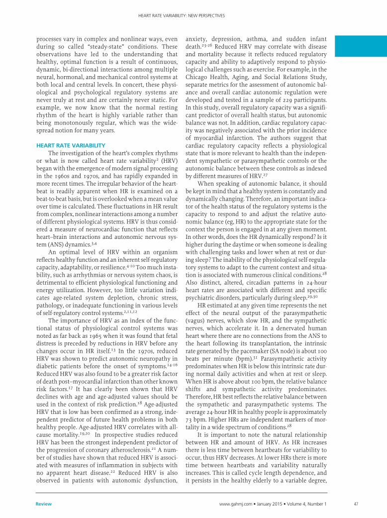
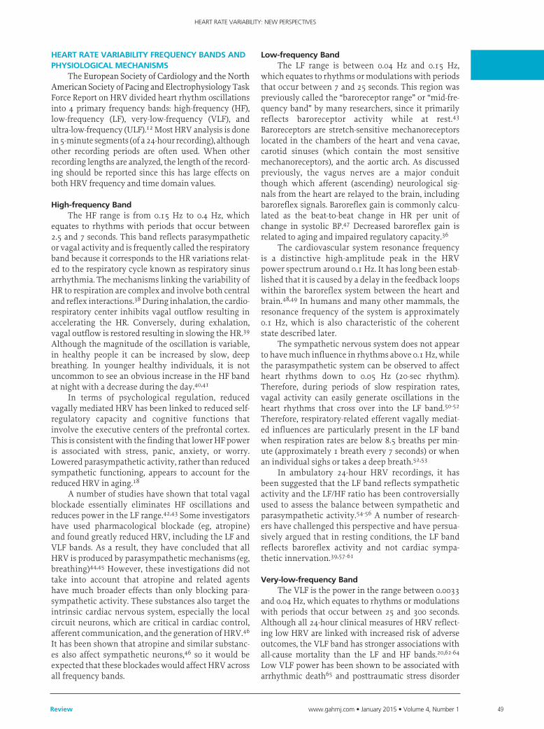
approaches, although the most commonly used are frequency domain (power spectral density) analysis and time domain analysis. The interactions between autonomic neural activity, BP, respiration, and higher level control systems produce both short and longer term rhythms in HRV measurements.4,12,37 The most common form for observing these changes is the HR tachogram, a plot of the sequence of time intervals between heartbeats (Figure 1).
High stress: public speaking
High energy expenditure: exercise
Figure 1Anexampleoftheheartrate(HR)tachogram,aplotofthesequenceoftimeintervalsbetweenheartbeatsoveran8-hourperiodinambulatoryrecordingtakenfroma36-year-oldmale.Eachofthetracesis1hourlong,withthestartingtimeofthehouronthelefthandsideofthefigure.Thetimebetweeneachverticallineis5minutes.Theverticalaxiswithineachofthehourlytracingsisthetimebetweenheartbeats(inter-beat-intervals)rangingbetween400and1200milliseconds(labelshownonsecondrow).Thehoursbeginningat10:45through12:45wereduringatimewhenhewasinalow-stressclassroomsetting.HisoverallHRincreased,andtherangeoftheHRVisconsiderablylessduringthehourstartingat13:45(publicspeaking),whenhewaspresentingtotheclass.Inthiscase,therelativeautonomicnervoussystembalanceisshiftedtosympatheticpredominanceduetotheemotionalstressaroundpresentingtoagroupofhispeers.Oncethepresentationcompletedneartheendofthehour,hisHRdroppedandnormalHRVwasrestored.Inthefollowinghours,hewaslisteningtootherspresentandprovidingfeedback.Inthehourstartingat17:45,hewasengagedinphysicalexercise(walkingupalongsteephill)startingabout20minutesintothehourwherehisHRisincreasedandtheHRVisreducedduetocycle-lengthdependenceeffects.
www.gahmj.com • January 2015 • Volume 4, Number 1 49Review
hEART RATE VARIABILITY: NEw PERSPEcTIVES
HEart ratE VarIaBILIty FrEQuEnCy Bands and pHysIOLOGICaL MECHanIsMs
The European Society of Cardiology and the North American Society of Pacing and Electrophysiology Task Force Report on HRV divided heart rhythm oscillations into 4 primary frequency bands: high-frequency (HF), low-frequency (LF), very-low-frequency (VLF), and ultra-low-frequency (ULF).12 Most HRV analysis is done in 5-minute segments (of a 24-hour recording), although other recording periods are often used. When other recording lengths are analyzed, the length of the record-ing should be reported since this has large effects on both HRV frequency and time domain values.
High-frequency Band The HF range is from 0.15 Hz to 0.4 Hz, which
equates to rhythms with periods that occur between 2.5 and 7 seconds. This band reflects parasympathetic or vagal activity and is frequently called the respiratory band because it corresponds to the HR variations relat-ed to the respiratory cycle known as respiratory sinus arrhythmia. The mechanisms linking the variability of HR to respiration are complex and involve both central and reflex interactions.38 During inhalation, the cardio-respiratory center inhibits vagal outflow resulting in accelerating the HR. Conversely, during exhalation, vagal outflow is restored resulting in slowing the HR.39 Although the magnitude of the oscillation is variable, in healthy people it can be increased by slow, deep breathing. In younger healthy individuals, it is not uncommon to see an obvious increase in the HF band at night with a decrease during the day.40,41
In terms of psychological regulation, reduced vagally mediated HRV has been linked to reduced self-regulatory capacity and cognitive functions that involve the executive centers of the prefrontal cortex. This is consistent with the finding that lower HF power is associated with stress, panic, anxiety, or worry. Lowered parasympathetic activity, rather than reduced sympathetic functioning, appears to account for the reduced HRV in aging.18
A number of studies have shown that total vagal blockade essentially eliminates HF oscillations and reduces power in the LF range.42,43 Some investigators have used pharmacological blockade (eg, atropine) and found greatly reduced HRV, including the LF and VLF bands. As a result, they have concluded that all HRV is produced by parasympathetic mechanisms (eg, breathing)44,45 However, these investigations did not take into account that atropine and related agents have much broader effects than only blocking para-sympathetic activity. These substances also target the intrinsic cardiac nervous system, especially the local circuit neurons, which are critical in cardiac control, afferent communication, and the generation of HRV.46 It has been shown that atropine and similar substanc-es also affect sympathetic neurons,46 so it would be expected that these blockades would affect HRV across all frequency bands.
Low-frequency BandThe LF range is between 0.04 Hz and 0.15 Hz,
which equates to rhythms or modulations with periods that occur between 7 and 25 seconds. This region was previously called the “baroreceptor range” or “mid-fre-quency band” by many researchers, since it primarily reflects baroreceptor activity while at rest.43 Baroreceptors are stretch-sensitive mechanoreceptors located in the chambers of the heart and vena cavae, carotid sinuses (which contain the most sensitive mechanoreceptors), and the aortic arch. As discussed previously, the vagus nerves are a major conduit though which afferent (ascending) neurological sig-nals from the heart are relayed to the brain, including baroreflex signals. Baroreflex gain is commonly calcu-lated as the beat-to-beat change in HR per unit of change in systolic BP.47 Decreased baroreflex gain is related to aging and impaired regulatory capacity.36
The cardiovascular system resonance frequency is a distinctive high-amplitude peak in the HRV power spectrum around 0.1 Hz. It has long been estab-lished that it is caused by a delay in the feedback loops within the baroreflex system between the heart and brain.48,49 In humans and many other mammals, the resonance frequency of the system is approximately 0.1 Hz, which is also characteristic of the coherent state described later.
The sympathetic nervous system does not appear to have much influence in rhythms above 0.1 Hz, while the parasympathetic system can be observed to affect heart rhythms down to 0.05 Hz (20-sec rhythm). Therefore, during periods of slow respiration rates, vagal activity can easily generate oscillations in the heart rhythms that cross over into the LF band.50-52 Therefore, respiratory-related efferent vagally mediat-ed influences are particularly present in the LF band when respiration rates are below 8.5 breaths per min-ute (approximately 1 breath every 7 seconds) or when an individual sighs or takes a deep breath.52,53
In ambulatory 24-hour HRV recordings, it has been suggested that the LF band reflects sympathetic activity and the LF/HF ratio has been controversially used to assess the balance between sympathetic and parasympathetic activity.54-56 A number of research-ers have challenged this perspective and have persua-sively argued that in resting conditions, the LF band reflects baroreflex activity and not cardiac sympa-thetic innervation.39,57-61
Very-low-frequency BandThe VLF is the power in the range between 0.0033
and 0.04 Hz, which equates to rhythms or modulations with periods that occur between 25 and 300 seconds. Although all 24-hour clinical measures of HRV reflect-ing low HRV are linked with increased risk of adverse outcomes, the VLF band has stronger associations with all-cause mortality than the LF and HF bands.20,62-64 Low VLF power has been shown to be associated with arrhythmic death65 and posttraumatic stress disorder
50 Volume 4, Number 1 • January 2015 • www.gahmj.com
GLOBAL ADVANCES IN HEALTH AND MEDICINE
Review
(PTSD).66 Additionally, low power in this band has been associated with high inflammation67,68 and has been correlated with low levels of testosterone. In contrast, other biochemical markers, such as those mediated by the hypothalamic-pituitary-adrenal (HPA) axis axis (eg, cortisol), did not.69 Longer time periods using 24-hour HRV recordings should be obtained to provide compre-hensive assessment of VLF and ULF fluctuations.70
Historically, the physiological explanation and mechanisms involved in the generation of the VLF component have not been as well defined as the LF and HF components. This region has been largely ignored even though it is the most predictive of adverse out-comes. Long-term regulatory mechanisms and ANS activity related to thermoregulation, the renin-angio-tensin system, and other hormonal factors appear to contribute to this band.71,72
Recent work by Armour has shed new light on the primary mechanisms underlying the VLF rhythm. This line of research began after some surprising results from a study looking at HRV in auto-transplanted hearts in dogs. In auto-transplants, the heart is removed and placed back in the same animal so there is no need for anti-rejection medications. The primary purpose of the study was to determine if the autonomic nerves re-innervated the heart posttransplant. Monthly 24-hour HRV recordings were done over a 1-year period on all the dogs with auto-transplanted hearts as well as the control dogs. The nerves did re-innervate but in a way that was not accurately reflected in HRV. It showed that the intrinsic cardiac nervous system has neuroplasticity and re-structured its neural connections. The truly sur-prising result was that these de-innervated hearts had higher levels of HRV, including HRV that is typically associated with respiration, than control dogs immedi-ately posttransplant. These levels were sustained over a 1-year period.73 This was unexpected as there is very little HRV in human transplant recipients.74
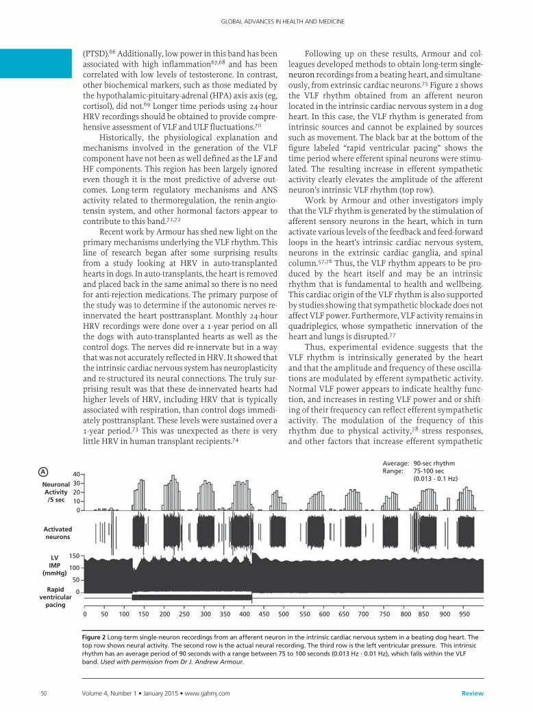
Following up on these results, Armour and col-leagues developed methods to obtain long-term single-neuron recordings from a beating heart, and simultane-ously, from extrinsic cardiac neurons.75 Figure 2 shows the VLF rhythm obtained from an afferent neuron located in the intrinsic cardiac nervous system in a dog heart. In this case, the VLF rhythm is generated from intrinsic sources and cannot be explained by sources such as movement. The black bar at the bottom of the figure labeled “rapid ventricular pacing” shows the time period where efferent spinal neurons were stimu-lated. The resulting increase in efferent sympathetic activity clearly elevates the amplitude of the afferent neuron’s intrinsic VLF rhythm (top row).
Work by Armour and other investigators imply that the VLF rhythm is generated by the stimulation of afferent sensory neurons in the heart, which in turn activate various levels of the feedback and feed-forward loops in the heart’s intrinsic cardiac nervous system, neurons in the extrinsic cardiac ganglia, and spinal column.57,76 Thus, the VLF rhythm appears to be pro-duced by the heart itself and may be an intrinsic rhythm that is fundamental to health and wellbeing. This cardiac origin of the VLF rhythm is also supported by studies showing that sympathetic blockade does not affect VLF power. Furthermore, VLF activity remains in quadriplegics, whose sympathetic innervation of the heart and lungs is disrupted.77
Thus, experimental evidence suggests that the VLF rhythm is intrinsically generated by the heart and that the amplitude and frequency of these oscilla-tions are modulated by efferent sympathetic activity. Normal VLF power appears to indicate healthy func-tion, and increases in resting VLF power and or shift-ing of their frequency can reflect efferent sympathetic activity. The modulation of the frequency of this rhythm due to physical activity,78 stress responses, and other factors that increase efferent sympathetic
Average: 90-sec rhythmRange: 75-100 sec (0.013 - 0.1 Hz)
NeuronalActivity/5 sec
Activatedneurons
LVIMP
(mmHg)
Rapidventricular
pacing
403020100
150
100
50
0
A
0 50 100 150 200 250 300 350 400 450 500 550 600 650 700 750 800 850 900 950
Figure 2Long-termsingle-neuronrecordingsfromanafferentneuronintheintrinsiccardiacnervoussysteminabeatingdogheart.Thetoprowshowsneuralactivity.Thesecondrowistheactualneuralrecording.Thethirdrowistheleftventricularpressure.Thisintrinsicrhythmhasanaverageperiodof90secondswitharangebetween75to100seconds(0.013Hz-0.01Hz),whichfallswithintheVLFband.Used with permission from Dr J. Andrew Armour.
www.gahmj.com • January 2015 • Volume 4, Number 1 51Review
hEART RATE VARIABILITY: NEw PERSPEcTIVES
activation can cause it to cross over into the lower region of the LF band during ambulatory monitoring or during short-term recordings when there is a sig-nificant emotional stressor.4
ultra-low-frequency BandThe ultra-low-frequency band (ULF) falls below
0.0033 Hz (333 seconds or 5.6 minutes). Oscillations or events in the heart rhythm with a period of 5 minutes or greater are reflected in this band and it can only be assessed with 24-hour and longer recordings.70 The cir-cadian oscillation in HR is the primary source of the ULF power, although other very slow-acting regulatory processes, such as core body temperature regulation, metabolism, and the renin-angiotensin system likely add to the power in this band.12 The Task Force Report on HRV suggests that 24-hour recordings should be divided into 5-minute segments and that HRV analysis should be performed on the individual segments prior to the calculation of mean values. This effectively filters out any oscillations with periods longer than 5 minutes. However, when spectral analysis is applied to entire 24-hour records, several lower frequency rhythms are
easily detected in healthy individuals.3 Circadian rhythms, core body temperature,
metabolism, hormones, and intrinsic rhythms gener-ated by the heart all contribute to lower frequency rhythms (eg, VLF and ULF) that extend below 0.04 Hz. In healthy individuals, there is an increase in VLF power that occurs during the night and peaks before waking.79,80 This increase in autonomic activity appears to correlate with the morning cortisol peak.
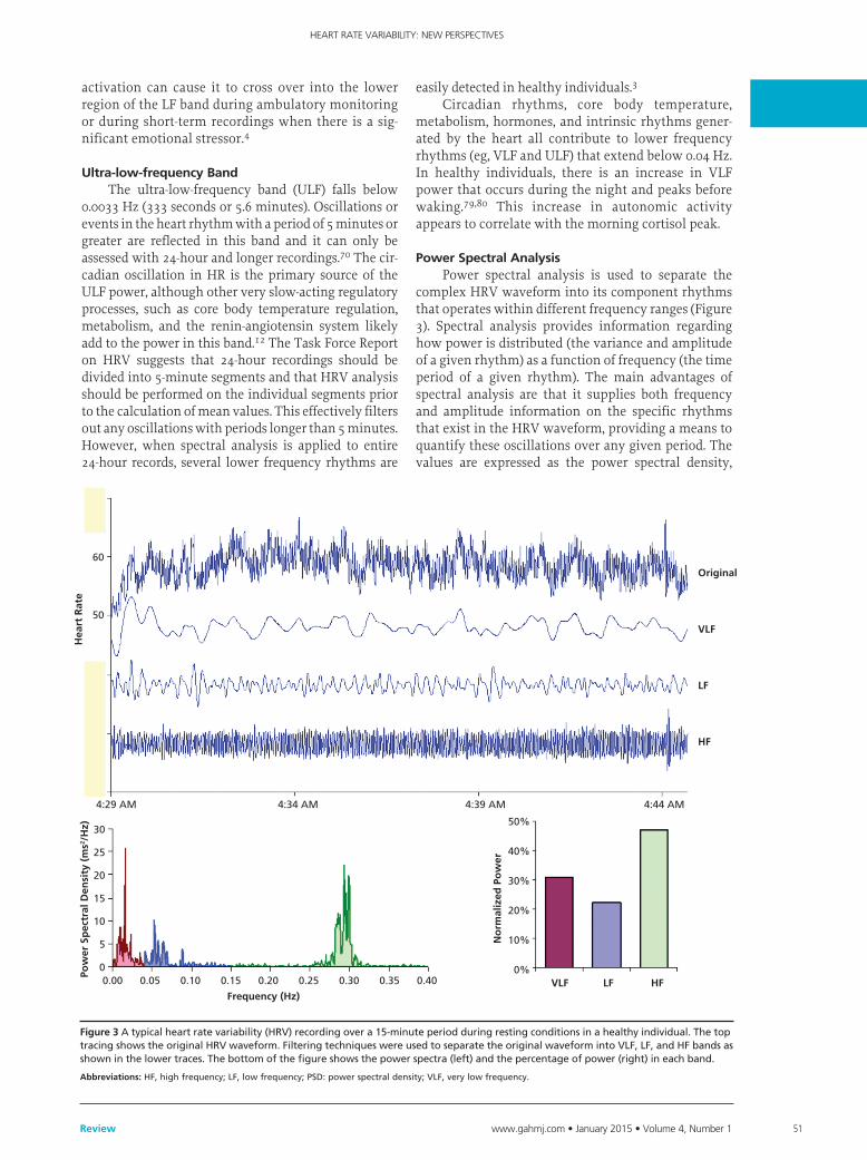
power spectral analysisPower spectral analysis is used to separate the
complex HRV waveform into its component rhythms that operates within different frequency ranges (Figure 3). Spectral analysis provides information regarding how power is distributed (the variance and amplitude of a given rhythm) as a function of frequency (the time period of a given rhythm). The main advantages of spectral analysis are that it supplies both frequency and amplitude information on the specific rhythms that exist in the HRV waveform, providing a means to quantify these oscillations over any given period. The values are expressed as the power spectral density,
Figure 3Atypicalheartratevariability(HRV)recordingovera15-minuteperiodduringrestingconditionsinahealthyindividual.ThetoptracingshowstheoriginalHRVwaveform.FilteringtechniqueswereusedtoseparatetheoriginalwaveformintoVLF,LF,andHFbandsasshowninthelowertraces.Thebottomofthefigureshowsthepowerspectra(left)andthepercentageofpower(right)ineachband.
abbreviations:HF,highfrequency;LF,lowfrequency;PSD:powerspectraldensity;VLF,verylowfrequency.
Original
VLF
LF
HF
60
50
Hea
rt R
ate
Pow
er S
pec
tral
Den
sity
(m
s2 /H
z)
4:29 AM 4:34 AM 4:39 AM 4:44 AM
30
25
20
15
10
5
0
No
rmal
ized
Po
wer
Frequency (Hz)VLF LF HF
50%
40%
30%
20%
10%
0%0.00 0.05 0.10 0.15 0.20 0.25 0.30 0.35 0.40
52 Volume 4, Number 1 • January 2015 • www.gahmj.com
GLOBAL ADVANCES IN HEALTH AND MEDICINE
Review
which is the area under the curve (peak) in a given bandwidth of the spectrum. The power or height of the peak at any given frequency indicates the amplitude and stability of the rhythm. The frequency reflects the period of time over which the rhythm occurs. For example, a 0.1 Hz frequency has a period of 10 seconds.
autonomic Balance and the Low-frequency:High-frequency ratio
The autonomic balance hypothesis assumes that the sympathetic and parasympathetic competitively regulate HR (accentuated antagonism), where increased sympathetic activity is paired with decreased parasympathetic activity. While some orthostatic challenges can produce reciprocal chang-es in sympathetic activation and vagal withdrawal, psychological stressors can also result in independent changes in sympathetic or parasympathetic activity. It is now generally accepted that both branches of the ANS are simultaneously active.27
The ratio of LF to HF power is controversial due to the issues regarding the LF band described above. It is often assumed that a low LF:HF ratio reflects greater parasympathetic activity relative to sympathetic activ-ity. However, this ratio is often shifted due to reduc-tions in LF power. Therefore, the LF:HR ratio should be interpreted with caution and the mean values of HF and LF power taken into consideration. In contrast, a high LF:HF ratio may indicate higher sympathetic activity relative to parasympathetic activity as can be observed when people engage in meeting a challenge that requires effort and increased sympathetic activa-tion. Alternatively, it can indicate increased parasym-pathetic activity as occurs during slow breathing. Again, the same cautions must be taken into consider-ation, especially in short-term recordings.
tIME dOMaIn MEasurEMEnts OF HEart ratE VarIaBILIty
Time domain measures are the simplest to calcu-late. Time domain measures do not provide a means to adequately quantify autonomic dynamics or determine the rhythmic or oscillatory activity generated by the dif-ferent physiological control systems. However, since they are always calculated the same way, data collected by different researchers are comparable but only if the recordings are exactly the same length of time and the data are collected under the same conditions. Time domain indices quantify the amount of variance in the inter-beat-intervals (IBI) using statistical measures. The three most important and commonly reported time domain measures are the standard deviation of normal-to-normal (SDNN), the SDNN index, and the root mean square of successive differences (RMSSD) are the most commonly reported metrics.
the standard deviation of the normal-to-normalThe SDNN is the standard deviation of the normal-
to-normal (NN) sinus-initiated IBIs measured in milli-
seconds. This measure reflects the ebb and flow of all the factors that contribute to HRV. In 24-hour recordings, the SDNN is highly correlated with ULF and total power.18 In short-term resting recordings, the primary source of the variation is parasympathetically mediated, especially with slow, deep breathing protocols. However, in ambulatory and longer term recordings the SDNN values are highly correlated with lower frequency rhythms.3 Thus, low age-adjusted values predict morbid-ity and mortality. For example, patients with moderate SDNN values (50-100 milliseconds) have a 400% lower risk of mortality than those with low values (0-50 milli-seconds) in 24-hour recordings.81,82
standard deviation of the normal-to-normal IndexThe SDNN index is the mean of the standard devia-
tions of all the NN intervals for each 5-minute segment. Therefore, this measurement only estimates variability due to the factors affecting HRV within a 5-minute period. In 24-hour HRV recordings, it is calculated by first dividing the 24-hour record into 288 five-minute segments and then calculating the standard deviation of all NN intervals contained within each segment. The SDNN index is the average of these 288 values.12 The SDNN index is believed to primarily measure auto-nomic influence on HRV. This measure tends to corre-late with VLF power over a 24-hour period.3
the root Mean square of successive differencesThe RMSSD is the root mean square of successive
differences between normal heartbeats. This value is obtained by first calculating each successive time dif-ference between heartbeats in milliseconds. Each of the values is then squared and the result is averaged before the square root of the total is obtained. The RMSSD reflects the beat-to-beat variance in HR and is the pri-mary time domain measure used to estimate the vagal-ly mediated changes reflected in HRV.12 The RMSSD is correlated with HF power and therefore also reflects self-regulatory capacity as discussed earlier.3
nEurOBIOLOGy OF sELF-rEGuLatIOnConsiderable evidence from clinical, physiologi-
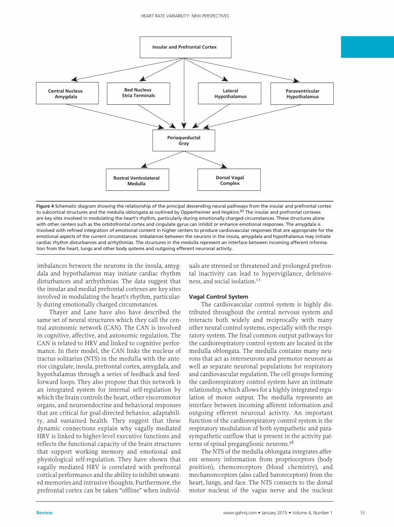
cal, and anatomical research has identified cortical, subcortical and medulla oblongata structures involved in self-regulation. Oppenheimer and Hopkins mapped a detailed hierarchy of cardiac con-trol structures among the cortex, amygdala and other subcortical structures, all of which can modify cardio-vascular-related neurons in the lower levels of the neuraxis (Figure 4).83 They suggest that the amygdala is involved with refined integration of emotional con-tent in higher centers to produce cardiovascular responses that are appropriate for the emotional aspects of the current circumstances.
The insular cortex and other centers such as the orbitofrontal cortex and cingulate gyrus can overcome (self-regulate) emotionally entrained responses by inhibiting or enhancing them. They also point out that
www.gahmj.com • January 2015 • Volume 4, Number 1 53Review
hEART RATE VARIABILITY: NEw PERSPEcTIVES
imbalances between the neurons in the insula, amyg-dala and hypothalamus may initiate cardiac rhythm disturbances and arrhythmias. The data suggest that the insular and medial prefrontal cortexes are key sites involved in modulating the heart’s rhythm, particular-ly during emotionally charged circumstances.
Thayer and Lane have also have described the same set of neural structures which they call the cen-tral autonomic network (CAN). The CAN is involved in cognitive, affective, and autonomic regulation. The CAN is related to HRV and linked to cognitive perfor-mance. In their model, the CAN links the nucleus of tractus solitarius (NTS) in the medulla with the ante-rior cingulate, insula, prefrontal cortex, amygdala, and hypothalamus through a series of feedback and feed-forward loops. They also propose that this network is an integrated system for internal self-regulation by which the brain controls the heart, other visceromotor organs, and neuroendocrine and behavioral responses that are critical for goal-directed behavior, adaptabili-ty, and sustained health. They suggest that these dynamic connections explain why vagally mediated HRV is linked to higher-level executive functions and reflects the functional capacity of the brain structures that support working memory and emotional and physiological self-regulation. They have shown that vagally mediated HRV is correlated with prefrontal cortical performance and the ability to inhibit unwant-ed memories and intrusive thoughts. Furthermore, the prefrontal cortex can be taken “offline” when individ-
uals are stressed or threatened and prolonged prefron-tal inactivity can lead to hypervigilance, defensive-ness, and social isolation.11
Vagal Control systemThe cardiovascular control system is highly dis-
tributed throughout the central nervous system and interacts both widely and reciprocally with many other neural control systems, especially with the respi-ratory system. The final common output pathways for the cardiorespiratory control system are located in the medulla oblongata. The medulla contains many neu-rons that act as interneurons and premotor neurons as well as separate neuronal populations for respiratory and cardiovascular regulation. The cell groups forming the cardiorespiratory control system have an intimate relationship, which allows for a highly integrated regu-lation of motor output. The medulla represents an interface between incoming afferent information and outgoing efferent neuronal activity. An important function of the cardiorespiratory control system is the respiratory modulation of both sympathetic and para-sympathetic outflow that is present in the activity pat-terns of spinal preganglionic neurons.38
The NTS of the medulla oblongata integrates affer-ent sensory information from proprioceptors (body position), chemoreceptors (blood chemistry), and mechanoreceptors (also called baroreceptors) from the heart, lungs, and face. The NTS connects to the dorsal motor nucleus of the vagus nerve and the nucleus
Insular and Prefrontal Cortex
Central Nucleus Amygdala
Bed Nucleus Stria Terminals
LateralHypothalamus
ParaventricularHypothalamus
PeriaqueductalGray
Rostral VentrolateralMedulla
Dorsal VagalComplex
Figure 4SchematicdiagramshowingtherelationshipoftheprincipaldescendingneuralpathwaysfromtheinsularandprefrontalcortextosubcorticalstructuresandthemedullaoblongataasoutlinedbyOppenheimerandHopkins.83Theinsularandprefrontalcortexesarekeysitesinvolvedinmodulatingtheheart’srhythm,particularlyduringemotionallychargedcircumstances.Thesestructuresalonewithothercenterssuchastheorbitofrontalcortexandcingulategyruscaninhibitorenhanceemotionalresponses.Theamygdalaisinvolvedwithrefinedintegrationofemotionalcontentinhighercenterstoproducecardiovascularresponsesthatareappropriatefortheemotionalaspectsofthecurrentcircumstances.Imbalancesbetweentheneuronsintheinsula,amygdalaandhypothalamusmayinitiatecardiacrhythmdisturbancesandarrhythmias.Thestructuresinthemedullarepresentaninterfacebetweenincomingafferentinforma-tionfromtheheart,lungsandotherbodysystemsandoutgoingefferentneuronalactivity.
54 Volume 4, Number 1 • January 2015 • www.gahmj.com
GLOBAL ADVANCES IN HEALTH AND MEDICINE
Review
ambiguous (NA). Neurocardiology research indicates that the efferent vagal fibers that innervate the heart are primarily A-fibers, the largest and fastest conduct-ing axons that originate from somata located primarily in the NA. The NA also receives and integrates informa-tion from the cortical and subcortical systems.38 Thus, the vagal regulatory centers respond to peripheral sen-sory (afferent) inputs and higher brain center inputs to adjust efferent neuronal outflows, which results in the vagally mediated beat-to-beat changes in HR.
Since BP regulation is a central role of the cardio-vascular system, the factors that alter BP also affect beat-to-beat fluctuations and therefore, the heart rhythms. Intrinsic cardiac afferent sensory neurons transduce and distribute mechanical and chemical information regarding the heart to the intrinsic car-diac nervous system.46 The afferent impulses from the intrinsic cardiac neurons travel via the vagal nerves to the nodose ganglia and then to the NTS. The NTS has connections with the NA and spinal cord resulting in modulation of activity patterns in both parasympa-thetic and sympathetic outflow to the heart and the blood vessels.38 There is controversy regarding any inhibitory role of parasympathetic efferent pregangli-onic neurons in the dorsal motor vagal (DMV) com-plex of the medulla as a number of anatomical studies suggest that virtually all efferent projections from the DMV are to subdiaphragmatic structures.83
The vagus nerves innervate the intrinsic cardiac nervous system. A few of these connections synapse on motor neurons in the intrinsic cardiac nervous system that project directly to the SA node (and other tissues in the heart) where they trigger acetylcholine release to slow HR.84 However, the majority of the efferent pre-ganglionic vagal neurons (~80%) connect to local cir-cuitry neurons in the intrinsic cardiac nervous system where motor information is integrated with inputs from mechanosensitive and chemosensory neurons in the heart.85 Thus, efferent sympathetic and parasympa-thetic activity is integrated in and with the activity occurring in the heart’s intrinsic nervous system. This includes the input signals from the mechanosensitive and chemosensory neurons within the heart, all of which ultimately contribute to beat-to-beat cardiac functional changes.46
The response time of a single efferent vagal impulse on the sinus node is very short and results in an immediate response that typically occurs within the cardiac cycle in which it occurs and affects only 1 or 2 heartbeats after its onset.33 After cessation of vagal stimulation, HR rapidly increases to its previous level. An increase in HR can also be achieved by reduced vagal activity (vagal withdrawal). Thus, any sudden change in HR, up or down, or between 1 beat and the next, are primarily parasympathetically mediated.33,34
In summary, the cardiorespiratory control system is complex, and information from many inputs is inte-grated at multiple levels of the system, all of which are important for the generation of normal beat-to-beat
variability in HR and BP. The medulla oblongata is the major structure integrating incoming afferent informa-tion from the heart, lungs and face with inputs from cortical and subcortical structures and is the source of the respiratory modulation of the activity patterns in sympathetic and parasympathetic outflow. The intrin-sic cardiac nervous system integrates mechanosensi-tive and chemosensitive neuron inputs with efferent information from both the sympathetic and parasym-pathetic inputs from the brain. As a complete system, it affects HRV, vasoconstriction, venoconstriction, and cardiac contractility in order to regulate HR and BP.33
afferent Modulation of Cardiac and Brain activityThe field of neurocardiology has extensively
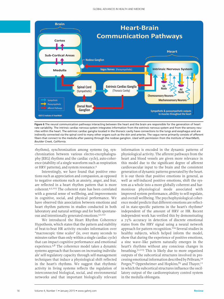
explored the anatomy and functions of the intrinsic cardiac nervous system along with its connections with the brain.75,86 While efferent regulation of the heart by the vagus nerves is generally well known, the majority of fibers in the vagus nerves are afferent in nature. Furthermore, more vagal fibers are related to cardiovascular pathways than other organs.87 Complex patterns of cardiovascular afferent nerve activity occur across time scales from milliseconds to minutes.88 The intrinsic cardiac nervous system has both short-term and long-term memory functions, which can influence HRV and afferent activity related to BP, rhythm, rate, and hormonal factors.70,88,89 The intrinsic cardiac neu-rons (sensory, interconnecting, afferent, and motor) can operate independently of central neuronal com-mand, and their network is sufficiently extensive to be characterized as its own “little brain” in the heart (Figure 5).84,90 The afferent nerves play a critical role in physiological regulation and affect the heart’s rhythm and HRV. Efferent sympathetic and parasympathetic activity is integrated in the heart’s intrinsic nervous system, with the signals arising from the mechanosen-sory and chemosensory neurons in the heart (Figure 6). The neural output of the intrinsic cardiac nervous sys-tem then travel to the brain via afferent pathways in the spinal column and vagus nerve. Intrinsic cardiac afferent neurons project to nodose and dorsal root gan-glia, the spinal cord, brainstem, hypothalamus, thala-mus, or amygdala and then to the cerebral cortex.4,46,91
John and Beatrice Lacey were the first to suggest a causal role of the heart in modulating cognitive func-tions such as sensory-motor and perceptual perfor-mance.92-94 They suggested that cortical functions are modulated via afferent input from pressure sensitive neurons in the heart, carotid arteries, and aortic arch.93 Their research focused on activity occurring within a single cardiac cycle, and they confirmed that cardio-vascular activity influences perception and cognitive performance. Research by Velden and Wölk later dem-onstrated that cognitive performance fluctuated at a rhythm around 10 Hz and showed that the modula-tion of cortical function via the heart’s influence was due to afferent inputs on the neurons in the thalamus, which globally synchronizes cortical activity.95,96 An
www.gahmj.com • January 2015 • Volume 4, Number 1 55Review
hEART RATE VARIABILITY: NEw PERSPEcTIVES
important aspect of their work was the finding that it is the “pattern and stability” (of the rhythm) of the heart’s afferent inputs, rather than the number of neu-ral bursts within the cardiac cycle, that are important in modulating thalamic activity, which in turn has global effects on brain function.
There has since been a growing body of research indicating that afferent information processed by the intrinsic cardiac nervous system can influence activi-ty in the frontocortical areas4,97,98 and motor cortex,58 affecting psychological factors such as attention level, motivation,99 perceptual sensitivity,100 and emotion-al processing.101
COHErEnCE The various concepts and measurements embraced
under the term coherence have become central to fields as diverse as quantum physics, cosmology, physiology, and brain and consciousness research.5 Coherence always implies connectedness, correlations, stability and efficient energy utilization. For example, we refer to people’s speech or thoughts as coherent if the words fit together well and incoherent if they are uttering mean-ingless nonsense or ideas that make no sense as a whole. In physics and physiology, the term coherence is used to describe the degree of synchronization between differ-ent oscillating systems. This type of coherence is called cross-coherence which occurs when two or more of the body’s oscillatory systems, such as respiration and heart rhythms, become entrained and operate at the same frequency. The term auto-coherence describes coherent activity within a single oscillatory system. An example is a system that exhibits sine wave like oscillations; the more stable the frequency, amplitude and shape, the higher the degree of coherence. When coherence is increased in a system that is coupled with other sys-
tems, it can pull the other systems into increased syn-chronization and more efficient function. For example, frequency pulling and entrainment can easily be seen between the heart, respiratory, and BP rhythms as well as between very-low-frequency brain rhythms, cranio-sacral rhythms, and electrical potentials measured across the skin.52,102
We (McCraty and colleagues) introduced the term physiological coherence to describe the degree of order, harmony, and stability in the various rhythmic activi-ties within living systems over any given time period.52 This harmonious order signifies a coherent system that has an efficient or optimal function directly related to the ease and flow in life processes. By contrast, an erratic, discordant pattern of activity denotes an inco-herent system whose function reflects stress and inef-ficient utilization of energy in life processes. Specifically, heart coherence (also referred to as cardiac coherence or resonance) can be measured by HRV analysis wherein a person’s heart rhythm pattern becomes more ordered and sine-wave like at a frequen-cy of around 0.1 Hz (10 seconds). A coherent heart rhythm is defined as a relatively harmonic, sine wave–like, signal with a very narrow, high-amplitude peak in the LF region of the HRV power spectrum with no major peaks in the VLF or HF regions. Coherence is assessed by identifying the maximum peak in the 0.04 Hz to 0.26 Hz range of the HRV power spectrum, calcu-lating the integral in a window 0.030 Hz wide, centered on the highest peak in that region, and then calculating the total power of the entire spectrum. The coherence ratio is formulated as: (Peak Power/[Total Power – Peak Power]).4 Physiological coherence includes specific approaches for quantifying the various types of coher-ence measures, such as cross-coherence (frequency entrainment between respiration, BP, and heart
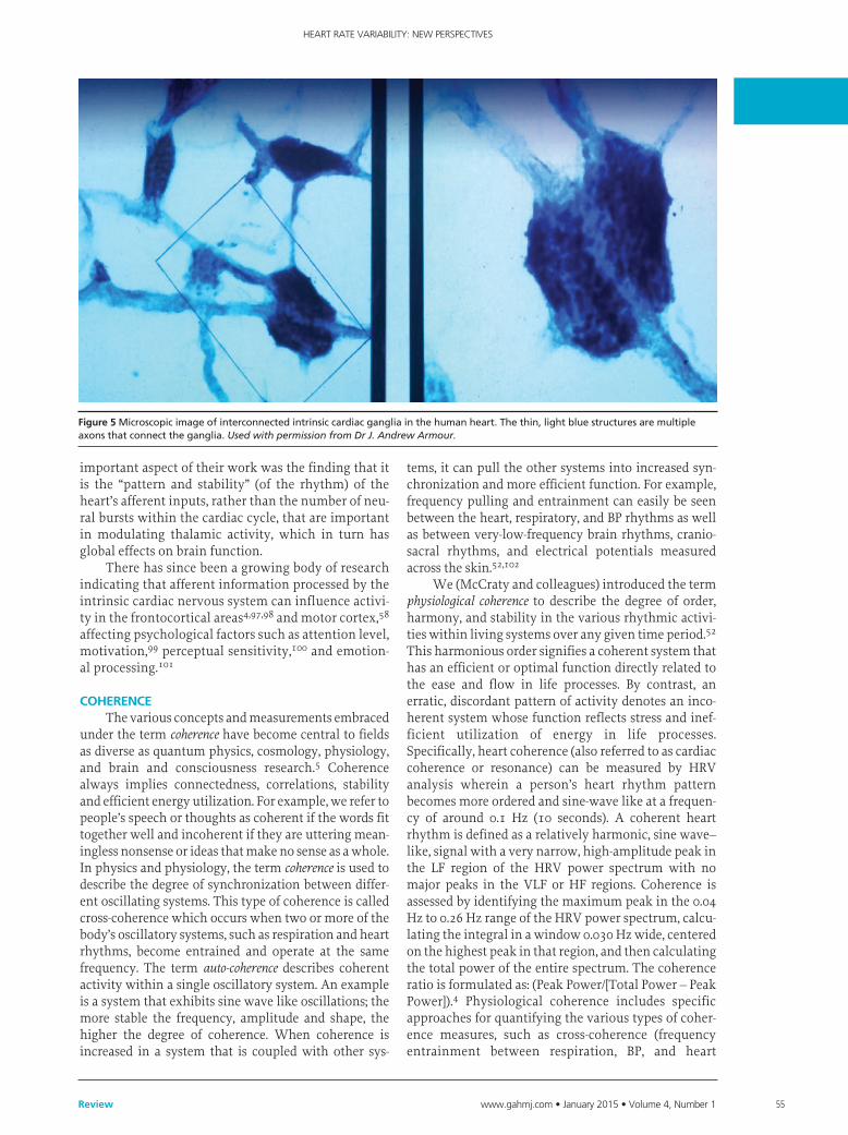
Figure 5Microscopicimageofinterconnectedintrinsiccardiacgangliainthehumanheart.Thethin,lightbluestructuresaremultipleaxonsthatconnecttheganglia.Used with permission from Dr J. Andrew Armour.
56 Volume 4, Number 1 • January 2015 • www.gahmj.com
GLOBAL ADVANCES IN HEALTH AND MEDICINE
Review
rhythms), synchronization among systems (eg, syn-chronization between various electro-encephalogra-phy [EEG] rhythms and the cardiac cycle), auto-coher-ence (stability of a single waveform such as respiration or HRV patterns), and system resonance.4
Interestingly, we have found that positive emo-tions such as appreciation and compassion, as opposed to negative emotions such as anxiety, anger, and fear, are reflected in a heart rhythm pattern that is more coherent.4,5,103 The coherent state has been correlated with a general sense of wellbeing, and improvements in cognitive, social, and physical performance. We have observed this association between emotions and heart rhythm patterns in studies conducted in both laboratory and natural settings and for both spontane-ous and intentionally generated emotions.52,102
We introduced the Heart Rhythm Coherence Hypothesis, which states that the pattern and stability of beat-to-beat HR activity encodes information over “macroscopic time scales” (ie, over many seconds to minutes rather than only within a single cardiac cycle) that can impact cognitive performance and emotional experience.4,8 The coherence model takes a dynamic systems approach that focuses on increasing individu-als’ self-regulatory capacity through self-management techniques that induce a physiological shift reflected in the heart’s rhythms. We suggest that rhythmic activity in living systems reflects the regulation of interconnected biological, social, and environmental networks and that important biologically relevant
information is encoded in the dynamic patterns of physiological activity. The afferent pathways from the heart and blood vessels are given more relevance in this model due to the significant degree of afferent cardiovascular input to the brain and the consistent generation of dynamic patterns generated by the heart. It is our thesis that positive emotions in general, as well as self-induced positive emotions, shift the sys-tem as a whole into a more globally coherent and har-monious physiological mode associated with improved system performance, ability to self-regulate, and overall wellbeing. The psychophysiological coher-ence model predicts that different emotions are reflect-ed in state-specific patterns in the heart’s rhythms4 independent of the amount of HRV or HR. Recent independent work has verified this by demonstrating a 75% accuracy in detection of discrete emotional states from the HRV signal using a neural network approach for pattern recognition.104 Several studies in healthy subjects, which helped inform the model, show that during the experience of positive emotions, a sine wave–like pattern naturally emerges in the heart’s rhythms without any conscious changes in breathing.52,103 This is likely due to more organized outputs of the subcortical structures involved in pro-cessing emotional information described by Pribram,59
Porges,60 Oppenheimer and Hopkins,83 and Thayer,11 in which the subcortical structures influence the oscil-latory output of the cardiorespiratory control system in the medulla oblongata.
Figure 6Theneuralcommunicationpathwaysinteractingbetweentheheartandthebrainareresponsibleforthegenerationofheartratevariability.Theintrinsiccardiacnervoussystemintegratesinformationfromtheextrinsicnervoussystemandfromthesensoryneu-riteswithintheheart.Theextrinsiccardiacganglialocatedinthethoraciccavityhaveconnectionstothelungsandesophagusandareindirectlyconnectedviathespinalcordtomanyotherorganssuchastheskinandarteries.Thevagusnerveprimarilyconsistsofafferentfibersthatconnecttothemedullaafterpassingthroughthenodoseganglion.Used with permission from the Institute of HeartMath, Boulder Creek, California.
www.gahmj.com • January 2015 • Volume 4, Number 1 57Review
hEART RATE VARIABILITY: NEw PERSPEcTIVES
Heart rate Variability Coherence Increases Vagal afferent traffic
One of the properties of sensory neurons is that they are most responsive to increases in rate of change in the function to which they are tuned to detect (eg, HR, BP).86 During periods of increased cardiac coher-ence, there is typically an increased range of variabili-ty in both BP and HR, which is detected as increases in the rate of change by the sensory neurons, resulting in increased firing rates that increase vagal afferent traf-fic. There is also a more ordered pattern of activity. A recent study using heartbeat-evoked potentials showed that using paced breathing at a 10-second rhythm increased both the range of HRV and the coherence in the rhythms as expected and also increased the N200 amplitude potential in the EEG heartbeat–evoked potentials, which indicates increased afferent input.61
Anatomical and stimulation studies have shown that the thalamic pain pathways in the spinal cord are inhibited by increases in vagal afferent nerve traffic over normal intrinsic levels.105-107 Several studies have demonstrated that teaching patients self-regulation techniques that increase HRV coherence is associated with reduced pain and physical activity limita-tions.108,109 In a study of patients with severe brain injury, it was found that emotion–self-regulation train-ing resulted in significantly higher coherence ratios and higher attention scores. Ratings of participants’ emotional control correlated with improved HRV coherence measures.110 Regular practice of HRV bio-feedback results in lasting improvements in baroreflex gain, independent of cardiovascular and respiratory effects. This indicates neuroplasticity within the baro-reflex system, likely within the intrinsic cardiac ner-vous system.111 Thus, repeated sessions of heart coher-ence practice can reset the baroreflex system resulting in increased afferent nerve activity noninvasively.
resilience and self-regulatory Capacity HRV also indicates psychological resiliency and
behavioral flexibility, reflecting an individual’s capac-ity to self-regulate and effectively adapt to changing social or environmental demands.27,112 A growing number of studies have specifically linked vagally mediated HRV to self-regulatory capacity,9,10,113 emo-tional regulation,114,115 social interactions,7,116 one’s sense of coherence,117 the personality character traits of self-directedness,118 and coping styles.119
More recently, several studies have shown an association between higher levels of vagally-mediated resting HRV and performance on cognitive perfor-mance tasks requiring the use of executive func-tions.11 HRV coherence can be increased in order to improve cognitive function4,120-122 as well as a wide range of clinical outcomes that have been shown to reduce healthcare costs.5,51,123-127
Porges suggests that the evolution of the ANS, specifically the vagus nerves, was central to the devel-
opment of emotional experience and the social engage-ment system. As human beings, we are not limited to fight, flight, or freeze responses. We can self-regulate and initiate pro-social behaviors when we encounter challenges, disagreements, or stressors. Porges sug-gests that the healthy function of the social engage-ment system depends upon the proper functioning of the vagus nerves, which act as a “vagal brake,” and that measurements of vagal activity could serve as a mark-er for one’s ability to self-regulate. His theory also sug-gests that the evolution and healthy function of the ANS determines the boundaries for the range of one’s emotional expression, quality of communication, and the ability to self-regulate emotions and behaviors.60
self-regulation techniques that Increase Cardiac Coherence
There is a paradigm shift occurring in the treat-ment of diverse disorders like depression, epilepsy, and pain by using vagal nerve stimulation, which stimulates afferent neural pathways.128-130 New per-spectives are emerging on behavioral intervention approaches that teach people self-regulation strategies that include a physiological aspect such as HRV bio-feedback and that naturally increase vagal traffic. For example, there are many studies showing that the practice of breathing at 6 breaths per minute, sup-ported by HRV biofeedback, induces the coherence rhythm and has a wide range of benefits.111,131-136
In addition to clinical applications, HRV coher-ence feedback training is often used to support self-regulation skill acquisition in educational, corporate, law enforcement, and military settings. Several sys-tems that assess the degree of coherence in the user’s heart rhythms are available. The majority of these systems—such as the emWavePro, or Inner Balance for iOS devices (HeartMath, Inc, Boulder Creek, California), Relaxing Rhythms (Wild Divine, Boulder City, Nevada), and the Stress Resilience Training System (Ease Interactive, San Diego, California)—use a noninvasive earlobe or finger pulse sensor and dis-play the user’s heart rhythm to provide feedback on their level of coherence.
Emotional self-regulation strategies may contrib-ute to improved health and performance. Alone or in combination with HRV coherence biofeedback train-ing, these strategies have been shown to increase resil-ience and accelerate recovery from stressors or trau-ma.5,8,136,137 Self-induced positive emotions can initi-ate a shift to increased cardiac coherence without any conscious intention to change the breathing rhythm.52,103 Typically, when people are able to self-activate a positive or calming feeling rather than remaining focused on their breathing, they enjoy the shift in feeling and are able to sustain high levels of coherence for much longer time periods.124
Heart-focused self-regulation techniques and assistive technologies that provide real-time HRV coherence feedback provide a systematic process for
58 Volume 4, Number 1 • January 2015 • www.gahmj.com
GLOBAL ADVANCES IN HEALTH AND MEDICINE
Review
self-regulating thoughts, emotions, and behaviors and increasing physiological coherence. Many of these techniques (eg, Heart-Focused Breathing, Freeze Frame, Quick Coherence139) are designed to enable people to intervene in the moment they start to experi-ence stress reactions or unproductive thoughts or emotions. With practice, one is able to use one of the techniques to shift into a more coherent physiological state before, during, and after challenging or adverse situations, thus optimizing mental clarity, emotional composure, and stability.
The first step in most of the techniques developed by the Institute of HeartMath is called Heart-Focused Breathing, which includes putting one’s attention in the center of the chest (area of the heart) and imagin-ing the breath is flowing in and out of the chest area while breathing a little slower and deeper than usual. Conscious regulation of one’s respiration at a 10-sec-ond rhythm (0.1Hz) increases cardiac coherence and starts the process of shifting into a more coherent state.4,124 With conscious control over breathing, an individual can slow the rate and increase the depth of the breathing rhythm. This takes advantage of physi-ological mechanisms to modulate efferent vagal activ-ity and thus the heart rhythm. This increases vagal afferent nerve traffic and increases the coherence (sta-bility) in the patterns of vagal afferent nerve traffic. In turn, this influences the neural systems involved in regulating sympathetic outflow, informing emotional experience, and synchronizing neural structures underlying cognitive processes.4
Several studies using various combinations of these self-regulation techniques have found signifi-cant correlations between HRV coherence and improvements in cognitive function and self-regulato-ry capacity. For example, a study of middle school students with attention deficit hyperactivity disorder showed a wide range of significant improvements in short and long-term memory, ability to focus, and sig-nificant improvements in behaviors both at home and in school.120 A study of 41 fighter pilots engaging in flight simulator tasks found a significant correlation between higher levels of performance and heart rhythm coherence as well as lower levels of frustra-tion.140 A study of recently returning soldiers from Iraq who were diagnosed with PTSD, found that rela-tively brief periods of HRV coherence training com-bined with practicing the Quick Coherence Technique resulted in significant improvements in the ability to self-regulate along with a wide range of cognitive func-tions. The degree of improvement correlated with increased cardiac coherence.121 Other studies have shown increases in parasympathetic activity (vagal tone),52 reductions in cortisol and increases in DHEA,127 lowered BP and stress measures in hyperten-sive populations,124,126 reduced healthcare costs,123 and significant improvements in functional capacity in patients with congestive heart failure.141 In addi-tion, a study of correctional officers showed reduc-
tions in systolic and diastolic BP, total cholesterol, fasting glucose, overall stress, anger, fatigue and hostil-ity.125 Similar results were obtained in several studies with police officers.138,142
In addition to the emotional self-regulation tech-niques, there are other approaches that also increase HRV coherence. For example, a study of Zen monks found that monks with greater experience in medita-tion tended to have more coherent heart rhythms dur-ing their resting recording, while the ones who had been monks for less than 2 years did not.143 A study of autogenic training also showed increased HRV coher-ence and found that cardiac coherence was strongly correlated with EEG alpha activity. The authors sug-gested that cardiac coherence could be a general mark-er for the meditative state.144 However, this does not suggest that all meditation or prayer styles increase coherence, unless the coherence state is driven by a focus on breathing at a 10-second rhythm or the acti-vation of a positive emotion.145-148 For example, a study examining HRV while reciting rosary or bead prayers and yoga mantras found that a coherent rhythm was produced by rhythmically breathing but not by random verbalization or breathing. The authors ascribed the mechanisms for this finding to a breath-ing pattern of 6-cycles per minute.149 In a study of the effects of five different types of prayer on HRV, it was found that all types of prayer elicited increased cardiac coherence. However, prayers of gratefulness and heart-felt love resulted in definitively higher coherence lev-els.148 It has also been shown that tensing the large muscles in the legs in a rhythmical manner at a 10-sec-ond rhythm can induce a coherent heart rhythm.150
COnCLusIOn HRV is an emergent property of interdependent
regulatory systems that operate on different time scales to adapt to environmental and psychological challenges. The physiological mechanisms that con-tribute to HRV are complex and involve the neuraxis that spans from the prefrontal and insular cortex to the intrinsic cardiac nervous system, with the medulla oblongata and intrinsic cardiac nervous system pro-viding major neural integration centers. HRV can be used as an index of the functional capacity of various regulatory systems and assessment of regulatory capacity may offer an alternative to autonomic bal-ance models. Since the HRV LF band primarily reflects the vagally mediated transmission between the heart and medulla, resting measurements should not be used as markers of sympathetic activity. Based on 24-hour monitoring, ULF and VLF rhythms are more strongly associated with overall health status than HF rhythms. New perspectives on mechanisms underly-ing the VLF rhythm suggest that the primary source of this rhythm is within the heart itself. Recent findings demonstrate the importance of the intrinsic cardiac nervous system and cardiac afferents in generating the heart rhythm and modulating the intervals between
www.gahmj.com • January 2015 • Volume 4, Number 1 59Review
hEART RATE VARIABILITY: NEw PERSPEcTIVES
heartbeats. Vagally-mediated HRV appears to repre-sent an index of psychological self-regulatory control, such that individuals with greater resting HRV have performed better on tests of executive function.
In addition to assessing regulatory capacity, HRV can also be used in the context of real-time feedback to help restore regulatory capacity. Heart rhythm coher-ence approaches train clients to produce auto-coher-ent heart rhythms with a single peak in the LF region (typically around 0.1 Hz) with no significant peaks in the VLF and HF regions. Emotional self-regulation strategies may contribute to improved client health and performance, alone, or in combination with HRV biofeedback training. Numerous studies have provid-ed evidence that coherence training consisting of intentional activation of positive emotions paired with HRV coherence feedback may facilitate signifi-cant improvements in wellness and wellbeing indica-tors in a variety of populations. acknowledgments
The authors express their profound thanks to Dr John Andrew Armour for his generous contributions to this article.
rEFErEnCEs1. Cannon WB. The James-Lange theory of emotion: a critical examination
and an alternative theory. Am J Psychol. 1987;100(3-4):567-86. 2. Singer DH, Martin GJ, Magid N, et al. Low heart rate variability and sudden
cardiac death. J Electrocardiol. 1988;21 Suppl:S46-55. 3. Shaffer F, McCraty R, Zerr CL. A healthy heart is not a metronome: an inte-
grative review of the heart’s anatomy and heart rate variability. Front Psychol. 2014;5:1040.
4. McCraty R, Atkinson M, Tomasino D, Bradley RT. The coherent heart: heart-brain interactions, psychophysiological coherence, and the emergence of system-wide order. Boulder Creek, CA: Institute of Heartmath; 2009.
5. McCraty R, Childre D. Coherence: bridging personal, social, and global health. Altern Ther Health Med. 2010;16(4):10-24.
6. Singer DH. High heart rate variability, marker of healthy longevity. Am J Cardiol. 2010;106(6):910.
7. Geisler FC, Kubiak T, Siewert K, Weber H. Cardiac vagal tone is associated with social engagement and self-regulation. Biol Psychol. 2013;93(2):279-86.
8. McCraty R, Zayas MA. Cardiac coherence, self-regulation, autonomic sta-bility, and psychosocial well-being. Front Psychol. 2014;5:1090.
9. Reynard A, Gevirtz R, Berlow R, Brown M, Boutelle K. Heart rate variability as a marker of self-regulation. Appl Psychophysiol Biofeedback. 2011;36(3):209-15.
10. Segerstrom SC, Nes LS. Heart rate variability reflects self-regulatory strength, effort, and fatigue. Psychol Sci. 2007;18(3):275-81.
11. Thayer JF, Hansen AL, Saus-Rose E, Johnsen BH. Heart rate variability, pre-frontal neural function, and cognitive performance: the neurovisceral integration perspective on self-regulation, adaptation, and health. Ann Behav Med. 2009;37(2):141-53.
12. Heart rate variability standards of measurement, physiological interpreta-tion, and clinical use. Task Force of the European Society of Cardiology and the North American Society of Pacing and Electrophysiology. Circulation. 1996;93(5):1043-65.
13. Hon EH, Lee ST. Electronic evaluations of the fetal heart rate VII: patterns preceding fetal death, further observations. Am J Obstet Gynecol. 1963;87:814-26.
14. Braune HJ, Geisendörfer U. Measurement of heart rate variations: influenc-ing factors, normal values and diagnostic impact on diabetic autonomic neuropathy. Diabetes Res Clin Pract. 1995;29(3):179-87.
15. Vinik AI, Maser RE, Mitchell BD, Freeman R. Diabetic autonomic neuropa-thy. Diabetes Care. 2003;26(5):1553-79.
16. Ewing DJ, Campbell IW, Clarke BF. Mortality in diabetic autonomic neu-ropathy. Lancet. 1976;1(7960):601-3.
17. Wolf MM, Varigos GA, Hunt D, Sloman JG. Sinus arrhythmia in acute myocardial infarction. Med J Aust. 1978;2(2):52-3.
18. Umetani K, Singer DH, McCraty R, Atkinson M. Twenty-four hour time domain heart rate variability and heart rate: relations to age and gender
over nine decades. J Am Coll Cardiol. 1998;31(3):593-601.19. Dekker JM, Schouten EG, Klootwijk P, Pool J, Swenne CA, Kromhout D.
Heart rate variability from short electrocardiographic recordings predicts mortality from all causes in middle-aged and elderly men. The Zutphen Study. Am J Epidemiol. 1997;145(10):899-908.
20. Tsuji H, Venditti FJ Jr, Manders ES, et al. Reduced heart rate variability and mortality risk in an elderly cohort. The Framingham Heart Study. Circulation. 1994;90(2):878-83.
21. Huikuri HV, Jokinen V, Syvänne M, et al. Heart rate variability and progres-sion of coronary atherosclerosis. Arterioscler Thromb Vasc Biol. 1999;19(8):1979-85.
22. Sajadieh A, Nielsen OW, Rasmussen V, Hein HO, Abedini S, Hansen JF. Increased heart rate and reduced heart-rate variability are associated with subclinical inflammation in middle-aged and elderly subjects with no apparent heart disease. Eur Heart J. 2004;25(5):363-70.
23. Kazuma N, Otsuka K, Matsuoka I, Murata M. Heart rate variability during 24 hours in asthmatic children. Chronobiol Int. 1997;14(6):597-606.
24. Carney RM, Blumenthal JA, Stein PK, et al. Depression, heart rate variabili-ty, and acute myocardial infarction. Circulation. 2001;104(17):2024-8.
25. Agelink MW, Boz C, Ullrich H, Andrich J. Relationship between major depression and heart rate variability. Clinical consequences and implica-tions for antidepressive treatment. Psychiatry Res. 2002;113(1-2):139-49.
26. Cohen H, Benjamin J. Power spectrum analysis and cardiovascular morbid-ity in anxiety disorders. Auton Neurosci. 2006;128(1-2):1-8.
27. Berntson GG, Norman GJ, Hawkley LC, Cacioppo JT. Cardiac autonomic balance versus cardiac regulatory capacity. Psychophysiology. 2008;45(4):643-52.
28. Palatini P. Elevated heart rate as a predictor of increased cardiovascular morbidity. J Hypertens Suppl. 1999;17(3):S3-10.
29. Stampfer HG. The relationship between psychiatric illness and the circadi-an pattern of heart rate. Aust N Z J Psychiatry. 1998;32(2):187-98.
30. Stampfer HG, Dimmitt SB. Variations in circadian heart rate in psychiatric disorders: theoretical and practical implications. Chronol Physiol Ther. 2013;3:41-50.
31. Opthof T. The normal range and determinants of the intrinsic heart rate in man. Cardiovasc Res. 2000;45(1):173-6.
32. Umetani K, Duda CL, Singer DH. Aging effects on cycle length dependence of heart rate variability. Proceedings of the 1996 Fifteenth Southern Biomedical Engineering Conference: Dayton, Ohio; 1996.
33. Hainsworth R. The control and physiological importance on heart rate. In: Malik M, Camm AJ, editors. Heart rate variability. Armonk, NY: Futura Publishing Company, Inc; 1995.
34. Lacey BC, Lacey JI. Two-way communication between the heart and the brain. Significance of time within the cardiac cycle. Am Psychol. 1978;33(2):99-113.
35. Felber Dietrich D, Schindler C, Schwartz J, et al. Heart rate variability in an ageing population and its association with lifestyle and cardiovascular risk factors: results of the SAPALDIA study. Europace. 2006;8(7):521-9.
36. Bristow JD, Gribbin B, Honour AJ, Pickering TG, Sleight P. Diminished baroreflex sensitivity in high blood pressure and ageing man. J Physiol. 1969;202(1):45P-6P.
37. Hirsch JA, Bishop B. Respiratory sinus arrhythmia in humans: how breath-ing pattern modulates heart rate. Am J Physiol. 1981;241(4):H620-9.
38. Hopkins D, Ellenberger H. Cardiorespiratory neurons in the mudulla oblongata: Input and output relationships. In: Armour JA, Ardell JL, edi-tors. Neurocardiology. New York: Oxford University Press; 1994:219-44.
39. Eckberg DL. Human sinus arrhythmia as an index of vagal outflow. J Appl Physiol Respir Environ Exerc Physiol. 1983;54(4):961-6.
40. Lombardi F, Sandrone G, Mortara A, et al. Linear and nonlinear dynamics of heart rate variability after acute myocardial infarction with normal and reduced left ventricular ejection fraction. Am J Cardiol. 1996;77(15):1283-8.
41. Otsuka K, Cornelissen G, Halberg F. Age, gender and fractal scaling in heart rate variability. Clin Sci (Lond). 1997;93(4):299-308.
42. Pomeranz B, Macaulay RJ, Caudill MA, et al. Assessment of autonomic function in humans by heart rate spectral analysis. Am J Physiol. 1985;248(1 Pt 2):H151-3.
43. Malliani A. Association of heart rate variability components with physio-logical regulatory mechanisms. In: Malik M, Camm AJ, editors. Heart Rate Variability. Armonk NY: Futura Publishing Company, Inc; 1995:173-188.
44. Taylor JA, Carr DL, Myers CW, Eckberg DL. Mechanisms underlying very-low-frequency RR-interval oscillations in humans. Circulation. 1998;98(6):547-55.
45. Reyes del Paso GA, Langewitz W, Mulder LJ, van Roon A, Duschek S. The utility of low frequency heart rate variability as an index of sympathetic cardiac tone: a review with emphasis on a reanalysis of previous studies. Psychophysiology. 2013;50(5):477-87.
46. Armour JA. Peripheral autonomic neuronal interactions in cardiac regula-tion. In: Neurocardiology. Armour JA, Ardell JL, editors. New York: Oxford University Press; 1994:219-44.
47. Bowers EJ, Murray A. Effects on baroreflex sensitivity measurements when
60 Volume 4, Number 1 • January 2015 • www.gahmj.com
GLOBAL ADVANCES IN HEALTH AND MEDICINE
Review
different protocols are used to induce regular changes in beat-to-beat inter-vals and systolic pressure. Physiol Meas. 2004;25(2):523-38.
48. deBoer RW, Karemaker JM, Strackee J. Hemodynamic fluctuations and baroreflex sensitivity in humans: a beat-to-beat model. Am J Physiol. 1987;253(3 Pt 2):H680-9.
49. Baselli G, Cerutti S, Badilini F, et al. Model for the assessment of heart peri-od variability interactions of respiration influences. Med Biol Eng Comput. 1994;32(2):143-52.
50. Ahmed AK, Harness JB, Mearns AJ. Respiratory control of heart rate. Eur J Appl Physiol. 1982;50:95-104.
51. Lehrer PM, Vaschillo E, Vaschillo B, et al. Heart rate variability biofeedback increases baroreflex gain and peak expiratory flow. Psychosom Med. 2003;65(5):796-805.
52. Tiller WA, McCraty R, Atkinson M. Cardiac coherence: a new, noninvasive measure of autonomic nervous system order. Altern Ther Health Med. 1996;2(1):52-65.
53. Brown TE, Beightol LA, Koh J, Eckberg DL. Important influence of respira-tion on human R-R interval power spectra is largely ignored. J Appl Physiol. 1993;75(5):2310-7.
54. Malliani A, Lombardi F, Pagani M, Cerutti S. Power spectral analysis of car-diovascular variability in patients at risk for sudden cardiac death. J Cardiovasc Electrophysiol. 1994;5(3):274-86.
55. Pal GK, Adithan C, Ananthanarayanan PH, et al. Sympathovagal imbal-ance contributes to prehypertension status and cardiovascular risks attrib-uted by insulin resistance, inflammation, dyslipidemia and oxidative stress in first degree relatives of type 2 diabetics. PLoS One. 2013;8(11):e78072.
56. Pagani M, Lombardi F, Guzzetti S, et al. Power spectral analysis of heart rate and arterial pressure variabilities as a marker of sympatho-vagal inter-action in man and conscious dog. Circ Res. 1986;59(2):178-93.
57. Kember GC, Fenton GA, Armour JA, Kalyaniwalla N. Competition model for aperiodic stochastic resonance in a Fitzhugh-Nagumo model of cardiac sensory neurons. Phys Rev E Stat Nonlin Soft Matter Phys. 2001;63(4 Pt 1):041911.
58. Svensson TH, Thorén P. Brain noradrenergic neurons in the locus coerule-us: inhibition by blood volume load through vagal afferents. Brain Res. 1979;172(1):174-8.
59. Pribram KH, Melges FT. Psychophysiological basis of emotion. In: Vinken PJ, Bruyn GW, editors. Handbook of clinical neurology. Amsterdam: North-Holland Publishing Company; 1969:316-41.
60. Porges SW. The polyvagal perspective. Biol Psychol. 2007;74(2):116-4361. MacKinnon S, Gevirtz R, McCraty R, Brown M. Utilizing heartbeat evoked
potentials to identify cardiac regulation of vagal afferents during emo-tion and resonant breathing. Appl Psychophysiol Biofeedback. 2013;38(4):241-55.
62. Schmidt H, Müller-Werdan U, Hoffmann T, et al. Autonomic dysfunction predicts mortality in patients with multiple organ dysfunction syndrome of different age groups. Crit Care Med. 2005;33(9):1994-2002.
63. Hadase M, Azuma A, Zen K, et al. Very low frequency power of heart rate variability is a powerful predictor of clinical prognosis in patients with congestive heart failure. Circ J. 2004;68(4):343-7.
64. Tsuji H, Larson MG, Venditti FJ Jr, et al. Impact of reduced heart rate vari-ability on risk for cardiac events. The Framingham Heart Study. Circulation. 1996;94(11):2850-5.
65. Bigger JT Jr, Fleiss JL, Steinman RC, Rolnitzky LM, Kleiger RE, Rottman JN. Frequency domain measures of heart period variability and mortality after myocardial infarction. Circulation. 1992;85(1):164-71.
66. Shah AJ, Lampert R, Goldberg J, Veledar E, Bremner JD, Vaccarino V. Posttraumatic stress disorder and impaired autonomic modulation in male twins. Biol Psychiatry. 2013;73(11):1103-10.
67. Lampert R, Bremner JD, Su S, et al. Decreased heart rate variability is asso-ciated with higher levels of inflammation in middle-aged men. Am Heart J. 2008;156(4):759.e1-7.
68. Carney RM, Freedland KE, Stein PK, et al. Heart rate variability and mark-ers of inflammation and coagulation in depressed patients with coronary heart disease. J Psychosom Res. 2007;62(4):463-7.
69. Theorell T, Liljeholm-Johansson Y, Björk H, Ericson M. Saliva testosterone and heart rate variability in the professional symphony orchestra after “public faintings” of an orchestra member. Psychoneuroendocrinology. 2007;32(6):660-8.
70. Kleiger RE, Stein PK, Bigger JT Jr. Heart rate variability: measurement and clinical utility. Ann Noninvas Electrocardiol. 2005;10(1):88-101.
71. Akselrod S, Gordon D, Ubel FA, Shannon DC, Berger AC, Cohen RJ. Power spectrum analysis of heart rate fluctuation: a quantitative probe of beat-to-beat cardiovascular control. Science. 1981;213(4504):220-2.
72. Cerutti S, Bianchi AM, Mainardi LT. Spectral analysis of the heart rate vari-ability signal. In: Malik M, Camm AJ, editors. Heart rate variability. Armonk NY. Futura Publishing Company, Inc;1995;63-74.
73. Murphy DA, Thompson GW, Ardell JL, et al. The heart reinnervates after transplantation. Ann Thorac Surg. 2000;69(6):1769-81.
74. Ramaekers D, Ector H, Vanhaecke J, van Cleemput J, van de Werf F. Heart rate variability after cardiac transplantation in humans. Pacing Clin Electrophysiol. 1996;19(12 Pt 1):2112-9.
75. Armour JA. Neurocardiology—anatomical and functional principles. Boulder Creek, CA: Institute of HeartMath; 2003.
76. Kember GC, Fenton GA, Collier K, Armour JA. Aperiodic stochastic reso-nance in a hysteretic population of cardiac neurons. Physical Rev. 2000;61(2):1816-24.
77. Berntson GG, Bigger JT Jr, Eckberg DL, et al. Heart rate variability: origins, methods, and interpretive caveats. Psychophysiology. 1997;34(6):623-48.
78. Bernardi L, Valle F, Coco M, Calciati A, Sleight P. Physical activity influenc-es heart rate variability and very-low-frequency components in Holter electrocardiograms. Cardiovasc Res. 1996;32(2):234-7.
79. Huikuri HV, Niemelä MJ, Ojala S, Rantala A, Ikäheimo MJ, Airaksinen KE. Circadian rhythms of frequency domain measures of heart rate variability in healthy subjects and patients with coronary artery disease. Effects of arousal and upright posture. Circulation. 1994;90(1):121-6.
80. Singh RB, Cornélissen G, Weydahl A, et al. Circadian heart rate and blood pressure variability considered for research and patient care. Int J Cardiol. 2003;87(1):9-28; discussion 29-30.
81. Stein PK, Domitrovich PP, Huikuri HV, Kleiger RE; Cast Investigators. Traditional and nonlinear heart rate variability are each independently associated with mortality after myocardial infarction. J Cardiovasc Electrophysiol. 2005;16(1):13-20.
82. Kleiger RE, Miller JP, Bigger JT Jr, Moss AJ. Decreased heart rate variability and its association with increased mortality after acute myocardial infarc-tion. Am J Cardiol. 1987;59(4):256-62.
83. Oppenheimer S, Hopkins D. Suprabulbar neuronal regulation of the heart. In: Armour JA, Ardell JL, editors. Neurocardiology. New York: Oxford University Press; 1994:309-41.
84. Armour JA. Anatomy and function of the intrathoracic neurons regulating the mammalian heart. In: Zucker IH, Gilmore JP, editors. Reflex control of the circulation. Boca Raton, FL: CRC Press; 1991:1-37.
85. Beaumont E, Salavatian S, Southerland EM, et al. Network interactions within the canine intrinsic cardiac nervous system: implications for reflex control of regional cardiac function. J Physiol. 2013;591(Pt 18):4515-33.
86. Armour JA, Ardell JL. Neurocardiology. New York: Oxford University Press; 1994:219-44.
87. Cameron OG. Visceral sensory neuroscience: interception. New York: Oxford University Press; 2002.
88. Armour JA, Kember GC. Cardiac sensory neurons. In: Armour JA, Ardell JL, editors. Basic and clinical neurocardiology. New York: Oxford University Press; 2004; 79-117.
89. Ardell JL, Cardinal R, Vermeulen M, Armour JA. Dorsal spinal cord stimu-lation obtunds the capacity of intrathoracic extracardiac neurons to trans-duce myocardial ischemia. Am J Physiol Regul Integr Comp Physiol. 2009;297(2):R470-7.
90. Armour JA. Potential clinical relevance of the ‘little brain’ on the mamma-lian heart. Exp Physiol. 2008;93(2):165-76.
91. Kukanova B, Mravec B. Complex intracardiac nervous system. Bratisl Lek Listy. 2006;107(3):45-51.
92. Lacey BC, Lacey JI. Studies of heart rate and other bodily processes in sen-sorimotor behavior. In: Obrist PA, Black AH, Brener J, Dicara LV, editors. Cardiovascular psychophysiology: current issues in response mecha-nisms, biofeedback, and methodology. Chicago: Aldine; 1974:538-64.
93. Lacey JI. Somatic response patterning and stress: Some revisions of activa-tion theory, In: Appley MH, Trumbull R, editors. Psychological stress: issues in research. New York: Appleton-Century-Crofts; 1967:14-42.
94. Lacey JI, Lacey BC. Some autonomic-central nervous system interrelation-ships. In: Black P, ed. Physiological correlates of emotion. New York: Academic Press; 1970:205-27.
95. Wölk C, Velden M. Detection variability within the cardiac cycle: toward a revision of the “baroreceptor hypothesis.” J Psychophysiol. 1987;1:61-5.
96. Wölk C, Velden M. Revision of the baroreceptor hypothesis on the basis of the new cardiac cycle effect. In: Bond NW, Siddle DAT, editors. Psychobiology: issues and applications. Amsterdam: Elsevier Science Publishers B.V.; 1989:371-379.
97. Lane RD, Reiman EM, Ahem GL, Thayer JF. Activity in medial prefrontal cortex correlates with vagal component of heart rate variability during emotion. Brain Cognit. 2001;47:97-100.
98. McCraty R, Atkinson M, Bradley RT. Electrophysiological evidence of intuition: Part 2. A system-wide process? J Altern Complement Med. 2004;10(2):325-36.
99. Schandry R, Montoya P. Event-related brain potentials and the processing of cardiac activity. Biol Psychol. 1996;42(1-2):75-85.
100. Montoya P, Schandry R, Müller A. Heartbeat evoked potentials (HEP): topog-raphy and influence of cardiac awareness and focus of attention. Electroencephalogr Clin Neurophysiol. 1993;88(3):163-72.
101. Zhang JX, Harper RM, Frysinger RC. Respiratory modulation of neuronal dis-charge in the central nucleus of the amygdala during sleep and waking states. Exp Neurol. 1986;91(1):193-207.
www.gahmj.com • January 2015 • Volume 4, Number 1 61Review
hEART RATE VARIABILITY: NEw PERSPEcTIVES
102. Bradley RT, Pribram KH. Communication and stability in social collectives. J Soc Evol Syst. 1998;21(1):29-80.
103. McCraty R, Atkinson M, Tiller WA, Rein G, Watkins AD. The effects of emo-tions on short-term power spectrum analysis of heart rate variability. Am J Cardiol. 1995;76(14):1089-93.
104. Leon, E, Clarke G, Callaghan V, Doctor F. Affect-aware behavior modeling and control inside an intelligent environment. Perv Mobile Comput. http://cswww.essex.ac.uk/staff/vic/papers/2010_PMCJ10%28AffectAwareBehaviour%29.pdf. Accessed December 22, 2015.
105. Foreman R. Organization of visceral input. In: Yaksh TL, Lynch C, Zapol WM, Maze M, Biebuyck JF, Saidman LJ, editors. Anesthesia: biologic foun-dations. Philadelphia: Lippincott-Raven Publishers; 1997:663-83.
106. Foreman RD. Vagal afferent modulation of cardiac pain. In: Levey MN, Schwartz PJ, editors. Vagal control of the heart: experimental basis and clini-cal implications. Armonk, New York: Futura Publishing Co; 1994:345-68.
107. Foreman RD. Organization of the spinothalamic tract as a relay for cardio-pulmonary sympathetic afferent fiber activity. Prog Sens Physiol. 1989;9:1-51.
108. Berry ME, Chapple IT, Ginsberg JP, Gleichauf KJ, Meyer JA, Nagpal ML. Non-pharmacological intervention for chronic pain in veterans: a pilot study of heart rate variability biofeedback. Global Adv Health Med. 2014;3(2):28-33.
109. Soer R, Vos D, Hofstra B, Reneman MF. Heart coherence training combined with back school in patients with chronic non-specific low back pain: first pragmatic clinical results. Appl Psychophysiol Biofeedback. 2014;39(3-4):259-67.
110. Kim S, Zemon V, Cavallo MM, Rath JF, McCraty R, Foley FW. Heart rate variability biofeedback, executive functioning and chronic brain injury. Brain Inj. 2013;27(2):209-22.
111. Lehrer P, Vaschillo E, Lu SE, et al. Heart rate variability biofeedback: effects of age on heart rate variability, baroreflex gain, and asthma. Chest. 2006;129(2):278-84.
112. Beauchaine T. Vagal tone, development, and Gray’s motivational theory: toward an integrated model of autonomic nervous system functioning in psychopathology. Dev Psychopathol. 2001;13(2):183-214.
113. Geisler F, Kubiak T. Heart rate variability predicts self‐control in goal pur-suit. Eur J Pers. 2009;23(8):623-33.
114. Appelhans B, LueckenL. Heart rate variability as an index of regulated emotional responding. Rev Gen Psychol. 2006;10(3):229-40.
115. Geisler F, Vennewald N, Kubiak T, Weber H. The impact of heart rate vari-ability on subjective well-being is mediated by emotion regulation. Pers Indiv Differ. 2010;49(7):723-8.
116. Smith TW, Cribbet MR, Nealey-Moore JB, et al. Matters of the variable heart: respiratory sinus arrhythmia response to marital interaction and associations with marital quality. J Pers Soc Psychol. 2011;100(1):103-19.
117. Nasermoaddeli A, Sekine M, Kagamimori S. Association between sense of coherence and heart rate variability in healthy subjects. Environ Health Prev Med. 2004;9(6):272-4.
118. Zohar A, Cloninger R, McCraty R. Personality and heart rate variability: exploring pathways from personality to cardiac coherence and health. Open J Soc Sci. 2013;1(6):32-9.
119. Ramaekers D, Ector H, Demyttenaere K, Rubens A, Van de Werf F. Association between cardiac autonomic function and coping style in healthy subjects. Pacing Clin Electrophysiol. 1998;21(8):1546-52.
120. Lloyd A, Brett D, Wesnes K. Coherence training in children with attention-deficit hyperactivity disorder: cognitive functions and behavioral changes. Altern Ther Health Med. 2010;16(4):34-42.
121. Ginsberg JP, Berry ME, Powell DA. Cardiac coherence and posttraumatic stress disorder in combat veterans. Altern Ther Health Med. 2010;16(4):52-60.
122. Bradley RT, McCraty R, Atkinson M, Tomasino D, Daugherty A, Arguelles L. Emotion self-regulation, psychophysiological coherence, and test anxi-ety: results from an experiment using electrophysiological measures. Appl Psychophysiol Biofeedback. 2010;35(4):261-83.
123. Bedell W, Kaszkin-Bettag M. Coherence and health care cost—RCA actuar-ial study: a cost-effectiveness cohort study. Altern Ther Health Med. 2010;16(4):26-31.
124. Alabdulgader AA. Coherence: a novel nonpharmacological modality for lowering blood pressure in hypertensive patients. Global Adv Health Med. 2012;1(2):56-64.
125. McCraty R, Atkinson M, Lipsenthal L, Arguelles L. New hope for correc-tional officers: an innovative program for reducing stress and health risks. Appl Psychophysiol Biofeedback. 2009;34(4):251-72.
126. McCraty R, Atkinson M, Tomasino D. Impact of a workplace stress reduc-tion program on blood pressure and emotional health in hypertensive employees. J Altern Complement Med. 2003;9(3):355-69.
127. McCraty R, Barrios-Choplin B, Rozman D, Atkinson M, Watkins AD. The impact of a new emotional self-management program on stress, emotions, heart rate variability, DHEA and cortisol. Integr Physiol Behav Sci. 1998;33(2):151-70.
128. Kosel M, Schlaepfer TE. Beyond the treatment of epilepsy: new applica-tions of vagus nerve stimulation in psychiatry. CNS Spectr. 2003;8(7):515-21.
129. Groves DA, Brown VJ. Vagal nerve stimulation: a review of its applications and potential mechanisms that mediate its clinical effects. Neurosci Biobehav Rev. 2005;29(3):493-500.
130. Mauskop A. Vagus nerve stimulation relieves chronic refractory migraine and cluster headaches. Cephalalgia. 2005;25(2):82-6.
131. Ratanasiripong P, Ratanasiripong N, Kathalae D. Biofeedback intervention for stress and anxiety among nursing students: a randomized controlled trial. ISRN Nurs. 2012;2012:827972.
132. Beckham AJ, Greene TB, Meltzer-Brody S. A pilot study of heart rate vari-ability biofeedback therapy in the treatment of perinatal depression on a specialized perinatal psychiatry inpatient unit. Arch Womens Ment Health. 2013;16(1):59-65.
133. Siepmann M, Aykac V, Unterdörfer J, Petrowski K, Mueck-Weymann M. A pilot study on the effects of heart rate variability biofeedback in patients with depression and in healthy subjects. Appl Psychophysiol Biofeedback. 2008;33(4):195-201.
134. Hallman DM, Olsson EM, von Schéele B, Melin L, Lyskov E. Effects of heart rate variability biofeedback in subjects with stress-related chronic neck pain: a pilot study. Appl Psychophysiol Biofeedback. 2011;36(2):71-80.
135. Henriques G, Keffer S, Abrahamson C, Horst SJ. Exploring the effectiveness of a computer-based heart rate variability biofeedback program in reduc-ing anxiety in college students. Appl Psychophysiol Biofeedback. 2011;36(2):101-12.
136. Lin G, et al. Heart rate variability biofeedback decreases blood pressure in prehypertensive subjects by improving autonomic function and barore-flex. J Altern Complement Med. 2012;18(2):143-52.
137. McCraty R Tomasino D. Coherence-building techniques and heart rhythm coherence feedback. New tools for stress reduction, disease prevention, and rehabilitation. In: Molinari E, Compare A, Parati G, editors. Clinical psychology and heart disease, Milan, Italy: Springer-Verlag; 2006.
138. McCraty R, Atkinson M. Resilience training program reduces physiologi-cal and psychological stress in police officers. Global Adv Health Med. 2012;1(5):44-66.
139. Childre D, Martin H. The HeartMath solution. San Francisco: Harper San Francisco; 1999.
140. Li W-C, Chiu FC, Kuo YS, Wu KJ. The investigation of visual attention and workload by experts and novices in the cockpit. In: Harris D, ed. Engineering psychology and cognitive ergonomics. Applications and ser-vices. Springer Berlin Heidelberg; 2013:167-76.
141. Luskin F, Reitz M, Newell K, Quinn T, Haskell W. A controlled pilot study of stress management training of elderly patients with congestive heart failure. Prev Cardiol. 2002;5(4):168-74.
142. Weltman G, Lamon J, Freedy E, Chartrand D. Police department personnel stress resilience training: an institutional case study. Global Adv Health Med. 2014;3(2):72-9.
143. Lehrer P, Sasaki Y, Saito Y. Zazen and cardiac variability. Psychosom Med. 1999;61(6):812-21.
144. Kim DK, Lee KM, Kim J, Whang MC, Kang SW. Dynamic correlations between heart and brain rhythm during Autogenic meditation. Front Hum Neurosci. 2013;7:414.
145. Peng CK, Mietus JE, Liu Y, et al. Exaggerated heart rate oscillations during two meditation techniques. Int J Cardiol. 1999;70(2):101-7.
146. Wu SD, Lo PC. Inward-attention meditation increases parasympathetic activity: a study based on heart rate variability. Biomed Res. 2008;29(5):245-50.
147. Phongsuphap S, Pongsupap Y. Analysis of heart rate variability during meditation by a pattern recognition method. http://cinc.org/archives/2011/pdf/0197.pdf. Accessed December 22, 2014.
148. Stanley R. Types of prayer, heart rate variablity and innate healing. Zygon. 2009;44(4):825-46.
149. Bernardi L Sleight P, Bandinelli G, et al. Effect of rosary prayer and yoga mantras on autonomic cardiovascular rhythms: Comparative study. BMJ. 2001;323:1446-9.
150. Lehrer P, Vaschillo E, Trost Z, France CR. Effects of rhythmical muscle ten-sion at 0.1Hz on cardiovascular resonance and the baroreflex. Biol Psychol. 2009;81(1):24-30.