Embed Size (px)
Citation preview

Helena Paula Fernandes Pereira
Outubro de 2009
Universidade do Minho
Escola de Ciências
Study of the role of monocarboxylatetransporters in colorectal carcinoma
Tese de MestradoGenética Molecular
Trabalho efectuado sob a orientação daProfessora Doutora Maria de Fátima Monginho Baltazar
Helena Paula Fernandes Pereira
Outubro de 2009
Universidade do Minho
Escola de Ciências
Study of the role of monocarboxylatetransporters in colorectal carcinoma

DECLARAÇÃO
Nome
Helena Paula Fernandes Pereira
Endereço electrónico: [email protected] Telefone: 00351966867670
Número do Bilhete de Identidade: 9877872
Título tese:
Study of the role of monocarboxylate transporters in colorectal carcinoma
Estudo do papel dos transportadores de monocarboxilatos no carcinoma colorectal
Orientador:
Maria de Fátima Monginho Baltazar
Ano de conclusão: 2009
Designação do Mestrado:
Mestrado em Genética Molecular
É AUTORIZADA A REPRODUÇÃO INTEGRAL DESTA TESE/TRABALHO APENAS PARA
EFEITOS DE INVESTIGAÇÃO, MEDIANTE DECLARAÇÃO ESCRITA DO INTERESSADO, QUE A TAL SE COMPROMETE;
Universidade do Minho, 20/10/2009 Assinatura: ________________________________________________________
iii
Acknowledgements/Agradecimentos
À Professora Doutora Fátima Baltazar, orientadora desta tese, um agradecimento muito
especial por todo o empenho, disponibilidade e acompanhamento durante todas as
etapas da realização desta tese. Foi de facto um enorme prazer desenvolver este trabalho
sob a sua orientação.
Aos Professores Doutores Rui Reis e Adhemar Longatto, quero agradecer a simpatia, a
oportunidade que me concederam e as boas condições de trabalho que me
proporcionaram.
A todos os colegas de laboratório (Sandra, Céline, Olga, Bruno, Inês, Marta, Ísis, Sara,
Ashley, Vera), obrigada pelo apoio, amizade e pelos momentos que passamos juntos.
Um agradecimento especial à Céline, pela paciência e ajuda prestada, sem ti teria sido
mais difícil.
A todas as pessoas que tive a oportunidade de conhecer no ICVS/ECS, obrigada por
tudo.
A todos os meus amigos, estrelas que iluminaram o meu caminho nas noites da minha
vida.
Ao meu irmão, Paulo, obrigada pelos incentivos e por me dizeres aquilo que por vezes
eu não queria ouvir. Admiro a tua coragem e força.
À minha mãe e ao meu pai, agradeço todo o amor e apoio que me deram. Este trabalho é
dedicado a vocês. Amo-vos.
Por último, ao meu melhor Amigo, minha paz, minha alegria, meu bem-querer. Sem Ti
nada seria possível. Obrigada.

v
SUMMARY
Study of the role of monocarboxylate transporters in colorectal
carcinoma
Colorectal carcinoma (CRC) is the second leading cause of cancer-related death
in industrialized countries. Despite 5-fluorouracil (5-FU) being one of the most efficient
therapeutics in the treatment of CRC, new strategies of combination of other agents
have been developed, aiming to improve its anti-tumour efficacy and decrease side-
effects. Similar to most malignant tumours, CRC is highly glycolytic, producing large
amounts of lactic acid, which is effluxed to the tumour microenvironment via lactate
transporters, namely monocarboxylate transporters (MCTs). MCTs are potential
therapeutic targets in solid tumours including CRCs.
The aim of this work was to explore the role of MCTs in CRC, by evaluating the
effects of MCT inhibitors on human colon carcinoma cell viability, migration, lactate
efflux, and MCTs expression. Furthermore, we intended to evaluate the effect of the
combination of the MCT inhibitor alfa-cyano-4-hidroxycinnamic (CHC), with 5-FU in
human colon carcinoma cell viability and migration.
Our results showed that CHC has an inhibitory effect on HCT-15 and Co-115
colon carcinoma cell viability being this effect greater in HCT-15 cells than in Co-115
cells. CHC also inhibited HCT-15 cell migration, reinforcing the sensitivity of these
cells to CHC. Furthermore, our results suggested that CHC was also able to decrease
lactate efflux and probably MCT1 expression in HCT-15 cells. On the other hand, AR-
C155858 had no effect on both HCT-15 and Co-115 cell viability, as indicated by MTT
assay and morphological analysis. However, it will be necessary to evaluate its effect on
cell viability, by trypan blue assay, and cell proliferation.
In this work, it was also demonstrated, for the first time, that CHC potentiated
the cytotoxic effect of 5-FU in HCT-15 and Co-115 colon carcinoma cells.
Our findings provide important evidence for the role of MCTs in CRC as well as
for the exploitation of MCTs as therapeutic targets in CRC. Additionally, our results
show evidences for the benefit of combining MCT inhibitors with conventional
anticancer chemotherapy.


vii
RESUMO
Estudo do papel dos transportadores de monocarboxilatos no carcinoma
colorectal
O carcinoma colorectal (CCR) é a segunda causa de morte por cancro nos países
industrializados. Apesar do 5-fluorouracilo (5-FU) ser uma das terapêuticas mais
eficazes no tratamento do CCR, novas estratégias de combinação de outros agentes têm
sido desenvolvidas, no sentido de diminuir as suas reacções adversas. Tal como a
maioria dos tumores malignos, o CCR é altamente glicolítico e produz grandes
quantidades de ácido láctico, que é transportado para o microambiente tumoral através
de transportadores de lactato, nomeadamente os transportadores de monocarboxilatos
(MCTs). Os MCTs são potenciais alvos terapêuticos em tumores sólidos, inclusive nos
CCRs.
O objectivo deste trabalho foi explorar o papel dos MCTs no CCR, através da
avaliação dos efeitos dos inibidores dos MCTs na viabilidade, migração, efluxo de
lactato e expressão dos MCTs em células humanas do carcinoma do cólon. Além disso,
avaliou-se o efeito da combinação do inibidor dos MCTs, ácido alfa-ciano-4-
hidroxicinâmico (CHC), com o 5-FU, na viabilidade e migração em células humanas do
carcinoma do cólon.
Os nossos resultados mostraram que o CHC tem um efeito inibitório na
viabilidade das células HCT-15 e Co-115, sendo este efeito maior nas células HCT-15.
O CHC inibiu também a migração das células HCT-15, reforçando a sua sensibilidade
ao CHC. Além disso, os nossos resultados também sugeriram que o CHC foi capaz de
diminuir o efluxo de lactato e provavelmente a expressão do MCT1. Por outro lado, o
AR-C155858 não teve efeito na viabilidade de ambas as linhas celulares, HCT-15 e Co-
115, como indicado pelo ensaio MTT. Contudo, será necessário avaliar o seu efeito na
viabilidade celular, pelo ensaio do trypan blue, bem como na proliferação celular.
Neste trabalho, foi também demonstrado, pela primeira vez, que o CHC
potenciou o efeito citotóxico do 5-FU nas células HCT-15 e Co-155.
Os nossos resultados fornecem evidências importantes para o papel dos MCTs
no CCR, bem como para a exploração dos MCTs como alvos terapêuticos. Além disso,
este estudo mostra evidências para o benefício da combinação de inibidores dos MCTs
com a quimioterapia convencional.


ix
Index
Summary ........................................................................................................................................ v
Resumo ........................................................................................................................................ vii
Abbreviations ............................................................................................................................... xi
Figures and Tables ...................................................................................................................... xiii
1 Introduction ......................................................................................................................... 1
1.1 Colorectal Cancer .......................................................................................................... 1
1.1.1 Risk factors .......................................................................................................... 1
1.1.2 Colorectal Carcinogenesis ................................................................................... 2
1.1.3 Histopathology ..................................................................................................... 5
1.1.4 Treatment ............................................................................................................. 6
1.1.4.1 Chemotherapeutic agents ................................................................................... 7
1.1.4.2 The need of new therapeutic strategies .............................................................. 9
1.2 Glucose Metabolism and Cancer ................................................................................. 10
1.2.1 Prevalence of aerobic glycolysis in cancer ........................................................ 11
1.2.2 Molecular mechanisms ...................................................................................... 13
1.2.3 Consequences of increased glucose metabolism ............................................... 15
1.3 Monocarboxylate ransporters (MCTs) ........................................................................ 15
1.3.1 MCT1 ................................................................................................................. 17
1.3.2 MCT2 ................................................................................................................. 18
1.3.3 MCT3 ................................................................................................................. 19
1.3.4 MCT4 ................................................................................................................. 19
1.3.5 Regulation of MCTs in cancer ........................................................................... 19
1.3.6 Inhibitors of MCTs ............................................................................................ 20
1.3.6.1 α-cyano-4-hydroxycinnamic acid (ACCA; CHC) ........................................... 21
1.3.6.2 Other inhibitors ................................................................................................ 22
1.3.6.3 New MCT1 specific inhibitors ........................................................................ 22
1.4 MCT1 and MCT4 as potential targets for CRC therapy .............................................. 23

x
1.5 Aims ............................................................................................................................ 24
2 Materials and methods ...................................................................................................... 25
2.1 Cell lines ...................................................................................................................... 25
2.2 Chemicals .................................................................................................................... 25
2.3 Cell culture conditions ................................................................................................. 25
2.4 Cell viability assays ..................................................................................................... 26
2.4.1 MTT dye reduction assay................................................................................... 26
2.4.2 Trypan blue dye exclusion assay ....................................................................... 26
2.5 Western blotting .......................................................................................................... 27
2.6 Immunocytochemistry ................................................................................................. 28
2.7 Lactate determination .................................................................................................. 29
2.8 Wound-healing assay ................................................................................................... 29
2.9 Statistical analysis........................................................................................................ 30
3 Results ................................................................................................................................. 31
3.1 Assessment of MCT1, MCT4 and CD147 expression in colon carcinoma cells ......... 31
3.2 Effect of CHC on the viability of colon carcinoma cells ............................................. 33
3.3 Effect of AR-C155858 on the viability of colon carcinoma cells ............................... 35
3.4 Effect of CHC on HCT-15 cell migration ................................................................... 36
3.5 Effect of CHC on HCT-15 cells lactate export ............................................................ 38
3.6 Effect of CHC on MCT1, MCT4 and CD147 protein expression in HCT-15 cells .... 39
3.7 Effect of CHC and 5-FU combined treatment on the viability of colon cancer cells .. 40
3.7.1 Effect of 5-FU on the viability of HCT-15 and Co-115 cells ............................ 41
3.7.2 Effect of 5-FU combined with CHC on the viability of HCT-15 and
Co-115 cells............... ........................................................................................ 42
3.8 Effect of CHC and 5-FU combined treatment on HCT-15 cell migration .................. 43
4 Discussion ........................................................................................................................... 45
5 Conclusion .......................................................................................................................... 48
6 Future perspectives ............................................................................................................ 49
7 References .......................................................................................................................... 50

xi
Abbreviations
CHC Alpha-cyano-4-hydroxycinnamate
CRC Colorectal carcinoma
DIDS 4,4’-diisothiocyanostilbene-2,2’-disulphonate
DMSO Dimethylsulfoxide
FBS Fetal bovine serum
FDG-PET Fluorodeoxyglucose positron emission tomography
5-FU 5-fluorouracil
GLUTs Glucose transporters
HIF Hypoxia-inducible factor
IC50 50% inhibition concentration
MCTs Monocarboxylate transporters
MTT 3-(4,5-dimethylthiazol-2-yl)-2,5-diphenyl tetrazolium bromide
NHE1 Na+/H+ exchangers
OXPHOS Oxidative phosphorylation
PBS Phosphate-buffered saline
pCMBS p-chloromercuribenzene sulphonate
SDS Sodium dodecyl sulphate
TBS Tris Buffered Saline
VEGF Vascular endothelial growth factor


xiii
Figures and Tables
Fig. 1. Progression model of colorectal cancer ............................................................................. 3
Fig. 2. Overview of the Wnt signaling pathway ........................................................................... 4
Fig. 3. Adenocarcinoma progression ............................................................................................ 6
Fig. 4. Mechanisms of action of 5-FU and its metabolites . .......................................................... 8
Fig. 5. Transport of glucose and its metabolites in mammalian cells ........................................ 10
Fig. 6. Positron-emission tomography imaging with 18fluorodeoxyglucose . ............................. 11
Fig. 7. Model for cell–environment interactions during carcinogenesis .................................... 13
Fig. 8. A summary of the HIF system ........................................................................................ 14
Fig. 9. Proposed membrane topology of the MCT family ......................................................... 16
Fig. 10. The proposed topology of CD147 .................................................................................. 18
Fig. 11. Hypothetical model of the topology of CD147 and MCT1in the plasma membrane . .. 18
Fig. 12. Structure of α-cyano-4-hydroxy cinnamic acid (CHC) .................................................. 22
Fig. 13. Western blot analysis of MCT1, MCT4 and CD147. .................................................... 32
Fig. 14. Immunocytochemical expression of MCT1 and MCT4 ............................................... 32
Fig. 15. Effect of CHC (5-25 mM) on colon carcinoma cell viability . ...................................... 34
Fig. 16. Effect of 10 mM CHC on HCT-15 colon carcinoma cell viability ............................... 34
Fig. 17. Effect of AR-C155858 on colon carcinoma cell viability ............................................. 35
Fig. 18. Migration of HCT-15 cells in response to 5-15 mM CHC ........................................... 36
Fig. 19. Cell migration in vitro in the presence of CHC (images) . ............................................ 37
Fig. 20. Effect of CHC on lactate export in colon carcinoma cells ............................................. 38
Fig. 21. Effect of treatment with 10 mM CHC on MCT1, MCT4 and CD147 expressions ...... 39
Fig. 22. Immunicytochemical expression of MCT1 and MCT4 in the presence of CHC .......... 40
Fig. 23. Effect of 5-FU on colon carcinoma cell viability. .......................................................... 41
Fig. 24. Effect of 5-FU + CHC on colon carcinoma cell viability .............................................. 42
Fig. 25. Migration of HCT-15 cells in response to 10 mM CHC and/or 1 mM 5-FU ................ 43
Fig. 26. Cell migration in vitro in the presence of 5-FU and/or CHC (images) ......................... 44
Table 1. Inhibitors of monocarboxylate transporters ................................................................. 21


Introduction
Study of the role of monocarboxylate transporters in colorectal carcinoma 1
1 Introduction
1.1 Colorectal Cancer
Colorectal carcinoma (CRC) is the third most common type of cancer in both
men and women and the second leading cause of cancer related-death in developed
countries [1]. The incidence of CRC in Europe is 58/100 000 per year and the mortality
is 30/100 000 per year [2]. Worldwide, the mortality rate is 8.9 per 1000 people for men
and 8.1 for women. These rates vary from 4.6 and 3.9, in less developed regions,
respectively to 27.5 and 25.1, in more developed regions, respectively [3]. The
probability of developing colorectal cancer rises sharply with age. In the younger
population, the risk of CRC is very low; between the ages of 45 and 49 years, the
incidence rate is approximately 20 per 100,000 for both males and females [4]. Among
those over 75 years of age, the incidence rate for CRC is over 300/100 000 per year for
males and over 200 for females [4]. The median age of patients at diagnosis is over 70
years [4].
1.1.1 Risk factors
Hereditary, experimental and epidemiological studies [5, 6] suggest that
colorectal cancer results from complex interactions between genetic and environmental
factors.
Approximately 75 percent of colorectal tumours are sporadic and occur in
people with no specific risk factors. The remaining 25 percent of cases develop in
people with significant risk factors. Most (15-20%) colorectal cancers occur in people
with either a personal history or a positive family history of polyps or colorectal cancer
[7]. The remaining cases develop in people with genetic predispositions, such as
hereditary non-polyposis colorectal cancer (HNPCC, 4-7%) or familial adenomatous
polyposis (FAP, 1%) or in people with inflammatory bowel disease (IBD, 1%) [8]. A
diet that is high in red meat and fat and low in vegetables, folate and fibre may increase
the risk of CRC [11]. A high intake of animal fat in the diet is linked with an increase in

Introduction
2 Study of the role of monocarboxylate transporters in colorectal carcinoma
faecal bile acids, such as cholic and deoxycholic acid. These bile acids act as potential
carcinogens on the colonic mucosa. In contrast to fat, fibre decreases bowel transit time
and therefore exposure of the bowel to these carcinogens [9]. Other risk factor
associated with colon cancer is lack of physical activity.
1.1.2 Colorectal Carcinogenesis
Colorectal cancer includes cancerous lesions in the colon, rectum and appendix.
Cancer cells can invade nearby tissues and organs and subsequently spread to distant
parts of the body (metastases). The liver and the lungs are common metastatic sites of
CRC [10].
The large majority of colorectal cancers develop from premalignant polyps,
commonly referred to as adenomas. These lesions consist in well-demarcated masses of
epithelial dysplasia, with uncontrolled cell division; most of them remain benign, but a
small fraction may evolve to malignancy. The malignant potential of adenomas of the
colon and rectum varies with size, histological type and grade of epithelial atypia. The
adenomatous polyp is usually small and has a low malignant potential, whereas tumours
with a villous structure are usually larger and have a much higher cancer rate. Severe
atypia is more common in villous adenomas than in adenomatous polyps [11].
Colorectal cancer typically progresses from normal epithelium through dysplasia
and adenoma stages to carcinoma in situ and finally to invasive cancer. Generally, CRC
results from the cumulative effect of multiple sequential genetic alterations (multistep
carcinogenesis) (Figure 1). These alterations can either be acquired, as happens in the
sporadic forms, or be inherited, as in genetic cancer predisposition syndromes [12, 13].
It is now widely accepted that activation of certain oncogenes and simultaneous
inactivation of a variety of tumour suppressor and DNA-repair genes are required for
tumour development and progression. In addition, epigenetic alterations by promoter
methylation have been found to play a major role in carcinogenesis of a large proportion
of sporadic colon cancers. Consequently, it is now apparent that multiple molecular
pathways exist in colorectal carcinogenesis in addition to the classic mechanisms [14].

Study of the role of monocarboxylate transporters in colorectal carcinoma
Figure 1. Progression model of colorectal cancer. The changes that occur in the outlined in this figure [Garnis et al, 2004].
Inactivating mutations of both copies (alleles) of the
(APC) gene, a tumour-suppressor gene on chromosome 5q,
events in colon cancer formation
protein ß-catenin, which, when activated, translocates to the nucleus and stimulates cell
proliferation by transcriptional activation of
proliferator-activated receptor delta
proliferation program is activated. In concert with other factors, the APC protein
cellular proliferation and promotes apoptosis by phosphorylating ß
its ubiquitination and degradation
the case of an inactivating mutation, APC
nuclear concentrations of ß
adenoma. Germline mutation of the
second APC allele cause the inherited familial adenomatous polyposis syndrome (FAP)
[17, 18]. This syndrome is characterized by the existence
colonic adenomatous polyps. If these polyps are left untreated, colorectal cancer
develops. Somatic (or acquired)
neoplasms, whether these cancers are
Mutation leading to dysregulation of the
an early event in colorectal carcinogenesis
12 encodes for a protein which transmits extracellular growth signals to the nucleus.
Introduction
nocarboxylate transporters in colorectal carcinoma
of colorectal cancer. The changes that occur in the progression of colorectal cancer, 2004].
Inactivating mutations of both copies (alleles) of the adenomatous polyposis coli
ppressor gene on chromosome 5q, mark one of the earliest
colon cancer formation [15]. The APC protein interacts with the intracellular
catenin, which, when activated, translocates to the nucleus and stimulates cell
proliferation by transcriptional activation of c-myc, cyclin D1 and the
activated receptor delta (PPARδ). Once ß-catenin levels increase
proliferation program is activated. In concert with other factors, the APC protein
cellular proliferation and promotes apoptosis by phosphorylating ß-cat
its ubiquitination and degradation through the proteosome pathway (Figure
the case of an inactivating mutation, APC-mediated ß-catenin degradation is lost and
nuclear concentrations of ß-catenin remain high, which results in the formation of the
Germline mutation of the APC gene and subsequent somatic mutation of the
allele cause the inherited familial adenomatous polyposis syndrome (FAP)
is characterized by the existence of hundreds to thousands of
colonic adenomatous polyps. If these polyps are left untreated, colorectal cancer
Somatic (or acquired) APC alterations are seen in most (85–
neoplasms, whether these cancers are of familial or sporadic origin [19].
Mutation leading to dysregulation of the K-RAS oncogene is also thought to be
colorectal carcinogenesis [20]. The oncogene K-RAS
12 encodes for a protein which transmits extracellular growth signals to the nucleus.
nocarboxylate transporters in colorectal carcinoma 3
progression of colorectal cancer are
adenomatous polyposis coli
k one of the earliest
with the intracellular
catenin, which, when activated, translocates to the nucleus and stimulates cell
and the peroxisome-
catenin levels increase, a cell
proliferation program is activated. In concert with other factors, the APC protein stops
catenin, leading to
Figure 2) [16]. In
catenin degradation is lost and
e formation of the
gene and subsequent somatic mutation of the
allele cause the inherited familial adenomatous polyposis syndrome (FAP)
of hundreds to thousands of
colonic adenomatous polyps. If these polyps are left untreated, colorectal cancer
–90%) colorectal
.
oncogene is also thought to be
on chromosome
12 encodes for a protein which transmits extracellular growth signals to the nucleus.

Introduction
4 Study of the role of monocarboxylate transporters in colorectal carcinoma
Mutations in K-RAS are always activating missense alterations that lead to continuous
growth signals. It has been demonstrated that mutations in K-RAS correlate with the size
of lesions and progression to dysplasia [21, 22]. Mutations of K-RAS are found in 50%
of large polyps and colorectal cancers [23].
Conversely, loss of heterozygosity (LOH) on the long arm of chromosome 18
(18q) occurs later in the sequence of development from adenoma to carcinoma. Loss of
the 18q region is thought to contribute to inactivation of the deleted in colorectal
carcinoma gene (DCC) tumour-suppressor gene [20]. More recent evidence suggests
that allelic loss on chromosome 18q may also contribute to the inactivation of SMAD-4
and SMAD-2 tumour-suppressor genes [21]. LOH on 18q occurs in approximately 70%
of all colon cancers, and this mutation may predict poor prognosis [20].
Figure 2. Overview of the Wnt signaling pathway [BIOCARTA, www.biocarta.com].
Introduction
Study of the role of monocarboxylate transporters in colorectal carcinoma 5
In addition, mutation of the tumour suppressor gene TP53 on chromosome 17p
appears to be a late phenomenon in tumorigenesis of colorectal cancer and can be found
in 50-60% of sporadic colorectal carcinomas [20, 24]. This mutation may allow the
growing tumour with multiple genetic alterations to evade cell cycle arrest and
apoptosis.
Subsequently, a third class of genes, DNA repair genes, has been implicated in
colorectal carcinogenesis [11]. DNA mismatch repair deficiency, due to germline
mutation of the hMSH2, hMLH1, hMSH6, hPMS1, hPMS2, or hMLH3 genes,
contributes to development of hereditary nonpolyposis colorectal cancer (HNPCC) [25].
Loss of DNA mismatch repair genes leads to a hypermutable state in which simple
repetitive DNA sequences, called microsatellites, are unstable during DNA replication,
giving rise to widespread alterations in these repeats [26]. Most of these alterations
occur in noncoding region of the genome, but a few in coding or promoter region of
genes involved in regulation of cell growth, such as type II TGF-ß receptor [27] and
BAX [28]. The majority of tumours in patients with HNPCC and 10% to 15% of
sporadic colon cancers exhibit microsatellite instability (MSI) [25].
Moreover, hypomethylation or hypermethylation of DNA sequences may alter
gene expression without nucleic acid mutation. The idea that epigenetic changes can be
a mechanism for altering gene expression and driving tumorigenesis has been supported
by previous work [29, 30]. In 1998, evidence begun to accumulate that hMLH1 was
inactivated by epigenetic modification (hypermethylation) of its promoter in most of the
sporadic tumours with MSI [31, 32].
1.1.3 Histopathology
The most common colorectal cancer cell type is adenocarcinoma which accounts
for 95% of cases. Other, rarer types include lymphoma, leiomyosarcoma and squamous
cell carcinoma [33, 34].
Adenocarcinoma is a malignant epithelial tumour, originating from glandular
epithelium of the colorectal mucosa. This tumour invades the wall, infiltrating the
muscularis mucosae, the submucosa and the muscularis propria (Figure 3) [35].
Adenocarcinoma cells describe irregular tubular structures, presenting multiple lumens,

6 Study of the role of
and reduced stroma ("back to back" aspect). Occasionally
which invades the interstitium produci
spaces) - Mucinous adenocarcinoma, poorly differentiated. If the mucus remains inside
the tumour cell, it pushes the nucleus at the
Cell adenocarcinoma. Depending on glandular architecture, cellular pleomorphism, and
mucosecretion of the predominant pattern, adenocarcinoma may present three degrees
of differentiation: well, moderately, and poorly differentiated
Figure 3. Adenocarcinoma progression [portal de oncologia português, www.pop.eu.com]
1.1.4 Treatment
The treatment for CRC depend
cancer, treatment may consist of
treatments such as chemotherapy or
Curative surgical treatment can be offered if the
multiple metastases, palliative
Introduction
Study of the role of monocarboxylate transporters in colorectal carcinoma
back to back" aspect). Occasionally, tumour cells
interstitium producing large pools of mucus (optically "empty"
adenocarcinoma, poorly differentiated. If the mucus remains inside
cell, it pushes the nucleus at the periphery ("signet-ring cell"
Depending on glandular architecture, cellular pleomorphism, and
mucosecretion of the predominant pattern, adenocarcinoma may present three degrees
of differentiation: well, moderately, and poorly differentiated [35, 36].
Adenocarcinoma progression [portal de oncologia português, www.pop.eu.com]
for CRC depends, to a large extent, on the cancer
t may consist of surgery alone while for more advanced cancers, other
chemotherapy or radiation therapy may also be required [
treatment can be offered if the tumour is localized.
palliative (non curative) resection of the primary
monocarboxylate transporters in colorectal carcinoma
cells secrete mucus,
(optically "empty"
adenocarcinoma, poorly differentiated. If the mucus remains inside
ring cell") - Signet Ring
Depending on glandular architecture, cellular pleomorphism, and
mucosecretion of the predominant pattern, adenocarcinoma may present three degrees
Adenocarcinoma progression [portal de oncologia português, www.pop.eu.com].
stage. For early
more advanced cancers, other
may also be required [37].
is localized. In case of
of the primary tumour is still

Introduction
Study of the role of monocarboxylate transporters in colorectal carcinoma 7
offered in order to reduce further morbidity caused by tumour bleeding, invasion, and
its catabolic effect [38].
Radiation therapy can be delivered preoperatively, intraoperatively, or
postoperatively and sometimes chemotherapy agents are used to increase the
effectiveness of radiation by sensitizing tumour cells [39].
Chemotherapy is used to reduce the likelihood of metastasis development,
decrease tumour size, or slow tumour growth. Chemotherapy is often applied after
surgery (adjuvant), before surgery (neo-adjuvant), or as the primary therapy (palliative)
[40]. The chemotherapeutic treatment of CRC has undergone revolutionary changes in
the last 5-10 years, with a variety of new drugs and regimens being either approved or
under investigation [41].
1.1.4.1 Chemotherapeutic agents
5-fluorouracil (5-FU), an antimetabolite fluoropyrimidine analogue, is one of the
most effective chemotherapeutic agents for colorectal carcinoma and has been used to
treat this type of tumour for about 50 years [42]. 5-FU and its metabolites possess a
number of different mechanisms of action (Figure 4). In vivo, fluorouracil is converted
to the active metabolite 5-fluoroxyuridine monophosphate (F-UMP); replacing uracil,
F-UMP incorporates into RNA and inhibits RNA processing, thereby inhibiting cell
growth. Another active metabolite, 5-5-fluoro-2'-deoxyuridine-5'-O-monophosphate (F-
dUMP), inhibits the activity of thymidylate synthase, resulting in the depletion of
thymidine triphosphate (TTP) and leading to inhibition of DNA synthesis and G1/S cell
cycle arrest [43]. Some data [44, 45] strongly supports the hypothesis that cell death in
intestinal epithelia or in human epithelial colon cancer cell lines requires 5-FU
metabolites to be incorporated into RNA. Cell death therefore occurs by p53-dependent
apoptosis.
As a single agent, 5-FU is only modestly active, producing a response rate of
15% in advanced colorectal cancer. Strategies of combination of other agents have been
developed, aiming to enhance the therapeutic efficiency of 5-FU and reduce the side-
effects [46]. In the 1980s, studies showed that the addition of leucovorin (LV; also
known as folinic acid) improved the efficacy of 5-FU without a large increase in

Introduction
8 Study of the role of monocarboxylate transporters in colorectal carcinoma
toxicity, thus 5-FU/LV became the standard of treatment for CRC [47, 48]. Today,
however, the chemotherapy for CRC is more likely to consist of 5-FU/LV in
combination with oxaliplatin, a third-generation platinum cytotoxic compound [49], or
irinotecan, an inhibitor of topoisomerase I [50]. Another drug, capecitabine, may also be
included. This is an orally administered fluropyrimidine, and a prodrug of 5-FU [48].
Figure 4. Mechanisms of action of 5-FU and its metabolites [Longley et al, 2003].
Recently, novel chemotherapeutic agents who target specific proteins in the
pathway of carcinogenesis such as cetuximab and bevacizumab have demonstrated
potential benefit. Bevacizumab is a recombinant humanised monoclonal antibody that
targets vascular endothelial growth factor (VEGF). It is thought that bevacizumab
inhibits angiogenesis (the formation of new blood vessels) by binding to VEGF.
Bevacizumab is currently licensed in combination with intravenous 5-FU/LV or
irinotecan plus intravenous 5-FU/LV in the first-line treatment of patients with

Introduction
Study of the role of monocarboxylate transporters in colorectal carcinoma 9
metastatic cancer of the colon or rectum [51, 52]. Cetuximab, a monoclonal antibody
that targets a protein called the epidermal growth factor receptor (EGFR), used in
combination with irinotecan, is indicated for the second- and subsequent-line treatment
of EGFR-expressing metastatic colorectal cancer in patients who are refractory to
irinotecan-based chemotherapy [53, 54]. EGFR is an ideal target as it is expressed in 25-
77% of colorectal tumours, and it regulates cell division, repair, survival, and
metastasis. [53].
Moreover, panitumumab was approved in 2006 for the treatment of EGFR-
expressing metastatic colorectal cancer in patients who have failed prior therapy [55].
Panitumumab is the first fully human monoclonal antibody that binds to EGFR. Of the
FDA-approved monoclonal antibodies targeting EGFR, panitumumab has a higher
binding affinity, is more potent in inhibiting EGFR, has a longer half-life, and is less
immunogenic than cetuximab [56].
1.1.4.2 The need of new therapeutic strategies
Despite advances in medical practices and the progress obtained with the
introduction of new cytotoxic agents, survival rates in cases of colorectal cancer have
changed little over the last 20 years [57]. Furthermore, for patients diagnosed with
metastatic CRC, the median survival time remains below 2 years and healing is often an
elusive goal. These facts highlight the need for more effective systemic therapies.
Proteomic analysis [58] and other published protein expression pattern changes
in CRC suggest that alterations of the following major metabolic pathways are involved
in the CRC tumorigenesis: elevated glycolysis, down-regulated gluconeogenesis,
decreased glucuronate metabolism, and impaired tricarboxylic acid cycle (Krebs cycle).
Furthermore, upregulation of glycolysis is an almost universal property of primary and
metastatic cancers and represents a major biochemical alteration associated with
malignant transformation. This alteration in cancer cell metabolism gives excellent
opportunities for the development of therapeutic strategies to preferentially induce
cancer cell death by targeting the glycolytic pathway [59]. In addition, cell culture
studies demonstrated that glycolytic tumour cells are uniquely sensitive to inhibition of

Introduction
10 Study of the role of monocarboxylate transporters in colorectal carcinoma
glycolysis, unlike their normal counterparts, suggesting a potential therapeutic window
[60].
1.2 Glucose Metabolism and Cancer
Well over a century ago, Pasteur first noted the inverse relationship between two
modes of glucose metabolism, finding that the absence of oxygen resulted in the
inhibition of oxidative phosphorylation (OXPHOS) and a switch to glycolysis for ATP
generation (the ‘Pasteur effect’ or anaerobic glycolysis) [61]. Under normoxic
conditions glucose is metabolized in mitochondria to H2O and CO2 producing about 36
moles of ATP per mole of glucose. Under anaerobic conditions glucose is metabolized
to lactic acid producing only 2 moles of ATP per mole of glucose (Figure 5). Note that
acid is produced by both processes but is increased in glycolytic pathways.
Figure 5. Transport of glucose and its metabolites in mammalian cells [Gatenby and Gillies, 2007]
Cancer cells are known to undergo a metabolic shift from oxidative to anaerobic
glycolysis as a response to intratumoral hypoxia. The inefficiency of anaerobic
metabolism is compensated by a several-fold increase in glucose flux. This
phenomenon is now routinely exploited for tumour imaging through FDG-PET
(18fluorodeoxyglucose positron emission tomography) [62]. FDG injected into the

Introduction
Study of the role of monocarboxylate transporters in colorectal carcinoma 11
bloodstream is taken up by glucose transporters on the cell surface and then
phosphorylated by hexokinases to form FDG-phosphate, thereby enabling visualization
of the tissues with the greatest glucose uptake and hexokinase activity (Figure 6) [63].
PET has confirmed that the great majority (>90%) of human primary and metastatic
tumours demonstrate increased glucose uptake indicating abnormal metabolism [64].
Figure 6. Positron-emission tomography imaging with 18fluorodeoxyglucose of a patient with lymphoma. The mediastinal nodes (purple arrow) and supra clavicular nodes (green arrows) show high uptake of 18fluorodeoxyglucose (FdG), showing that tumours in these nodes have high levels of FdG uptake. The bladder (yellow arrow) also has high activity, because of excretion of the radionuclide [Gatenby and Gillies, 2004].
1.2.1 Prevalence of aerobic glycolysis in cancer
In the 1920s, Otto Warburg made the surprising finding that tumour cells, unlike
their normal counterparts, utilize glycolysis instead of mitochondrial oxidative
phosphorylation for glucose metabolism even when in normoxia, a phenomenon
historically known as the Warburg effect or aerobic glycolysis [65].
Introduction
12 Study of the role of monocarboxylate transporters in colorectal carcinoma
However, the proliferative advantages gained from altered glucose metabolism
are far from clear. Firstly, anaerobic respiration is more than an order of magnitude less
efficient than its aerobic counterpart, producing only 2 ATP per glucose in comparison
to approximately 36 ATP (Figure 5). Secondly, glycolysis increases acid production
resulting in a highly acidic extra-cellular environment [66]. This results in local toxicity
including cell death and extra-cellular matrix degradation due to release of proteolytic
enzymes [67]. Thus, the dynamics that leads to adoption of aerobic glycolysis as a
typical component of the malignant phenotype remains unknown.
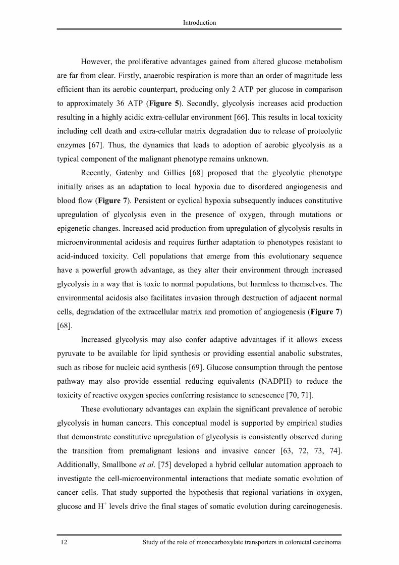
Recently, Gatenby and Gillies [68] proposed that the glycolytic phenotype
initially arises as an adaptation to local hypoxia due to disordered angiogenesis and
blood flow (Figure 7). Persistent or cyclical hypoxia subsequently induces constitutive
upregulation of glycolysis even in the presence of oxygen, through mutations or
epigenetic changes. Increased acid production from upregulation of glycolysis results in
microenvironmental acidosis and requires further adaptation to phenotypes resistant to
acid-induced toxicity. Cell populations that emerge from this evolutionary sequence
have a powerful growth advantage, as they alter their environment through increased
glycolysis in a way that is toxic to normal populations, but harmless to themselves. The
environmental acidosis also facilitates invasion through destruction of adjacent normal
cells, degradation of the extracellular matrix and promotion of angiogenesis (Figure 7)
[68].
Increased glycolysis may also confer adaptive advantages if it allows excess
pyruvate to be available for lipid synthesis or providing essential anabolic substrates,
such as ribose for nucleic acid synthesis [69]. Glucose consumption through the pentose
pathway may also provide essential reducing equivalents (NADPH) to reduce the
toxicity of reactive oxygen species conferring resistance to senescence [70, 71].
These evolutionary advantages can explain the significant prevalence of aerobic
glycolysis in human cancers. This conceptual model is supported by empirical studies
that demonstrate constitutive upregulation of glycolysis is consistently observed during
the transition from premalignant lesions and invasive cancer [63, 72, 73, 74].
Additionally, Smallbone et al. [75] developed a hybrid cellular automation approach to
investigate the cell-microenvironmental interactions that mediate somatic evolution of
cancer cells. That study supported the hypothesis that regional variations in oxygen,
glucose and H+ levels drive the final stages of somatic evolution during carcinogenesis.
Introduction
Study of the role of monocarboxylate transporters in colorectal carcinoma 13
They proposed that the phenotypic adaptations to the sequence of hypoxia-glycolysis-
acidosis are necessary to form an invasive cancer [75].
Figure 7. Model for cell–environment interactions during carcinogenesis, giving the stages of tumour growth and their associated physiological states. Normal epithelial (grey), hyperplastic (pink), hypoxic (blue), glycolytic (green) and motile (yellow) cells [Gatenby and Gillies, 2004].
These results suggest that tumour prevention strategies aimed at interrupting
glycolysis and the resulting cellular adaptations could be explored to delay or prevent
transition of in situ to invasive cancer. Furthermore, in advanced metastatic cancers,
understanding of the molecular and physiological causes and consequences of
upregulated glycolysis may lead to targeted therapies [76].
1.2.2 Molecular mechanisms
The increase in tumour glucose uptake is achieved by up-regulating the
expression of glycolytic enzymes and glucose transporters [77]. The molecular basis for
evolution of the glycolytic phenotype has been clarified by recent advances in

Introduction
14 Study of the role of monocarboxylate transporters in colorectal carcinoma
understanding the hypoxia-inducible factor, HIF-1, system [78]. Hypoxia and HIF-1
increase virtually all the enzymes in the glycolytic pathway, as well as the glucose
transporters 1 and 3 (GLUT1, GLUT3) [79], and other hypoxia-related genes such as
those involved in angiogenesis, iron metabolism and cell proliferation/survival (Figure
8) [80].
Figure 8. A summary of the HIF system demonstrating several factors which govern the level of HIF-1α and some of its protean effects on cell metabolism, survival, proliferation and their microenvironment [Gatenby and Gillies, 2007].
HIF-1 is a heterodimeric complex consisting of a hypoxically inducible subunit
HIF-1α and a constitutively expressed subunit HIF-1β [81]. Although HIF-1β is
constitutively expressed and its mRNA and protein are maintained at constant levels
regardless of oxygen availability [82], HIF-1α protein has a short half-life (t1/2 ~ 5 min)
and is highly regulated by oxygen [83]. The transcription and synthesis of HIF-1α are
constitutive and seem not to be affected by oxygen [81, 82, 84]. However, in normoxia,
the HIF-1α proteins are rapidly degraded, resulting in essentially no detectable HIF-1α
protein [81]. During hypoxia, HIF-1α becomes stabilized and translocates from the
cytoplasm to the nucleus, where it dimerizes with HIF-1β, and the HIF complex formed
becomes transcriptional active [82, 85]. The activated HIF complex then associates with
hypoxia response elements (HREs) in the regulatory regions of target genes and binds
the transcriptional coactivators to induce gene expression [86].

Introduction
Study of the role of monocarboxylate transporters in colorectal carcinoma 15
Furthermore, the glycolysis metabolic products, such as lactate and pyruvate,
have been reported to cause HIF-1α accumulation under normoxia and regulate
hypoxia-inducible gene expression, establishing a potential positive feedback loop
which may be critical to aerobic glycolysis [87]. However, HIF activity can also be
stabilized in the presence of oxygen by growth factors (Figure 8) that also participate in
carcinogenesis, including RAS, HSP 90, Cox 2, HER, and the AKT/mTOR pathway
[88]. Because of this, HIF can be characterized as being reversibly increased by
physiologic stress (e.g., hypoxia) or by hormonal growth factor stimulation or as being
constitutively active under normoxic conditions through heritable alterations, such as
activated oncogenes.
1.2.3 Consequences of increased glucose metabolism
One consequence of this dependence on glycolysis is the need for metabolically
active tumours to efflux the accumulating lactic acid to the tumour microenvironment to
prevent intracellular acidosis [89, 90]. Cellular acidosis has been shown to be a trigger
in the early phase of apoptosis and leads to activation of endonucleases inducing DNA
fragmentation. In addition, cellular pH is crucial for biological functions such as cell
proliferation [90], invasion [91], metastasis [92] and drug resistance [93]. The
observation that the intracellular pH of tumours is more alkaline than that of normal
tissues [94, 95], even in the presence of an acidic tumour microenvironment, suggests
the existence of an active mechanism for lactate/H+ efflux. Although several
transporters including Na+/H+ exchangers (NHE1), vacuolar adenosine triphosphatases,
anion exchangers, and lactate/H+ symporters (monocarboxylate transporters [MCTs])
are involved in pH homeostasis in mammalian cells [89], MCTs are thought to be most
important for pH regulation within rapidly metabolizing, highly glycolytic tumour cells
[96, 97].
1.3 Monocarboxylate Transporters (MCTs)
MCTs are members of the SLC16 gene family [98]. This family includes 14
proteins related to each other by sequence homology. The first four members, MCT1-

Introduction
16 Study of the role of monocarboxylate transporters in colorectal carcinoma
MCT4 have been experimentally demonstrated to transport aliphatic monocarboxylates,
a diverse group of compounds which includes many important metabolites such as
lactate, pyruvate and ketone bodies. This transport is controlled by hydrogen ion and
substrate gradients, thus facilitating transport of MCTs either in (influx) or out (efflux)
of the cell. Lactate efflux is especially important for cells and tissues which generate
large quantities of lactic acid as a result of glycolysis (e.g. red and white blood cells,
skeletal muscle, and most tumour cells). Lactate influx is of value to tissues where it is
being utilized (via pyruvate) as a respiratory fuel (brain, heart, red skeletal muscle), as a
gluconeogenic substrate in liver, or under fasting conditions, as a glycerol-neogenic
substrate in white adipose tissue [99]. Since lactate is a substrate for MCT1-MCT4
transporters, considerable attention has been given to the potential role of these
transporters in the handling of lactate in tumour cells.
MCTs have 12 transmembrane domains with the N- and C-termini located in the
cytoplasm [98, 100]. The transmembrane domains (TMDs) are highly conserved
between isoforms with the greatest sequence variations observed in the C-terminus and
the large intracellular loop between TMDs 6 and 7, which has a range of 29–105 amino
acid residues (Figure 9) [100]. This observed variability is common to transporters with
12 TMDs and it is thought that these sequence variations are related to substrate
specificity or regulation of transport activity [100, 101].
Figure 9. Proposed membrane topology of the MCT family. Adapted from Halestrap and Meredith, 2004.
Regulation of MCTs has been demonstrated to occur via transcriptional,
translational and post-transcriptional mechanisms [102-104]. These regulatory pathways

Introduction
Study of the role of monocarboxylate transporters in colorectal carcinoma 17
appear to be age- and tissue dependent, which further complicates the understanding of
these pathways [103, 104]. Some MCTs require an ancillary protein which can be
involved in cellular localization [105] or protein–protein interactions [106]; however,
the role of these accessory proteins in overall transporter function is not yet completely
understood [105].
1.3.1 MCT1
MCT1 is the most well-studied and functionally characterized member of the
MCT family, largely due to the fact that it is the only monocarboxylate transporter
expressed in human erythrocytes, and it also has the widest tissue distribution. MCT1 is
ubiquitously expressed but is especially prominent in heart and red muscle where it is
upregulated in response to increased work, suggesting an important role in lactic acid
oxidation [101, 107, 108, 109].
Transport kinetics have been thoroughly explored using lactate for this isoform
and have demonstrated that it functions as a proton-dependent cotransporter/exchanger
[110, 111]. Transport occurs by ordered sequential binding with association of a proton
followed by lactate binding. The complex is translocated across the membrane and the
lactate and proton are released sequentially. Since the transporter functions as an
exchanger, transport can occur bidirectionally; however, it is primarily responsible for
the uptake of substrates [101].
Relatively few studies have been conducted to assess the regulation of MCTs.
Studies have indicated that altered physiological conditions and the presence of
xenobiotics may alter the regulation of MCTs, in addition to altered expression at
different developmental stages (112–114). MCT1 expression undergoes transcriptional,
post-transcriptional and post-translational regulation and appears to be regulated in a
tissue-specific manner [102-104]. In colonic epithelium, exposure to butyrate resulted in
a concentration- and time dependent increase in MCT1 mRNA, protein expression and a
corresponding increase in butyrate transport [115]. These data suggest the possibility of
altered transcriptional regulation; however, the authors further demonstrated increased
transcript stability indicating additional post-transcriptional regulation mechanisms
[115]. High concentrations of lactate have also been demonstrated to increase MCT1

Introduction
18 Study of the role of monocarboxylate transporters in colorectal carcinoma
mRNA and protein levels in L6 cells [116]. In contrast, treatment with testosterone
resulted in increased skeletal muscle MCT1 protein expression and lactate transport in
the absence of mRNA changes suggesting the importance of post-transcriptional
regulation [103].
MCT1 is further regulated by its association with the cell surface glycoprotein
CD147 (Basigin), which has a single transmembrane domain with the C-terminus
located in the cytosol (Figure 10) [117, 118]. Topology studies suggest that one MCT1
molecule interacts with a single CD147 molecule with subsequent dimerization with
another MCT1/CD147 pair (Figure 11) [111]. The initial association of CD147 and
MCT1 is required for the translocation of MCT1 to the plasma membrane [117].
Furthermore, more recent studies indicate that CD147 is also required for catalytic
activity of MCT1 [119].
1.3.2 MCT2
MCT2 is a high-affinity transporter for monocarboxylates and its expression
under normal conditions is restricted to tissues such as the liver, kidney, and neurons
that take up lactate rather than release it. In humans, expression of MCT2 is more
restricted than MCT1, with the greatest expression observed in the testis [120]. In both
rodents and humans, MCT2 splice variants have been detected in a species and tissue-
Figure 10. The proposed topology of CD147, the ancillary protein that associates with MCT1 and MCT4. Adapted from Halestrap and Meredith, 2004.
Figure 11. Hypothetical model of the topology of CD147 and MCT1 in the plasma membrane [Wilson et al, 2002].

Introduction
Study of the role of monocarboxylate transporters in colorectal carcinoma 19
dependent manner suggesting that transcriptional and post-transcriptional regulation
pathways play an important role in the tissue specificity of this isoform [120-122].
Similar to MCT1, MCT2 requires an accessory protein for translocation to the plasma
membrane. However, MCT2 requires gp70 (EMBIGIN), not CD147 [105]. MCT2
possesses high affinity for lactate and most likely functions in the import of lactate into
cells.
1.3.3 MCT3
MCT3 is believed to have the most restricted distribution of any MCT with
expression in the basolateral membrane of the retinal pigment epithelium (RPE) and the
choroid plexus epithelium (CPE) in humans, rodents and chickens [123-125]. However,
recent studies demonstrated MCT3 expression in vascular smooth muscle cell lines
[126], human aorta [126] and human kidney [127], suggesting that MCT3 mRNA may
be more widely distributed than originally thought.
1.3.4 MCT4
In contrast to MCT1, MCT4 is predominantly expressed in highly glycolytic
cells such as white muscle and white blood cells suggesting that its physiological
function is lactate efflux [101, 128, 129]. MCT4 localization at the plasma membrane is
dependent on CD147 expression, which is consistent with results obtained for MCT1
[114]. The role of MCT4 in lactate efflux is further supported by its high expression in
the placenta where it is involved in the transfer of lactate into the maternal circulation
[98]. While there is a great degree of overlap in the substrate specificity of MCT1 and
MCT4, these two isoforms differ in their substrate affinities with MCT4 having lower
affinities for a range of monocarboxylates.
1.3.5 Regulation of MCTs in cancer
MCT1 is the most widely expressed member of its family in normal tissues and
is expressed, also, in a variety of human cancer cell lines and in human cancers,

Introduction
20 Study of the role of monocarboxylate transporters in colorectal carcinoma
including breast, head and neck, and lung cancers [130] as well as neuroblastoma [113],
brain [131], alveolar sarcoma of soft tissues [132], colon [130, 133, 134], melanoma
[135], pancreatic [136], cervical [137], and gastric [138] cancers.
MCT4 is expressed under normal conditions in tissues that are glycolytic in
nature with resultant increased production of lactate [98]. It has low affinity for lactate,
thus making it an ideal transporter for export of lactate when this metabolite is
generated at high levels as occurs in glycolytic tissues. In spite of the fact that the
kinetic features of MCT4 are suitable for lactate export, the expression of this
transporter in cancer is controversial. Some studies have found no evidence of cancer-
associated expression of MCT4 [133, 139]. However, more recent studies have
demonstrated that MCT4 is upregulated in cancer and in tumour cells [114, 134, 137].
In accordance with these recent findings, the gene coding for MCT4 has been found to
be upregulated by hypoxia through a HIF-1α-dependent mechanism [140].
The expression of MCT2 is reduced in tumour cells [133, 134]. Since tumour
cells release lactate rather than import it, downregulation of MCT2 in tumour cells
correlates well with the metabolic phenotype of tumour cells. Thus, MCT2, which is
involved in the uptake of monocarboxylates into the cells in normal metabolism [141],
does not appear to have an important role in highly glycolytic cancer cells. Furthermore,
the gene coding for MCT2 lacks binding sites for HIF-1 [140].
Broad MCT distribution among human cancers opens promising therapeutic
perspectives for the development and clinical evaluation of pharmacological MCT
inhibitors. Furthermore, a number of reports have attributed to MCTs a role in lactate
efflux in tumour cells with active aerobic glycolysis [113, 142, 143, 144, 145]. These
studies showed further evidence of lethal intracellular acidification upon MCT
inhibition in vitro. Thus, MCTs can constitute attractive targets for cancer therapy.
1.3.6 Inhibitors of MCTs
Since MCTs are transmembrane proteins exposed to the extracellular milieu,
they are prone to targeting by systemic application of small-molecule inhibitors. Several
chemicals are known to inhibit the function of MCTs (Table 1) [110]. Those inhibitors
fall into four broad categories: (1) bulky or aromatic monocarboxylates which act as

Introduction
Study of the role of monocarboxylate transporters in colorectal carcinoma 21
competitive inhibitors (e.g. phenyl-pyruvate and α-cyano-4-hydroxycinnamate (CHC));
(2) amphiphilic compounds with divergent structures (e.g. quercetin and phloretin); (3)
some 4,4’-substituted stilbene-2,2’-disulphonates (e.g. DIDS); and (4) miscellaneous
inhibitors including thiol reagents such as p-chloromercuribenzene sulphonate
(pCMBS) and amino reagents (e.g. pyridoxal phosphate and phenylglyoxal [98].
Table 1. Inhibitors of monocarboxylate transporters.
MCT Isoform Inhibitor References
MCT1 CHC pCMBS Phloretin Quercetin DIDS AR-C155858
[147] [105] [110] [110] [150] [149]
MCT2 CHC Phloretin
[147] [110]
MCT4 CHC pCMBS Phroretin Fluvastatin Atorvastatin Lovastatin
[147] [105] [110] [151] [151] [151]
Simvastatin [151]
1.3.6.1 α-cyano-4-hydroxycinnamic acid (ACCA; CHC)
Some "classic" inhibitors of monocarboxylate transporters are derivatives of
cinnamic acid, first identified by Halestrap and co-workers for their effect on isolated
mitochondrial pyruvate transport [146] and by Lehninger and co-workers on intact
Ehrich ascites tumour [147]. Others studies [142, 144] have indicated cinnamic acid
derivates to be competitive inhibitors of lactate transport in tumours, with α-cyano-4-
hydroxycinnamic acid (ACCA; CHC) (Figure 12) as one of the most potent inhibitors
of lactate transport, with a Ki of 0.5 mM. A study [148] evaluating its therapeutic
efficacy against malignant glioma indicates CHC to be an effective cytotoxic and
cytostatic agent both in vitro and in vivo, but at mM concentrations (≥ 10 mM) due to its
high ki against lactate. This study on glioma cells also indicated that CHC remains

22 Study of the role of
extracellular. Thus, the inhibitory effect of CHC was restricted to the glioma plasma
membrane monocarboxylate transport
inhibited by CHC, i.e., the mitochondrial pyruvate carrier (MPC) were not affected. A
recent in vivo study [130] demonstrated that the inhibitory effect of CHC is restricted to
tumour cells expressing MCT1
concentrations of CHC up to 125 mM
Figure 12.
1.3.6.2 Other inhibitors
The stilbene derivatives such
(DIDS) act as reversible
eventually becomes irreversible on prolonged incubation, reflecting covalent
modification of the transporter
thiol and amino reagents, especially the organomercurial thiol reagent p
chloromercuribenzene sulphonate
inhibition is not of MCT1 and MCT4
In contrast to other MCTs, lactate transport via MCT4 is inhibited by a range of statin
drugs which may play a role in cytotoxicities obser
1.3.6.3 New MCT1 specific inhibitors
A group of small molecular weight compounds, synthesized by AstraZeneca,
were recently identified as potent and selective inhibitors of MCT1 in activated human
T cells. These compounds produced specific inhibition of human T lymphocyte
Introduction
Study of the role of monocarboxylate transporters in colorectal carcinoma
extracellular. Thus, the inhibitory effect of CHC was restricted to the glioma plasma
e monocarboxylate transport. Intracellular transporters known to be strongly
inhibited by CHC, i.e., the mitochondrial pyruvate carrier (MPC) were not affected. A
] demonstrated that the inhibitory effect of CHC is restricted to
r cells expressing MCT1 at the plasma membrane. That study also showed that
concentrations of CHC up to 125 mM were not toxic in mice (animal models).
. Structure of α-cyano-4-hydroxy cinnamic acid (CHC).
inhibitors
The stilbene derivatives such as 4,4’-diisothiocyanostilbene-2,2
inhibitors of MCT1 in erythrocytes. Inhibition by DIDS
eventually becomes irreversible on prolonged incubation, reflecting covalent
modification of the transporter [150]. In addition, MCT1 and MCT4 are
reagents, especially the organomercurial thiol reagent p
chloromercuribenzene sulphonate (pCMBS). Interestingly it has been shown that this
and MCT4 directly, but of the ancillary protein
In contrast to other MCTs, lactate transport via MCT4 is inhibited by a range of statin
drugs which may play a role in cytotoxicities observed with statin administration [
New MCT1 specific inhibitors
small molecular weight compounds, synthesized by AstraZeneca,
were recently identified as potent and selective inhibitors of MCT1 in activated human
T cells. These compounds produced specific inhibition of human T lymphocyte
monocarboxylate transporters in colorectal carcinoma
extracellular. Thus, the inhibitory effect of CHC was restricted to the glioma plasma
. Intracellular transporters known to be strongly
inhibited by CHC, i.e., the mitochondrial pyruvate carrier (MPC) were not affected. A
] demonstrated that the inhibitory effect of CHC is restricted to
plasma membrane. That study also showed that
mice (animal models).
2,2’-disulphonate
Inhibition by DIDS
eventually becomes irreversible on prolonged incubation, reflecting covalent
and MCT4 are inhibited by
reagents, especially the organomercurial thiol reagent p-
(pCMBS). Interestingly it has been shown that this
rotein CD147 [105].
In contrast to other MCTs, lactate transport via MCT4 is inhibited by a range of statin
ved with statin administration [151].
small molecular weight compounds, synthesized by AstraZeneca,
were recently identified as potent and selective inhibitors of MCT1 in activated human
T cells. These compounds produced specific inhibition of human T lymphocyte

Introduction
Study of the role of monocarboxylate transporters in colorectal carcinoma 23
proliferation in vitro with nanomolar potency [149]. This study also demonstrated that
the effects of MCT1 inhibitors in activated T cells, which rely on aerobic glycolysis for
energy, seem to be mediated by the blockade in lactate efflux rather than by effects on
pH, by transport of alternative substrates or via CD147 [149].
1.4 MCT1 and MCT4 as potential targets for CRC therapy
CRCs, in common with many other malignancies, have enhanced glucose
utilization and glycolytic metabolism [152-154], producing high amounts of lactic acid,
which is effluxed to the tumour microenvironment via lactate transporters, namely
monocarboxylate transporters (MCTs).
There are evidences for the upregulation of MCTs in colorectal carcinomas [130,
133, 134]. Koukourakis et al. [133] reported an increase in MCT1 expression in tumour
cells, which is supported by the metabolic alterations induced by anaerobic glycolysis.
That study also assessed the expression of MCT2 and MCT4, finding a strong
cytoplasmic expression of MCT2 in cancer cells, but a weak expression of MCT4 in the
tumour environment. Pinheiro et al. [134] reported an increase in expression of MCT1,
MCT2 and MCT4 in tumour cells when comparing with the adjacent normal epithelium.
In that study, the authors also found a significant gain of membrane expression for
MCT1 and MCT4 and loss of plasma membrane expression for MCT2 in tumour cells.
Taking into consideration the lactate affinities to the different MCT isoforms, MCT1
and MCT4 expression in the membrane is in agreement with the metabolic alterations
observed in colorectal cancer cells. That is, MCT1 and MCT4 need to be at the plasma
membrane to export the accumulating acids in highly glycolytic tumour cells [134].
Those findings point to MCT1 and MCT4 as playing an important role in highly
glycolytic CRC cells. Thus, targeting MCT1 and MCT4 activity may debilitate such
tumours via disruption of aerobic glycolysis and promoting apoptosis, suggesting that
MCT inhibitors may be particularly effective against CRC. Moreover, inhibiting MCTs
would not only induce apoptosis due to cellular acidosis, but would also lead to
reduction in tumour angiogenesis [155], invasion [91], and metastasis [92].

Introduction
24 Study of the role of monocarboxylate transporters in colorectal carcinoma
1.5 Aims
The first purpose of this work was to explore the role of MCTs in CRC, by
evaluating the effects of MCT inhibitors in human colon carcinoma cell viability,
migration, lactate efflux, and MCT expression. The second purpose was to investigate
the effects of combination of CHC (MCT inhibitor) with 5-FU (conventional
chemotherapy) in human colon carcinoma cell viability and migration.
Thus, the specific aims were:
– Characterization of MCT1, MCT4 and CD147 expression in colorectal
carcinoma cell lines by Western blotting and immunocytochemistry.
– Evaluate the effect of MCT inhibitors on:
a) Cell viability by MTT assay and/or trypan blue assay;
b) Cell migration by wound-healing assay;
c) MCT activity;
d) MCT expression by Western blotting and immunocytochemistry.
– Investigate the effect of the combined treatment with CHC and 5-FU on:
a) Cell viability by trypan blue assay;
b) Cell migration by wound-healing assay.

Material and methods
Study of the role of monocarboxylate transporters in colorectal carcinoma 25
2 Materials and methods
2.1 Cell lines
The human colon carcinoma-derived cell lines HCT-15 and Co-115 were kindly
provided by Dr. Raquel Seruca, IPATIMUP, Oporto.
2.2 Chemicals
5-FU, a commonly used anti-cancer drug for the treatment of colorectal
carcinoma, and CHC, a competitive inhibitor of lactate/pyruvate transporters, were
purchased from Sigma-Aldrich (St. Louis, MO, USA). AR-C155858, a specific
monocarboxylate transporter-1 (MCT-1) inhibitor, was synthesized and gently provided
by AstraZeneca (R&D Charnwood, UK).
5-FU, CHC and AR-C155858 were dissolved in 100% dimethylsulfoxide
(DMSO) at the concentrations of 2 M, 5 M and 10 mM, respectively, and further diluted
in RPMI 1640 medium (Gibco, Invitrogen, USA) to the desired final concentrations.
The pH of the CHC solutions were adjusted to 7.4 with NaOH and all solutions were
sterilized by filtration (0.20 µm syringe filter units, Starstedt, Germany) before use.
Control cells were incubated with vehicle (DMSO). The final concentration of DMSO
was maintained at 0.5%.
2.3 Cell culture conditions
HCT-15 and Co-115 colon carcinoma cells were cultured in RPMI 1640 medium
(Gibco) supplemented with 10% (v/v) fetal bovine serum (FBS, Gibco, Invitrogen,
USA) and 1% (v/v) penicillin-streptomycin solution (Invitrogen, USA) and incubated at
37 ºC in a humidified atmosphere containing 5% CO2. Cells were subcultured every
three days and maintained in a log-phase growth.

Materials and methods
26 Study of the role of monocarboxylate transporters in colorectal carcinoma
2.4 Cell viability assays
Cell viability was measured with both MTT (3-(4,5-dimethylthiazol-2-yl)-2,5-
diphenyl tetrazolium bromide) dye reduction [Cell Proliferation Kit I (MTT), Roche,
Mannheim, Germany] and Trypan Blue dye exclusion assays.
2.4.1 MTT dye reduction assay
This assay is based on the cleavage of the yellow tetrazolium salt MTT to purple
formazan crystals by metabolic active cells. This cellular reduction involves the
pyridine nucleotide cofactors NADH and NADPH.
HCT-15 and Co-115 cells were seeded in 96-well plates (6×10³/100 µl/well) and
incubated at 37 ºC in a 5% CO2 atmosphere for 24 hours. To examine the effects of the
treatment with 5-FU, CHC or AR-C155858, the spent media were removed and 0.01-10
mM 5-FU, 5-25 mM CHC, or 1-10 µM AR-C155858 were added. Controls were
performed with DMSO alone. After 24 hours of incubation, 10 µl MTT labelling
reagent (final concentration 0.5 mg/ml) was added to each well and microplates were
incubated for 4 hours at 37ºC. Subsequently, 100 µl of solubilisation solution (10% SDS
in 0.01 M HCl) were added to each well for solubilisation of the purple formazan
crystals and the mixture incubated at 37 ºC overnight. Spectrophotometrical
absorbances were measured using a microplate reader (Model 450, Bio-Rad). The
wavelength to measure absorbance of the formazan product and the reference
wavelength were 570 nm and 750 nm, respectively. To determine cell viability, percent
viability was calculated as (OD experiment/OD control) × 100 (%). Results are
presented as mean ± SD of three independent experiments.
2.4.2 Trypan Blue dye exclusion assay
Trypan Blue is a dye which cannot enter healthy cells, but can permeate
compromised membranes of dying cells. Dye exclusion was measured with 0.4% trypan
blue in 0.85% saline (Invitrogen).

Material and methods
Study of the role of monocarboxylate transporters in colorectal carcinoma 27
HCT-15 and Co-115 cells were seeded in 24-well plates (2.5×10⁴/500 µl/well)
and incubated at 37 ºC in a 5% CO2 atmosphere for 24 hours. Spent media were
removed and to examine the effects of the treatment with CHC, cells were treated with
5-25 mM CHC for 24 hours. To determine the effect of CHC exposure time, HCT-15
cells were treated with 10 mM CHC for 6-48 hours. To address the effects of 5-FU and
CHC co-treatment, cells were treated with 1 mM 5-FU (IC50) and increasing
concentrations of CHC (5-25 mM) for 24 hours. Controls were performed with DMSO
alone. The supernatant from each well was recovered and 75 µl trypsin (0.05% trypsin,
0.53 mM ethylenediamine tetra-acetic acid·4Na, Invitrogen) was added to each well and
incubated at 37ºC for 10-20 minutes. Subsequently, 150 µl of fresh medium was added
to quench the trypsin activity, and cells were recovered and pooled with the culture
supernatant. Aliquots of the cell suspensions were diluted 1:1 in trypan blue solution.
Clear and blue cells were counted with a hemocytometer under a bright-field
microscope, and the viable fraction was estimated by dividing the number of clear cells
by the total number of cells. Results are presented as mean ± SD of three independent
experiments.
2.5 Western blotting
The characterization of MCT1, MCT4 and CD147 protein expression in
untreated HCT-15 and Co-115 cells and HCT-15 CHC treated cells were evaluated by
western blotting.
HCT-15 and Co-115 cells were cultured in 6-well plates at a density of 3 × 10⁵
cells per well and incubated at 37 ºC in a 5% CO2 atmosphere for 48 hours. To evaluate
the effect of CHC on MCT1, MCT4 and CD147 protein expressions, HCT-15 cells were
treated with 10 mM CHC and incubated for further 24 hours. Controls were performed
with DMSO alone. Cells were washed with PBS and quickly scraped from the well with
50 µl lysing buffer (150 mM NaCl, 50 mM Tris-HCl, pH 7.5, 0.1 mM EDTA, 1%
Triton X-100, 1% Nonidet P-40), supplemented with protease inhibitor cocktail (Roche,
Mannheim, Germany), and homogenized 10 minutes on ice. Cells were then centrifuged
at 3000 rpm, for 15 minutes at 4ºC, to remove cell debris. Supernatants were collected
and protein concentrations were quantified using a Bio-Rad DC protein assay (Bio-Rad

Materials and methods
28 Study of the role of monocarboxylate transporters in colorectal carcinoma
Laboratories, Inc., Hercules, CA, USA). BSA was used as a protein standard. Twenty
micrograms of total protein from each cell lysate were separated on a 10% (w/v)
polyacrylamide gel and electrotransferred to nitrocellulose membranes (Amersham
Pharmacia Biotech, Piscataway, NJ, USA). Membranes were blocked with 0.1%
Tween-20, 5% (w/v) non-fat dry milk in TBS for 1 hour at room temperature.
Membranes were then incubated overnight at 4ºC with primary polyclonal antibodies
for MCT1 (AB3538P, Chemicon International, Temecula, CA, USA), MCT4
(AB3316P, Chemicon International), and CD147 (18-7344, Zymed, Invitrogen,
Carsbad, CA, USA) diluted 1:200 for MCT1 and MCT4, and 1:750 for CD147, in TBS,
0.1% Tween-20 and 1% (w/v) non-fat dry milk. After washing, membranes were
incubated 1 hour at room temperature with secondary antibody conjugated with IgG
horseradish peroxidise (Santa Cruz Biotechnology, Santa Cruz, CA, USA), diluted
1:10 000 in TBS, 0.1% Tween-20 and 1% (w/v) non-fat dry milk. Subsequently,
immunoreactive bands were visualized by chemiluminescence (Supersignal West Femto
kit, Pierce, Rockford, IL, USA). Band area intensity was quantified using ImageJ/Java,
a free software downloaded from http://rsb.info.nih.gov/ij/. ß-actin was used as loading
control.
2.6 Immunocytochemistry
Characterization of MCT1 and MCT4 protein expression in untreated HCT-15
and Co-115 cells and HCT-15 CHC treated cells were evaluated by
immunocytochemistry.
HCT-15 and Co-115 cells were grown on coverslips at 30% confluence. To
evaluate the effect of CHC in MCT1 and MCT4 protein expression, HCT-15 cells were
treated with 10 mM CHC for 24 hours at 37 ºC. Controls were performed with DMSO
alone. Untreated and treated cells grown on coverslips were fixed with PBS containing
4% (w/v) paraformaldehyde for 15 minutes at room temperature and then permeabilized
in PBS containing 0.05% Triton X-100 for 4 minutes. Cells were incubated with 0.3%
hydrogen peroxide in methanol for 30 minutes, to inactivate endogenous peroxidases,
and washed in PBS. Cells were then incubated for 20 minutes with Normal Horse
Serum (Vector, Burlingame, CA, USA), to block non-specific binding, and incubated

Material and methods
Study of the role of monocarboxylate transporters in colorectal carcinoma 29
overnight at room temperature with primary polyclonal antibodies for MCT1
(AB3538P, Chemicon International) and MCT4 (AB3316P, Chemicon International),
diluted 1:200. Cells were then sequentially washed in PBS and incubated with
biotinylated universal secondary antibody (Vector) for 30 minutes, R.T.U. Vectastain
ABC elite reagent (Vector), for 45 minutes at 37 ºC and developed with 3,3’- Diamino-
benzidine (DAB+ Substrate System, DakoCytomation, Carpinteria, CA, USA) for 10
minutes. Cells were counterstained with haematoxylin for 5 seconds and coverslips
were permanently mounted on slides using an aqueous solution (Biomeda corp., Foster
City, CA, USA). Images were acquired in an Olympus BX 61 microscope (Tokyo,
Japan).
2.7 Lactate determination
An enzymatic colorimetric assay (Lactate, LO-POD. Enzymatic colorimetric,
Spinreact, S.A.U., St. Esteve de Bas, Spain) was used for lactate determination in CHC
treated and untreated cell culture media.
HCT-15 cells were seeded in 24-well plates (2.5×10⁴/500 µl/well) and incubated
at 37 ºC in a 5% CO2 atmosphere for 24 hours. To examine the effects of the treatment
with CHC, spent media was removed and cells were incubated with 10 mM CHC.
Controls were performed with DMSO alone. Supernatant was collected at 2, 6, and 24
hours and assayed using the enzymatic colorimetric assay. In 96-well plates, 2 µl of
each sample were mixed with 200 µl of working reagent and incubated for 10 min. at
room temperature (15-25ºC). Spectrophotometrical absorbances were measured at 490
nm using a microplate reader (Model 450, Bio-Rad). Results are presented as mean ±
SD of three independent experiments.
2.8 Wound-healing assay
In vitro wound healing assays were used to assess the effect of 5-FU and CHC,
alone and in combination, on HCT-15 cell migration. This method mimics cell
migration during wound healing in vivo.
Materials and methods
30 Study of the role of monocarboxylate transporters in colorectal carcinoma
HCT-15 cells were seeded in 12-well plates (4.5 × 10⁵/well) in RPMI containing
10% FBS and grown until 90% confluent. Cells were then starved for 24 hours, and two
linear wounds were scratched with a p200 pipette tip. Cells were then washed with PBS
and to examine the effect of the treatment with CHC alone, cells were treated with 5 -
15 mM CHC. To address the effects of 5-FU and CHC co-treatment, cells were treated
with the combination 1mM 5-FU and 10 mM CHC. Controls were performed with
DMSO alone. Wounds were photographed using a microscope coupled with a camera at
0 and 24 hours post-treatment. Wound regions were marked and photographed at the
same region each time. The images acquired for each sample were analyzed
quantitatively using the ImageJ/Java software (http://rsb.info.nih.gov/ij/). Wound sizes
were measured at five distinct sites from the microphotographs taken at 24 h and
expressed as a percentage of the value at 0 h. Results are presented as mean ± SD of
three independent experiments.
2.9 Statistical analysis
Data were expressed as mean ± standard deviation (SD). One-way ANOVA,
followed by Bonferroni post test was used to perform statistical analysis, using
GraphPad Prism 4.0 software, and P-values <0.05 were considered statistically
significant in all experiments.

Results
Study of the role of monocarboxylate transporters in colorectal carcinoma 31
3 Results
3.1 Assessment of MCT1, MCT4 and CD147 expression in colon
carcinoma cells
Western blotting and immunocytochemistry were performed to assess
MCT1/MCT4 expression in HCT-15 and Co-115 colon carcinoma cell lines. Total
protein extracts from HCT-15 and Co-115 colon carcinoma-derived cell lines were
profiled. Western blot analysis demonstrated that HCT-15 and Co-115 cells expressed
both MCT1 and MCT4 (Figure 13). Protein bands were detected around 50 kDa, which
is consistent with the molecular weight described for these two MCT isoforms [131].
Functional MCT transporter complexes require association with ancillary
proteins. It was reported that CD147 forms complexes with MCT1 and MCT4, which is
essential for MCT membrane expression and catalytic activity [117]. Thus, expression
of CD147 was also evaluated. Western analysis demonstrated that both HCT-15 and
Co-115 cells expressed CD147 (Figure 14). Both high glycosylated (HG) CD147 and
low glycosylated (LG) CD147 were detected in the blots.
HCT-15 and Co-115 cells were also analyzed for MCT1 and MCT4
immunocytochemical expression. MCT1 and MCT4 were present in both HCT-15 and
Co-115 cell lines (Figure 14). MCT1 and MCT4 were expressed in the cytoplasm of
both cell lines, however, a plasma membrane expression of MCT1 was only observed in
HCT-15 cells (Figure 14 A).

32 Study of the role of
Figure 13. Western blot analysis of MCT1, MCT4 and CD147 in HCTcarcinoma cell lines. ß-actin was used as internal loading control.
Figure 14. Immunocytochemical expression of MCT1MCT1 plasma membrane staining (A) was observed in HCT
MCT1
HCT-15
MCT4
A
C
Results
Study of the role of monocarboxylate transporters in colorectal carcinoma
Western blot analysis of MCT1, MCT4 and CD147 in HCT-15 and Coactin was used as internal loading control.
Immunocytochemical expression of MCT1 and MCT4 in HCT-15 and Co-115 colon carcinoma cells. MCT1 plasma membrane staining (A) was observed in HCT-15 cells.
Co-115
B
D
monocarboxylate transporters in colorectal carcinoma
15 and Co-115 colon
115 colon carcinoma cells.

Results
Study of the role of monocarboxylate transporters in colorectal carcinoma 33
3.2 Effect of CHC on the viability of colon carcinoma cells
CHC, a competitive inhibitor of MCTs, has an inhibitory effect in the viability of
some cancer cells [130, 144]. To evaluate the effect of CHC on the viability of HCT-15
and Co-115 colon carcinoma cells, both cell lines were treated with different
concentrations of CHC (5-25 mM) and cell viability was assessed by MTT assay.
CHC was able to decrease both HCT-15 and Co-115 cell viability in a dose-
dependent manner (Figure 15). When treated with 5-25 mM CHC, the viability of
HCT-15 cells decreased from 95.3% to 4.0% (Figure 15 A). In Co-115 cells, the
viability of cells for the same concentrations decreased from 93.1% to 34.0% (Figure
15 B). A significant decrease in the percentage of viable cells was observed for HCT-15
and Co-115 cells treated with 15 mM, 20 mM, and 25 mM CHC (p<0.05). The CHC
concentrations responsible for 50% growth inhibition (IC50) were 16.77 mM and 19.24
mM for HCT-15 and Co-115 cells, respectively.
MTT dye reduction assay was reported to give a fourfold higher level of
viability than trypan blue dye exclusion assay in MCT-target cells [141]. Thus, cell
viability of CHC-treated cells was further evaluated by trypan blue assay to compare
with MTT assay data. In general, trypan blue assays indicated lower cell viabilities
when compared with MTT assays (Figure 15 C and D). When treated with 5-25 mM
CHC, the viability of HCT-15 cells decreased from 78.0% to 4.6% (Figure 15 C). In
Co-115 cells, the viability of cells for 10-25 mM CHC decreased from 73.5% to 32.0%
(Figure 15 D). A significant decrease in HCT-15 and Co-115 cell viability could be
observed when treated with 10 mM to 25 mm CHC (p<0.05). The inhibitory effect of
CHC was higher in HCT-15 cells when compared with Co-115 cells, being the CHC
IC50 values 9.99 mM and 16.18 mM for HCT-15 and Co-115 cells, respectively.
The influence of CHC exposure time was also evaluated in HCT-15 cells. The
trypan blue assay was used since it indicated lower viabilities when compared with
MTT assay in CHC-treated cells. A significant decrease in cell viability was achieved
from 12 to 48 hours (p<0.05) of treatment with 10 mM CHC (Figure 16). The lowest
percentage in viable cells was obtained after 24 hours of exposure.

Results
34 Study of the role of monocarboxylate transporters in colorectal carcinoma
Figure 15. Effect of CHC on colon carcinoma cell viability. HCT-15 (A and C) and Co-115 (B and D) colon carcinoma cells were incubated with different CHC concentrations for 24 hours. Cell viability was determined by MTT assay (A and B) and trypan blue assay (C and D). Values are means ± SD of three independent experiments performed in triplicates. *p<0.05, compared to control.
Figure 16. Effect of CHC on colon carcinoma cell viability. HCT-15 colon carcinoma cells were incubated with 10 mM CHC up to 48 hours. Cell viability was determined by trypan blue assay. Values are means ± SD of one experiment performed in triplicate. *p<0.05, compared to 0 hours.
0
20
40
60
80
100
120
Control 5 10 15 20 25
Cell viability (% control)
CHC (mM)
MTT assay in HCT-15
0
20
40
60
80
100
120
Control 5 10 15 20 25
Cell viability (% control)
CHC (mM)
MTT assay in Co-115
0
20
40
60
80
100
120
140
Control 5 10 15 20 25
Cell viability (% control)
CHC (mM)
Trypan blue assay in HCT-15
0
20
40
60
80
100
120
140
Control 10 15 20 25
Cell viability (% control)
CHC (mM)
Trypan blue assay in Co-115
0
20
40
60
80
100
120
0 6 12 24 36 48
Cell viability (% untreated cells)
Time (h)
Trypan blue assay in HCT-15
*
*
*
*
* *
*
* * *
*
* * *
* *
* *
B
C
A
D

Results
Study of the role of monocarboxylate transporters in colorectal carcinoma 35
3.3 Effect of AR-C155858 on the viability of colon carcinoma cells
The effect of AR-C155858, a specific inhibitor of MCT1, on the viability of
HCT-15 and Co-115 was also tested. Twenty-four hours after exposure to AR-C155858,
cell viability was assessed by MTT assay. Incubations with 1 µM, 5 µM, and 10 µM
AR-C155858 did not have any effect on the viability of HCT-15 and Co-115 cells
(Figure 17). Analysis by light microscopy also revealed no changes in morphology and
cell number in AR-C155858 treated cells when compared with untreated cells (data not
shown), confirming the results obtained in MTT assay.
Figure 17. Effect of AR-C155858 on colon carcinoma cells viability. HCT-15 (A) and Co-115 (B) colon carcinoma cells were incubated with different AR-C155858 concentrations for 24 hours. Cell viability was determined by MTT assay. Values are means ± SD of three independent experiments performed in triplicates.
0
20
40
60
80
100
120
Control 1 5 10
Cell viability (%control)
AR-C155858 (µM)
MTT assay in HCT-15
0
20
40
60
80
100
120
Control 1 5 10
Cell viability (% control)
AR-C155858 (µM)
MTT assay in Co-115A B

Results
36 Study of the role of monocarboxylate transporters in colorectal carcinoma
3.4 Effect of CHC on HCT-15 cell migration
To evaluate the influence of CHC on the migration capacity of HCT-15 cells
(more sensitive to CHC) a wound-healing assay was performed. Cells were treated with
different concentrations of CHC (5-15 mM) for 24 hours and the wound size was
compared between untreated (control) and CHC treated cells. As shown in Figure 18
and 19, after treatment with 10 mM or 15 mM CHC (24 hours) the wound size was
47.6% ± 8.4% and 52.9% ± 5.2%, respectively, whereas in the untreated control, the
wound was 24% ± 6.6% of its original size. Thus, a significant increase in wound size
was observed for CHC treated cells (10, 15 mM) in comparison to control (p<0.05).
Figure 18. Migration of HCT-15 cells in response to 5-15 mM CHC was measured in a wound-healing assay. The width of the wound was measured at five distinct sites from the microphotographs taken at 24 h and expressed as percentage of the value at 0 h. Data are means ± SD of three independent experiments, each performed in triplicate. *p<0.05, compared to 0 mM CHC.
0
10
20
30
40
50
60
70
0 5 10 15
Wound size (%control T0)
CHC (mM)
* *
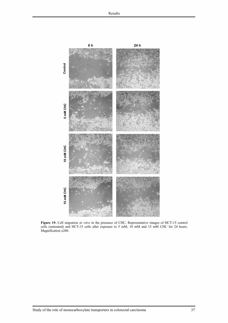
Study of the role of monocarboxylate transporters in colorectal carcinoma
Figure 19. Cell migration in vitro cells (untreated) and HCT-15 cells after exposure to 5 mM, 10 mM and 15 mM CHC for 24 hours. Magnification x200.
Control
5 mM CHC
10 mM CHC
15 mM CHC
Results
nocarboxylate transporters in colorectal carcinoma
in vitro in the presence of CHC. Representative images of HCT15 cells after exposure to 5 mM, 10 mM and 15 mM CHC for 24 hours.
0 h 24 h
nocarboxylate transporters in colorectal carcinoma 37
in the presence of CHC. Representative images of HCT-15 control 15 cells after exposure to 5 mM, 10 mM and 15 mM CHC for 24 hours.
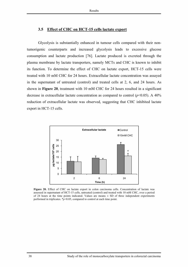
Results
38 Study of the role of monocarboxylate transporters in colorectal carcinoma
3.5 Effect of CHC on HCT-15 cells lactate export
Glycolysis is substantially enhanced in tumour cells compared with their non-
tumorigenic counterparts and increased glycolysis leads to excessive glucose
consumption and lactate production [76]. Lactate produced is excreted through the
plasma membrane by lactate transporters, namely MCTs and CHC is known to inhibit
its function. To determine the effect of CHC on lactate export, HCT-15 cells were
treated with 10 mM CHC for 24 hours. Extracellular lactate concentration was assayed
in the supernatant of untreated (control) and treated cells at 2, 6, and 24 hours. As
shown in Figure 20, treatment with 10 mM CHC for 24 hours resulted in a significant
decrease in extracellular lactate concentration as compared to control (p<0.05). A 40%
reduction of extracellular lactate was observed, suggesting that CHC inhibited lactate
export in HCT-15 cells.
Figure 20. Effect of CHC on lactate export in colon carcinoma cells. Concentration of lactate was assessed in supernatant of HCT-15 cells, untreated (control) and treated with 10 mM CHC, over a period of 24 hours at the time points indicated. Values are means ± SD of three independent experiments performed in triplicates. *p<0.05, compared to control at each time point.
0
5
10
15
20
25
30
2 6 24
µg lactate/104cells
Time (h)
Extracellular lactate Control
10mM CHC
*

Study of the role of monocarboxylate transporters in colorectal carcinoma
3.6 Effect of CHC on MCT1, MCT4 and CD147 protein expression
levels in HCT
To check if the effects of CHC reflect direct
protein expression levels, expression of these
immunocytochemistry in HCT
hours. As shown in Figure 21
MCT1 but not both MCT4 and CD147. The decrease of MCT1
treated cells was of 40% compared with untreated (control) cells.
The effect of CHC on MCT1 and MCT4 protein expressions was also analyzed
by immunocytochemistry.
similar in untreated and CHC treated cells (
western blot the decrease of MCT1 expression in CHC treated cells was not evident.
Furthermore, the decrease of MCT1 plasma membrane expression was not obse
either.
Figure 21. Effect of treatment with 10 mM CHC (HCT-15 cells, using western blot.
Results
nocarboxylate transporters in colorectal carcinoma
Effect of CHC on MCT1, MCT4 and CD147 protein expression
evels in HCT-15 cells
To check if the effects of CHC reflect directly on MCT1, MCT4 and CD147
protein expression levels, expression of these proteins was assessed by western blot and
immunocytochemistry in HCT-15 cells untreated and treated with 10 mM CHC for 24
Figure 21, by western blot, CHC decreased the expression of
MCT1 but not both MCT4 and CD147. The decrease of MCT1 expression in CHC
treated cells was of 40% compared with untreated (control) cells.
The effect of CHC on MCT1 and MCT4 protein expressions was also analyzed
by immunocytochemistry. MCT1 and MCT4 immunoexpressions were
similar in untreated and CHC treated cells (Figure 22). In contrast to the results from
western blot the decrease of MCT1 expression in CHC treated cells was not evident.
Furthermore, the decrease of MCT1 plasma membrane expression was not obse
of treatment with 10 mM CHC (24 hours) on MCT1, MCT4 and CD147 expression15 cells, using western blot. ß-actin was used as loading control.
nocarboxylate transporters in colorectal carcinoma 39
Effect of CHC on MCT1, MCT4 and CD147 protein expression
on MCT1, MCT4 and CD147
proteins was assessed by western blot and
15 cells untreated and treated with 10 mM CHC for 24
, by western blot, CHC decreased the expression of
expression in CHC
The effect of CHC on MCT1 and MCT4 protein expressions was also analyzed
s were apparently
). In contrast to the results from
western blot the decrease of MCT1 expression in CHC treated cells was not evident.
Furthermore, the decrease of MCT1 plasma membrane expression was not observed
on MCT1, MCT4 and CD147 expressions in

40 Study of the role of
Figure 22. Immunicytochemical expression of MCT1 and MCT4 in the presence of CHC.of HCT-15 control cells (untreated) and HCT
3.7 Effect of CHC and 5
colon carcinoma
To address the question whether CHC may potenciate the cytotoxic effect of 5
FU in colon carcinoma cells, the effect of combined treatment with CHC and 5
the viability of HCT-15 and Co
values were determined for HCT
effect of 5-FU and CHC co-
HCT-15 (control)
MCT1
MCT4
Results
Study of the role of monocarboxylate transporters in colorectal carcinoma
Immunicytochemical expression of MCT1 and MCT4 in the presence of CHC. Representative images 15 control cells (untreated) and HCT-15 cells after exposure to 10 mM CHC for 24 hours.
CHC and 5-FU combined treatment on the viability
colon carcinoma cells
To address the question whether CHC may potenciate the cytotoxic effect of 5
FU in colon carcinoma cells, the effect of combined treatment with CHC and 5
15 and Co-115 cells was assessed. Thus, as a first step, IC50
values were determined for HCT-15 and Co-115 cells treated with 5-FU and then
-treatment on viability of both cell lines was evaluated.
(control) HCT-15 (10 mM CHC)
monocarboxylate transporters in colorectal carcinoma
Representative images 15 cells after exposure to 10 mM CHC for 24 hours.
on the viability of
To address the question whether CHC may potenciate the cytotoxic effect of 5-
FU in colon carcinoma cells, the effect of combined treatment with CHC and 5-FU on
d. Thus, as a first step, IC50
FU and then, the
treatment on viability of both cell lines was evaluated.
(10 mM CHC)

Results
Study of the role of monocarboxylate transporters in colorectal carcinoma 41
3.7.1 Effect of 5-FU on the viability of HCT-15 and Co-115 cells
To assess the cytotoxic effect of 5-FU, various concentrations of 5-FU were
added to the media of HCT-15 and Co-115 colon carcinoma cells. Twenty-four hours
after exposure to 5-FU, cell viability was assessed by MTT assay. 5-FU decreased
HCT-15 and Co-115 cell viability in a dose-dependent manner (Figure 23). When
treated with 0.01-10 mM 5-FU, the viability of HCT-15 cells decreased from 79.0% to
35.0% (Figure 23 A). HCT-15 cells treated with 0.01 to 10 mM 5-FU presented a
significant decrease in the percentage of viable cells relative to control (p<0.05). In Co-
115 cells, the viability of cells for the same concentrations of 5-FU decreased from
82.1% to 34.4% (Figure 23 B). Treatment of Co-115 cells with 0.1 to 10 mM 5-FU
induced a significant decrease in cell viability (p<0.05). The cytotoxic effect of 5-FU
was similar in both cell lines, being the 5-FU IC50 values 1.6 mM and 1.5 mM for
HCT-15 and Co-115 cells, respectively.
Figure 23. Effect of 5-FU on colon carcinoma cell viability. HCT-15 (A) and Co-115 (B) colon carcinoma cells were incubated with different 5-FU concentrations (0.01-10 mM) for 24 hours. Cell viability was determined by MTT assay. Values are means ± SD of three independent experiments performed in triplicate. *p<0.05, compared to control.
0
20
40
60
80
100
120
Control 0.01 0.1 1 10
Cell viability (% control)
5-FU (mM)
MTT assay in HCT-15
0
20
40
60
80
100
120
Control 0.01 0.1 1 10
Cell viability (% control)
5-FU (mM)
MTT assay in Co-115
*
A
* *
*
B
*
*
*

Results
42 Study of the role of monocarboxylate transporters in colorectal carcinoma
3.7.2 Effect of 5-FU combined with CHC on the viability of HCT-15 and
Co-115 cells
To evaluate the effect of the combined treatment with 5-FU and CHC on cell
viability, HCT-15 and Co-115 cells were treated with 1 mM 5-FU (IC50) and different
concentrations of CHC. Twenty-four hours after treatment, cell viability was assessed
by trypan blue assay.
CHC increased the cytotoxic effect of 5-FU in HCT-15 and Co-115 cell viability
in a dose-dependent manner (Figure 24). Treatment of HCT-15 cells with 1 mM 5-FU
combined with CHC decreased significantly the percentage of cell viability compared
with 1 mM 5-FU alone for all concentrations tested (p<0.05). Co-115 cells treated with
1 mM 5-FU combined with CHC presented a significant decrease in the percentage of
viable cells relative to 1 mM 5-FU alone only for 25 mM CHC (p<0.05). Thus, the
cytotoxic effect of the combined treatment (5-FU plus CHC) was more evident for
HCT-15 than for Co-115 cells.
Figure 24. Effect of 5-FU + CHC on colon carcinoma cell viability. HCT-15 (A) and Co-115 (B) colon carcinoma cells were incubated with 1 mM 5-FU and different CHC concentrations for 24 hours. Cell viability was determined by Trypan blue assay. Values are means ± SD of three independent experiments performed in triplicates. *p<0.05, compared to 1 mM 5-FU (0 mM CHC).
0
20
40
60
80
100
120
Cell viability (% control)
Trypan blue assay in HCT-15
0
20
40
60
80
100
120
Cell viability (% control)
Trypan blue assay in Co-115
CHC (mM)
5-FU (mM)
0
0
0
0
CHC (mM)
5-FU (mM)
0
1
0
1
10
1
15
1
20
1
25
1
5
1
10
1
15
1
20
1
A B
* * *
*
*

Results
Study of the role of monocarboxylate transporters in colorectal carcinoma 43
3.8 Effect of CHC and 5-FU combined treatment on HCT-15 cell
migration
In order to assess the effect of combined treatment of 5-FU and CHC on cell
migration, a wound-healing assay was performed. HCT-15 cells were treated with 1
mM 5-FU and 10 mM CHC, alone and in combination, for 24 hours. As shown in
Figure 25 and 26, after incubation with either 10 mM CHC or 1 mM 5-FU, a significant
increase in wound size was observed in comparison to control (p<0.05). After treatment
with 10 mM CHC or 1 mM 5-FU, the wound size was 47.6% ± 8.4% and 57.6% ±
2.4%, respectively, whereas in the untreated control, the wound was 24% ± 6.6% of its
original size. However, with the combined treatment with 5-FU and CHC, the wound
size did not increase significantly when compared with either 5-FU or CHC alone.
Figure 25. Migration of HCT-15 cells in response to 10 mM CHC and/or 1 mM 5-FU was measured in a wound-healing assay. The width of the wound was measured at five distinct sites from the microphotographs taken at 24 h and expressed as a percentage of the value at 0 h. Data are the means ± SD of three independent experiments each performed in triplicate. *p<0.05, compared to control (no treatment).
0
20
40
60
80
100
Wound size (% Control T0)
CHC (mM)
5-FU (mM)
0
0
10
0
0
1
10
1
* *
*

44 Study of the role of
Figure 26. Cell migration in vitro 15 control cells (untreated) and HCTin combination, for 24 hours. Magnification x200.
Control
10 mM CHC
1 mM 5-FU
1 mM 5-FU + 10 mM CHC
Results
Study of the role of monocarboxylate transporters in colorectal carcinoma
in vitro in the presence of 5-FU and/or CHC. Representative images of HCT15 control cells (untreated) and HCT-15 cells after exposure to 10 mM CHC and 1 mM 5in combination, for 24 hours. Magnification x200.
0 h 24 h
monocarboxylate transporters in colorectal carcinoma
FU and/or CHC. Representative images of HCT-exposure to 10 mM CHC and 1 mM 5-FU, alone and

Discussion
Study of the role of monocarboxylate transporters in colorectal carcinoma 45
4 Discussion
Similar to most malignant tumours, colorectal carcinoma (CRC) is highly
glycolytic, producing large amounts of lactic acid, which is effluxed to the tumour
microenvironment via lactate transporters, namely monocarboxylate transporters
(MCTs). Thus, inhibition of lactate efflux in these tumours to cause internal
acidification presents an attractive therapeutic approach. The consequent increase in
extracellular pH will also lead to reduction in tumour angiogenesis [155], invasion [91],
and metastasis [92]. Some findings point to MCT1 and MCT4 as playing important
roles in highly glycolytic CRC cells [133, 134]. Thus, MCT inhibitors may be
particularly effective against those tumours. CHC, a low affinity competitive inhibitor
of MCTs, has been reported to have an inhibitory effect in cell viability of highly
glycolytic tumours [130, 148]. AR-C155858 was recently identified as a potent and
selective inhibitor of MCT1 in activated T cells [149]. In this study, AR-C155858
showed a potent inhibitory effect on T lymphocyte proliferation in vitro and in vivo,
resulting in significant suppression of alloimmune responses in immune-mediated
animal models of disease [149]. However, there are no reports on the effect of this
compound in cancer. AR-C155858 was provided by AstraZeneca for the present study.
In this work, the effects of CHC and AR-C155858 were studied in two different
human colon carcinoma cell lines. First, MCT1, MCT4 and CD147 expressions were
assessed in both colon carcinoma cell lines and after demonstrating their expression in
both HCT-15 and Co-115 cells, the effect of CHC and AR-C155858 on cell viability
was assessed. Both MTT and trypan blue assays indicated that CHC decreased the
viability of both HCT-15 and Co-115 cell lines in a dose-dependent manner, although a
higher level of viability was indicated by MTT assay. Mathupala et al [145] also
observed a disparity between the two cell viability assays in MCT-targeted cells. They
then assumed that the higher level of viability obtained by MTT may indicate an
increased level of NADH in MCT-targeted cells, resulting from changes in the
pyruvate-lactate equilibrium that follow accumulating intracellular lactate levels. From
these findings and considering the results of Mathupala et al [145], it was decided to use
trypan blue assay in subsequent CHC experiments. The inhibitory effect of CHC was
higher in HCT-15 cells than in Co-115 cells, as indicated by the trypan blue assay. It
Discussion
46 Study of the role of monocarboxylate transporters in colorectal carcinoma
was reported that the antitumour efficacy of CHC, known to inhibit MCT1 with
approximately 10-fold selectivity compared to other MCTs, is restricted to tumour cells
expressing MCT1 at the plasma membrane [129, 130]. In this study, we observed that
MCT1 was expressed in the cytoplasm of both cell lines but only in the plasma
membrane of HCT-15 cells. Thus, the presence of MCT1 in HCT-15 plasma membrane
could probably explain the higher sensitivity of these cells to CHC. We also observed
that the migration of HCT-15 cells was inhibited by CHC, reinforcing the sensitivity of
these cells to CHC, this time on the migration capacity of cells. AR-C155858 had no
impact on both HCT-15 and Co-115 cell viability, as indicated by MTT assay, and
morphological analysis. However, trypan blue assay demonstrated be more suitable to
assess MCT-target cell viability. It would then be important to further evaluate the
effect of this drug on viability of HCT-15 and Co-115 cells with trypan blue assay, as
well as on proliferation.
Another purpose of the present study was to assess if the inhibitory effect of
CHC in HCT-15 cells viability was mediated by MCTs. Thus, the effects of CHC on
both lactate export and MCT1, MCT4 and CD147 (MCT1/4 chaperone) protein
expression levels were assessed. In the present study, CHC decreased extracellular
lactate in HCT-15 cells after 24 hours of treatment, suggesting that CHC inhibited
lactate efflux in these cells. Our results are similar to the study by Colen et al [148].
According to that study, CHC inhibited the glioma cells capacity to efflux lactate. In the
present work, CHC also decreased the expression of MCT1, as indicated by western
blot analysis. Our results suggest that CHC has a direct effect both in function and
expression of MCT1. However, only one experiment was done in western blot analysis.
Thus, the effect of CHC on MCT protein expression levels will have to be confirmed in
a future work. A number of reports have attributed to MCTs a role in lactate efflux in
tumour cells [113, 142, 143, 144, 145], and those studies showed further evidence of
lethal intracellular acidification upon MCT inhibition. Thus, we can deduce that this
inhibition of both lactate efflux and MCT1 expression by CHC may induce an increase
in intracellular pH promoting a decrease of HCT-15 cell viability. In contrast, by
immunocytochemistry, the decrease of MCT1 expression was not evident, neither in the
plasma membrane.
Although 5-FU is currently the first-line agent for colorectal cancer, the response
rate to this chemotherapeutic agent in adjuvant treatment is less than 15%. This lack of

Discussion
Study of the role of monocarboxylate transporters in colorectal carcinoma 47
acceptable response has stimulated intensive efforts to develop new cancer drugs and
new combined regimens in colorectal cancer treatment [46]. Recently, a combined
CHC/radiation therapy in glioma cells showed an enhanced level of radiation-induced
cell kill [148]. Other recent study showed that sulindac sulfide, an active metabolite of
sulindac, potentiated the inhibitory effect of 5-FU on colorectal cancer cell survival,
parallel to the induction of apoptosis [156]. On the other hand, Lim et al [157]
demonstrated that celecoxib, a cyclooxygenase-2 (COX-2) specific inhibitor, attenuated
cytotoxic effect of 5-FU in colon cancer cells, parallel to prevention of apoptosis. The
inhibitory properties of CHC, observed in the present work, suggest that CHC may
potentiate the cytotoxic effect of 5-FU in colon cancer cells. The final purpose of this
study was then to examine the effect of CHC on 5-FU-induced decrease in cell viability
in HCT-15 and Co-115 cell lines. As expected, 5-FU given alone inhibited both HCT-
15 and Co-115 cell viability. Addition of CHC to 1 mM 5-FU increased the cytotoxic
effect of 5-FU in a dose-dependent manner, being the cytotoxic effect of the
combination more evident in HCT-15 cells. Thus, we also studied the effect of the
combination in HCT-15 cell migration. The studied compounds given alone were able
to inhibit the migratory capacity of cells, however the combined treatment did not
increase the inhibitory effect of 5-FU. The potentiation of 5-FU cytotoxic effect by
CHC is not surprising as by itself it has inhibitory potential in colon cancer cell
viability. Thus, when 5-FU and CHC are applied simultaneously they may mutually
enhance their activity against CRC cells.
To further characterize the therapeutic value of CHC as well as the combination
5-FU plus CHC, it would be also important to evaluate their effect on other tumour
features, such as proliferation (e.g. BrDU assay), apoptosis and invasion.
Conclusion
48 Study of the role of monocarboxylate transporters in colorectal carcinoma
5 Conclusion
The current work was based on the fact that MCT1 and MCT4 expression is
increased in CRC [133, 134], representing attractive targets in CRC drug therapy.
Additionally, CHC, a competitive inhibitor of MCTs, has already been explored as
potential anticancer drug [130, 148]. In the present study, it was demonstrated that CHC
has an inhibitory effect on colon cancer cell viability, cell migration, lactate efflux, and
probably on MCT1 expression.
In this study, we also investigated, for the first time, whether CHC has the
potential to be used with 5-FU as a combined chemotherapeutic agent for the treatment
of CRC. CHC was found to potentiate the cytotoxic effect of 5-FU in human colon
cancer cells viability. Our study shows evidence for the benefit of combining MCT
inhibitors with conventional anticancer chemotherapy.
Although additional studies are necessary concerning other aspects of tumour
cells, such as proliferation, invasion, and apoptosis, our results showed important
evidence for the role of MCTs, especially MCT1, in CRC as well as for the exploitation
of MCTs as therapeutic targets in CRC.

Future prespectives
Study of the role of monocarboxylate transporters in colorectal carcinoma 49
6 Future perspectives
In the near future it will be necessary to carry out further experiments to
complement the work here presented, such as:
– Evaluation of AR-C155858 effects on cell viability, by trypan blue assay, lactate
efflux, cell migration, and MCTs expression, in colon carcinoma cells.
– Evaluation of the effects of both MCT inhibitors alone and in combination with
5-FU on other tumour features such as proliferation (e.g. BrDU assay), invasion
(Transwell migration assay), and apoptosis (Anexin V/PI by flow cytometry).
– Assessment of the effects of both MCT inhibitors on cell viability and
proliferation of normal colon cells, so as to evaluate the selectivity of the
proposed treatment for cancer cells.

References
50 Study of the role of monocarboxylate transporters in colorectal carcinoma
7 References
1. Jemal A, Siegel R, Ward E, Hao Y, Xu J, Murray T, et al (2008) Cancer statistics. CA
Cancer J Clin 58:71-96.
2. Van Cutsen EJD, Kataja VV (2005) EMSO Minimun clinical recommendations for
diagnosis, adjuvant treatment and follow-up of colon cancer. Ann Oncol 16:i16-i17.
3. Parkin DM, Bray F, Ferlay J, Pisani P (2001) Estimating the world cancer burden:
Globocan 2000. Int J Cancer 94:153-156.
4. Quinn M, Babb M, Broch A, et al (2001) Cancer Trends in England and Wales 1950–
1999. Studies in Medical and Population Subjects. London: Office for National Statistics.
5. Reddy B, Engle A, Katsifis S, Simi B, Bartram HP, Perrino P, et al (1989) Biochemical
epidemiology of colon cancer: effect of types of dietry fiber on fecal mutagens, acid, and
neutral sterols in healthy subjects. Cancer Res 49:4629–4635.
6. Potter JD (1992) Reconciling the epidemiology, physiology, and molecular biology of
colon cancer. JAMA 268:1573–1577.
7. Johns LE, Houlston RS (2001) A systematic review and meta-analysis of familial
colorectal cancer risk. Am J Gastroenterol 96:2992–3003.
8. Lynch HT, de la Chapelle A (2003) Hereditary colorectal cancer. N Engl J Med 348:919-
932.
9. Sandler R, Lyles C, Peipins L, et al (1993) Diet and risk of colorectal adenomas:
macronutrients, cholesterol, and fiber. J Natl Cancer Inst 85:884-891.
10. National Cancer Institute. (http://www.cancer.gov/), accessed in October 2009.
11. Muto T, Bussey HJR, Morson BC (1975) The evolution of cancer of the colon and
rectum. Cancer 36: 2251–2270.
12. Houlston RS (2001) What we could do now: molecular pathology of colorectal cancer.
Mol Pathol 54:206-214.
13. Fearnhead NS, Wilding JL, Bodmer WF (2002) Genetics of colorectal cancer: hereditary
aspects and overview of colorectal tumorigenesis. Br Med Bull 64:27-43.
14. Arnold CN, Goel A, Blum HE, Boland CR (2005) Molecular pathogenesis of colorectal
cancer. Cancer 104:2035-2047.
15. Powell SM, Zilz N, Beazer-Barclay Y, Bryan TM, Hamilton SR, Thibodeau SN,
Vogelstein B, Kinzler KW (1992) APC mutations occur early during colorectal
tumorigenesis. Nature 359:235-237.
References
Study of the role of monocarboxylate transporters in colorectal carcinoma 51
16. Oving IM, Clevers HC (2002) Molecular causes of colon cancer. Eur J Clin Invest
32:448–457.
17. Ichii S, Takeda S, Horii A, Nakatsuru S, Miyoshi Y, Emi M, Fujiwara Y, Koyama K,
Furuyama J, Utsunomiya J, Nakamura Y (1993) Detailed analysis of genetic alterations
in colorectal 67 tumors from patients with and without familial adenomatous polyposis
(FAP). Oncogene 8:2399-2405.
18. Groden J, Thliveris A, Samowitz W, Carlson M, Gelbert L, Albertsen H, Joslyn G,
Stevens J, Spirio L, Robertson M, Sargeant L, Krapcho K, Wolff E, Burt R, Hughes JP,
Warrington J, McPherson J, Wasmuth J, Le Paslier D, Abderrahim H, Cohen D, Leppert
M, White R (1991) Identification and characterization of the familial adenomatous
polyposis coli gene. Cell 66:589-600.
19. Kinzler KW, Vogelstein B (1996) Lessons from hereditary colorectal cancer. Cell
87:159-170.
20. Vogelstein B, Fearon ER, Hamilton SR, et al (1988) Genetic alterations during
colorectal-tumor development. N Engl J Med 319:525-532.
21. Fearon ER, Vogelstein B (1990) A genetic model for colorectal tumorigenesis. Cell
61:759–767.
22. Arber N, Shapira I, Ratan J, Stern B, Hibshoosh H, Moshkowitz M, Gammon M, Fabian
I, Halpern Z (2000) Activation of c-K-ras mutations in human gastrointestinal tumors.
Gastroenterology 118:1045-1050.
23. Bos JL, Fearon ER, Hamilton SR, et al (1987) Prevalence of ras gene mutations in human
colorectal cancers. Nature 327:293–297.
24. Soussi T, Dehouche K, Béroud C (2000) p53 Website and analysis of p53 gene mutations
in human cancer: Forging a link between epidemiology and carcinogenesis. Hum Mutat
15:105-113.
25. Marra G, Boland CR (1995) Hereditary nonpolyposis colorectal cancer: the syndrome,
the genes, and historical perspectives. J Natl Cancer Inst 87:1114–1125.
26. Ionov Y, Peinado MA, Malkhosyan S, Shibata D, Perucho M (1993) Ubiquitous somatic
mutations in simple repeated sequences reveal a new mechanism for colonic
carcinogenesis. Nature 363:558–561.
27. Markowitz S, Wang J, Myeroff L, et al (1995) Inactivation of the type II TGF-beta
receptor in colon cancer cells with microsatellite instability. Science 268:1336-1338.

References
52 Study of the role of monocarboxylate transporters in colorectal carcinoma
28. Rampino N, Yamamoto H, Ionov Y, et al (1997) Somatic frameshift mutations in the
BAX gene in colon cancers of the microsatellite mutator phenotype. Science 275: 967-
969.
29. Suzuki H, Gabrielson E, Chen W, Anbazhagan R, van Engeland M, Weijenberg MP,
Herman JG, Baylin SB (2002) A genomic screen for genes upregulated by demethylation
and histone deacetylase inhibition in human colorectal cancer. Nat Genet 31:141-149.
30. Young J, Biden KG, Simms LA, Huggard P, Karamatic R, Eyre HJ, Sutherland GR,
Herath N, Barker M, Anderson GJ, Fitzpatrick DR, Ramm GA, Jass JR, Leggett BA
(2001) HPP1: a transmembrane protein-encoding gene commonly methylated in
colorectal polyps and cancers. Proc Natl Acad Sci USA 98:265-270.
31. Herman JG, Umar A, Polyak K, et al (1998) Incidence and functional consequences of
hMLH1 promoter hypermethylation in colorectal carcinoma. Proc Natl Acad Sci USA
95:6870–6875.
32. Kane MF, Loda M, Gaida GM, et al (1997) Methylation of the hMLH1 promoter
correlates with lack of expression of hMLH1 in sporadic colon tumors and mismatch
repair defective human tumor cell lines. Cancer Res 57:808–811.
33. Midgley R, Kerr D (1999) Colorectal cancer. Lancet 353:391-399.
34. Anagnostopoulos G, Sakorafas GH, Kostopoulos P, et al (2005) Squamous cell
carcinoma of the rectum: a case report and review of the literature. Eur J Cancer Care
(Engl) 14:70-4.
35. Pathology atlas (in Romanian). (http://www.pathologyatlas.ro/Colon%20Cancer.html),
accessed in October 2009.
36. Buetow P, Buck, J (2006) "Colorectal Adenocarcinoma." RadioGraphics 15.1 (Jan.
1995). Archives of the AFIP.
37. Skibber JM, Eng C (2006) Colon, rectal and anal cancer management. In: Chang AE,
Ganz PA, Hayes DF, et al (eds.) Oncology. An evidence-based approach. New York:
Springer Science+Business Media, Inc., 704-732.
38. Saltz LB (2005) Metastatic colorectal cancer: is there one standard approach? Oncology
19:1147-1154.
39. Gerard A, Buyse M, Nordlinger B, et al (1988) Preoperative radiotherapy as adjuvant
treatment in rectal cancer. Final results of a randomized study of the European
Organization for Research and Treatment of Cancer (EORTC). Ann Surg 208:606-614.

References
Study of the role of monocarboxylate transporters in colorectal carcinoma 53
40. Fisher B, Wolmark N, Rockette H, et al (1988) Postoperative adjuvant chemotherapy or
radiation therapy for rectal cancer: results from NSABP protocol R-01. J Natl Cancer
Inst 80:21-29.
41. Boulikas T, Pantos A, Bellis E, Christofis P (2007) Designing platinum compounds in
cancer: structures and mechanisms Cancer Therapy 5:537-583.
42. Heidelberger C, Chaudhuri NK, Danneberg P, Mooren D, Griesbach L, Duschinsky R et
al (1957) Fluorinated pyrimidines, a new class of tumor-inhibitory compounds. Nature
179:663–666.
43. Yoshikawa R, Kusunoki M, Yanagi H, Noda M, Furuyama JI, Yamamura T, Hashimoto-
Tamaoki T (2001) Dual antitumor effects of 5-fluorouracil on the cell cycle in colorectal
carcinoma cells: a novel target mechanism concept for pharmacokinetic modulating
chemotherapy. Cancer Res 61:1029-1037.
44. Pritchard DM, Watson AJM, Potten CS, Jackman AL, Hickman JA (1997) Inhibition by
uridine but not thymidine of p53-dependent intestinal apoptosis initiated by 5-
fluorouracil: evidence for the involvement of RNA perturbation. Proc Natl Acad Sci
94:1795–1799.
45. Pritchard DM, Potten CS, Hickman JA (1998) The relationships between p53-dependent
apoptosis, inhibition of proliferation, and 5-fluorouracil-induced histopathology in
murine intestinal epithelia. Cancer Res 58:5453–5465.
46. Kim R, Yamaguchi Y, and Toge T (2002) Adjuvant therapy for colorectal carcinoma.
Anticancer Res 22:2413–2418.
47. Gray RG, Barnwell J, Hills R et al (2004) QUASAR: a randomized study of adjuvant
chemotherapy (CT) vs observation including 3238 colorectal cancer patients. J Clin
Oncol 22:3501a.
48. Terstriep S, Grothey A (2006) First- and second-line therapy of metastatic colorectal
cancer. Expert Rev Anticancer Ther 6:921-930.
49. Oxaliplatin: summary of product characteristics. (2005) Electron Medicines
Compendium. (http://emc.medicines.org.uk/), accessed in October 2009.
50. Irinotecan: summary of product characteristics. (2005) Electron Medicines Compendium.
(http://emc.medicines.org.uk/), accessed in October 2009.
51. Kabbinavar F, Schulz J, McCleod M, Pattel T, Hamm J, Hecht JR, et al (2005) Addition
of bevacizumab to bolus fluorouracil and leucovorin in first-line metastatic colorectal
cancer: results of a randomized Phase II trial. J Clin Oncol 23:3697–3705.

References
54 Study of the role of monocarboxylate transporters in colorectal carcinoma
52. Giantonio B (2004) Bevacizumab (anti-VEGF) plus IFL (irinotecan, fluorouracil,
leucovorin) as front-line therapy for advanced colorectal cancer (advCRC): updated
results from the Eastern Cooperative Oncology Group (ECOG) Study E2200. 2004
Gastrointestinal Cancers Symposium, Abstract No. 289.
53. Ciardiello F, Tortora G (2003) Epidermal growth factor receptor (EGFR) as a target in
cancer therapy: understanding the role of receptor expression and other molecular
determinants that could influence the response to anti-EGFT drugs. Eur J Cancer
39:1348–1354.
54. Corrie P (2005) The MABEL study. An open, uncontrolled, multicentre, phase II study of
Erbitux (cetuximab) in combination with irinotecan in patients with metastatic colorectal
carcinoma expressing the epidermal growth factor receptor and having progressed on a
defined irinotecan based regimen as most recent treatment. National Research Register.
55. U. S. Food and Drug Administration. (http://www.fda.gov/), accessed in October 2009.
56. Goodin S (2007) Development of monoclonal antibodies for the treatment of colorectal
cancer. Bull Cancer 94:S211-219.
57. Ozols RF, Herbst RS, Colson YL, et al (2007) Clinical Cancer Advances 2006: Major
Research Advances in Cancer Treatment, Prevention, and Screening—A Report From the
American Society of Clinical Oncology. J Clin Oncol 25:146-162.
58. Bi X, Lin Q, Foo TW, et al (2006) Proteomic Analysis of Colorectal Cancer Reveals
Alterations in Metabolic Pathways. Molecular & Cellular Proteomics 5: 1119-1130.
59. Chen Z, Lu W, Garcia-Prieto C, Huang P (2007) The Warburg effect and its cancer
therapeutic implications. J Bioenerg Biomembr 39:267-274.
60. Shaw RJ (2006) Glucose metabolism and cancer. Curr Opin Cell Biol 18:598-608.
61. Lehninger AL, Cox MM, Nelson DL (1993) Principles of Biochemistry. New York:
Worth Publishers.
62. Czernin J, Phelps ME (2002) Positron emission tomography scanning: current and future
applications. Annu Rev Med 53:89–112.
63. Gambhir SS (2002) Molecular imaging of cancer with positron emission tomography.
Nat Rev Cancer 2:683–693.
64. Di Chiro G, Hatazawa J, Katz DA, Rizzoli HV, De Michele DJ (1987) Glucose
utilization by intracranial meningiomas as an index of tumor aggressivity and probability
of recurrence: a PET study. Radiology 164:521–526.
65. Warburg O (1956) On the origin of cancer cells. Science 123:309–314.

References
Study of the role of monocarboxylate transporters in colorectal carcinoma 55
66. Schornack PA, Gillies RJ (2003) Contributions of cell metabolism and H+ diffusion to the
acidic pH of tumors. Neoplasia 5:135–145.
67. Rohzin J, Sameni M, Ziegler G, Sloane, BF (1994) Pericellular pH affects distribution
and secretion of cathepsin B in malignant cells. Cancer Res 54:6517–6525.
68. Gatenby RA, Gillies RJ (2004) Why do cancers have high aerobic glycolysis? Nat Rev
Cancer 4:891–899.
69. Homem de Bittencourt PI, Peres CM, Yano MM, Hiratea MH, Curi R (1993) Pyruvate is
a lipid precursor for rat lymphocytes in culture: Evidence for a lipid exporting capacity.
Biochem Mol Biol Int 30:631–641.
70. Kondoh H, Lieonart ME, Bernard D, Gil J (2007) Protection from oxidative stress by
enhanced glycolysis; a possible mechanism of cellular immortalization. Histol
Histopathol 22:85–90.
71. Kondoh H, Lieonart ME, Gil J, Beach D, Peters G (2005) Glycolysis and cellular
immortalization Drug Discov Today 2:263–267.
72. Abbey CK, Borowsky AD, Gregg JP, Cardiff RD, Cherry SR (2006) Preclinical imaging
of mammary intraepithelial neoplasia with positron emission tomography. J Mammary
Gland Biol Neoplasia 11:137–149.
73. Yasuda S, Fuji H, Nakahara T, Nishiumi N, Takahashi W, Ide M, et al (2001) 18F-FDG
PET detection of colonic adenomas. J Nucl Med 42:989–992.
74. Abbey CK, Borowsky AD, McGoldrick ET, Gregg JP, Maglowsky JE, Cardiff RD, et al
(2004) In vivo PET imaging of progression of transformation in a mouse model of
mammary neoplasia. Proc Natl Acad Sci USA 101:11438–11443.
75. Smallbone K, Gatenby RA, Gillies RJ, Maini PK, Gavaghan DJ (2007) Metabolic
changes during carcinogenesis: Potential impact on invasiveness. J Theor Biol 244:703-
713.
76. Gatenby RA, Gillies RJ (2007) Glycolysis in cancer: a potential target for therapy. IJBCB
39:1358-1366.
77. Wenger RH (2002) Cellular adaptation to hypoxia: O2-sensing protein hydroxylases,
hypoxia-inducible transcription factors, and O2-regulated gene expression. FASEB J
16:1151–1162.
78. Ryan HE, Lo J, Johnson RS (1998) HIF-1 alpha is required for solid tumor formation and
embryonic vascularization. EMBO J 17:3005–3015.

References
56 Study of the role of monocarboxylate transporters in colorectal carcinoma
79. Chen C, Pore N, Behrooz A, Ismail-Beigi F, Maity A (2001) Regulation of glut1 mRNA
by hypoxia-inducible factor-1. Interaction between H-ras and hypoxia. J Biol Chem
276:9519–9525.
80. Semenza GL (1998) Hypoxia-inducible factor 1: master regulator of O2 homeostasis.
Curr Opin Genet Dev 8:588–594.
81. Wang GL, Jiang BH, Rue EA, Semenza GL (1995) Hypoxia-inducible factor 1 is a basic-
helix-loop-helix-PAS heterodimer regulated by cellular O2 tension. Proc Natl Acad Sci
USA 92:5510–5514.
82. Kallio PJ, Pongratz I, Gradin K, McGuire J, Poellinger L (1997) Activation of hypoxia-
inducible factor 1alpha: posttranscriptional regulation and conformational change by
recruitment of the Arnt transcription factor. Proc Natl Acad Sci USA 94:5667–5672.
83. Salceda S, Caro J (1997) Hypoxia-inducible factor 1 (HIF-1α) protein is rapidly degraded
by the ubiquitin-proteasome system under normoxic conditions. Its stabilization by
hypoxia depends on redox-induced changes. J Biol Chem 272:22642–22647.
84. Wiesener MS, Turley H, Allen WE, Willam C, Eckardt KU, Talks KL, Wood SM, Gatter
KC, Harris AL, Pugh CW, et al (1998) Induction of endothelial PAS domain protein-1 by
hypoxia: characterization and comparison with hypoxia-inducible factor-1alpha. Blood
92:2260–2268.
85. Huang LE, Arany Z, Livingston DM, Bunn HF (1996) Activation of hypoxia inducible
transcription factor depends primarily upon redox-sensitive stabilization of its alpha
subunit. J Biol Chem 271:32253–32259.
86. Lando D, Peet DJ, Whelan DA, Gorman JJ, Whitelaw ML (2002b) Asparagine
hydroxylation of the HIF transactivation domain a hypoxic switch. Science (Wash DC)
295:858–861.
87. Lu H, Forbes RA, Verma A (2002) Hypoxia-inducible factor 1 activation by aerobic
glycolysis implicates the Warburg effect in carcinogenesis. J Biol Chem 277:23111–
23115.
88. Sang N, Stiehl DP, Bohensky J, Leshchinsky V, Sriinivas JC (2003) MAPK signaling up-
regulated the activity of hypoxia-inducible factor by its effects on p300. J Biol Chem
278:14013–14019.
89. Grinstein S, Rotin D, Mason MJ (1989) Na+/H+ exchange and growth factor induced
cytosolic pH changes: Role in cellular proliferation. Biochim Biophys Acta 988:73–97.
90. Shrode LD, Tapper H, Grinstein S (1997) Role of intracellular pH in proliferation,
transformation, and apoptosis. J Bioenerg Biomembr 29:393–399.

References
Study of the role of monocarboxylate transporters in colorectal carcinoma 57
91. Martinez-Zaguilan R, Seftor EA, Seftor RE, et al (1996) Acidic pH enhances the invasive
behavior of human melanoma cells. Clin Exp Metastasis 14:176–186.
92. Schlappack OK, Zimmermann A, Hill RP (1991) Glucose starvation and acidosis: effect
on experimental metastatic potential, DNA content and MTX resistance of murine
tumour cells. Br J Cancer 64:663–670.
93. Tredan O, Galmarini CM, Patel K, Tannock IF (2007) Drug resistance and the solid
tumor microenvironment. J Natl Cancer Inst 99:1441–1454.
94. Gillies RJ, Lynch RM (2001) Frontiers in the measurement of cell and tissue pH.
Novartis Found Symp 240:7–19.
95. Griffiths JR (1991) Are cancer cells acidic? Br J Cancer 64:425–427.
96. Gillies RJ, Martinez-Zaguilan R, Martinez GM, Serrano R, Perona R (1990) Tumorigenic
3T3 cells maintain an alkaline intracellular pH under physiological conditions. Proc Natl
Acad Sci USA 87:7414–7418.
97. Stubbs M, Bhujwalla ZM, Tozer GM, Rodrigues LM, Maxwell RJ, Morgan R, Howe FA,
Griffiths JR (1992) An assessment of 31P MRS as a method of measuring pH in rat
tumours. NMR Biomed 5:351–359.
98. Halestrap AP, Meredith D (2004) The SLC16 gene family - from monocarboxylate
transporters (MCTs) to aromatic amino acid transporters and beyond. Eur J Physiol
447:619−628.
99. Beale EG, Hammer RE, Antoine B, Forest C (2002) Glyceroneogenesis comes of age.
FASEB J 16:1695-1696.
100. Halestrap AP, Price NT (1999) The proton-linked monocarboxylate transporter (MCT)
family: structure, function and regulation. Biochem J 343:281–299 (1999).
101. Juel C, Halestrap AP (1999) Lactate transport in skeletal muscle - role and regulation of
the monocarboxylate transporter. J Physiol 517:633–642.
102. Cuff MA, Shirazi-Beechey SP (2002) The human monocarboxylate transporter, MCT1:
genomic organization and promoter analysis. Biochem Biophys Res Commun 292:1048–
1056.
103. Enoki T, Yoshida Y, Lally J, Hatta H, Bonen A (2006) Testosterone increases lactate
transport, monocarboxylate transporter (MCT) 1 and MCT4 in rat skeletal muscle. J
Physiol 577:433–443.
104. Chenal J, Pellerin L (2007) Noradrenaline enhances the expression of the neuronal
monocarboxylate transporter MCT2 by translational activation via stimulation of
PI3K/Akt and the mTOR/S6K pathway. J Neurochem 102:389–397.
References
58 Study of the role of monocarboxylate transporters in colorectal carcinoma
105. Wilson MC, Meredith D, Fox JE, Manoharan C, Davies AJ, Halestrap AP (2005) Basigin
(CD147) is the target for organomercurial inhibition of monocarboxylate transporter
isoforms 1 and 4: the ancillary protein for the insensitive MCT2 is EMBIGIN (gp70). J
Biol Chem 280:27213–27221.
106. Watanabe-Kaneko K, Sonoda T, Miyagi Y, Yamashita T, Okuda K, Kawamoto S (2007)
The synaptic scaffolding protein Delphilin interacts with monocarboxylate transporter 2.
Neuroreport 18:489–493.
107. McCullagh KJA, Poole RC, Halestrap AP, Tipton KF, O’Brien M, Bonen A (1997)
Chronic electrical stimulation increases MCT1 and lactate uptake in red and white
skeletal muscle. Am J Physiol 273:239-246.
108. Pilegaard H, Asp S (1998) Effect of prior eccentric contractions on lactate/H+ transport in
rat skeletal muscle. Am J Physiol 276:554-559.
109. Pilegaard, H., Domino, K., Noland, T., Juel, C., Hellsten, Y., Halestrap, A. P. &
Bangsbo, J. (1999a). Effect of high intensity exercise training on lactate/H+ transport
capacity in human skeletal muscle. Am J Physiol 276:255-261.
110. Poole RC, Halestrap AP (1993) Transport of lactate and other monocarboxylates across
mammalian plasma membranes. Am. J. Physiol 264:761–782.
111. Broer S, Schneider HP, Broer A, Rahman B, Hamprecht B, Deitmer JW (1998)
Characterization of the monocarboxylate transporter 1 expressed in Xenopus laevis
oocytes by changes in cytosolic pH. Biochem J 333:167–174.
112. Enerson BE, Drewes LR (2003) Molecular features, regulation, and function of
monocarboxylate transporters: implications for drug delivery. J Pharm Sci 92:1531–
1544.
113. Fang J, Quinones QJ, Holman TL, et al (2006) The H+-linked monocarboxylate
transporter (MCT1/SLC16A1): a potential therapeutic target for high-risk neuroblastoma.
Mol Pharmacol 70:2108–2115.
114. Gallagher SM, Castorino JJ, Wang D, Philp NJ (2007) Monocarboxylate transporter 4
regulates maturation and trafficking of CD147 to the plasma membrane in the metastatic
breast cancer cell line MDA-MB-231. Cancer Res 67:4182–4189.
115. Cuff MA, Lambert DW, Shirazi-Beechey SP (2002) Substrate-induced regulation of the
human colonic monocarboxylate transporter, MCT1. J Physiol 539:361–371.
116. Hashimoto T, Hussien R, Oommen S, Gohil K, Brooks GA (2007) Lactate sensitive
transcription factor network in L6 cells: activation of MCT1 and mitochondrial
biogenesis. Faseb J 21:2602–2612.

References
Study of the role of monocarboxylate transporters in colorectal carcinoma 59
117. Kirk P, Wilson MC, Heddle C, Brown MH, Barclay AN, Halestrap AP (2000) CD147 is
tightly associated with lactate transporters MCT1 and MCT4 and facilitates their cell
surface expression. Embo J 19:3896–3904.
118. Wilson MC, Meredith D, Halestrap AP (2002) Fluorescence resonance energy transfer
studies on the interaction between the lactate transporter MCT1 and CD147 provide
information on the topology and stoichiometry of the complex in situ. J Biol Chem
277:3666–3672.
119. Manoharan C, Wilson MC, Sessions RB, Halestrap AP (2006) The role of charged
residues in the transmembrane helices of monocarboxylate transporter 1 and its ancillary
protein basigin in determining plasma membrane expression SLC16A Transport Family
and catalytic activity. Mol Membr Biol 23:486–498.
120. Lin RY, Vera JC, Chaganti RS, Golde DW (1998) Human monocarboxylate transporter 2
(MCT2) is a high affinity pyruvate transporter. J Biol Chem 273:28959–28965.
121. Jackson VN, Price NT, Carpenter L, Halestrap AP (1997) Cloning of the
monocarboxylate transporter isoform MCT2 from rat testis provides evidence that
expression in tissues is species-specific and may involve post-transcriptional regulation.
Biochem J 324:447–453.
122. Pellerin L, Pellegri G, Martin JL, Magistretti PJ (1998) Expression of monocarboxylate
transporter mRNAs in mouse brain: support for a distinct role of lactate as an energy
substrate for the neonatal vs. adult brain. Proc Natl Acad Sci USA 95:3990–3995.
123. Philp NJ, Wang D, Yoon H, Hjelmeland LM (2003) Polarized expression of
monocarboxylate transporters in human retinal pigment epithelium and ARPE-19 cells.
Invest. Ophthalmol Vis Sci 44:1716–1721.
124. Philp NJ, Yoon H, Grollman EF (1998) Monocarboxylate transporter MCT1 is located in
the apical membrane and MCT3 in the basal membrane of rat RPE. Am J Physiol
274:1824–1828.
125. Grollman EF, Philp NJ, McPhie P, Ward RD, Sauer B (2000) Determination of transport
kinetics of chick MCT3 monocarboxylate transporter from retinal pigment epithelium by
expression in genetically modified yeast. Biochemistry 3:9351–9357.
126. Zhu S, Goldschmidt-Clermont PJ, Dong C (2005) Inactivation of monocarboxylate
transporter MCT3 by DNA methylation in atherosclerosis. Circulation 112:1353–1361.
127. Wang Q, Lu Y, Yuan M, Darling IM, Repasky EA, Morris ME (2006) Characterization
of monocarboxylate transport in human kidney HK-2 cells. Mol Pharm 3:675–685.

References
60 Study of the role of monocarboxylate transporters in colorectal carcinoma
128. Wilson MC, Jackson VN, Heddle C, et al (1998) Lactic acid efflux from white skeletal
muscle is catalyzed by the monocarboxylate transporter isoform MCT4. J Biol Chem
273:15920–15926.
129. Manning Fox JE, Meredith D, Halestrap AP (2000) Characterisation of human
monocarboxylate transporter 4 substantiates its role in lactic acid efflux from skeletal
muscle. J Physiol 529:285–293.
130. Sonveaux P, et al (2008) Targeting lactate-fueled respiration selectively kills hypoxic
tumor cells in mice. J Clin Invest 118:3930–3942.
131. Froberg MK, et al (2001) Expression of monocarboxylate transporter MCT1 in normal
and neoplastic human CNS tissues. Neuroreport 12:761–765.
132. Ladanyi M, Antonescu CR, Drobnjak M, Baren A, Lui MY, Golde DW, Cordon-Cardo C
(2002) The precrystalline cytoplasmic granules of alveolar soft part sarcoma contain
monocarboxylate transporter 1 and CD147. AJP 160:1215–1221
133. Koukourakis MI, Giatromanolaki A, Harris AL, Sivridis E (2006) Comparison of
metabolic pathways between cancer cells and stromal cells in colorectal carcinomas: a
metabolic survival role for tumor-associated stroma. Cancer Res 66:632–637.
134. Pinheiro C, et al (2008) Increased expression of monocarboxylate transporters 1, 2, and 4
in colorectal carcinomas. Virchows Arch 452:139–146.
135. Su J, Chen X, Kanekura T (2009) A CD147-targeting siRNA inhibits the proliferation,
invasiveness, and VEGF production of human malignant melanoma cells by down-
regulating glycolysis. Cancer Lett 273:140-147.
136. Schneiderhan W, et al (2009) CD147 Silencing Inhibits Lactate Transport and Reduces
Malignant Potential of pancreatic cancer cells in in-vivo and in-vitro Models. Gut
137. Pinheiro C, et al (2008) Increasing Expression of Monocarboxylate Transporters 1 and 4
Along Progression to Invasive Cervical Carcinoma. Intern J Gynec Pathol 27:568-574.
138. Pinheiro C, et al (2009) The prognostic value of CD147/EMMPRIN is associated with
monocarboxylate transporter 1 co-expression in gastric cancer. Europ J Cancer 45:2418-
2424.
139. Lambert DW, Wood IS, Ellis A, Shirazi-Beechey SP (2002) Molecular changes in the
expression of human colonic nutrient transporters during the transition from normality to
malignancy. Br J Cancer 86:1262−1269.
140. Ullah MS, Davies AJ, Halestrap AP (2006) The plasma membrane lactate transporter
MCT4, but not MCT1, is up-regulated by hypoxia through a HIF-1α- dependent
mechanism. J Biol Chem 281:9030−9037.

References
Study of the role of monocarboxylate transporters in colorectal carcinoma 61
141. Broer S, Broer A, Schneider HP, Stegen C, Halestrap AP, Deitmer JW (1999)
Characterization of the high-affinity monocarboxylate transporter MCT2 in Xenopus
laevis oocytes. Biochem J 341:529–535.
142. Wahl ML, et al (2002) Regulation of intracellular pH in human melanoma: potential
therapeutic implications. Mol Cancer Ther 1:617–628.
143. Belt JA, Thomas JA, Buchsbaum RN, Racker E (1979) Inhibition of lactate transport and
glycolysis in Ehrlich ascites tumor cells by bioflavonoids. Biochemistry 18:3506–3511.
144. Coss RA, Storck CW, Daskalakis C, Berd D, Wahl ML (2003) Intracellular acidification
abrogates the heat shock response and compromises survival of human melanoma cells.
Mol Cancer Ther 2:383–388.
145. Mathupala SP, Parajuli P, Sloan AE (2004) Silencing of monocarboxylate transporters
via small interfering ribonucleic acid inhibits glycolysis and induces cell death in
malignant glioma: an in vitro study. Neurosurgery 55:1410–1419.
146. Halestrap AP, Denton RM (1974) Specific inhibition of pyruvate transport in rat liver
mitochondria and human erythrocytes by alpha-cyano-4-hydroxycinnamate. Bioch J
138:313–316.
147. Spencer TL, Lehninger AL (1976) L-lactate transport in Ehrlich ascites-tumour cells.
Biochem J 154:405–414.
148. Colen CB, Seraji-Bozorgzad N, Marples B, Galloway MP, Sloan AE, Mathupala SP
(2006) Metabolic remodeling of malignant gliomas for enhanced sensitization during
radiotherapy: an in vitro study. Neurosurgery 59:1313–1324.
149. Murray CM, et al (2005) Monocarboxylate transporter MCT1 is a target for
immunosuppression. Nat Chem Biol 1:371-376.
150. Poole RC, Halestrap AP (1991) Reversible and irreversible inhibition, by
stilbenedisulphonates, of lactate transport into rat erythrocytes. Identification of some
new high-affinity inhibitors. Bioch J 275:307–312.
151. Kobayashi M, Otsuka Y, Itagaki S, Hirano T, Iseki K (2006) Inhibitory effects of statins
on human monocarboxylate transporter 4. Int J Pharm 317:19–25.
152. Holm E, Hagmuller E, Staedt U, Schlickeiser G, Gunther H-J, Leweling H, Tokus M,
Kollmar HB (1995) Substracte balances across colonic carcinomas in humans. Cancer
Res 55:1373-1378.
153. Yasuda S, Takahashi W, Takagi S, Ide M, Shohtsu A (1998) Primary colorectal cancers
detected with PET. Jpn J Clin Oncol 28:638–640.
References
62 Study of the role of monocarboxylate transporters in colorectal carcinoma
154. Valk PE, Abella-Columna E, Haseman MK, et al (1999) Whole-body PET imaging with
[18F]fluorodeoxyglucose in management of recurrent colorectal cancer. Arch Surg
134:503–511.
155. Fukumura D, Xu L, Chen Y, Gohongi T, Seed B, Jain RK (2001) Hypoxia and acidosis
independently up-regulate vascular endothelial growth factor transcription in brain
tumors in vivo. Cancer Res 61:6020–6024.
156. Flis S, Splawinski J (2009) Inhibitory effects of 5-fluorouracil and oxaliplatin on human
colorectal cancer cell survival are synergistically enhanced by sulindac sulfide.
Anticancer Res 29:435-442.
157. Lim YJ, Rhee JC, Bae YM, Chun WJ (2007) Celecoxib attenuates 5-fluorouracil-induced
apoptosis in HCT-15 and HT-29 human colon cancer cells. World J Gastroenterol
13:1947-1952.