Embed Size (px)
Citation preview
Helix-Inducing r-Aminoisobutyric Acid in Opioid Mimetic Deltorphin CAnalogues
Sharon D. Bryant,*,† Remo Guerrini,⊥ Severo Salvadori,⊥ Clementina Bianchi,‡ Roberto Tomatis,‡Martti Attila,§ and Lawrence H. Lazarus†
Peptide Neurochemistry Group, National Institute of Environmental Health Sciences,Research Triangle Park, North Carolina 27709, Department of Pharmaceutical Science and Biotechnology Center andInstitute of Pharmacology, University of Ferrara, I-44100 Ferrara, Italy, and Faculty of Veterinary Medicine,Department of Clinical Sciences, Pharmacology and Toxicology, University of Helsinki, Helsinki, Finland FIN-00014
Received January 24, 1997X
The achiral symmetric R-aminoisobutyric acid (Aib) replaced the critical N-terminal residuesof the amphibian skin opioid deltorphin C (H-Tyr-D-Ala-Phe-Asp-Val-Val-Gly-NH2) withoutdetriment to the physicochemical requirements for δ opioid receptor recognition. Substitutionsby the R,R-dialkyl amino acid in place of D-Ala2 or Phe3, or both, exhibited high δ receptoraffinity (Kiδ ) 0.12-3.6 nM) and 5-9-fold greater selectivity (Kiµ/Kiδ ) 5000-8500) than theparent compound. This is the first definitive demonstration that the D-chirality of alanineand the aromaticity of phenylalanine are replaceable by an achiral R,R-dialkylated residuewithout detrimental effects on ligand binding. Incorporation of the mono-R-alkyl amino acidL- or D-Ala at the third position also produced highly selective δ ligands (Kiµ/Kiδ ) 2000-3500), albeit with reduced δ affinities (Kiδ ) 6-15 nM). Replacement of the anionic residueAsp4 by Aib yielded an opioid peptide that fit two-site binding models for the δ receptor (η )0.763; P < 0.0001) and displayed dual high affinity for both δ and µ receptors, emphasizingthe repulsive effect by a negative charge at µ receptor sites and the insignificance of Asp for δaffinity. Molecular dynamics conformational analyses suggested that Aib residues causeddistinct changes in deltorphin C secondary structure when substituted for D-Ala2, Asp4, andsimultaneously D-Ala2 and Phe3 but not when substituted for Phe3. These conformationalchanges might be critical factors for the proper orientation of reactive constituents of residuesin the N-terminal region of deltorphin C. Disparities between binding data and functionalbioassays of [Aib3] indicated that Phe3 was required for bioactivity in mouse vas deferens butnot for interaction with δ opioid receptors in rat brain membranes.
Introduction1
An R,R-dialkylated residue, R-aminoisobutyric acid (R-methylalanine, Aib), typically found in peptaibol anti-biotics,2 is known to stabilize R- and 310-helices inpeptides with 7-20 residues.2-7 X-ray diffraction stud-ies demonstrated that heptapeptides with Aib positionedadjacent to non-helix-forming valine residues adoptedhelical conformations.2-5 Modifications of peptides withAib substitutions are useful for exploring the impact ofsecondary conformational changes on the structure-activity relationships between ligands and their re-ceptors based on biological activity5,8 and receptorbinding.9-11
The structure-activity relationships of opioid recep-tors and their ligands are of great interest since thedistinct location and conformation of the binding sitesof the three (δ, κ, µ) seven-transmembrane G-protein-coupled opioid receptor subtypes are not known and thebioactive conformations of their ligands remain to beelucidated. Furthermore the design of highly selectivepeptides is critical for determining the distinguishablefunctions between the opioid receptor subtypes as wellas providing potential therapeutics associated with the
opioid system.12 Examples of such applications includemodulation of immunity,13 pain abatement,14 preventionof morphine tolerance,15 alcohol dependency,16 andtreatment of autism.17
The amphibian skin peptides, deltorphins A (H-Tyr-D-Met-Phe-His-Leu-Met-Asp-NH2), B (H-Tyr-D-Ala-Phe-Glu-Val-Val-Gly-NH2), and C (H-Tyr-D-Ala-Phe-Asp-Val-Val-Gly-NH2), provide scaffolds for probing opioidstructure-activity relationships since they exhibit higheraffinity and selectivity for the δ opioid receptor18 thanthe endogenous enkephalins (H-Tyr-Gly-Gly-Phe-Leu-OH and H-Tyr-Gly-Gly-Phe-Met-OH).19 Although thereare several studies concerning Aib containing enkepha-lin derivatives9-11 with moderate opioid activity, thereare none at the time of this study involving the more δselective deltorphins. Here we present Aib substitutionsin deltorphin C to explore its effect on δ opioid receptorbinding properties, biological activity, and ligand con-formation.
Rationale
The primary amino acid sequence responsible for highopioid receptor selectivity lies within the N-terminaltetrapeptide domain of the naturally occurring deltor-phins,20 which share the common sequence H-Tyr1-D-Xaa2-Phe3 with the µ selective dermorphins.18 Chang-ing the chirality of the first three to four residuesreduced receptor binding by orders of magnitude21-27
and virtually eliminated bioactivity.23,25 Modificationof electronic properties of the Phe3 benzyl side chain28
* Corresponding author: NIEHS, P.O. Box 12233, MD: C3-04,Research Triangle Park, NC 27709. Phone: (919) 541-4962. Fax: (919)541-0626. E-mail: [email protected].
† NIEHS.⊥ Department of Pharmaceutical Science and Biotechnology Center.‡ Institute of Pharmacology.§ University of Helsinki.X Abstract published in Advance ACS Abstracts, July 1, 1997.
2579J. Med. Chem. 1997, 40, 2579-2587
S0022-2623(97)00053-8 CCC: $14.00 © 1997 American Chemical Society
or replacement by a variety of unnatural Phe-relatedamino acids and heterocyclic compounds29-31 both in-creased and decreased the δ affinity of deltorphin C.Substitutions by noncharged amino acids for the anionicGlu and Asp residues in the fourth position of deltor-phins B and C, respectively, maintained δ affinity andincreased µ affinity resulting in nonselective ana-logues.27,32,33
Aib could conceivably replace D-Ala2 in deltorphin Csince the symmetric carbon affords an equal possibilityfor the L- or D-configuration2 and Aib adopts (φ, ψ)values that overlap those of D-Ala in the Ramachandranmap.3,4 The R,R-dimethyl groups of Aib might furtheroffset the hydrophobicity of the phenyl side chain of Phe3since the converse was observed when L-Phe replacedAib residues in Aib-rich peptides.5 It is feasible thatAib can substitute for the critical residues in theN-terminal region of deltorphin C without detrimentallyaffecting the physicochemical requirements for receptorrecognition and at the same time induce conformationalchanges that influence receptor recognition. In otherpeptides the R,R-dimethyl groups restricted backboneorientation (φ, ψ) to the 310-helical and right and leftR-helical regions of the Ramachandran plot,3,4 andorientation of the peptide bonds (N-C′) preceding andfollowing Aib residues was trans (180).2
Although the 3-dimensional structure of deltorphinC was investigated using proton nuclear magneticresonance (1H NMR) spectroscopy34,35 and computa-tional modeling methods,27,35,36 its solid state structureremains unresolved. Cumulative data from 1H NMRindicated type I and II′ â-turns in the N-terminalregion,34,35,37,38 while computational modeling,27,35-37
indicated type I, II′, and III â-turns, demonstrating thatdeltorphin C is highly flexible in solution, and no firmconclusions exist about its bioactive conformation(s).However, the presence of CR,R-dialkyl linear or cyclicR-amino acids6 and the CR,R-dialkyl 1-aminocycloalkane-1-carboxylic acids39 are known to form 310- or R-helicesin crystalline peptides2-4 and helices and â-turns inenkephalin analogues.10,11 The symmetrical dialkylatedAib residue could impart helicity in deltorphin Canalogues providing evidence that opioid receptor bind-ing sites interact with helices or at least well-orderedstructures which might be an important factor forreceptor recognition.27,30,33,36 This structural informa-tion is useful for the refinement of a δ opioid agonistpharmacophore and for further development of highlyselective opioid ligands. Thus, this study exploresconformational motifs adopted by Aib analogues ofdeltorphin C (Figure 1) using molecular dynamicssimulations without specifying bioactive conformations
and assesses the impact of these substitutions onreceptor binding and biological activity.
Results
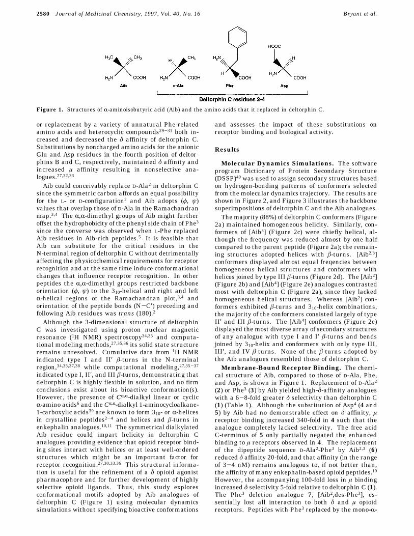
Molecular Dynamics Simulations. The softwareprogram Dictionary of Protein Secondary Structure(DSSP)40 was used to assign secondary structures basedon hydrogen-bonding patterns of conformers selectedfrom the molecular dynamics trajectory. The results areshown in Figure 2, and Figure 3 illustrates the backbonesuperimpositions of deltorphin C and the Aib analogues.The majority (88%) of deltorphin C conformers (Figure
2a) maintained homogeneous helicity. Similarly, con-formers of [Aib3] (Figure 2c) were chiefly helical, al-though the frequency was reduced almost by one-halfcompared to the parent peptide (Figure 2a); the remain-ing structures adopted helices with â-turns. [Aib2,3]conformers displayed almost equal freqencies betweenhomogeneous helical structures and conformers withhelices joined by type III â-turns (Figure 2d). The [Aib2](Figure 2b) and [Aib4] (Figure 2e) analogues contrastedmost with deltorphin C (Figure 2a), since they lackedhomogeneous helical structures. Whereas [Aib2] con-formers exhibited â-turns and 310-helix combinations,the majority of the conformers consisted largely of typeII′ and III â-turns. The [Aib4] conformers (Figure 2e)displayed the most diverse array of secondary structuresof any analogue with type I and I′ â-turns and bendsjoined by 310-helix and conformers with only type III,III′, and IV â-turns. None of the â-turns adopted bythe Aib analogues resembled those of deltorphin C.Membrane-Bound Receptor Binding. The chemi-
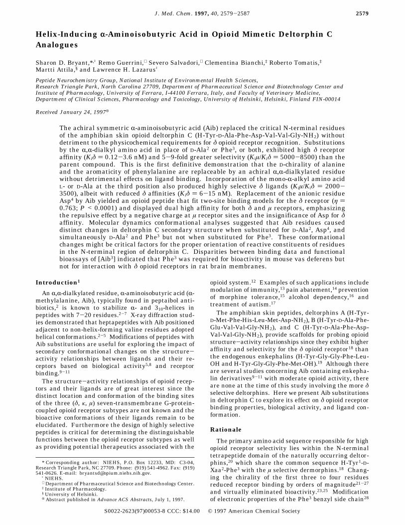
cal structure of Aib, compared to those of D-Ala, Phe,and Asp, is shown in Figure 1. Replacement of D-Ala2(2) or Phe3 (3) by Aib yielded high-δ-affinity analogueswith a 6-8-fold greater δ selectivity than deltorphin C(1) (Table 1). Although the substitution of Asp4 (4 and5) by Aib had no demonstrable effect on δ affinity, µreceptor binding increased 340-fold in 4 such that theanalogue completely lacked selectivity. The free acidC-terminus of 5 only partially negated the enhancedbinding to µ receptors observed in 4. The replacementof the dipeptide sequence D-Ala2-Phe3 by Aib2,3 (6)reduced δ affinity 20-fold, and that affinity (in the rangeof 3-4 nM) remains analogous to, if not better than,the affinity of many enkephalin-based opioid peptides.19However, the accompanying 100-fold loss in µ bindingincreased δ selectivity 5-fold relative to deltorphin C (1).The Phe3 deletion analogue 7, [Aib2,des-Phe3], es-sentially lost all interaction to both δ and µ opioidreceptors. Peptides with Phe3 replaced by the mono-R-
Figure 1. Structures of R-aminoisobutyric acid (Aib) and the amino acids that it replaced in deltorphin C.
2580 Journal of Medicinal Chemistry, 1997, Vol. 40, No. 16 Bryant et al.
methyl amino acid L-Ala3 (8) or its D-isomer (9) (Table1) exhibited decreased affinity for both δ and µ receptorsbut nonetheless exhibited a doubling (8) or quadrupling(9) of δ selectivity.
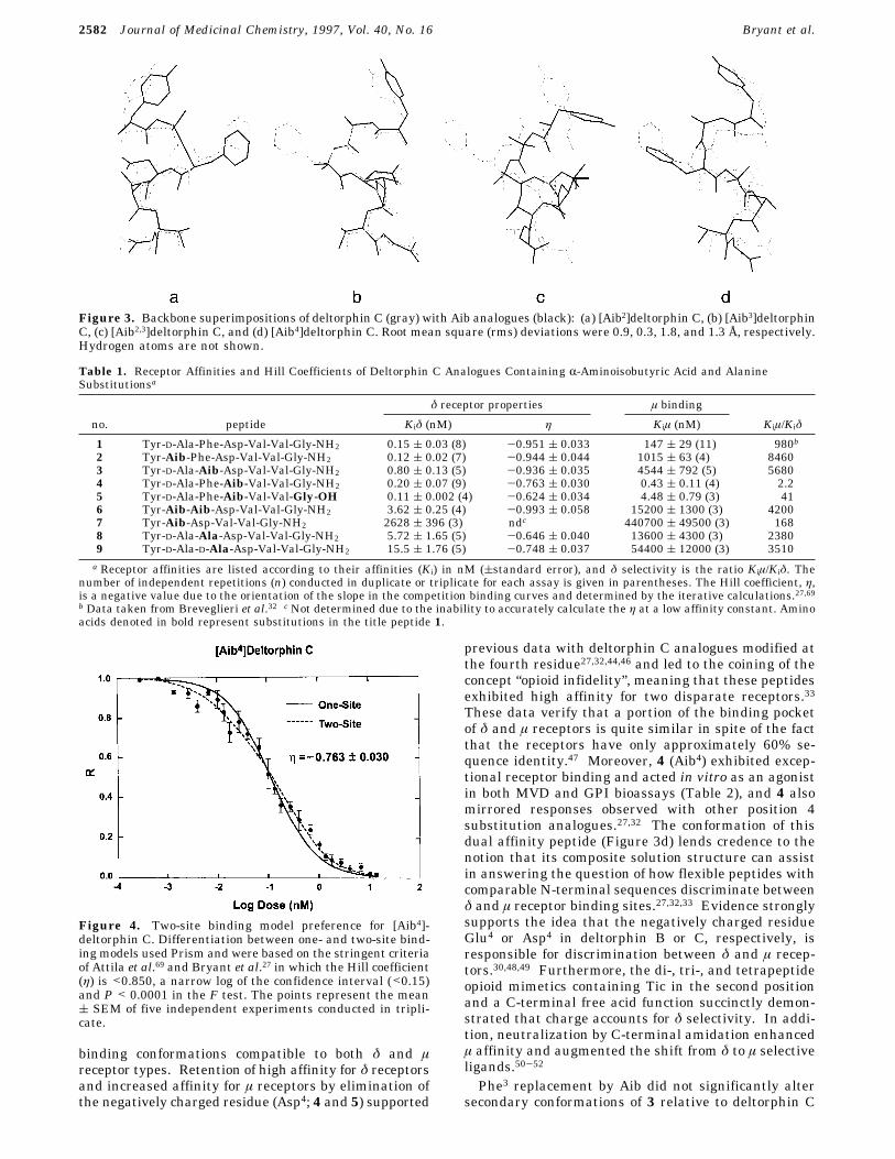
The Hill coefficients (η) of 4, 5, 8, and 9 for the δreceptor displayed heterogeneous binding as illustratedwith [Aib4]deltorphin C (4) (Figure 4). Differences of<0.15 in log of the 95% confidence interval in the ηdetermination with P < 0.0001 in which F tests ) 15.1,25.1, 25.5, and 14.1 for 4, 5, 8, and 9, respectively,indicated fits to a two-site binding model for the δreceptor. Moreover, the fit of 4 to µ receptors cor-responded to a one-site binding model (η ) 0.999 (0.048) as previously observed with all of the CR,R-dialkylcyclic amino acids at µ receptors.32 Analogues 1, 2, 3,and 6 were described by iterative calculations to fit asimple, bimolecular, one-site binding model (F rangedfrom 0.0 to 0.5 and P > 0.6 are nonsignificant fits for atwo-site model).Functional Biological Activity in Vitro. Phar-
macological activity in vitro of 1, 2, and 4 on therelaxation of mouse vas deferens (MVD) indicatedagonist activities with IC50 values and δ selectivity(Table 2) that supported the receptor binding data inrat brain membranes (Table 1). The relationship be-tween the high δ and µ affinity of [Aib4]deltorphin (4)in rat brain synaptosomes and the high δ and µ activityrecorded with MVD and guinea pig ileum (GPI), respec-tively, indicated a dual mode of action at disparateopioid receptors. However, the low activity in MVD of[Aib3] 3 and [Aib2,3] 6 was inconsistent with the high δaffinity observed in the synaptosomal preparations.Those data deviated by factors of 70-130 as noted inother bioassay and receptor data.29,30-32,41-43
Discussion
Enhanced selectivity and bioactivity exhibited by theAib analogues suggested that they provided a better fitto the δ opioid receptor binding site than deltorphin C,and that might reflect a preference for more stable, well-ordered ligand binding conformations. Conformers of2 and 4 contrasted most with deltorphin C since theylacked homogeneous helical structures (Figure 2b,e).The removal of the chiral D-Ala and the bulky Aspresidues increased conformational flexibility, while both2 and 4 exhibited the highest δ affinities (Table 1) andbioactive potencies (Table 2) on MVD. This implied thatconformational change through increased flexibilityrelative to the parent peptide facilitated receptor inter-action or perhaps increased its probability of adoptingbioactive conformations.Replacement of the critical D-enantiomer in the
second position by Aib substantially enhanced δ selec-tivity and δ agonist bioactivity. Normally, amino acidreplacements at this key position (depending upon thenature of the residue) either caused substantial lossesin receptor affinity,21,22,26,44 were moderately toler-ated,26,32,45 or eliminated bioactivity entirely.23-25 In-terestingly, replacement of D-Ala in deltorphin C withthe achiral CR,R-dialkyl amino acid 1-aminocyclohexane-1-carboxylic acid32 decreased δ affinity 16-fold while theAib residue substitution of a similar scaffold slightlyenhanced δ opioid receptor affinity relative to the parentpeptide. This phenomenon further underscores thesensitivity of the δ opioid receptor binding site to thephysical dimension as well as orientation of the sidechain at the second position.The high dual affinity for δ and µ receptors by 4
indicated that Aib substitution for Asp accommodated
Figure 2. Secondary structural features of deltorphin Canalogues analyzed using the program DSSP.40 Frequencyindicates the ratio between the total number of structuresanalyzed and the number of times that a particular secondarystructure was observed.
MD of δ Selective Aib Deltorphin C Analogues Journal of Medicinal Chemistry, 1997, Vol. 40, No. 16 2581
binding conformations compatible to both δ and µreceptor types. Retention of high affinity for δ receptorsand increased affinity for µ receptors by elimination ofthe negatively charged residue (Asp4; 4 and 5) supported
previous data with deltorphin C analogues modified atthe fourth residue27,32,44,46 and led to the coining of theconcept “opioid infidelity”, meaning that these peptidesexhibited high affinity for two disparate receptors.33These data verify that a portion of the binding pocketof δ and µ receptors is quite similar in spite of the factthat the receptors have only approximately 60% se-quence identity.47 Moreover, 4 (Aib4) exhibited excep-tional receptor binding and acted in vitro as an agonistin both MVD and GPI bioassays (Table 2), and 4 alsomirrored responses observed with other position 4substitution analogues.27,32 The conformation of thisdual affinity peptide (Figure 3d) lends credence to thenotion that its composite solution structure can assistin answering the question of how flexible peptides withcomparable N-terminal sequences discriminate betweenδ and µ receptor binding sites.27,32,33 Evidence stronglysupports the idea that the negatively charged residueGlu4 or Asp4 in deltorphin B or C, respectively, isresponsible for discrimination between δ and µ recep-tors.30,48,49 Furthermore, the di-, tri-, and tetrapeptideopioid mimetics containing Tic in the second positionand a C-terminal free acid function succinctly demon-strated that charge accounts for δ selectivity. In addi-tion, neutralization by C-terminal amidation enhancedµ affinity and augmented the shift from δ to µ selectiveligands.50-52
Phe3 replacement by Aib did not significantly altersecondary conformations of 3 relative to deltorphin C
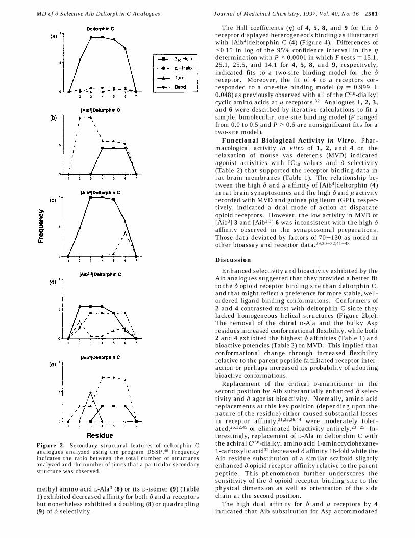
Figure 3. Backbone superimpositions of deltorphin C (gray) with Aib analogues (black): (a) [Aib2]deltorphin C, (b) [Aib3]deltorphinC, (c) [Aib2,3]deltorphin C, and (d) [Aib4]deltorphin C. Root mean square (rms) deviations were 0.9, 0.3, 1.8, and 1.3 Å, respectively.Hydrogen atoms are not shown.
Table 1. Receptor Affinities and Hill Coefficients of Deltorphin C Analogues Containing R-Aminoisobutyric Acid and AlanineSubstitutionsa
δ receptor properties µ binding
no. peptide Kiδ (nM) η Kiµ (nM) Kiµ/Kiδ
1 Tyr-D-Ala-Phe-Asp-Val-Val-Gly-NH2 0.15 ( 0.03 (8) -0.951 ( 0.033 147 ( 29 (11) 980b2 Tyr-Aib-Phe-Asp-Val-Val-Gly-NH2 0.12 ( 0.02 (7) -0.944 ( 0.044 1015 ( 63 (4) 84603 Tyr-D-Ala-Aib-Asp-Val-Val-Gly-NH2 0.80 ( 0.13 (5) -0.936 ( 0.035 4544 ( 792 (5) 56804 Tyr-D-Ala-Phe-Aib-Val-Val-Gly-NH2 0.20 ( 0.07 (9) -0.763 ( 0.030 0.43 ( 0.11 (4) 2.25 Tyr-D-Ala-Phe-Aib-Val-Val-Gly-OH 0.11 ( 0.002 (4) -0.624 ( 0.034 4.48 ( 0.79 (3) 416 Tyr-Aib-Aib-Asp-Val-Val-Gly-NH2 3.62 ( 0.25 (4) -0.993 ( 0.058 15200 ( 1300 (3) 42007 Tyr-Aib-Asp-Val-Val-Gly-NH2 2628 ( 396 (3) ndc 440700 ( 49500 (3) 1688 Tyr-D-Ala-Ala-Asp-Val-Val-Gly-NH2 5.72 ( 1.65 (5) -0.646 ( 0.040 13600 ( 4300 (3) 23809 Tyr-D-Ala-D-Ala-Asp-Val-Val-Gly-NH2 15.5 ( 1.76 (5) -0.748 ( 0.037 54400 ( 12000 (3) 3510a Receptor affinities are listed according to their affinities (Ki) in nM ((standard error), and δ selectivity is the ratio Kiµ/Kiδ. The
number of independent repetitions (n) conducted in duplicate or triplicate for each assay is given in parentheses. The Hill coefficient, η,is a negative value due to the orientation of the slope in the competition binding curves and determined by the iterative calculations.27,69b Data taken from Breveglieri et al.32 c Not determined due to the inability to accurately calculate the η at a low affinity constant. Aminoacids denoted in bold represent substitutions in the title peptide 1.
Figure 4. Two-site binding model preference for [Aib4]-deltorphin C. Differentiation between one- and two-site bind-ing models used Prism and were based on the stringent criteriaof Attila et al.69 and Bryant et al.27 in which the Hill coefficient(η) is <0.850, a narrow log of the confidence interval (<0.15)and P < 0.0001 in the F test. The points represent the mean( SEM of five independent experiments conducted in tripli-cate.
2582 Journal of Medicinal Chemistry, 1997, Vol. 40, No. 16 Bryant et al.
(Figure 2c) supporting previous observations that Aibwas replaceable by Phe in Aib-rich peptides.5 However,Aib replacement of Phe3 exhibited a 120-fold loss ofMVD potency (Table 2), while it was not detrimental torat brain δ opioid receptor affinity (Table 1). Enkepha-lin analogues with modifications at Phe4 also displayeddifferentiation between δ opioid receptors in the ratbrain and MVD.53 Apparently the aromaticity of Phe3as well as the orientation of the aromatic residue wascritical for δ receptor activation in the peripheraltissues, but hydrophobicity was sufficient for interactionwith receptors in the rat brain membranes. Phe3substitution by other aromatic residues, such as Aic andAtc, resulted in remarkable MVD bioactivities in vitrofurther supporting this observation.30,41,42 In contrast,hydrophobicity associated with Aib sufficiently replacedthe aromatic benzyl ring of Phe3 for interaction with therat brain δ opioid receptor, or the geometry of thebinding site was more accommodating to the less bulkyR,R-dimethyl groups of Aib. In this regard, reduced δaffinities were observed when Phe3 was replaced by thelarger branched chain aliphatic amino acids Val, Leu,and Ile.31 On the other hand an R-monoalkyl amino acid(L-Ala) replacement of Phe3 resulted in a 40-fold loss inδ receptor affinity (Table 1), and D-Ala substitution atthis position rendered a 100-fold loss (8 and 9). Theeffects of the Ala analogues differed from those of thetitle peptide 1 and the Aib3 analogue 3, although thereduced δ affinities and the two-site binding model(except Ac5c3)32 closely resembled those of the CR,R-dialkyl cyclic amino acid-substituted deltorphin ana-logues (Table 1). It is possible that the side chain of L-or D-Ala3 either was physically incompatible with thereceptor binding pocket or produced conformationalchanges that affected the interaction of other side chainswith the receptor.Increased flexibility of 6 observed during MD simula-
tions, in which conformers adopted helices as well asvarious â-turn secondary structures, may account forthe diminished δ affinity and dramatic loss in bioactiv-ity. Although δ affinity was decreased 20-fold, thisvalue was still comparable to many enkephalin anddeltorphin derivatives.19,30,31 The dramatic decrease inbioactivity in the peripheral tissue is largely due to theloss of aromatic phenylalanine and possibly a confor-mational change. As observed in peptide 3 the loss ofaromaticity resulted in decreased interaction with theMVD opioid receptors, and modifications of Phe4 inenkephalin derivatives53 resulted in decreased MVDbioactivity.These combined data confirm that the physical di-
mension, orientation, and hydrophobicity of side chainsin the N-terminal region of deltorphin C stronglyinfluence interaction with δ opioid receptors while the
aromaticity of the residue located at the third positionis necessary for MVD bioactivity. Likewise, peptideconformation is important for positioning the physico-chemical properties of the residue side chains in theN-terminal region of deltorphin C (Tables 1 and 2) foroptimal interaction with opioid receptors. Other studieson structural mimetics emphasized that amphiphilicstructures provide well-defined placement of residueswhich would enable greater efficiency in receptor bind-ing and that a â-folded structure is more likely presentin bioactive peptides.54 For example, the [Leu5]en-kephalin analogue N,N-diallyl-Tyr-Aib-Aib-Phe-Leu-OH, a δ opioid antagonist,9 exhibited a tightly foldeddouble â-bend conformation,55 while [Aib3]DPDPE dis-played a type I or III′ â-turn at residues 2-5.10 Byanalogy the many â-turns observed in 2 and 4 (Figure2) could represent conformations that are important foropioid ligand-receptor interaction.
ConclusionsAib substitutions appear to induce conformational
changes in deltorphin C analogues and increase peptidehydrophobicity. These properties combined may providegreater compatibility with a binding site tethered in ahighly localized lipophilic milieu consisting of residueswith aromatic or hydrophobic side chains.56,57 Althoughthe location and 3-dimensional arrangement of the δopioid receptor binding site are not known, site-directedmutagenesis of δ receptors indicated that the opioidligand enters a transmembrane channel to interact withregions of the receptor embedded in the membrane lipidbilayer.57-59 Ligands adopting R-helical or amphiphilicstructures might compliment this binding site. On theother hand, while these data appear to be consistentwith the concept of “message” and “address” domainsin peptides,60 in which charge in a membrane moderatesopioid peptide recognition,61 the evidence presented inthis paper indicates that ligand conformation plays adefinitive role in aligning the peptide within the recep-tor. For example, in the case of the rat brain receptorassay, while single Aib substitutions for residues atpositions 2 and 3 (peptides 2 and 3) were not detrimen-tal to δ opioid affinity, the simultaneous substitutionsof those residues (peptide 6) resulted in decreased δopioid affinity indicating that a conformational changeinfluenced δ receptor interaction. Whereas MD confor-mational analyses do not definitively confirm that Aibinduced helical conformations commonly observed incrystalline Aib-containing peptides,2-8 they do suggestthat Aib residues caused distinct changes in deltorphinC secondary structure (Figures 2 and 3) when substi-tuted for D-Ala2, Asp4, and simultaneously D-Ala2 andPhe3 but not when substituted for Phe3 alone. Theseconformational changes are critical factors for the proper
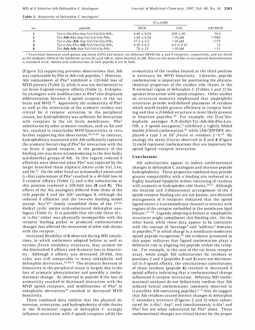
Table 2. Bioactivity of Deltorphin C Analoguesa
IC50 (nM)
no. peptide MVD GPI GPI/MVD
1 Tyr-D-Ala-Phe-Asp-Val-Val-Gly-NH2 0.46 ( 0.24 420 ( 95 9132 Tyr-Aib-Phe-Asp-Val-Val-Gly-NH2 1.44 ( 0.24 >10 µM >70003 Tyr-D-Ala-Aib-Asp-Val-Val-Gly-NH2 57.3 ( 5.5 >10 µM >1754 Tyr-D-Ala-Phe-Aib-Val-Val-Gly-NH2 0.36 ( 0.3 4.5 ( 0.35 136 Tyr-Aib-Aib-Asp-Val-Val-Gly-NH2 70 ( 23 >10 µM >21
a Functional bioassays used guinea pig ileum (GPI) and mouse vas deferens (MVD) for µ and δ receptors, respectively, and are listedas the midpoint (50%) in the inhibition curves (IC50) in nM or, when denoted, in µM. Data are the mean of four to six separate determinations( standard error. Amino acid substitutions in title peptide 1 are in bold.
MD of δ Selective Aib Deltorphin C Analogues Journal of Medicinal Chemistry, 1997, Vol. 40, No. 16 2583
orientation of reactive constituents of residues in theN-terminal region, which includes the -OH of thephenolic side chain of Tyr1, the free acid of Asp4, theprotonated nitrogen of the N-terminal amine, and thearomaticity of Tyr1 and Phe3. The hydroxyl group ofthe Tyr side chain and the N-terminal amine areconsidered to be involved in hydrogen bonding with thereceptor,52,62 while the negative charge of Asp is involvedin selectivity between δ and µ receptors27,32,33,41,45-47 andthe aromatic sides chains of Tyr1 and Phe3 may influ-ence receptor binding through hydrophobic stacking,π-π forces, or cation-π interactions.62,63 Thus, peptideconformations in concert with the physicochemicalproperties of the N-terminal residues are synergisticallyinvolved in the proper alignment of opioid ligands inthe receptor pocket and act in the differentiationbetween receptor types.41,62
Experimental SectionMaterials. Rink resin [4-(2′,4′-dimethoxyphenyl)-Fmoc-
(aminomethyl)phenoxy resin, 0.47 mmol/g], Fmoc-Gly-Wangresin (p-benzyloxycarbonyl alcohol resin, 0.5 mmol/g), DAGO,and DPDPE were obtained from Bachem California (Torrence,CA); the Fmoc-protected amino acids were either products ofBachem or Novabiochem AG (Germany). The Pico-Tag systemwas from Millipore (Waltham, MA). [3H]DAGO (60 Ci/mmol)was a product of Amersham (Arlington, IL) and [3H]DPDPE(28.1 Ci/mmol) from NEN-DuPont (Boston, MA). Prism (ver-sion 1.03) and InPlot (version 4.03) are programs by GraphPadsoftware (San Diego, CA).Solid Phase Peptide Synthesis. Peptides were synthe-
sized by solid phase methods using a Milligen 9050 synthesizerusing 0.1 g of Rink resin and were mixed with glass beads(1:15, w/w). Peptides were assembled using Fmoc-protectedamino acids with 4-fold excess for 1 h with each of the couplingreagents, DIPCI and HOBt. Double coupling was required inthe dipeptide sequences Asp-Val, Val-Val, Aib-Val, and Xaa-Aib. Boc-Tyr(t-Bu)-Aib-Aib-OH (infra vide) was used in 4-foldexcess during a 14 h acylation step by solid phase methods.The free acid peptide (compound 5) and 0.1 g of Fmoc-Gly-Wang resin were mixed with glass beads (1:15, w/w). Theprocedures for the synthesis of the peptide amides are reportedelsewhere.28,30Solution Phase Synthesis of Boc-Tyr(t-Bu)-Aib-Aib-
OH. The synthesis of compound 6 was prepared by synthesiz-ing the protected tripeptide Boc-Tyr(t-Bu)-Aib-Aib-OH bysolution methods according to Schmitt and Jung64 since weinitially failed to obtain coupling between Fmoc-Aib and theH-Aib-peptide-resin.a. Boc-Tyr(t-Bu)-Aib-Aib-OMe. Boc-Tyr(t-Bu)-OH (3.4
g, 10 mmol) and HOBt (1.7 g, 11 mmol) were dissolved in DMF(50 mL) and activated with water soluble carbodiimide (1.9 g,11 mmol). After 30 min, the dipeptide ester hydrochloride,H-Aib-Aib-OMe (2.38 g, 10 mmol),64,65 and triethylamine (1.4mL, 10 mmol) were added. After 12 h at room temperature,the solvent was removed in vacuo and the residue diluted withethyl acetate (100 mL) and washed sequentially with 10% citricacid, 5% NaHCO3, and a solution of saturated NaCl. Afterdrying over Na2SO4, the ethyl acetate was evaporated and thepeptide crystallized with ether at -10 °C: yield 3.0 g (58%);mp 135-136 °C; [R]20D +22.8 (MeOH); Rf (III) 0.75; K′ 20.1;1H NMR (CDCl3) δ 1.33 (s, 6H), 1.40 (s, 9H), 1.42 (s, 9H), 1.49(s, 3H), 1.51 (s, 3H), 3.02 (d, 2H), 3.70 (s, 3H), 4.15 (q, 1H),5.0 (m, 1H), 6.15 (bs, 1H), 6.94 (d, 2H, J ) 8.44 Hz), 7.05 (d,2H, J ) 8.51 Hz), 7.13 (s, 1H).b. Boc-Tyr(t-Bu)-Aib-Aib-OH. Boc-Tyr(t-Bu)-Aib-Aib-
OMe (2.5 g, 5 mmol) in methanol (20 mL) was saponified atroom temperature with 2 N NaOH (10 mL). After a 24 hreaction, neutralization, and evaporation of the solvent, thesolution was acidified and extracted with ethyl acetate, washedwith a saturated solution of NaCl, and dried with Na2SO4; thetripeptide was crystallized from ether. Analytical values: mp176-177 °C; [R]20D +19.1 (MeOH); Rf (I) 0.78, (III) 0.3; K′ 19.97.
No signals were obtained from the methyl ester at δ 3.70 inthe NMR spectra.Purification. All peptides were cleaved from the resin
(solid phase synthesis) by treatment with TFA/H2O/triethyl-silane (88:5:7, v/v/v) at room temperature for 1 h. Crudepeptides were purified by reversed-phase chromatographyusing a Waters Delta Prep 3000 column (30 × 3 cm, 15 µmparticle size). The peptides were eluted with a gradient of0-60% mobile phase B over 25 min at a flow rate of 30 mL/min using mobile phases A (10% acetonitrile in 0.1% TFA) andB (60% acetonitrile in 0.1% TFA). Analytical HPLC analyseswere performed on a Bruker liquid chromatography LC-21instrument using a Waters Pico-Tag C18 column (150 × 3.9mm, 5 µm particle size) equipped with a Bruker LC 313 UVvariable wavelength detector. Recording and quantificationwere accomplished with a chromatographic data processorcoupled to an Epson computer system (QX-10). The capacityfactor (K′) of each peptide was determined using HPLCconditions in the above solvent systems with a linear gradientfrom 0% to 100% mobile phase B in 25 min at a flow rate of 1mL/min. All analogues showed less than 1% impurities whenmonitored at 220 nm.Analytical Determinations. Amino acid analysis was
carried out using PITC (Pico-Tag methodology) as the aminoacid derivatization reagent. Aib was not quantitatively de-termined during amino acid analyses. Lyophilized samplesof peptides (50-1000 pmol) were placed in heat-treatedborosilicate tubes (50 × 4 mm), sealed, and hydrolyzed using200 µL of 6 N HCl containing 1% phenol in the Pico-Tagworkstation for 1 h at 150 °C. The Pico-Tag column wasemployed to separate the PITC-amino acid derivatives. Thinlayer chromatography used precoated plates of silica gel F254in the following solvent systems: I, 1-butanol/acetic acid/H2O(3:1:1, v/v/v); II, ethyl acetate/pyridine/acetic acid/H2O (6:2:0.6:1.1, v/v/v/v); III, chloroform/benzene/methanol (17:1:2).Ninhydrin (1%), fluorescamine, and chlorine reagent were usedas detection sprays.Melting points were determined on a Reicher-Kofler ap-
paratus and are uncorrected. Optical rotations were deter-mined in a Perkin-Elmer 241 polarimeter with a 10 cm cellusing methanol at a peptide concentration of 1%. Molecularweights of the compounds were determined by a triple-stagequadrupole mass spectrometer (TSQ 700, Finnigan MAT)equipped with a pneumatic electrospray (ion-spray) interface,and the data were compiled using a DEC 5000/125 computer.Proton NMR resonance spectra were recorded on a Bruker 200MHz spectrometer using tetramethylsilane as an internalstandard.Molecular Dynamics Simulations. Molecular dynamics
(MD) simulations of deltorphin C and [Aib2]-, [Aib3]-, [Aib2,3]-,and [Aib4]deltorphin C analogues were performed on a SiliconGraphics Indigo2 computer system using AMBER (version 3.0,revision A).66 The amino acid R-aminoisobutyric acid and theamine functional group (-NH2) were built using the prepmodule based on the prep input of glycine and valine alreadycontained in AMBER. Starting structures incorporated thedihedrals φ (rotation about N-CR), ψ (C′-CR), ø1 (CR-Câ), andø2 (Câ-Cγ) derived from 1H NMR data of deltorphin C(unpublished data). Values: ψ1 ) 139°, ø11 ) 179°, ø12 )-106°; φ2 ) 55°, ψ2 ) -143°, ø21 ) 53°; φ3 ) -49°, ψ3 ) -41°,ø31 ) -51°, ø32 ) -119°; φ4 ) -53°, ψ4 ) -24°, ø41 ) 151°, ø42) 92°; φ5 ) -50°, ψ5 ) -38°, ø51 ) 174°; φ6 ) -56°, ψ6 ) -27°,ø61 ) 179°; φ7 ) -63°. The backbone dihedrals (φ, ψ) of Aibwere assigned the values of the residue it replaced. The totalcharge on each peptide was neutral.Energy minimization lasted 500 hundred cycles with a
distance dependent dielectric (ε ) r) and 9 Å nonbondedcutoffs. Restart coordinates were output every 25 cycles, andthe nonbonded pair list was updated every 50 steps. Steepestdescent and conjugate gradient algorithms were used for theenergy minimizations with a step length of 0.001 fs, aconvergence criterion of 1 × 10⁻⁷ kcal/mol, and a gradientconvergence of 0.10 kcal/mol‚Å. Peptides were solvated inthree shells of TIP3P Monte Carlo water molecules underperiodic boundary conditions, and the solvent was energyminimized (before and after MD) for 500 cycles with constant
2584 Journal of Medicinal Chemistry, 1997, Vol. 40, No. 16 Bryant et al.
dielectric (ε ) 1) followed by 20 ps of MD under constantpressure (1 atm) and temperature (300 K). The time step was0.001 ps with 1000 steps/run, and a 0.4 ps time constant forheat bath coupling with a 0.6 ps pressure relaxation time and9 Å nonbonded cutoffs were incorporated. The nonbonded pairlist was updated every 20 ps, and information was output every100 steps. The solvent and peptide systems were energyminimized for 500 cycles with the same parameters as withthe solvent energy minimization followed by 100 ps of MD onthe peptide and solvent with the same parameters as thesolvent dynamics except that the coordinates were written tofile every 200 steps. Conformers generated every 4 ps duringthe 100 ps MD simulation were assigned secondary structuresusing the program Dictionary of Protein Secondary Structure(DSSP).40 The DSSP algorithm is principally based on hydro-gen-bonding patterns and defines 310-helical structures ashaving hydrogen bonds between Oi f H-Ni+3, R-helical as Oi
f H-Ni+4, turns as Oi f H-Ni+3, Oi f H-Ni+4, or Oi f H-Ni+5,and bends as Oi f H-Ni+2. Distances, RCi- RCi+3, and dihedralangles, φ and ψ, of the i+1 and i+2 bend residues weremeasured for structures characterized by DSSP as turns orbends and compared to values reported from X-ray crystal-lography of 29 proteins67 for classification of turn types.Receptor Binding Assays. Receptor affinities of deltor-
phin C analogues were assessed using competitive bindingassays labeled with either [3H]DPDPE (6.3 nM) for the δ sitesor [3H]DAGO (1.28 nM) for the µ sites according to publishedmethods.26-28,30 Excess unlabeled peptides (2 µM) saturatedthe opioid binding sites in order to obtain a baseline value.Duplicate tubes contained preincubated rat brain synaptoso-mal membranes in equilibrium assays containing 50 mMHEPES, pH 7.5, 5 mM MgCl2, glycerol, and protease inhibi-tors21 for 120 min at room temperature (22-23 °C). Incubationmixtures were trapped in the glass fiber filters and rapidlywashed within 5 s with 3 × 2 mL of ice-cold buffer containing0.01% BSA. In the duplicate assays, labeled peptides weredisplaced using 6-14 concentrations of each analogue to covera 1000-fold range in peptide dose. Competitive inhibitionconstants (Ki) were derived from the IC50 values based on theequations of Cheng and Prusoff.68
Statistical Analysis of Binding Site Models. Determi-nation of the Hill coefficients (η) and statistical analyses ofthe binding data utilized receptor assays conducted in tripli-cate using 25-35 peptide dosages covering differences inconcentrations of 300-500-fold either side of the Ki. Competi-tion curves were tested for fits to one- or two-site bindingmodels as detailed by Attila et al.69 and Bryant et al.27 usingPrism (version 1.02). In the assignment of fits to either a one-or two-site binding model, stringent iterative calculations wereonly considered valid for the two-site model when the Hillcoefficients were <0.85 with a narrow log of the 95% confidenceinterval (<0.15) and an F test in which P < 0.0001.Bioassays. Functional pharmacological assays were con-
ducted according to Salvadori et al.28 using a 2-3 cm portionof GPI in a 20 mL organ bath containing 70 µM hexametho-nium bromide and 0.125 µM mepyramine maleate aeratedwith 95% O2/5% CO2 at 36 °C for µ receptors. GPI wasstimulated transmurally with 0.5 ms square-wave pulses at0.1 Hz in which the stimulus was 1.5 times that necessary toproduce a maximal twitch (∼30 V) and recorded at a magni-fication ratio of 1:15. For δ receptors, a single MVD wassuspended in 4 mL of modified Kreb’s solution aerated with95% O2/5% CO2 at 33 °C with the twitch induced by fieldstimulation (0.1 Hz for 1 ms at 40 V) recorded with anisometric transducer. Dose-response curves were preparedfor each analogue in comparison to known compounds for eachtissue preparation (dermorphin or morphine for GPI anddeltorphin C for MVD). The Ke values for naloxone or N,N-diallyl-Tyr-Aib-Aib-Phe-Leu-OH (ICI 174,864) were in therange of 1-2 nM as detailed previously.28
Acknowledgment. S. Salvadori was supported inpart by grants from CNR Progetto Finalizzato ChimicaFine e Secondaria II and MURST. We thank T. A.Darden for his assistance in designing the DAT files for
the AMBER prep input, as well as R. Langenbach andL. Pedersen for their critical assessment of this study.We are grateful to Y. Okada and T. Odagami for theirassistance during the preparation of this manuscriptand to S. Fuller, E. M. Leadem, F. T. Lyndon, and R.Williams for their continuing support in the retrievalof published material.
Supporting Information Available: Analytical proper-ties of the R-aminoisobutyric acid-containing analogues ofdeltorphin C including the capacity factor (K′) and the massspectrometry molecular weights (MH+), the AMBER prep filesfor the Aib and -NH2 residues, and the secondary structureassignments for the MD conformers (5 pages). Orderinginformation is given on any current masthead page.
References(1) Symbols and abbreviations are in accord with the recommenda-
tion of the IUPAC-IUB Commission of Nomenclature (J. Biol.Chem. 1972, 247, 977). Other abbreviations: AMBER, assistedmodel building with energy refinement; Aib, R-aminoisobutyricacid; Boc, tert-butyloxycarbonyl; DAGO, H-Tyr-D-Ala-Gly-N-methyl-Phe-Gly-CH2OH; t-Bu, tert-butyl; deltorphin A (deltor-phin or dermenkephalin), H-Tyr-D-Met-Phe-His-Leu-Met-Asp-NH2; deltorphin C, H-Tyr-D-Ala-Phe-Asp-Val-Val-Gly-NH2;deltorphin B, [Glu4]deltorphin C; dermorphin, H-Tyr-D-Ala-Phe-Gly-Tyr-Pro-Ser-NH2; DIPCI, 1,3-diisopropylcarbodiimide; DP-DPE, [D-Pen2,5]enkephalin, H-Tyr-[D-Pen-Gly-Phe-D-Pen]-OH;DSSP, dictionary of protein secondary structure; Ot-Bu, tert-butyl ester; Fmoc, 9-fluorenylmethyloxycarbonyl; GPI, guineapig ileum; HOBt, 1-hydroxybenzotriazole; MD, molecular dy-namics; Pen, â,â-dimethylcysteine; Tic, 1,2,3,4-tetrahydro-3-isoquinoline-3-carboxylic acid.
(2) Toniolo, C.; Bonora, G. M.; Bavoso, A.; Benedetti, E.; di Blasio,B.; Pavone, V.; Pedone, C. Preferred Conformations of PeptidesContaining R,R-Disubstituted R-Amino Acids. Biopolymers 1983,22, 205-215.
(3) Karle, I. L.; Balaram, P. Structural Characteristics of R-HelicalPeptide Molecules Containing Aib Residues. Biochemistry 1990,29, 6747-6756.
(4) Marshall, G. R.; Hodgkin, E. E.; Langs, D. A.; Smith, G. D.;Zabrocki, J.; Leplawy, M. T. Factors Governing Helical Prefer-ence of Peptides Containing Multiple R,R-Dialkyl Amino Acids.Proc. Natl. Acad. Sci. U.S.A. 1990, 87, 487-491.
(5) Basu, G.; Bagchi, K.; Kuki, A. Conformational Preferences ofOligopeptides Rich in R-Aminoisobutyric Acid. I. Observation ofa 310/R-Helical Transition Upon Sequence Permutation. Biopoly-mers 1991, 31, 1763-1774.
(6) Bindra, V. A.; Kuki, A. Conformational Preferences of Oligopep-tides Rich in R-Aminoisobutyric Acid. III. Design, Synthesis andHydrogen Bonding in 310-Helices. Int. J. Pept. Protein Res. 1994,44, 539-548.
(7) (a) Benedetti, E.; di Blasio, B.; Pavone, V.; Pedone, C.; Santini,A.; Crisma, M.; Valle, G.; Toniolo, C. Structural versatility ofpeptides from CR,R-Dialkylated Glycines: Linear Ac3c Homo-Oligopeptides. Biopolymers 1989, 28, 175-184. (b) Karle, I. L.Flexibility in Peptide Molecules and Restraints Imposed byHydrogen Bonds, the Aib Residue, and Core Inserts. Biopolymers1996, 40, 157-180.
(8) di Blasio, B.; Pavone, V.; Lombardi, A.; Pedone, C.; Benedetti,E. Noncoded Residues as Building Blocks in the Design ofSpecific Secondary Structures: Symmetrically DisubstitutedGlycines and â-Alanine. Biopolymers 1993, 33, 1037-1049.
(9) Cotton, R.; Giles, M. G.; Miller, L.; Shaw, J. S.; Timms, D. ICI174864: A Highly Selective Antagonist for the Opioid δ-Recep-tor. Eur. J. Pharmacol. 1984, 97, 331-332.
(10) Haaseth, R. C.; Sobczyk-Kojiro, K.; Medzihradsky, F.; Smith, C.B.; Mosberg, H. I. Single Residue Modifications of the DeltaOpioid Receptor Selective Peptide, [D-Pen2,D-Pen5]-Enkephalin(DPDPE). Int. J. Pept. Protein Res. 1990, 36, 139-146.
(11) (a) Sudha, T. H.; Balaram, P. Conformational Flexibility in En-kephalins: Solvent Dependent Transitions in Peptides with Gly-Gly Segments Detected by Circular Dichroism. FEBS Lett. 1981,134, 32-36. (b) Prasad, V. B. V.; Sudha, T. S.; Balaram, P.Molecular Structure of Boc-Aib-Aib-Phe-Met-NH2-DMSO. AFragment of a Biologically Active Enkephalin Analogue. J.Chem. Soc., Perkin Trans. 1983, I, 417-421. (c) Sudha, T. S.;Balaram, P. Stabilization of â-Turn Conformations in Enkepha-lins. Int. J. Pept. Protein Res. 1983, 21, 381-383. (d) Kimura,S.; Sasaki-Yagi, Y.; Imanishi, Y. Receptor Selectivity of En-kephalin Analogs Carrying Artificial Address Peptides. Int. J.Pept. Protein Res. 1990, 35, 550-556.
(12) Olsen, G. A.; Olsen, R. D.; Kastin, A. J. Endogenous Opiates.Peptides 1996, 17, 1421-1466.
MD of δ Selective Aib Deltorphin C Analogues Journal of Medicinal Chemistry, 1997, Vol. 40, No. 16 2585
(13) (a) Arakawa, K.; Akami, T.; Okamoto, M.; Akioka, K.; Nakai, I.;Oka, T.; Nagase, H. Immunosuppression by Delta Opioid Recep-tor Antagonist. Transplant. Proc. 1993, 25, 738-740. (b) Chien,S.; Oeltgren, P. R.; Diana, J. N.; Salley, R. K.; Su, T.-P. Extensionof Tissue Survival Time in Multiorgan Block Preparation witha Delta Opioid DADLE ([D-Ala2,D-Leu2]-Enkephalin). J. Thorac.Cadiovasc. Surg. 1994, 107, 964-967. (c) Caroleo, M. C.;Arbitrio, M.; Melchiorri, D.; Nistico, G. A Reappraisal of the Roleof the Various Opioid Subtypes in Cell-Mediated Immunity.Neuroimmunodulation 1994, 1, 141-147.
(14) (a) Hammond, D. L. Pharmacological Mechanisms of PainModulation. An Update on δ Receptors. In Current and Emerg-ing Issues in Cancer Pain: Research and Practice; Chapman,C. R., Foley, K. M., Eds.; Raven Press: New York, 1993; pp 175-183. (b) Ziegler, D. K. Opiate and Opioid Use in Patients withRefractory Headache. Cephalagia 1995, 14, 5-10. (c) Portenoy,R. K. Opioid Therapy for Chronic Nonmalignant Pain: A Reviewof the Critical Issues. J. Pain Sympt. Manag. 1996, 11, 203-217.
(15) Abdelhamid, E. E.; Sultana, M.; Portoghese, P. S.; Takemori,A. E. Selective Blockage of Delta Opioid Receptors Prevents theDevelopment of Morphine Tolerance and Dependence in Mice.J. Pharmacol. Exp. Ther. 1991, 258, 299-303.
(16) (a) Froelich, J. C.; Li, T.-K. Opioid Peptides. In Recent Develop-ments in Alcoholism, Volume II: Ten Years of Progress; Galanter,M., Ed.; Plenum Press: New York, 1995; pp 187-205. (b)Terenius, L. Alcohol Addiction (Alcoholism) and the OpioidSystem. Alcohol 1996, 13, 31-34.
(17) Gillberg, C. Endogenous Opioids and Opiate Antagonists inAutism: A Brief Review of Empirical Findings and Implicationsfor Clinicians. Dev. Med. Child Neurol. 1995, 37, 239-245.
(18) (a) Erspamer, V. The Opioid Peptides of the Amphibian Skin.Int. J. Dev. Neurosci. 1992, 10, 3-30. (b) Lazarus, L. H.; Attila,M. The Toad, Ugly and Venomous, Wears Yet a Precious Jewelin His Skin. Prog. Neurobiol. 1993, 41, 473-507.
(19) Hruby, V. J.; Gehrig, C. A. Recent Developments in the Designof Receptor Specific Opioid Peptides. Med. Res. Rev. 1989, 9,343-401.
(20) (a) Lazarus, L. H.; Salvadori, S.; Tomatis, R.; Wilson, W. E.Opioid Receptor Selectivity Reversal in Deltorphin TetrapeptideAnalogues. Biochem. Biophys. Res. Commun. 1991, 178, 110-115. (b) Charpentier, S.; Sagan, S.; Delfour, A.; Nicolas, P.Dermenkephalin and Deltorphin I Reveal Similarities withinLigand-Binding Domains of µ- and δ-Opioid Receptors and anAdditional Address Subsite on the δ-Receptor. Biochem. Biophys.Res. Commun. 1991, 179, 1161-1168.
(21) Lazarus, L. H.; de Castiglione, R.; Guglietta, A.; Wilson, W. E.Highly Selective δ-Opioid Receptor Peptide from Preprodermor-phin Gene Sequence. J. Biol. Chem. 1989, 264, 3047-3050.
(22) Mor, A.; Delfour, A.; Sagan, S.; Amiche, M.; Pradelles, P.; Rossier,J.; Nicolas, P. Isolation of Dermenkephalin from AmphibianSkin, a High-Affinity δ-Selective Opioid Heptapeptide Contain-ing a D-Amino Acid Residue. FEBS Lett. 1989, 255, 269-274.
(23) Kreil, G.; Barra, D.; Simmaco, M.; Erspamer, V.; FalconieriErspamer, G.; Negri, L.; Severini, C.; Corsi, R.; Melchiorri, P.Deltorphin, A Novel Amphibian Skin Peptide with High Selec-tivity and Affinity for δ Opioid Receptors. Eur. J. Pharmacol.1989, 162, 123-128.
(24) Erspamer, V.; Melchiorri, P.; Falconieri Erspamer, G.; Negri,L.; Corsi, R.; Severini, C.; Barra, D.; Simmaco, M.; Kreil, G. Del-torphins: A Family of Naturally Occurring Peptides with HighAffinity and Selectivity for 2 Opioid Binding Sites. Proc. Natl.Acad. Sci. U.S.A. 1989, 86, 5188-5192.
(25) Richter, K.; Egger, R.; Negri, L.; Corsi, R.; Severini, C.; Kreil,G. cDNAs Encoding [D-Ala2]Deltorphin Precursors from Skinof Phyllomedusa Bicolor Also Contain Genetic Information forThree Dermorphin-Related Opioid Peptides. Proc. Natl. Acad.Sci. U.S.A. 1990, 87, 4836-4839.
(26) Lazarus, L. H.; Salvadori, S.; Balboni, G.; Tomatis, R.; Wilson,W. E. Stereospecificity of Amino Acid Side Chains in DeltorphinDefines Binding to Opioid Receptors. J. Med. Chem. 1992, 35,1222-1227.
(27) Bryant, S. D.; Attila, M.; Salvadori, S.; Guerrini, R.; Lazarus,L. H. Molecular Dynamics Conformations of Deltorphin Ana-logues Advocated δ Opioid Binding Site Models. Peptide Res.1994, 7, 175-184.
(28) Salvadori, S.; Bianchi, C.; Lazarus, L. H.; Scaranari, V.; Attila,M.; Tomatis, R. Para-Substituted Phe3 Deltorphin Analogues:Enhanced Selectivity of Halogenated Derivatives for δ OpioidReceptor Sites. J. Med. Chem. 1992, 35, 4651-4657.
(29) Schiller, P. W.; Nguyen, T. M.-D.; Weltrowska, G.; Lemieux, C.;Chung, N. N. Development of [D-Ala2]Deltorphin I Analogs withExtraordinary Delta Receptor Selectivity. In New Leads inOpioid Research; van Ree, J. M., Mulder, A. H., Wiegant, V. M.,van Wimersma Greidanus, T. B., Eds.; Excerpta Medica: Am-sterdam, 1990; pp 288-290.
(30) Salvadori, S.; Bryant, S. D.; Bianchi, C.; Balboni, G.; Attila, M.;Lazarus, L. H. Phe3-Substituted Analogues of Deltorphin C.Spatial Conformation and Topography of the Aromatic Ring inPeptide Recognition by δ Opioid Receptors. J. Med. Chem. 1993,36, 3748-3756.
(31) (a) Heyl, D. L.; Schmitter, S. J.; Bouzit, H.; Johnson, T. W.; Hepp,A. M.; Kurtz, K. R.; Mousigian, C. Substitution of Aromatic andNonaromatic Amino Acids for the Phe3 Residue in the δ-SelectiveOpioid Peptide Deltorphin I: Effects on Binding Affinity andSelectivity. Int. J. Pept. Protein Res. 1994, 44, 420-426. (b) Heyl,D. L.; Dandabathula, M.; Kurtz, K. R.; Mousigian, C. OpioidReceptor Binding Requirements for the δ-Selective PeptideDeltorphin I: Phe3 Replacement with Ring-Substituted andHeterocyclic Amino Acids. J. Med. Chem. 1995, 38, 1242-1246.
(32) Breveglieri, A.; Guerrini, R.; Salvadori, S.; Bianchi, C.; Bryant,S. D.; Attila, M.; Lazarus, L. H. Design and Synthesis of1-Aminocycloalkane-1-carboxylic Acid Substituted DeltorphinAnalogues: Unique δ and µ Opioid Activity in Modified Peptides.J. Med. Chem. 1996, 39, 773-780.
(33) Lazarus, L. H.; Bryant, S. D.; Salvadori, S.; Attila, M.; Jones,L. S. Opioid Infidelity: Novel Opioid Peptides with Dual HighAffinity for δ- and µ-Receptors. Trends Neurosci. 1996, 19, 31-35.
(34) Balboni, G.; Marastoni, M.; Picone, D.; Salvadori, S.; Tancredi,T.; Temussi, P. A.; Tomatis, R. New Features of the δ OpioidReceptor: Conformational Properties of Deltorphin I Analogues.Biochem. Biophys. Res. Commun. 1990, 169, 617-622.
(35) Amodeo, P.; Motta, A.; Tancredi, T.; Salvadori, S.; Tomatis, R.;Picone, D.; Saviano, G.; Temussi, P. A. Solution Structure ofDeltorphin I at 265 K: A Quantitative NMR Study. Peptide Res.1992, 5, 48-55.
(36) Bryant, S. D.; Salvadori, S.; Attila, M.; Lazarus, L. H. Topo-graphical Conformations of the Deltorphins Predicate δ OpioidReceptor Affinity. J. Am. Chem. Soc. 1993, 115, 8503-8504.
(37) (a) Temussi, P. A.; Picone, D.; Tancredi, T.; Tomatis, R.;Salvadori, S.; Marastoni, M.; Balboni, G. Conformational Prop-erties of Deltorphin: New Features of the δ-Opioid Receptors.FEBS Lett. 1989, 247, 283-288. (b) Tancredi, T.; Temussi, P.A.; Picone, D.; Amodeo, P.; Tomatis, R.; Salvadori, S.; Marastoni,M.; Santagada, V.; Balboni, G. New insights on µ/δ Selectivityof Opioid Peptides: Conformational Analysis of DeltorphinAnalogues. Biopolymers 1991, 31, 751-760.
(38) Duchnesne, D.; Naim, M.; Nicolas, P.; Baron, D. Folding Trendsin a Flexible Peptide: Two Dimensional NMR* Study of Del-torphin-I, a δ Selective Opioid Heptapeptide. Biochem. Biophys.Res. Commun. 1993, 195, 630-636.
(39) Balaji, V. N.; Ramnarayan, K.; Chan, M. F.; Rao, S. N. Confor-mational Studies on Model Peptides with 1-Aminocyclopropane-1-carboxylic Acid Residues. Peptide Res. 1994, 7, 60-71.
(40) Kabsch, W.; Sander, C. Dictionary of Protein Secondary Struc-ture: Pattern Recognition of Hydrogen-Bonded and GeometricalFeatures. Biopolymers 1983, 22, 2577-2637.
(41) Schiller, P. W.; Nguyen, T. M.-D.; Chung, N. N.; Lemieux, C.Dermorphin Analogues Carrying an Increased Positive NetCharge in their “Message” Domain Display Extremely High µOpioid Receptor Selectivity. J. Med. Chem. 1989, 32, 698-703.
(42) Schiller, P. W.; Weltrowska, G.; Nguyen, T. M.-D.; Wilkes, B.C.; Chung, N. N.; Lemieux, C. Conformationally RestrictedDeltorphin Analogues. J. Med. Chem. 1992, 35, 3956-3561.
(43) Ronai, A. Z.; Magyar, A.; Orosz, G.; Borsodi, A.; Benyhe, S.; Toth,G.; Mako, EÄ .; K tay, E.; Babka, E.; Medzihradszky, K. OpioidAntagonist Properties of the Highly δ-Receptor-Selective BOC-Tyr-Pro-Gly-Phe-Leu-Thr(OtBu) Peptide and of its Phe1 andMet1Analogues. Arch. Int. Pharmocodyn. 1995, 330, 361-369.
(44) (a) Amiche, M.; Sagan, S.; Mor, A.; Delfour, A.; Nicolas, P.Dermenkephalin (Tyr-D-Met-Phe-His-Leu-Met-Asp-NH2): A Po-tent and Fully Specific Agonist for the δ Opioid Receptor. Mol.Pharmacol. 1989, 35, 774-779. (b) Sagan, S.; Amiche, M.;Delfour, A.; Camus, A.; Mor, A.; Nicolas, P. Differential Contri-bution of C-Terminal Regions of Dermorphin and Dermenkepha-lin to Opioid-Sites Selection and Binding Potency. Biochem.Biophys. Res. Commun. 1989, 163, 726-732. (c) Melchiorri, P.;Negri, L.; Falconieri-Erspamer, G.; Severini, C.; Corsi, R.; Soaje,M.; Erspamer, V.; Barra, D. Structure-Activity Relationships ofthe δ-Opioid Selective Agonists, Deltorphins. Eur. J. Pharmacol.1991, 195, 201-207.
(45) Schmidt, R.; Chung, N. N.; Lemieux, C.; Schiller, P. W. Tic2-Substitution in Dermorphin, Deltorphin I and Dynorphin AAnalogues: Effect on Opioid Receptor Binding and OpioidActivity In Vitro. Reg. Pept. 1994, 54, 259-260.
(46) Salvadori, S.; Guerrini, R.; Forlani, V.; Bryant, S. D.; Attila, M.;Lazarus, L. H. Prerequisite for His4 in Deltorphin A for High δOpioid Receptor Selectivity. Amino Acids 1994, 7, 291-304.
(47) (a) Evans, C. J.; Keith, D. E., Jr.; Morrison, H.; Magendzo, K.;Edwards, R. H. Cloning of a Delta Opioid Receptor by FunctionalExpression. Science 1992, 258, 1952-1955. (b) Kieffer, B. L.;Befort, K.; Gaveriaux-Ruff, C.; Hirth, C. G. The δ-OpioidReceptor: Isolation of a cDNA by Expression Cloning andPharmacological Characterization. Proc. Natl. Acad. Sci. U.S.A.1992, 89, 12048-12052. (c) Chen, Y.; Mestek, A.; Liu, J.; Yu, L.
2586 Journal of Medicinal Chemistry, 1997, Vol. 40, No. 16 Bryant et al.
Molecular Cloning and Functional Expression of Mu-OpioidReceptor from Rat Brain. Biochem. J. 1993, 95, 625-628. (d)Fukuda, K.; Kato, S.; Mori, K.; Nishi, M.; Takashima, H. PrimaryStructures and Expression from cDNAs of Rat Opioid Receptorδ- and µ-Subtypes. FEBS Lett. 1993, 327, 311-314. (e) Yasuda,K.; Raynor, K.; Kong, H.; Breder, C. D.; Takeda, J.; Resine, T.;Bell, G. I. Cloning and Functional Comparison of κ and δ OpioidReceptors fromMouse Brain. Proc. Natl. Acad. Sci. U.S.A. 1993,90, 6736-6740.
(48) Lazarus, L. H.; Salvadori, S.; Santagada, V.; Tomatis, R.; Wilson,W. E. Function of Negative Charge in the “Address Domain” ofDeltorphins. J. Med. Chem. 1991, 34, 1350-1359.
(49) Sagan, S.; Charpentier, S.; Delfour, A.; Amiche, M.; Nicolas, P.The Aspartic Acid in Deltorphin I and Dermenkephalin Pro-motes Targeting to δ-Opioid Receptor Independently of ReceptorBinding. Biochem. Biophys. Res. Commun. 1992, 187, 1203-1210.
(50) (a) Schiller, P. W.; Nguyen, T. M.-D.; Weltrowska, G.; Wilkes,B. C.; Marsden, B. J.; Lemieux, C.; Chung, N. N. DifferentialStereochemical Requirements of µ vs. δ Opioid Receptors forLigand Binding and Signal Transduction: Development of aClass of Potent and Highly δ-Selective Peptide Antagonists. Proc.Natl. Acad. Sci. U.S.A. 1992, 89, 11871-11875. (b) Schiller, P.W.; Weltroska, G.; Nguyen, T. M.-D.; Wilkes, B. C.; Chung, N.N.; Lemieux, C. TIPP [Ψ]: a Highly Potent and Stable Pseudopep-tide δ Opioid Receptor Antagonist with Extraordinary δ Selec-tivity. J. Med. Chem. 1993, 36, 3182-3187. (c) Mosberg, H. I.;Omnass, J. R.; Sobczyk-Kojiro, K.; Dua, R.; Ho, J. C.; Ma, W.;Bush, R.; Mousigian, C.; Lomize, A. Pharmacophore Elementsof the TIPP Class of Delta Opioid Receptor Antagonists. Lett.Pept. Sci. 1994, 1, 69-72.
(51) Temussi, P. A.; Salvadori, S.; Amodeo, P.; Bianchi, C.; Guerrini,R.; Tomatis, R.; Lazarus, L. H.; Picone, D.; Tancredi, T. SelectiveOpioid Dipeptides. Biochem. Biophys. Res. Commun. 1994, 198,933-939.
(52) Salvadori, S.; Attila, M.; Balboni, G.; Bianchi, C.; Bryant, S. D.;Crescenzi, O.; Guerrini, R.; Picone, D.; Tancredi, T.; Temussi,P. A.; Lazarus, L. H. δ Opioidmimetic Antagonists: Prototypesfor Designing a New Generation of Ultraselective Opioid Pep-tides. Mol. Med. 1995, 1, 678-689.
(53) Vaughn, L. K.; Wire, W. S.; Davis, P.; Shimohigashi, Y.; Toth,G.; Knapp, R. J.; Hruby, V. J.; Burks, T. F.; Yamamura, H. I.Differentiation Between Rat Brain and Mouse Vas Deferens δOpioid Receptors. Eur. J. Pharmacol. 1990, 177, 99-101.
(54) Marraud, M.; Aubry, A. Crystal Structures of Peptides andModified Peptides. Biopolymers 1996, 40, 45-83.
(55) Flippen-Anderson, J. L.; George, C.; Deschamps, J. R.; Reddy,P. A.; Lewin, A.; Brine, G. A. X-ray Structures of the δ OpioidAntagonist TIPP and a Protected Derivative of the δ OpioidAntagonist ICI 174,864. Lett. Pept. Sci. 1994, 1, 107-115.
(56) Befort, K.; Tabbara, L.; Kling, D.; Maigret, B.; Kieffer, B. Roleof Aromatic Transmembrane Residues of the δ-Opioid Receptorin Ligand Recognition. J. Biol. Chem. 1996, 271, 10161-10168.
(57) Valiquette, M.; Vu, H. K.; Yue, S. Y.; Wahlestedt, C.; Walker,
P. Involvement of Trp-284, Val-296, and Val-297 of the Humanδ-Opioid Receptor in Binding of δ-Selective Ligands. J. Biol.Chem. 1996, 271, 18789-18796.
(58) (a) Surratt, C. K.; Johnson, P. S.; Moriwaki, A.; Seidleck, B. K.;Blaschak, C. J.; Wang, J. B.; Uhl, G. R. µ Opiate Receptor.Charged Transmembrane Domain Amino Acids for AgonistRecognition and Intrinsic Activity. J. Biol. Chem. 1994, 269,20548-20553. (b) Kong, H.; Raynor, K.; Yasuda, K.; Moe, S. T.;Portoghese, P.; Bell, G. I.; Reisine, T. A Single Residue, AsparticAcid 95, in the δ Opioid Receptor Specifies Selective HighAffinity Agonist Binding. J. Biol. Chem. 1993, 268, 23055-23058.
(59) Befort, K.; Tabbara, L.; Bausch, S.; Chavkin, C.; Evans, C.;Kieffer, B. The Conserved Aspartate Residue in the ThirdPutative Transmembrane Domain of the δ-Opioid Receptor isnot the Anionic Counterpart for Cationic Opiate Binding but isa Constituent of the Receptor Binding Site. Mol. Pharmacol.1996, 49, 216-223.
(60) Schwyzer, R. ACTH: A Short Introductory Review. Ann. N. Y.Acad. Sci. 1977, 297, 3-26.
(61) Schwyzer, R. Molecular Mechanism of Opioid Receptor Selection.Biochemistry 1986, 25, 6335-6342.
(62) (a) Balboni, G.; Guerrini, R.; Salvadori, S.; Tomatis, R.; Bianchi,C.; Attila, M.; Bryant, S. D.; Lazarus, L. H. Opioid Diketo-piperazines: Synthesis and Activity of a Prototypic Class ofUnique Opioid Antagonists. Biol. Chem. 1997, 378, 19-22. (b)Bryant, S. D.; Balboni, G.; Guerrini, R.; Salvadori, S.; Tomatis,R.; Lazarus, L. H. Opioid Diketopiperazines: Refinement of theδ Opioid Antagonist Pharmacophore. Biol. Chem. 1997, 378,107-114.
(63) Dougherty, D. A. Cation-π Interactions in Chemistry and Biol-ogy: A New View of Benzene, Phe, Tyr, and Trp. Science 1996,271, 163-168.
(64) Schmitt, H.; Jung, G. Total Synthesis of the R-Helical Eicos-apeptide Antibiotic Alamethicin. Liebigs Ann. Chem. 1985, 321-344.
(65) Leplawy, M. T.; Jones, D. S.; Kenner, G. W.; Shepard, R. C.Synthesis of Peptides Derived From R-Methylalanine. Tetrahe-dron 1960, 11, 39-51.
(66) Singh, U. C.; Weiner, D. K.; Caldwell, J. W.; Kollman, P. A.AMBER (version 3.1); Department of Pharmaceutical Chemistry,University of California: San Francisco, CA, 1988.
(67) Chou, P. Y.; Fasman, G. â-Turn in Proteins. J. Mol. Biol. 1977,115, 135-175.
(68) Cheng, Y.-C.; Prusoff, W. H. Relationships Between the Inhibi-tion Constant (Ki) and the Concentration of Inhibition whichCauses 50 Per Cent Inhibition (I50) of an Enzymatic Reaction.Biochem. Pharmacol. 1973, 22, 3099-3108.
(69) Attila, M.; Salvadori, S.; Balboni, G.; Bryant, S. D.; Lazarus, L.H. Synthesis and Receptor Binding Analysis of DermorphinHepta-, Hexa- and Pentapeptide Analogues. Evidence for One-and Two-Site Binding Models for the µ-Opioid Receptors. Int.J. Pept. Protein Res. 1993, 42, 550-559.
JM9700530
MD of δ Selective Aib Deltorphin C Analogues Journal of Medicinal Chemistry, 1997, Vol. 40, No. 16 2587


![Synthèse des analogues de l’[azaPhe4]-GHRP-6 comme](https://img.pdfslide.tips/doc/110x75/618c27ade26e05549a04661f/synthse-des-analogues-de-lazaphe4-ghrp-6-comme-.jpg)