Embed Size (px)
Citation preview

432 - Acta Cirúrgica Brasileira - Vol. 24 (6) 2009
2 - PRELIMINARY NOTEModels, Biological
Hemosiderin. A new marker for sentinel lymph node identification1
Hemossiderina. Um novo marcador para identificação do linfonodo sentinela
Luiz Gonzaga Porto PinheiroI, Renato Santos de Oliveira FilhoII, Paulo Henrique Diógenes VasquesIII, Pedro Henrique de OliveiraFilgueiraIV, Douglas Henning Pinheiro AragãoIV, Pedro Macedo Esmeraldo BarbosaIV, Hugo Enrique Orsini BeserraV, RaissaVasconcelos CavalcanteV
I PhD, Associate Professor, Department of Surgery, Faculty of Medicine, UFC, Fortaleza - CE, Brazil.II PhD, Affiliate Professor, Division Plastic Surgery, Department of Surgery, Federal University of Sao Paulo (UNIFESP), Brazil.III Fellow Master degree, Postgraduate Program, Department of Surgery, UFC, Fortaleza - CE, Brazil.IV Graduate student, Medicine Faculty, UFC, Fortaleza - CE, Brazil.V Graduate student, Veterinary Faculty, State University of Ceara (UECE), Brazil.
ABSTRACTPurpose: To evaluate and present our initial results of a new marker (hemosiderin) for mammary sentinel lymph node identification inan experimental model. Methods: Skins mapped like a lymphatic duct draining to the axilla in patients submitted to breast biopsy, in ourmastology service, stimulated us to try it in an animal model (female dogs). Our theory was that some blood derivate (hemosiderin) wascaptured by macrophages and accessed the lymphatic ducts in direction to the axilla. Six female dogs of no defined race were studied. Weinjected 0,2 ml of technetium on both superior mammary glands. After ten minutes, a 2,5 ml solution of hemolized blood (hemosiderin)from the own animal was injected in the subareolar lymphatic plexus on the left superior mammary gland and 2,5 ml of patentblue concomitantly and equally on the contralateral gland. Ten minutes after, incisions on both axilas were made to search, throughthe lymphatic mapping and a gamma probe, the sentinel lymph nodes. Results: Seven brown sentinel lymph nodes were indentifiedand also radiomarked on the left axilla. Six blue sentinel lymph nodes were identified and also radiomarked on the right axilla.Conclusion: Preliminary studies of a potential new dye for sentinel lymph node identification are presented. It may be the change of thecurrent use of the blue dyes and their severe side-effects on patients submitted to sentinel lymph node biopsies.Key words: Breast Neoplasms. Sentinel Lymph Node Biopsy. Hemosiderin.
RESUMOObjetivo: Avaliar e apresentar resultados preliminares de um novo marcador (hemossiderina) para a identificação de linfonodos sentinelamamários em um modelo experimental. Métodos: Durante acompanhamento de dois casos de biópsias excisionais de tumores da mama,no nosso serviço de mastologia, observou-se trajeto pigmentado no quadrante inferior externo daquelas mamas, sugerindo ser marcaçãocutânea do ducto de drenagem linfática a partir da papila mamária em direção a axila homolateral. Levantamos a hipótese que umderivado sanguíneo (hemossiderina) foi capturado por macrófagos obtendo acesso aos ductos linfáticos em direção à axila. Seis cadelassem raça definida foram estudadas. Injeção de 0,2 ml de tecnécio foi realizada em ambas as mamas superiores. Após 10 minutos, umasolução de 2,5 ml de sangue hemolizado (hemossiderina) do próprio animal foi injetado no plexo linfático subpapilar da mama esquerdae 2,5 ml de azul patente na mama contralateral concomitantemente e igualmente. Após mais 10 minutos, incisões axilares foram realizadaspara a procura, pela coloração e com um gama probe, dos linfonodos sentinela. Resultados: Sete linfonodos sentinela castanhos eradiomarcados foram identificados na axila esquerda. Seis linfonodos sentinela azuis e radiomarcados foram identificados na axiladireita. Conclusão: São apresentados estudos preliminares de um potencial novo marcador para identificação do linfonodo sentinela.Este poderá mudar o uso dos corantes vitais e de seus efeitos adversos em pacientes submetidos à biópsia do linfonodo sentinela.Descritores: Neoplasias da Mama. Biópsia de Linfonodo Sentinela. Hemossiderina.1Research performed at Experimental Animal Laboratory, Department of Surgery, Faculty of Medicine, Federal University of Ceara (UFC), Fortaleza,Brazil.
Introduction
The incidence of female breast cancer has increased0.5% since the year 2000, as reported by the InternationalAgency Research on Cancer. This number is greater in developingcountries due to the increase in life expectancy and change inbehavior resulting in increased exposure to risk factors.
Lymph node staging is an early event carried out duringinitial patient evaluation in developed countries. Some 20% to 30%of all cases are diagnosed quite early (in situ lesions) with negativeaxillary nodes in 79% of the patients at this stage1. These valuesare somewhat different in developing countries where more than50% of the cases are diagnosed at advanced stages of the disease.Late stages (III and IV) of breast cancer are seen in 60% of women

Hemosiderin. A new marker for sentinel lymph node identification
Acta Cirúrgica Brasileira - Vol. 24 (6) 2009 - 433
who seek medical attention at the Maternidade-Escola (FederalUniversity of Ceara) Breast Unit. Lack of adequate surveillanceprograms, the reduced number of specialists and the small numberof educational programs may account for these numbers.
Increased survival rates have been reported lately.According to Parkin2, the overall survival rate is 91% after thefirst year and 65% after five years in Europe. These values increaseto 96.8% after the first year in the United States. As primaryprevention of breast cancer is not available as yet, early detectionand treatment in the initial phase of the disease are, therefore,the most important measures for its control3.
Available diagnostic methods include mammography,breast ultrassonography, sentinel lymph node biopsy, andfine-needle aspiration biopsy among others4,5. With the definitionof the sentinel lymph node as the first one which receives thedrainage of the tumoral area6, it was possible to ensure means forthe proper staging of the illness and the therapeutic approachestablishing less invasive surgery techniques.
Currently, it is admitted that the presence of metastaticlymph nodes is the main predictor factor for prognosis of the breastmalignant neoplasia course and subsequent therapeutic program.Lymph nodes are also valuable for staging breast cancer. In therecent past dissection of the axillary lymph nodes was required forthis matter, resulting in a series of additional complications7.
The principles of the sentinel lymph node were first used byCabanas (1973) in penis cancer. Morton et al.8 used this principleson the identification of melanoma metastasis. He used Patent bluedye. Giuliano et al.9 described its use for the breast cancer, alsousing the vital blue dye for lymphatic mapping and then, Krag etal.10 additionally used a radioguided model with a gamma probeincreasing the accuracy on the identification of the sentinellymph node.
The results of the researches on breast sentinel lymphnode are encouraging, although there are some methods whichare controversial11. One of these is the vital blue dye use for thelymphatic mapping which may cause severe hypersensitivityreactions on patients12. Other dyes are used, as methylene bluewhich may also cause serious reactions, including skin and fatnecrosis in the site of injection13.
During the management of two patients submitted tobreast biopsy in our service, we observed their skin mapped asthe lymphatic ducts draining to axillary lymph nodes. Thisobservation stimulated us to try an animal model which wasdescribed for our group. Our theory was that some blood derivate(hemosiderin) was captured by macrophages and accessed thelymphatic duct in direction to the axilla (Figure 1). For thesereasons, the purpose of this paper is to present our initial resultsof a new marker for sentinel lymph node identification.
FIGURE 1 – Skin mapped as a lymphatic duct in direction to the axilla
434 - Acta Cirúrgica Brasileira - Vol. 24 (6) 2009
Pinheiro LGP et al
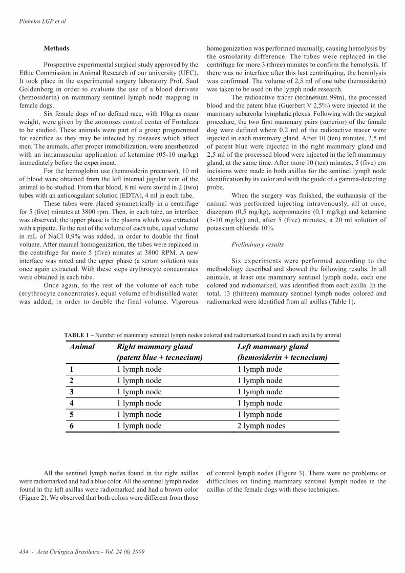
Methods
Prospective experimental surgical study approved by theEthic Commission in Animal Research of our university (UFC).It took place in the experimental surgery laboratory Prof. SaulGoldenberg in order to evaluate the use of a blood derivate(hemosiderin) on mammary sentinel lymph node mapping infemale dogs.
Six female dogs of no defined race, with 10kg as meanweight, were given by the zoonoses control center of Fortalezato be studied. These animals were part of a group programmedfor sacrifice as they may be infected by diseases which affectmen. The animals, after proper immobilization, were anesthetizedwith an intramuscular application of ketamine (05-10 mg/kg)immediately before the experiment.
For the hemoglobin use (hemosiderin precursor), 10 mlof blood were obtained from the left internal jugular vein of theanimal to be studied. From that blood, 8 ml were stored in 2 (two)tubes with an anticoagulant solution (EDTA), 4 ml in each tube.
These tubes were placed symmetrically in a centrifugefor 5 (five) minutes at 3800 rpm. Then, in each tube, an interfacewas observed; the upper phase is the plasma which was extractedwith a pipette. To the rest of the volume of each tube, equal volumein mL of NaCl 0,9% was added, in order to double the finalvolume. After manual homogenization, the tubes were replaced inthe centrifuge for more 5 (five) minutes at 3800 RPM. A newinterface was noted and the upper phase (a serum solution) wasonce again extracted. With these steps erythrocyte concentrateswere obtained in each tube.
Once again, to the rest of the volume of each tube(erythrocyte concentrates), equal volume of bidistilled waterwas added, in order to double the final volume. Vigorous
homogenization was performed manually, causing hemolysis bythe osmolarity difference. The tubes were replaced in thecentrifuge for more 3 (three) minutes to confirm the hemolysis. Ifthere was no interface after this last centrifuging, the hemolysiswas confirmed. The volume of 2,5 ml of one tube (hemosiderin)was taken to be used on the lymph node research.
The radioactive tracer (technetium 99m), the processedblood and the patent blue (Guerbert V 2,5%) were injected in themammary subareolar lymphatic plexus. Following with the surgicalprocedure, the two first mammary pairs (superior) of the femaledog were defined where 0,2 ml of the radioactive tracer wereinjected in each mammary gland. After 10 (ten) minutes, 2,5 mlof patent blue were injected in the right mammary gland and2,5 ml of the processed blood were injected in the left mammarygland, at the same time. After more 10 (ten) minutes, 5 (five) cmincisions were made in both axillas for the sentinel lymph nodeidentification by its color and with the guide of a gamma-detectingprobe.
When the surgery was finished, the euthanasia of theanimal was performed injecting intravenously, all at once,diazepam (0,5 mg/kg), acepromazine (0,1 mg/kg) and ketamine(5-10 mg/kg) and, after 5 (five) minutes, a 20 ml solution ofpotassium chloride 10%.
Preliminary results
Six experiments were performed according to themethodology described and showed the following results. In allanimals, at least one mammary sentinel lymph node, each onecolored and radiomarked, was identified from each axilla. In thetotal, 13 (thirteen) mammary sentinel lymph nodes colored andradiomarked were identified from all axillas (Table 1).
TABLE 1 – Number of mammary sentinel lymph nodes colored and radiomarked found in each axilla by animal
Animal Right mammary gland
(patent blue + tecnecium)
Left mammary gland
(hemosiderin + tecnecium)
1 1 lymph node 1 lymph node
2 1 lymph node 1 lymph node
3 1 lymph node 1 lymph node
4 1 lymph node 1 lymph node
5 1 lymph node 1 lymph node
6 1 lymph node 2 lymph nodes
All the sentinel lymph nodes found in the right axillaswere radiomarked and had a blue color. All the sentinel lymph nodesfound in the left axillas were radiomarked and had a brown color(Figure 2). We observed that both colors were different from those
of control lymph nodes (Figure 3). There were no problems ordifficulties on finding mammary sentinel lymph nodes in theaxillas of the female dogs with these techniques.

Hemosiderin. A new marker for sentinel lymph node identification
Acta Cirúrgica Brasileira - Vol. 24 (6) 2009 - 435
FIGURE 3 – Sentinel lymph nodes marked with hemosiderin at left (bold arrows) and with patentblue at right (thin arrows). NSL = Non-sentinel lymph node.
Discussion
Sleth14, while discussing, in hiswork, the annual rise on the incidence ofadverse reactions to blue dyes duringsentinel lymph node biopsies, the clearlyquantifiable allergic risks and thecontinuous evolution of the sentinellymph node study in other tumors, askedif it was not the time to change the dye.
Nowadays, reactions to vitaldyes are being more frequently reporteddue to its increased use in lymphaticmappings and in sentinel lymph nodebiopsies in patients with breast cancerand other malignancies12,15. Cases andmeta-analysis of anaphylactic reactionsare described due to the use of blue dyesin sentinel lymph node identification.The incidence of anaphylactic reactionsto isosulfan blue or patent blue varyfrom 0,6% to 2,7% in patients submittedto sentinel lymph node biopsy andmust be known by the surgeon and theanesthesiologist16.
Mertes et al.15 reported 14clinical cases of anaphylaxis induced bydyes during the years 2004 and 2006 infour different allergoanesthesia frenchcenters. All patients suffered hypersensitivereactions to patent blue V during sentinellymph node biopsy due to breast cancer.In this study, 43% of patients sufferedlife-threatening reactions (cardiovascularcollapse, tachycardia or bradycardia,arrhythmias and severe bronchospasm)and 64% of these patients requiredprolonged continuous epinephrineinfusion and transference to an intensivecare unit. The authors recommended theuse of dermatologic tests for thesemarkers before surgery.
Wohrl et al .17 described anear-fatal case of anaphylaxis due to thepatent blue V dye in sentinel lymph nodeidentification. A patient with melanomasuffered cardiac arrest for 10 minutes andrequired mechanic cardio-pulmonaryreanimation measures after the intradermalinjection of this dye.
The preliminary results of ourexperimental work showed that the bloodderivate (hemosiderin) was capable ofbeing used along with the radiomarker( t e c h n e t i u m 9 9 m ) a n d c o l o r i n gwonderfully the sentinel lymph node offemale dogs.
FIGURE 2 – Sentinel lymph node marked with hemosiderin in vivo

436 - Acta Cirúrgica Brasileira - Vol. 24 (6) 2009
Pinheiro LGP et al
The use of the blood derivate (hemosiderin) with theradiomarker also showed equal results comparing the use of thepatent blue with the radiomarker in identifying the mammarysentinel lymph nodes of the female dog because they were alwaysfound in both axillas.
The hemosiderin, a blood-derived marker, theoreticallydoes not cause side effects as allergic reactions. Besides, it maydecrease personal and healthcare spendings, reduce the morbidityof the procedure and the emotional suffering, along with otherindirect vantages.
Conclusion
Preliminary studies of a potential new dye for sentinellymph node identification are presented. It may be the change ofthe current use of the blue dyes and their severe side-effects onpatients submitted to sentinel lymph node biopsies.
References
1. Holland JF, Frei E, Kufe DW, Bast RC. Principles of medical oncology.6ed. Philadelphia: Saunders; 2001.2. Parkin DM, Bray FI, Devesa SS. Cancer burden in the year 2000. Theglobal picture. Eur J Cancer. 2001;37:64-6.3. Instituto Nacional do Câncer. Estimativas da incidência e mortalidadepor câncer no Brasil em 2008. [cited 2009 April 12]. Available from URL:http://www.inca.gov.br/estimativa/2008/index.asp4. Moore MP, Kinne DW. Is axillary lymph node dissection necessary inthe routine management of breast cancer? Yes. Important Adv Oncol.1996;12:245-50.5. Morton DL, Ollila DW. Critical review of the sentinel node hypothesis.Surgery. 1999;126:815-9.6. Cabanas RM. An approach for the treatment of penile carcinoma.Cancer. 1977;39(2):456-66.
7. Veronesi U, Paganelli G, Niale G, Galimberti V, Luini A, Zurrida S,Robertson C, Sacchini V, Veronesi P, Orvieto E, De Cicco C, Intra M, TosiG, Scarpa D. Sentinel lymph node biopsy and axillary dissection in breastcancer: results in a large series. J Natl Cancer Inst. 1999;91:368-73.8. Morton DL, Wen DR, Wong JH, Economou JS, Cagle LA, Storm FK,Foshag LJ, Cochran AJ. Technical details of intraoperative lymphaticmapping for early stage melanoma. Arch Surg. 1992;127:392-9.9. Giuliano AE, Kirgan DM, Guenther JM, Morton DL. Lymphaticmapping and sentinel lymphadenectomy for breast cancer. Ann Surg.1994;220:391-401.10. Krag DN, Weaver DL, Alex JC, Fairbank JT. Surgical resection andradiolocalization of the sentinel lymph node in breast cancer using a gammaprobe. Surg Oncol. 1993;2:335-9.11. Pinheiro LGP, Moraes MO, Soares AH, Lopes AJT, Naguére MASP,Gondim FAL, Brandão CB, Nascimento DCH, Soares JPH, Silva JMM.Estudo Experimental de linfonodo sentinela na mama da cadela com azulpatente e tecnécio Tc99. Acta Cir Bras. 2003;18(6):545-52.12. Mertes PM, Malinovsky JM, Mouton-Faivre C, Bonnet-Boyer MC,Benhaijoub A, Lavaud F, Valfrey J, O’Brien J, Pirat P, Lalourcey L, DemolyP. Anaphylaxis to dyes during the perioperative period: reports of 14clinical cases. J Allergy Clin Immunol. 2008; 122:348-52.13. Salhab M, Al Sarakbi W, Mokbel K. Skin and fat necrosis of the breastfollowing methylene blue dye injection for sentinel node biopsy in apatient with breast cancer. Int Semin Surg Oncol. 2005;2:26.14. Sleth JC. Un accident anaphylactoïde imputé au bleu patenté. Faut-ilchanger de colorant? Ann Fr Anesth Reanim. 2008;27:515.15. Mulan MH, Deacock SJ, Quiney NF, Kissin MW. Anaphylaxis to patentblue dye during sentinel lymph node biopsy for breast cancer. Eur J SurgOncol. 2001;27:218-9.16. Scherer K, Studer W, Figueiredo V, Bircher AJ. Anaphylaxis to isosulfanblue and cross-reactivity to patent blue V: case report and review of thenomenclature of vital blue dyes. Ann Allergy Asthma Immunol.2006;96:497-500.17. Wohrl S, Focke M, Hinterhuber G, Stingl G, Binder M. Near-fatalanaphylaxis to patent blue V. Br J Dermatol. 2004;150:1037-8.
Conflict of interest: noneFinancial source: none
Correspondence:Luiz Gonzaga Porto PinheiroDepartamento de CirurgiaRua Prof. Costa Mendes 1608/ 3o andar60430-140 Fortaleza – CE BrazilPhone: (55 85)3366-8063Fax: (55 85)[email protected]
Received: April 14, 2009Review: June 10, 2009
Accepted: July 15, 2009
How to cite this articlePinheiro LGP, Oliveira Filho RS, Vasques PHD, Filgueira PHO, Aragão DHP, Barbosa PME, Beserra HEO, Cavalcante RV. Hemosiderin.A new marker for sentinel lymph node identification. Acta Cir Bras. [serial on the Internet] 2009 Nov-Dec;24(6). Available from URL:http://www.scielo.br/acb