Embed Size (px)
Citation preview
Instructions for use
Title Hepatectomy for Hepatocellular Carcinoma with Bile Duct Tumor Thrombus, Including Cases with ObstructiveJaundice
Author(s) Orimo, Tatsuya; Kamiyama, Toshiya; Yokoo, Hideki; Wakayama, Kenji; Shimada, Shingo; Tsuruga, Yosuke;Kamachi, Hirofumi; Taketomi, Akinobu
Citation Annals of surgical oncology, 23(8), 2627-2634https://doi.org/10.1245/s10434-016-5174-7
Issue Date 2016-08
Doc URL http://hdl.handle.net/2115/66904
Rights The original publication is available at www.springerlink.com
Type article (author version)
File Information AnnSurgOncol23_2627.pdf
Hokkaido University Collection of Scholarly and Academic Papers : HUSCAP
1
Hepatectomy for hepatocellular carcinoma with bile duct tumor thrombus, including
cases with obstructive jaundice
Tatsuya Orimo*, Toshiya Kamiyama, Hideki Yokoo, Kenji Wakayama, Shingo Shimada,
Yosuke Tsuruga, Hirofumi Kamachi, Akinobu Taketomi
Department of Gastroenterological Surgery I, Hokkaido University Graduate School of
Medicine, Sapporo, Japan
*Corresponding author:
Tatsuya Orimo, MD, PhD
Department of Gastroenterological Surgery I
Hokkaido University Graduate School of Medicine
North 15-West 7, Kita-Ku, Sapporo, Hokkaido 060-8638, Japan
Tel: +81-11-706-5927; Fax: +81-11-717-7515; Email: [email protected]
Running title: HCC with bile duct tumor thrombus
Conflicts of interest: None to report.

2
Synopsis
Hepatectomy should be considered for hepatocellular carcinoma with bile duct tumor
thrombus.
3
Abstract
Purpose: The purpose of this study was to evaluate the short- and long-term outcomes of
hepatectomy for hepatocellular carcinoma (HCC) with bile duct tumor thrombus (BDTT),
including cases with obstructive jaundice.
Methods: We reviewed 42 HCC patients with BDTT, including 6 patients who needed
preoperative biliary drainage due to obstructive jaundice and 732 HCC patients without
BDTT. We analyzed the impact of BDTT on the surgical outcomes and assessed the
outcomes of hepatectomy for patients presenting with obstructive jaundice.
Results: HCC patients with BDTT had increased alpha-fetoprotein expression, larger
tumors, more portal vein invasion status and were almost all stage III or IV. The survival of
the HCC patients with BDTT was significantly inferior to that of the patients without BDTT
(p = 0.0003). There was no significant difference in survival between HCC patients with or
without BDTT when the two groups were matched by stage (p = 0.3366). HCC patients
with BDTT that presented with obstructive jaundice demonstrated similar outcomes as HCC
patients with BDTT that did not present with obstructive jaundice in terms of the overall
survival rate (p = 0.5469). The perioperative outcomes of HCC patients with BDTT did not
depend on the presence or absence of preoperative jaundice. No patients in either BDTT
group demonstrated 90-day mortality in this study.
Conclusions: Hepatectomy should be considered for HCC patients with BDTT, even for

4
cases with obstructive jaundice because equivalent surgical outcomes as HCC without
BDTT can be achieved.
5
Background
Hepatocellular carcinoma (HCC) with bile duct tumor thrombus (BDTT) is rare in
comparison with HCC with portal vein thrombus.1 HCC with portal vein thrombus is often
recognized and its prognosis is poor, but the prognosis of HCC with BDTT is not well
recognized due to its rarity. In particular, HCC with BDTT showing obstructive jaundice is
called icteric hepatoma, and this type of HCC is known to present difficult problems in the
differential diagnosis of conditions such as advanced liver cirrhosis and biliary tract cancer.2
HCC with BDTT showing obstructive jaundice leads to severe symptoms such as
cholangitis and hemobilia, and can cause hepatic failure.3-5 However, it is difficult to
determine the surgical indications for patients with obstructive jaundice because jaundice
due to advanced liver cirrhosis is a contraindication for hepatectomy. There have been
several studies on HCC with BDTT and it is clear that surgical approach for HCC with
BDTT is important. 6-8 However, it is presently unclear whether or not surgical treatment is
valid for HCC with BDTT that presents with obstructive jaundice because the short- and
long-term outcomes of such cases are unknown. In addition, the optimal surgical approach
for HCC patients with BDTT— whether or not to preserve the bile duct —is also unclear.
In this study, we reviewed HCC patients with and without BDTT and analyzed the
clinicopathological features and surgical outcomes. Furthermore, we reviewed the surgical
management of patients with BDTT that present with obstructive jaundice and evaluated the
6
validity of the surgical approaches for this type of HCC.
Patients and Methods
Between January 1996 and May 2015, 774 HCC patients underwent several types
of hepatectomy at the Department of Gastroenterological Surgery I, Hokkaido University
Hospital. Forty-two of these patients (5.4%) had HCC with BDTT that was
histopathologically diagnosed. BDTT positive patients were defined as those who had
BDTT in the biliary tree or the intrahepatic bile duct histopathologically. BDTT negative
patients were defined as those who did not have any BDTT in the biliary tree or the
intrahepatic bile duct. Microscopic BDTT was defined as BDTT which developed in more
than just the second branch of the intrahepatic bile duct. Macroscopic BDTT was defined as
BDTT which existed in the second or the first branch of the biliary tree, or the common bile
duct. There were 21 microscopic BDTT and 21 macroscopic BDTT cases in this study. Of
the 42 BDTT cases, there were 6 HCC patients with BDTT who demonstrated obstructive
jaundice, which required preoperative biliary drainage. Macroscopic portal vein tumor
thrombus (PVTT) was defined as PVTT involving the first or the second branches or main
trunk of the portal vein. There were 20 macroscopic PVTT cases among HCC patients with
BDTT and 82 macroscopic PVTT cases among HCC patients without BDTT in this study.
The median follow-up period of these patients was 45.4 months (range = 0.2–233.2
7
months).
This study was approved by the institutional review board of Hokkaido University
Hospital (approval number: 015-0251). All analyses in this study were performed in
accordance with the ethical guidelines of Hokkaido University Hospital.
Preoperative management
Preoperative management was performed according to our previous report.9 Our
algorithm, which incorporates the indocyanine green retention rate at 15 minutes (ICGR15)
and remnant liver volume, was used to determine the operative procedure, as previously
described. 9 If the ICGR15 is less than 15% and the resected liver volume is less than 60%,
hemihepatectomy or extended hemihepatectomy can be performed. However, if the
ICGR15 is less than 15% and the resected liver volume is greater than 60%, then
percutaneous transhepatic portal embolization is performed before surgery. For patients
with an ICGR15 of 15% to 20%, sectionectomy can be performed; for patients with an
ICGR15 of 20% to 25%, segmentectomy can be performed; and for patients with an
ICGR15 of 25% to 40%, a limited resection can be performed. If the ICGR15 is more than
40%, hepatectomy is contraindicated. For patients with BDTT that presented with
obstructive jaundice, biliary drainage was performed first, and hepatic functional reserve
was evaluated after the serum bilirubin level was < 2 mg/dl. A liver resection was secondly
8
performed when possible according to our algorithm.
Surgical methods
The surgical methods used for liver resection were previously described.9
Anatomical resection was defined as a resection in which the lesion(s) are anatomically
completely removed based on Couinaud’s classification (segmentectomy, sectionectomy
and hemihepatectomy or extended hemihepatectomy). We preserved the bile duct in all
HCC patients with BDTT. Our surgical procedure is similar to bile duct-preserving surgery
reported by Yamamoto et al. 10 We cut the bile duct, peeled off the tumor thrombus, closed
the bile duct incision site by running sutures with 5-0 or 6-0 absorbable monofilament
thread, and inserted a C-tube into the cystic duct to decompress the bile duct. We performed
cholangiography to verify the absence of the tumor thrombus. Hepatectomy for HCC with
BDTT in this study included two right trisectionectomies, one left trisectionectomy, five
extended right hepatectomies, four extended left hepatectomies, 13 right hepatectomies,
eight left hepatectomies, one central bisectionectomy, two right anterior sectionectomies,
two right posterior sectionectomies, two left lateral sectionectomies, and two partial
resections.
Statistical analysis
9
The correlation between BDTT and the clinicopathological features was evaluated
using the Fisher exact test for categorical variables or the Mann-Whitney U test for
continuous variables. The overall survival rates and time to recurrence were calculated
using the Kaplan-Meier method and compared between groups using the log-rank test.
Potential prognostic factors were identified by univariate analysis using the log-rank test.
Independent prognostic factors were evaluated using a Cox proportional-hazards regression
model. In this study, p < 0.05 was considered significant. Statistical analyses were
performed using JMP (version 12 for Windows; SAS Institute, Cary, NC).
Results
Differences in the clinicopathological features and surgical outcomes according to the
presence or absence of BDTT
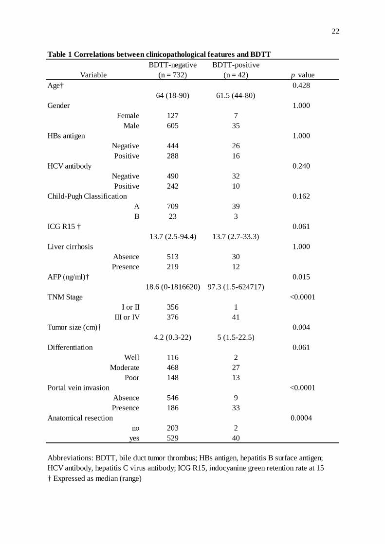
The clinicopathological features of the HCC patients with and without BDTT are
presented in Table 1. The median age, sex, proportion of hepatitis B surface antigen,
proportion of hepatitis C virus antibody, Child-Pugh Classification, ICGR15, and proportion
of liver cirrhosis in the patients in the BDTT-negative and BDTT-positive groups were
similar. On the other hand, HCC patients with BDTT vs without BDTT demonstrated
significantly different alpha-fetoprotein expression, TNM stage, tumor size, portal vein
invasion status, and proportion of anatomical resection. In terms of histological
10
differentiation, there were no significant differences between the 2 groups, but there was a
tendency toward poorer differentiation in the BDTT-positive group (p = 0.061) (Table 1).
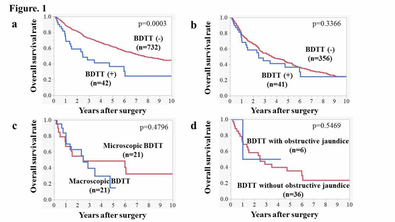
The overall 1-, 3-, and 5-year survival rates in the HCC patients with BDTT were 75.1%,
44.9%, and 36.6% in comparison with 89.0%, 72.4%, and 61.9% in the HCC patients
without BDTT, respectively (p = 0.0003) (Fig. 1A). The median survival times (MST) of
the HCC patients with and without BDTT were 2.46 and 7.62 years, respectively. Almost all
HCC patients with BDTT (41 of 42 patients) were stage III or IV, and therefore when
limited to stage III or IV patients there was no significant difference in the survival rate
between HCC patients with or without BDTT (p = 0.3366) (Fig. 1B).
Risk factors for survival and recurrence in HCC patients with BDTT
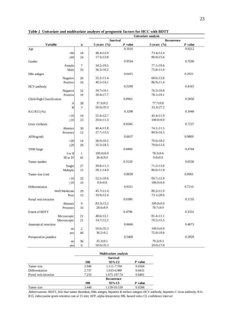
In HCC patients with BDTT, univariate analysis revealed that tumor size,
differentiation, and portal vein invasion were significant prognostic factors for survival, and
tumor size was a significant prognostic factor for recurrence (Table 2). Multivariate analysis
revealed that tumor size, differentiation, and portal vein invasion were independent
prognostic factors for survival, and tumor size for recurrence (Table 2). In terms of the
extent of BDTT, there was no significant difference in the survival rate between HCC
patients with microscopic BDTT and HCC patients with macroscopic BDTT (p = 0.4796),
and the MST values were 2.43 and 2.46 years, respectively (Table 2; Fig. 1C). The median
11
time to recurrence was 0.48 years for microscopic BDTT patients and 1.24 years for
macroscopic BDTT patients, but this was also not significantly different (p = 0.3331) (Table
2). Among HCC patients with BDTT, there was no significant difference in the survival rate
between patients with and without macroscopic PVTT (p = 0.0820). Among HCC patients
without BDTT, there was a significant difference in survival between those with and
without macroscopic PVTT (p < 0.0001).
Recurrence sites and treatments after hepatectomy for HCC with BDTT
The sites of first recurrence after hepatectomy and treatments are listed in Table 3.
The most frequent site of recurrence was the liver (15 of 24 patients; 62.5%), and
intrahepatic recurrence was most commonly treated using transcatheter arterial
chemoembolization (TACE) (10 of 24 patients; 41.6%). In our present study, there was 1
case of recurrence, which developed in the form of BDTT. As a relapse treatment, this
BDTT was peeled off and the bile duct was preserved. Following this relapse treatment, the
patient has been without recurrence for 19 months.
Perioperative surgical outcome for patients with BDTT that present with obstructive
jaundice
There were 6 patients in our current study series who needed preoperative biliary
12
drainage due to jaundice (Table 4). Four patients needed endoscopic nasobiliary drainage,
and 2 patients needed percutaneous transhepatic biliary drainage. The maximum serum
bilirubin level before biliary drainage ranged from 3.4–22.7 mg/dl, with a median
concentration of 6.6 mg/dl. Liver resection for cases with obstructive jaundice included 5
right hepatectomies and 1 right anterior sectionectomy. There were no significant
differences in the overall survival rate (p = 0.5469) among HCC patients with BDTT in
terms of the presence or absence of preoperative jaundice (Table 2; Fig. 1D). The
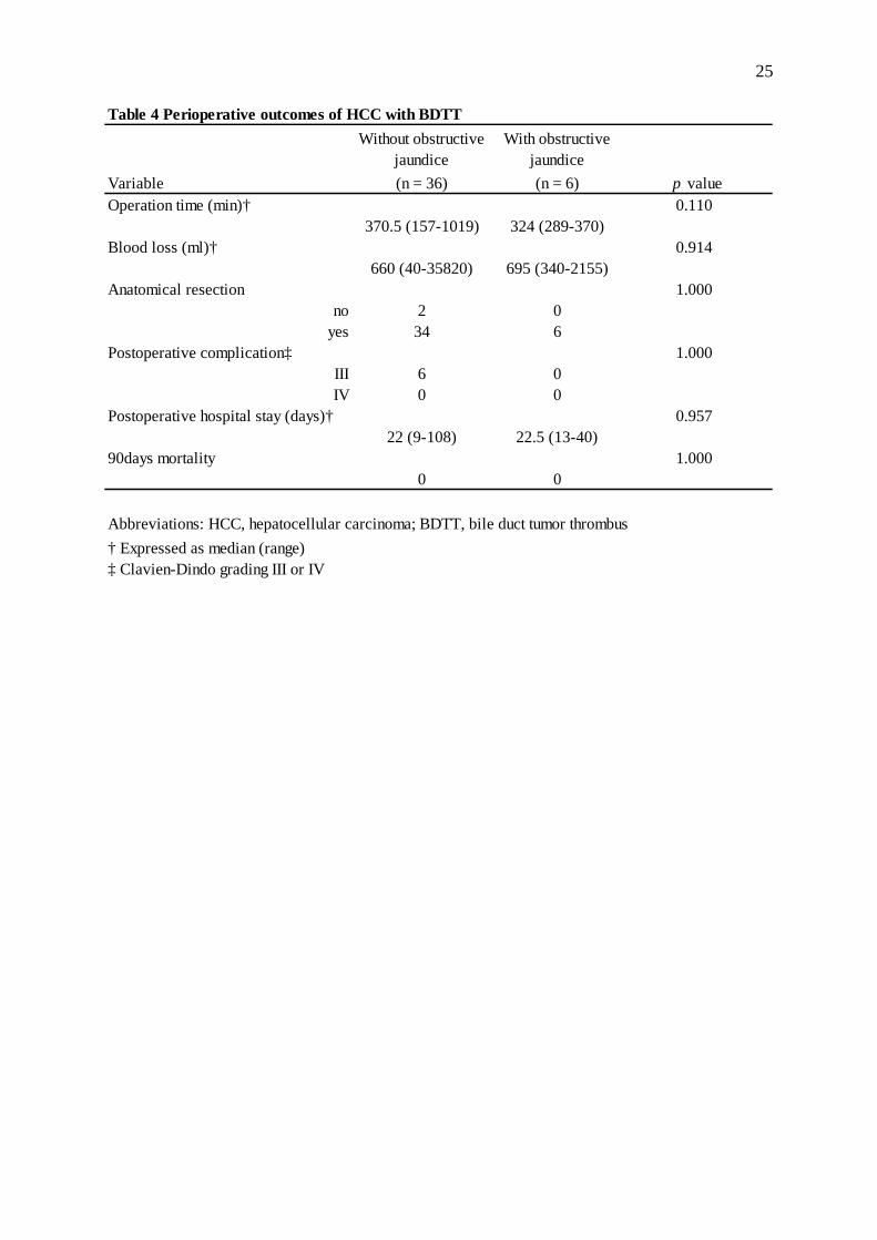
perioperative outcomes for HCC with BDTT are presented in Table 4. There were no
significant differences between the 2 groups depending on the presence or absence of
preoperative jaundice in terms of operation time, blood loss, anatomical resection,
postoperative complications, or the postoperative hospital stays. In addition, there were no
90-day mortalities in either study group.
Discussion
HCC with BDTT is a rare phenomenon that occurs in about 2.5–3.4% of patients
with HCC.6-8 Yeh et al reported the relationship between the pathogenesis of HCC with
BDTT and the microRNA-200 family. 11 HCC with BDTT reportedly demonstrates
pathological features such as a higher incidence of vascular invasion and less histological
differentiation.6, 11, 12 Our present results (Table 1) are consistent with these earlier findings.
13
The 5-year survival rate of HCC patients with BDTT is reportedly about 30%.7,8,12
In our current series, the 5-year survival rate of HCC patients with BDTT was 36.6%, and
there was no significant difference in survival between HCC patients with or without BDTT
when the two groups were matched by stage. Wong et al also reported that HCC patients
with or without BDTT demonstrated similar survival rates using their matching criteria.7
Macrovascular invasion, including portal vein invasion and hepatic vein invasion, is
correlated with poor prognosis in HCC patients.13-17 Regarding BDTT, in contrast, Esaki et
al reported that macroscopic bile duct invasion demonstrates a favorable impact on patient
outcomes in HCC patients with BDTT. 8 In our current series, there was no significant
difference found in the survival rate and time to recurrence between HCC patients with
microscopic or macroscopic BDTT. These data indicated a lower malignant potential of
BDTT in comparison with vascular invasion, and that macroscopic BDTT is not a
contraindication for hepatectomy. Our results also indicate that tumor size, differentiation,
and portal vein invasion are independent prognostic factors for survival, and that tumor size
is an independent factor for recurrence. Kasai et al have reported previously that major
vascular invasion is a negative prognostic indicator for HCC patients with BDTT.18 These
results suggest that BDTT alone does not affect prognosis and only does so in conjunction
with other prognostic factors, such as vascular invasion, poor differentiation, or large size.
Regarding a hepatectomy for HCC with BDTT, there is an issue about whether or
14
not to preserve the bile duct. Wong et al proposed extrahepatic bile duct resection for HCC
with BDTT in order to minimize bile duct recurrence.7 However, according to several
reports, routine bile duct resection is not recommended unless direct invasion of the BDTT
into the bile duct is suspected.8,12,18 Shiomi et al reported that the survival of their bile
duct-preserved group was similar to that of their bile duct-resected group.19 Recently,
Yamamoto et al reported using bile duct-preserving surgery for HCC with BDTT—called
the “peeling off technique”10—which is similar to our surgical approach. One reason why
preserving the bile duct is important is that BDTT demonstrates expansive growth and does
not usually adhere to the bile duct wall.10 The other reason is that treatments for recurrence,
such as TACE or radiofrequency ablation (RFA), are restricted after resecting the common
bile duct because liver abscess formation after TACE and RFA is relatively common when
an underlying bilioenteric anastomosis is present.20,21 As indicated in Table 3, intrahepatic
recurrence is the most common recurrence after liver resection for HCC, and TACE can be
performed in this circumstance after preserving the bile duct. Thus, by preserving the bile
duct, the peeling-off technique is suitable for HCC with BDTT. However, careful
observations must be made to check for BDTT-type recurrence. There was one patient with
BDTT in our current series who presented with recurrence in the bile duct, and a salvage
operation was performed to peel off the BDTT. No recurrence was noted thereafter in this
case. Bile duct-preserving hepatectomy for HCC with BDTT may cause this type of
15
recurrence because of the direct invasion of the BDTT into the bile duct, with the BDTT
remaining in the small branches of the bile ducts with intraoperative seeding along them.
The selection criteria for preserving or resecting the bile duct is thus still unclear, and
further studies are needed.
Jaundice affects the functional liver reserve, and cases with obstructive jaundice
often presents with cholangitis and hemobilia that can cause hepatic failure.3-5 Therefore,
biliary drainage is mandatory before hepatectomy for patients with BDTT that present with
obstructive jaundice. We perform preoperative biliary drainage at our hospital in accordance
with the preoperative management of biliary tract cancer.22 Biliary drainage of the remnant
liver is performed until the serum bilirubin is < 2 mg/dl because a higher level than this is a
contraindication for hepatectomy according to Makuuchi’s criteria, and hepatectomy is
performed if the functional liver reserve is sufficient.23 In our present study, perioperative
outcomes were similar between the groups depending on the presence or absence of
preoperative jaundice. The postoperative complications according to the Clavien-Dindo
classification24 and the periods of the postoperative hospital stays were also similar between
our 2 study groups, and there were no 90-day mortalities in either group. These data suggest
that hepatectomy for patients with BDTT that present with obstructive jaundice can safely
be performed if sufficient functional liver reserve remains after biliary drainage.
In conclusion, a hepatectomy should be considered for HCC patients with BDTT,

16
including cases with obstructive jaundice, because equivalent surgical outcomes as HCC
without BDTT can be achieved if their functional liver reserve is sufficient.
17
References
1. Ikai I, Arii S, Kojiro M, et al. Reevaluation of prognostic factors for survival after liver
resection in patients with hepatocellular carcinoma in a Japanese nationwide survey.
Cancer. 2004;101:796-802.
2. Kojiro M, Kawabata K, Kawano Y, Shirai F, Takemoto N, Nakashima T. Hepatocellular
carcinoma presenting as intrabile duct tumor growth: a clinicopathologic study of 24
cases. Cancer. 1982; 49:2144-7.
3. Suh YG, Kim do Y, Han KH, Seong J. Effective biliary drainage and proper treatment
improve outcomes of hepatocellular carcinoma with obstructive jaundice. Gut Liver.
2014; 8:526-35.
4. Minami Y, Kudo M. Hepatocellular carcinoma with obstructive jaundice: endoscopic
and percutaneous biliary drainage. Dig Dis. 2012; 30:592-7.
5. Sugiyama G, Okabe Y, Ishida Y, et al. Evaluation of endoscopic biliary stenting for
obstructive jaundice caused by hepatocellular carcinoma. World J Gastroenterol. 2014;
20:6968-73.
6. Satoh S, Ikai I, Honda G, et al. Clinicopathologic evaluation of hepatocellular
carcinoma with bile duct thrombi. Surgery. 2000; 128:779-83.
7. Wong TC, Cheung TT, Chok KS, et al. Outcomes of hepatectomy for hepatocellular
carcinoma with bile duct tumour thrombus. HPB (Oxford). 2015; 17:401-8.
18
8. Esaki M, Shimada K, Sano T, Sakamoto Y, Kosuge T, Ojima H. Surgical results for
hepatocellular carcinoma with bile duct invasion: a clinicopathologic comparison
between macroscopic and microscopic tumor thrombus. J Surg Oncol. 2005; 90:226-32.
9. Kamiyama T, Nakanishi K, Yokoo H, et al. Perioperative management of hepatic
resection toward zero mortality and morbidity: analysis of 793 consecutive cases in a
single institution. J Am Coll Surg. 2010; 211:443-9.
10. Yamamoto S, Hasegawa K, Inoue Y, et al. Bile duct preserving surgery for
hepatocellular carcinoma with bile duct tumor thrombus. Ann Surg. 2015; 261:e123-5.
11. Yeh TS, Wang F, Chen TC, Yeh CN, Yu MC, Jan YY, Chen MF. Expression profile of
microRNA-200 family in hepatocellular carcinoma with bile duct tumor thrombus. Ann
Surg. 2014; 259:346-54.
12. Moon DB, Hwang S, Wang HJ, et al. Surgical outcomes of hepatocellular carcinoma
with bile duct tumor thrombus: a Korean multicenter study. World J Surg. 2013;
37:443-51.
13. Bruix J, Sherman M. Management of hepatocellular carcinoma. Hepatology. 2005;
42:1208-36.
14. Minagawa M, Makuuchi M, Takayama T, Ohtomo K. Selection criteria for hepatectomy
in patients with hepatocellular carcinoma and portal vein tumor thrombus. Ann Surg.
2001; 233:379-84.
19
15. Kamiyama T, Nakanishi K, Yokoo H, et al. Efficacy of preoperative radiotherapy to
portal vein tumor thrombus in the main trunk or first branch in patients with
hepatocellular carcinoma. Int J Clin Oncol. 2007; 12:363-8.
16. Inoue Y, Hasegawa K, Ishizawa T, et al. Is there any difference in survival according to
the portal tumor thrombectomy method in patients with hepatocellular carcinoma?
Surgery. 2009; 145:9-19.
17. Kokudo T, Hasegawa K, Yamamoto S, et al. Surgical treatment of hepatocellular
carcinoma associated with hepatic vein tumor thrombosis. J Hepatol. 2014; 61:583-8.
18. Kasai Y, Hatano E, Seo S, Taura K, Yasuchika K, Uemoto S. Hepatocellular carcinoma
with bile duct tumor thrombus: surgical outcomes and the prognostic impact of
concomitant major vascular invasion. World J Surg. 2015; 39:1485-93.
19. Shiomi M, Kamiya J, Nagino M, et al. Hepatocellular carcinoma with biliary tumor
thrombi: aggressive operative approach after appropriate preoperative management.
Surgery. 2001; 129:692-8.
20. Woo S, Chung JW, Hur S, Joo SM, Kim HC, Jae HJ, Park JH. Liver abscess after
transarterial chemoembolization in patients with bilioenteric anastomosis: frequency and
risk factors. AJR Am J Roentgenol. 2013; 200:1370-7.
21. Iida H, Aihara T, Ikuta S, Yamanaka N. Risk of abscess formation after liver tumor
radiofrequency ablation: a review of 8 cases wtih a history of enterobiliary anastomosis.
20
Hepatogastroenterology. 2014; 61:1867-70.
22. Miyazaki M, Yoshitomi H, Miyakawa S, et al. Clinical practice guidelines for the
management of biliary tract cancers 2015: the 2nd English edition. J Hepatobiliary
Pancreat Sci. 2015; 22:249-73.
23. Makuuchi M, Kosuge T, Takayama T, Yamazaki S, Kakazu T, Miyagawa S, Kawasaki
S. Surgery for small liver cancers. Semin Surg Oncol. 1993; 9:298-304.
24. Clavien PA, Barkun J, de Oliveira ML, et al. The Clavien-Dindo classification of
surgical complications: five-year experience. Ann Surg. 2009; 250:187-96.
21
Figure Legends
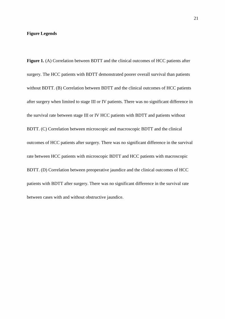
Figure 1. (A) Correlation between BDTT and the clinical outcomes of HCC patients after
surgery. The HCC patients with BDTT demonstrated poorer overall survival than patients
without BDTT. (B) Correlation between BDTT and the clinical outcomes of HCC patients
after surgery when limited to stage III or IV patients. There was no significant difference in
the survival rate between stage III or IV HCC patients with BDTT and patients without
BDTT. (C) Correlation between microscopic and macroscopic BDTT and the clinical
outcomes of HCC patients after surgery. There was no significant difference in the survival
rate between HCC patients with microscopic BDTT and HCC patients with macroscopic
BDTT. (D) Correlation between preoperative jaundice and the clinical outcomes of HCC
patients with BDTT after surgery. There was no significant difference in the survival rate
between cases with and without obstructive jaundice.
22
Table 1 Correlations between clinicopathological features and BDTT BDTT-negative BDTT-positive
Variable (n = 732) (n = 42) p valueAge† 0.428
64 (18-90) 61.5 (44-80)Gender 1.000
Female 127 7Male 605 35
HBs antigen 1.000Negative 444 26Positive 288 16
HCV antibody 0.240Negative 490 32Positive 242 10
Child-Pugh Classification 0.162A 709 39B 23 3
ICG R15 † 0.06113.7 (2.5-94.4) 13.7 (2.7-33.3)
Liver cirrhosis 1.000Absence 513 30Presence 219 12
AFP (ng/ml)† 0.01518.6 (0-1816620) 97.3 (1.5-624717)
TNM Stage <0.0001I or II 356 1
III or IV 376 41Tumor size (cm)† 0.004
4.2 (0.3-22) 5 (1.5-22.5)Differentiation 0.061
Well 116 2Moderate 468 27
Poor 148 13Portal vein invasion <0.0001
Absence 546 9Presence 186 33
Anatomical resection 0.0004no 203 2
yes 529 40
† Expressed as median (range)
Abbreviations: BDTT, bile duct tumor thrombus; HBs antigen, hepatitis B surface antigen;HCV antibody, hepatitis C virus antibody; ICG R15, indocyanine green retention rate at 15
23
Variable nAge
<60 18>60 24
GenderFemale 7
Male 35HBs antigen
Negative 26Positive 16
HCV antibodyNegative 32Positive 10
Child-Pugh ClassificationA 39B 3
ICG R15 (%)<10 19>10 23
Liver cirrhosisAbsence 30Presence 12
AFP(ng/ml)<20 14>20 28
TNM StageI or II 1
III or IV 41Tumor number
Single 27Multiple 15
Tumor size (cm)<10 32>10 10
DifferentiationWell/Moderate 29
Poor 13Portal vein invasion
Absence 9Presence 33
Extent of BDTTMicroscopic 21Macroscopic 21
Anatomical resectionno 2
yes 40Preoperative jaundice
no 36yes 6
Tumor sizeDifferentiationPortal vein invasion
Tumor size
0.3298 0.304855.4±12.7 45.4±11.9
76.2±10.830.4±17.7
33.3±27.2
27.7±13.5
79.0±11.6
78.3±9.6
Table 2 Univariate and multivariate analyses of prognostic factors for HCC with BDTT
Recurrence
37.3±9.250.0±35.3
40.4±11.8
0.5299
5-years (%)
73.4±12.4
78.1±19.1
80.0±15.6
77.1±19.675.8±11.0
68.6±13.886.9±11.4
77.7±9.8
74.1±11.5
100.0±0.0
80.9±16.5
76.6±18.2
0.0±0.0
71.2±13.086.6±11.8
69.7±12.9100.0±0.0
80.2±11.073.1±20.6
100.0±0.078.7±9.9
100.0±0.075.0±10.6
79.2±9.320.0±17.8
P value0.9212
0.7030
0.2021
0.5858
0.7237
0.4165
0.9809
0.4744
0.0536
0.0001
0.7210
0.1135
0.4673
0.2828
SurvivalUnivariate analysis
P value0.1616
0.9534
0.6415
0.0963
0.6566
0.6637
0.6800
0.3120
0.0039
0.0321
0.0380
0.6666
0.5469
5-years (%)
48.4±12.917.5±13.8
34.2±19.536.3±10.2
35.2±11.440.5±14.1
39.7±10.1
83.3±15.2
36.0±10.235.5±18.5
100.0±0.036.4±9.0
39.8±11.328.1±14.9
20.6±11.3
28.0±8.9
50.0±35.336.2±9.2
35.3±9.150.0±35.3
52.5±10.60.0±0.0
45.7±11.419.9±12.4
0.4796 0.333148.6±12.1 81.4±11.114.7±12.2 70.5±15.5
HR 95% CI P value2.940 1.111-7.704 0.03042.737 1.033-6.989 0.04317.235 1.075-197.74 0.0401
Abbreviations: BDTT, bile duct tumor thrombus; HBs antigen, hepatitis B surface antigen; HCV antibody, hepatitis C virus antibody; ICGR15, indocyanine green retention rate at 15 min; AFP, alpha-fetoprotein; HR, hazard ratio; CI, confidence interval
Multivariate analysisSurvival
RecurrenceHR 95% CI P value
3.448 1.139-10.539 0.0288

24
Table 3 Recurrence site and its treatment after hepatectomy for HCC with BDTTTreatment (Number of cases)
The first recurrence site TACE Resection Systemic chemotherapy Radiation Best supportive care TotalLiver 10 2 0 0 3 15
Lung 0 2 1 0 0 3
Liver and lung 0 0 0 0 2 2
Bone 0 0 0 1 1 2
Lymph node 0 0 0 1 0 1
Bile duct 0 1 0 0 0 1
Total 10 5 1 2 6 24
Abbreviations: BDTT, bile duct tumor thrombus; TACE, transcatheter arterial chemoembolization
25
Table 4 Perioperative outcomes of HCC with BDTTWithout obstructive
jaundiceWith obstructive
jaundiceVariable (n = 36) (n = 6) p valueOperation time (min)† 0.110
370.5 (157-1019) 324 (289-370)Blood loss (ml)† 0.914
660 (40-35820) 695 (340-2155)Anatomical resection 1.000
no 2 0yes 34 6
Postoperative complication‡ 1.000III 6 0IV 0 0
Postoperative hospital stay (days)† 0.95722 (9-108) 22.5 (13-40)
90days mortality 1.0000 0
† Expressed as median (range)‡ Clavien-Dindo grading III or IV
Abbreviations: HCC, hepatocellular carcinoma; BDTT, bile duct tumor thrombus