Embed Size (px)
Citation preview
Heterochrony of cuticular differentiation in eusocialcorbiculate bees
Moysés ELIAS-NETO1, Ana L. O. NASCIMENTO
1, Ana M. BONETTI
2,
Fabio S. NASCIMENTO1, Sidnei MATEUS
1, Carlos A. GARÓFALO
1,
Márcia M. G. BITONDI1
1Departamento de Biologia; Faculdade de Filosofia, Ciências e Letras de Ribeirão Preto, Universidade de SãoPaulo, Av. Bandeirantes 3900, 14040-901 Ribeirao Preto, Sao Paulo, Brazil
2Instituto de Genética e Bioquímica, Universidade Federal de Uberlândia, Rua Acre, bloco 2E, 38400-902Uberlandia, Minas Gerais, Brazil
Received 28 June 2013 – Revised 15 October 2013 – Accepted 30 October 2013
Abstract – The exoskeleton (cuticle) of insects varies widely in shape, biomechanical properties, andfunctions, which are inherent to the biological species and developmental stage, besides showing a wealth ofarchitectural specializations and nuances in the different body regions. The morphological pattern ofexoskeleton maturation was studied in eusocial and solitary bees, including species of all three eusocial tribesof corbiculate bees as follows: Apini, Meliponini, and Bombini. The results showed striking differences in therate of cuticle maturation between the solitary bees that leave the nest soon after the adult ecdysis, and theeusocial bees that take longer to leave the colony for foraging activities. There was a clear delay in the post-ecdysial cuticle tanning (melanization and sclerotization) in the eusocial bee species in comparison to thesolitary species, suggesting adaptation to sociality and to the protective environment of the colony. Suchheterochrony of cuticle maturation seems a conserved ontogenetic trait related to the way of life in corbiculatebees. The data generated a basic framework of exoskeleton maturation in corbiculate bees, under ontogeneticand evolutionary approaches, and give experimental support for further research on adaptation to the colonyenvironment.
exoskeleton / tanning / eusociality / Apinae / solitary bees
1. INTRODUCTION
The cuticular exoskeleton is a multifunction-al structure because in addition to the support itprovides the insects, it allows locomotion andmovements, protects against predators, andprevents desiccation, besides functioning in themechanical digestion carried out by mouthparts,
and in the sensorial perception via antennae andsensorial setae (Hepburn 1985).
Exoskeleton differentiation is a remarkableevent of insect ontogenesis and occurs througha process known as tanning, meaning theprogressive darkening (melanization) and hard-ening (sclerotization) of the newly-secretedcuticle (Andersen 2005). Ontogenetically, twobasic types of exoskeleton can be identified inthe honey bees, the flexible and colorless(unpigmented) larval and pupal exoskeletons,and the rigid (highly sclerotized) and pigmentedadult exoskeleton. Melanin pigments are depos-ited into the adult cuticle, which becomesincreasingly sclerotized, as it differentiates
Corresponding author: M. Elias-Neto,[email protected];M. Bitondi, [email protected] editor: Monique Gauthier
Apidologie Original article* INRA, DIB and Springer-Verlag France, 2013DOI: 10.1007/s13592-013-0254-1
underneath the pupal cuticle. In the honey bee,melanization starts in the developing adultcuticle 80 h after pupal cuticle apolysis, ap-proximately (Michelette and Soares 1993).Apolysis progressively occurs along the anteri-or–posterior body axis, and adult cuticle depo-sition and tanning follow this pattern. Thus,cuticle tanning is first seen in the head andthorax. The abdomen is weakly pigmented andsclerotized until close to the adult ecdysis.
Cuticular melanization and sclerotizationoccur simultaneously through the same biosyn-thetic pathway starting with hydroxylation oftyrosine to dopa, followed by a step requiringthe catalytic activity of dopa-decarboxylase fordopamine production. Dopamine is a precursorin the synthesis of melanin, catalyzed byphenoloxidases, and in the synthesis ofsclerotizing agents. Sclerotization starts whenN-acetyldopamine and N-β-alanyldopamine areoxidized to quinones by the action ofphenoloxidases and laccases. Quinones thenreact with cuticular proteins, thus stabilizingcuticle structure. These reactions can occurbefore the ecdysis (pre-ecdysial sclerotization)or after ecdysis (post-ecdysial sclerotization)(review in Andersen 2010). In the honeybee,the cuticle deposited before the adult ecdysis isless sclerotized than the post-ecdysial cuticle(Andersen et al. 1981) thus indicating thatsclerotization proceeds throughout part of theadult stage. A detailed description of themorphogenesis of the adult integument (epider-mis and its associated cuticle) in the honeybee,using conventional histology and light micros-copy, evidenced that exoskeleton maturationcontinues and is intensified after ecdysis to theadult stage (Elias-Neto et al. 2009).
We have observed that the rate of post-ecdysial cuticle tanning differs among beespecies. This variation in the temporal patternof exoskeleton tanning, or heterochrony, mayrepresent an adaptive mechanism related to thebee lifestyle. Heterochrony is defined as anyevolutionary change in the temporal pattern ofexpression of a certain characteristic (West-Eberhard 2003), meaning the shift (acceleration
or delay) in the relative timing of developmentalevents. Heterochrony is a phenomenon of greatimportance in the evolution, both in the frequencyof occurrence and the origin of significant evolu-tionary changes (Gould 1977). New approachesand tools for the study of heterochrony at cellularand molecular levels have provided opportunitiesto a deeper understanding on the basis of the shiftsin the timing of ontogenetic events (Smith 2003).
In this context, the evolution of insect socialityrepresents remarkable scenery for testing hypothe-ses related to adaptive mechanisms of exoskeletonmaturation. The protective environment of thecolony possibly has led to a loosening of theselective pressures over the cuticular differentia-tion, which enables, for instance, the emergence ofadults with a relatively immature exoskeleton.Complete maturation is only attained several dayslater, when the eusocial bees become foragers. Incontrast, the solitary bees leave the nest soon afterthe emergence from the brood cells, and thisevidently demands a fully mature exoskeleton.Therefore, morphological markers of exoskeletontanning, such as the dynamics of melanization andsclerotization, are useful to investigate whether thecomplexity of the evolution of eusociality includedthe slowdown in the rate of exoskeletonmaturation.
Co r b i c u l a t e b e e s (Ho l ome t a bo l a ,Hymenoptera, Apidae) are a monophyleticgroup (Michener 2007), supported by a seriesof synapomorphies, including the presence of acorbicula (basket-shaped structure in the fe-male tibia which is used for pollen transport).Such bee group forms a distinct clade withinApinae (Apidae). It includes four tribes asfollows: Euglossini (“orchid bees”), Bombini(“bumblebees”), Apini (“honey bees”), andMeliponini (“stingless bees”). Euglossini spe-cies are solitary, communal or primitivelysocial; Bombini species are primitively euso-cial (except for the Psithyrus, which comprisessocial parasites) (Silveira et al. 2002);Meliponini and Apini show true sociality(eusociality) (Michener 2007).
Except for a few studies in Apis mellifera(Thompson 1978; Elias-Neto et al. 2009), othercorbiculate bee species have not been studied asregards to developmental morphology of the
M. Elias-Neto et al.
exoskeleton, despite a series of reports (com-piled in Cruz-Landim and Abdalla 2002) oncharacterization of exocrine glands associatedwith the epidermis and cuticle.
Species of the bee tribes above mentioned wereused to investigate whether the temporal shift incuticle tanning reflects the evolutionary transition toeusociality. In a comparative approach, the purposeof the present workwas to investigate the hypothesisthat the heterochrony of cuticular maturation is aconserved ontogenetic phenomenon related to thesocial environment in corbiculate bees.
2. MATERIALS AND METHODS
2.1. Bees
This study included species of all three eusocialtribes of corbiculate bees as follows: Apini (A.mellifera), Meliponini (Frieseomelitta varia,Tetragonisca angustula, Scaptotrigona aff. depilis,Melipona scutellaris), and Bombini (Bombus morio).The outgroup consisted of the solitary bees Centrisanalis (Centridini) and Tetrapedia diversipes(Tetrapediini). Females (workers) of these specieswere collected soon after the adult ecdysis (newly-emerged bees) and when they were foraging.
The bees were collected at the Campus of the SãoPaulo University in Ribeirão Preto. The species A.mellifera, F. varia, T. angustula, S. aff. depilis, and M.scutellaris were obtained in the experimental apiary/meliponary of the Department of Genetics. Specimens ofB. morio were collected from a colony at the Laboratoryof Ecology, Department of Biology, where trap-nests ofC. analis and T. diversipes are also maintained.
The criteria for the selection of the species werebased on the representativeness of different evolu-tionary lineages. Previous studies on the biology ofMeliponini, Bombini, and solitary species, whichenabled their maintenance and collection (Camillo etal. 1995; Nogueira-Neto 1997; Jesus and Garófalo2000; Goulson 2006), also influenced our choice.
2.2. Integument sections
Histological sections were prepared using theintegument dissected from the dorsal region of thethorax of newly-emerged and forager bees. The
thoracic sections were made in a longitudinal plane,cutting the dorsal portion of the thorax (includingpronotum, mesonotum, and metanotum) at the mid-line. The cuticle pieces were fixed for 24 h at 4 °C in4 % paraformaldehyde in phosphate buffer (0.1 M,pH 7.3). This was followed by dehydration insuccessively higher concentrations (70, 80, 90, and95 %) of ethanol in water (v/v) for 30 min in eachsolution. The cuticles were then embedded inmethacrylate resin. Sections of 5 μm were obtainedwith the Microm HM200 Ergostar Microtome, andwere stained with methylene blue and basic fuchsinfor 3 min, followed by a rapid washing in distilledwater. Sections were mounted in Entellan (Merck)and examined and photographed using an opticalmicroscope (Axioskop II photomicroscope, Zeiss). Atleast three samples of each developmental stage wereused in the histological preparations. The best-qualityphotographs (mesonotum) were presented.
2.3. Whole mounts
Whole mounts were also prepared with the dorsalportion of the thorax (pronotum, mesonotum, andmetanotum). The thoracic integument samples weregently rinsed in Ringer saline, cleaned of excess fatbody and muscles, and mounted on slides using pureglycerol. The whole mounts were immediatelyexamined in the Axioskop II photomicroscope andphotographed. At least three samples were obtainedfor each stage of development. The best-qualityphotographs (mesonotum) were shown.
3. RESULTS
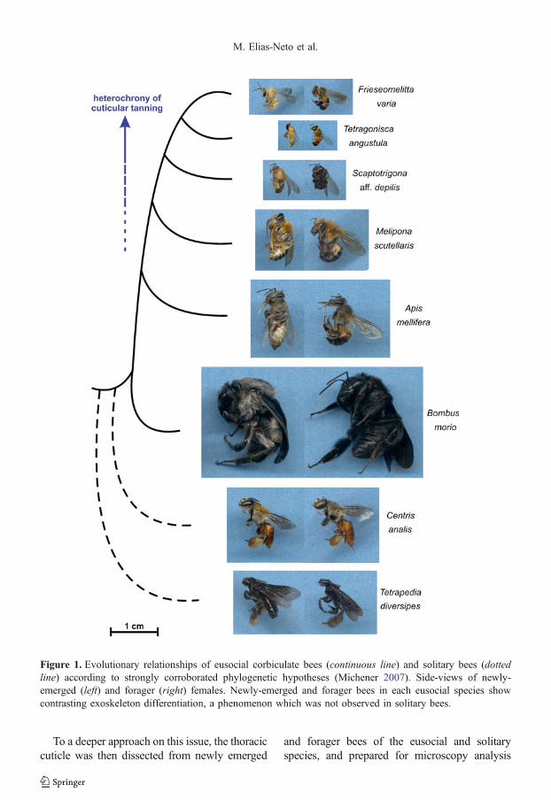
Forager bees from the eusocial species A.mellifera, F. varia, T. angustula, S. aff. depilis,M. scutellaris, and B. morio showed in a lesseror greater degree, depending on the species, adarker and stiffer cuticular exoskeleton thannewly-emerged bees. Such difference was notobserved in the solitary bee species (C. analisand T. diversipes) (Figure 1). These intra- andinterspecific comparisons highlighted that incontrast to the solitary bees, the exoskeleton ofeusocial bees takes longer to differentiate as acompletely mature structure.
Cuticle differentiation in eusocial bees
To a deeper approach on this issue, the thoraciccuticle was then dissected from newly emerged
and forager bees of the eusocial and solitaryspecies, and prepared for microscopy analysis
Figure 1. Evolutionary relationships of eusocial corbiculate bees (continuous line) and solitary bees (dottedline) according to strongly corroborated phylogenetic hypotheses (Michener 2007). Side-views of newly-emerged (left) and forager (right) females. Newly-emerged and forager bees in each eusocial species showcontrasting exoskeleton differentiation, a phenomenon which was not observed in solitary bees.
M. Elias-Neto et al.
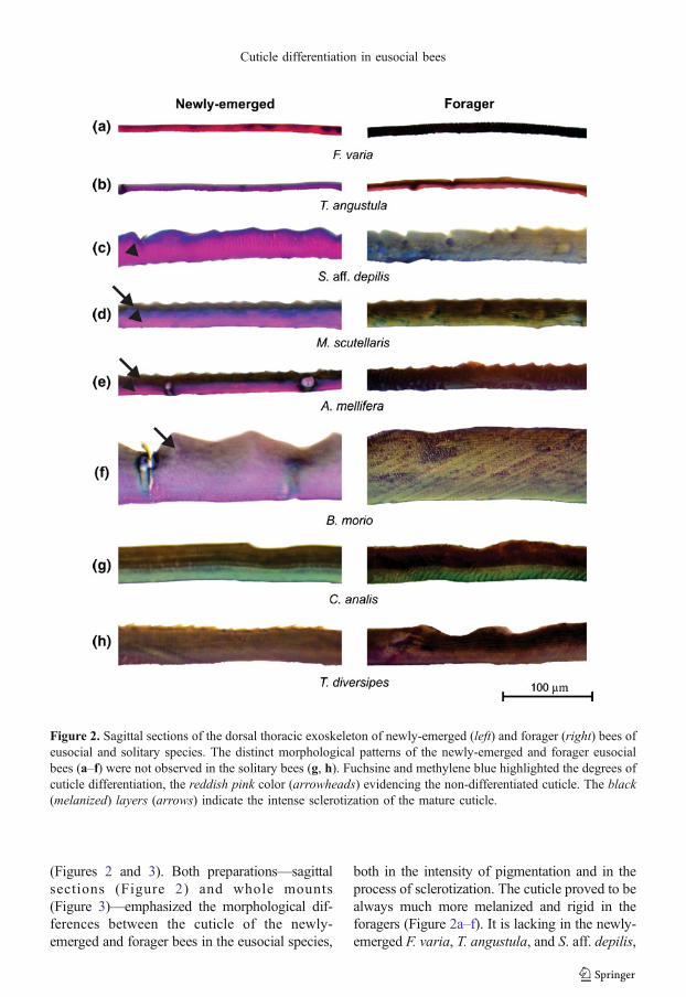
(Figures 2 and 3). Both preparations—sagittalsections (Figure 2) and whole mounts(Figure 3)—emphasized the morphological dif-ferences between the cuticle of the newly-emerged and forager bees in the eusocial species,
both in the intensity of pigmentation and in theprocess of sclerotization. The cuticle proved to bealways much more melanized and rigid in theforagers (Figure 2a–f). It is lacking in the newly-emerged F. varia, T. angustula, and S. aff. depilis,
Figure 2. Sagittal sections of the dorsal thoracic exoskeleton of newly-emerged (left) and forager (right) bees ofeusocial and solitary species. The distinct morphological patterns of the newly-emerged and forager eusocialbees (a–f) were not observed in the solitary bees (g, h). Fuchsine and methylene blue highlighted the degrees ofcuticle differentiation, the reddish pink color (arrowheads) evidencing the non-differentiated cuticle. The black(melanized) layers (arrows) indicate the intense sclerotization of the mature cuticle.
Cuticle differentiation in eusocial bees
the melanized superficial layer, which is onlyevident in the foragers. In spite of the presence ofsuch a layer in the cuticle of newly-emerged beesof the other eusocial species, M. scutellaris, A.mellifera, and B. morio (Figure 2d–f, arrows), it iscomparatively thicker in the foragers of thesespecies. Themelanized surface of the cuticle is notalways well-delimited in the stained cross sec-tions, thus making the comparison less obvious insome species, likeB. morio (Figure 2f). Even so, itis clear that a completely differentiated (mature)cuticle occurs in the eusocial bees only severaldays after the emergence. In contrast, the solitaryspecies, C. analis and T. diversipes, collected atthe emergence or when they were foraging,showed very similar cuticle patterns, which arecharacterized by a thick melanized layer(Figure 2g, h).
In addition to the differences in melanization,cuticle sections of the newly-emerged eusocialbees showed a pink-reddish layer that varies inthickness among the different species (Figure 2a–f,arrowheads). This layer undergoes intense modi-fications in its chemical properties during cuticularmaturation as deduced by the change in its color inforagers of the eusocial species. The pink-reddishcolor thus marks the yet undifferentiated layer ofthe cuticle.
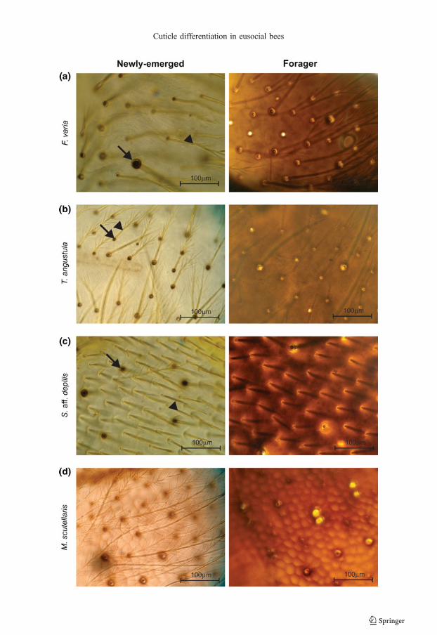
In the whole mount preparations (Figure 3),there was also a clear distinction between thecuticular patterns of the newly-emerged andforager bees of the eusocial species (Figure 3a–f),which was not observed in the solitary species(Figure 3g, h). F. varia, T. angustula, and S. aff.depilis showed the highest degree of cuticularmaturation heterochrony (Figure 3a–c). Foragersof these species have a brownish cuticle with ahomogeneous distribution of pigments, whereas inthe newly-emerged bees, pigmentation was con-centrated in the setae and setal sockets.
Compared to F. varia, T. angustula, and S.aff. depilis, the eusocial species M. scutellaris,A. mellifera, and B. morio showed a moderatedegree of cuticular maturation heterochrony,although indubitably pigmentation is muchmore intense in the foragers (Figure 3d–f).
The newly-emerged and forager bees of thesolitary species, C. analis and T. diversipes,
showed cuticles morphologically very similar,without apparent differences in the pigmenta-tion pattern (Figure 3g, h).
4. DISCUSSION
The present study included six species ofBombini, Meliponini, and Apini, which representbranches of the corbiculate phylogenetic tree.Although there is some controversy about thephylogeny of corbiculate bees (Lockhart andCameron 2001; Danforth et al. 2013), a stronglycorroborated grouping, both by the combined use ofmorphological and molecular characters (Chavarríaand Carpenter 1994) and also by behavioralevidence (Noll 2002), consists of Euglossini +(Bombini + (Meliponini +Apini). This classificationwas already seen in previous classical reviews(Michener 1944; Kerr and Esch 1965).
In addition to the six social species, our studyincluded two solitary species, which comprisethe majority of bee species (Batra 1984; Silveiraet al. 2002), and were characterized by thefemale independence during nest construction.There is no cooperation or division of laboramong the females of a same generation and,usually, there is no overlap in generationsbetween parents and offspring. The massiveprovisioning of food ensures the completedevelopment of the larvae toward the adultstage (Michener 1974). In general, the solitarybees dig their nests mainly in the ground or intree trunks, although about 5 % of the speciesnest in preexisting cavities (Krombein 1967).Such feature has benefited bee biology studies,since females are drawn to nesting in artificially
�Figure 3. Whole preparations of the dorsal thorac-ic integument of newly-emerged (left) and forager(right) bees of the eusocial (F. varia, T. angustula,S. aff. depilis, M. scutellaris, A. mellifera, B.morio) and solitary (C. analis, T. diversipes)species. The cuticle is much more intenselypigmented in foragers than in the newly-emergedof each eusocial species (a–f); newly-emerged andforagers of the solitary species showed a similardegree of cuticle pigmentation (g, h). Setae(arrowheads) and setal sockets (arrows).
M. Elias-Neto et al.
Cuticle differentiation in eusocial bees

Figure 3. continued.
M. Elias-Neto et al.
built cavities, the trap-nests (Camillo et al.1995). More important in the context of thepresent study, and in contrast to the eusocialbees, the solitary bees leave the nest immedi-ately after emerging as adults. Their cuticularexoskeleton, therefore, is ready to face theexternal environmental conditions.
The delay in exoskeleton tanning, or matura-tion, is apparently a conserved ontogeneticphenomenon among the eusocial corbiculatebees. We noticed distinct degrees of cuticularmaturation heterochrony between the adults ofthe phylogenetically related eusocial speciesincluded in this study. As an example, in F.varia, which shows the greater degree ofcuticular heterochrony, an extreme eusocial traitis also observed, such as the permanent sterilityof the workers (Da Cunha and Campos 1993;Boleli et al. 1999). Conversely, the primitivelyeusocial bee B. morio shows a more subtlecuticular heterochrony. The existence of suchgradient is hypothetical and new studies involv-ing other species are needed to obtain a moreelucidative scenario on this subject.
There is a straight association between sociallife and the nest, and any theory that addressessociality evolution must consider some previousadaptations which favor gregarious behaviorand inhibit dispersion behaviors (Nowak et al.2010). The immature cuticle at the moment ofadult emergence acts in both ways. The fragileand slightly pigmented exoskeleton keeps thebees from leaving the nest because it does notprovide enough body protection against thedamaging effects of the external environment(developmental constraint). At the same time,the plastic cuticle, from the ontogenetic point ofview, possibly allows for the modeling ofcuticular communication systems between thecolony members (developmental plasticity).
An incipient and plastic cuticle seems anadvantage during the time window that goesfrom emergence (brood cell-to-indoor nesttransition) to the age at onset of foraging (nest-to-field transition). It is exactly during this timeinterval that the social life in the colonymanifests more intensely. As an organ thatmakes the interface between the organism and
the environment, the cuticle is an importantsource of semiochemicals, such as hydrocar-bons, for social communication. The cuticularhydrocarbons show discriminated abilities forrecognition of nestmates, and cuticle fromyoung and forager bees of social species differin the profiles of these compounds (Kather et al.2011; Falcon 2013). Therefore, the relativelyimmature cuticle of the newly-emerged andyoung bees is possibly advantageous for theirrecognition and acceptance by the nestmates,and integration in the colony. Like melanizationand sclerotization, the blends of hydrocarbonsare markers of exoskeleton maturation, since itcan distinguish the immature from the maturecuticle. In this sense, variations in hydrocarbonprofiles are consistent with the hypothesis thatthe cuticle of eusocial bees only becomescompletely mature when they start foragingactivities.
The lateness in exoskeleton maturation is heretentatively assigned to the way of life within aprotective nest with a humid environment andcontrolled temperature (Winston 1991), bothcertainly contributing for the cuticle waterproof-ing function. In addition, honeybees show mech-anisms for protection against diseases, such ashygienic behavior, that confer a degree of socialimmunity (Evans and Spivak 2010) and protectcolony members (including those with an imma-ture cuticle) against parasites and microbialpathogens. Therefore, an immature exoskeletonat the moment of emergence from the brood celldoes not appear to be a disadvantage to theeusocial bee as to its protection.
Thus, the cuticle of young eusocial bees isevidently adapted to the within-hive environ-ment where they live and exert tasks ascooperative care of the brood during the firsthalf of adulthood. At the right time, it achievesthe structural complexity needed for the intenseforaging activities usually carried out during thesecond half of adulthood.
Changes in bee behavior and physiologyduring age-related task transition from insideto outside the nest have been linked to extensivechanges in gene expression in the brain (Amentet al. 2008, 2011). However, there are no studies
Cuticle differentiation in eusocial bees
on global gene expression in the cuticle-producing epidermis during transition for forag-ing. Honeybee genes involved in adult cuticleformation and maturation, such as the genesencoding a structural cuticular protein(AmelCPR14) (Soares et al. 2007), a laccaseenzyme (Amlac2) (Elias-Neto et al. 2010), and asubunit of the neurohormone bursicon (unpub-lished data), showed expression profiles shiftedtoward the adult stage. This suggests that thesegenes are involved in the post-ecdysial matura-tion of the exoskeleton.
Cuticular maturation heterochrony in beespossibly has arisen by the combined influencesof environmental and genetic factors.Heterochrony can be caused by environmentalstimuli, which may generate intraspecific alter-ations in the life cycle, caste, or gender(Miyazaki et al. 2010) and even in speciationevents (Gould 1977). The environment does notact in the evolutionary process solely throughnatural selection, but also as an active agent inthe development (West-Eberhard 1989, 2005).Therefore, cuticular maturation postponement ineusocial bees may have been induced by colonyenvironment, and possibly is associated withdelayed activation of genes involved in cuticleformation and maturation. Further transcriptome-wide analysis using integuments of newly-emerged and forager bees from solitary andeusocial species has the potential to determinethe extension of expression of genes involved inexoskeleton maturation, and whether this expres-sion differs dependently on the bee lifestyle. Inaddition to this approach, other question on thismatter is whether the cuticle reaches the completematurity even in bee workers that permanentlyremain into the hive without ever leaving forforaging, as exceptionally verified (Seehuus et al.2006; Amdam 2011). Such knowledge may helpaddress whether cuticular maturation is dependentupon the social context.
The acquisition of eusociality is a greatevolutionary transition, in which complexinteraction systems resulted in the emergenceof new properties, such as social homeostasis(Maynard Smith and Szathmáry 1995). Theorigin and fixation of heterochrony of cutic-
ular maturation in eusocial bee speciesperhaps may be understandable if we lookfor the concept of colony as an adaptiveunity (Seeley 1989) subject to natural selec-tion acting in different levels of biologicalorganization—multilevel selection (Krauseand Ruxton 2002; Korb and Heinze 2004).
Certainly, comparative studies along a gradi-ent of sociality to include solitary, semi-social,and eusocial bee species can actually revealunique aspects of exoskeleton maturation in thecontext of evolution of sociality.
ACKNOWLEDGMENTS
We thank L.R. Aguiar and J. Souza for technicalassistance in the apiary/meliponary. We also thankV.L.C. Figueiredo for providing assistance with thehistological preparations. This research was support-ed by the Fundação de Amparo à Pesquisa do Estadode São Paulo (FAPESP: 05/03926-5; 10/16380-9),which also provided a fellowship (07/08300-2; 12/09108-6) to M. Elias-Neto.
Hétérochronie de la différenciation cuticulaire chezles abeilles corbiculées eusociales
Exosquelette / tannage / eusocialité / Apinae / abeillessolitaires
Heterochronie in der Differenzierung der Kutikulabei eusozialen corbiculaten Bienen
Exoskelett / Aushärtung / Eusozialität / Apinae /solitäre Bienen
REFERENCES
Amdam, G.V. (2011) Social context, stress, and plasticityof aging. Aging Cell 10, 18–27
Ament, S.A., Corona, M., Pollock, H.S., Robinson, G.E.(2008) Insulin signaling is involved in the regulationof worker division of labor in honey bee colonies.Proc. Natl. Acad. Sci. 105, 4226–4231
Ament, S.A., Velarde, R.A., Kolodkin, M.H., Moyse, D.,Robinson, G.E. (2011) Neuropeptide Y-like signalingand nutritionally mediated gene expression and behav-ior in the honeybee. Insect Mol. Biol. 20, 335–345
M. Elias-Neto et al.
Andersen, S.O. (2005) Cuticular sclerotization andtanning. In: Gilbert, L.I., Iatrou, K., Gill, S. (eds.)Comprehensive molecular insect science, vol. 4, pp.145–170. Elsevier Press, Oxford
Andersen, S.O. (2010) Insect cuticular sclerotization: areview. Insect Biochem. Mol. Biol. 40, 166–178
Andersen, S.O., Thompson, P.R., Hepburn, H.R. (1981)Cuticular sclerotization in the honeybee (Apismellifera adansonii). J. Comp. Physiol. 145, 17–20
Batra, S.W.T. (1984) Solitary bees. Sci. Am. 250, 86–93
Boleli, I.C., Simões, Z.L.P., Bitondi, M.M.G. (1999) Celldeath in ovarioles causes permanent sterility inFrieseomelitta varia worker bees. J. Morphol. 242,271–282
Camillo, E., Garófalo, C.A., Serrano, J.C., Muccillo, G.(1995) Diversidade e abundância sazonal de abelhase vespas solitárias em ninhos-armadilha (Hymenop-tera, Apocrita, Aculeata). Rev. Bras. Entomol. 39,459–470
Chavarría, G., Carpenter, J.M. (1994) “Total evidence”and the evolution of highly social bees. Cladistics10, 229–258
Cruz-Landim, C., Abdalla, F.C. (eds.) (2002) Glândulasexócrinas das abelhas. FUNPEC, Ribeirão Preto
Da Cunha, M.A.S., CAMPOS, L.A.O. (1993)Desenvolvimento ovariano em operárias deFrieseomelitta varia varia (Lep 1836) (Hymenop-tera, Apidae). Rev. Bras. Biol. 53, 63–69
Danforth, B.N., Cardinal, S., Praz, C., Almeida, E.A.B.,Michez, D. (2013) The impact of molecular data onour understanding of bee phylogeny and evolution.Annu. Rev. Entomol. 58, 57–78
Elias-Neto, M., Soares, M.P.M., Bitondi, M.M.G. (2009)Changes in integument structure during the imaginalmolt of the honeybee. Apidologie 40, 29–39
Elias-Neto, M., Soares, M.P.M., Simões, Z.L.P.,Hartfelder, K., Bitondi, M.M.G. (2010) Develop-mental characterization, function and regulation of aLaccase2 encoding gene in the honeybee, Apismellifera (Hymenoptera, Apinae). Insect Biochem.Mol. Biol. 40, 241–251
Evans, J.D., Spivak, M. (2010) Socialized medicine:individual and communal disease barriers in honey-bees. J. Invertebr. Pathol. 103, S62–S72
Falcon, T.L. (2013) Maturação cuticular em Apis mellifera:Perfis de hidrocarbonetos cuticulares, expressão eevolução de desaturases e elongases, p. 119p. MScThesis, Universidade de São Paulo, Brazil
Gould, S.J. (1977) Ontogeny and phylogeny. HarvardUniversity Press, Cambridge
Goulson, D. (2006) Bumblebees: Behavior and ecology.Oxford University Press, New York
Hepburn, H.R. (1985) Structure of the integument.In: Kerkut, G.A., Gilbert, L.I. (eds.) Comprehen-sive insect physiology, biochemistry and pharma-cology, vol. 3, pp. 1–58. Pergamon Press, Oxford
Jesus, B.M.V., Garófalo, C.A. (2000) Nesting behaviorof Centris (Heterocentris) analis (Fabricius) insoutheastern Brazil (Hymenoptera, Apidae,Centridini). Apidologie 31, 503–515
Kather, R., Drijfhout, F.P., Martin, S.J. (2011) Taskgroup differences in cuticular lipids in the honey beeApis mellifera. J. Chem. Ecol. 37, 205–212
Kerr, W.E., Esch, H. (1965) Comunicação entre asabelhas sociais brasileiras e sua contribuição para oentendimento da sua evolução. Ciênc. Cult. 17, 529–538
Korb, J., Heinze, J. (2004) Multilevel selection and socialevolution of insect societies. Naturwissenschaften 91,291–304
Krause, J., Ruxton, G.D. (2002) Living in groups.Oxford University Press, New York
Krombein, K.V. (1967) Trap-nesting wasps and bees: lifehistories, nests, and associates. Smithsonian Press,Washington
Lockhart, P.J., Cameron, S.A. (2001) Trees for bees.Trends Ecol. Evol. 16, 84–88
Maynard Smith, J., Szathmáry, E. (1995) Themajor transitionsin evolution. W. H. Freeman Spektrum, Oxford
Michelette, E.R.F., Soares, A.E.E. (1993) Characteriza-tion of preimaginal developmental stages in Afri-canized honeybee workers (Apis mellifera L).Apidologie 24, 431–440
Michener, C.D. (1944) Comparative external morphology,phylogeny, and a classification of the bees (Hymenop-tera). Bull. Am. Mus. Nat. Hist. 82, 151–326
Michener, C.D. (1974) The social behavior of the bees—Acomparative study. Harvard University Press, Cambridge
Michener, C.D. (2007) The bees of the world. JohnsHopkins University Press, Baltimore
Miyazaki, S., Murakami, T., Kubo, T., Azuma, N.,Higashi, S., Miura, T. (2010) Ergatoid queendevelopment in the ant Myrmecina nipponica:modular and heterochronic regulation of castedifferentiation. Proc. R. Soc. B 277, 1953–1961
Nogueira-Neto, P. (1997) Vida e criação de abelhasindígenas sem ferrão. Editora Nogueirapis, São Paulo
Noll, F.B. (2002) Behavioral phylogeny of corbiculateApidae (Hymenoptera; Apinae), with special refer-ence to social behavior. Cladistics 18, 137–153
Nowak, M.A., Tarnita, C.E., Wilson, E.O. (2010) Theevolution of eusociality. Nature 466, 1057–1062
Seehuus, S.-C., Krekling, T., Amdam, G.V. (2006)Cellular senescence in honeybee brain is largelyindependent of chronological age. Exp. Gerontol.41, 1117–1125
Seeley, T.D. (1989) The honeybee colony as a superor-ganism. Am. Sci. 77, 546–553
Silveira, F.A., Melo, G.A.R., Almeida, E.A.B. (2002)Abelhas brasileiras: sistemática e identificação. F. A.Silveira, Belo Horizonte
Cuticle differentiation in eusocial bees
Smith, K.K. (2003) Time’s arrow: heterochrony and theevolution of development. Int. J. Dev. Biol. 47, 613–621
Soares, M.P.M., Elias-Neto, M., Simões, Z.L.P., Bitondi,M.M.G. (2007) A cuticle protein gene in thehoneybee: expression during development and inrelation to the ecdysteroid titer. Insect Biochem.Mol. Biol. 37, 1272–1282
Thompson, P.R. (1978) Histological development ofcuticle in the worker honeybee, Apis melliferaadansonii. J. Apic. Res. 17, 32–40
West-Eberhard, M.J. (1989) Phenotypic plasticity andthe origins of diversity. Annu. Rev. Ecol. Syst. 20,249–278
West-Eberhard, M.J. (2003) Developmental plasticity andevolution. Oxford University Press, New York
West-Eberhard, M.J. (2005) Developmental plasticityand the origin of species differences. Proc. Natl.Acad. Sci. 102, 6543–6549
Winston, M.L. (1991) The biology of the honeybee.Harvard University Press, USA
M. Elias-Neto et al.


![WAG Bees 160210 Presentation[1]](https://img.pdfslide.tips/doc/110x75/577d275b1a28ab4e1ea3b79d/wag-bees-160210-presentation1.jpg)