Embed Size (px)
Citation preview
High Value Care of GI
Bleeding
Bennie R. Upchurch MD, FACP, FASGE, AGAF, FACG
Medical Director, GI Bleeding Program, Division of Gastroenterology, Nutrition, and Hepatology
Ohio State University Wexner Medical Center
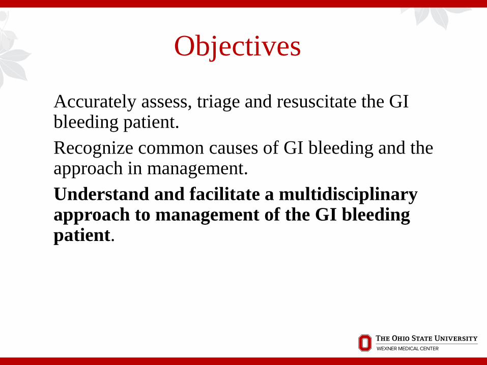
Objectives
• Accurately assess, triage and resuscitate the GI bleeding patient.
• Recognize common causes of GI bleeding and the approach in management.
• Understand and facilitate a multidisciplinary approach to management of the GI bleeding patient.
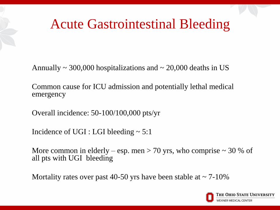
Acute Gastrointestinal Bleeding
• Annually ~ 300,000 hospitalizations and ~ 20,000 deaths in US
• Common cause for ICU admission and potentially lethal medical emergency
• Overall incidence: 50-100/100,000 pts/yr
• Incidence of UGI : LGI bleeding ~ 5:1
• More common in elderly – esp. men > 70 yrs, who comprise ~ 30 % of all pts with UGI bleeding
• Mortality rates over past 40-50 yrs have been stable at ~ 7-10%
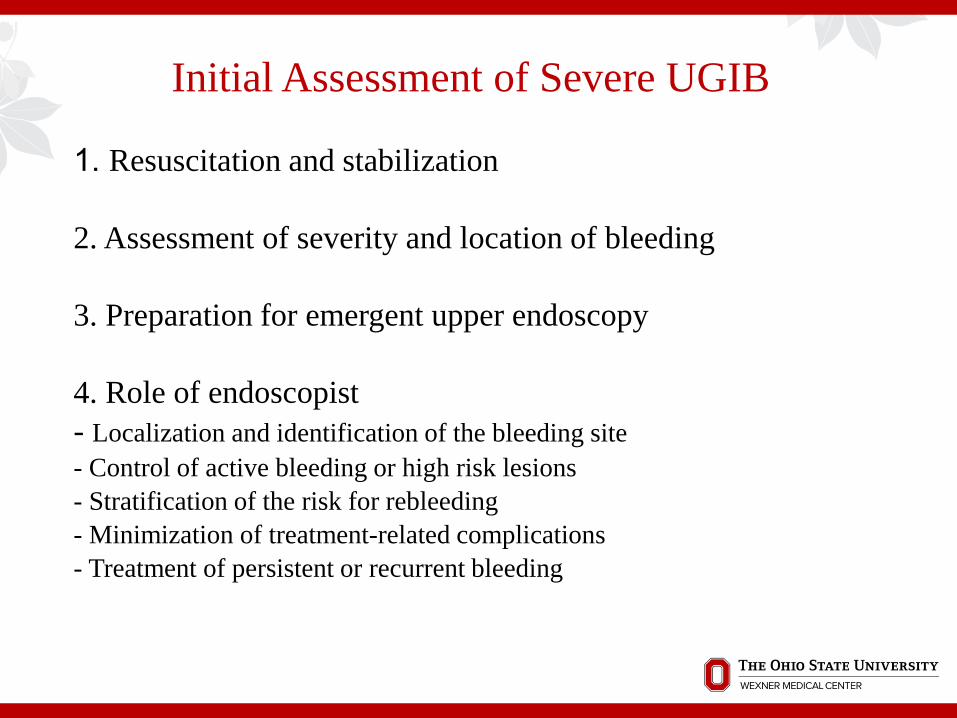
Initial Assessment of Severe UGIB
1. Resuscitation and stabilization
2. Assessment of severity and location of bleeding
3. Preparation for emergent upper endoscopy
4. Role of endoscopist
- Localization and identification of the bleeding site
- Control of active bleeding or high risk lesions
- Stratification of the risk for rebleeding
- Minimization of treatment-related complications
- Treatment of persistent or recurrent bleeding
Initial Evaluation - History
- Age: Elderly ( ischemia, cancer, diverticula)
Young ( ulcers, esophagitis, varices)
- Prior GI Bleeding
- Previous gastrointestinal disease
- Previous GI surgery
- Underlying Medical Disorders ( esp - liver disease, CKD)
- Meds : NSAIDS - ASA/Anticoagulant use
- Symptoms: Abdominal pain, fever, wt loss, anorexia, epistaxis, hematuria etc
- Allergies
Physical examination
- Hemodynamics with a thorough cardiopulmonary exam
- Skin ( spider angiomata, purpura, cutaneous telangiectasias /pigmentation)
- Abdomen ( ascites, tenderness, masses )
- Digital Rectal exam
Initial Patient Care/Management
- Appropriate IV access: 2 large bore I.V catheters
- IV fluids (NS/LR) and/or blood product resuscitation.
( Target HCT 30% in elderly/ 25-30% in young adults and pts with in
Portal HTN )
- Continuous cardio-pulm monitoring for those with coronary risk factors
with supplemental O2.
- Frequent vital signs / urine output monitoring.
- Consider intubation in those with altered mental status or brisk bleeding.
Labs and Studies
– Complete blood count ( check MCV) and electrolytes
( BUN >> Cr ), Iron studies.
– Coagulation Panel: PT/INR
– Type + screen or type + cross-match blood
– EKG for patients > 50 yrs or risk factors for heart
disease.
– Abdominal radiographs usually not indicated

Naso Gastric Lavage
- 79% sensitivity and 55% specificity for active bleeding
- Positive NGL does not provide etiology
- A non bloody aspirate in ~ 25% of UGIB
- A bile aspirate does not R/O UGIB
- NO evidence based data that NGL affects outcome
Predictors of Mortality
• Increasing age - Age > 70 yrs
• Concurrent active major organ disease
• Preexisting hospitalization ( mortality rate ~ 34% )
• Passing frequent frank blood. Esp if - Shock or Orthostatic hypotension
• Variceal vs. Non variceal ( eg. Variceal bleed has mortality rate of 30% during initial hosp, with 1 yr mortality rate of ~ 60% )
• Requiring emergency surgery for GIB
• Active bleeding / Transfusion requirement
– 4 or more red cell units in the first 24 hours
– 2 or more units for rebleeding event
– 6-8 units total
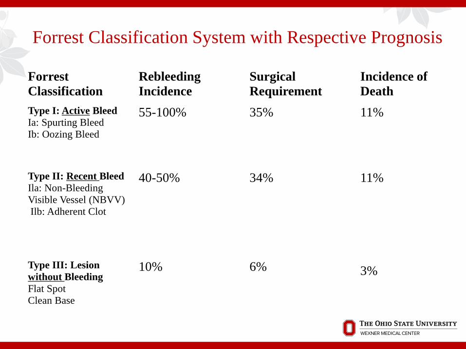
Forrest Classification System with Respective Prognosis
Forrest
Classification
Rebleeding
Incidence
Surgical
Requirement
Incidence of
Death
Type I: Active Bleed
Ia: Spurting Bleed
Ib: Oozing Bleed
55-100% 35% 11%
Type II: Recent Bleed
Ila: Non-Bleeding
Visible Vessel (NBVV)
Ilb: Adherent Clot
40-50% 34% 11%
20-30% 10% 7%
Type III: Lesion
without Bleeding
Flat Spot
Clean Base
10% 6% 3%
5% 0.5% 2%
Return to Article
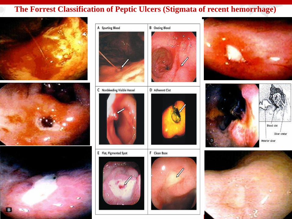
The Forrest Classification of Peptic Ulcers (Stigmata of recent hemorrhage)
Prognostic Features of GD Ulcers
• Ulcer size > 1 cm is associated with increased re-bleeding and mortality
• Endoscopic hemostasis is less successful in ulcers > 2 cm in size
• Greatest re-bleeding risk from ulcers is within first 72 hours
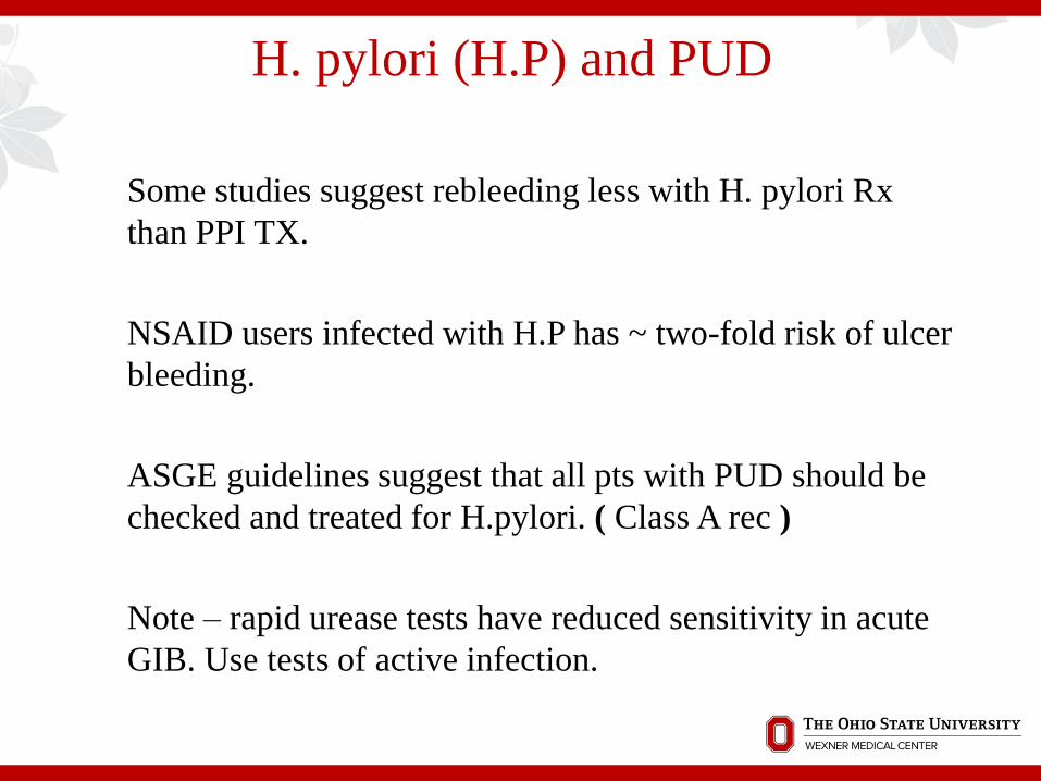
H. pylori (H.P) and PUD
• Some studies suggest rebleeding less with H. pylori Rx
than PPI TX.
• NSAID users infected with H.P has ~ two-fold risk of ulcer
bleeding.
• ASGE guidelines suggest that all pts with PUD should be
checked and treated for H.pylori. ( Class A rec )
• Note – rapid urease tests have reduced sensitivity in acute
GIB. Use tests of active infection.to r/o infection.
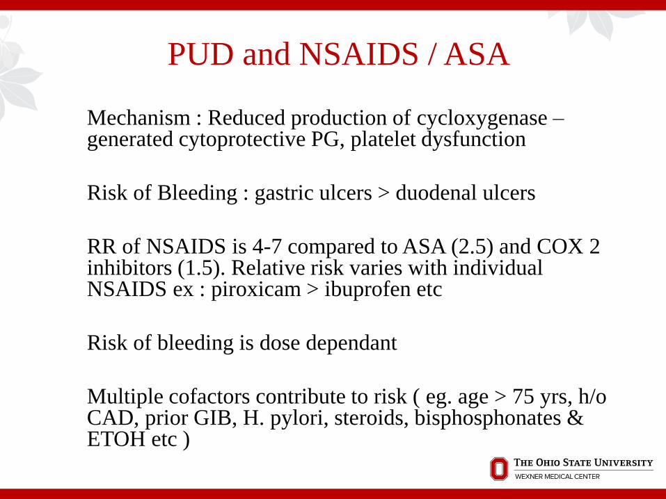
PUD and NSAIDS / ASA
• Mechanism : Reduced production of cycloxygenase –generated cytoprotective PG, platelet dysfunction
• Risk of Bleeding : gastric ulcers > duodenal ulcers
• RR of NSAIDS is 4-7 compared to ASA (2.5) and COX 2 inhibitors (1.5). Relative risk varies with individual NSAIDS ex : piroxicam > ibuprofen etc
• Risk of bleeding is dose dependant
• Multiple cofactors contribute to risk ( eg. age > 75 yrs, h/o CAD, prior GIB, H. pylori, steroids, bisphosphonates & ETOH etc )
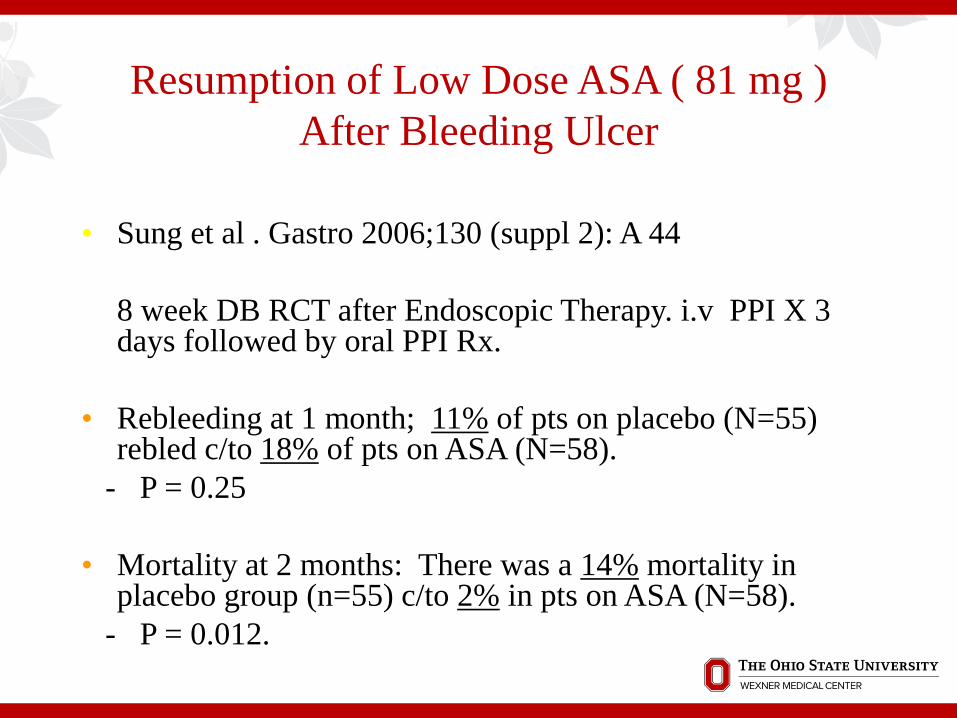
Resumption of Low Dose ASA ( 81 mg )
After Bleeding Ulcer
• Sung et al . Gastro 2006;130 (suppl 2): A 44
• 8 week DB RCT after Endoscopic Therapy. i.v PPI X 3 days followed by oral PPI Rx.
• Rebleeding at 1 month; 11% of pts on placebo (N=55) rebled c/to 18% of pts on ASA (N=58).
- P = 0.25
• Mortality at 2 months: There was a 14% mortality in placebo group (n=55) c/to 2% in pts on ASA (N=58).
- P = 0.012.
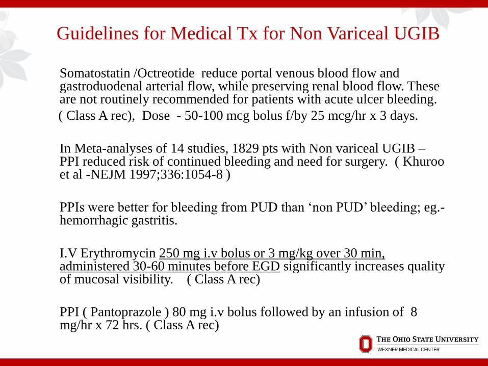
Guidelines for Medical Tx for Non Variceal UGIB
• Somatostatin /Octreotide reduce portal venous blood flow and gastroduodenal arterial flow, while preserving renal blood flow. These are not routinely recommended for patients with acute ulcer bleeding.
( Class A rec), Dose - 50-100 mcg bolus f/by 25 mcg/hr x 3 days.
• In Meta-analyses of 14 studies, 1829 pts with Non variceal UGIB – PPI reduced risk of continued bleeding and need for surgery. ( Khuroo et al -NEJM 1997;336:1054-8 )
• PPIs were better for bleeding from PUD than ‘non PUD’ bleeding; eg.- hemorrhagic gastritis.
• I.V Erythromycin 250 mg i.v bolus or 3 mg/kg over 30 min, administered 30-60 minutes before EGD significantly increases quality of mucosal visibility. ( Class A rec)
• PPI ( Pantoprazole ) 80 mg i.v bolus followed by an infusion of 8 mg/hr x 72 hrs. ( Class A rec)
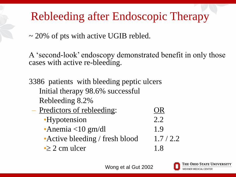
Rebleeding after Endoscopic Therapy
• ~ 20% of pts with active UGIB rebled.
• A ‘second-look’ endoscopy demonstrated benefit in only those cases with active re-bleeding.
• 3386 patients with bleeding peptic ulcers
– Initial therapy 98.6% successful
– Rebleeding 8.2%
– Predictors of rebleeding: OR
•Hypotension 2.2
•Anemia <10 gm/dl 1.9
•Active bleeding / fresh blood 1.7 / 2.2
• 2 cm ulcer 1.8
Wong et al Gut 2002
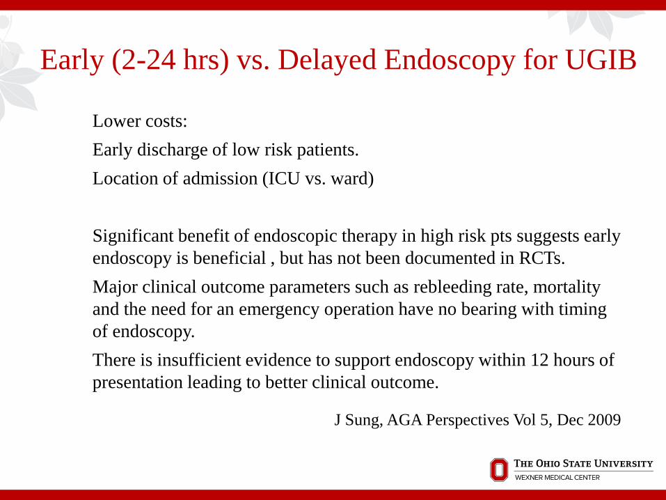
Early (2-24 hrs) vs. Delayed Endoscopy for UGIB
• Lower costs:
- Early discharge of low risk patients.
- Location of admission (ICU vs. ward)
• Significant benefit of endoscopic therapy in high risk pts suggests early
endoscopy is beneficial , but has not been documented in RCTs.
• Major clinical outcome parameters such as rebleeding rate, mortality
and the need for an emergency operation have no bearing with timing
of endoscopy.
• There is insufficient evidence to support endoscopy within 12 hours of
presentation leading to better clinical outcome.
J Sung, AGA Perspectives Vol 5, Dec 2009
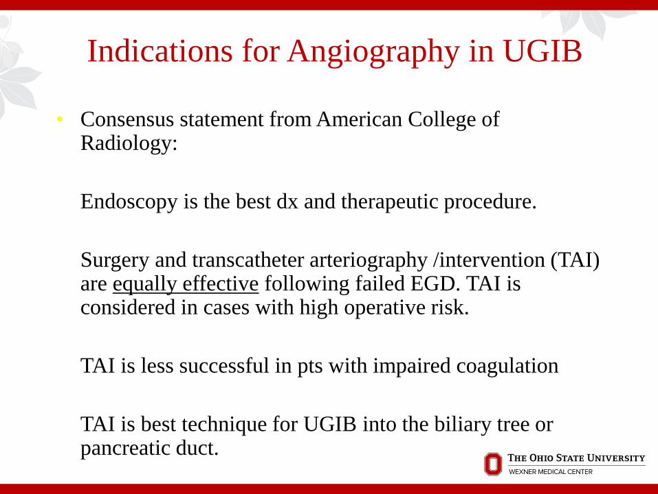
Indications for Angiography in UGIB
• Consensus statement from American College of Radiology:
- Endoscopy is the best dx and therapeutic procedure.
- Surgery and transcatheter arteriography /intervention (TAI) are equally effective following failed EGD. TAI is considered in cases with high operative risk.
- TAI is less successful in pts with impaired coagulation
- TAI is best technique for UGIB into the biliary tree or pancreatic duct.
Angiographic Therapy
• Bleeding should be > 0.5 ml/hr.
• Selective Intra-arterial vasopression – not used now.
Risks: Brady-arrhythmias, ischemia etc
• Selective occlusion of bleeding arteries with gelfoam, beads, tissues adhesives and coils etc are used.
• Rebleeding is common, and complications such as ischemia, infarction, perforation and abscess etc are prominent.
• CTA, MRA, CTe
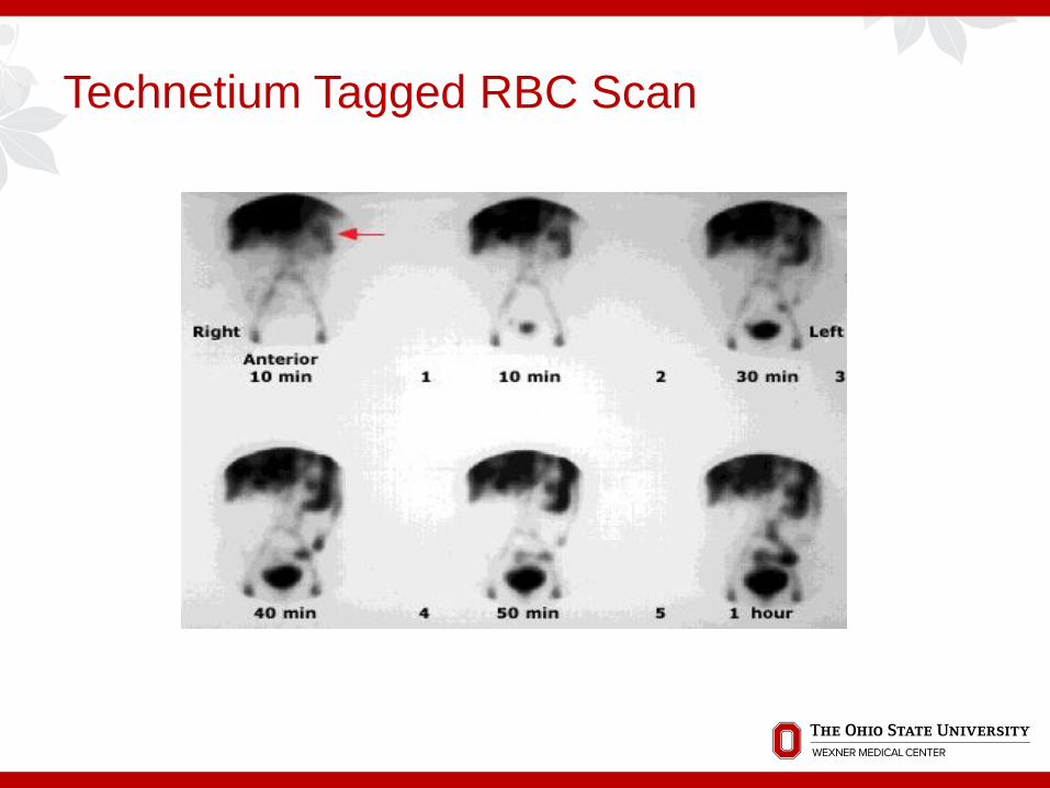
Technetium Tagged RBC Scan
Surgical Therapy for UGIB- When ?
• Role is controversial.
• Is usually considered in high risk cases when ;
1) HD instability even after > 3 units PRBC transfusions
2) TWO unsuccessful EGDs/attempts at hemostasis
3) Shock with recurrent hemorrhage
4) Continuous bleeding with transfusion requirements of > 3 units PRBC / day.
Surgical Therapy
• Typically pts are severely ill and mortality is ~ 25 % c/to ~ 10% in un-operated pts.
• Primary objective is not to cure ulcer disease but stop hemorrhage. Acid-reducing procedures may be added.
• A large RCT trial of 92 pts – demonstrated that after initial failure of Endo Tx – an endoscopic re-treatment reduced the need for surgery without increasing death and had fewer complications than surgery.
• At this time, no data from current endoscopic era supports early surgery except - A-E fistula, bleeding benign tumors and severe GAVE
Acute Lower GI Bleeding
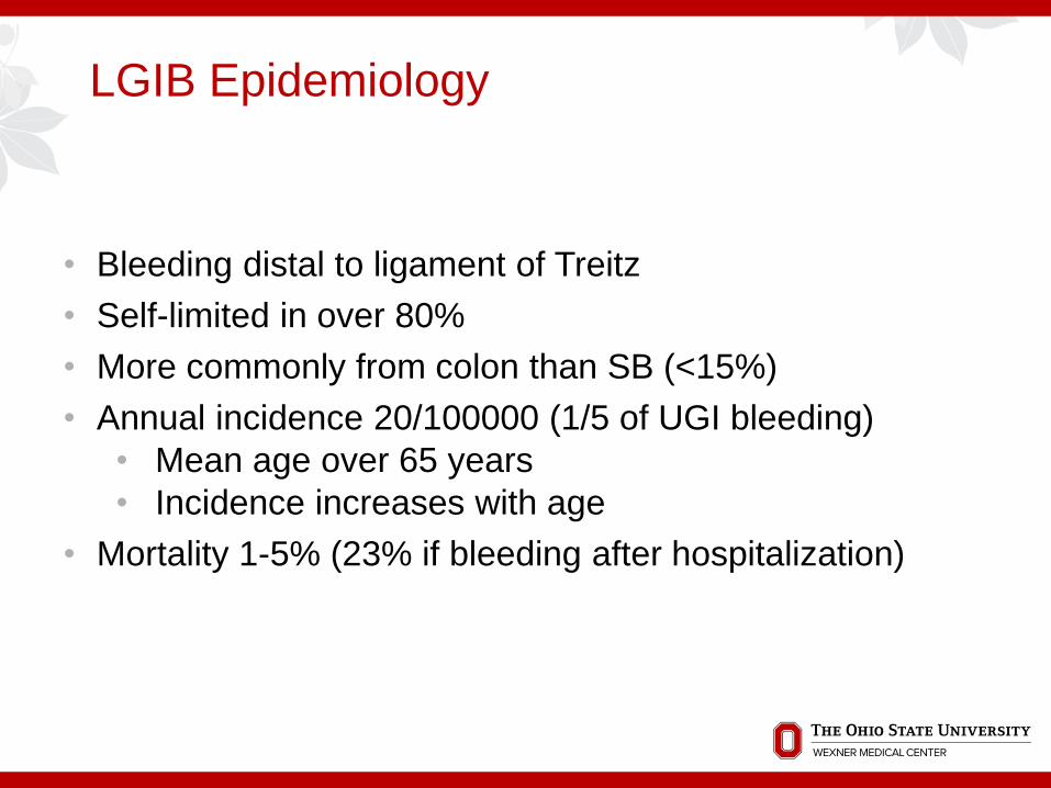
LGIB Epidemiology
• Bleeding distal to ligament of Treitz
• Self-limited in over 80%
• More commonly from colon than SB (<15%)
• Annual incidence 20/100000 (1/5 of UGI bleeding)
• Mean age over 65 years
• Incidence increases with age
• Mortality 1-5% (23% if bleeding after hospitalization)
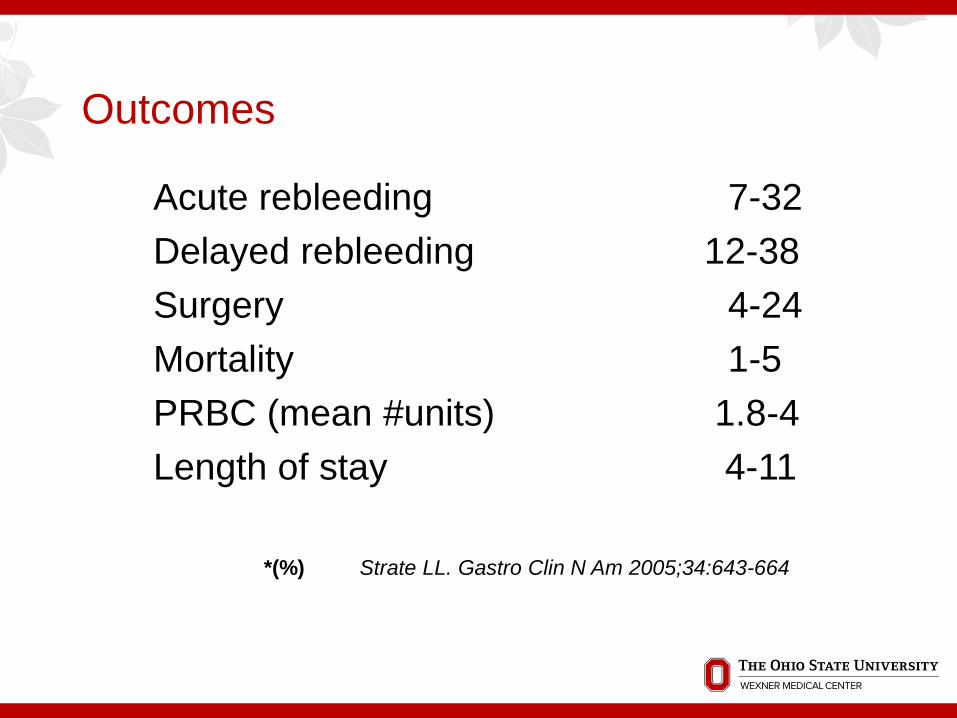
Outcomes
Acute rebleeding 7-32
Delayed rebleeding 12-38
Surgery 4-24
Mortality 1-5
PRBC (mean #units) 1.8-4
Length of stay 4-11
*(%) Strate LL. Gastro Clin N Am 2005;34:643-664
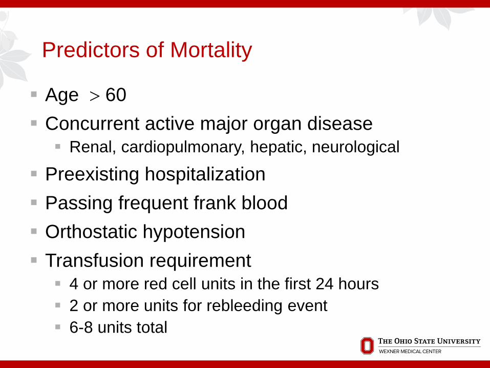
Predictors of Mortality
Age > 60
Concurrent active major organ disease
Renal, cardiopulmonary, hepatic, neurological
Preexisting hospitalization
Passing frequent frank blood
Orthostatic hypotension
Transfusion requirement
4 or more red cell units in the first 24 hours
2 or more units for rebleeding event
6-8 units total
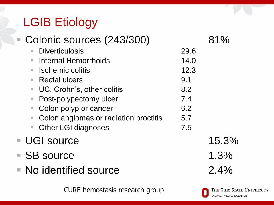
LGIB Etiology
Colonic sources (243/300) 81% Diverticulosis 29.6
Internal Hemorrhoids 14.0
Ischemic colitis 12.3
Rectal ulcers 9.1
UC, Crohn’s, other colitis 8.2
Post-polypectomy ulcer 7.4
Colon polyp or cancer 6.2
Colon angiomas or radiation proctitis 5.7
Other LGI diagnoses 7.5
UGI source 15.3%
SB source 1.3%
No identified source 2.4%
CURE hemostasis research group
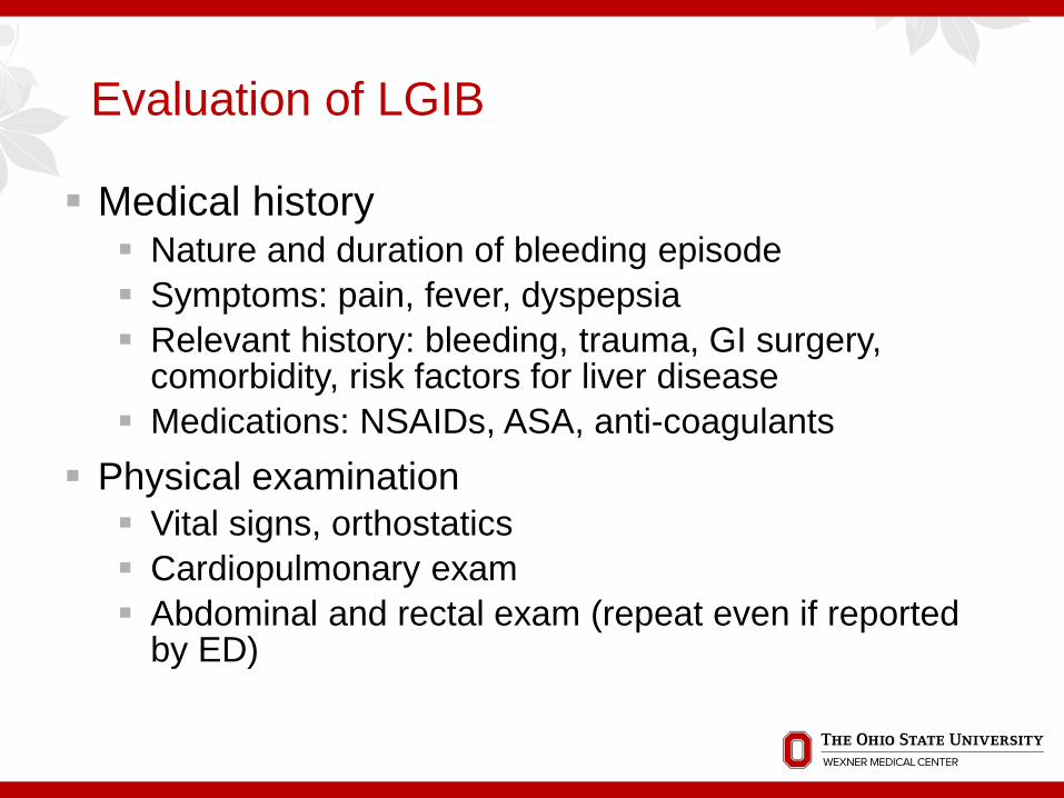
Evaluation of LGIB
Medical history Nature and duration of bleeding episode
Symptoms: pain, fever, dyspepsia
Relevant history: bleeding, trauma, GI surgery, comorbidity, risk factors for liver disease
Medications: NSAIDs, ASA, anti-coagulants
Physical examination
Vital signs, orthostatics
Cardiopulmonary exam
Abdominal and rectal exam (repeat even if reported by ED)
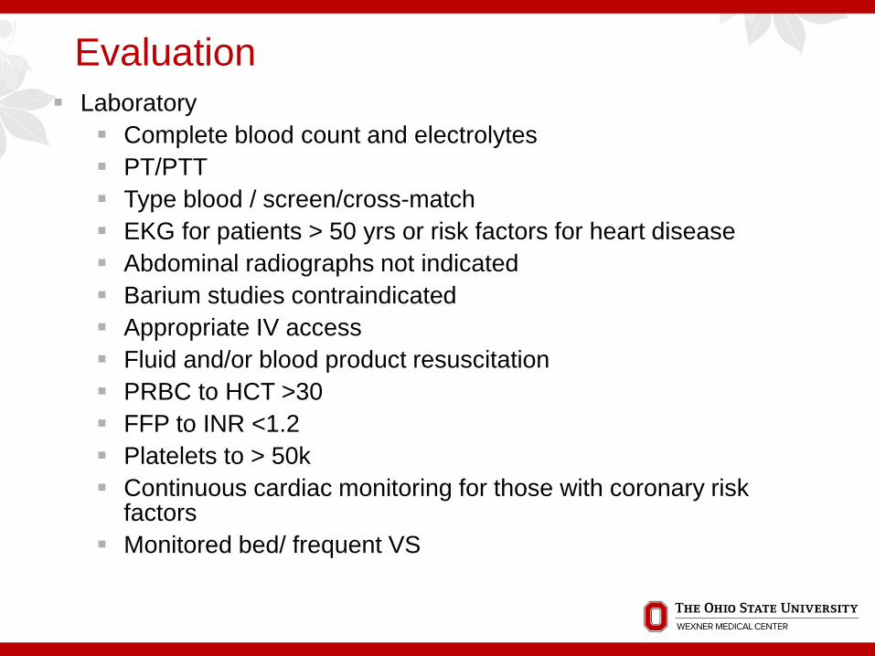
Evaluation Laboratory
Complete blood count and electrolytes
PT/PTT
Type blood / screen/cross-match
EKG for patients > 50 yrs or risk factors for heart disease
Abdominal radiographs not indicated
Barium studies contraindicated
Appropriate IV access
Fluid and/or blood product resuscitation
PRBC to HCT >30
FFP to INR <1.2
Platelets to > 50k
Continuous cardiac monitoring for those with coronary risk factors
Monitored bed/ frequent VS
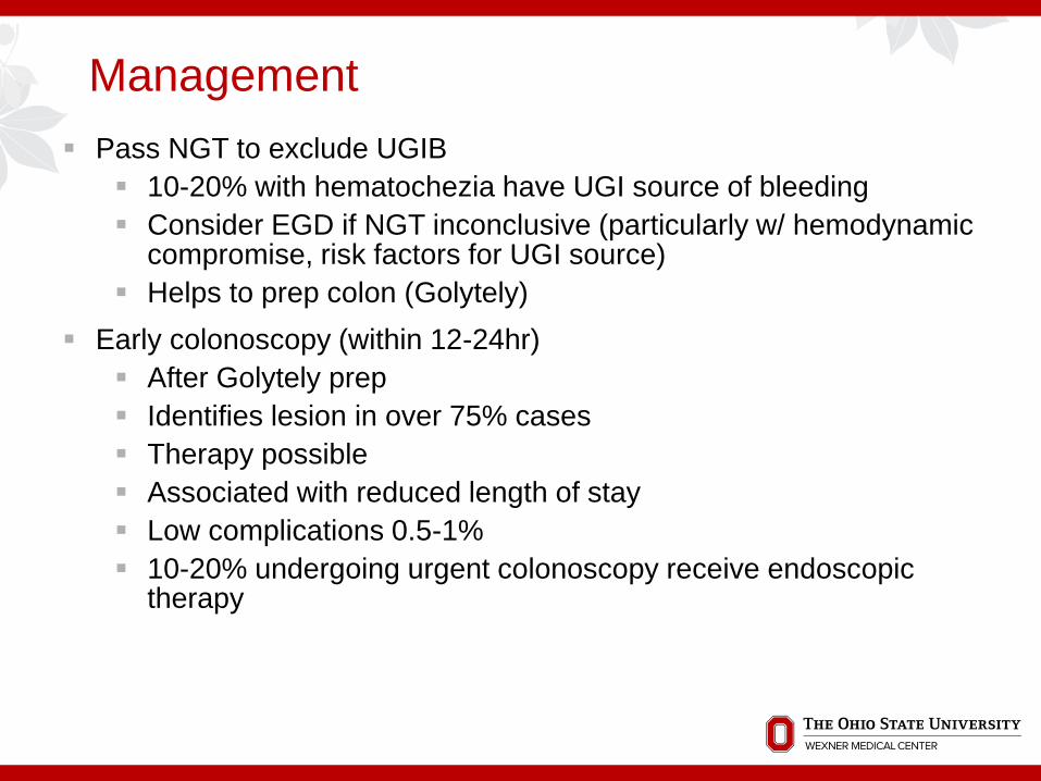
Management
Pass NGT to exclude UGIB
10-20% with hematochezia have UGI source of bleeding
Consider EGD if NGT inconclusive (particularly w/ hemodynamic compromise, risk factors for UGI source)
Helps to prep colon (Golytely)
Early colonoscopy (within 12-24hr)
After Golytely prep
Identifies lesion in over 75% cases
Therapy possible
Associated with reduced length of stay
Low complications 0.5-1%
10-20% undergoing urgent colonoscopy receive endoscopic therapy
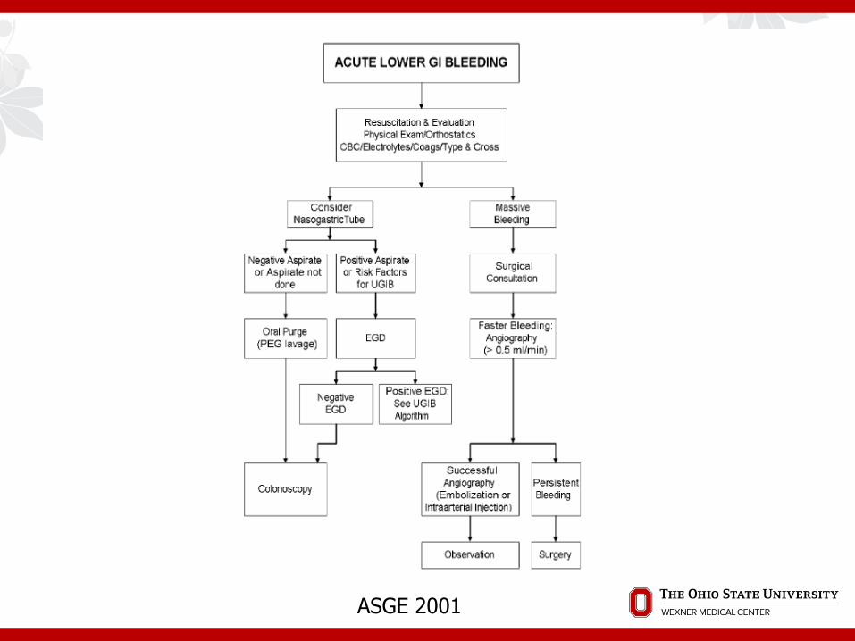
ASGE 2001
Vascular Ectasia
Multiple, mainly right colon
Associated with advanced age, CRF, Valvular heart disease
Intermittent hematochezia, massive bleeding rare
Argon Plasma Coagulation effective, widely used
All lesions should be treated
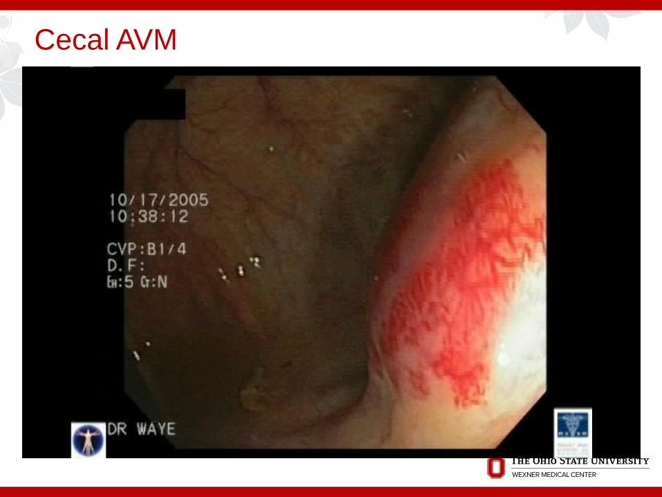
Cecal AVM
Radiation Proctopathy
Intermittent hematochezia with tenesmus
9m to 4 yrs after XRT for prostate or gyn cancers
Multiple telangiectasias on endoscopy
APC most effective All visible lesions treated and repeated Q 4 weeks, full bowel
prep advised, mean 2.9 sessions (1-8)
Post procedure rectal pain and cramps possible
Post procedure rectal ulcers Rx with 5 ASA suppositories
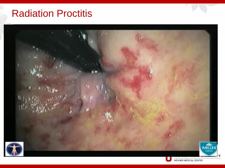
Radiation Proctitis
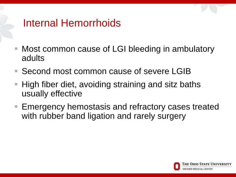
Internal Hemorrhoids
Most common cause of LGI bleeding in ambulatory adults
Second most common cause of severe LGIB
High fiber diet, avoiding straining and sitz baths usually effective
Emergency hemostasis and refractory cases treated with rubber band ligation and rarely surgery
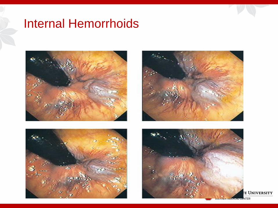
Internal Hemorrhoids
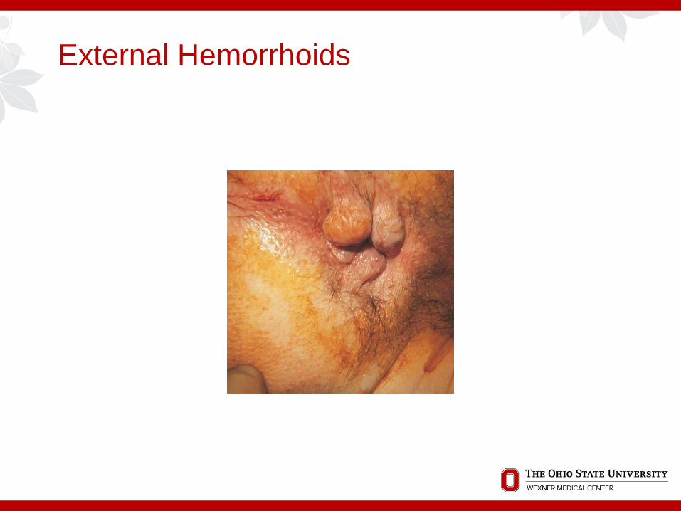
External Hemorrhoids
Ischemic Colitis
Results from sudden and often temporary reduction in mesenteric blood flow
Sudden lower abdominal pain followed w/in 24 hrs by bloody diarrhea
Segmental involvement of water shed areas-splenic flexure and descending colon
Endoscopy mainly diagnostic - mucosal erythema, friability, necrotic ulcerations
Supportive management
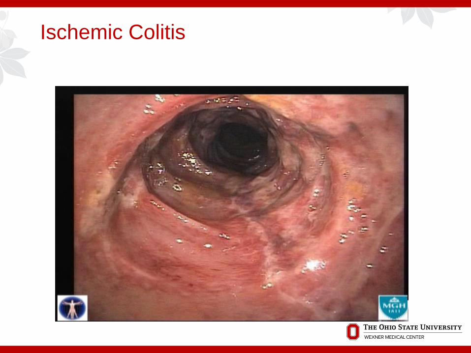
Ischemic Colitis
Small Bowel Bleeding
Small Bowel Bleeding
• 5-7 % of obscure GI bleeding
• Proximal jejunum angioectasia
• Similar to right colon AVMs
• Capsule endoscopy
• Balloon assisted enteroscopy
• Double balloon endoscopy
• Single balloon enteroscopy
• Intraoperative enteroscopy
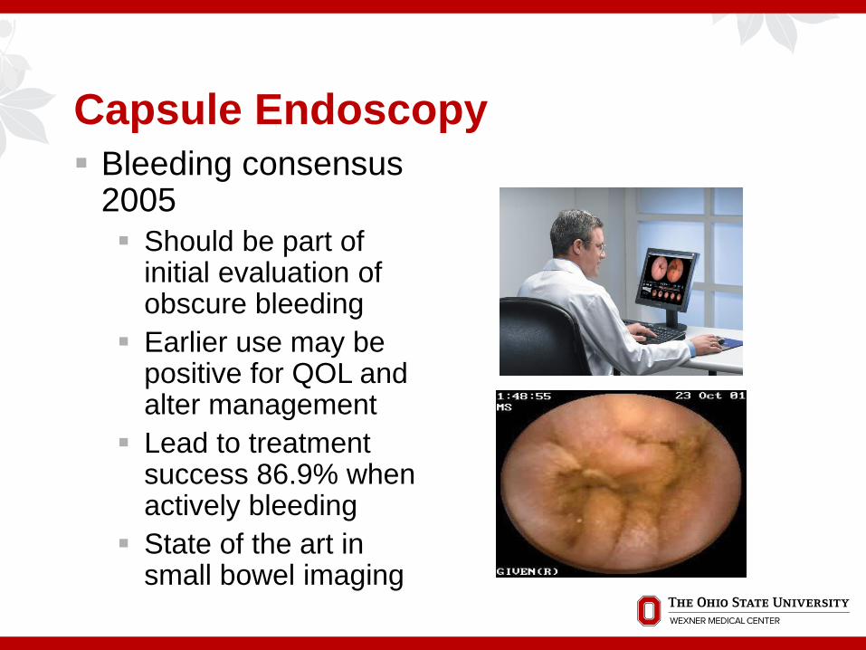
Capsule Endoscopy Bleeding consensus
2005
Should be part of initial evaluation of obscure bleeding
Earlier use may be positive for QOL and alter management
Lead to treatment success 86.9% when actively bleeding
State of the art in small bowel imaging
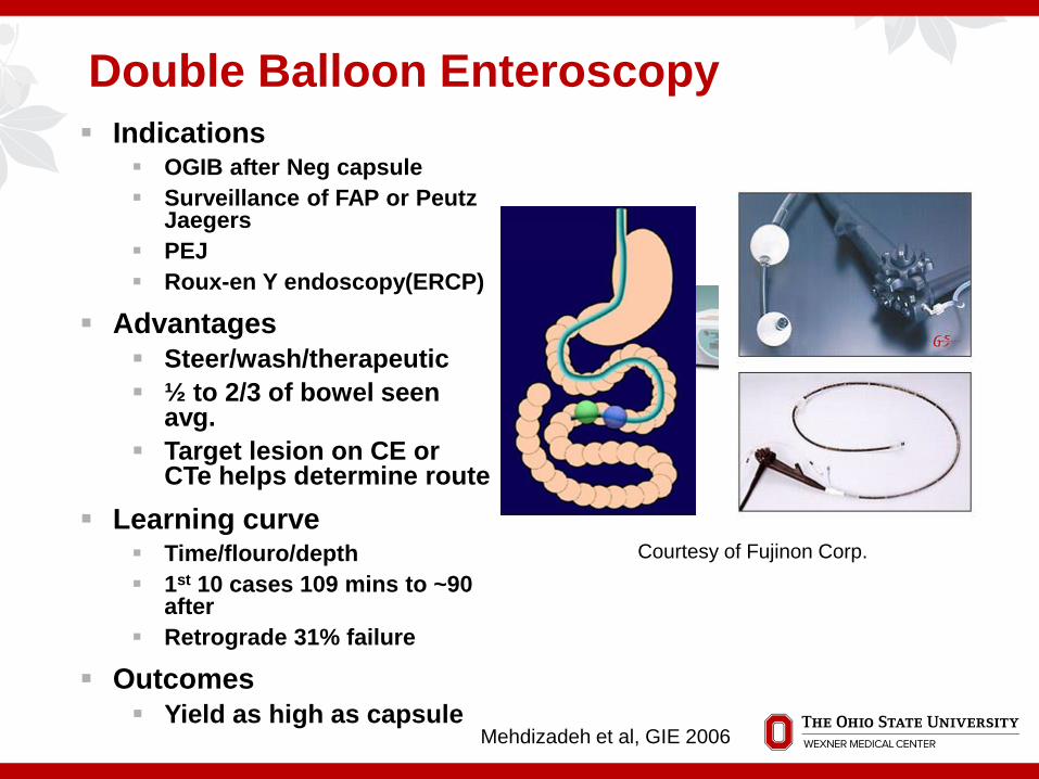
Double Balloon Enteroscopy Indications
OGIB after Neg capsule
Surveillance of FAP or Peutz Jaegers
PEJ
Roux-en Y endoscopy(ERCP)
Advantages
Steer/wash/therapeutic
½ to 2/3 of bowel seen avg.
Target lesion on CE or CTe helps determine route
Learning curve Time/flouro/depth
1st 10 cases 109 mins to ~90 after
Retrograde 31% failure
Outcomes
Yield as high as capsule
Courtesy of Fujinon Corp.
Mehdizadeh et al, GIE 2006
Conclusions:
•Medical stabilization
•Signs and symptoms help to localize
•Direct the investigation
•Alter risk factors
•Value = Benefits/Costs
Thank You