Embed Size (px)
Citation preview
How to Diagnose a Uterine Non-Hodgkin’s Lymphoma?: A Case Report
Uterus Non-Hodgkirı Lenfomasına Nasıl Tanı Koyalım?:Olgu Sunumu
Tayfun GÜNGÖR1, Adnan ŞİMŞEK2, Serap AKBAY3, Emel Üçgül ÇAVUŞOĞLU4, Ümit BİLGE2
1 Dr. Zekai Tahir Burak Kadın Sağlığı Eğitim ve Araştırma Hastanesi, Jinekolojik Onkoloji Kliniği, ANKARA2 Dr. Zekai Tahir Burak Kadın Sağlığı Eğitim ve Araştırma Hastanesi, Kadın Doğum Kliniği, ANKARA3 Dr. Zekai Tahir Burak Kadın Sağlığı Eğitim ve Araştırma Hastanesi, Patoloji Kliniği, ANKARA4 Dr. Zekai Tahir Burak Kadın Sağlığı Eğitim ve Araştırma Hastanesi, Medikal Onkoloji Kliniği, ANKARA
SUMMARY
Lymphoma of the female genital tract (FGT) is uncommon. This diagnosis, in such an unusual location, is very difficult to establish. Herein we report a case of FGT lymphoma and describe it ’s ıvay to diagnosis. A 37 year-old-woman was referred to our institution due to undiagnosed cervicat mass. Two consecutive biopsies had been taken from the mass, both of which yield- ed inflamation. An unusual-looking bulky cervicat mass was seen and the upper part of vagina and parametrial tissues were deeply infiltrated. On magnetic resonance imaging a uterine mass measuring 7.0 cm x 8.8 cm x 2.3 cm was protruding into the vagina. A pathologic sized lymph node in the left iliac region was detected. The vaginal wall was thickened, 3.3 cm in diam eter. Punch biopsy was reported as regenerative changes. After thata core biopsy was taken from a different area on the mass, from vvhich biopsy was not taken before. Histopathological analysis retrieved a diffuse large celi non-Hodgkin's lymphoma according to REAL classification. Computed tomography (CT) and flourodeoxyglucose-18 positron emission tomography (FDG-PET) was used for staging. According to Ann Arbor staging System, stage was lleb and after 6 courses of R-CHOP and additional 2 cours- es of rituximab therapy, patient is on complete remission. İn case of peivic masses with negative biopsy results, core biopsy should be preferred instead of using punch or excisionai method. Uterine lymphomas may be easily masked by inflamation or misdiagnosed as inflamation unless it is not suspected. Probably using both CT and FDG-PET is the best for proper staging.
Key VVords: Core biopsy, diagnosis, lymphoma, uterus.
ÖZET
Kadın genital sistem lenfoması oldukça nadir görülmektedir. Bu sıra dışı yerleşimde lenfoma tanısı koymak da oldukça zordur. Bu çalışmada bir kadın genital sistem lenfomasına nasıl tanı koyduğumuzu sunmaktayız. Otuz yedi yaşındaki bir kadın hasta, merkezimize tanımlanamayan servikal kitle nedeniyle sevk edildi. Ultrasonografide, sıra dışı görünen dev bir sen/ikal kitlenin vajinanın üst kısmını ve parametriyal dokuları in filtre ettiği saptandı. Magnetik rezonans görüntülemede 7.0 cm x 8.8 cm x 2.3 cm boyutlarında uterustan kaynaklanan bir kitlenin vajinadan protruze olduğu saptandı. Kitleden alınan iki biyopsi infla- masyon olarak raporlandı. Sol iliyak bölgede patolojik boyutta bir lenf nodu saptandı. Vajina duvarı kalınlaşmıştı ve 3.3 cm çapa ulaşmıştı. “Punch” biyopsi sonucu rejenaratif değişiklikler olarak geldi. Daha sonra kitlenin daha önce biyopsi alınmamış bir bölgesinden alman kor biyopsi sonucu REAL sınıflamasına göre difüz büyük hücreli non-Hodgkin lenfoma olarak çıktı. Çekilen flo- rodeoksiglukoz-pozitron emisyon tomografi/bilgisayarlı tomografi (FDG-PET/BT) sonucuna göre Ann-Arbor evre lleb uterin non- Hodgkin lenfoma olarak evrelenen hasta 6 kür R-CHOP + 2 kür rituksimab tedavisi sonucu tam remisyonda izlenmektedir. Biyopsi sonucu negatif olan pelvik kitlelerde eksizyoner ya da “punch” biyopsi yerine kor biyopsi tercih edilmelidir. Bu olguda ısrarlı biyopsilerle hasta total abdominal histerektomi gibi gereksiz organ kaybına yol açan cerrahilerden kaçınılmıştır. Eğer şüphe edilmezse uterus lenfomaları kolayca inflamasyon tarafından maskelenebilir veya inflamasyon olarak tanımlanabilir. FDG-PET/BT en iyi evreleme yöntemidir.
Anahtar Kelimeler: Kor biyopsi, tanı, lenfoma, uterus.
41
How to Diagnose a Uterine Non-Hodgkin’s Lymphoma?: A Case Report
INTRODUCTION
Malignant lymphoma of the female genital tract (FGT) is a very rare disease. The incidence of uterine lymphoma is estimated to be less than 0.5% of ali non-Hodgkin’s lymphomas (NHL) (1). Revievv of the literatüre vvould suggest that one in 175 female extra- nodal lymphomas is likely to originate in FGT (2). FGT lymphoma is very difficult to diagnose, it can easily be misdiagnosed as usual type cervical cancer and due to it’s systemic nature, it’s management and treatment approach is quite different than usual type cervical cancer. Herein we report a complicated case of FGT lymphoma and describe it’s long and bother- some diagnostic way.
CASE REPORT
A 37 year-old-woman was referred to our institu- tion due to undiagnosed cervical mass. The outside pap smear cytology was normal. We leamt that two consecutive biopsies had been taken from the mass. The first was an excisional biopsy and yielded acute inflamation and the second was a punch biopsy which was reported as active chronic inflamation. She was complaining about menstrual irregularity, dysuria, pelvic pain, difficulty with urination, fever, nausea and vomiting. Her past medical history was completely uneventful. The initial physical examina- tion revealed a suprapubic mass extending near umblicus level. Since urinary globe was suspected, the bladder was catheterized and 3500 mİ urine was drained. An unusual-looking bulky cervical mass was seen and the upper part of vagina and parametrial tis- sues were deeply infiltrated on bi-manual examina- tion. Sonography revealed a hypoechoic heteroge- neous pelvic mass measuring 12 cm in diameter and mild pelvicaliceal dilatation. Laboratory tests indicat- ed mild anemia and seriously impaired kidney func- tions. Thereby the case was consulted with nephrolo- gy department and subsequently she undervvent hemodialysis.
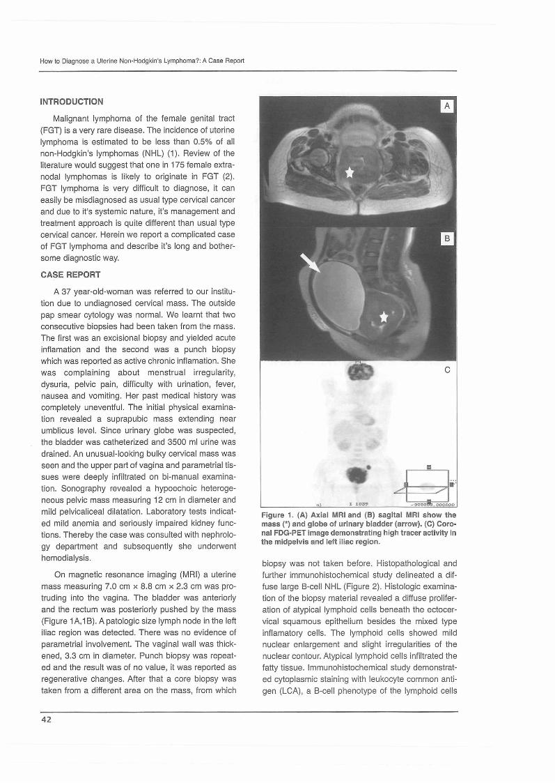
On magnetic resonance imaging (MRI) a uterine mass measuring 7.0 cm x 8.8 cm x 2.3 cm was pro- truding into the vagina. The bladder was anteriorly and the rectum was posteriorly pushed by the mass (Figüre 1A,1B). Apatologic size lymph node in the left iliac region was detected. There was no evidence of parametrial involvement. The vaginal wall was thick- ened, 3.3 cm in diameter. Punch biopsy was repeat- ed and the result was of no value, it was reported as regenerative changes. After that a core biopsy was taken from a different area on the mass, from which
Figüre 1. (A) Axial MRI and (B) sagital MRI show the mass (*) and globe of urinary bladder (arrovv). (C) Coro- nal FDG-PET image demonstrating high tracer activity in the midpelvis and left iliac region.
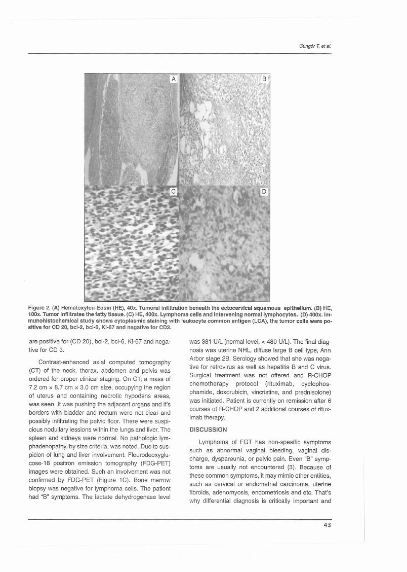
biopsy was not taken before. Histopathological and further immunohistochemical study delineated a dif- fuse large B-cell NHL (Figüre 2). Histologic examina- tion of the biopsy material revealed a diffuse prolifer- ation of atypical lymphoid cells beneath the ectocer- vical squamous epitheiium besides the mixed type inflamatory cells. The lymphoid cells shovved mild nuclear enlargement and slight irregularities of the nuclear contour. Atypical lymphoid cells infiltrated the fatty tissue. İmmunohistochemical study demonstrat- ed cytoplasmic staining with leukocyte common anti- gen (LCA), a B-cell phenotype of the lymphoid cells
42
Güngör T, et al.
Figüre 2, (A) Hematoxylen-Eosin (HE), 40x. Tumoral infiltration beneath the ectocervical squamous epithelium. (B) HE, 100x. Tumor infiltrates the fatty tissue. (C) HE, 400x. Lymphoma cells and intervening normal lymphocytes. (D) 400x. Im- munohistochemical study shows cytoplasmic staining with leukocyte common antigen (LCA), the tumor cells vvere po- sitive for CD 20, bcl-2, bcl-6, Ki-67 and negative for CD3.
are positive for (CD 20), bcl-2, bcl-6, Ki-67 and negative for CD 3.
Contrast-enhanced axial computed tomography (CT) of the neck, thorax, abdomen and pelvis was ordered for proper clinical staging. On CT; a mass of 7.2 cm x 8.7 cm x 3.0 cm size, occupying the region of uterus and containing necrotic hypodens areas, was seen. İt was pushing the adjacent organs and it’s borders with bladder and rectum vvere not clear and possibly infiltrating the pelvic floor. There vvere suspi- cious nodullary lessions vvithin the lungs and liver. The spleen and kidneys vvere normal. No pathologic lym- phadenopathy, by size criteria, was noted. Due to sus- picion of lung and liver involvement. Flourodeoxyglu- cose-18 positron emission tomography (FDG-PET) images vvere obtained. Such an involvement vvas not confirmed by FDG-PET (Figüre 1C). Bone marrovv biopsy vvas negative for lymphoma cells. The patient had “B” symptoms. The lactate dehydrogenase level
vvas 381 U/L (normal level, < 480 U/L). The final diag- nosis vvas uterine NHL, diffuse large B celi type, Ann Arbor stage 2B. Serology shovved that she vvas negative for retrovirus as well as hepatitis B and C virüs. Surgical treatment vvas not offered and R-CHOP chemotherapy protocol (rituximab, cyclophos- phamide, doxorubicin, vincristine, and prednisolone) vvas initiated. Patient is currently on remission after 6 courses of R-CHOP and 2 additional courses of ritux- imab therapy.
DISCUSSION
Lymphoma of FGT has non-spesific symptoms such as abnormal vaginal bleeding, vaginal dis- charge, dyspareunia, or pelvic pain. Even “B” symptoms are usually not encountered (3). Because of these common symptoms, it may mimic other entities, such as cervical or endometrial carcinoma, uterine fibroids, adenomyosis, endometriosis and ete. That’s vvhy differential diagnosis is critically important and
43
How to Diagnose a Uterine Non-Hodgkin’s Lymphoma?: A Case Report
histopathological examination is necessary to reach the definitive diagnosis because non of the imaging modalities is reliable by this means. Unfortunateiy due to the fact that most lymphomas are subepithelial unless there is ulceration; the cervical smear is most- ly negative and it’s very hard to obtain adequate tis- sue sample by conventional biopsy techniques such as punch and excisional biopsy (4). Such an experi- ence was also reported by Van Renterghem et al. (1). İn the present case; the cervical smear was normal and the first three biopsies revealed no malignancy. The first two biopsies were performed at outside and the third biopsy was performed by us. After yielding the regenerative changes result on the third biopsy, we understood that we had taken it from the previous biopsy areas. So we decided to take the next biopsy from a different area from vvhich any biopsies were not taken before and a core biopsy was necessary to penetrate the thickened vaginal wall, vvhich vvas seen on the MRI. Thereby we diagnosed the nature of mass. That’s vvay vve recommend that in case of pelvic masses vvith negative biopsy results, core biopsy should be preferred instead of using punch or exci- sional method again. İn our opinion this inference is the main learning of the present case.
As knovvn lymphoma is the malignant transforma- tion of lymphocytes. These cells are the predominant elements of inflamation, principally in chronic condi- tions. İn the cervix inflamatory processes are very common especially due to infections and foreign bod- ies like intrauterine device, condoms, lubricants and ete. Hence in such localizations, atypical lenfoid pro- liferations like lymphoma may be easily masked by inflamation or misdiagnosed as inflamation unless it is not suspeeted. For sure, this probability is not strong but in case of pelvic masses, biopsies of vvhich repetitively reported as inflamation; then attention must be orientated to the morphology of lymphocytes to seek for atypical features and vvhen such features are encountered then immunohistochemical markers should be used. From this point of vievv, vve conclud- ed that in such unusual locations like FGT lymphoma diagnosis needs strong suspicion.
CT is commonly used for the staging of NHL (5). CT enables accurate measurement of both tumour size and extent, and provides information that can be used to plan an appropriate therapeutic regimen as vvell as follovv response to treatment. Somehovv in the present case, CT did not shovv the left iliac lym- phadenopathy vvhich vvas deteeted by MRI and veri- fied to be malignant tissue by FDG-PET. Actually in this case FDG-PET vvas ordered to investigate liver and lung involvement, it helped us to see that men- tioned lymphadenopathy vvas involved by the dis- ease. So vve don’t knovv vvhich one of the tvvo areas, the uterus or the left iliac lymph node, is the primary source and if vve only relied on CT then our staging vvould be Ann Arbor Stage 1B vvhich vvas more advanced in fact. This result let us to think the supe- riority of FDG-PET över the CT, in fact Delbeke has postulated that these tvvo modalities have compara- ble sensitivities (6). We believe that probably using these tvvo modalities together is the best for proper staging.
REFERENCES
1. Van Renterghem N, De Paepe P, Van den Broecke R, Bourgain C, Serreyn R. Primary lymphoma of the cervix uteri: A diagnostic chattenge. Report of tvvo cases and revievv of the literatüre. Eur J Gynaecol Oncol 2005;26:36-8.
2. Grace A, O’Connell N, Byrne P, et al. Malignant lymphoma of the cervix: An unusual presentation and a rare disease. Eur J Gynecol Oncol 1999;20:26-8.
3. Al Talib R, Sworn M, Ramsey A, et al. Primary cervical lymphoma: The role of cervical eytology. Cytopathology 1996;7: 173-7.
4. King J, Elkhalifa M, Michael C. Malignant lymphoma identi- fied on cervical cytotogic smear vvith immunophenotypic analysis. Açta Cytol 1997;41:1228-30.
5. Fishman EK, Kuhlman JE, Jones RJ. CT of lymphoma: Spectrum of disease. Radiographics 1991;11:647-69.
6. Delbeke K. Oncological applications of FDG PET imaging: Brain tumors, colorectal cancer, lymphoma and melanoma. J Nucl Med 1999;40:591-603.
44