Embed Size (px)
Citation preview
Instructions for use
Title Human Amnion-Derived Mesenchymal Stem Cell Transplantation Ameliorates Dextran Sulfate Sodium-Induced SevereColitis in Rats
Author(s) Onishi, Reizo; Ohnishi, Shunsuke; Higashi, Ryosuke; Watari, Michiko; Yamahara, Kenichi; Okubo, Naoto; Nakagawa,Koji; Katsurada, Takehiko; Suda, Goki; Natsuizaka, Mitsuteru; Takeda, Hiroshi; Sakamoto, Naoya
Citation Cell transplantation, 24(12): 2601-2614
Issue Date 2015-12-18
Doc URL http://hdl.handle.net/2115/60537
Rights(URL) http://creativecommons.org/licenses/by-nc/3.0/
Type article
File Information s14.pdf
Hokkaido University Collection of Scholarly and Academic Papers : HUSCAP
Cell Transplantation, Vol. 24, pp. 2601–2614, 2015 0963-6897/15 $90.00 + .00Printed in the USA. All rights reserved. DOI: http://dx.doi.org/10.3727/096368915X687570Copyright Ó 2015 Cognizant Comm. Corp. E-ISSN 1555-3892 www.cognizantcommunication.com
2601
Received January 31, 2015; final acceptance February 22, 2015. Online prepub date: March 25, 2015.Address correspondence to Shunsuke Ohnishi, M.D., Ph.D., Department of Gastroenterology and Hepatology, Hokkaido University Graduate School of Medicine, N15, W7, Kita-ku, Sapporo 060-8638, Japan. Tel: +81-11-716-1161; Fax: +81-11-706-7867; E-mail: [email protected]
Human Amnion-Derived Mesenchymal Stem Cell Transplantation Ameliorates Dextran Sulfate Sodium-Induced Severe Colitis in Rats
Reizo Onishi,* Shunsuke Ohnishi,* Ryosuke Higashi,† Michiko Watari,‡ Kenichi Yamahara,§ Naoto Okubo,† Koji Nakagawa,† Takehiko Katsurada,* Goki Suda,* Mitsuteru Natsuizaka,*
Hiroshi Takeda,† and Naoya Sakamoto*
*Department of Gastroenterology and Hepatology, Hokkaido University Graduate School of Medicine, Sapporo, Japan†Laboratory of Pathophysiology and Therapeutics, Faculty of Pharmaceutical Sciences, Hokkaido University, Sapporo, Japan
‡Department of Gynecology, Tenshi Hospital, Sapporo, Japan§Department of Regenerative Medicine and Tissue Engineering, National Cerebral and Cardiovascular
Center Research Institute, Osaka, Japan
Mesenchymal stem cells (MSCs) are a valuable cell source in regenerative medicine. Recently, several studies have shown that MSCs can be easily isolated from human amnion. In this study, we investigated the thera-peutic effect of human amnion-derived MSCs (AMSCs) in rats with severe colitis. Colitis was induced by the administration of 8% dextran sulfate sodium (DSS) from day 0 to day 5, and AMSCs (1 × 10⁶ cells) were transplanted intravenously on day 1. Rats were sacrificed on day 5, and the colon length and histological coli-tis score were evaluated. The extent of inflammation was evaluated using quantitative reverse transcription-polymerase chain reaction (qRT-PCR) and immunohistochemistry. The effect of AMSCs on the inflammatory signals was investigated in vitro. AMSC transplantation significantly ameliorated the disease activity index score, weight loss, colon shortening, and the histological colitis score. mRNA expression levels of proinflam-matory cytokines such as tumor necrosis factor (TNF)-a, interleukin (IL)-1b, and migration inhibitory factor (MIF) were significantly decreased in the rectums of AMSC-treated rats. In addition, the infiltration of mono-cytes/macrophages was significantly decreased in AMSC-treated rats. In vitro experiments demonstrated that activation of proinflammatory signals induced by TNF-a or lipopolysaccharide (LPS) in immortalized murine macrophage cells (RAW264.7) was significantly attenuated by coculturing with AMSCs or by culturing with a conditioned medium obtained from AMSCs. Although the phosphorylation of IkB induced by TNF-a or LPS was not inhibited by the conditioned medium, nuclear translocation of NF-kB was significantly inhibited by the conditioned medium. Taken together, AMSC transplantation provided significant improvement in rats with severe colitis, possibly through the inhibition of monocyte/macrophage activity and through inhibition of NF-kB activation. AMSCs could be considered as a new cell source for the treatment of severe colitis.
Key words: Mesenchymal stem cells (MSCs); Amnion; Colitis; Macrophages; NF-kB
INTRODUCTION
Inflammatory bowel diseases (IBDs) are increasing worldwide; Crohn’s disease and ulcerative colitis are two main types of IBD (4,5,25). IBD is generally treated with immunosuppressive drugs, such as prednisone, tumor necrosis factor (TNF) inhibitors, azathioprine, methotrex-ate, or mercaptopurine, depending on the level of sever-ity (24). However, severe and complex cases may require surgery, including bowel resection and ileostomy (24).
Mesenchymal stem cells (MSCs) are multipotent cells that can differentiate into a variety of lineages, including bone, cartilage, or fat, and are present in adult tissue (27). At present, MSCs have been investigated in regenerative
medicine because of their differentiation ability and their potential to improve damaged tissues by the secretion of a variety of growth factors and anti-inflammatory mol-ecules (21,35). The efficacy of autologous and alloge-neic MSC transplantation in patients with IBD has been recently reported (11,20).
The fetal membrane consists of amnion and chorion, which envelops the developing fetus. Although human fetal membrane is usually discarded as medical waste after delivery, fetal tissues have been found to be rich sources of MSCs (2,16). Systemic administration of amnion-derived MSCs (AMSCs) improved rats with hindlimb ischemia (17), myocarditis (18,26), glomerulonephritis (33), and
2602 ONISHI ET AL.
ischemia/reperfusion-induced acute kidney injury (34) by inducing angiogenesis and anti-inflammatory effects.
In this study, we investigated whether the administra-tion of human AMSCs improves dextran sulfate sodium (DSS)-induced severe colitis in rats and explored its underlying mechanisms.
MATERIALS AND METHODS
Animals
The experimental protocol was approved by the Animal Care and Use Committees of Hokkaido University. Eight-week-old male Sprague–Dawley rats were procured from Japan SLC (Hamamatsu, Japan); one rat was housed per cage in a temperature-controlled room (24°C) on a 12 h/ 12 h light/dark cycle. All rats had ad libitum access to standard pellets.
Induction and General Assessment of Severe Colitis
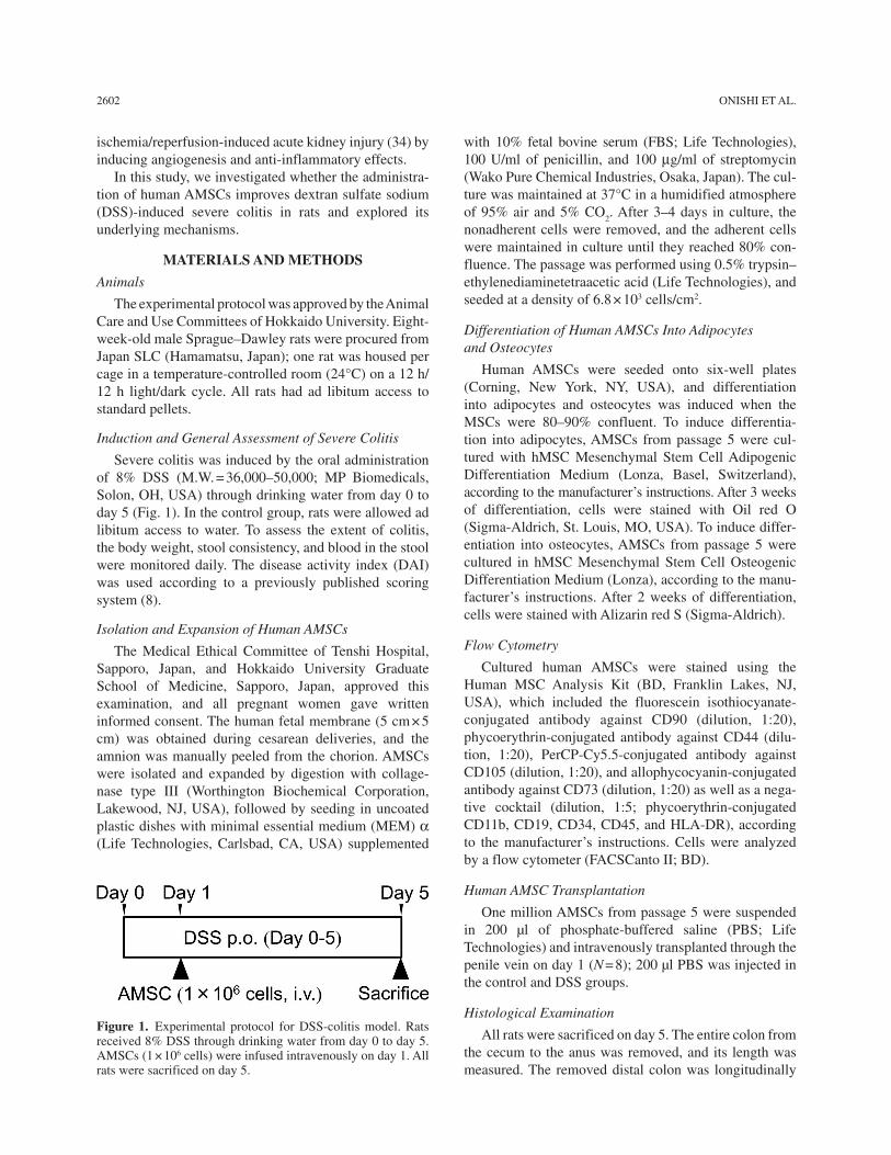
Severe colitis was induced by the oral administration of 8% DSS (M.W. = 36,000–50,000; MP Biomedicals, Solon, OH, USA) through drinking water from day 0 to day 5 (Fig. 1). In the control group, rats were allowed ad libitum access to water. To assess the extent of colitis, the body weight, stool consistency, and blood in the stool were monitored daily. The disease activity index (DAI) was used according to a previously published scoring system (8).
Isolation and Expansion of Human AMSCs
The Medical Ethical Committee of Tenshi Hospital, Sapporo, Japan, and Hokkaido University Graduate School of Medicine, Sapporo, Japan, approved this examination, and all pregnant women gave written informed consent. The human fetal membrane (5 cm × 5 cm) was obtained during cesarean deliveries, and the amnion was manually peeled from the chorion. AMSCs were isolated and expanded by digestion with collage-nase type III (Worthington Biochemical Corporation, Lakewood, NJ, USA), followed by seeding in uncoated plastic dishes with minimal essential medium (MEM) a (Life Technologies, Carlsbad, CA, USA) supplemented
with 10% fetal bovine serum (FBS; Life Technologies), 100 U/ml of penicillin, and 100 mg/ml of streptomycin (Wako Pure Chemical Industries, Osaka, Japan). The cul-ture was maintained at 37°C in a humidified atmosphere of 95% air and 5% CO
2. After 3–4 days in culture, the
nonadherent cells were removed, and the adherent cells were maintained in culture until they reached 80% con-fluence. The passage was performed using 0.5% trypsin–ethylenediaminetetraacetic acid (Life Technologies), and seeded at a density of 6.8 × 10³ cells/cm2.
Differentiation of Human AMSCs Into Adipocytes and Osteocytes
Human AMSCs were seeded onto six-well plates (Corning, New York, NY, USA), and differentiation into adipocytes and osteocytes was induced when the MSCs were 80–90% confluent. To induce differentia-tion into adipocytes, AMSCs from passage 5 were cul-tured with hMSC Mesenchymal Stem Cell Adipogenic Differentiation Medium (Lonza, Basel, Switzerland), according to the manufacturer’s instructions. After 3 weeks of differentiation, cells were stained with Oil red O (Sigma-Aldrich, St. Louis, MO, USA). To induce differ-entiation into osteocytes, AMSCs from passage 5 were cultured in hMSC Mesenchymal Stem Cell Osteogenic Differentiation Medium (Lonza), according to the manu-facturer’s instructions. After 2 weeks of differentiation, cells were stained with Alizarin red S (Sigma-Aldrich).
Flow Cytometry
Cultured human AMSCs were stained using the Human MSC Analysis Kit (BD, Franklin Lakes, NJ, USA), which included the fluorescein isothiocyanate-conjugated antibody against CD90 (dilution, 1:20), phycoerythrin-conjugated antibody against CD44 (dilu-tion, 1:20), PerCP-Cy5.5-conjugated antibody against CD105 (dilution, 1:20), and allophycocyanin-conjugated antibody against CD73 (dilution, 1:20) as well as a nega-tive cocktail (dilution, 1:5; phycoerythrin-conjugated CD11b, CD19, CD34, CD45, and HLA-DR), according to the manufacturer’s instructions. Cells were analyzed by a flow cytometer (FACSCanto II; BD).
Human AMSC Transplantation
One million AMSCs from passage 5 were suspended in 200 μl of phosphate-buffered saline (PBS; Life Technologies) and intravenously transplanted through the penile vein on day 1 (N = 8); 200 μl PBS was injected in the control and DSS groups.
Histological Examination
All rats were sacrificed on day 5. The entire colon from the cecum to the anus was removed, and its length was measured. The removed distal colon was longitudinally
Figure 1. Experimental protocol for DSS-colitis model. Rats received 8% DSS through drinking water from day 0 to day 5. AMSCs (1 × 10⁶ cells) were infused intravenously on day 1. All rats were sacrificed on day 5.
HUMAN AMNION MSCs IMPROVE SEVERE COLITIS IN RATS 2603
opened, rinsed with PBS, and excised for histological examination. Specimens were fixed in 40 g/L of formal-dehyde saline (Wako Pure Chemical Industries), embed-ded in paraffin, and cut into 5-μm sections. Tissue sections were stained with hematoxylin and eosin (H&E; Wako Pure Chemical Industries) and microscopically examined by an independent observer. The tissues were scored in a blinded fashion by an examiner, on a 0- to 40-point scale based on the parameters of inflammation severity (0–3), inflammation extent (0–3), and crypt damage (0–4). Each of these parameters were multiplied by the percentage involvement (1 = 0–25%; 2 = 25–50%; 3 = 50–75%, and 4 = 75–100%), as described previously (30,31,36).
Immunohistochemical Examination
To assess the infiltration of monocytes/macrophages, neutrophils, and T lymphocytes, the tissue sections were stained with anti-rat CD68 monoclonal antibody (dilu-tion, 1:50; AbD Serotec, Kidlington, UK), anti-rat CD163 monoclonal antibody (dilution, 1:150; AbD Serotec), anti-myeloperoxidase antibody (dilution, 1:300; Thermo Scientific, Waltham, MA, USA), and anti-rat CD3 anti-body (dilution, 1:50; BD), respectively, for 40 min. Ten random fields on a section from each rat were photo-graphed, and the number of CD68-, CD163-, myeloper-oxidase (MPO)- and CD3-positive cells per low-powered field were counted, respectively.
RNA Isolation and Quantitative Reverse-Transcription Polymerase Chain Reaction (qRT-PCR)
Total RNA of the rat rectum or cultured cells was extracted using the RNeasy Mini Kit (Qiagen, Hilden, Germany); 1 μg of the total RNA was reverse-transcribed into cDNA using the QuantiTect Reverse Transcription Kit (Qiagen). PCR amplification was performed using a 25-μl reaction mixture that contained 1 μl of cDNA and 12.5 μl of Platinum SYBR Green PCR Mix (Invitrogen, Carlsbad, CA, USA). b-Actin mRNA that was ampli-fied from the same samples served as an internal control. After initial denaturation at 95°C for 2 min, a two-step cycle procedure was used (denaturation at 95°C for 15 s, annealing and extension at 60°C for 1 min) for 40 cycles in a 7700 Sequence Detector (Applied Biosystems, Foster City, CA, USA). Gene expression levels were deter-mined using the comparative threshold cycle (DDCt) method with b-actin as an endogenous control. Data were analyzed with Sequence Detection Systems soft-ware (Applied Biosystems). All primers were purchased from Qiagen (QuantiTect Primer Assays, b-actin; Rn_Actb_1_SG, TNF-a; Rn_Tnf_1_SG, IL-1b; Rn_Ilb_1_SG, MIF; Rn_Mif_1_SG, IL-6; Rn_Il-6_1_SG, MCP-1; Rn_Ccl2_1_SG, CXCL-1; Rn_Cxcl1_1_SG, CXCL-2; Rn_Cxcl2_1_SG, IFN-g; Rn_Ifn-g_SG) and bioinformati-cally validated.
Measurement of Rat Serum Monocyte Chemoattractant Protein-1 (MCP-1)
Rat serum MCP-1 concentration was measured using the Quantikine ELISA Kit (R&D Systems, Minneapolis, MN, USA), following the manufacturer’s instructions.
Inflammatory Reaction After Treatment With Lipopolysaccharide (LPS)
RAW264.7 cells (immortalized murine macrophage cells obtained from American Type Tissue Collection, Manassas, VA, USA) and AMSCs were cultured in a humidified chamber at 37°C in a mixture of 95% air and 5% CO
2. A conditioned medium was collected by cultur-
ing subconfluent AMSCs with serum-free MEMa for 48 h. RAW264.7 cells were stimulated with 100 ng/ml of LPS (Sigma-Aldrich) for 24 h under coincubation with AMSCs (1:10 or 1:5 AMSCs to RAW264.7 cells) or were cultured with the conditioned medium without dilution. Subsequently, total RNA and culture supernatant were collected. Expression levels and production of TNF-a and MCP-1 were measured by qPT-PCR (QuantiTect Primer Assays; Qiagen) and the enzyme-linked immuno-sorbent assay (ELISA), respectively. mRNA expression levels were expressed relative to controls. TNF-a and MCP-1 concentrations in the culture media were mea-sured using the Quantikine ELISA Kit (R&D Systems), following the manufacturer’s instructions.
Immunofluorescent Staining
To investigate the nuclear translocation of NF-kB, RAW264.7 cells were seeded in a type I collagen-coated eight-chamber slide (BD), at a density of 1 × 10⁴ cells/chamber. The next day, the culture medium was changed to conditioned medium for 3 h and stimulated with 20 ng/ ml of LPS for 60 min. Cells were fixed with 4% para-formaldehyde (Wako Pure Chemical Industries) at 4°C for 30 min and permeabilized with 0.2% Triton X-100 (MP Biomedicals). After background inhibition with 3% bovine serum albumin (Sigma-Aldrich), cells were labeled with anti-NF-kB polyclonal antibody (1:100; Cell Signaling Technology, Danvers, MA, USA) at 4°C for 60 min. After washing with PBS, the cells were stained with Alexa Fluor® 488-conjugated goat anti-rabbit IgG (1:2,000; Invitrogen) at room temperature for 60 min, followed by staining with 4¢6¢-diamidino-2-phenylindole (DAPI; 1:2,000; Dojindo Laboratories, Kumamoto, Japan). Fluorescence images were obtained using a fluorescent microscope (BZ-9000; Keyence, Osaka, Japan).
Western Blot Analysis
To investigate the phosphorylation of IkB, human embryonic kidney cells (HEK293, provided by the RIKEN BRC through the National Bio-Resource Project
2604 ONISHI ET AL.
of the MEXT, Japan) or their derivatives, which were stably transfected with the human TLR4a, MD2, and CD14 genes (293/hTLR4A-MD2-CD14; InvivoGen, San Diego, CA, USA), were plated at a density of 1 × 10⁵ cells in 12-well plates (Corning) and cultured with complete medium. The next day, culture medium was changed to the conditioned medium, and cells were incubated for 3 h, then cells were treated with 10 ng/ml of mouse recombinant TNF-a (R&D Systems) or 10 ng/ ml of LPS for up to 60 min, and washed with ice-cold Tris-buffered saline (TBS; Wako Pure Chemical Industries) (−) and lysed in 1× SDS sample buffer (Wako Pure Chemical Industries) containing 2% 2-mercaptoeth-anol (Wako Pure Chemical Industries). The samples were heated at 95°C for 5 min and then subjected to sodium dodecyl sulfate-polyacrylamide gel electropho-resis (SDS-PAGE; Bio-Rad Laboratories, Hercules, CA, USA). The separated proteins were transferred to Immobilon-P polyvinylidene difluoride (PVDF) mem-branes (Millipore, Bedford, MA, USA), which were subsequently incubated in TBS with 0.05% Tween 20 (TBST; Wako Pure Chemical Industries) comprising 5% dried nonfat milk (Wako Pure Chemical Industries) at room temperature for 30 min. Membranes were probed with primary antibodies for phospho-IkB (1:2,000; Cell Signaling Technology), IkB (1:2,000; Cell Signaling Technology), and bound antibodies were detected with per-oxidase-labeled rabbit or mouse antibodies (1:10,000; Jackson ImmunoResearch Laboratories, West Grove, PA, USA) and visualized using Immobilon Western horse-radish peroxidase (HRP) substrate detection reagents (Millipore).
Transient Transfection and Reporter Gene Assay
HEK293 or 293/hTLR4A-MD2-CD14 cells were plated at a density of 5 × 10⁴ cells in 24-well plates (Corning) containing 500 μl of culture medium. After incubation for 24 h at 37°C, cells were transfected with 10 ng of luciferase plasmid DNA with 10 ng of Renilla pGL4.75(hRluc/CMV) vector (Promega, Madison, WI, USA) as an internal control and 200 ng of plasmid DNA containing five copies of an NF-kB response element that drove the transcription of the luciferase reporter gene luc2P [pGL4.32(luc2P/NF-kBRE/Hygro), Promega], using Lipofectamine® LTX (Invitrogen). After 24 h of incuba-tion at 37°C, cells were treated with 10 ng/ml of mouse recombinant TNF-a or 10 ng/ml of LPS for 6 h; reporter gene assay was performed using the Dual Luciferase® Reporter Assay System (Promega). Luminescence inten-sity was measured using a GloMax®-Multi Detection System (Promega) according to manufacturer’s instruc-tions. Transcription activity was normalized according to the Renilla luciferase activity. These experiments were performed in triplicate.
Statistical Analysis
Data were expressed as mean ± SEM. Parameters among the groups were compared by one-way ANOVA, followed by Tukey’s test. Comparisons of the time course of the body weight and DAI were made by two-way ANOVA for repeated measures, followed by Tukey’s test. These differ-ences were considered significant at p < 0.05.
RESULTS
Characterization of Human AMSCs
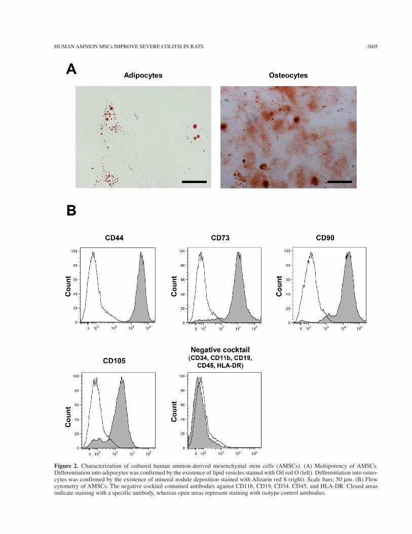
To evaluate the multipotency of human AMSCs, we induced differentiation of cultured AMSCs into adipo-cytes and osteocytes. AMSCs differentiated into adipo-cytes and osteocytes, as demonstrated by Oil red O and Alizarin red S staining, respectively (Fig. 2A). Flow cytometry of cultured AMSCs demonstrated that they expressed CD44, CD73, CD90, and CD105, but not CD34, CD11b, CD19, CD45, and HLA-DR, which is characteristic of MSCs (Fig. 2B) (10).
Effect of AMSC Transplantation on Body Weight, DAI, and Colon Length in DSS-Induced Severe Colitis
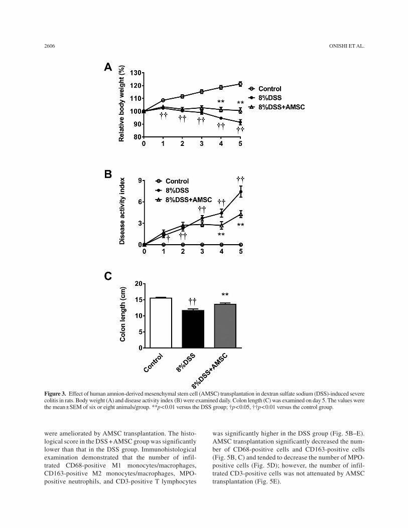
In the DSS group, the body weight gradually decreased on day 3 onward; however, the decrease in body weight was suppressed and significantly attenuated in the DSS + AMSC group on days 4 and 5 (Fig. 3A). DAI gradually increased in the DSS group, whereas it was significantly ameliorated on days 4 and 5 by AMSC transplantation (Fig. 3B). On day 5, colon length was significantly shortened by 8% DSS; however, the length was significantly improved by AMSC transplantation (Fig. 3C). These findings were consistent across AMSCs obtained from three different donors (data not shown).
Effect of AMSC Transplantation on Colonic mRNA Expression Levels of Inflammatory Mediators in the Rectum
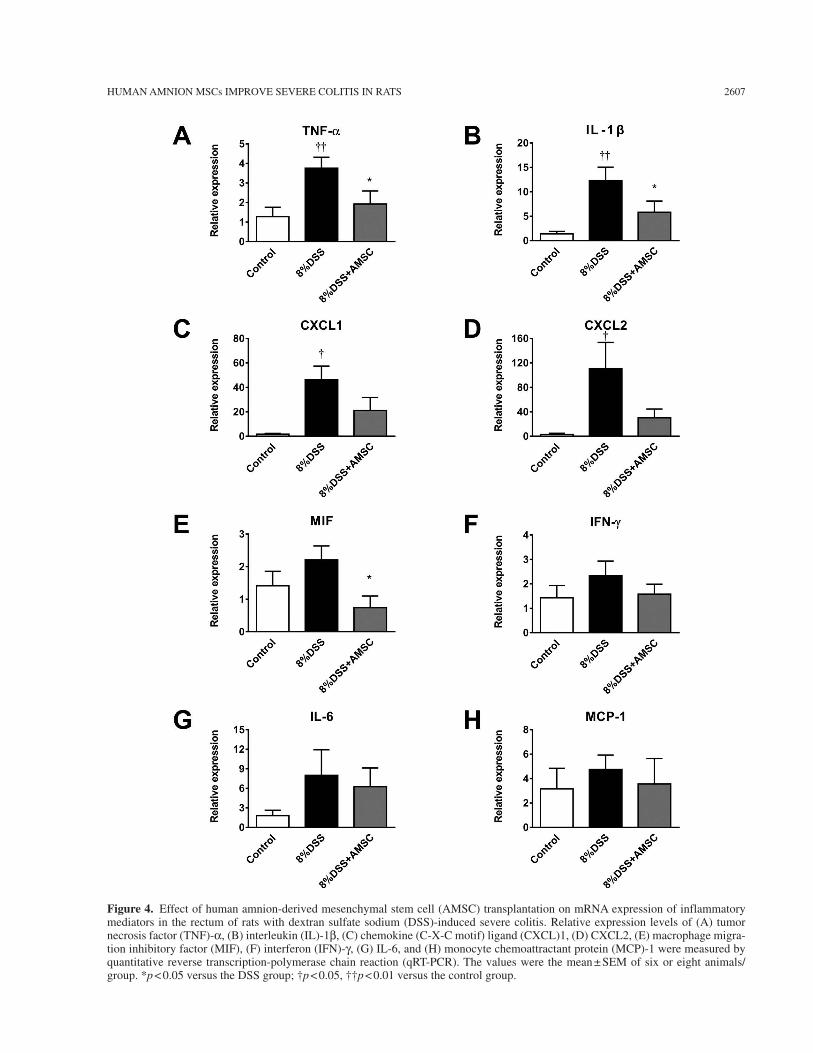
In the DSS group, mRNA expression levels of inflam-matory mediators such as TNF-a, IL-1b, chemokine (C-X-C motif) ligand (CXCL)1, and CXCL2 were significantly increased (Fig. 4A–D). AMSC trans-plantation significantly decreased expression levels of TNF-a, IL-1b, and macrophage migration inhibitory factor (MIF) (Fig. 4A, B, and E). Although not sig-nificantly, AMSC transplantation tended to decrease expression levels of CXCL1, CXCL2, and IFN-g (Fig. 4C, D, and F).
Histological Changes by AMSC Transplantation in 8% DSS-Treated Rats
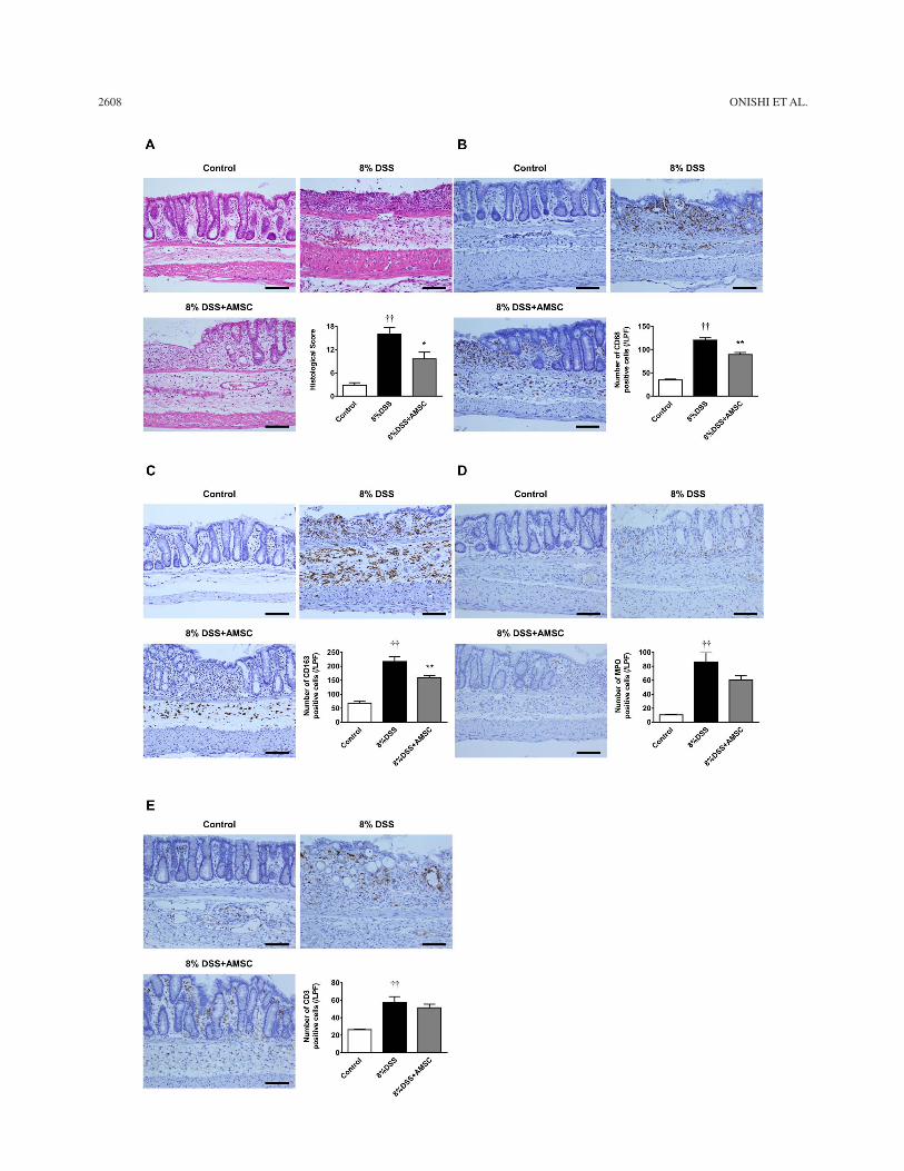
Histological changes in the DSS group included severe transmural inflammation, loss of crypts, and infil-tration of inflammatory cells (Fig. 5A). These findings
HUMAN AMNION MSCs IMPROVE SEVERE COLITIS IN RATS 2605
Figure 2. Characterization of cultured human amnion-derived mesenchymal stem cells (AMSCs). (A) Multipotency of AMSCs. Differentiation into adipocytes was confirmed by the existence of lipid vesicles stained with Oil red O (left). Differentiation into osteo-cytes was confirmed by the existence of mineral nodule deposition stained with Alizarin red S (right). Scale bars: 50 mm. (B) Flow cytometry of AMSCs. The negative cocktail contained antibodies against CD11b, CD19, CD34, CD45, and HLA-DR. Closed areas indicate staining with a specific antibody, whereas open areas represent staining with isotype control antibodies.
2606 ONISHI ET AL.
were ameliorated by AMSC transplantation. The histo-logical score in the DSS + AMSC group was significantly lower than that in the DSS group. Immunohistological examination demonstrated that the number of infil-trated CD68-positive M1 monocytes/macrophages, CD163-positive M2 monocytes/macrophages, MPO-positive neutrophils, and CD3-positive T lymphocytes
was significantly higher in the DSS group (Fig. 5B–E). AMSC transplantation significantly decreased the num-ber of CD68-positive cells and CD163-positive cells (Fig. 5B, C) and tended to decrease the number of MPO-positive cells (Fig. 5D); however, the number of infil-trated CD3-positive cells was not attenuated by AMSC transplantation (Fig. 5E).
Figure 3. Effect of human amnion-derived mesenchymal stem cell (AMSC) transplantation in dextran sulfate sodium (DSS)-induced severe colitis in rats. Body weight (A) and disease activity index (B) were examined daily. Colon length (C) was examined on day 5. The values were the mean ± SEM of six or eight animals/group. **p < 0.01 versus the DSS group; †p < 0.05, ††p < 0.01 versus the control group.
HUMAN AMNION MSCs IMPROVE SEVERE COLITIS IN RATS 2607
Figure 4. Effect of human amnion-derived mesenchymal stem cell (AMSC) transplantation on mRNA expression of inflammatory mediators in the rectum of rats with dextran sulfate sodium (DSS)-induced severe colitis. Relative expression levels of (A) tumor necrosis factor (TNF)-a, (B) interleukin (IL)-1b, (C) chemokine (C-X-C motif) ligand (CXCL)1, (D) CXCL2, (E) macrophage migra-tion inhibitory factor (MIF), (F) interferon (IFN)-g, (G) IL-6, and (H) monocyte chemoattractant protein (MCP)-1 were measured by quantitative reverse transcription-polymerase chain reaction (qRT-PCR). The values were the mean ± SEM of six or eight animals/group. *p < 0.05 versus the DSS group; †p < 0.05, ††p < 0.01 versus the control group.
2608 ONISHI ET AL.
HUMAN AMNION MSCs IMPROVE SEVERE COLITIS IN RATS 2609
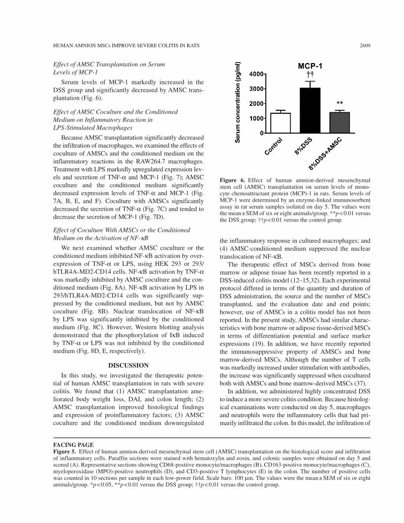
Effect of AMSC Transplantation on Serum Levels of MCP-1
Serum levels of MCP-1 markedly increased in the DSS group and significantly decreased by AMSC trans-plantation (Fig. 6).
Effect of AMSC Coculture and the Conditioned Medium on Inflammatory Reaction in LPS-Stimulated Macrophages
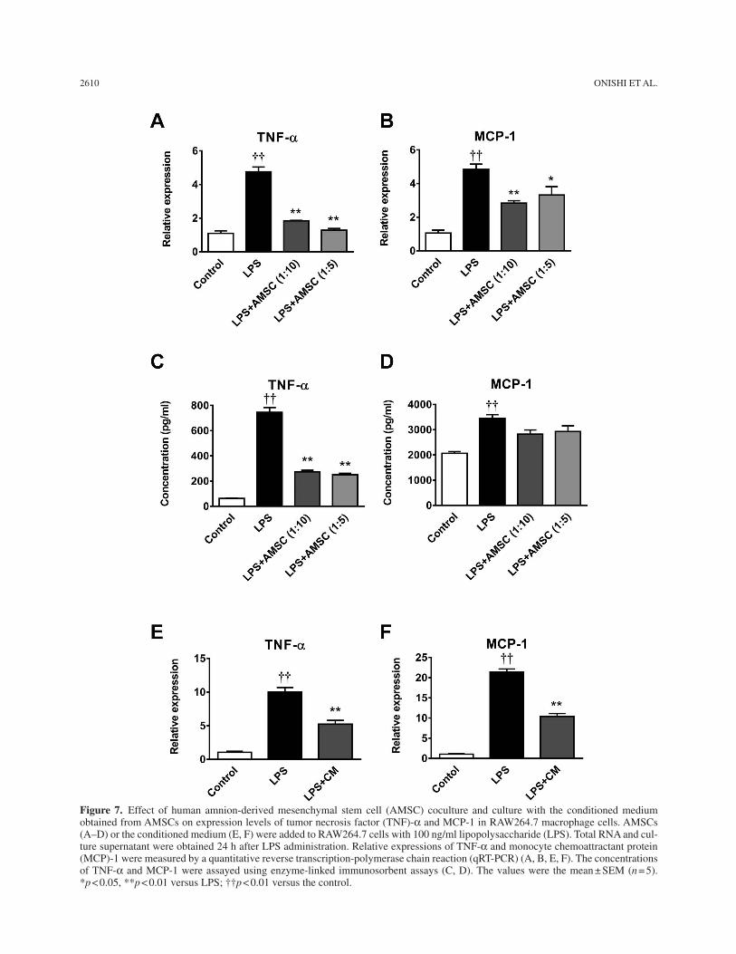
Because AMSC transplantation significantly decreased the infiltration of macrophages, we examined the effects of coculture of AMSCs and the conditioned medium on the inflammatory reactions in the RAW264.7 macrophages. Treatment with LPS markedly upregulated expression lev-els and secretion of TNF-a and MCP-1 (Fig. 7); AMSC coculture and the conditioned medium significantly decreased expression levels of TNF-a and MCP-1 (Fig. 7A, B, E, and F). Coculture with AMSCs significantly decreased the secretion of TNF-a (Fig. 7C) and tended to decrease the secretion of MCP-1 (Fig. 7D).
Effect of Coculture With AMSCs or the Conditioned Medium on the Activation of NF-kB
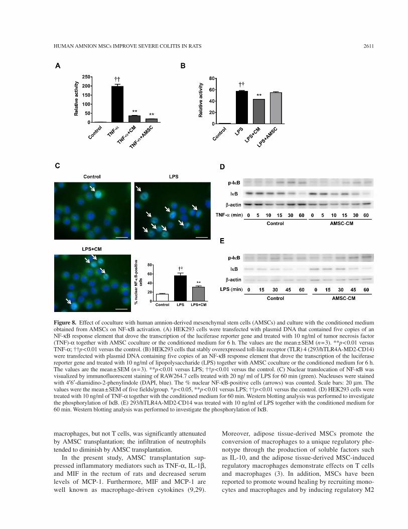
We next examined whether AMSC coculture or the conditioned medium inhibited NF-kB activation by over-expression of TNF-a or LPS, using HEK 293 or 293/hTLR4A-MD2-CD14 cells. NF-kB activation by TNF-a was markedly inhibited by AMSC coculture and the con-ditioned medium (Fig. 8A). NF-kB activation by LPS in 293/hTLR4A-MD2-CD14 cells was significantly sup-pressed by the conditioned medium, but not by AMSC coculture (Fig. 8B). Nuclear translocation of NF-kB by LPS was significantly inhibited by the conditioned medium (Fig. 8C). However, Western blotting analysis demonstrated that the phosphorylation of IkB induced by TNF-a or LPS was not inhibited by the conditioned medium (Fig. 8D, E, respectively).
DISCUSSION
In this study, we investigated the therapeutic poten-tial of human AMSC transplantation in rats with severe colitis. We found that (1) AMSC transplantation ame-liorated body weight loss, DAI, and colon length; (2) AMSC transplantation improved histological findings and expression of proinflammatory factors; (3) AMSC coculture and the conditioned medium downregulated
the inflammatory response in cultured macrophages; and (4) AMSC-conditioned medium suppressed the nuclear translocation of NF-kB.
The therapeutic effect of MSCs derived from bone marrow or adipose tissue has been recently reported in a DSS-induced colitis model (12–15,32). Each experimental protocol differed in terms of the quantity and duration of DSS administration, the source and the number of MSCs transplanted, and the evaluation date and end points; however, use of AMSCs in a colitis model has not been reported. In the present study, AMSCs had similar charac-teristics with bone marrow or adipose tissue-derived MSCs in terms of differentiation potential and surface marker expressions (19). In addition, we have recently reported the immunosuppressive property of AMSCs and bone marrow-derived MSCs. Although the number of T cells was markedly increased under stimulation with antibodies, the increase was significantly suppressed when cocultured both with AMSCs and bone marrow-derived MSCs (37).
In addition, we administered highly concentrated DSS to induce a more severe colitis condition. Because histolog-ical examinations were conducted on day 5, macrophages and neutrophils were the inflammatory cells that had pri-marily infiltrated the colon. In this model, the infiltration of
Figure 6. Effect of human amnion-derived mesenchymal stem cell (AMSC) transplantation on serum levels of mono-cyte chemoattractant protein (MCP)-1 in rats. Serum levels of MCP-1 were determined by an enzyme-linked immunosorbent assay in rat serum samples isolated on day 5. The values were the mean ± SEM of six or eight animals/group. **p < 0.01 versus the DSS group; ††p < 0.01 versus the control group.
FACING PAGEFigure 5. Effect of human amnion-derived mesenchymal stem cell (AMSC) transplantation on the histological score and infiltration of inflammatory cells. Paraffin sections were stained with hematoxylin and eosin, and colonic samples were obtained on day 5 and scored (A). Representative sections showing CD68-positive monocyte/macrophages (B), CD163-positive monocyte/macrophages (C), myeloperoxidase (MPO)-positive neutrophils (D), and CD3-positive T lymphocytes (E) in the colon. The number of positive cells was counted in 10 sections per sample in each low-power field. Scale bars: 100 mm. The values were the mean ± SEM of six or eight animals/group. *p < 0.05, **p < 0.01 versus the DSS group; ††p < 0.01 versus the control group.
2610 ONISHI ET AL.
Figure 7. Effect of human amnion-derived mesenchymal stem cell (AMSC) coculture and culture with the conditioned medium obtained from AMSCs on expression levels of tumor necrosis factor (TNF)-a and MCP-1 in RAW264.7 macrophage cells. AMSCs (A–D) or the conditioned medium (E, F) were added to RAW264.7 cells with 100 ng/ml lipopolysaccharide (LPS). Total RNA and cul-ture supernatant were obtained 24 h after LPS administration. Relative expressions of TNF-a and monocyte chemoattractant protein (MCP)-1 were measured by a quantitative reverse transcription-polymerase chain reaction (qRT-PCR) (A, B, E, F). The concentrations of TNF-a and MCP-1 were assayed using enzyme-linked immunosorbent assays (C, D). The values were the mean ± SEM (n = 5). *p < 0.05, **p < 0.01 versus LPS; ††p < 0.01 versus the control.
HUMAN AMNION MSCs IMPROVE SEVERE COLITIS IN RATS 2611
macrophages, but not T cells, was significantly attenuated by AMSC transplantation; the infiltration of neutrophils tended to diminish by AMSC transplantation.
In the present study, AMSC transplantation sup-pressed inflammatory mediators such as TNF-a, IL-1b, and MIF in the rectum of rats and decreased serum levels of MCP-1. Furthermore, MIF and MCP-1 are well known as macrophage-driven cytokines (9,29).
Moreover, adipose tissue-derived MSCs promote the conversion of macrophages to a unique regulatory phe-notype through the production of soluble factors such as IL-10, and the adipose tissue-derived MSC-induced regulatory macrophages demonstrate effects on T cells and macrophages (3). In addition, MSCs have been reported to promote wound healing by recruiting mono-cytes and macrophages and by inducing regulatory M2
Figure 8. Effect of coculture with human amnion-derived mesenchymal stem cells (AMSCs) and culture with the conditioned medium obtained from AMSCs on NF-kB activation. (A) HEK293 cells were transfected with plasmid DNA that contained five copies of an NF-kB response element that drove the transcription of the luciferase reporter gene and treated with 10 ng/ml of tumor necrosis factor (TNF)-a together with AMSC coculture or the conditioned medium for 6 h. The values are the mean ± SEM (n = 3). **p < 0.01 versus TNF-a; ††p < 0.01 versus the control. (B) HEK293 cells that stably overexpressed toll-like receptor (TLR) 4 (293/hTLR4A-MD2-CD14) were transfected with plasmid DNA containing five copies of an NF-kB response element that drove the transcription of the luciferase reporter gene and treated with 10 ng/ml of lipopolysaccharide (LPS) together with AMSC coculture or the conditioned medium for 6 h. The values are the mean ± SEM (n = 3). **p < 0.01 versus LPS; ††p < 0.01 versus the control. (C) Nuclear translocation of NF-kB was visualized by immunofluorescent staining of RAW264.7 cells treated with 20 ng/ ml of LPS for 60 min (green). Nucleuses were stained with 4¢6¢-diamidino-2-phenylindole (DAPI, blue). The % nuclear NF-kB-positive cells (arrows) was counted. Scale bars: 20 mm. The values were the mean ± SEM of five fields/group. *p < 0.05, **p < 0.01 versus LPS; ††p < 0.01 versus the control. (D) HEK293 cells were treated with 10 ng/ml of TNF-a together with the conditioned medium for 60 min. Western blotting analysis was performed to investigate the phosphorylation of IkB. (E) 293/hTLR4A-MD2-CD14 was treated with 10 ng/ml of LPS together with the conditioned medium for 60 min. Western blotting analysis was performed to investigate the phosphorylation of IkB.
2612 ONISHI ET AL.
macrophages through the action of several enzymes such as prostaglandin E2 (PGE2) and COX-2 that were produced by MSCs (19). PGE2 is a well-known immune modulator in bone marrow MSCs (1), and our group has recently reported that amnion MSCs, but not chorion-derived MSCs, secrete a significant amount of PGE2, particularly when cocultured with human CD4+ T cells (37). M2 macrophages promote the resolution of inflam-mation and tissue repair by releasing IL-10, secreting trophic factors, and enhancing apoptotic cell clearance (23). However, in the present study, immunohistological staining with CD68 and CD163 showed that the infil-tration of both M1 and M2 macrophages was inhibited by AMSC transplantation. Therefore, it appeared that AMSCs did not induce the repolarization of M1 mac-rophages to M2 macrophages.
Our in vitro study demonstrated that AMSC cocul-ture and the conditioned medium significantly decreased expression levels of TNF-a and MCP-1 from RAW264.7 cells. The production of inflammatory molecules in LPS-activated RAW264.7 cells was significantly suppressed by coincubation with human placental multipotent mes-enchymal stromal cells (hPMSCs) (6). Those findings showed that the anti-inflammatory effect of hPMSCs was mediated, at least in part, by PGE2 via a myeloid differen-tiation primary response gene 88-dependent pathway. In the present study, the conditioned medium inhibited the transcriptional activation of NF-kB by TNF-a. Because IkB phosphorylation by TNF-a or LPS was not inhibited, the conditioned medium may have suppressed the inflam-matory signal of NF-kB at the step of nuclear transloca-tion. Further studies are required to clarify the underlying mechanism.
Recently, several studies related to cell therapy using human MSCs in IBD have been reported. Administration of autologous bone marrow-derived MSCs has been reported to be safe and feasible in the treatment of refrac-tory luminal Crohn’s disease (11). The authors showed that MSC transplantation contributed to amelioration of clinical symptoms and endoscopic findings without any critical side effects. In addition, local injection of autolo-gous bone marrow-derived MSCs has been reported to be feasible, safe, and successful in treating Crohn’s dis-ease with fistuling (7). Moreover, allogeneic transplan-tation of MSCs, obtained from bone marrow of healthy donors, and umbilical cords, obtained at normal deliver-ies, was safe and may contribute to clinical improvement in patients with refractory Crohn’s disease and ulcerative colitis (20). Other clinical trials using human MSCs as a cell-based therapy in IBD are being conducted in several countries (http://www.clinicaltrials.gov).
Recent reports have suggested a similar efficacy for AMSCs in several other diseases. It has been demonstrated that intravenous infusion of human AMSCs ameliorates
inflammation and fibrosis in the lung induced by bleo-mycin in mice (22). In addition, human AMSCs have been reported to have the potential to reduce liver fibrosis induced by carbon tetrachloride administration in mice (38). Furthermore, very recently, a first-in-human pilot study using fetal membrane-derived MSCs has been con-ducted to treat nine patients with steroid-refractory acute graft-versus-host disease (GVHD); the fetal membrane-derived MSCs appeared safe for intravenous infusion to most patients, and the overall response rate in severe refractory acute GVHD appeared to be similar to the rate observed while using bone marrow-derived MSCs (28). This is encouraging because it is rather invasive to aspi-rate bone marrow or obtain adipose tissue from donors, and patients with refractory IBD may require repeated cell therapy for a long time. Furthermore, AMSCs are immediately available when needed by a patient because they can be stored in advance.
In conclusion, human AMSC transplantation amelio-rated the inflammatory response in a rat model of severe colitis, possibly through the suppression of macrophage activity. Considering that fetal membrane is generally treated as medical waste and can be obtained without an invasive procedure, human AMSC transplantation may be a therapeutic strategy for the treatment of severe colitis.
ACKNOWLEDGMENTS: This study was supported by a Grant-in-Aid for Young Scientists (B) from the Japan Society for the Promotion of Science (JSPS) and by the Ministry of Health Labour and Welfare. The authors declare no conflicts of interest.
REFERENCESAggarwal, S.; Pittenger, M. F. Human mesenchymal stem 1. cells modulate allogeneic immune cell responses. Blood 105:1815–1822; 2005.Alviano, F.; Fossati, V.; Marchionni, C.; Arpinati, M.; Bonsi, 2. L.; Franchina, M.; Lanzoni, G.; Cantoni, S.; Cavallini, C.; Bianchi, F.; Tazzari, P. L.; Pasquinelli, G.; Foroni, L.; Ventura, C.; Grossi, A.; Bagnara, G. P. Term amniotic membrane is a high throughput source for multipotent mes-enchymal stem cells with the ability to differentiate into endothelial cells in vitro. BMC Dev. Biol. 7:11; 2007.Anderson, P.; Souza-Moreira, L.; Morell, M.; Caro, M.; 3. O’Valle, F.; Gonzalez-Rey, E.; Delgado, M. Adipose-derived mesenchymal stromal cells induce immunomodu-latory macrophages which protect from experimental colitis and sepsis. Gut 62:1131–1141; 2013.Baumgart, D. C.; Carding, S. R. Inflammatory bowel dis- 4. ease: Cause and immunobiology. Lancet 369:1627–1640; 2007.Burisch, J.; Munkholm, P. Inflammatory bowel disease epi- 5. demiology. Curr. Opin. Gastroenterol. 29:357–362; 2013.Chen, C. P.; Tsai, P. S.; Huang, C. J. Anti-inflammation effect 6. of human placental multipotent mesenchymal stromal cells is mediated by prostaglandin E2 via a myeloid differentiation primary response gene 88-dependent pathway. Anesthesiology 117:568–579; 2012.Ciccocioppo, R.; Bernardo, M. E.; Sgarella, A.; 7. Maccario, R.; Avanzini, M. A.; Ubezio, C.; Minelli, A.;
HUMAN AMNION MSCs IMPROVE SEVERE COLITIS IN RATS 2613
Alvisi, C.; Vanoli, A.; Calliada, F.; Dionigi, P.; Perotti, C.; Locatelli, F.; Corazza, G. R. Autologous bone marrow- derived mesenchymal stromal cells in the treatment of fis-tulising Crohn’s disease. Gut 60:788–798; 2011.Cooper, H. S.; Murthy, S. N.; Shah, R. S.; Sedergran, D. J. 8. Clinicopathologic study of dextran sulfate sodium experi-mental murine colitis. Lab. Invest. 69:238–249; 1993.de Jong, Y. P.; Abadia-Molina, A. C.; Satoskar, A. R.; Clarke, 9. K.; Rietdijk, S. T.; Faubion, W. A.; Mizoguchi, E.; Metz, C. N.; Alsahli, M.; ten Hove, T.; Keates, A. C.; Lubetsky, J. B.; Farrell, R. J.; Michetti, P.; van Deventer, S. J.; Lolis, E.; David, J. R.; Bhan, A. K.; Terhorst, C. Development of chronic colitis is dependent on the cytokine MIF. Nat. Immunol. 2:1061–1066; 2001.Dominici, M.; Le Blanc, K.; Mueller, I.; Slaper-Cortenbach, 10. I.; Marini, F.; Krause, D.; Deans, R.; Keating, A.; Prockop, D.; Horwitz, E. Minimal criteria for defining multipotent mesenchymal stromal cells. The International Society for Cellular Therapy position statement. Cytotherapy 8:315–317; 2006.Duijvestein, M.; Vos, A. C.; Roelofs, H.; Wildenberg, 11. M. E.; Wendrich, B. B.; Verspaget, H. W.; Kooy-Winkelaar, E. M.; Koning, F.; Zwaginga, J. J.; Fidder, H. H.; Verhaar, A. P.; Fibbe, W. E.; van den Brink, G. R.; Hommes, D. W. Autologous bone marrow-derived mesenchymal stromal cell treatment for refractory luminal Crohn’s disease: Results of a phase I study. Gut 59:1662–1669; 2010.Duijvestein, M.; Wildenberg, M. E.; Welling, M. M.; 12. Hennink, S.; Molendijk, I.; van Zuylen, V. L.; Bosse, T.; Vos, A. C.; de Jonge-Muller, E. S.; Roelofs, H.; van der Weerd, L.; Verspaget, H. W.; Fibbe, W. E.; te Velde, A. A.; van den Brink, G. R.; Hommes, D. W. Pretreatment with interferon-gamma enhances the therapeutic activity of mes-enchymal stromal cells in animal models of colitis. Stem Cells 29:1549–1558; 2011.Gonzalez-Rey, E.; Anderson, P.; Gonzalez, M. A.; Rico, L.; 13. Buscher, D.; Delgado, M. Human adult stem cells derived from adipose tissue protect against experimental colitis and sepsis. Gut 58:929–939; 2009.Gonzalez, M. A.; Gonzalez-Rey, E.; Rico, L.; Buscher, D.; 14. Delgado, M. Adipose-derived mesenchymal stem cells allevi-ate experimental colitis by inhibiting inflammatory and auto-immune responses. Gastroenterology 136:978–989; 2009.He, X. W.; He, X. S.; Lian, L.; Wu, X. J.; Lan, P. Systemic 15. infusion of bone marrow-derived mesenchymal stem cells for treatment of experimental colitis in mice. Dig. Dis. Sci. 57:3136–3144; 2012.In ‘t Anker, P. S.; Scherjon, S. A.; Kleijburg-van der Keur, C.; 16. de Groot-Swings, G. M.; Claas, F. H.; Fibbe, W. E.; Kanhai, H. H. Isolation of mesenchymal stem cells of fetal or mater-nal origin from human placenta. Stem Cells 22:1338–1345; 2004.Ishikane, S.; Ohnishi, S.; Yamahara, K.; Sada, M.; Harada, K.; 17. Mishima, K.; Iwasaki, K.; Fujiwara, M.; Kitamura, S.; Nagaya, N.; Ikeda, T. Allogeneic injection of fetal membrane-derived mesenchymal stem cells induces therapeutic angiogenesis in a rat model of hind limb ischemia. Stem Cells 26:2625–2633; 2008.Ishikane, S.; Yamahara, K.; Sada, M.; Harada, K.; Kodama, 18. M.; Ishibashi-Ueda, H.; Hayakawa, K.; Mishima, K.; Iwasaki, K.; Fujiwara, M.; Kangawa, K.; Ikeda, T. Allogeneic admin-istration of fetal membrane-derived mesenchymal stem cells attenuates acute myocarditis in rats. J. Mol. Cell. Cardiol. 49:753–761; 2010.
Le Blanc, K.; Mougiakakos, D. Multipotent mesenchymal 19. stromal cells and the innate immune system. Nat. Rev. Immunol. 12:383–396; 2012.Liang, J.; Zhang, H.; Wang, D.; Feng, X.; Wang, H.; Hua, B.; 20. Liu, B.; Sun, L. Allogeneic mesenchymal stem cell transplan-tation in seven patients with refractory inflammatory bowel disease. Gut 61:468–469; 2012.Meirelles Lda, S.; Fontes, A. M.; Covas, D. T.; Caplan, 21. A. I. Mechanisms involved in the therapeutic properties of mesenchymal stem cells. Cytokine Growth Factor Rev. 20:419–427; 2009.Moodley, Y.; Vaghjiani, V.; Chan, J.; Baltic, S.; Ryan, M.; 22. Tchongue, J.; Samuel, C. S.; Murthi, P.; Parolini, O.; Manuelpillai, U. Anti-inflammatory effects of adult stem cells in sustained lung injury: A comparative study. PLoS One 8:e69299; 2013.Mosser, D. M.; Edwards, J. P. Exploring the full spectrum 23. of macrophage activation. Nat. Rev. Immunol. 8:958–969; 2008.Mowat, C.; Cole, A.; Windsor, A.; Ahmad, T.; Arnott, I.; 24. Driscoll, R.; Mitton, S.; Orchard, T.; Rutter, M.; Younge, L.; Lees, C.; Ho, G. T.; Satsangi, J.; Bloom, S.; IBD Section of the British Society of Gastroenterology. Guidelines for the management of inflammatory bowel disease in adults. Gut 60:571–607; 2011.Ng, S. C.; Bernstein, C. N.; Vatn, M. H.; Lakatos, P. L.; 25. Loftus, E. V., Jr.; Tysk, C.; O’Morain, C.; Moum, B.; Colombel, J. F.; Epidemiology and Natural History Task Force of the International Organization of Inflammatory Bowel Disease. Geographical variability and environmen-tal risk factors in inflammatory bowel disease. Gut 62:630–649; 2013.Ohshima, M.; Yamahara, K.; Ishikane, S.; Harada, K.; 26. Tsuda, H.; Otani, K.; Taguchi, A.; Miyazato, M.; Katsuragi, S.; Yoshimatsu, J.; Kodama, M.; Kangawa, K.; Ikeda, T. Systemic transplantation of allogenic fetal membrane-de-rived mesenchymal stem cells suppresses Th1 and Th17 T cell responses in experimental autoimmune myocarditis. J. Mol. Cell. Cardiol. 53:420–428; 2012.Pittenger, M. F.; Mackay, A. M.; Beck, S. C.; Jaiswal, R. K.; 27. Douglas, R.; Mosca, J. D.; Moorman, M. A.; Simonetti, D. W.; Craig, S.; Marshak, D. R. Multilineage potential of adult human mesenchymal stem cells. Science 284:143–147; 1999.Ringdén, O.; Erkers, T.; Nava, S.; Uzunel, M.; Iwarsson, E.; 28. Conrad, R.; Westgren, M.; Mattsson, J.; Kaipe, H. Fetal membrane cells for treatment of steroid-refractory acute graft-versus-host disease. Stem Cells 31:592–601; 2013.Rollins, B. J. Chemokines. Blood 90:909–928; 1997.29. Sartor, R. B. Mechanisms of disease: Pathogenesis of 30. Crohn’s disease and ulcerative colitis. Nat. Clin. Pract. Gastroenterol. Hepatol. 3:390–407; 2006.Singh, K.; Chaturvedi, R.; Barry, D. P.; Coburn, L. A.; 31. Asim, M.; Lewis, N. D.; Piazuelo, M. B.; Washington, M. K.; Vitek, M. P.; Wilson, K. T. The apolipoprotein E-mimetic peptide COG112 inhibits NF-kappaB signaling, proinflam-matory cytokine expression, and disease activity in murine models of colitis. J. Biol. Chem. 286:3839–3850; 2011.Tanaka, F.; Tominaga, K.; Ochi, M.; Tanigawa, T.; Watanabe, 32. T.; Fujiwara, Y.; Ohta, K.; Oshitani, N.; Higuchi, K.; Arakawa, T. Exogenous administration of mesenchymal stem cells ameliorates dextran sulfate sodium-induced coli-tis via anti-inflammatory action in damaged tissue in rats. Life Sci. 83:771–779; 2008.
2614 ONISHI ET AL.
Tsuda, H.; Yamahara, K.; Ishikane, S.; Otani, K.; Nakamura, 33. A.; Sawai, K.; Ichimaru, N.; Sada, M.; Taguchi, A.; Hosoda, H.; Tsuji, M.; Kawachi, H.; Horio, M.; Isaka, Y.; Kangawa, K.; Takahara, S.; Ikeda, T. Allogenic fetal membrane-derived mesenchymal stem cells contribute to renal repair in experimental glomerulonephritis. Am. J. Physiol. Renal Physiol. 299:F1004–1013; 2010.Tsuda, H.; Yamahara, K.; Otani, K.; Okumi, M.; Yazawa, 34. K.; Kaimori, J. Y.; Taguchi, A.; Kangawa, K.; Ikeda, T.; Takahara, S.; Isaka, Y. Transplantation of allogenic fetal membrane-derived mesenchymal stem cells protect against ischemia-reperfusion-induced acute kidney injury. Cell Transplant. 23:889–899; 2014.Uccelli, A.; Moretta, L.; Pistoia, V. Mesenchymal stem 35. cells in health and disease. Nat. Rev. Immunol. 8:726–736; 2008.
Williams, K. L.; Fuller, C. R.; Dieleman, L. A.; DaCosta, 36. C. M.; Haldeman, K. M.; Sartor, R. B.; Lund, P. K. Enhanced survival and mucosal repair after dextran sodium sulfate–induced colitis in transgenic mice that overexpress growth hormone. Gastroenterology 120:925–937; 2001.Yamahara, K.; Harada, K.; Ohshima, M.; Ishikane, S.; 37. Ohnishi, S.; Tsuda, H.; Otani, K.; Taguchi, A.; Soma, T.; Ogawa, H.; Katsuragi, S.; Yoshimatsu, J.; Harada-Shiba, M.; Kangawa, K.; Ikeda, T. Comparison of angiogenic, cytoprotective, and immunosuppressive properties of human amnion- and chorion-derived mesenchymal stem cells. PLoS One 9:e88319; 2014.Zhang, D.; Jiang, M.; Miao, D. Transplanted human amni-38. otic membrane-derived mesenchymal stem cells ameliorate carbon tetrachloride-induced liver cirrhosis in mouse. PLoS One 6:e16789; 2011.