Embed Size (px)
Citation preview
1919Journal of Cell Science 110, 1919-1934 (1997)Printed in Great Britain © The Company of Biologists Limited 1997JCS4150
Hydra regeneration from recombined ectodermal and endodermal tissue
II. Differential stability in the ectodermal and endodermal epithelial organization
Motohide Murate 1,*, Yasuyuki Kishimoto 1,†, Tsutomu Sugiyama 2,‡, Toshitaka Fujisawa 2,§, Hiromi Takahashi-Iwanaga 3,¶ and Toshihiko Iwanaga 3,II
1Department of Life Sciences, The Graduate University for Advanced Studies, Mishima 411, Japan2National Institute of Genetics, Mishima 411, Japan3School of Medicine, Niigata University, Niigata 951, Japan
*Present address: Frontier Research Program, The Institute of Physical and Chemical Research (RIKEN), Wako, Saitama 351-01, Japan†Present address: Max-Planck Institute for Developmental Biology, 72076 Tübingen, Germany‡Present address: Ishinomaki Senshu University, Ishinomaki 986-80, Japan§Author for correspondence (e-mail: [email protected])¶Present address: School of Medicine, Hokkaido University, Sapporo 060, JapanIIPresent address: Department of Veterinary Medicine, Hokkaido University, Sapporo 060, Japan
Hydra tissue consists of the ectodermal and the endoder-mal layers. When the two layers were separated byprocaine treatment and then recombined, the ectodermalepithelial cells spread as a single cell layer over theendoderm as in epiboly in vertebrate embryogenesis, andthe resultant spherical structure subsequently regeneratedinto a complete hydra. In this study, light and electronmicroscopy were used to examine the structural changeswhich took place in the cells and tissue during this epibolicectodermal spreading process. Within a few hours aftertissue recombination, the endoderm underwent dramaticchanges; it lost its epithelial sheet organization, and turnedinto a mass of irregularly shaped cells without the apical-basal cell polarity initially present. In contrast, theectoderm maintained its basic epithelial sheet organizationas it spread over the endoderm. Later, the endodermalepithelial cells reorganized themselves into a single-layered
epithelial sheet underneath the spreading ectodermal layer.The resultant spherical structure consisted of a single layerof ectodermal epithelial cells outside, a single layer of endo-dermal epithelial cells inside, and an empty cavity in thecenter as in normal hydra tissue. This structure regener-ated into hydra in the following days. These and otherobservations demonstrate that the two-layered epithelialsheet organization is highly dynamic, and that its stabilityis maintained by strong interactions between the two layersin normal hydra. It is suggested that this dynamic natureof the hydra tissue, particularly the high plasticity of theendodermal epithelial sheet organization, may be animportant element for the high regenerative capacity of thisorganism.
Key words: Hydra, Epiboly, Regeneration, Electron microscopy,Epithelial organization
SUMMARY
eren-ereheor
dn-ne. toce
e atto-ireingof
INTRODUCTION
The body plan of hydra is very simple (Campbell and Bod1983). Its main body column is a hollow cylinder consisting an outer layer of ectodermal epithelial cells and an inner laof endodermal epithelial cells. The two layers are separateda thick layer of extracellular matrix, the mesoglea. A head cosisting of a hypostome and tentacles is located at one enthe body column, while a foot with a basal disk and pedunexists at the other end.
Epp et al. (1979, 1986) showed that ectodermal and endermal layers can be separated from each other by treatmwith an anesthetizing drug, procaine, and that the separatissue layers can be reassembled to regenerate into a new hThis reassembly system was used to show that both ectoderm and endoderm are involved in determining tpolarity of regeneration from the reassembled tissue (Smid Tardent, 1982), and that gland cells present in the endod
e,ofyer byn-
d ofcle
do-entted
ydra.theheanderm
can arise by differentiation from the interstitial cells initiallylocated in the ectoderm (Bode et al., 1987).
In these studies, the ectodermal and endodermal tissues wseparated from each other without altering the original cylidrical organization of each tissue. The separated tissues wthen reassembled by slipping the endodermal cylinder into tcentral hollow space of the ectodermal cylinder in a parallel anti-parallel direction.
In a previous study (Kishimoto et al., 1996), we modifiethe procedure by deliberately destroying the cylindrical cofiguration of one or both of the tissues separated by procaiThe two tissue pieces were then recombined to allow themregenerate. We then examined the interaction which took plabetween the recombined ectodermal and endodermal tissuthe light microscope level. Soon after recombination, the ecdermal tissue spread as a thin layer to cover the entendoderm in 1-2 days. This process of ectodermal spreadover the endoderm was termed ‘epiboly’ after the name
1920
e
M. Murate and others
similar processes in early embryogenesis in various ver-tebrates, including chick (New, 1959), amphibian (Keller,1980), and fish (Trinkaus, 1984). During the epibolic process,extensive cell rearrangement took place in the tissue, particu-larly in the interface between the recombined ectoderm andendoderm.
In the present study, we used light and electron microscopyto examine structural changes which took place in the ecto-dermal and endodermal epithelial cells during the epibolicprocess. We found that both the ectoderm and endodermunderwent major structural changes, but in significantlydifferent ways. The ectoderm maintained its basic epithelialsheet organization, whereas the endoderm lost the epithelialsheet organization within a few hours after separation from theectoderm, and turned into a mass of irregularly shaped cells.These cells later reestablished an epithelium underneath thespreading ectoderm.
The roles of epithelial and interstitial cell lineages in hydraregeneration are discussed based on observations made in thisand related previous studies.
MATERIALS AND METHODS
Strain and cultureHydra strain and culture conditions used in this study were describedin detail by Kishimoto et al. (1996). Briefly, strain 105 of Hydra mag-nipapillata (Sugiyama and Fujisawa, 1977) was cultured at a lowdensity in large flat plastic trays at 18°C. They were fed daily tosatiation with freshly hatched brine shrimp nauplii, and a few hourslater transferred to new trays containing fresh culture solution. Rela-tively young polyps cultured for 7-8 days after detachment fromparental polyps were harvested 24-30 hours after the last feeding, andused for preparing the ectodermal and endodermal tissue describedbelow.
Ectodermal and endodermal tissuesThe procedures originally described by Epp et al. (1979) and modifiedby Kishimoto et al. (1996) were used to separate the ectodermal andendodermal tissues and then to recombine the separated tissues.
Equal volumes of a 1% solution of procaine-HCl (Wako PureChemicals, Tokyo) in distilled water, hyper-osmotic (70 mOsmol) saltsolution for tissue dissociation and cell reaggregation (DM solution)(Gierer et al., 1972), and modified M solution for hydra culture (mMsolution) (Sugiyama and Fujisawa, 1977) were mixed and adjusted topH 4.5 (A solution) or pH 2.5 (B solution) immediately before use.Heads and feet were removed from 7-8 day old hydra by amputationsmade below the tentacle ring and above the budding zone (a in Fig.1).
To prepare the ectodermal tissue, the body column tissue obtainedwas treated in A solution for 5 minutes, followed by treatment in Bsolution for 1G minutes, both at 4°C. The treated tissue was gentlytransferred to DM solution kept at 18°C. Within several minutes, theectodermal tissue contracted into a ring-shaped tissue at one end ofthe rod-shaped endoderm (c in Fig. 1). Using a pair of forceps withfine tips, the two tissue pieces were teased apart to obtain the ecto-dermal tissue (d in Fig. 1). To prepare the endodermal tissue, the bodycolumn tissue was treated in the same way, except for treatment in Bsolution for 6 minutes instead of 1G minutes. This treatment producedpartial disintegration of the ectodermal tissue whereas the endoder-mal tissue remained in the rod-shape in DM solution. The former wasflushed away from the latter by a stream of liquid directed against theformer using a Pasteur pipette. The rod-shaped endodermal tissue (ein Fig. 1) obtained in this way was used.
Recombination of the ectodermal and endodermal tissuesThe ring-shaped ectodermal tissue obtained by procaine treatment wasradially cut into 6-8 equal-sized pieces (d in Fig. 1), and the freshlycut-surface of one of the pieces (f in Fig. 1) was gently placed in directcontact with an endodermal tissue (e in Fig. 1). A firm adhesion wasestablished between them in less than half an hour (g in Fig. 1). Therecombined tissue produced in this way was allowed to regenerate.The entire process was carried out in DM solution.
Scanning electr on micr oscop ySpecimens of recombined tissue were harvested at various times afterrecombination, and fixed with 2.5% glutaraldehyde in 0.1 Mphosphate buffer, pH 7.5, at 4°C for overnight or longer. The fixedspecimens were rinsed in distilled water, conductively stained by thetannin-osmium method described by Murakami (1974), dehydratedthrough a graded series of ethanol, transferred to isoamyl acetate,dried at critical point using CO2, coated with gold-palladium vapor,and examined in a Hitachi S-450 LB SEM at an acceleration voltageof 10 kV.
Transmission electr on micr oscop ySpecimens were fixed in 2.5% glutaraldehyde in 0.1 M cacodylatebuffer, pH 7.5, at 4°C for overnight or longer. The fixed samples werepost fixed in 1% OsO4 for 1G hours, dehydrated through a gradedseries of ethanol and propylene oxide, and embedded in Araldite resin.Semithin sections were cut at a thickness of 0.5 µm and stained withToluidine blue for examination under a light microscope. Ultrathinsections were cut at a thickness of 95 nm using an ultramicrotomewith a diamond knife, double-stained with uranyl acetate and leadcitrate, and examined under a Hitachi H-7000 TEM at an accelerationvoltage of 75 kV.
Layouts of photographs were made electronically using a NikonCool Scan and a computer program Adobe Photoshop 3.0J (AdobeSystems Inc., Mountain View, California), and printouts were madeusing a Pictrography 3000 (Fuji Photo Film, Tokyo).
RESULTS
An outline of the experiment carried out in this study is illus-trated in Fig. 1. Hydra body column tissue (b in Fig. 1) wastreated with procaine to separate the ectodermal and endoder-mal tissue layers from each other (c in Fig. 1) (see Materialsand Methods). The ring-shaped ectodermal tissue (d in Fig. 1)was cut into smaller pieces (f in Fig. 1), and one of them wasplaced in the middle of a rod-shaped piece of endodermaltissue (e in Fig. 1). The two tissue types established a firmadhesion quickly (g in Fig. 1). This was followed by gradualspreading of the ectodermal tissue as a thin layer over theendoderm (h and i in Fig. 1). During this process, the endodermgradually rounded up. The resultant structure formed in 1-2days was a small sphere completely enveloped by ectodermaltissue (j in Fig. 1). This structure regenerated into a new hydrain the following 3-4 days (Kishimoto et al., 1996).
At various times in the first 1-2 day period, the recombinedtissues undergoing the epibolic process (g-j in Fig. 1) wereharvested, fixed, and examined by light, scanning and trans-mission electron microscopy. Based on the results obtained, wedivided the morphogenetic processes in this period into 4stages. Stage I represents initial adhesion soon after tissucontact. Stages II, III and IV represent the early, middle andlate phases, respectively, of the ectodermal spreading over theendoderm. Photographs of typical specimens from each ofthese stages are presented below.
1921Hydra regeneration
AA
Procaine
Ectoderm
Endoderm
a
b cd
f
e g
hij
treatment
AA
Fig. 1. An outline of the experiment carried out inthis study. See main text for explanation (alsoMaterials and Methods).
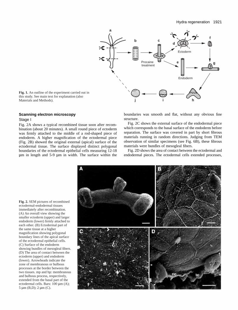
Scanning electr on micr oscop y Stage IFig. 2A shows a typical recombined tissue soon after recom-bination (about 20 minutes). A small round piece of ectodermwas firmly attached to the middle of a rod-shaped piece ofendoderm. A higher magnification of the ectodermal piece(Fig. 2B) showed the original external (apical) surface of theectodermal tissue. The surface displayed distinct polygonalboundaries of the ectodermal epithelial cells measuring 12-18µm in length and 5-9 µm in width. The surface within the
Fig. 2. SEM pictures of recombinedectodermal-endodermal tissuesimmediately after recombination.(A) An overall view showing thesmaller ectoderm (upper) and largerendoderm (lower) firmly attached toeach other. (B) Ectodermal part ofthe same tissue at a highermagnification showing polygonalboundary lines of the apical surfaceof the ectodermal epithelial cells.(C) Surface of the endodermshowing bundles of mesogleal fibers.(D) The area of contact between theectoderm (upper) and endoderm(lower). Arrowheads indicate thezone of membranous or bulbousprocesses at the border between thetwo tissues. mp and bp: membranousand bulbous process, respectively,extended from the basal part of theectodermal cells. Bars: 100 µm (A);5 µm (B,D); 2 µm (C).
boundaries was smooth and flat, without any obvious finestructure.
Fig. 2C shows the external surface of the endodermal piecewhich corresponds to the basal surface of the endoderm beforeseparation. The surface was covered in part by short fibrousmaterials running in random directions. Judging from TEMobservation of similar specimens (see Fig. 6B), these fibrousmaterials were bundles of mesogleal fibers.
Fig. 2D shows the area of contact between the ectodermal andendodermal pieces. The ectodermal cellsextended processes,
1922 M. Murate and others
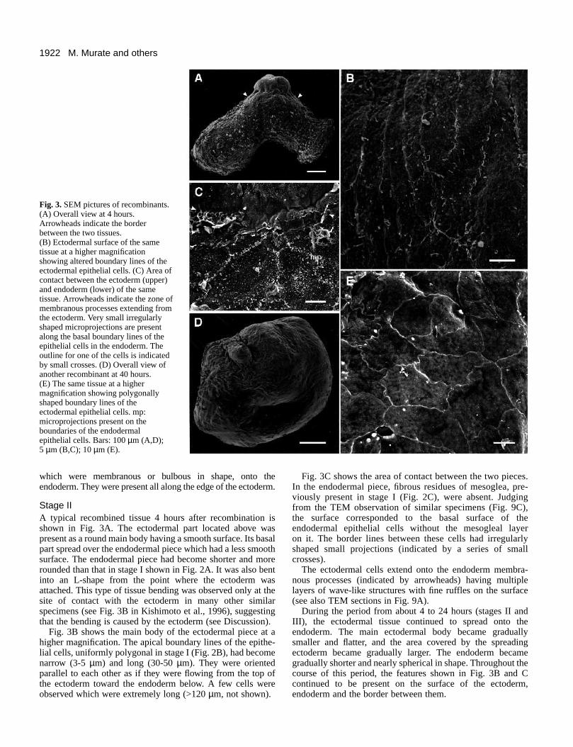
Fig. 3.SEM pictures of recombinants.(A) Overall view at 4 hours.Arrowheads indicate the borderbetween the two tissues.(B) Ectodermal surface of the sametissue at a higher magnificationshowing altered boundary lines of theectodermal epithelial cells. (C) Area ofcontact between the ectoderm (upper)and endoderm (lower) of the sametissue. Arrowheads indicate the zone ofmembranous processes extending fromthe ectoderm. Very small irregularlyshaped microprojections are presentalong the basal boundary lines of theepithelial cells in the endoderm. Theoutline for one of the cells is indicatedby small crosses. (D) Overall view ofanother recombinant at 40 hours.(E) The same tissue at a highermagnification showing polygonallyshaped boundary lines of theectodermal epithelial cells. mp:microprojections present on theboundaries of the endodermalepithelial cells. Bars: 100 µm (A,D);5 µm (B,C); 10 µm (E).
which were membranous or bulbous in shape, onto theendoderm. They were present all along the edge of the ectoderm.
Stage IIA typical recombined tissue 4 hours after recombination isshown in Fig. 3A. The ectodermal part located above waspresent as a round main body having a smooth surface. Its basalpart spread over the endodermal piece which had a less smoothsurface. The endodermal piece had become shorter and morerounded than that in stage I shown in Fig. 2A. It was also bentinto an L-shape from the point where the ectoderm wasattached. This type of tissue bending was observed only at thesite of contact with the ectoderm in many other similarspecimens (see Fig. 3B in Kishimoto et al., 1996), suggestingthat the bending is caused by the ectoderm (see Discussion).
Fig. 3B shows the main body of the ectodermal piece at ahigher magnification. The apical boundary lines of the epithe-lial cells, uniformly polygonal in stage I (Fig. 2B), had becomenarrow (3-5 µm) and long (30-50 µm). They were orientedparallel to each other as if they were flowing from the top ofthe ectoderm toward the endoderm below. A few cells wereobserved which were extremely long (>120 µm, not shown).
Fig. 3C shows the area of contact between the two pieces.In the endodermal piece, fibrous residues of mesoglea, pre-viously present in stage I (Fig. 2C), were absent. Judgingfrom the TEM observation of similar specimens (Fig. 9C),the surface corresponded to the basal surface of theendodermal epithelial cells without the mesogleal layer on it. The border lines between these cells had irregularlyshaped small projections (indicated by a series of smallcrosses).
The ectodermal cells extend onto the endoderm membra-nous processes (indicated by arrowheads) having multiplelayers of wave-like structures with fine ruffles on the surface(see also TEM sections in Fig. 9A).
During the period from about 4 to 24 hours (stages II andIII), the ectodermal tissue continued to spread onto theendoderm. The main ectodermal body became graduallysmaller and flatter, and the area covered by the spreadingectoderm became gradually larger. The endoderm becamegradually shorter and nearly spherical in shape. Throughout thecourse of this period, the features shown in Fig. 3B and Ccontinued to be present on the surface of the ectoderm,endoderm and the border between them.
1923Hydra regeneration
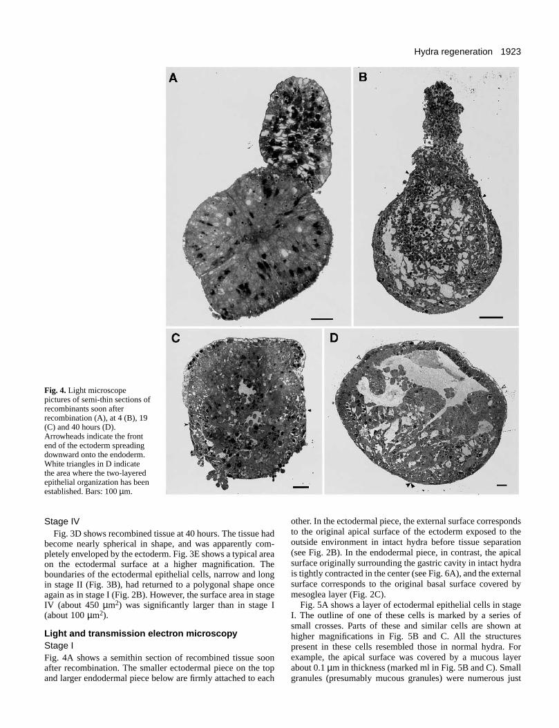
Fig. 4.Light microscopepictures of semi-thin sections ofrecombinants soon afterrecombination (A), at 4 (B), 19(C) and 40 hours (D).Arrowheads indicate the frontend of the ectoderm spreadingdownward onto the endoderm.White triangles in D indicatethe area where the two-layeredepithelial organization has beenestablished. Bars: 100 µm.
Stage IVFig. 3D shows recombined tissue at 40 hours. The tissue had
become nearly spherical in shape, and was apparently com-pletely enveloped by the ectoderm. Fig. 3E shows a typical areaon the ectodermal surface at a higher magnification. Theboundaries of the ectodermal epithelial cells, narrow and longin stage II (Fig. 3B), had returned to a polygonal shape onceagain as in stage I (Fig. 2B). However, the surface area in stageIV (about 450 µm2) was significantly larger than in stage I(about 100 µm2).
Light and transmission electr on micr oscop y Stage IFig. 4A shows a semithin section of recombined tissue soonafter recombination. The smaller ectodermal piece on the topand larger endodermal piece below are firmly attached to each
other. In the ectodermal piece, the external surface correspondsto the original apical surface of the ectoderm exposed to theoutside environment in intact hydra before tissue separation(see Fig. 2B). In the endodermal piece, in contrast, the apicalsurface originally surrounding the gastric cavity in intact hydrais tightly contracted in the center (see Fig. 6A), and the externalsurface corresponds to the original basal surface covered bymesoglea layer (Fig. 2C).
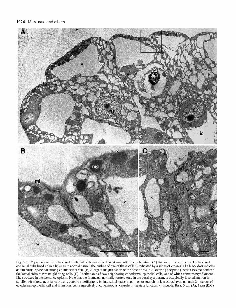
Fig. 5A shows a layer of ectodermal epithelial cells in stageI. The outline of one of these cells is marked by a series ofsmall crosses. Parts of these and similar cells are shown athigher magnifications in Fig. 5B and C. All the structurespresent in these cells resembled those in normal hydra. Forexample, the apical surface was covered by a mucous layerabout 0.1 µm in thickness (marked ml in Fig. 5B and C). Smallgranules (presumably mucous granules) were numerous just
1924 M. Murate and others
Fig. 5. TEM pictures of the ectodermal epithelial cells in a recombinant soon after recombination. (A) An overall view of several ectodermalepithelial cells lined up in a layer as in normal tissue. The outline of one of these cells is indicated by a series of crosses. The black dots indicatean interstitial space containing an interstitial cell. (B) A higher magnification of the boxed area in A showing a septate junction located betweenthe lateral sides of two neighboring cells. (C) Another area of two neighboring endodermal epithelial cells, one of which contains myofilament-like structure in the lateral cytoplasm. Note that the filaments, normally located only in the basal cytoplasm, is ectopically located and run inparallel with the septate junction. em: ectopic myofilament; is: interstitial space; mg: mucous granule; ml: mucous layer; n1 and n2: nucleus ofectodermal epithelial cell and interstitial cell, respectively; nc: nematocyst capsule; sj: septate junction; v: vacuole. Bars: 5 µm (A); 1 µm (B,C).
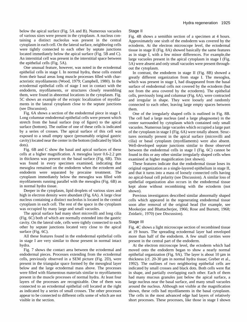
1925Hydra regeneration
below the apical surface (Fig. 5A and B). Numerous vacuolesof various sizes were present in the cytoplasm. A nucleus con-taining a distinct nucleolus was present in the centralcytoplasm in each cell. On the lateral surface, neighboring cellswere tightly connected to each other by septate junctionslocated immediately below the apical surface (Fig. 5B and C).An interstitial cell was present in the interstitial space betweenthe epithelial cells (Fig. 5A).
One unusual feature, however, was noted in the ectodermalepithelial cells in stage I. In normal hydra, these cells extendfrom their basal areas long muscle processes filled with char-acteristic myofilaments (Wood, 1979; Campbell, 1980). In theectodermal epithelial cells of stage I not in contact with theendoderm, myofilaments, or structures closely resemblingthem, were found in abnormal locations in the cytoplasm. Fig.5C shows an example of the ectopic localization of myofila-ments in the lateral cytoplasm close to the septate junctions(see Discussion).
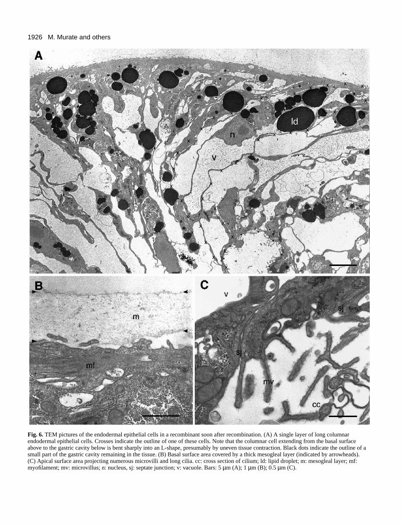
Fig. 6A shows a section of the endodermal piece in stage I.Long columnar endodermal epithelial cells were present whichstretch from the basal surface (top of figure) to the apicalsurface (bottom). The outline of one of these cells is indicatedby a series of crosses. The apical surface of this cell wasexposed to a small empty space (presumably original gastriccavity) located near the center in the bottom (indicated by blackdots).
Fig. 6B and C show the basal and apical surfaces of thesecells at a higher magnification. A mesoglea layer 0.6-2.2 µmin thickness was present on the basal surface (Fig. 6B). Thiswas found in every specimen examined, indicating thatmesoglea remained on the endoderm when the ectoderm andendoderm were separated by procaine treatment. Thecytoplasm immediately below the mesoglea was filled withmyofilaments running parallel with the mesoglea (Fig. 6B) asin normal hydra tissue.
Deeper in the cytoplasm, lipid droplets of various sizes andhigh in electron density were abundant (Fig. 6A). A large clearnucleus containing a distinct nucleolus is located in the centralcytoplasm in each cell. The rest of the space in the cytoplasmwas occupied by many large and small vacuoles.
The apical surface had many short microvilli and long cilia(Fig. 6C) both of which are normally extended into the gastriccavity. On the lateral side, cells were tightly connected to eachother by septate junctions located very close to the apicalsurface (Fig. 6C).
All these features found in the endodermal epithelial cellsin stage I are very similar to those present in normal intacthydra.
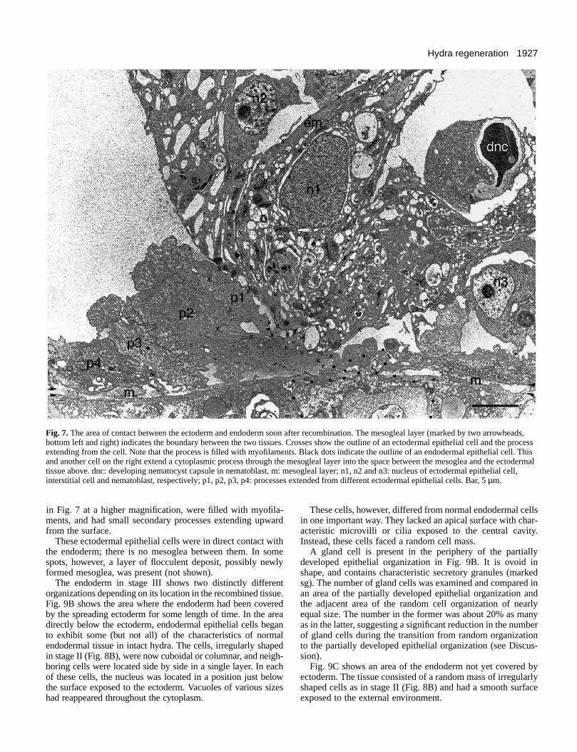
Fig. 7 shows the contact area between the ectodermal andendodermal pieces. Processes extending from the ectodermalcells, previously observed in a SEM picture (Fig. 2D), werepresent in the triangular space formed by the mesogleal layerbelow and the large ectodermal mass above. The processeswere filled with filamentous materials similar to myofilamentspresent in the muscle processes of normal hydra. At least fourlayers of the processes are recognizable. One of them wasconnected to an ectodermal epithelial cell located at the rightas indicated by a series of small crosses. The other processesappear to be connected to different cells some of which are notvisible in the section.
Stage IIFig. 4B shows a semithin section of a specimen at 4 hours.Approximately one sixth of the endoderm was covered by theectoderm. At the electron microscope level, the ectodermaltissue in stage II (Fig. 8A) showed basically the same featuresas in stage I, with a few minor differences. For example,thelarge vacuoles present in the apical cytoplasm in stage I (Fig.5A) were absent and only small vacuoles were present through-out the cytoplasm.
In contrast, the endoderm in stage II (Fig. 8B) showed agreatly different organization from stage I. The mesoglea,which was present in stage I, had disappeared from the basalsurface of endodermal cells not covered by the ectoderm (butnot from the area covered by the ectoderm). The epithelialcells, previously long and columnar (Fig. 6A), were contractedand irregular in shape. They were loosely and randomlyconnected to each other, leaving large empty spaces betweenthem.
One of the irregularly shaped cells is outlined in Fig. 8B.The cell had a large nucleus (and a large phagosome) in thecenter surrounded by cytoplasm which contained only smallvacuoles. Numerous large vacuoles which occupied a large partof the cytoplasm in stage I (Fig. 6A) were totally absent. Struc-tures normally present in the apical surface (microvilli andcilia) or basal cytoplasm (myofilaments) were also absent.Well-developed septate junctions similar to those observedbetween the endodermal cells in stage I (Fig. 6C) cannot befound in this or any other similar irregularly shaped cells whenexamined at higher magnification (not shown).
These features indicate that the endodermal tissue loses itsoriginal organization as a single-layered epithelium in stage II,and that it turns into a mass of loosely connected cells havingno apical-basal cell polarity (see Discussion). A similar loss ofepithelial organization also occurs in the endodermal tissuekept alone without recombining with the ectoderm (notshown).
Previous investigators described similar abnormally shapedcells which appeared in the regenerating endodermal tissuesoon after removal of the original head (for example, seeMookerjee and Bhattacherjee, 1966; Rose and Burnett, 1968;Znidaríc, 1970) (see Discussion).
Stage IIIFig. 4C shows a light microscope section of recombined tissueat 19 hours. The spreading ectodermal layer had envelopedmore than half of the endoderm. A few small cavities werepresent in the central part of the endoderm.
At the electron microscope level, the ectoderm which hadmoved onto the endoderm began to show a nearly normalepithelial organization (Fig. 9A). The layer is about 10 µm inthickness (cf. 20-30 µm in normal hydra tissue; Greber et al.,1992). The outlines of two neighboring epithelial cells areindicated by small crosses and black dots. Both cells were flatin shape, and partially overlapping each other. Each of themhad many mucous granules just below the apical surface, alarge nucleus near the basal surface, and many small vacuolesaround the nucleus. Although not visible at the magnificationshown, these cells had myofilaments in the basal cytoplasm.The cells in the most advanced edge had layers of relativelyshort processes. These processes, like those in stage I shown
1926 M. Murate and others
Fig. 6. TEM pictures of the endodermal epithelial cells in a recombinant soon after recombination. (A) A single layer of long columnarendodermal epithelial cells. Crosses indicate the outline of one of these cells. Note that the columnar cell extending from the basal surfaceabove to the gastric cavity below is bent sharply into an L-shape, presumably by uneven tissue contraction. Black dots indicate the outline of asmall part of the gastric cavity remaining in the tissue. (B) Basal surface area covered by a thick mesogleal layer (indicated by arrowheads).(C) Apical surface area projecting numerous microvilli and long cilia. cc: cross section of cilium; ld: lipid droplet; m: mesogleal layer; mf:myofilament; mv: microvillus; n: nucleus, sj: septate junction; v: vacuole. Bars: 5 µm (A); 1 µm (B); 0.5 µm (C).
1927Hydra regeneration
Fig. 7. The area of contact between the ectoderm and endoderm soon after recombination. The mesogleal layer (marked by two arrowheads,bottom left and right) indicates the boundary between the two tissues. Crosses show the outline of an ectodermal epithelial cell and the processextending from the cell. Note that the process is filled with myofilaments. Black dots indicate the outline of an endodermal epithelial cell. Thisand another cell on the right extend a cytoplasmic process through the mesogleal layer into the space between the mesoglea and the ectodermaltissue above. dnc: developing nematocyst capsule in nematoblast, m: mesogleal layer; n1, n2 and n3: nucleus of ectodermal epithelial cell,interstitial cell and nematoblast, respectively; p1, p2, p3, p4: processes extended from different ectodermal epithelial cells. Bar, 5 µm.
in Fig. 7 at a higher magnification, were filled with myofila-ments, and had small secondary processes extending upwardfrom the surface.
These ectodermal epithelial cells were in direct contact withthe endoderm; there is no mesoglea between them. In somespots, however, a layer of flocculent deposit, possibly newlyformed mesoglea, was present (not shown).
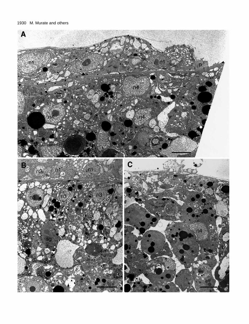
The endoderm in stage III shows two distinctly differentorganizations depending on its location in the recombined tissue.Fig. 9B shows the area where the endoderm had been coveredby the spreading ectoderm for some length of time. In the areadirectly below the ectoderm, endodermal epithelial cells beganto exhibit some (but not all) of the characteristics of normalendodermal tissue in intact hydra. The cells, irregularly shapedin stage II (Fig. 8B), were now cuboidal or columnar, and neigh-boring cells were located side by side in a single layer. In eachof these cells, the nucleus was located in a position just belowthe surface exposed to the ectoderm. Vacuoles of various sizeshad reappeared throughout the cytoplasm.
These cells, however, differed from normal endodermal cellsin one important way. They lacked an apical surface with char-acteristic microvilli or cilia exposed to the central cavity.Instead, these cells faced a random cell mass.
A gland cell is present in the periphery of the partiallydeveloped epithelial organization in Fig. 9B. It is ovoid inshape, and contains characteristic secretory granules (markedsg). The number of gland cells was examined and compared inan area of the partially developed epithelial organization andthe adjacent area of the random cell organization of nearlyequal size. The number in the former was about 20% as manyas in the latter, suggesting a significant reduction in the numberof gland cells during the transition from random organizationto the partially developed epithelial organization (see Discus-sion).
Fig. 9C shows an area of the endoderm not yet covered byectoderm. The tissue consisted of a random mass of irregularlyshaped cells as in stage II (Fig. 8B) and had a smooth surfaceexposed to the external environment.

1928 M. Murate and others
1929Hydra regeneration
Fig. 8. The recombinants in stage II. (A) The ectodermal tissue at 2hours. Crosses and black dots indicate the outlines of an ectodermalepithelial cell and a large interstitial space containing 4 developingnematoblasts, respectively. (B) The endodermal tissue at 4 hoursconsisting of a random mass of irregularly shaped cells. Crossesindicate the outline of one of these cells. dnc: developing nematocystcapsule in nematoblast; ld: lipid droplet; n1, n2, n3, and n4: nucleusof ectodermal epithelial cell, interstitial cell, nematoblast, andirregularly shaped endodermal cell, respectively; p: phagosomecontained in the irregularly shaped cell. Bars: 10 µm (A); 5 µm (B).
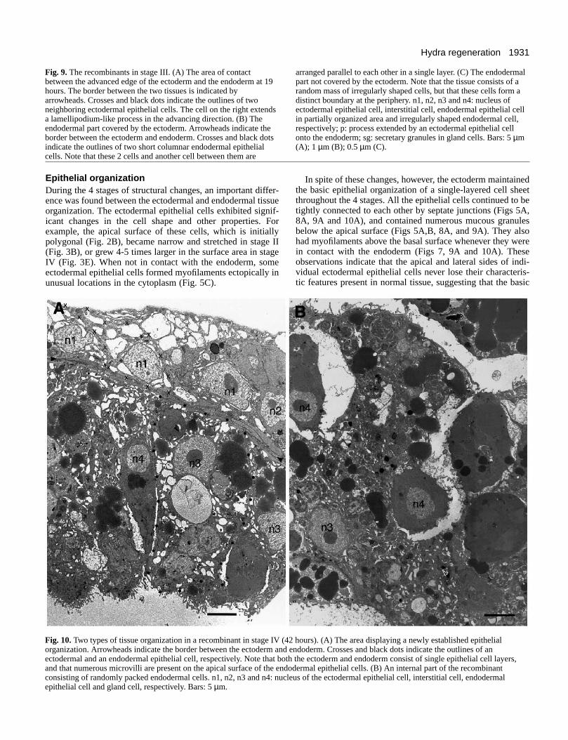
Stage IVFig. 4D shows a light microscope section of a typical recom-binant at 42 hours. A thin layer of the ectoderm had completelyenveloped the endoderm. About two fifths of the recombinantin the upper side showed an organization of two cell layers withan empty cavity below it as in normal hydra. The remainingpart showed a more complex organization consisting ofpartially organized tissue and many empty spaces.
Fig. 10A shows the arrangement of individual cells in thewell-organized area. The ectodermal and endodermal cellsboth showed an organization similar to that in intact hydra. Theectoderm consisted of a single layer of epithelial cells whichpartially overlap each other. Each cell had a large nucleus witha distinct nucleolus, and many vacuoles in the cytoplasm.Small mucous granules were present in the apical cytoplasm,and muscle processes were located just above the basalmembrane. Cells were tightly joined together by septatejunctions located just below the apical surface.
The endodermal part consisted of a single layer of columnarepithelial cells and a large empty cavity below. These cells hadcilia and numerous microvilli on the apical surface, many smallempty vacuoles in the apical cytoplasm, and a thin layer ofmyofibers close to the basal surface. Cells were joined tightlyby septate junctions located near the apical surface. A glandcell also occurred in the cell layer, projecting its apical tip intothe central cavity (Fig. 10A).
Newly formed mesoglea 0.7-1.3 µm in thickness was presentbetween the ectoderm and endoderm throughout the area of theorganized layered structure (compared to 0.5-2 µm in normalhydra tissue; Haynes et al., 1968).
Fig. 10B shows a representative area in the complex organ-ization in the main endodermal mass. Many cells were tightlybut randomly packed together, and surrounded by large emptyspaces and connecting narrow empty spaces. Cell surfacesexposed to the empty space had numerous microvilli similar tothe apical surfaces of normal endodermal cells. However,myofibers characteristic of the basal cytoplasm could not befound in any of these cells.
DISCUSSION
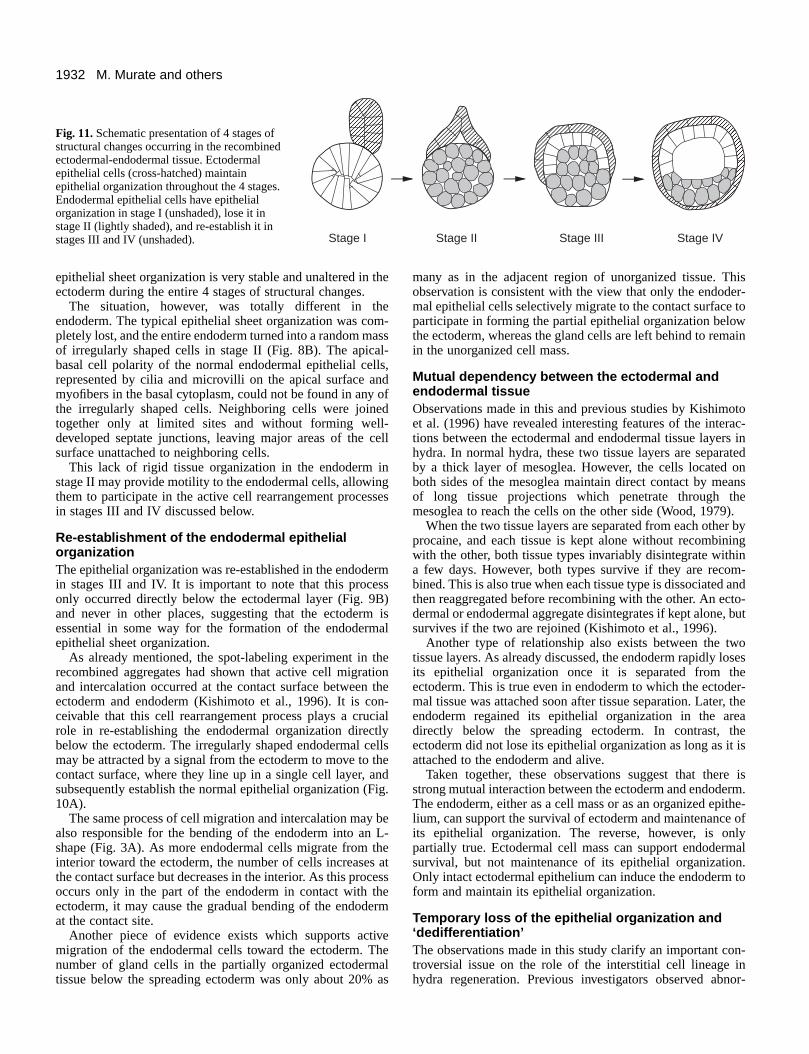
Four sta ges of structural c hang esThe present study has shown a dramatic series of structuralchanges in the ectodermal-endodermal tissue recombinant ofhydra. The changes observed are divided into 4 stages asschematically shown in Fig. 11.
Stage I occupied a short period immediately after tissuerecombination. A firm adhesion was established between the
ectoderm and endoderm (Figs 2A and 4A). The ectodermalepithelial cells started to extend membranous or bulbousprocesses onto the endoderm (Figs 2D and 7).
Stage II began immediately following stage I, and lasted forabout 24 hours. About a quarter of the endoderm was coveredby the ectoderm (Fig. 4B). The endodermal epithelial cells lostepithelial organization and turned into a random mass of ir-regularly shaped cells (Fig. 8B).
Stage III started at about 18 hours, partially overlapping withstage II. Half to three-quarters of the endoderm was coveredby the ectoderm (Fig. 4C). Epithelial-like organization beganto be restored in the part of the endoderm directly covered bythe ectoderm (Fig. 9B).
Stage IV started at about 20-30 hours. The endoderm wascompletely enveloped by the ectoderm (Figs 3D and 4D). Thetwo-layered tissue organization similar to that present innormal hydra was re-established in a part of the recombinants(Fig. 10A).
Migrator y ability of the ectodermal epithelial cellsIn stages I to III, the ectodermal epithelial cells located at theadvanced edge had processes filled with myofilaments (Figs2D, 3C, 7 and 9A). These processes are probably a modifiedform of the muscle processes of the ectodermal epithelial cells.In intact hydra, the ectodermal muscle processes are long andnarrow in shape, and extend longitudinally onto the mesogleaalong the hydra axis (Otto, 1977). In the recombinant, theywere short and irregular in shape, and extended onto themesoglea or directly onto the endoderm in apparently randomdirections (Fig. 2D).
The processes in the recombinant exhibited a fine ruffledstructure on the surface (Figs 3C and 9A), and contained adense network of myofilaments (Fig. 7). In these respects, theyappear similar to lamellipodia of migrating cells in culture(Abercrombie et al., 1971; Rajaraman et al., 1974), suggestinga possibility that these processes may provide locomotiveability f or the epibolic ectodermal spreading over theendoderm.
Evidence exists, however, which suggests that the epibolicectodermal spreading may be driven by a differentmechanism. Kishimoto et al. (1996) examined movement ofindividual cells during the epiboly by spot vital staining ofcells on the recombined ectodermal and endodermal cellaggregates. The results had shown that extensive cellrearrangement took place in the interface between the recom-bined two aggregates. Cells initially located in the inside ofthe endodermal aggregate migrated to the contact surface, andplaced themselves among the cells located in the contactsurface. This cell intercalation produced a gradual growth ofthe contact surface area, suggesting that this process isresponsible, at least in part, for producing the spreading ofthe ectoderm.
Thus, two different mechanisms, not mutually exclusive, canbe considered as the driving force for the epiboly. One is activelocomotion of the ectodermal epithelial cells over theendoderm. The other is passive spreading of the ectodermalcells as the result of growth of the contact surface areagenerated by the endodermal epithelial cell migration andintercalation Whether or not either of them really plays acrucial role in the epibolic ectodermal spreading remains to beexamined.
1930 M. Murate and others
1931Hydra regeneration
Fig. 9. The recombinants in stage III. (A) The area of contactbetween the advanced edge of the ectoderm and the endoderm at 19hours. The border between the two tissues is indicated byarrowheads. Crosses and black dots indicate the outlines of twoneighboring ectodermal epithelial cells. The cell on the right extendsa lamellipodium-like process in the advancing direction. (B) Theendodermal part covered by the ectoderm. Arrowheads indicate theborder between the ectoderm and endoderm. Crosses and black dotsindicate the outlines of two short columnar endodermal epithelialcells. Note that these 2 cells and another cell between them are
arranged parallel to each other in a single layer. (C) The endodermalpart not covered by the ectoderm. Note that the tissue consists of arandom mass of irregularly shaped cells, but that these cells form adistinct boundary at the periphery. n1, n2, n3 and n4: nucleus ofectodermal epithelial cell, interstitial cell, endodermal epithelial cellin partially organized area and irregularly shaped endodermal cell,respectively; p: process extended by an ectodermal epithelial cellonto the endoderm; sg: secretary granules in gland cells. Bars: 5 µm(A); 1 µm (B); 0.5 µm (C).
Epithelial or ganizationDuring the 4 stages of structural changes, an important differ-ence was found between the ectodermal and endodermal tissueorganization. The ectodermal epithelial cells exhibited signif-icant changes in the cell shape and other properties. Forexample, the apical surface of these cells, which is initiallypolygonal (Fig. 2B), became narrow and stretched in stage II(Fig. 3B), or grew 4-5 times larger in the surface area in stageIV (Fig. 3E). When not in contact with the endoderm, someectodermal epithelial cells formed myofilaments ectopically inunusual locations in the cytoplasm (Fig. 5C).
Fig. 10.Two types of tissue organization in a recombinant in stage IV (4organization. Arrowheads indicate the border between the ectoderm andectodermal and an endodermal epithelial cell, respectively. Note that botand that numerous microvilli are present on the apical surface of the enconsisting of randomly packed endodermal cells. n1, n2, n3 and n4: nuclepithelial cell and gland cell, respectively. Bars: 5 µm.
In spite of these changes, however, the ectoderm maintainedthe basic epithelial organization of a single-layered cell sheetthroughout the 4 stages. All the epithelial cells continued to betightly connected to each other by septate junctions (Figs 5A,8A, 9A and 10A), and contained numerous mucous granulesbelow the apical surface (Figs 5A,B, 8A, and 9A). They alsohad myofilaments above the basal surface whenever they werein contact with the endoderm (Figs 7, 9A and 10A). Theseobservations indicate that the apical and lateral sides of indi-vidual ectodermal epithelial cells never lose their characteris-tic features present in normal tissue, suggesting that the basic
2 hours). (A) The area displaying a newly established epithelial endoderm. Crosses and black dots indicate the outlines of anh the ectoderm and endoderm consist of single epithelial cell layers,dodermal epithelial cells. (B) An internal part of the recombinanteus of the ectodermal epithelial cell, interstitial cell, endodermal
1932 M. Murate and others
AAAAAAAAAAAA
AAAAAAAAAAAA
AAAAAAAAAAAAAAAA
AAAAAAAAAAAA
AAAAAAAAAAAAAAAAAAAAAAAAA
Stage I Stage II Stage III Stage IV
Fig. 11. Schematic presentation of 4 stages ofstructural changes occurring in the recombinedectodermal-endodermal tissue. Ectodermalepithelial cells (cross-hatched) maintainepithelial organization throughout the 4 stages.Endodermal epithelial cells have epithelialorganization in stage I (unshaded), lose it instage II (lightly shaded), and re-establish it instages III and IV (unshaded).
epithelial sheet organization is very stable and unaltered in theectoderm during the entire 4 stages of structural changes.
The situation, however, was totally different in theendoderm. The typical epithelial sheet organization was com-pletely lost, and the entire endoderm turned into a random massof irregularly shaped cells in stage II (Fig. 8B). The apical-basal cell polarity of the normal endodermal epithelial cells,represented by cilia and microvilli on the apical surface andmyofibers in the basal cytoplasm, could not be found in any ofthe irregularly shaped cells. Neighboring cells were joinedtogether only at limited sites and without forming well-developed septate junctions, leaving major areas of the cellsurface unattached to neighboring cells.
This lack of rigid tissue organization in the endoderm instage II may provide motility to the endodermal cells, allowingthem to participate in the active cell rearrangement processesin stages III and IV discussed below.
Re-estab lishment of the endodermal epithelialorganizationThe epithelial organization was re-established in the endodermin stages III and IV. It is important to note that this processonly occurred directly below the ectodermal layer (Fig. 9B)and never in other places, suggesting that the ectoderm isessential in some way for the formation of the endodermalepithelial sheet organization.
As already mentioned, the spot-labeling experiment in therecombined aggregates had shown that active cell migrationand intercalation occurred at the contact surface between theectoderm and endoderm (Kishimoto et al., 1996). It is con-ceivable that this cell rearrangement process plays a crucialrole in re-establishing the endodermal organization directlybelow the ectoderm. The irregularly shaped endodermal cellsmay be attracted by a signal from the ectoderm to move to thecontact surface, where they line up in a single cell layer, andsubsequently establish the normal epithelial organization (Fig.10A).
The same process of cell migration and intercalation may bealso responsible for the bending of the endoderm into an L-shape (Fig. 3A). As more endodermal cells migrate from theinterior toward the ectoderm, the number of cells increases atthe contact surface but decreases in the interior. As this processoccurs only in the part of the endoderm in contact with theectoderm, it may cause the gradual bending of the endodermat the contact site.
Another piece of evidence exists which supports activemigration of the endodermal cells toward the ectoderm. Thenumber of gland cells in the partially organized ectodermaltissue below the spreading ectoderm was only about 20% as
many as in the adjacent region of unorganized tissue. Thisobservation is consistent with the view that only the endoder-mal epithelial cells selectively migrate to the contact surface toparticipate in forming the partial epithelial organization belowthe ectoderm, whereas the gland cells are left behind to remainin the unorganized cell mass.
Mutual dependenc y between the ectodermal andendodermal tissueObservations made in this and previous studies by Kishimotoet al. (1996) have revealed interesting features of the interac-tions between the ectodermal and endodermal tissue layers inhydra. In normal hydra, these two tissue layers are separatedby a thick layer of mesoglea. However, the cells located onboth sides of the mesoglea maintain direct contact by meansof long tissue projections which penetrate through themesoglea to reach the cells on the other side (Wood, 1979).
When the two tissue layers are separated from each other byprocaine, and each tissue is kept alone without recombiningwith the other, both tissue types invariably disintegrate withina few days. However, both types survive if they are recom-bined. This is also true when each tissue type is dissociated andthen reaggregated before recombining with the other. An ecto-dermal or endodermal aggregate disintegrates if kept alone, butsurvives if the two are rejoined (Kishimoto et al., 1996).
Another type of relationship also exists between the twotissue layers. As already discussed, the endoderm rapidly losesits epithelial organization once it is separated from theectoderm. This is true even in endoderm to which the ectoder-mal tissue was attached soon after tissue separation. Later, theendoderm regained its epithelial organization in the areadirectly below the spreading ectoderm. In contrast, theectoderm did not lose its epithelial organization as long as it isattached to the endoderm and alive.
Taken together, these observations suggest that there isstrong mutual interaction between the ectoderm and endoderm.The endoderm, either as a cell mass or as an organized epithe-lium, can support the survival of ectoderm and maintenance ofits epithelial organization. The reverse, however, is onlypartially true. Ectodermal cell mass can support endodermalsurvival, but not maintenance of its epithelial organization.Only intact ectodermal epithelium can induce the endoderm toform and maintain its epithelial organization.
Temporar y loss of the epithelial or ganization and‘dediff erentiation’The observations made in this study clarify an important con-troversial issue on the role of the interstitial cell lineage inhydra regeneration. Previous investigators observed abnor-
1933Hydra regeneration
mally shaped cells similar to the irregularly shaped cellsobserved in this study. They appeared in the endoderm of theregenerating tissue soon after the removal of the original head(Mookerjee and Bhattacherjee, 1966; Znidaric, 1970), or in theendoderm separated from the ectoderm by enzyme treatment(Davis et al., 1966). These cells were referred to by variousnames such as ‘embryonic cells’ (Rose and Burnett, 1968),‘neoblasts’ (Lui and Znidaríc, 1968) or ‘amoeboid interstitialcells’ (Mookerjee and Bhattacherjee, 1966). As implied bytheir names, these cells were thought to arise by ‘dedifferenti-ation’ from endodermal cells, and have the ability to differen-tiate into other cell types (Davis et al., 1966; Mookerjee andBhattacherjee, 1966; Znidaríc, 1970). In short, they were con-sidered to be the equivalent of the blastema cells or neoblastswhich appear in regenerating tissue of urodele amphibians orplanarians, respectively.
We now suggest that these cells are identical to the irregu-larly shaped endodermal epithelial cells observed in thisstudy. Although the abnormally shaped cells observed byprevious workers may appear similar to normal interstitialcells at light microscope level, distinct morphological differ-ences are present in the two cell types at the electron micro-scope level. For example, the abnormally shaped cells alwayscontain numerous small vacuoles and sometimes phagosomes(for example see the marked cell in Fig. 8B), whereas suchstructures are never found in any of the interstitial cells (forexample see the n2 cell in Fig. 5A). In addition, there is noevidence which directly shows that the abnormally shapedcells have a strong ability to proliferate or to later turn intodifferent cell types other than the endodermal epithelial (orgland) cells.
Furthermore, it is well established that the interstitial celllineage is not essential for hydra regeneration. Epithelial hydracompletely free of interstitial stem cells, nerve cells or nema-tocytes can regenerate nearly as well as normal hydra (Marcumand Campbell, 1978; Sugiyama and Fujisawa, 1978). Thus, thecellular mechanisms of regeneration in hydra differ funda-mentally from those in urodele amphibians or planarians. Inthe latter organisms, blastema cells or neoblasts accumulate inthe regenerating tissue, and are thought to play a crucial rolein the strong regenerative abilities of these animals (for reviewsee Chernoff and Stocum, 1995). In contrast, cell dedifferenti-ation and redifferentiation plays little or no role in hydra regen-eration.
At present, the roles of the irregularly shaped endodermalepithelial cells in hydra regeneration is uncertain. One mayspeculate that the remarkably high plasticity of the endodermaltissue organization in hydra is correlated in some way to thestrong regenerative capacity of this organism.
Contr ol of epithelial or ganizationMorphogenetic processes of epithelial sheet formation arewidely observed in vivo in various embryonic systems and invitro in many types of cultured cells. In the case of chick skintissue, loss of epithelial organization occurs by removal of thebasement membrane from its tissue, suggesting the importanceof the basement membrane for maintenance of the epithelialorganization in this system (Dodson, 1967).
In the present system, however, loss of epithelial organiz-ation takes place in the endoderm to which the basementmembrane (mesoglea) is attached, but not in the ectoderm
which is free from the basement membrane after procaine sep-aration. Therefore, the basement membrane may not be tooimportant, but direct interaction with the ectoderm may becrucially important for the maintenance and formation of theendodermal epithelial organization in hydra.
In the case of Drosophila, two genes have been identifiedwhich play crucial roles in controlling epithelial organization(Knust, 1994). One is crumbswhich encodes a large trans-membrane protein localized in the apical surface of ectodermalepithelial tissue. Mutation in this gene results in the loss ofapical-basal polarity (Tepass et al., 1990; Tepass and Knust,1990), whereas its over-expression leads to expansion of theapical surface and reduction of the baso-lateral surface in theectodermal epithelial tissue (Wodarz et al., 1995). Mutation inthe other gene, stardust, also produces loss of cell polarity. Fur-thermore, zonula adherens are not properly formed in bothmutants (Grawe et al., 1996).
Hydra may have genes which play similar roles to these twoDrosophila genes. When the endoderm is separated from theectoderm, functioning of such genes in the endoderm may bedisrupted to convert the endodermal tissue into a mass of irregu-larly shaped cells without apical-basal polarity. The identity ofsuch genes in hydra, however, is unknown at present.
We thank Dr M. Hatta for valuable discussions and Drs R. D.Campbell and C. N. David for critical reading of the manuscript. Thiswork was supported in part by Grants-in-Aids for Scientific Researchfrom the Ministry of Education, Science and Culture, Japan to T.S.(06454689, 072832210) and to T.F (06640803).
REFERENCES
Abercrombie, M., Heaysman, J. E. M. and Pegrum, S. M. (1971). Thelocomotion of fibroblasts in culture. IV. Electron microscopy of the leadinglamella. Exp. Cell Res. 67, 359-367.
Bode, H. R., Heimfeld, S., Chow, M. A. and Huang, L. W. (1987). Gland cellsarise by differentiation from interstitial cells in Hydra attenuata. Dev. Biol.122, 577-585.
Campbell, R. D. (1980). Role of muscle processes in hydra morphogenesis. InDevelopmental and Cellular Biology of Coelenterates(ed. P. Tardent and R.Tardent), pp. 421-428. Elsevier/North-Holland Medical Press, Amsterdam.
Campbell, R. D. and Bode, H. R. (1983). Terminology for morphology andcell types. InHydra: Research Methods (ed. H. M. Lenhoff), pp. 5-14.Plenum Press, NY.
Chernoff, E. A. G. and Stocum, D. L. (1995). Developmental aspects of spinalcord and limb regeneration. Dev. Growth Differ. 37, 133-147.
Davis, L. E., Burnett, A. L., Haynes, J. F. and Mumaw, V. R. (1966). Ahistological and ultrastructural study of dedifferentiation andredifferentiation of digestive and gland cells in Hydra viridis. Dev. Biol. 14,307-329.
Dodson, J. W. (1967). The differentiation of epidermis. I. The interrelationshipof epidermis and dermis in embryonic chick skin. J. Embryol. Exp. Morphol.17, 83-105.
Epp, L. G., Tardent, P. and Bänninger, R. (1979). Isolation and observationof tissue layers in Hydra attenuata Pall. (Cnidaria, Hydrozoa). Trans. Am.Microsc. Soc. 98, 392-400.
Epp, L., Smid, I. and Tardent, P. (1986). Synthesis of the mesoglea byectoderm and endoderm in reassembled hydra. J. Morphol. 189, 271-279.
Gierer, A., Berking, S., Bode, H., David, C. N., Flick, K., Hansmann, G.,Schaller, H. and Trenkner, E. (1972). Regeneration of hydra fromreaggregated cells. Nature New Biol. 239, 98-101.
Grawe, F., Wodarz, A., Lee, B., Knust, E. and Skaer, H. (1996). TheDrosophilagenes crumbsand stardustsare involved in the biogenesis ofadherens junctions. Development 122, 951-959.
Greber, M. J., David, C. N. and Holstein, T. W. (1992). A quantitative methodfor separation of living Hydra cells. Roux’s Arch. Dev. Biol. 201, 296-300.
Haynes, J. F., Burnett, A. L. and Davis, L. E. (1968). Histological and

1934 M. Murate and others
ultrastructural study of the muscular and nervous systems in Hydra. I. Themuscular system and the mesoglea. J. Exp. Zool. 167, 283-294.
Keller, R. E. (1980). The cellular basis of epiboly: An SEM study of deep-cellrearrangement during gastrulation in Xenopus laevis. J. Embryol. Exp.Morphol. 60, 201-234.
Kishimoto, Y., Murate, M. and Sugiyama, T. (1996). Hydra regenerationfrom recombined ectodermal and endodermal tissue. I. Epibolic ectodermalspreading is driven by cell intercalation. J. Cell Sci. 109, 763-772.
Knust, E. (1994). Control of epithelial cell polarity in Drosophila. TrendsGenet. 10, 275-280.
Lui, A. and Znidaríc , D. (1968). Das Gastroderm im Prozeß der Regenerationder Hydra. Roux´ Arch. Entwicklungsmech. Org. 160, 1-8.
Mar cum, B. A. and Campbell, R. D. (1978). Development of hydra lackingnerve and interstitial cells. J. Cell Sci. 29, 17-33.
Mookerjee, S. and Bhattacherjee, S. (1966). Cellular mechanics in hydroidregeneration. Roux’ Arch. Entwicklungsmech. Org. 157, 1-20.
Mur akami, T. (1974). A revised tannin-osmium method for non-coatedscanning electron microscope specimens. Arch. Histol. Jpn36, 189-193.
New, D. A. T. (1959). The adhesive properties and expansion of the chickblastoderm. J. Embryol. Exp. Morphol. 7, 146-164.
Noda, K. (1971). Reconstitution of dissociated cells of hydra. Zool. Mag. 80,99-101.
Otto, J. J. (1977). Orientation and behavior of epithelial cell muscle processesduring Hydra budding. J. Exp. Zool.202, 307-322.
Rajaraman, R., Rounds, D. E., Yen, S. P. S. and Rembaum, A.(1974). Ascanning electron microscope study of cell adhesion and spreading in vitro.Exp. Cell Res.88, 327-339.
Rose, P. G. and Burnett, A. L. (1968). An electron microscopic and
radioautographic study of hypostomal regeneration in Hydra viridis. Roux’sArch.Dev. Biol.161, 298-318.
Smid, I. and Tardent, P. (1982). The influences of ecto- and endoderm indetermining the axial polarity of Hydra attenuataPall. (Cnidaria, Hydrozoa).Roux’s Arch. Dev. Biol. 191, 64-67.
Sugiyama, T. and Fujisawa, T. (1977). Genetic analysis of developmentalmechanisms in hydra. I. Sexual reproduction of Hydra magnipapillata andisolation of mutants. Dev. Growth Differ. 19, 187-200.
Sugiyama, T. and Fujisawa, T. (1978). Genetic analysis of developmentalmechanisms in hydra. II. Isolation and characterization of an interstitial cell-deficient strain. J. Cell Sci. 29, 35-52.
Tepass, U., Theres, C. and Knust, E. (1990). crumbs encodes an EGF-likeprotein expressed on apical membranes of Drosophila epithelial cells andrequired for organization of epithelia. Cell 61, 787-798.
Tepass, U. and Knust, E. (1990). Phenotypic and developmental analysis ofmutations at the crumbs locus, a gene required for the development ofepithelia in Drosophila melanogaster. Roux’s Arch. Dev. Biol. 199, 189-206.
Tr inkaus, J. P. (1984). Mechanism of Fundulusepiboly - a current view. Am.Zool.24, 673-688.
Wodarz, A., Hinz, U., Engelbert, M. and Knust, E. (1995). Expression ofcrumbsconfers apical character of plasma membrane domains of ectodermalepithelia of Drosophila. Cell 82, 67-76.
Wood, R. L. (1979). The fine structure of the hypostome and mouth of hydra. II.Transmission electron microscopy. Cell Tiss. Res. 199, 319-338.
Znidaríc , D. (1970). Comparison of the regeneration of the hypostome with thebudding process in Hydra littoralis. Roux’s Arch. Dev. Biol. 166, 45-53.
(Received 28 November 1996 - Accepted 6 June 1997)