Embed Size (px)
Citation preview
polymers
Review
Hydrogels-Assisted Cell Engraftment for Repairingthe Stroke-Damaged Brain: Chimera or Reality
Daniel González-Nieto 1,2,3,*, Laura Fernández-García 1, José Pérez-Rigueiro 1,3,4,Gustavo V. Guinea 1,3,4 ID and Fivos Panetsos 5,6 ID
1 Center for Biomedical Technology, Universidad Politécnica de Madrid, 28040 Madrid, Spain;[email protected] (L.F.-G.); [email protected] (J.P.-R.);[email protected] (G.V.G.)
2 Departamento de Tecnología Fotónica y Bioingeniería, ETSI Telecomunicaciones,Universidad Politécnica de Madrid, 28040 Madrid, Spain
3 Biomedical Research Networking Center in Bioengineering Biomaterials and Nanomedicine (CIBER-BBN),28040 Madrid, Spain
4 Departamento de Ciencia de Materiales, ETSI Caminos, Canales y Puertos,Universidad Politécnica de Madrid 28040 Madrid, Spain
5 Neurocomputing and Neurorobotics Research Group: Faculty of Biology and Faculty of Optics,Universidad Complutense de Madrid, 28040 Madrid, Spain; [email protected]
6 Instituto de Investigación Sanitaria, Hospital Clínico San Carlos Madrid, IdISSC, 28040 Madrid, Spain* Correspondence: [email protected]; Tel.: +34-913364653
Received: 20 December 2017; Accepted: 11 February 2018; Published: 13 February 2018
Abstract: The use of advanced biomaterials as a structural and functional support for stem cells-basedtherapeutic implants has boosted the development of tissue engineering applications in multipleclinical fields. In relation to neurological disorders, we are still far from the clinical reality of restoringnormal brain function in neurodegenerative diseases and cerebrovascular disorders. Hydrogelpolymers show unique mechanical stiffness properties in the range of living soft tissues such asnervous tissue. Furthermore, the use of these polymers drastically enhances the engraftment of stemcells as well as their capacity to produce and deliver neuroprotective and neuroregenerative factors inthe host tissue. Along this article, we review past and current trends in experimental and translationalresearch to understand the opportunities, benefits, and types of tentative hydrogel-based applicationsfor the treatment of cerebral disorders. Although the use of hydrogels for brain disorders has beenrestricted to the experimental area, the current level of knowledge anticipates an intense developmentof this field to reach clinics in forthcoming years.
Keywords: biomaterials; hydrogels; stem cells; stroke; brain repair
1. Introduction
In recent decades, considerable progress has been made in the development of experimentaltherapies to treat different brain disorders. Among the different approaches examined, cell therapyhas been configured as a viable option for restoring the damaged brain. Tissue recovery afterdamage has been associated with the mobilization and integration of new cerebral cells derived fromexogenous sources. Alternatively, endogenous brain repair mechanisms may also be stimulated by thetransplantation of different cell populations with the ability to secrete factors that mobilize endogenousneurogenesis. Cell transplantation through a systemic intravascular route has been regularly testedin animal models and brain injury patients. Intra-venous and intra-arterial administrations areconsidered relatively feasible to perform and are less invasive than cerebral implantation. However,the systemic route is handicapped by the inefficient mobilization of therapeutic cells from the blood
Polymers 2018, 10, 184; doi:10.3390/polym10020184 www.mdpi.com/journal/polymers
Polymers 2018, 10, 184 2 of 37
towards the brain tissue given that most cells are retained in the pulmonary capillaries, spleen, liver,and kidneys [1,2], therefore limiting the number of cells able to reach the brain by crossing theblood-brain barrier which separates the circulatory system from the brain tissue. Despite its higherinvasiveness, cerebral implantation is the most promising strategy because of the possibility of graftinga greater number of therapeutic cells in the area of interest, which might be restricted to or nearthe area of the lesion [3–8]. The relevance of this approach has been supported in later years bythe progressive expansion of the number of clinical trials performing intracerebral cell implants inpatients with cerebral damage using different populations of stem cells and progenitors of embryonic,hematopoietic, and mesenchymal sources.
However, even the cerebral route is not free of difficulties. Among them, we should mention thereduced temporality of the graft due to the poor survival ratio of the cells as well as their dispersiontowards regions far away from the area of interest, thus diluting any therapeutic effect. It is assumedthat in response to injury the brain tissue is converted into a hostile microenvironment not only forthe brain itself, but also for the grafting of different cell populations that generally do not survivebeyond a few weeks after implantation [5,6,8]. The lack of survival factors combined with cell deathsignals from reactive astrocytes, microglia and peripheral leukocytes not only contribute to damagethe still alive brain tissue in perilesional areas but most likely also constitute the main causes for poorcell engraftment.
In regenerative neuroscience, the combined implantation of cells and different biomaterials toincrease the viability of cellular grafts constitutes a very promising approach in full expansion [9].In this context hydrogels rise as a powerful and versatile group of architectonic elements for cellencapsulation and brain reconstruction due to their special chemical and physical structures withstiffness modules in the range of the soft tissues such as the brain. Given the increasing variety ofapplications and experimental models for the development of biomaterials-based stem cell therapiesfor neurological disorders, this review focuses on current knowledge on the use of hydrogels-basedcell therapy for ischemic brain injury (ischemic stroke), which represents the most frequent typeof disabling neurological pathology [10]. An additional objective is to provide a global view ofthe biomedical problems as well as of the past and current trends in the related experimental andtranslational research. Finally, we aim at highlighting the opportunities, type of applications andpossible benefits of hydrogels-based cellular therapies for ischemic brain injuries. For a deeperunderstanding of the application of hydrogels and therapeutic cells in the treatment of other lessfrequent but not less important neurological disorders, several comprehensive reviews have beenincluded in the Reference Section [11–14].
2. Clinical Scenario and Challenges to Solve
Cerebrovascular diseases encompass a set of syndromes of different etiology and severity that leadto transient or permanent disorders of brain function. Pathologically, cerebrovascular diseases may bea consequence of: (i) alterations in cerebral blood flow; (ii) disturbances in blood circulation that modifyblood flow and pressure; and (iii) disorders of cardiac function. Stroke represents a sudden onset ofcerebrovascular disease and occurs in most cases through a vascular obstructive process (ischemic;~85% of stroke patients) or by the rupture of one or more brain blood vessels and extravasation ofblood (effusion) into the extravascular space (hemorrhagic; ~15% of stroke patients), both leading toa reduction or total abolition of blood supply to the brain. Epidemiologically, stroke is a leading causeof adult disability and cognitive impairment and is the second-leading global cause of death behindischemic heart disease [10,15]. It is estimated that after stroke a substantial proportion of patients(25–50%) remain dependent for at least one daily task up to six months after injury [16] producingincalculable personal, family, and social costs worldwide. The main risk factors for stroke includehigh blood pressure, ischemic heart disease, diabetes mellitus, disorders of heart rhythm, high bloodcholesterol and lipids, smoking, physical inactivity, diet and weight, family history, and geneticbackground. Due to the progressive increase in life expectancy, it is assumed that in the next few
Polymers 2018, 10, 184 3 of 37
decades there will be a parallel increase in the number of stroke cases (confirmed by the fact thatstroke incidence has increased by up to 20% in the last few years [15,17]) increasing the susceptibilityof suffering this vascular affectation at an even earlier age [18].
Mitigation of stroke-originated brain damages strongly depends on the recognition of signsand symptoms in the hospital emergency area and the consequent rapidity of medical intervention.Hemiparesis, loss of sensation, impaired speech, vertigo, and gait disturbances represent the initialclinical signs [19,20]. In ischemic stroke, the early reestablishment of blood flow in the obstructedvessel is a strong determinant for the clinical evolution of the patient. The intravenous injectionof the tissue plasminogen activator (tPA) alone or in combination with surgical procedures suchas endovascular thrombectomy for the recanalization of the occluded vessel are currently the maintherapeutic approaches for eliminating the obstructive clot [19,21–23]. However, the percentage ofstroke patients that can get benefit from these two treatments is extremely low. Due to the shorttherapeutic window (3.0–4.5 h after the onset of the attack) and the risk of complications, only 6%of the affected people are eligible for these treatments. In addition, tPA is effective in less than 10%of the treated cases, reducing the possible beneficiaries to less than 0.6% of the affected population.Endovascular thrombectomy is even less effective, being also very limited the number of teamsspecialized in this type of intervention.
Patients who survive a stroke and do not receive adequate treatment within this narrowtherapeutic window or when its reception is ineffective often show disabilities with different degreesof affectation [24]. For these patients, no therapies are currently available for the repair of the damagedbrain or for the promotion of a satisfactory degree of functional recovery [25]. Physical and/orcognitive rehabilitation, transcranial magnetic (TMS) or direct-current (tDCS) stimulation may leadto a functional improvement in a reduced number of patients during the chronic phase of disease.There are contradictory results among different groups on the efficacy of these types of approachesmost likely related to the heterogeneity and low number of patients participating in the clinical trialsand the lack of consensus in the methodologies used [26–29]. Instead of reconstructing the neural tissue,neuro-rehabilitation and training strategies promote the emergence of new circuitry in non-injuredareas surrounding (perilesional) the damaged tissue. This perilesional tissue may take control ofspecific functions that were lost and initially dependent of the injured areas (“vicarious” function),as has been demonstrated previously in pioneering studies with monkeys and humans [30,31].However, and despite contradictions [32], neuro-rehabilitation and training strategies may resultvery useful for the functional improvement of stroke patients particularly when applied in synergywith other promising therapeutic alternatives, as for example biomaterials-based therapy.
Cell therapy has been proposed as a strong and convenient strategy for the reconstruction of thedamaged brain tissue or guide cerebral plasticity in perilesional areas enhancing functional recovery.Transplanted cells should modulate recovery mechanisms similar to those acting in spontaneouslyhealing or rehabilitation improvements. The first objective of cell therapy is to provide a sourceof transplanted exogenous cells that can differentiate into the main components of nervous tissue(neurons, astrocytes, oligodendrocytes, and vascular endothelial cells). Additionally, transplantedcells should stimulate endogenous self-repairing mechanisms and produce biomolecules to promoteneurogenesis and angiogenesis. A variety of stem cells, progenitors and differentiated cells havebeen engrafted in different models of brain damage. Since the first studies using cells from neural orhematopoietic origin to promote recovery in ischemic animals [33–37], in the last two decades, we havewitnessed a growing number of clinical trials with different types of cell populations transplantedeither systemically or intracerebrally [38–47]. In the next section, we discuss the timeline for stroketreatment after two decades of experimental and clinical research in cell-based therapies and weintroduce elements for reasoning about the use of hydrogels based on different materials as a supportfor cell engraftment and function.
Polymers 2018, 10, 184 4 of 37
3. Therapeutic Intervention after Stroke: What Have Two Decades of Cell Therapy ResearchTaught Us?
The use of cell therapies for the treatment of human diseases dates back to the 1950s, specificallyin the area of hemopathies. In those years, two independent groups reported how the intravenousadministration of hematopoietic stem cells and progenitors from spleen or bone marrow favored thehematopoietic regeneration of mice previously subjected to myeloablation by lethal irradiation [48,49].Thomas et al. were pioneers in performing the first cell-based therapy by transplanting bone marrowin six human patients previously mieloablated with irradiation or chemotherapy to treat leukemiaand multiple myeloma [50]. Since then, cell therapy has been configured as a plausible strategy fortissue and organ regeneration in other human disorders, mostly by using undifferentiated cells withcharacteristic self-renewal and potency properties, the so-called stem cells (SCs). In addition to bonemarrow transplantation, which probably constitutes one of the most relevant cell-based therapieswith proven efficacy in the treatment of malignant disorders related to hematopoietic dysfunctions,cell transplantation has been approved for other diseases such as the use of limbic stem cells (lSCs)to regenerate the cornea after ocular burns [51] or the use of mesenchymal stem cells (mSCs) to treatperianal fistulas in Crohn’s Disease [52]. Very recently transgenic SCs have been used clinically toproduce skin implants for junctional epidermolysis bullosa disease [53]. The number of clinical studieshas grown exponentially. For example in the case of mSCs more than 300 clinical trials have been carriedout in the last years to evaluate the repair potential of this cell population in bone, cartilage, heart, lung,liver, kidney, autoimmune, gastrointestinal and neurological disorders [54]. However, despite greatadvances in preclinical and clinical environments, the market for cell-regenerative medicine is still inits infancy and no more than 10 marketing licenses have been granted in the European Union to treatdiseases not related with cerebral disorders [55]. Another example of the slow transition to the regularuse of SCs in patients is illustrated by the first approval by the US Food and Drug Administration(FDA) of a cell-based gene therapy to treat lymphoblastic leukemia on August 2017 [56].
In the area of cerebral disorders and specifically in stroke, during the last two decades, numerousstudies with animals have validated the efficiency of different cell populations (mainly stem cells andprogenitors of diverse tissue sources) in promoting functional recovery after brain injury. In addition,considerable progress has been made in the study of the molecular and cellular substrates responsiblefor this recovery. Knowledge obtained in last few years about the action mechanisms of the differentSCs as well as about their therapeutic limitations moved this field into a new scenario, the combinedimplantation of SCs with different biomaterials of natural or artificial origin. Generally, differentstem cells and progenitors (SCs/P) are capable to differentiate into progressively more maturecell phenotypes (neurons, astrocytes, and oligodendrocytes) as well as to secrete different trophicfactors [57,58]. Both abilities can be exploited therapeutically: (i) to protect neurons from a hypoxichostile environment and decrease the post-ischemia inflammatory response [59,60]; (ii) to favor thereplacement (restitution) of damaged neurons with new neural cells creating new circuitry in theinjured areas [8,61,62]; and (iii) to enhance tissue remodeling mechanisms in perilesional regions of thedamaged brain. This last one is especially relevant for functional recovery after cerebral ischemia [3].
When a cerebral infarction occurs the injured area is anatomically and functionally categorizedin a central nucleus where blood flow has been totally interrupted and brain tissue evolves towardsinfarcted condition (infarct core) and a perilesional area where the blood flow has be reduced butnot abolished containing structurally intact but functionally inactive neural networks (ischemicpenumbra) [63]. If the reduced blood flow persists, penumbra is irreversibly damaged whilereperfusion with an adequate blood flow speeds down the ischemic process. After the damagedarea has been established the perilesional tissue might undergo functional modifications (plasticity)to sustain post-stroke functional recovery as has been reported in humans subjects and animalmodels [64].
Polymers 2018, 10, 184 5 of 37
3.1. Stem Cells for Neuroprotection and Repair of the Injured Brain
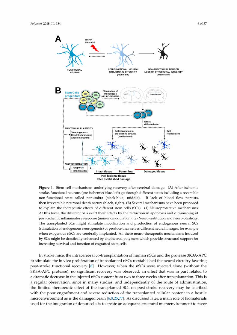
Over the last two decades, different type of cells (mostly multi- and pluripotent stem cells)have been used as therapeutic tools to enhance nervous tissue regeneration and brain plasticityphenomena (Figure 1). After the initial injury different SCs might neuroprotect the brain by thereduction in apoptosis and diminishing of post-ischemic inflammatory response that commonlyexacerbates the brain damage propagating injury towards healthy tissue areas. The transplanted SCsmight also stimulate the mobilization and production of endogenous neural SCs and progenitors in thesubventricular zone (SVZ) and hippocampus (stimulation of endogenous neurogenesis) or producethemselves different neural lineages, for example when exogenous nSCs are cerebrally implanted.Exogenous SCs- or endogenous SCs-derived newborn neural cells (NNcells) might replace damagedcells creating new circuitry in the infarcted tissue or might be integrated in pre-existing neural networksin the perilesional tissue. Apart from NNcells integration, other forms of functional plasticity supportedby the paracrine action of SCs (secretion of factors) occur at structural level, including the formationof new synapses (synaptogenesis), dendritic branching and growth and axonal sprouting favoringneuronal connectivity in the perilesional tissue, which have strong potential for plasticity after braindamage. In addition, different SCs may stimulate angiogenesis creating an optimal vascular networkmicroenvironment for cell replacement, neural integration and plasticity. Many of these mechanismscan be favored by engineered polymers which provide structural and functional support for theengrafted stem cells.
Among the various types of stem cells, neural stem cells (nSCs) and neural precursors (nPCs)were initially established as the ideal cell populations for the repair of central nervous system. Duringembryonic development, neurogenesis through natural and pre-existing nSCs and nPCs principallyoccurs in two main brain regions: the subventricular zone (SVZ) and the ventral and lateral ganglioniceminences (VGE and LGE). The first region (SVZ) is the origin of the largest population of neocorticalneurons while the ganglionic eminences give rise to the neurons of the basal ganglia and the neocorticalinterneurons [65]. In contrast, in the adult brain the main neurogenic niches are restricted to theventricular/subventricular region as well as to the subgranular region (SGZ) of the dentate gyrus ofthe hippocampus [66]. Adult neurogenesis is regulated over several stages including maintenanceand self-renewal of nSCs [67], proliferation and migration of nPCs, as wells as survival, maturation,and cellular integration in already established circuits or in ex novo built neural networks. After braininjury, endogenous nPCs migrate to the zones of damage [68,69] following the chemoattracting signalssecreted by astrocytes, microglia, and vascular endothelial cells [66]. However, the number, recruitmentand integration of neural cells are insufficient for the reestablishment of the neurological functions.Thus, although present, the endogenous neurogenesis is extremely inefficient which explains the lackof substantial post-stroke spontaneous recovery in humans and animals.
The implantation of nSCs has been considered a therapeutic option to favor the production andintegration of terminal neural cells in either the core or the perilesional regions [70–72]. Additionally,donor nSCs stimulate endogenous neurogenesis [72,73] as well as angiogenesis [74]. Part of nSCsregenerative effects can be explained by their secretoma activity including the release of differentproteins such as vascular endothelial growth factor (VEGF), brain derived neurotrophic factor (BDNF),nerve growth factor (NGF), and tumor necrosis factor alpha (TNF-α) among others [75,76]. To providesome representative examples of the therapeutic use of nSCs, in one study the implantation of humannSCs increased axonal transport, dendritic ramifications and axonal growth and propagation, all ofthese known to be mechanisms that contribute to the increase of neuronal plasticity, promoting thefunctional recovery of rats with focal ischemia [3].
Polymers 2018, 10, 184 6 of 37
BRAIN
DAMAGE
FUNCTIONAL
NEURON
NON-FUNCTIONAL NEURON
STRUCTURAL INTEGRITY
(reversible)
NON-FUNCTIONAL NEURON
LOSS OF STRUCTURAL INTEGRITY
(irreversible)
A
B
Inflammation
Cell
replacement
Stem Cells
progenitors
Damaged tissuePenumbraIntact tissue
Peri-lesional tissue
after established damage
Stimulation of
endogenous
NEUROGENESIS
Cell integration in
pre-existing circuits
(peri-lesional)
Sinaptogenesis
Dendritic branching
Axonal sprouting
Apoptosis
NEUROPROTECTION
SVZ Hippocampus
FUNCTIONAL PLASTICITY
Neural
differentiation
Figure 1. Stem cell mechanisms underlying recovery after cerebral damage. (A) After ischemicstroke, functional neurons (pre-ischemic; blue, left) go through different states including a reversiblenon-functional state called penumbra (black-blue, middle). If lack of blood flow persists,then irreversible neuronal death occurs (black, right). (B) Several mechanisms have been proposedto explain the therapeutic effects of different stem cells (SCs). (1) Neuroprotective mechanisms:At this level, the different SCs exert their effects by the reduction in apoptosis and diminishing ofpost-ischemic inflammatory response (immunomodulation). (2) Neuro-restitution and neuro-plasticity:The transplanted SCs might stimulate mobilization and production of endogenous neural SCs(stimulation of endogenous neurogenesis) or produce themselves different neural lineages, for examplewhen exogenous nSCs are cerebrally implanted. All these neuro-therapeutic mechanisms inducedby SCs might be drastically enhanced by engineered polymers which provide structural support forincreasing survival and function of engrafted stem cells.
In stroke mice, the intracerebral co-transplantation of human nSCs and the protease 3K3A-APCto stimulate the in vivo proliferation of transplanted nSCs reestablished the neural circuitry favoringpost-stroke functional recovery [8]. However, when the nSCs were injected alone (without the3K3A-APC protease), no significant recovery was observed, an effect that was in part related toa dramatic decrease in the injected nSCs content from two to three weeks after transplantation. This isa regular observation, since in many studies, and independently of the route of administration,the limited therapeutic effect of the transplanted SCs on post-stroke recovery may be ascribedwith the poor engraftment and severe reduction of the transplanted cellular content in a hostilemicroenvironment as is the damaged brain [6,8,25,77]. As discussed later, a main role of biomaterialsused for the integration of donor cells is to create an adequate structural microenvironment to favor
Polymers 2018, 10, 184 7 of 37
the survival, retention, and cross-talk of the implanted cells with the host nervous tissue. AlthoughnSCs-based regenerative therapies are very promising, the most significant disadvantages for their usein clinics are their scarce availability and difficulty of ex vivo expansion as well as the immunogenicityof allogenic transplants [78].
Embryonic stem cells (eSCs) constitute an alternative type of SCs that have been commonly usedin a wide variety of studies. They are pluripotent cells capable of self-renewing and differentiatinginto any cellular phenotype of the organism including the neural lineage. The ability of eSCs topromote post-ischemic functional recovery has been demonstrated in several studies [79,80]. Howeverthe ethical limitations derived from its isolation from embryonic tissue [81] as well as the malignanttransformation of this cellular phenotype and its tendency to frequently form teratomas in vivo [82]makes them less suitable for clinical applications.
Mesenchymal stem cells (mSCs), bone marrow mononuclear cells (BMmCs), and inducedpluripotent stem cells (iPSCs) represent an alternative to nSCs and eSCs since they can beisolated, generated and expanded with no excessive difficulty. These cell populations have shownneuroprotective and neuro-regenerative effects in different animal models. They can be isolated orgenerated from the patients themselves therefore allowing autologous transplantation and avoidinggraft rejection. Among the potential benefits of mSCs, iPSCs, and BMmCs it is worth mentioning themodulation of the brain tissue microenvironment through the secretion of several growth factors thatregulate the immune response, limit astrogliosis and microgliosis, stimulate endogenous neurogenesisand angiogenesis or reduce apoptosis by decreasing the oxidative stress [83–85].
The iPSCs hold many of the functional properties found in eSCs, exhibiting similar morphology,the endogenous expression of pluripotency genes and their ability to self-renew or differentiate intoprogenitors, precursors, and mature cells of different germinal origin including neural cells [86].Differentiated somatic cells can be reprogrammed to pluripotency (iPSCs) by treatment with definedfactors. Several groups have been able to induce pluripotency easily and non-invasively from differentsomatic cells including fibroblasts, bone marrow, adipose tissue, and peripheral leukocytes [87–89].Chen et al. found in a rat focal ischemia model that after the combined subdural transplantation ofiPSCs and fibrin glue, there was a significant reduction in the infarct volume and a greater functionalrecovery in the animals examined with the rotarod test [90]. It has also been reported in infarctedrats that the intracerebral transplantation of iPSCs in both the affected and non-affected hemispheresinduced their migration to the damaged area and subsequent differentiation into neural cells thatenhanced post-stroke sensorimotor function recovery [91,92]. However, results are not conclusive sincein other studies the iPSCs did not produce a substantial recovery of infarcted animals and the incidenceof tumorigenesis was relatively elevated [93]. Thus, some limitations inherent to their genotype ofiPSCs should be surpassed such as the possible mutations in cancer-related genes, aneuploidy andDNA aberrant methylation causing genomic instability, which have been associated with the formationof tumors and therefore incompatible with their clinical translation [93–95].
The BMmCs constitute a heterogeneous group of mature hematopoietic cells (B cells, T cells,and monocytes) and a small proportion of stem cells and hematopoietic progenitors, which togetherwith stem cells and progenitors of mesenchymal origin (mSCs) represent a promising approachfor the treatment of cerebrovascular disorders. BMmCs increase neurogenesis [62,96] and cerebralplasticity [97] as well as favor blood flow to the damaged tissue by increasing angiogenesis,thus creating a microenvironment suitable for the migration and integration of neural cells andthe replacement of lost tissue promoting functional recovery in infarcted animals.
3.2. Mesenchymal Stem Cells
mSCs are by far one of the most commonly employed cell populations. mSCs constitutea heterogeneous non-hematopoietic cell population which were first characterized by Friedenstein et al.in 1970 [98]. Although these cells can be isolated from different tissues including fat, umbilical cordand bone, it is in the bone marrow where they are present in a higher content [99], providing signaling
Polymers 2018, 10, 184 8 of 37
for hematopoietic stem cell survival and function [100,101]. The special functional characteristics ofmSCs and their strong potential for the treatment of cerebral disorders including stroke made themone of the better studied cell populations [35–37,102–109]. The wide variety of studies came througha significant advance in the knowledge of the neuroprotective and neurorepair mechanisms regulatedby the paracrine activity of the mSCs. Pioneer studies reported mSCs are able to differentiate intoneural phenotypes [110,111] but later studies have discredited the direct transdifferentiation of mSCsinto neural lineages [112]. Current consensus is that most mSCs therapeutic effects are due to thesecretome activity of these cells, which, independently of the administration route (cerebral, nasal,and intra-arterial/venous), is able to modulate the main neuroprotection mechanisms, i.e., post-strokeimmunoresponse, inflammation and apoptosis [113].
mSCs may reduce inflammatory response and apoptosis by decreasing the levels ofpro-inflammatory molecules such as interleukins IL-1β and IL-6A, or TNF-α [114–116].Immunomodulation of the damaged brain is important to reduce the inflammatory responseminimizing injury and promoting recovery [117]. Inhibition of inflammation has been related to theincreased levels of neurogenesis [118]. mSCs participate in regulating the M1/M2 activation balance bybiasing microglia differentiation towards the anti-inflammatory M2 state for resolution of inflammationand tissue repair in detriment of the pro-inflammatory M1 phenotype [119,120] and mSCs implantationhas also been correlated with a reduced astrogliosis [121]. mSCs secretome also promotes angiogenesis,neurogenesis, and sinaptogenesis which in turn facilitate post-stroke brain repair and plasticity [122].Angiogenesis in the perilesional tissue plays a crucial role in the survival and regeneration (restitution)of the damaged tissue. Intravenous administration of mSCs increases VEGF levels in rat ischemic tissueand provokes a strong development of microvasculature in the perilesional cortex of the infarctedhemisphere [102,123]. Angiogenesis as well as neurogenesis might be promoted by factors released bymSCs, including NGF, BDNF, VEGF, Glial-cell-derived-neurotrophic factor (GDNF), placental growthfactor (PFG), or stromal-derived-factor (SDF-1) [2,6,102,124,125]. After ischemic damage, immatureDoublecortine (DCX) positive neurons migrate from their neurogenic niches (SVZ) to areas of theperilesional tissue, deviating from their natural route to the olfactory bulb [126,127]. After thromboticinfarction in mice, the intracerebral transplantation of mSCs in the striatum was associated withincreasing neurogenesis determined by an increment of cell proliferation and the content of DCX+ cellsin areas surrounding the infarct core as well as with vascularization in the damaged area accompaniedby a reduction of glial scar [6]. Finally, mSCs favor neuronal plasticity by increasing the number ofsynapses in both damaged and intact hemispheres in models of unilateral infarctions [128,129]. Axonalsprouting and myelin formation are essential for the reconstruction of the damaged neural circuits andcould also be modulated by the implantation of mSCs in the brain [130].
Most of the above mentioned neurotherapeutic mechanisms are related to the factors released bythe mSCs, which might have a certain parallelism with the secretome activity present in other types ofstem cells with known neuroprotective and neuroregenerative potential. While many of these factorscan be secreted by exocytosis which constitutes a main mechanism for releasing extracellular proteinsor neurotransmitters in neurons, recent studies suggest that part of the paracrine action of mSCs maybe accomplished by cell-derived vesicles also called exosomes that could transport mRNAs, miRNAsand proteins with tissue repairing potential [131,132]. In vitro studies have shown that exosomesstimulate the axonal growth of cortical neurons [133] as well as favor angiogenesis. Intravenousadministration of mSCs in infarcted rats enhanced functional recovery through the microRNA 133b,which is present in exosomes released from mSCs [134]. In this study MiR-133b promoted axonalplasticity and neurite remodeling in the perilesional cortex [134].
3.3. Optimal Time Window and Best Administration Route
Although stem cell implantation in experimental models of stroke has been performed evenseveral weeks after cerebral damage [61,135] significant post-treatment recovery is usually observedwith cell implantation within 24 h to a few days after injury. This might be explained by the existence of
Polymers 2018, 10, 184 9 of 37
a putative optimal intervention window for promoting recovery, neuro-restitution, and neuro-plasticityafter brain injury consensually restricted to the first post-stroke days [5,6,8]. For example, physicalrehabilitation is powerful when starts in the first 1–2 weeks after brain damage while no significantpost-stroke improvement has been observed when started several weeks post-stroke [136]. However,many stroke patients continue improving for years after brain injury bringing to debate the limits ofsuch temporal window. With respect to the SCs, there is no consensus regarding the best temporalpoint for their implantation [137].
The optimal route for cell implantation has still to be defined, although cerebral administrationreports better benefits than that of intra-venous and intra-arterial infusion [137]. Surprisingly,the number of studies comparing the efficacy of different routes of administration using the samecellular phenotype and brain damage model is quite limited. In terms of therapeutic efficacyintra-arterial administration is comparable to intra-arterial infusion [138] and different types of SCshave been employed in models of cerebral infarction where, in many cases, functional improvementhas been observed [102,104,114,138]. Although both routes of administration are relatively safe andless invasive than cerebral implantation, one main limitation of systemic intravascular transplantationis the reduced migration of the implanted cells towards the brain, which might be trapped in thepulmonary capillaries, spleen, kidney, or liver, thus diminishing its therapeutic effect [1]. In addition,systemic administration is not free of complications and risks, for example because of the formation ofpulmonary and brain microembolisms that are mostly related to cell size and concentration as wellas to infusion velocity [1,139,140]. In contrast, intranasal delivery of SCs is safe, feasible, and poorlyinvasive [141]. Cells administered via this route must pass across the intranasal mucous epitheliumtowards the brain tissue until they reach the area where therapeutic effect is needed [142]. However,the mucociliary tissue represents a barrier for particles and microorganisms where cells might gettrapped, which obliges to increase the cellular doses to achieve a measurable therapeutic effect [142].Up to date no clinical trials have been performed with this delivery route and it is unknown if the largemigratory distance between mucociliary tissue and brain in humans represents a serious limitation [1].
Cerebral implants are more invasive than other transplantation routes. In general, the implantationof cells in the rodent brain has not been translated into important side effects. However, the humanbrain is extremely sensible to surgical manipulation and minor damages could create profoundfunctional deficits beyond previous stroke-originated dysfunctions [143]. However, cerebral implantsrepresent the most efficient approaches to promote post-stroke functional recovery [6,8,144,145].They provide precision in terms of graft location as well as control of the number of the implantedcells. Furthermore, cerebral implants circumvent the blood–brain-barrier (BBB) which constitutes thestronger limitation for the transplanted cells when they are infused systemically or intranasally.The cerebral implants are also of relevance when hydrogels are used to assist the function oftherapeutic cells. In a low percentage of patients SCs implantation has been related with importantside effects such as focal hemorrhage, chronic subdural hematomes, seizures and psychomotorexacerbation [1,41,47,146]. However, cerebral implants might be justified by weighing the benefits ofquality of life of the individual patient against the infrequent potentially life-threatening side effects.When considering the cerebral route there is also a lack of consensus regarding the best place for cellimplantation to achieve a better recovery. Few cells survive beyond two or three weeks after cerebraltransplantation [5,6,8,77,78], but the rate of survival is higher when SCs transplantation is performed inperilesional areas or far away from the cavity lesion [5,8]. After brain injury, the inflammatory responsecreates a hostile environment deleterious for both, the brain itself and the engrafted cells, making graftlocation critical for the survival of the implanted cells [5]. Short-lasting SCs survival inevitably leadsto a non-durable therapeutic effect (cell differentiation and factors secretion). Similarly, if structuraland functional changes of perilesional tissue are essential for plasticity and recovery, then SCstransplantation in these areas could be counterproductive. To overcome some of these obstacles,hydrogels-integrated SCs and factors have been implanted in the stroke cavity observing behavioralimprovement in the animals treated despite a priori inhospitability of the infarcted area [144,147]
Polymers 2018, 10, 184 10 of 37
or have been applied epi-cortically (to the brain cortical surface) in a theoretically less invasiveapproach [7].
3.4. Clinical Scenario: Where Are We Now?
The safety and tolerability of the implantation of SCs in patients with cerebral damage has beenwidely demonstrated for different types of SCs and for all administrative routes [38–47] althoughsome adverse effects have been observed in a minor fraction of treated patients [41,47]. Results aboutthe neuro-therapeutic efficacy of the SCs are not conclusive, possibly because of the small numberof patients included in the studies and the variability of the location of the stroke. In one of the firstclinical trials, Bang et al., proved both the safety and efficacy of the intravenous transplantation ofautologous mSCs in stroke-injured patients. They reported a reduction of the neurological deficitsin the large majority of the treated patients [38] but their conclusions raised concerns because ofthe reduced number of subjects and the lack of adequate controls [148]. Subsequent studies havedemonstrated the safety and efficacy of the systemic administration of mSCs, here too, a reduction ofpost-stroke functional deficits was observed [44,149]. Recently, a comparison was made between theeffect of intravenous injection of mSCs with respect to the combined co-transplantation of mSCsplus nSCs through the cerebellomedullary cistern [150]. After two-years follow-up, no seriousadverse effects were observed (neurological infections, tumor formation) except for low fever (30% ofpatients) and temporary dizziness, and most patients showed clinical improvements [150]. However,the reduced number of patients (six subjects) limited the conclusions of the study. The safety and thetherapeutic effects of systemic and intra-cerebral transplantation of BMmCs, nSCs, and mSCs havebeen corroborated by several other studies [41,46,47,151,152]. Collectively, there is a need of clinicaltrials involving a high number of patients, more homogenous and with better inclusion/exclusioncriteria [153]. In addition, a greater number of pre-clinical studies are required to further investigatethe neuro-therapeutic mechanisms of action of different SCs. The cellular and molecular substratesresponsible for recovery are in general not well understood. In a few experimental studies, relevantmechanistic information for recovery has been obtained, which has been related to the production ofnSC-derived newborn neurons and their functional integration with the host tissue [8], and structuralchanges (plasticity) related to axonal sprouting and dendritic branching of the host neurons afterimplantation with human neural progenitors and their secretome activity [3]. In contrast, the modestfunctional recovery observed in preclinical and clinical studies could originate from the cells implantedsystemically and intra-nasally not migrating to the brain tissue with all the required efficacy [137].In the case of cerebral transplantation poor recovery could be related to a progressive dispersionof the SCs outside the implanted area and to a higher mortality of the transplanted cells in theinflammatory post-ischemic brain tissue [5,6,77,124,154]. Poor engraftment of SCs into the brain isbeing tentatively resolved with different approaches, for example the combined implantation of SCsand factors that increase donor cell expansion [8] or the use of different biomaterials to assist cellsurvival and function [144], as we will discuss in the next section.
4. Hydrogels-Assisted Cell Therapies for Brain Stroke
The use of biomaterials to assist cell therapy after brain injury is merely restricted to theexperimental arena, which raises important concerns on whether we are really seeding a reasonedtechnology for future use in patients, or if we are facing an unreachable scenario. Preclinical datahave supported the efficacy of the co-transplantation of hydrogels and cells to repair the damagedbrain [135,144,155,156] and a growing number of clinical trials have demonstrated the feasibility andrelative safety of cerebral implants with different SCs [39]. Thus, the available evidence envisagesa horizon where different biomaterials, mostly in the form of hydrogels, could be useful in enhancingcellular engraftment to assist SCs therapeutic effects.
The concept of hydrogels for biomedical applications comes from the 1960s by using copolymersof glycolmethacrylates [157,158] to design structures with adjustable water content (>90% water),
Polymers 2018, 10, 184 11 of 37
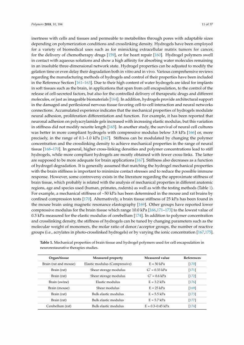
inertness with cells and tissues and permeable to metabolites through pores with adaptable sizesdepending on polymerization conditions and crosslinking density. Hydrogels have been employedfor a variety of biomedical uses such as for mimicking extracellular matrix tumors for cancer,for the delivery of chemotherapy drugs [159], or for heart repair [160]. Hydrogel polymers swellin contact with aqueous solutions and show a high affinity for absorbing water molecules remainingin an insoluble three-dimensional network state. Hydrogel properties can be adjusted to modify thegelation time or even delay their degradation both in vitro and in vivo. Various comprehensive reviewsregarding the manufacturing methods of hydrogels and control of their properties have been includedin the Reference Section [161–163]. Due to their high content of water hydrogels are ideal for implantsin soft tissues such as the brain, in applications that span from cell encapsulation, to the control of therelease of cell-secreted factors, but also for the controlled delivery of therapeutic drugs and differentmolecules, or just as imageable biomaterials [164]. In addition, hydrogels provide architectural supportin the damaged and perilesional nervous tissue favoring cell-to-cell interaction and neural networksconnections. Accumulated experience indicates that the mechanical properties of hydrogels modulateneural adhesion, proliferation differentiation and function. For example, it has been reported thatneuronal adhesion on polyacrylamide gels increased with increasing elastic modulus, but this variationin stiffness did not modify neurite length [165]. In another study, the survival of neural cell cultureswas better in more compliant hydrogels with compressive modulus below 3.8 kPa [166] or, moreprecisely, in the range of 0.1–1.0 kPa [167]. Stiffness can be modulated by changing the polymerconcentration and the crosslinking density to achieve mechanical properties in the range of neuraltissue [168–170]. In general, higher cross-linking densities and polymer concentrations lead to stiffhydrogels, while more compliant hydrogels are mostly obtained with fewer cross-links. The latterare supposed to be more adequate for brain applications [167]. Stiffness also decreases as a functionof hydrogel degradation. It is generally assumed that matching the hydrogel mechanical propertieswith the brain stiffness is important to minimize contact stresses and to reduce the possible immuneresponse. However, some controversy exists in the literature regarding the approximate stiffness ofbrain tissue, which probably is related with the analysis of mechanical properties in different anatomicregions, age and species used (human, primates, rodents) as well as with the testing methods (Table 1).For example, a mechanical stiffness of ~50 kPa has been determined in the mouse and rat brains byconfined compression tests [170]. Alternatively, a brain tissue stiffness of 25 kPa has been found inthe mouse brain using magnetic resonance elastography [169]. Other groups have reported lowercompressive modulus for the brain tissue which range 10.0 kPa [166,171–173] to the lowest value of0.3 kPa measured for the elastic modulus of cerebellum [174]. In addition to polymer concentrationand crosslinking density, the stiffness of hydrogels can be tuned by changing parameters such as themolecular weight of monomers, the molar ratio of donor/acceptor groups, the number of reactivegroups (i.e., acrylates in photo-crosslinked hydrogels) or by varying the ionic concentration [167,175].
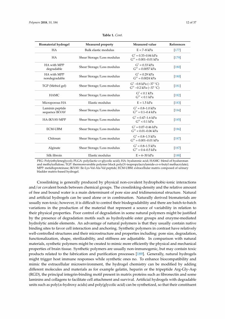
Table 1. Mechanical properties of brain tissue and hydrogel polymers used for cell encapsulation inneurorestaurative therapies studies.
Organ/tissue Measured property Measured value References
Brain (rat and mouse) Elastic modulus (Compressive) E = 50 kPa [170]
Brain (rat) Shear storage modulus G′ = 0.33 kPa [171]
Brain (rat) Shear storage modulus G′ = 0.6 kPa [172]
Brain (swine) Elastic modulus E = 3.2 kPa [176]
Brain (mouse) Shear modulus E = 25 kPa [169]
Brain (rat) Bulk elastic modulus E = 5.5 kPa [173]
Brain (rat) Bulk elastic modulus E = 5.7 kPa [177]
Cerebellum (rat) Bulk elastic modulus E = 0.3–0.45 kPa [174]
Polymers 2018, 10, 184 12 of 37
Table 1. Cont.
Biomaterial hydrogel Measured property Measured value References
HA Bulk elastic modulus E = 7–8 kPa [177]
HA Shear Storage/Loss modulus G′ = 0.33–0.84 kPaG′ ′ = 0.001–0.01 kPa [179]
HA with MPPdegradable Shear Storage/Loss modulus G′ = 0.33 kPa
G′ ′ = 0.0057 kPa [180]
HA with MPPnondegradable Shear Storage/Loss modulus G′ = 0.29 kPa
G′ ′ = 0.0024 kPa [180]
TGP (Mebiol gel) Shear Storage/Loss modulus G′ ~0.8 kPa (~37 ◦C)G′ ′ ~0.2 kPa (~37 ◦C) [181]
HAMC Shear Storage/Loss modulus G′ < 0.1 kPaG′ ′ < 0.1 kPa [182]
Microporous HA Elastic modulus E = 1.5 kPa [183]
Laminin peptidesequence IKVAV Shear Storage/Loss modulus G′ = 0.8–1.0 kPa
G′ ′ = 0.1–0.4 kPa [184]
HA-IKVAV-MPP Shear Storage/Loss modulus G′ = 0.47–1.6 kPaG′ ′ < 0.1 kPa [185]
ECM-UBM Shear Storage/Loss modulus G′ = 0.07–0.46 kPaG′ ′ < 0.01–0.06 kPa [186]
Chitosan Shear Storage/Loss modulus G′ = 0.8–1.5 kPaG′ ′ = 0.001–0.01 kPa [187]
Alginate Shear Storage/Loss modulus G′ = 0.8–1.5 kPaG′ ′ = 0.4–0.5 kPa [187]
Silk fibroin Elastic modulus E = 6–30 kPa [188]
PEG: Poly(ethyleneglycol); PLGA: poly(lactic-co-glycolic acid); HA: hyaluronic acid; HAMC: blend of hyaluronanand methylcellulose; TGP: thermoreversible polymer block poly(N-isopropylacrylamide-co-n-butyl methacrylate);MPP: metalloproteinase; IKVAV: Ile-Lys-Val-Ala-Val peptide; ECM-UBM: extracellular matrix composed of urinarybladder matrix-based hydrogel.
Crosslinking is generally produced by physical non-covalent hydrophobic-ionic interactionsand/or covalent bonds between chemical groups. The crosslinking-density and the relative amountof free and bound water is a main determinant of pore size and tridimensional structure. Naturaland artificial hydrogels can be used alone or in combination. Naturally derived biomaterials areusually non-toxic; however, it is difficult to control their biodegradability and there are batch-to-batchvariations in the production of the material that represent a source of variability in relation totheir physical properties. Poor control of degradation in some natural polymers might be justifiedby the presence of degradation motifs such as hydrolysable ester groups and enzyme-mediatedhydrolytic amide elements. An advantage of natural polymers is that they usually contain nativebinding sites to favor cell interaction and anchoring. Synthetic polymers in contrast have relativelywell-controlled structures and their microstructure and properties including: pore size, degradation,functionalization, shape, sterilizability, and stiffness are adjustable. In comparison with naturalmaterials, synthetic polymers might be created to mimic more efficiently the physical and mechanicalproperties of brain tissue. Synthetic polymers are usually non-inmunogenic, but may contain toxicproducts related to the fabrication and purification processes [189]. Generally, natural hydrogelsmight trigger host immune responses while synthetic ones no. To enhance biocompatibility andmimic the extracellular microenvironment, the hydrogel chemistry can be modified by addingdifferent molecules and materials as for example gelatin, heparin or the tripeptide Arg-Gly-Asp(RGD), the principal integrin-binding motif present in matrix proteins such as fibronectin and somelaminins and collagens to facilitate cell attachment and survival. Artificial hydrogels with degradableunits such as poly(α-hydroxy acids) and poly(glycolic acid) can be synthetized, so that their constituent
Polymers 2018, 10, 184 13 of 37
monomers dissociate in contact with water. Alternatively, other hydrogels, such as poly(ε-caprolactone)can be also enzymatically degraded by lipases.
One of the first studies using biomaterials for in vivo applications was performed in the1980s [190]. Yannas and Burke performed skin implants in guinea pigs using collagen membranes andobserved a limited inflammatory response by detecting mono- and polynucleated cells. This immuneresponse against the implanted biomaterial could be minimized by crosslinking collagen withglucosaminoglucan. Later, Vacanti et al. cultured mouse and rat hepatocytes into artificial polymerscomposed of polyglactin, polyorthoesters, and polyanhydride for subsequent liver transplantation inrats, demonstrating that hepatocytes remained viable up to 14 days after in vivo implantation [191].In the same period, a pioneer study showed prolonged in vivo survival and function of pancreatic isletsencapsulated into cross-linked alginate microcapsules [192]. These studies provided some conceptualbasis for the development of tissue engineering. Since then, a wide variety of applications in the fieldof regenerative medicine for different tissues and organs has been reported using hydrogel-basedbiomaterials to enclose different cells, drugs and different growth factors.
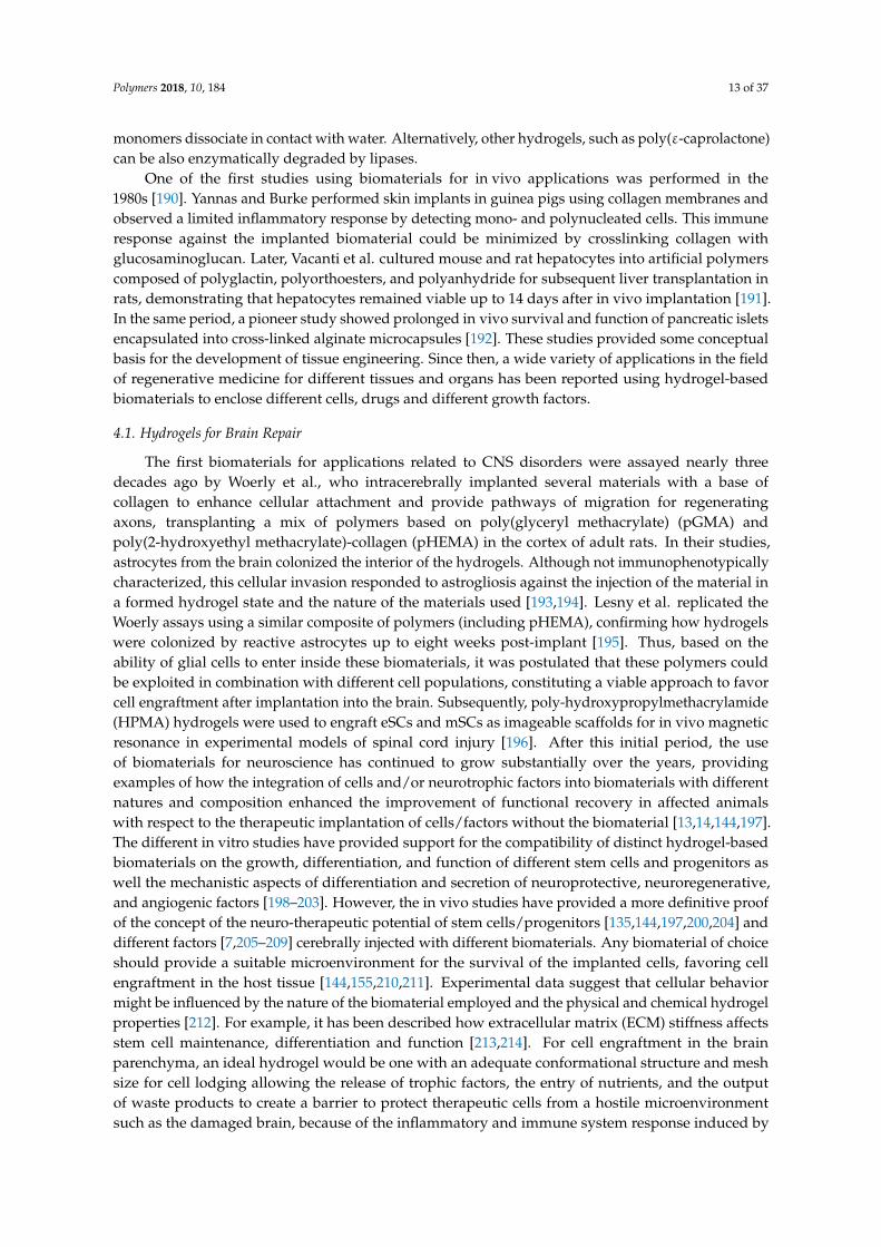
4.1. Hydrogels for Brain Repair
The first biomaterials for applications related to CNS disorders were assayed nearly threedecades ago by Woerly et al., who intracerebrally implanted several materials with a base ofcollagen to enhance cellular attachment and provide pathways of migration for regeneratingaxons, transplanting a mix of polymers based on poly(glyceryl methacrylate) (pGMA) andpoly(2-hydroxyethyl methacrylate)-collagen (pHEMA) in the cortex of adult rats. In their studies,astrocytes from the brain colonized the interior of the hydrogels. Although not immunophenotypicallycharacterized, this cellular invasion responded to astrogliosis against the injection of the material ina formed hydrogel state and the nature of the materials used [193,194]. Lesny et al. replicated theWoerly assays using a similar composite of polymers (including pHEMA), confirming how hydrogelswere colonized by reactive astrocytes up to eight weeks post-implant [195]. Thus, based on theability of glial cells to enter inside these biomaterials, it was postulated that these polymers couldbe exploited in combination with different cell populations, constituting a viable approach to favorcell engraftment after implantation into the brain. Subsequently, poly-hydroxypropylmethacrylamide(HPMA) hydrogels were used to engraft eSCs and mSCs as imageable scaffolds for in vivo magneticresonance in experimental models of spinal cord injury [196]. After this initial period, the useof biomaterials for neuroscience has continued to grow substantially over the years, providingexamples of how the integration of cells and/or neurotrophic factors into biomaterials with differentnatures and composition enhanced the improvement of functional recovery in affected animalswith respect to the therapeutic implantation of cells/factors without the biomaterial [13,14,144,197].The different in vitro studies have provided support for the compatibility of distinct hydrogel-basedbiomaterials on the growth, differentiation, and function of different stem cells and progenitors aswell the mechanistic aspects of differentiation and secretion of neuroprotective, neuroregenerative,and angiogenic factors [198–203]. However, the in vivo studies have provided a more definitive proofof the concept of the neuro-therapeutic potential of stem cells/progenitors [135,144,197,200,204] anddifferent factors [7,205–209] cerebrally injected with different biomaterials. Any biomaterial of choiceshould provide a suitable microenvironment for the survival of the implanted cells, favoring cellengraftment in the host tissue [144,155,210,211]. Experimental data suggest that cellular behaviormight be influenced by the nature of the biomaterial employed and the physical and chemical hydrogelproperties [212]. For example, it has been described how extracellular matrix (ECM) stiffness affectsstem cell maintenance, differentiation and function [213,214]. For cell engraftment in the brainparenchyma, an ideal hydrogel would be one with an adequate conformational structure and meshsize for cell lodging allowing the release of trophic factors, the entry of nutrients, and the outputof waste products to create a barrier to protect therapeutic cells from a hostile microenvironmentsuch as the damaged brain, because of the inflammatory and immune system response induced by
Polymers 2018, 10, 184 14 of 37
injury. In addition, porosity of hydrogels can be tuned creating pores large enough to allow neuriteextension and vascularization between the host brain and the implanted hydrogel [167]. As hydrogelsare designed for soft tissues such as the brain, they are mechanically weak and difficult to handle informed hydrogel state. However, hydrogels can be formed in situ under physiological conditions oncea polymer (or mix of them) have been implanted as a liquid in the brain, making it easy to incorporatecells and factors before gelation, thus reducing the invasiveness of hydrogel implantation insteadof using a formed hydrogel state [188,215]. This strategy allows, for example, the hydrogel to fillcompletely amorphous cavities as the result of injury, whereas formed hydrogels are not suitable forthis type of applications.
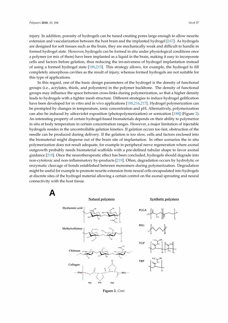
In this regard, one of the basic design parameters of the hydrogel is the density of functionalgroups (i.e., acrylates, thiols, and polyesters) in the polymer backbone. The density of functionalgroups may influence the space between cross-links during polymerization, so that a higher densityleads to hydrogels with a tighter mesh structure. Different strategies to induce hydrogel gelificationhave been developed for in vitro and in vivo applications [188,216,217]. Hydrogel polymerization canbe prompted by changes in temperature, ionic concentration and pH. Alternatively, polymerizationcan also be induced by ultraviolet exposition (photopolymerization) or sonication [188] (Figure 2).An interesting property of certain hydrogel-based biomaterials depends on their ability to polymerizein situ at body temperature in certain concentration ranges. However, a major limitation of injectablehydrogels resides in the uncontrollable gelation kinetics. If gelation occurs too fast, obstruction of theneedle can be produced during delivery. If the gelation is too slow, cells and factors enclosed intothe biomaterial might disperse out of the brain site of implantation. In other scenarios the in situpolymerization does not result adequate, for example in peripheral nerve regeneration where axonaloutgrowth probably needs biomaterial scaffolds with a pre-defined tubular shape to favor axonalguidance [218]. Once the neurotherapeutic effect has been concluded, hydrogels should degrade intonon-cytotoxic and non-inflammatory by-products [219]. Often, degradation occurs by hydrolytic orenzymatic cleavage of bonds established between monomers during polymerization. Degradationmight be useful for example to promote neurite extension from neural cells encapsulated into hydrogelsat discrete sites of the hydrogel material allowing a certain control on the axonal sprouting and neuralconnectivity with the host tissue.
Polymers 2018, 10, x FOR PEER REVIEW 14 of 37
hydrogel [167]. As hydrogels are designed for soft tissues such as the brain, they are mechanically
weak and difficult to handle in formed hydrogel state. However, hydrogels can be formed in situ
under physiological conditions once a polymer (or mix of them) have been implanted as a liquid in
the brain, making it easy to incorporate cells and factors before gelation, thus reducing the
invasiveness of hydrogel implantation instead of using a formed hydrogel state [188,215]. This
strategy allows, for example, the hydrogel to fill completely amorphous cavities as the result of
injury, whereas formed hydrogels are not suitable for this type of applications.
In this regard, one of the basic design parameters of the hydrogel is the density of functional
groups (i.e., acrylates, thiols, and polyesters) in the polymer backbone. The density of functional
groups may influence the space between cross-links during polymerization, so that a higher density
leads to hydrogels with a tighter mesh structure. Different strategies to induce hydrogel gelification
have been developed for in vitro and in vivo applications [188,216,217]. Hydrogel polymerization
can be prompted by changes in temperature, ionic concentration and pH. Alternatively,
polymerization can also be induced by ultraviolet exposition (photopolymerization) or sonication
[188] (Figure 2). An interesting property of certain hydrogel-based biomaterials depends on their
ability to polymerize in situ at body temperature in certain concentration ranges. However, a major
limitation of injectable hydrogels resides in the uncontrollable gelation kinetics. If gelation occurs
too fast, obstruction of the needle can be produced during delivery. If the gelation is too slow, cells
and factors enclosed into the biomaterial might disperse out of the brain site of implantation. In
other scenarios the in situ polymerization does not result adequate, for example in peripheral nerve
regeneration where axonal outgrowth probably needs biomaterial scaffolds with a pre-defined
tubular shape to favor axonal guidance [218]. Once the neurotherapeutic effect has been concluded,
hydrogels should degrade into non-cytotoxic and non-inflammatory by-products [219]. Often,
degradation occurs by hydrolytic or enzymatic cleavage of bonds established between monomers
during polymerization. Degradation might be useful for example to promote neurite extension
from neural cells encapsulated into hydrogels at discrete sites of the hydrogel material allowing a
certain control on the axonal sprouting and neural connectivity with the host tissue.
A
Formed hydrogelPolymeric solution
B Temperature
Ionic strength
Functionalization
Sonication
Synthetic polymersNatural polymers
Photo-polymerization
Ph
ysi
cal
and
ch
emic
al
cro
ss-l
ink
ing
in
itia
tors
PLGA
TGP
TRP
PEGMethylcellulose
Hyaluronic acid
Chitosan
Collagen
Figure 2. Cont.
Polymers 2018, 10, 184 15 of 37Polymers 2018, 10, x FOR PEER REVIEW 15 of 37
A
Formed hydrogelPolymeric solution
B Temperature
Ionic strength
Functionalization
Sonication
Synthetic polymersNatural polymers
Photo-polymerization
Ph
ysi
cal
and
ch
emic
al
cro
ss-l
ink
ing
in
itia
tors
PLGA
TGP
TRP
PEGMethylcellulose
Hyaluronic acid
Chitosan
Collagen
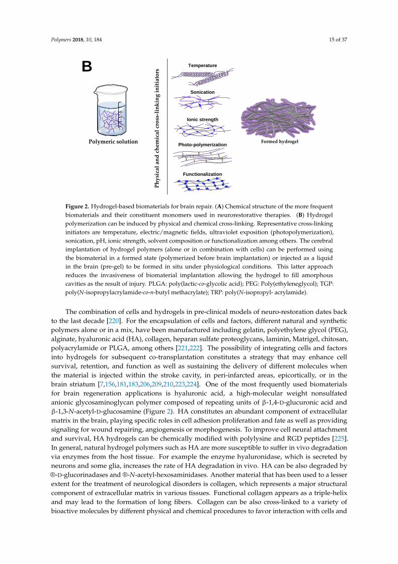
Figure 2. Hydrogel-based biomaterials for brain repair. (A) Chemical structure of the more frequent
biomaterials and their constituent monomers used in neurorestorative therapies. (B) Hydrogel
polymerization can be induced by physical and chemical cross-linking. Representative cross-linking
initiators are temperature, electric/magnetic fields, ultraviolet exposition (photopolymerization),
sonication, pH, ionic strength, solvent composition or functionalization among others. The cerebral
implantation of hydrogel polymers (alone or in combination with cells) can be performed using the
biomaterial in a formed state (polymerized before brain implantation) or injected as a liquid in the
brain (pre-gel) to be formed in situ under physiological conditions. This latter approach reduces the
invasiveness of biomaterial implantation allowing the hydrogel to fill amorphous cavities as the
result of injury. PLGA: poly(lactic-co-glycolic acid); PEG: Poly(ethyleneglycol); TGP:
poly(N-isopropylacrylamide-co-n-butyl methacrylate); TRP: poly(N-isopropyl- acrylamide).
The combination of cells and hydrogels in pre-clinical models of neuro-restoration dates back
to the last decade [220]. For the encapsulation of cells and factors, different natural and synthetic
polymers alone or in a mix, have been manufactured including gelatin, polyethylene glycol (PEG),
alginate, hyaluronic acid (HA), collagen, heparan sulfate proteoglycans, laminin, Matrigel, chitosan,
polyacrylamide or PLGA, among others [221,222]. The possibility of integrating cells and factors
into hydrogels for subsequent co-transplantation constitutes a strategy that may enhance cell
survival, retention, and function as well as sustaining the delivery of different molecules when the
material is injected within the stroke cavity, in peri-infarcted areas, epicortically, or in the brain
striatum [7,156,181,183,206,209,210,223,224]. One of the most frequently used biomaterials for brain
regeneration applications is hyaluronic acid, a high-molecular weight nonsulfated anionic
glycosaminoglycan polymer composed of repeating units of β-1,4-D-glucuronic acid and
β-1,3-N-acetyl-D-glucosamine (Figure 2). HA constitutes an abundant component of extracellular
matrix in the brain, playing specific roles in cell adhesion proliferation and fate as well as providing
signaling for wound repairing, angiogenesis or morphogenesis. To improve cell neural attachment
and survival, HA hydrogels can be chemically modified with polylysine and RGD peptides [225]. In
general, natural hydrogel polymers such as HA are more susceptible to suffer in vivo degradation
via enzymes from the host tissue. For example the enzyme hyaluronidase, which is secreted by
neurons and some glia, increases the rate of HA degradation in vivo. HA can be also degraded by
® -D-glucorinadases and ® -N-acetyl-hexosaminidases. Another material that has been used to a
lesser extent for the treatment of neurological disorders is collagen, which represents a major
structural component of extracellular matrix in various tissues. Functional collagen appears as a
triple-helix and may lead to the formation of long fibers. Collagen can be also cross-linked to a
variety of bioactive molecules by different physical and chemical procedures to favor interaction
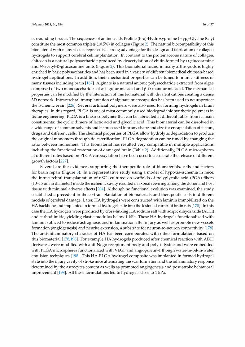
with cells and surrounding tissues. The sequences of amino acids Proline (Pro)-Hydroxyproline
Figure 2. Hydrogel-based biomaterials for brain repair. (A) Chemical structure of the more frequentbiomaterials and their constituent monomers used in neurorestorative therapies. (B) Hydrogelpolymerization can be induced by physical and chemical cross-linking. Representative cross-linkinginitiators are temperature, electric/magnetic fields, ultraviolet exposition (photopolymerization),sonication, pH, ionic strength, solvent composition or functionalization among others. The cerebralimplantation of hydrogel polymers (alone or in combination with cells) can be performed usingthe biomaterial in a formed state (polymerized before brain implantation) or injected as a liquidin the brain (pre-gel) to be formed in situ under physiological conditions. This latter approachreduces the invasiveness of biomaterial implantation allowing the hydrogel to fill amorphouscavities as the result of injury. PLGA: poly(lactic-co-glycolic acid); PEG: Poly(ethyleneglycol); TGP:poly(N-isopropylacrylamide-co-n-butyl methacrylate); TRP: poly(N-isopropyl- acrylamide).
The combination of cells and hydrogels in pre-clinical models of neuro-restoration dates backto the last decade [220]. For the encapsulation of cells and factors, different natural and syntheticpolymers alone or in a mix, have been manufactured including gelatin, polyethylene glycol (PEG),alginate, hyaluronic acid (HA), collagen, heparan sulfate proteoglycans, laminin, Matrigel, chitosan,polyacrylamide or PLGA, among others [221,222]. The possibility of integrating cells and factorsinto hydrogels for subsequent co-transplantation constitutes a strategy that may enhance cellsurvival, retention, and function as well as sustaining the delivery of different molecules whenthe material is injected within the stroke cavity, in peri-infarcted areas, epicortically, or in thebrain striatum [7,156,181,183,206,209,210,223,224]. One of the most frequently used biomaterialsfor brain regeneration applications is hyaluronic acid, a high-molecular weight nonsulfatedanionic glycosaminoglycan polymer composed of repeating units of β-1,4-D-glucuronic acid andβ-1,3-N-acetyl-D-glucosamine (Figure 2). HA constitutes an abundant component of extracellularmatrix in the brain, playing specific roles in cell adhesion proliferation and fate as well as providingsignaling for wound repairing, angiogenesis or morphogenesis. To improve cell neural attachmentand survival, HA hydrogels can be chemically modified with polylysine and RGD peptides [225].In general, natural hydrogel polymers such as HA are more susceptible to suffer in vivo degradationvia enzymes from the host tissue. For example the enzyme hyaluronidase, which is secreted byneurons and some glia, increases the rate of HA degradation in vivo. HA can be also degraded by®-D-glucorinadases and ®-N-acetyl-hexosaminidases. Another material that has been used to a lesserextent for the treatment of neurological disorders is collagen, which represents a major structuralcomponent of extracellular matrix in various tissues. Functional collagen appears as a triple-helixand may lead to the formation of long fibers. Collagen can be also cross-linked to a variety ofbioactive molecules by different physical and chemical procedures to favor interaction with cells and
Polymers 2018, 10, 184 16 of 37
surrounding tissues. The sequences of amino acids Proline (Pro)-Hydroxyproline (Hyp)-Glycine (Gly)constitute the most common triplets (10.5%) in collagen (Figure 2). The natural biocompatibility of thisbiomaterial with many tissues represents a strong advantage for the design and fabrication of collagenhydrogels to support cerebral cell implantation. In contrast to the proteinaceous nature of collagen,chitosan is a natural polysaccharide produced by deacetylation of chitin formed by D-glucosamineand N-acetyl-D-glucosamine units (Figure 2). This biomaterial found in many arthropods is highlyenriched in basic polysaccharides and has been used in a variety of different biomedical chitosan-basedhydrogel applications. In addition, their mechanical properties can be tuned to mimic stiffness ofmany tissues including brain [187]. Alginate is a natural anionic polysaccharide extracted from algaecomposed of two monosaccharides of α-L-guluronic acid and β-D-mannuronic acid. The mechanicalproperties can be modified by the interaction of this biomaterial with divalent cations creating a dense3D network. Intracerebral transplantation of alginate microcapsules has been used to neuroprotectthe ischemic brain [226]. Several artificial polymers were also used for forming hydrogels in braintherapies. In this regard, PLGA is one of most commonly used biodegradable synthetic polymers intissue engineering. PLGA is a linear copolymer that can be fabricated at different ratios from its mainconstituents: the cyclic dimers of lactic acid and glycolic acid. This biomaterial can be dissolved ina wide range of common solvents and be processed into any shape and size for encapsulation of factors,drugs and different cells. The chemical properties of PLGA allow hydrolytic degradation to producethe original monomers through de-esterification. PLGA degradation can be tuned by changing theratio between monomers. This biomaterial has resulted very compatible in multiple applicationsincluding the functional restoration of damaged brain (Table 2). Additionally, PLGA microspheresat different rates based on PLGA carboxylation have been used to accelerate the release of differentgrowth factors [227].
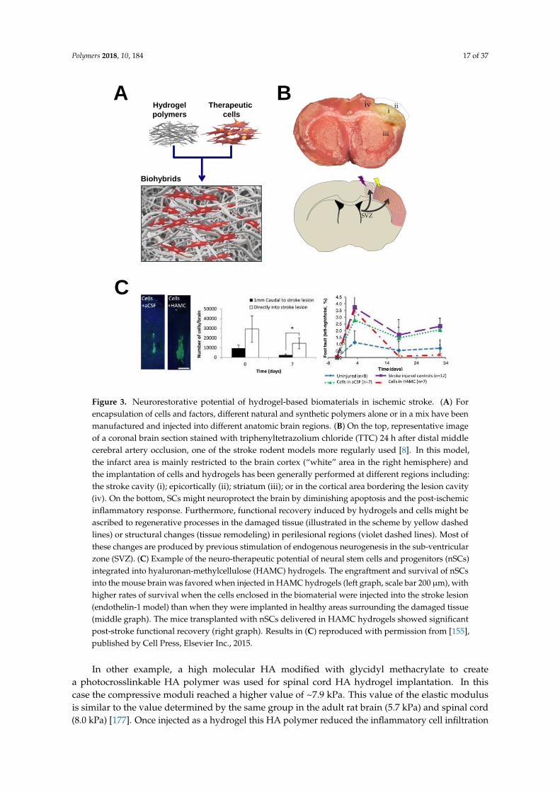
Several are the evidences supporting the therapeutic role of biomaterials, cells and factorsfor brain repair (Figure 3). In a representative study using a model of hypoxia-ischemia in mice,the intracerebral transplantation of nSCs cultured on scaffolds of polyglycolic acid (PGA) fibers(10–15 µm in diameter) inside the ischemic cavity resulted in axonal rewiring among the donor and hosttissue with minimal adverse effects [204]. Although no functional evolution was examined, the studyestablished a precedent in the co-transplantation of biomaterials and therapeutic cells in differentmodels of cerebral damage. Later, HA hydrogels were constructed with laminin immobilized on theHA backbone and implanted in formed hydrogel state into the lesioned cortex of brain rats [178]. In thiscase the HA hydrogels were produced by cross-linking HA sodium salt with adipic dihydrazide (ADH)and carbodiimide, yielding elastic modulus below 1 kPa. These HA hydrogels functionalized withlaminin sufficed to reduce astrogliosis and inflammation after injury as well as promote new vesselsformation (angiogenesis) and neurite extension, a substrate for neuron-to-neuron connectivity [178].The anti-inflammatory character of HA has been corroborated with other formulations based onthis biomaterial [178,198]. For example HA hydrogels produced after chemical reaction with ADHderivates, were modified with anti-Nogo receptor antibody and poly-L-lysine and were embeddedwith PLGA microspheres functionalized with VEGF and angiopoietin-1 though water-in-oil-in-wateremulsion techniques [198]. This HA-PLGA hydrogel composite was implanted in formed hydrogelstate into the injury cavity of stroke mice attenuating the scar formation and the inflammatory responsedetermined by the astrocytes content as wells as promoted angiogenesis and post-stroke behavioralimprovement [198]. All these formulations led to hydrogels close to 1 kPa.
Polymers 2018, 10, 184 17 of 37Polymers 2018, 10, x FOR PEER REVIEW 17 of 37
A BHydrogel
polymers
Therapeutic
cells
Biohybrids
C
iiv
iii
ii
SVZ
Figure 3. Neurorestorative potential of hydrogel-based biomaterials in ischemic stroke. (A) For
encapsulation of cells and factors, different natural and synthetic polymers alone or in a mix have
been manufactured and injected into different anatomic brain regions. (B) On the top, representative
image of a coronal brain section stained with triphenyltetrazolium chloride (TTC) 24 h after distal
middle cerebral artery occlusion, one of the stroke rodent models more regularly used [8]. In this
model, the infarct area is mainly restricted to the brain cortex (“white” area in the right hemisphere)
and the implantation of cells and hydrogels has been generally performed at different regions
including: the stroke cavity (i); epicortically (ii); striatum (iii); or in the cortical area bordering the
lesion cavity (iv). On the bottom, SCs might neuroprotect the brain by diminishing apoptosis and
the post-ischemic inflammatory response. Furthermore, functional recovery induced by hydrogels
and cells might be ascribed to regenerative processes in the damaged tissue (illustrated in the
scheme by yellow dashed lines) or structural changes (tissue remodeling) in perilesional regions
(violet dashed lines). Most of these changes are produced by previous stimulation of endogenous
neurogenesis in the sub-ventricular zone (SVZ). (C) Example of the neuro-therapeutic potential of
neural stem cells and progenitors (nSCs) integrated into hyaluronan-methylcellulose (HAMC)
hydrogels. The engraftment and survival of nSCs into the mouse brain was favored when injected in
HAMC hydrogels (left graph, scale bar 200 μm), with higher rates of survival when the cells
enclosed in the biomaterial were injected into the stroke lesion (endothelin-1 model) than when they
were implanted in healthy areas surrounding the damaged tissue (middle graph). The mice
transplanted with nSCs delivered in HAMC hydrogels showed significant post-stroke functional
recovery (right graph). Results in (C) reproduced with permission from [155], published by Cell
Press, Elsevier Inc., 2015.
In other example, a high molecular HA modified with glycidyl methacrylate to create a
photocrosslinkable HA polymer was used for spinal cord HA hydrogel implantation. In this case
the compressive moduli reached a higher value of ~7.9 kPa. This value of the elastic modulus is
similar to the value determined by the same group in the adult rat brain (5.7 kPa) and spinal cord
(8.0 kPa) [177]. Once injected as a hydrogel this HA polymer reduced the inflammatory cell
Figure 3. Neurorestorative potential of hydrogel-based biomaterials in ischemic stroke. (A) Forencapsulation of cells and factors, different natural and synthetic polymers alone or in a mix have beenmanufactured and injected into different anatomic brain regions. (B) On the top, representative imageof a coronal brain section stained with triphenyltetrazolium chloride (TTC) 24 h after distal middlecerebral artery occlusion, one of the stroke rodent models more regularly used [8]. In this model,the infarct area is mainly restricted to the brain cortex (“white” area in the right hemisphere) andthe implantation of cells and hydrogels has been generally performed at different regions including:the stroke cavity (i); epicortically (ii); striatum (iii); or in the cortical area bordering the lesion cavity(iv). On the bottom, SCs might neuroprotect the brain by diminishing apoptosis and the post-ischemicinflammatory response. Furthermore, functional recovery induced by hydrogels and cells might beascribed to regenerative processes in the damaged tissue (illustrated in the scheme by yellow dashedlines) or structural changes (tissue remodeling) in perilesional regions (violet dashed lines). Most ofthese changes are produced by previous stimulation of endogenous neurogenesis in the sub-ventricularzone (SVZ). (C) Example of the neuro-therapeutic potential of neural stem cells and progenitors (nSCs)integrated into hyaluronan-methylcellulose (HAMC) hydrogels. The engraftment and survival of nSCsinto the mouse brain was favored when injected in HAMC hydrogels (left graph, scale bar 200 µm), withhigher rates of survival when the cells enclosed in the biomaterial were injected into the stroke lesion(endothelin-1 model) than when they were implanted in healthy areas surrounding the damaged tissue(middle graph). The mice transplanted with nSCs delivered in HAMC hydrogels showed significantpost-stroke functional recovery (right graph). Results in (C) reproduced with permission from [155],published by Cell Press, Elsevier Inc., 2015.
In other example, a high molecular HA modified with glycidyl methacrylate to createa photocrosslinkable HA polymer was used for spinal cord HA hydrogel implantation. In thiscase the compressive moduli reached a higher value of ~7.9 kPa. This value of the elastic modulusis similar to the value determined by the same group in the adult rat brain (5.7 kPa) and spinal cord(8.0 kPa) [177]. Once injected as a hydrogel this HA polymer reduced the inflammatory cell infiltration
Polymers 2018, 10, 184 18 of 37
and gliosis on the surrounding tissue after spinal cord injury. In another study, a biodegradableporous gelatin-siloxane 3D scaffold formed by the integration of gelatin and 3-glycidoxypropyltrimethoxysilane was used to provide the gradual secretion of EGF/FGF neurothrophic factors [228].When this material was implanted alone into the brain, the inflammatory response determined bythe microglia content was minimal, thus supporting the biocompatibility of this biomaterial scaffold.The combined implantation of EGF/FGF with this biohybrid promoted tissue regeneration inferred bythe accumulation of 5-Bromo-2′-deoxyuridine-proliferative cells, dendrite elongation, and endothelialvascular cell expansion, suggestive of active angiogenesis [228].
Table 2. In vivo studies using different cell populations in combination with distinct biomaterials torestore the functionality loss after brain stroke.
Stroke model,specie
Biomaterial, cell population,site of implantation Therapeutic effects References
CCAO, mouse PGA, nSCs, infarct cavity Increasing axonal rewiring, reduction ofinflammation and glial scar formation [204]
MCAO, mouse TGP, mSCs, brain surfaceIncreasing engraftment of transplanted cells,
increasing neuronal differentiation, nofunctional improvement
[181]
MCAO, mouse HA alone, infarct cavityReduction of inflammation and glial scar,
enhanced perilesional vascularization,stimulation of endogenous neurogenesis
[183]
MCAO, rat PLGA, nSCs, infarct cavity Cavity size reduction, increasing engraftmentof transplanted cells, de novo tissue formation [156]
MCAO, rat Matrigel, eSCs-nPCs, infarct cavity
Reduction in lesion size, increasing survival oftransplanted cells, neuronal and astroglial
differentiation, neuronal migration,improvement of behavioral outcome
[135]
MCAO, rat Col I , nSCs, infarct cavity
Increasing survival of transplanted cells,neuronal differentiation, increasing
synaptogenesis, improvement ofbehavioral outcome
[229]
MCAO, rat PLGA-VEGF, nSCs, infarct cavity Hipervascularization linked with astrocyticdifferentiation, limited neuronal commitment [230]
MCAO, rat TRP, mSCs, brain surface Improvement of motor function [231]
PTS, mouse HA, iPSCs –nPCs, infarct cavity
Enhancing survival of transplanted cells,reduction of post-stroke inflammation,
increasing neuronal andastrocytic differentiation
[180,232]
PTS, mouse HA-heparin-Col, eSCs-nPCs,infarct cavity
Increasing survival of transplanted cells,reduction of inflammation and glial scar [233]
ET-1, mouse HAMC, nSCs, infarct cavity
Increasing survival of transplanted cells,astrocytic differentiation, limited neuronal andoligodendrocyte commitment, improvement of
behavioral outcome
[155]
ET-1, ratSelf-assembling IKVAV peptide(laminin epitope), eSCs-nPCs,
infarct cavity
Increasing survival of transplanted cells, tissueregeneration, cell adhesion and axonal growth,
improvement of behavioral outcome[184]
CCAO: common carotid artery occlusion stroke model; MCAO: middle cerebral artery occlusion strokemodel; PTS: Photothrombotic stroke model; ET-1: endothelin-1 stroke model; PGA: poly(glycolic acid); PLGA:poly(lactic-co-glycolic acid); Col I: collagen type I; HA: hyaluronic acid; HAMC: blend of hyaluronan andmethylcellulose; TGP: thermoreversible polymer block poly(N-isopropylacrylamide-co-n-butyl methacrylate);TRP: thermoreversible poly(N-isopropyl- acrylamide); IKVAV: Ile-Lys-Val-Ala-Val peptide; nSCs: neural stemcells; nPCs: neural progenitor cells; mSCs: mesenchymal stem cells; iPSCs: induced pluripotent stem cells;eSCs: embryonic stem cells; VEGF: Vascular endothelial growth factor.
As a result of the promising outlook of the therapeutic approach, an increasing number ofbiomaterials in different formats have been assayed and extended in a wide variety of experimentalbrain damage models in the last decade (Table 2). Some of the most salient outcomes in neurorestorativetherapies for stroke using advanced polymers in combination with different therapeutic cells aresummarized below. For example, Jin et al. showed in rats that transplantation in the infarcted cavityof human eSCs-derived neuronal progenitors mixed with a suspension of liquid Matrigel for in
Polymers 2018, 10, 184 19 of 37
situ gelation at body temperature improved sensorimotor, memory, and learning deficits after focalischemia induced by occlusion of the middle cerebral artery [135]. Only the exclusive combination ofcells and Matrigel was associated with a reduced cavity size, better survival of transplanted cells whenintegrated into the biomaterial and improvement of functional outcome. Although the experimentaltreatment was initiated three weeks after infarction, the behavioral improvement observed in theanimals suggest that it is possible to rescue the brain damage at even longer times after injury,when the infarction and functional deficits has been definitively established and the inflammationis resolved [135]. In other study, injectable PLGA microparticles were fabricated through singleoil-in-water emulsion technique and functionalized with allylamine and coated with fibronectin toprovide structural support for nSCs. Once implanted into the stroke cavity new cerebral tissue wasgenerated and cell morphology and behavior of transplanted cells was dependent on cell positionrelative to the infarct core, suggesting that the inhospitable core influenced on transplanted cellsdifferentially with respect to the effect exerted by the perilesional tissue [156]. When these PLGAmicroparticles were modified with VEGF, endogenous endothelial cells chemo-attraction, migrationand proliferation were observed to develop new vascular networks that might sustain the function ofa de novo tissue within the stroke cavity [230]. A different combination of thiol-modified sodium HAcross-linked with heparin sulfate, collagen (gelatin) and polyethylene glycol diacrylate has been usedto transplant eCS-derived neural progenitors into the lesion cavity of stroke mice [233]. This hydrogelmatrix was able to polymerize at body temperature after brain injection. Although the mechanicalproperties of this biomaterial were not characterized in this study, it was shown that this mixtureof polymers did not cause deformation of the brain after transplantation suggesting that the elasticproperties were in the range of cerebral tissue. In addition, the hydrogel promoted the survival oftransplanted cells and their differentiation into astrocytes and neurons in vivo. Another interestingfinding was that the hydrogel alone diminished the post-stroke inflammatory reaction, a remarkablecharacteristic of HA-based biomaterials [233]. In yet another study, HA hydrogels with storagemodulus in the range of 0.3–0.8 kPa favored the in vitro neural differentiation of iPSCs-derivednSCs [179]. In the in vivo context the same group used HA hydrogels functionalized througha Michael addition of acrylates present in the HA backbone with di-cysteins present in a matrixmetalloproteinase degradable peptides [180]. These combined HA hydrogels presented a storagemodulus of 0.3 kPa. When cerebrally injected in liquid format in combination with iPSCs-derivednSCs for in situ polymerization into the infarct cavity, the inflammatory response exerted by thisHA-based biomaterial was minimal, in contrast to the greater inflammatory response observed aftertransplantation of HA polymers with storage moduli in excess of 1.0 kPa [180]. Thus, it is accepted thatmechanical characteristics of hydrogels influence the degree of biocompatibility of the biomaterial andits integration into the host tissue [224], and stiffness properties close to the brain tissue are generallyassociated with lower inflammation and/or minor toxic effects. Despite this HA hydrogel causedminimal inflammation however it did not enhance survival of transplanted cells although significantneuronal differentiation was promoted [180]. Similarly, a fast (~10 min) in situ gelation polymer-basedhydrogel composed of acrylated-HA, matrix metalloproteinase degradable and non-degradablemotifs, adhesion peptides and heparin functionalized with thiol groups was used for cerebralimplantation of iPSCs-derived nSCs [232]. This HA-hydrogel, with an elastic modulus in the range of0.1–1.0 kPa, favored the survival of iPSCs-derived nSCs promoting glial and neuronal differentiation.By contrast, a copolymer of poly(N-isopropylacrylamide) and poly(ethyleneglycol) has been usedto satisfactorily engraft mSCs into the mouse brain. This thermosensitive hydrogel with a sol/geltransition temperature of ~20 ◦C and storage modulus in the range of 0.5–1.0 kPa was implanted asa fully formed hydrogel over the brain cortex surface. Although no significant post-stroke recoverywas observed after treatment in the interval of eight weeks of analysis, no significant inflammatoryresponse induced by this specific copolymer was detected either [181]. In contrast, post-strokefunctional recovery was efficiently enhanced in rats after epicortical transplantation of a anotherreversible temperature-responsive polymer (cell-sheet format) based on poly(N-isopropylacrylamide)
Polymers 2018, 10, 184 20 of 37
in combination with mesenchymal progenitor stromal cells [231]. In another study with rats,nSCs were cerebrally transplanted together with porous 3D collagen sponges, which enhanced thesurvival of engrafted cells and synapse formation as well as improved neurological function afterstroke [229]. An interesting observation with this study was that the collagen degraded completelythirty days after the biomaterial implantation, which might suppose a limitation with this biomaterial ifpersistent therapeutic stimulation is needed. Other combinations of biomaterials have been also tested.For example, a fast-gelling injectable in situ hydrogel mixture of HA modified with acetic hydrazidethrough carbodiimide chemistry and methylcellulose (MC), favored nSCs engraftment in the damagedbrain, enhancing post-stroke motor recovery in an endothelin-1 mouse stroke model [155] (Figure 3).In addition, the transplanted nSCs differentiated into astrocytes and to a minor extent gave neuronsand myelinating oligodendrocytes [155]. Although the mechanical properties of this hydrogel matrixwere not assessed in this study, polymers with the same composition (HA and MC), but with slightlydifferent proportions, showed elastic moduli inferior to 0.1 kPa [182]. In contrast, a blend of HA andMC injectable hydrogel, that was tested in the same stroke rodent model (endothelin-1), did not resultin better survival of cerebrally transplanted iPS-derived neuroepithelial stem cells. It was hypothesizedthat the lack of a therapeutic effect might be attributable to the stresses produced by the flow of the cellsalong the needle, in addition to the extensional flow of cells in the transition syringe/needle duringcerebral injection [200]. Very recently an interesting finding has been reported after the implantation ofan advanced injectable porous HA hydrogel in the stroke cavity of infarcted mice after middle cerebralartery occlusion [183]. In a similar way to [180], this hydrogel was synthesized through carbodiimidechemistry to introduce acrylamide groups on the HA backbone. The biomaterial was functionalizedwith several peptides (including the RGD tri-peptide) and then cross-linked through Michael-typeaddition using di-cysteins present in matrix metalloproteinase degradable peptides giving a hydrogelscaffold with an elastic modulus of 1.5 kPa [183]. One difference with HA hydrogels of limited-porosityis that the implantation of this porous HA hydrogels was translated into a post-stroke reduced scarthickness, decreasing inflammatory microglia, and enhanced perilesional vascularization. In parallel,these porous HA gels stimulated the infiltration and migration of endogenous neural progenitorscells from the SVZ (neurogenic niche) towards the perilesional and stroke cavity regions, which couldbe related to an increasing infiltration of astrocytes [183]. Recently, a self-assembling peptide-basedscaffold composed of repeating units of the IKVAV peptide sequence present in the major ECM proteinlaminin has been engineered [184]. This binding domain sequence (IKVAV) has been involved inneuronal adhesion, migration, proliferation differentiation and neurite guidance and outgrowth [234].The IKVAV-injectable hydrogel with rheological elastic shear moduli below 1.0 kPa was implantedtogether with human eSCs-derived cortical progenitors into the lesion cavity of stroke rats (endothelin-1model). This approach favored long-term survival of eSCs-derived cortical progenitors as well aspromoted neuroprotection, cell replacement through the appearance of de novo tissue and supportedcell adhesion and axonal growth, all of them mechanisms that contributed to enhance post-ischemiafunctional recovery [184]. The IKVAV sequence has been also previously used to functionalizeHA-based polymers [185]. In this case, IKVAV peptides and metalloproteinase were immobilizedto acryl groups of acrylated-HA through Michael type reaction giving HA hydrogels with elasticmodulus in the range of 0.5–1.6 kPa. These copolymers were mixed with BDNF through electrostaticinteractions to sustain BNDF release, and were also combined with human mSCs, enhancing nerveregeneration and recovery in a model of spinal cord injury in rats [185]. The biomaterial compositionand stiffness of the implanted hydrogels not only affects the macrophages/microglia infiltration(a measure of inflammation) modifying the microglia M1/M2 balance [186], but also impacts on the insitu formation and retention of hydrogels in the stroke cavity, as it has been for example demonstratedin rats transplanted with an injectable ECM urinary bladder matrix-based hydrogel with storagemodulus in the range of 0.07–0.4 kPa [186]. In this case, injection of higher ECM concentrations(>3.0 mg/mL) gelled within the stroke cavity, resulting in an extensive distribution and retention ofthe biomaterial within the cavity while reduced ECM concentrations were associated with permeation
Polymers 2018, 10, 184 21 of 37
of ECM into the perilesional tissue [186,235]. Interestingly, when higher concentrations of this ECMhydrogel (>8.0 mg/mL) were injected into the stroke cavity, the biomaterial provided structuralsupport to the surrounding healthy host brain tissue reducing the lesion cavity [235]. Collectively,these studies help us understand the importance of the design of a hydrogel with a defined structureand composition to provide a directed interaction with the cerebral microenvironment upon thepathological state resulting in minor damage and enhancing repair after injury. The applicability ofstem cells and biomaterials has been tested in other neurological diseases of sudden onset, for example,traumatic brain injury (TBI). In rats, one week after TBI, the transplantation of a biodegradableinjectable collagen gel favored donor mSCs survival and retention in the lesion core [236]. AlthoughmSCs alone enhanced functional recovery, this effect was potentiated with the inclusion of this cellpopulation in the biomaterial. In other example, the survival of nSCs was potentiated across eightweeks when this cell population was transplanted into the lesion cavity together with laminin/collagen-or fibronectin/collagen-based scaffolds with in situ gelation [237]. Similarly, this strategy inducedprogressive behavioral improvement after TBI in mice [237].
Across the different studies, the benefits of implanting SCs together with different biomaterialshas been related to the modification (reduction) of post-stroke damaged areas; the formation of newvessels (angiogenesis) in the infarcted and peri-infarcted regions; or in the neuro-restitution andplasticity phenomena. In the case of angiogenesis, it is extensively accepted that vascular formationstimulates endogenous recovery mechanisms, creating an adequate microenvironment for endogenousproliferation migration and differentiation, thus supporting neural restitution, synaptogenesis, axonalgrowth, and neuro-plasticity [238]. These main therapeutic mechanisms are probably the result ofthe increasing survival and longer retention of implanted cells in the brain tissue, which is assistedby the different biomaterials used, favoring a persistent release of different neurotrophic factorsresponsible for the observed effects. Alternative approaches based on the direct implantation ofdifferent neurotherapeutic factors and hydrogels have been also developed and studied [239,240].For example, two inducers of angiogenesis, VEGF and angiopoietin-1, have been implanted togetherwith HA and PLGA in stroke models [198]. An enhanced stimulation of endogenous nSCs in parallelwith the proliferation of neural progenitors have been found after the controlled release of cyclosporin Avia a combination of HA and MC applied epicortically in rats submitted to stroke [7]. Post-strokeneurogenesis and axonal connectivity in the perilesional area promoted motor recovery after thecontrolled release of BDNF through HA hydrogels [206]. In another example, a hyaluronan/heparansulfate hydrogel was used to control the delivery of antibodies against ephrin A, enhancing axonalsprouting and functional recovery [145].
4.2. Toxicity and Adverse Concerns
A deep reflection on the possible clinical applicability of the synthetic and/or natural biomaterialsthat are being used in the current brain damage models is necessary given that, with most biomaterials,the short- and long-term adverse effects derived from the implant into the brain are unknown.Many studies have focused on examining the therapeutic effects of a particular biomaterial alone or incombination with different cell populations without considering the analysis of additional cytotoxicityand inflammation induced by the material itself. In many cases, the compatibility of the biomaterialswith brain-derived neural cell populations has been tested exclusively in vitro. However, in vitrostudies offer a reductionist view of the tolerability of biomaterials with respect to in vivo approacheswhere it is possible to examine how the biomaterial can be integrated with the brain analyzing thehost bioresponse towards the graft. For example, HA, one of the most frequently used biomaterialsfor experimental brain repair, does not produce significant astrogliosis, microgliosis, or increaseneuronal death in the normal brain [145]. In fact, some tunable HA hydrogels with non-restrictedpermeability reduced the post-stroke inflammatory response [183]. However certain fragments ofhyaluronan can trigger the innate immune response activating alloimmunity [241]. In this context,any biomaterial should be non-toxic both as a fully polymerized hydrogel and as any by-product that
Polymers 2018, 10, 184 22 of 37
may be generated during its degradation. In this regard, the accumulation of HA has been relatedto aging processes and demyelinating diseases such as multiple sclerosis. Some forms of hyaluronanproduced by astrocytes accumulate in demyelinated lesions avoiding the remyelination of neuronalcircuits after brain damage [242,243]. Although a clear positive effect of HA promoting post-strokerecovery (including the reduction of inflammation) has been reported, reasonable doubts emerge withthe usefulness of this biomaterial for application in other neurological disorders when consideringthe clinical perspective. As discussed previously, other biomaterials with proven neuro-therapeuticpotential are collagen, chitosan, PLGA, or Matrigel. However, the use of collagen as a biomedicalimplant raises safety concerns related to the possible contamination of this biomaterial with viruses andprions, the latter related with neurodegenerative diseases such as bovine spongiform encephalopathy.Collagen is difficult to obtain due to its limited sources and can be degraded easily, which is a handicapif a prolonged neurotherapeutic effect is needed. PLGA degrades into by-products that mightexacerbate inflammation, therefore producing additional brain damage after brain injury [244,245].In the case of chitosan, this biomaterial shows fast degradation and some preparations cause allergicreactions due to residual components [246]. Matrigel, a solubilized mix of ECM proteins extractedfrom the Englebreth-Holm-Swarm mouse sarcoma, is not adequate for clinical application despite theproven experimental therapeutic potential of nSCs embedded in this material to enhance post-strokefunctional improvement [135]. Fibrin hydrogels might support cells adhesion and growth; however,the poor stiffness, fast degradation, and shrinkage of this biomaterial represent a limitation for theclinics. In this context, silk fibroin constitutes an interesting biomaterial for stem cells/factors therapyin brain injuries and neurodegenerative diseases. This flexible and adaptable biomaterial has beenused for years in a variety of biomedical applications [247,248]. We have recently developed anin situ fast-gelling silk fibroin hydrogel through the sonication-induced of regenerated silk fibroinsolutions [188]. This hydrogel, with elastic modulus in the range of 6–30 kPa [188], favors stem cellsurvival in vitro and in vivo (Figure 4) [249] and can be injected in the brain tissue without causingsignificant inflammatory or neural death responses in an interval of four weeks of analysis [188]. In ourspecific study, after brain-silk-fibroin transplantation, we did not find impairments of sensorimotor andcognitive capacities or sleep-wake disturbances [188]. However, we do not have any evidence regardingwhether this silk fibroin biomaterial might be therapeutically more efficient than other alternativebiomaterials. From our point of view, in terms of clinical applicability, the in vivo biocompatibility ofdifferent materials with brain tissue should not be checked exclusively by the examination of tissularinflammatory or cytotoxicity responses without addressing important aspects of brain functionality.For example, it would be widely recommended to perform animal behavioral testing to examinepossible sensorimotor, memory, and learning abnormalities as a consequence of the biomaterialimplantation or even to further the study of structural and functional connectivity by using anatomictracers, electrophysiology, and brain imaging-based approaches [188,235]. In contrast, there arealso a very limited number of studies comparing the in vivo toxicity of different biomaterials aswell as the therapeutic efficacy under the same brain damage models. In this context one wouldexpect a comparison between different biomaterial candidates which are under study for brainrepair following brain damage. However, no sufficient effort has been made in this sense creatinga lack of consensus with respect to which biomaterials look most promising. In one in vitro study itwas found that nSCs migration, proliferation and differentiation were better supported by lamininmatrices and Matrigel than fibronectin or poly-L-ornithine [250]. In other example, a comparisonbetween different HA and PEG hydrogel formulations was undertaken to examine the survival,growth and differentiation of neural progenitor cells [251]. We do not have enough informationregarding both the optimal site of cerebral implantation and temporal point of intervention to achievethe best cell-biomaterial engraftment/therapeutic effect compromise after brain injury. Post-strokerecovery has been largely demonstrated in a majority of studies implanting cells and biomaterials butfunctional outcome was evaluated with different readouts and distinct brain damage models beingunknown which therapeutic conditions might be more favorable [56]. In most cases, the molecular and
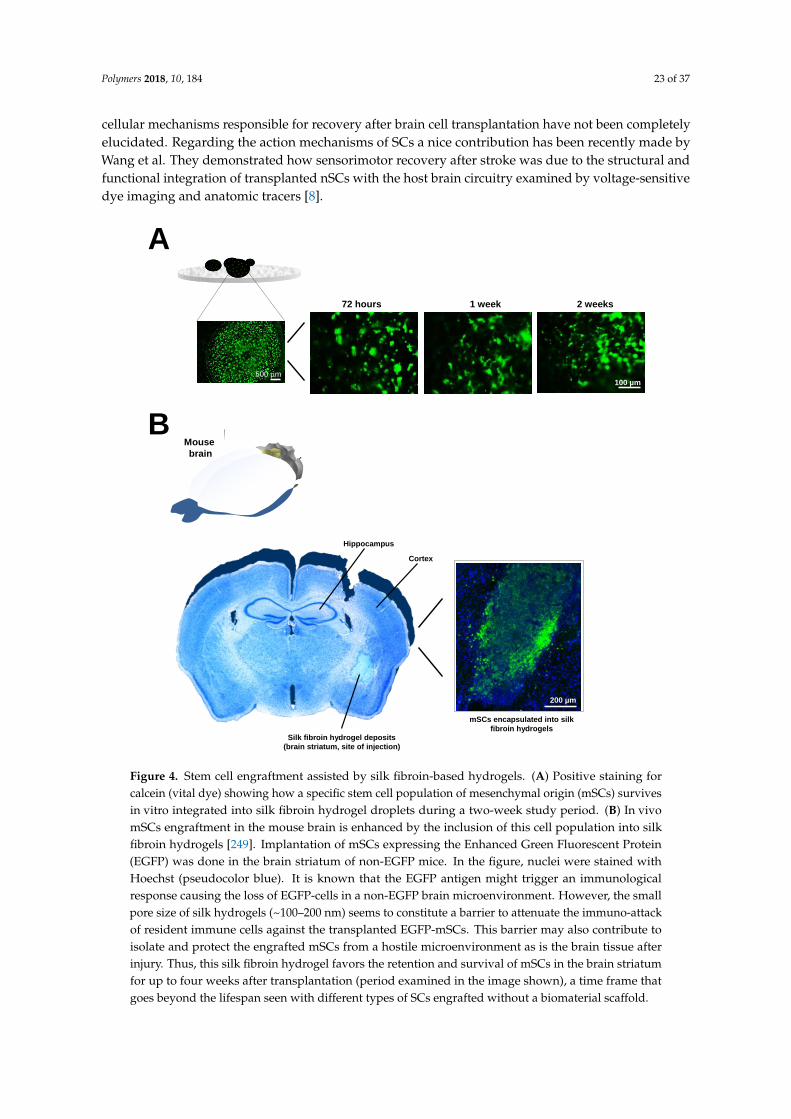
Polymers 2018, 10, 184 23 of 37
cellular mechanisms responsible for recovery after brain cell transplantation have not been completelyelucidated. Regarding the action mechanisms of SCs a nice contribution has been recently made byWang et al. They demonstrated how sensorimotor recovery after stroke was due to the structural andfunctional integration of transplanted nSCs with the host brain circuitry examined by voltage-sensitivedye imaging and anatomic tracers [8].
Polymers 2018, 10, x FOR PEER REVIEW 23 of 37
cells and biomaterials but functional outcome was evaluated with different readouts and distinct
brain damage models being unknown which therapeutic conditions might be more favorable [56].
In most cases, the molecular and cellular mechanisms responsible for recovery after brain cell
transplantation have not been completely elucidated. Regarding the action mechanisms of SCs a
nice contribution has been recently made by Wang et al. They demonstrated how sensorimotor
recovery after stroke was due to the structural and functional integration of transplanted nSCs with
the host brain circuitry examined by voltage-sensitive dye imaging and anatomic tracers [8].
A
BMouse
brain
Hippocampus
Cortex
Silk fibroin hydrogel deposits
(brain striatum, site of injection)
72 hours 1 week
100 µm
2 weeks
500 µm
200 µm
mSCs encapsulated into silk
fibroin hydrogels
Figure 4. Stem cell engraftment assisted by silk fibroin-based hydrogels. (A) Positive staining for
calcein (vital dye) showing how a specific stem cell population of mesenchymal origin (mSCs)
survives in vitro integrated into silk fibroin hydrogel droplets during a two-week study period. (B)
In vivo mSCs engraftment in the mouse brain is enhanced by the inclusion of this cell population
into silk fibroin hydrogels [249]. Implantation of mSCs expressing the Enhanced Green Fluorescent
Protein (EGFP) was done in the brain striatum of non-EGFP mice. In the figure, nuclei were stained
with Hoechst (pseudocolor blue). It is known that the EGFP antigen might trigger an immunological
response causing the loss of EGFP-cells in a non-EGFP brain microenvironment. However, the small
pore size of silk hydrogels (~100–200 nm) seems to constitute a barrier to attenuate the
immuno-attack of resident immune cells against the transplanted EGFP-mSCs. This barrier may also
contribute to isolate and protect the engrafted mSCs from a hostile microenvironment as is the brain
tissue after injury. Thus, this silk fibroin hydrogel favors the retention and survival of mSCs in the
brain striatum for up to four weeks after transplantation (period examined in the image shown), a
time frame that goes beyond the lifespan seen with different types of SCs engrafted without a
biomaterial scaffold.
Figure 4. Stem cell engraftment assisted by silk fibroin-based hydrogels. (A) Positive staining forcalcein (vital dye) showing how a specific stem cell population of mesenchymal origin (mSCs) survivesin vitro integrated into silk fibroin hydrogel droplets during a two-week study period. (B) In vivomSCs engraftment in the mouse brain is enhanced by the inclusion of this cell population into silkfibroin hydrogels [249]. Implantation of mSCs expressing the Enhanced Green Fluorescent Protein(EGFP) was done in the brain striatum of non-EGFP mice. In the figure, nuclei were stained withHoechst (pseudocolor blue). It is known that the EGFP antigen might trigger an immunologicalresponse causing the loss of EGFP-cells in a non-EGFP brain microenvironment. However, the smallpore size of silk hydrogels (~100–200 nm) seems to constitute a barrier to attenuate the immuno-attackof resident immune cells against the transplanted EGFP-mSCs. This barrier may also contribute toisolate and protect the engrafted mSCs from a hostile microenvironment as is the brain tissue afterinjury. Thus, this silk fibroin hydrogel favors the retention and survival of mSCs in the brain striatumfor up to four weeks after transplantation (period examined in the image shown), a time frame thatgoes beyond the lifespan seen with different types of SCs engrafted without a biomaterial scaffold.

Polymers 2018, 10, 184 24 of 37
5. Concluding Remarks
Cures for stroke and other CNS injuries through engineered biomaterials will have a majorpositive impact in heavy and long-term burdens on patients and healthcare systems. The developmentof advanced biomaterials for cerebral damage responds to a major clinical need to provide patientswith an efficient technological solution. Across the last two decades of experimental research, it hasbeen possible to partially restore functionality loss after brain injury by using biomaterials of differentcomposition and format to favor the engraftment of cells with neuro-therapeutic abilities. Despite thevaluable contribution of behavioral studies in examining the improvement of functional outcomesin animals, in many cases, the molecular and cellular substrates underlying recovery are unknown,making it necessary to further investigate these mechanisms to rationally identify those targets whosemodulation through cells and biomaterials might lead to greater therapeutic benefit. In addition,it would also be ideal to advance the screening of those more promising biomaterials such as thosethat are poorly inmmunogenic and less cytotoxic with controlled stiffness properties to facilitate theirintegration with the brain tissue through minimally invasive procedures. Tolerability of hydrogelsshould be examined using a multidimensional approach that could initially include preliminaryin vitro studies, and the subsequent implantation in animals to analyze the in vivo bio-response atboth the structural and functional level. It is also essential to examine concerns regarding the besttemporal point of intervention and site of implantation to achieve an efficient post-brain-damagetherapeutic response.
Although cerebral implantation of biomaterials-based hydrogels has not been applied in humansubjects after brain injury, the antecedents presented in the preclinical (stem cells plus biomaterials)and clinical (stem cells) scenarios outline a magnificent opportunity for the treatment of CNS disordersusing distinct biomaterials. In recent years, the fast evolution of this field predisposes the continuationof the search, characterization, and improvement of advanced hydrogels to clinical translation.
Acknowledgments: The authors apologize to colleagues whose important work was not directly cited due to spacelimitations. We express our gratitude to Masaru Okabe (Osaka University, Japan) and Jose Carlos Segovia (CIEMAT,Madrid, Spain) who donated to us the C57BL/6-Tg(CAG-EGFP)C14-Y01-FM131Osb mice for intracerebraltransplantation of mSCs expressing EGFP. This study was supported by the community of Madrid (grantNeurocentro-B2017/BMD-3760) and funds from the Spanish Ministry of Economy and Competitiveness(MAT2016-79832-R and MAT2015-66666-C3-3-R).
Conflicts of Interest: The authors declare no conflict of interest.
References
1. Boltze, J.; Arnold, A.; Walczak, P.; Jolkkonen, J.; Cui, L.; Wagner, D.C. The Dark Side of the Force—Constraintsand Complications of Cell Therapies for Stroke. Front. Neurol. 2015, 6, 155. [CrossRef] [PubMed]
2. Bao, X.; Feng, M.; Wei, J.; Han, Q.; Zhao, H.; Li, G.; Zhu, Z.; Xing, H.; An, Y.; Qin, C.; et al. Transplantation ofFlk-1+ human bone marrow-derived mesenchymal stem cells promotes angiogenesis and neurogenesis aftercerebral ischemia in rats. Eur. J. Neurosci. 2011, 34, 87–98. [CrossRef] [PubMed]
3. Andres, R.H.; Horie, N.; Slikker, W.; Keren-Gill, H.; Zhan, K.; Sun, G.; Manley, N.C.; Pereira, M.P.; Sheikh, L.A.;McMillan, E.L.; et al. Human neural stem cells enhance structural plasticity and axonal transport in theischaemic brain. Brain 2011, 134, 1777–1789. [CrossRef] [PubMed]
4. Darsalia, V.; Kallur, T.; Kokaia, Z. Survival, migration and neuronal differentiation of human fetal striataland cortical neural stem cells grafted in stroke-damaged rat striatum. Eur. J. Neurosci. 2007, 26, 605–614.[CrossRef] [PubMed]
5. Kelly, S.; Bliss, T.M.; Shah, A.K.; Sun, G.H.; Ma, M.; Foo, W.C.; Masel, J.; Yenari, M.A.; Weissman, I.L.;Uchida, N.; et al. Transplanted human fetal neural stem cells survive, migrate, and differentiate in ischemicrat cerebral cortex. Proc. Natl. Acad. Sci. USA 2004, 101, 11839–11844. [CrossRef] [PubMed]
6. Mora-Lee, S.; Sirerol-Piquer, M.S.; Gutierrez-Perez, M.; Gomez-Pinedo, U.; Roobrouck, V.D.; Lopez, T.;Casado-Nieto, M.; Abizanda, G.; Rabena, M.T.; Verfaille, C.; et al. Therapeutic effects of hMAPC and hMSCtransplantation after stroke in mice. PLoS ONE 2012, 7, e43683. [CrossRef] [PubMed]
Polymers 2018, 10, 184 25 of 37
7. Tuladhar, A.; Morshead, C.M.; Shoichet, M.S. Circumventing the blood-brain barrier: Local delivery ofcyclosporin A stimulates stem cells in stroke-injured rat brain. J. Control. Release 2015, 215, 1–11. [CrossRef][PubMed]
8. Wang, Y.; Zhao, Z.; Rege, S.V.; Wang, M.; Si, G.; Zhou, Y.; Wang, S.; Griffin, J.H.; Goldman, S.A.; Zlokovic, B.V.3K3A-activated protein C stimulates postischemic neuronal repair by human neural stem cells in mice.Nat. Med. 2016, 22, 1050–1055. [CrossRef] [PubMed]
9. Hoag, H. Drug delivery: Brain food. Nature 2014, 510, S6–S7. [CrossRef] [PubMed]10. Benjamin, E.J.; Blaha, M.J.; Chiuve, S.E.; Cushman, M.; Das, S.R.; Deo, R.; de Ferranti, S.D.; Floyd, J.;
Fornage, M.; Gillespie, C.; et al. Heart Disease and Stroke Statistics-2017 Update: A Report From theAmerican Heart Association. Circulation 2017, 135, e146–e603. [CrossRef] [PubMed]
11. Cooke, M.J.; Vulicb, K.; Shoichet, M.S. Design of biomaterials to enhance stem cell survival when transplantedinto the damaged central nervous system. Soft Matter 2010, 6, 4988–4998. [CrossRef]
12. Khaing, Z.Z.; Thomas, R.C.; Geissler, S.A.; Schmidt, C.E. Advanced biomaterials for repairing the nervoussystem: What can hydrogels do for the brain? Materialstoday 2014, 17, 332–340. [CrossRef]
13. Lim, T.C.; Spector, M. Biomaterials for Enhancing CNS Repair. Transl. Stroke Res. 2017, 8, 57–64. [CrossRef][PubMed]
14. Orive, G.; Anitua, E.; Pedraz, J.L.; Emerich, D.F. Biomaterials for promoting brain protection, repair andregeneration. Nat. Rev. Neurosci. 2009, 10, 682–692. [CrossRef] [PubMed]
15. GBD-2013-study. Global, regional, and national age-sex specific all-cause and cause-specific mortality for240 causes of death, 1990–2013: A systematic analysis for the Global Burden of Disease Study 2013. Lancet2015, 385, 117–171. [CrossRef]
16. O’Mahony, P.G.; Thomson, R.G.; Dobson, R.; Rodgers, H.; James, O.F. The prevalence of stroke and associateddisability. J. Public Health Med. 1999, 21, 166–171. [CrossRef] [PubMed]
17. Krishnamurthi, R.V.; Feigin, V.L.; Forouzanfar, M.H.; Mensah, G.A.; Connor, M.; Bennett, D.A.; Moran, A.E.;Sacco, R.L.; Anderson, L.M.; Truelsen, T.; et al. Global and regional burden of first-ever ischaemicand haemorrhagic stroke during 1990–2010: Findings from the Global Burden of Disease Study 2010.Lancet Glob. Health 2013, 1, e259–e281. [CrossRef]
18. Smajlovic, D. Strokes in young adults: Epidemiology and prevention. Vasc. Health Risk Manag. 2015, 11,157–164. [CrossRef] [PubMed]
19. Brott, T.; Bogousslavsky, J. Treatment of acute ischemic stroke. N. Engl. J. Med. 2000, 343, 710–722. [CrossRef][PubMed]
20. Hankey, G.J. Stroke. Lancet 2017, 389, 641–654. [CrossRef]21. Campbell, B.C.; Mitchell, P.J. Endovascular therapy for ischemic stroke. N. Engl. J. Med. 2015, 372, 2365–2366.
[CrossRef] [PubMed]22. Goyal, M.; Menon, B.K.; van Zwam, W.H.; Dippel, D.W.; Mitchell, P.J.; Demchuk, A.M.; Davalos, A.;
Majoie, C.B.; van der Lugt, A.; de Miquel, M.A.; et al. Endovascular thrombectomy after large-vesselischaemic stroke: A meta-analysis of individual patient data from five randomised trials. Lancet 2016, 387,1723–1731. [CrossRef]
23. Saver, J.L.; Goyal, M.; Bonafe, A.; Diener, H.C.; Levy, E.I.; Pereira, V.M.; Albers, G.W.; Cognard, C.; Cohen, D.J.;Hacke, W.; et al. Stent-retriever thrombectomy after intravenous t-PA vs. t-PA alone in stroke. N. Engl.J. Med. 2015, 372, 2285–2295. [CrossRef] [PubMed]
24. Emberson, J.; Lees, K.R.; Lyden, P.; Blackwell, L.; Albers, G.; Bluhmki, E.; Brott, T.; Cohen, G.; Davis, S.;Donnan, G.; et al. Effect of treatment delay, age, and stroke severity on the effects of intravenous thrombolysiswith alteplase for acute ischaemic stroke: A meta-analysis of individual patient data from randomised trials.Lancet 2014, 384, 1929–1935. [CrossRef]
25. George, P.M.; Steinberg, G.K. Novel Stroke Therapeutics: Unraveling Stroke Pathophysiology and Its Impacton Clinical Treatments. Neuron 2015, 87, 297–309. [CrossRef] [PubMed]
26. Corbetta, D.; Imeri, F.; Gatti, R. Rehabilitation that incorporates virtual reality is more effective thanstandard rehabilitation for improving walking speed, balance and mobility after stroke: A systematicreview. J. Physiother. 2015, 61, 117–124. [CrossRef] [PubMed]
27. Laver, K.E.; George, S.; Thomas, S.; Deutsch, J.E.; Crotty, M. Virtual reality for stroke rehabilitation.Cochrane Database Syst. Rev. 2015, CD008349. [CrossRef] [PubMed]
Polymers 2018, 10, 184 26 of 37
28. Levy, R.; Ruland, S.; Weinand, M.; Lowry, D.; Dafer, R.; Bakay, R. Cortical stimulation for the rehabilitation ofpatients with hemiparetic stroke: A multicenter feasibility study of safety and efficacy. J. Neurosurg. 2008,108, 707–714. [CrossRef] [PubMed]
29. Levy, R.M.; Harvey, R.L.; Kissela, B.M.; Winstein, C.J.; Lutsep, H.L.; Parrish, T.B.; Cramer, S.C.;Venkatesan, L. Epidural Electrical Stimulation for Stroke Rehabilitation: Results of the Prospective,Multicenter, Randomized, Single-Blinded Everest Trial. Neurorehabil. Neural Repair 2016, 30, 107–119.[CrossRef] [PubMed]
30. Jaillard, A.; Martin, C.D.; Garambois, K.; Lebas, J.F.; Hommel, M. Vicarious function within the humanprimary motor cortex? A longitudinal fMRI stroke study. Brain 2005, 128, 1122–1138. [CrossRef] [PubMed]
31. Nudo, R.J.; Wise, B.M.; SiFuentes, F.; Milliken, G.W. Neural substrates for the effects of rehabilitative trainingon motor recovery after ischemic infarct. Science 1996, 272, 1791–1794. [CrossRef] [PubMed]
32. Krucoff, M.O.; Rahimpour, S.; Slutzky, M.W.; Edgerton, V.R.; Turner, D.A. Enhancing Nervous SystemRecovery through Neurobiologics, Neural Interface Training, and Neurorehabilitation. Front. Neurosci. 2016,10, 584. [CrossRef] [PubMed]
33. Borlongan, C.V.; Tajima, Y.; Trojanowski, J.Q.; Lee, V.M.; Sanberg, P.R. Transplantation of cryopreservedhuman embryonal carcinoma-derived neurons (NT2N cells) promotes functional recovery in ischemic rats.Exp. Neurol. 1998, 149, 310–321. [CrossRef] [PubMed]
34. Chen, J.; Li, Y.; Chopp, M. Intracerebral transplantation of bone marrow with BDNF after MCAo in rat.Neuropharmacology 2000, 39, 711–716. [CrossRef]
35. Chen, J.; Li, Y.; Wang, L.; Lu, M.; Zhang, X.; Chopp, M. Therapeutic benefit of intracerebral transplantationof bone marrow stromal cells after cerebral ischemia in rats. J. Neurol. Sci. 2001, 189, 49–57. [CrossRef]
36. Chen, J.; Li, Y.; Wang, L.; Zhang, Z.; Lu, D.; Lu, M.; Chopp, M. Therapeutic benefit of intravenousadministration of bone marrow stromal cells after cerebral ischemia in rats. Stroke 2001, 32, 1005–1011.[CrossRef] [PubMed]
37. Li, Y.; Chopp, M.; Chen, J.; Wang, L.; Gautam, S.C.; Xu, Y.X.; Zhang, Z. Intrastriatal transplantation of bonemarrow nonhematopoietic cells improves functional recovery after stroke in adult mice. J. Cereb. BloodFlow MeTable 2000, 20, 1311–1319. [CrossRef] [PubMed]
38. Bang, O.Y.; Lee, J.S.; Lee, P.H.; Lee, G. Autologous mesenchymal stem cell transplantation in stroke patients.Ann. Neurol. 2005, 57, 874–882. [CrossRef] [PubMed]
39. Borlongan, C.V. Preliminary Reports of Stereotaxic Stem Cell Transplants in Chronic Stroke Patients. Mol. Ther.2016, 24, 1710–1711. [CrossRef] [PubMed]
40. Borlongan, C.V. Age of PISCES: Stem-cell clinical trials in stroke. Lancet 2016, 388, 736–738. [CrossRef]41. Kondziolka, D.; Steinberg, G.K.; Wechsler, L.; Meltzer, C.C.; Elder, E.; Gebel, J.; Decesare, S.; Jovin, T.;
Zafonte, R.; Lebowitz, J.; et al. Neurotransplantation for patients with subcortical motor stroke: A phase 2randomized trial. J. Neurosurg. 2005, 103, 38–45. [CrossRef] [PubMed]
42. Kondziolka, D.; Wechsler, L.; Goldstein, S.; Meltzer, C.; Thulborn, K.R.; Gebel, J.; Jannetta, P.; DeCesare, S.;Elder, E.M.; McGrogan, M.; et al. Transplantation of cultured human neuronal cells for patients with stroke.Neurology 2000, 55, 565–569. [CrossRef] [PubMed]
43. Kordower, J.H.; Freeman, T.B.; Snow, B.J.; Vingerhoets, F.J.; Mufson, E.J.; Sanberg, P.R.; Hauser, R.A.;Smith, D.A.; Nauert, G.M.; Perl, D.P.; et al. Neuropathological evidence of graft survival and striatalreinnervation after the transplantation of fetal mesencephalic tissue in a patient with Parkinson’s disease.N. Engl. J. Med. 1995, 332, 1118–1124. [CrossRef] [PubMed]
44. Lee, J.S.; Hong, J.M.; Moon, G.J.; Lee, P.H.; Ahn, Y.H.; Bang, O.Y. A long-term follow-up study of intravenousautologous mesenchymal stem cell transplantation in patients with ischemic stroke. Stem Cells 2010, 28,1099–1106. [CrossRef] [PubMed]
45. Lindvall, O.; Sawle, G.; Widner, H.; Rothwell, J.C.; Bjorklund, A.; Brooks, D.; Brundin, P.; Frackowiak, R.;Marsden, C.D.; Odin, P.; et al. Evidence for long-term survival and function of dopaminergic grafts inprogressive Parkinson’s disease. Ann. Neurol. 1994, 35, 172–180. [CrossRef] [PubMed]
46. Savitz, S.I.; Dinsmore, J.; Wu, J.; Henderson, G.V.; Stieg, P.; Caplan, L.R. Neurotransplantation of fetal porcinecells in patients with basal ganglia infarcts: A preliminary safety and feasibility study. Cerebrovasc. Dis. 2005,20, 101–107. [CrossRef] [PubMed]
Polymers 2018, 10, 184 27 of 37
47. Steinberg, G.K.; Kondziolka, D.; Wechsler, L.R.; Lunsford, L.D.; Coburn, M.L.; Billigen, J.B.; Kim, A.S.;Johnson, J.N.; Bates, D.; King, B.; et al. Clinical Outcomes of Transplanted Modified Bone Marrow-DerivedMesenchymal Stem Cells in Stroke: A Phase 1/2a Study. Stroke 2016, 47, 1817–1824. [CrossRef] [PubMed]
48. Jacobson, L.O.; Simmons, E.L.; Marks, E.K.; Eldredge, J.H. Recovery from radiation injury. Science 1951, 113,510–511. [CrossRef] [PubMed]
49. Lorenz, E.; Uphoff, D.; Reid, T.R.; Shelton, E. Modification of irradiation injury in mice and guinea pigs bybone marrow injections. J. Natl. Cancer Inst. 1951, 12, 197–201. [CrossRef] [PubMed]
50. Thomas, E.D.; Lochte, H.L., Jr.; Lu, W.C.; Ferrebee, J.W. Intravenous infusion of bone marrow in patientsreceiving radiation and chemotherapy. N. Engl. J. Med. 1957, 257, 491–496. [CrossRef] [PubMed]
51. Rama, P.; Matuska, S.; Paganoni, G.; Spinelli, A.; De Luca, M.; Pellegrini, G. Limbal stem-cell therapy andlong-term corneal regeneration. N. Engl. J. Med. 2010, 363, 147–155. [CrossRef] [PubMed]
52. Panes, J. Stem Cell Therapy for Perianal Fistulas in Crohn’s Disease. Gastroenterol Hepatol (N.Y.) 2016, 12,637–640.
53. Hirsch, T.; Rothoeft, T.; Teig, N.; Bauer, J.W.; Pellegrini, G.; De Rosa, L.; Scaglione, D.; Reichelt, J.;Klausegger, A.; Kneisz, D.; et al. Regeneration of the entire human epidermis using transgenic stemcells. Nature 2017, 551, 327–332. [CrossRef] [PubMed]
54. Trounson, A.; McDonald, C. Stem Cell Therapies in Clinical Trials: Progress and Challenges. Cell Stem Cell2015, 17, 11–22. [CrossRef] [PubMed]
55. Abou-El-Enein, M.; Elsanhoury, A.; Reinke, P. Overcoming Challenges Facing Advanced Therapies in theEU Market. Cell Stem Cell 2016, 19, 293–297. [CrossRef] [PubMed]
56. Khalil, D.N.; Smith, E.L.; Brentjens, R.J.; Wolchok, J.D. The future of cancer treatment: Immunomodulation,CARs and combination immunotherapy. Nat. Rev. Clin. Oncol. 2016, 13, 273–290. [CrossRef] [PubMed]
57. Gervois, P.; Wolfs, E.; Ratajczak, J.; Dillen, Y.; Vangansewinkel, T.; Hilkens, P.; Bronckaers, A.; Lambrichts, I.;Struys, T. Stem Cell-Based Therapies for Ischemic Stroke: Preclinical Results and the Potential ofImaging-Assisted Evaluation of Donor Cell Fate and Mechanisms of Brain Regeneration. Med. Res. Rev.2016, 36, 1080–1126. [CrossRef] [PubMed]
58. Lees, J.S.; Sena, E.S.; Egan, K.J.; Antonic, A.; Koblar, S.A.; Howells, D.W.; Macleod, M.R. Stem cell-basedtherapy for experimental stroke: A systematic review and meta-analysis. Int. J. Stroke 2012, 7, 582–588.[CrossRef] [PubMed]
59. Bernstock, J.D.; Peruzzotti-Jametti, L.; Ye, D.; Gessler, F.A.; Maric, D.; Vicario, N.; Lee, Y.J.; Pluchino, S.;Hallenbeck, J.M. Neural stem cell transplantation in ischemic stroke: A role for preconditioning and cellularengineering. J. Cereb. Blood Flow MeTable 2017, 37, 2314–2319. [CrossRef] [PubMed]
60. Dirnagl, U.; Iadecola, C.; Moskowitz, M.A. Pathobiology of ischaemic stroke: An integrated view.Trends Neurosci. 1999, 22, 391–397. [CrossRef]
61. Ramos-Cabrer, P.; Justicia, C.; Wiedermann, D.; Hoehn, M. Stem cell mediation of functional recovery afterstroke in the rat. PLoS ONE 2010, 5, e12779. [CrossRef] [PubMed]
62. Taguchi, A.; Soma, T.; Tanaka, H.; Kanda, T.; Nishimura, H.; Yoshikawa, H.; Tsukamoto, Y.; Iso, H.;Fujimori, Y.; Stern, D.M.; et al. Administration of CD34+ cells after stroke enhances neurogenesis viaangiogenesis in a mouse model. J. Clin. Invest. 2004, 114, 330–338. [CrossRef] [PubMed]
63. Astrup, J.; Symon, L.; Branston, N.M.; Lassen, N.A. Cortical evoked potential and extracellular K+ and H+ atcritical levels of brain ischemia. Stroke 1977, 8, 51–57. [CrossRef] [PubMed]
64. Murphy, T.H.; Corbett, D. Plasticity during stroke recovery: From synapse to behaviour. Nat. Rev. Neurosci.2009, 10, 861–872. [CrossRef] [PubMed]
65. Rallu, M.; Corbin, J.G.; Fishell, G. Parsing the prosencephalon. Nat. Rev. Neurosci. 2002, 3, 943–951. [CrossRef][PubMed]
66. Kaneko, N.; Sawada, M.; Sawamoto, K. Mechanisms of neuronal migration in the adult brain. J. Neurochem.2017, 141, 835–847. [CrossRef] [PubMed]
67. Obernier, K.; Cebrian-Silla, A.; Thomson, M.; Parraguez, J.I.; Anderson, R.; Guinto, C.; Rodas Rodriguez, J.;Garcia-Verdugo, J.M.; Alvarez-Buylla, A. Adult Neurogenesis Is Sustained by Symmetric Self-Renewal andDifferentiation. Cell Stem Cell 2018, 22, 221–234. [CrossRef] [PubMed]
68. Arvidsson, A.; Collin, T.; Kirik, D.; Kokaia, Z.; Lindvall, O. Neuronal replacement from endogenousprecursors in the adult brain after stroke. Nat. Med. 2002, 8, 963–970. [CrossRef] [PubMed]
Polymers 2018, 10, 184 28 of 37
69. Reumers, V.; Deroose, C.M.; Krylyshkina, O.; Nuyts, J.; Geraerts, M.; Mortelmans, L.; Gijsbers, R.;Van den Haute, C.; Debyser, Z.; Baekelandt, V. Noninvasive and quantitative monitoring of adult neuronalstem cell migration in mouse brain using bioluminescence imaging. Stem Cells 2008, 26, 2382–2390. [CrossRef][PubMed]
70. Chu, K.; Kim, M.; Park, K.I.; Jeong, S.W.; Park, H.K.; Jung, K.H.; Lee, S.T.; Kang, L.; Lee, K.; Park, D.K.; et al.Human neural stem cells improve sensorimotor deficits in the adult rat brain with experimental focalischemia. Brain Res. 2004, 1016, 145–153. [CrossRef] [PubMed]
71. Riess, P.; Zhang, C.; Saatman, K.E.; Laurer, H.L.; Longhi, L.G.; Raghupathi, R.; Lenzlinger, P.M.; Lifshitz, J.;Boockvar, J.; Neugebauer, E.; et al. Transplanted neural stem cells survive, differentiate, and improveneurological motor function after experimental traumatic brain injury. Neurosurgery 2002, 51, 1043–1052,discussion 1043–1052. [CrossRef] [PubMed]
72. Zhu, J.M.; Zhao, Y.Y.; Chen, S.D.; Zhang, W.H.; Lou, L.; Jin, X. Functional recovery after transplantationof neural stem cells modified by brain-derived neurotrophic factor in rats with cerebral ischaemia. J. Int.Med. Res. 2011, 39, 488–498. [CrossRef] [PubMed]
73. Hattiangady, B.; Shuai, B.; Cai, J.; Coksaygan, T.; Rao, M.S.; Shetty, A.K. Increased dentate neurogenesis aftergrafting of glial restricted progenitors or neural stem cells in the aging hippocampus. Stem Cells 2007, 25,2104–2117. [CrossRef] [PubMed]
74. Augestad, I.L.; Nyman, A.K.G.; Costa, A.I.; Barnett, S.C.; Sandvig, A.; Haberg, A.K.; Sandvig, I. Effects ofNeural Stem Cell and Olfactory Ensheathing Cell Co-transplants on Tissue Remodelling After TransientFocal Cerebral Ischemia in the Adult Rat. Neurochem. Res. 2017, 42, 1599–1609. [CrossRef] [PubMed]
75. Harms, K.M.; Li, L.; Cunningham, L.A. Murine neural stem/progenitor cells protect neurons against ischemiaby HIF-1alpha-regulated VEGF signaling. PLoS ONE 2010, 5, e9767. [CrossRef] [PubMed]
76. Roitbak, T.; Li, L.; Cunningham, L.A. Neural stem/progenitor cells promote endothelial cell morphogenesisand protect endothelial cells against ischemia via HIF-1alpha-regulated VEGF signaling. J. Cereb. BloodFlow MeTable 2008, 28, 1530–1542. [CrossRef] [PubMed]
77. Bliss, T.; Guzman, R.; Daadi, M.; Steinberg, G.K. Cell transplantation therapy for stroke. Stroke 2007, 38,817–826. [CrossRef] [PubMed]
78. Goldman, S.A. Stem and Progenitor Cell-Based Therapy of the Central Nervous System: Hopes, Hype,and Wishful Thinking. Cell Stem Cell 2016, 18, 174–188. [CrossRef] [PubMed]
79. Wei, L.; Cui, L.; Snider, B.J.; Rivkin, M.; Yu, S.S.; Lee, C.S.; Adams, L.D.; Gottlieb, D.I.; Johnson, E.M., Jr.;Yu, S.P.; et al. Transplantation of embryonic stem cells overexpressing Bcl-2 promotes functional recoveryafter transient cerebral ischemia. Neurobiol. Dis. 2005, 19, 183–193. [CrossRef] [PubMed]
80. Yanagisawa, D.; Qi, M.; Kim, D.H.; Kitamura, Y.; Inden, M.; Tsuchiya, D.; Takata, K.; Taniguchi, T.;Yoshimoto, K.; Shimohama, S.; et al. Improvement of focal ischemia-induced rat dopaminergic dysfunctionby striatal transplantation of mouse embryonic stem cells. Neurosci. Lett. 2006, 407, 74–79. [CrossRef][PubMed]
81. Thomson, J.A.; Itskovitz-Eldor, J.; Shapiro, S.S.; Waknitz, M.A.; Swiergiel, J.J.; Marshall, V.S.; Jones, J.M.Embryonic stem cell lines derived from human blastocysts. Science 1998, 282, 1145–1147. [CrossRef][PubMed]
82. Solter, D. From teratocarcinomas to embryonic stem cells and beyond: A history of embryonic stem cellresearch. Nat. Rev. Genet. 2006, 7, 319–327. [CrossRef] [PubMed]
83. Honmou, O.; Onodera, R.; Sasaki, M.; Waxman, S.G.; Kocsis, J.D. Mesenchymal stem cells: Therapeuticoutlook for stroke. Trends Mol. Med. 2012, 18, 292–297. [CrossRef] [PubMed]
84. Janowski, M.; Walczak, P.; Date, I. Intravenous route of cell delivery for treatment of neurological disorders:A meta-analysis of preclinical results. Stem Cells Dev. 2010, 19, 5–16. [CrossRef] [PubMed]
85. Li, Y.; Chopp, M. Marrow stromal cell transplantation in stroke and traumatic brain injury. Neurosci. Lett.2009, 456, 120–123. [CrossRef] [PubMed]
86. Li, L.; Chao, J.; Shi, Y. Modeling neurological diseases using iPSC-derived neural cells : IPSC modeling ofneurological diseases. Cell Tissue Res. 2017, 371, 143–151. [CrossRef] [PubMed]
87. Seki, T.; Yuasa, S.; Oda, M.; Egashira, T.; Yae, K.; Kusumoto, D.; Nakata, H.; Tohyama, S.; Hashimoto, H.;Kodaira, M.; et al. Generation of induced pluripotent stem cells from human terminally differentiatedcirculating T cells. Cell Stem Cell 2010, 7, 11–14. [CrossRef] [PubMed]
Polymers 2018, 10, 184 29 of 37
88. Takahashi, K.; Yamanaka, S. Induction of pluripotent stem cells from mouse embryonic and adult fibroblastcultures by defined factors. Cell 2006, 126, 663–676. [CrossRef] [PubMed]
89. Tat, P.A.; Sumer, H.; Jones, K.L.; Upton, K.; Verma, P.J. The efficient generation of induced pluripotent stem(iPS) cells from adult mouse adipose tissue-derived and neural stem cells. Cell Transplant. 2010, 19, 525–536.[CrossRef] [PubMed]
90. Chen, S.J.; Chang, C.M.; Tsai, S.K.; Chang, Y.L.; Chou, S.J.; Huang, S.S.; Tai, L.K.; Chen, Y.C.; Ku, H.H.;Li, H.Y.; et al. Functional improvement of focal cerebral ischemia injury by subdural transplantation ofinduced pluripotent stem cells with fibrin glue. Stem. Cells Dev. 2010, 19, 1757–1767. [CrossRef] [PubMed]
91. Jiang, M.; Lv, L.; Ji, H.; Yang, X.; Zhu, W.; Cai, L.; Gu, X.; Chai, C.; Huang, S.; Sun, J.; et al. Inductionof pluripotent stem cells transplantation therapy for ischemic stroke. Mol. Cell Biochem. 2011, 354, 67–75.[CrossRef] [PubMed]
92. Yuan, T.; Liao, W.; Feng, N.H.; Lou, Y.L.; Niu, X.; Zhang, A.J.; Wang, Y.; Deng, Z.F. Human inducedpluripotent stem cell-derived neural stem cells survive, migrate, differentiate, and improve neurologicfunction in a rat model of middle cerebral artery occlusion. Stem Cell Res. Ther. 2013, 4, 73. [CrossRef][PubMed]
93. Kawai, H.; Yamashita, T.; Ohta, Y.; Deguchi, K.; Nagotani, S.; Zhang, X.; Ikeda, Y.; Matsuura, T.; Abe, K.Tridermal tumorigenesis of induced pluripotent stem cells transplanted in ischemic brain. J. Cereb. BloodFlow MeTable 2010, 30, 1487–1493. [CrossRef] [PubMed]
94. Lamm, N.; Ben-David, U.; Golan-Lev, T.; Storchova, Z.; Benvenisty, N.; Kerem, B. Genomic Instability inHuman Pluripotent Stem Cells Arises from Replicative Stress and Chromosome Condensation Defects.Cell Stem Cell 2016, 18, 253–261. [CrossRef] [PubMed]
95. Lister, R.; Pelizzola, M.; Kida, Y.S.; Hawkins, R.D.; Nery, J.R.; Hon, G.; Antosiewicz-Bourget, J.; O’Malley, R.;Castanon, R.; Klugman, S.; et al. Hotspots of aberrant epigenomic reprogramming in human inducedpluripotent stem cells. Nature 2011, 471, 68–73. [CrossRef] [PubMed]
96. Nakano-Doi, A.; Nakagomi, T.; Fujikawa, M.; Nakagomi, N.; Kubo, S.; Lu, S.; Yoshikawa, H.; Soma, T.;Taguchi, A.; Matsuyama, T. Bone marrow mononuclear cells promote proliferation of endogenous neuralstem cells through vascular niches after cerebral infarction. Stem Cells 2010, 28, 1292–1302. [CrossRef][PubMed]
97. Shyu, W.C.; Lin, S.Z.; Chiang, M.F.; Su, C.Y.; Li, H. Intracerebral peripheral blood stem cell (CD34+)implantation induces neuroplasticity by enhancing beta1 integrin-mediated angiogenesis in chronic strokerats. J. Neurosci. 2006, 26, 3444–3453. [CrossRef] [PubMed]
98. Friedenstein, A.J.; Chailakhjan, R.K.; Lalykina, K.S. The development of fibroblast colonies in monolayercultures of guinea-pig bone marrow and spleen cells. Cell Tissue Kinet. 1970, 3, 393–403. [CrossRef] [PubMed]
99. Bianco, P. “Mesenchymal” stem cells. Annu. Rev. Cell Dev. Biol. 2014, 30, 677–704. [CrossRef] [PubMed]100. Gonzalez-Nieto, D.; Li, L.; Kohler, A.; Ghiaur, G.; Ishikawa, E.; Sengupta, A.; Madhu, M.; Arnett, J.L.;
Santho, R.A.; Dunn, S.K.; et al. Connexin-43 in the osteogenic BM niche regulates its cellular composition andthe bidirectional traffic of hematopoietic stem cells and progenitors. Blood 2012, 119, 5144–5154. [CrossRef][PubMed]
101. Mendez-Ferrer, S.; Michurina, T.V.; Ferraro, F.; Mazloom, A.R.; Macarthur, B.D.; Lira, S.A.; Scadden, D.T.;Ma’ayan, A.; Enikolopov, G.N.; Frenette, P.S. Mesenchymal and haematopoietic stem cells form a uniquebone marrow niche. Nature. 2010, 466, 829–834. [CrossRef] [PubMed]
102. Chen, J.; Zhang, Z.G.; Li, Y.; Wang, L.; Xu, Y.X.; Gautam, S.C.; Lu, M.; Zhu, Z.; Chopp, M. Intravenousadministration of human bone marrow stromal cells induces angiogenesis in the ischemic boundary zoneafter stroke in rats. Circ. Res. 2003, 92, 692–699. [CrossRef] [PubMed]
103. Horie, N.; Pereira, M.P.; Niizuma, K.; Sun, G.; Keren-Gill, H.; Encarnacion, A.; Shamloo, M.; Hamilton, S.A.;Jiang, K.; Huhn, S.; et al. Transplanted stem cell-secreted vascular endothelial growth factor effects poststrokerecovery, inflammation, and vascular repair. Stem Cells 2011, 29, 274–285. [CrossRef] [PubMed]
104. Ishizaka, S.; Horie, N.; Satoh, K.; Fukuda, Y.; Nishida, N.; Nagata, I. Intra-arterial cell transplantationprovides timing-dependent cell distribution and functional recovery after stroke. Stroke 2013, 44, 720–726.[CrossRef] [PubMed]
105. Li, G.; Yu, F.; Lei, T.; Gao, H.; Li, P.; Sun, Y.; Huang, H.; Mu, Q. Bone marrow mesenchymal stem cell therapyin ischemic stroke: Mechanisms of action and treatment optimization strategies. Neural Regen. Res. 2016, 11,1015–1024. [CrossRef] [PubMed]
Polymers 2018, 10, 184 30 of 37
106. Li, Y.; Chen, J.; Chopp, M. Adult bone marrow transplantation after stroke in adult rats. Cell Transplant. 2001,10, 31–40. [CrossRef] [PubMed]
107. Scheibe, F.; Klein, O.; Klose, J.; Priller, J. Mesenchymal stromal cells rescue cortical neurons from apoptoticcell death in an in vitro model of cerebral ischemia. Cell Mol. Neurobiol. 2012, 32, 567–576. [CrossRef][PubMed]
108. Scheibe, F.; Ladhoff, J.; Huck, J.; Grohmann, M.; Blazej, K.; Oersal, A.; Baeva, N.; Seifert, M.; Priller, J. Immuneeffects of mesenchymal stromal cells in experimental stroke. J. Cereb. Blood Flow MeTable 2012, 32, 1578–1588.[CrossRef] [PubMed]
109. Shinozuka, K.; Dailey, T.; Tajiri, N.; Ishikawa, H.; Kaneko, Y.; Borlongan, C.V. Stem cell transplantation forneuroprotection in stroke. Brain Sci. 2013, 3, 239–261. [CrossRef] [PubMed]
110. Kopen, G.C.; Prockop, D.J.; Phinney, D.G. Marrow stromal cells migrate throughout forebrain and cerebellum,and they differentiate into astrocytes after injection into neonatal mouse brains. Proc. Natl. Acad. Sci. USA1999, 96, 10711–10716. [CrossRef] [PubMed]
111. Lepski, G.; Jannes, C.E.; Strauss, B.; Marie, S.K.; Nikkhah, G. Survival and neuronal differentiationof mesenchymal stem cells transplanted into the rodent brain are dependent upon microenvironment.Tissue Eng. Part A 2010, 16, 2769–2782. [CrossRef] [PubMed]
112. Alvarez-Dolado, M.; Pardal, R.; Garcia-Verdugo, J.M.; Fike, J.R.; Lee, H.O.; Pfeffer, K.; Lois, C.; Morrison, S.J.;Alvarez-Buylla, A. Fusion of bone-marrow-derived cells with Purkinje neurons, cardiomyocytes andhepatocytes. Nature 2003, 425, 968–973. [CrossRef] [PubMed]
113. Kokaia, Z.; Martino, G.; Schwartz, M.; Lindvall, O. Cross-talk between neural stem cells and immune cells:The key to better brain repair? Nat. Neurosci. 2012, 15, 1078–1087. [CrossRef] [PubMed]
114. Leu, S.; Lin, Y.C.; Yuen, C.M.; Yen, C.H.; Kao, Y.H.; Sun, C.K.; Yip, H.K. Adipose-derived mesenchymal stemcells markedly attenuate brain infarct size and improve neurological function in rats. J. Transl. Med. 2010, 8,63. [CrossRef] [PubMed]
115. Meisel, R.; Zibert, A.; Laryea, M.; Gobel, U.; Daubener, W.; Dilloo, D. Human bone marrow stromal cellsinhibit allogeneic T-cell responses by indoleamine 2,3-dioxygenase-mediated tryptophan degradation. Blood2004, 103, 4619–4621. [CrossRef] [PubMed]
116. Zhu, Y.; Guan, Y.M.; Huang, H.L.; Wang, Q.S. Human umbilical cord blood mesenchymal stem celltransplantation suppresses inflammatory responses and neuronal apoptosis during early stage of focalcerebral ischemia in rabbits. Acta Pharmacol. Sin. 2014, 35, 585–591. [CrossRef] [PubMed]
117. Burns, T.C.; Verfaillie, C.M.; Low, W.C. Stem cells for ischemic brain injury: A critical review. J. Comp. Neurol.2009, 515, 125–144. [CrossRef] [PubMed]
118. Hoehn, B.D.; Palmer, T.D.; Steinberg, G.K. Neurogenesis in rats after focal cerebral ischemia is enhanced byindomethacin. Stroke 2005, 36, 2718–2724. [CrossRef] [PubMed]
119. Hegyi, B.; Kornyei, Z.; Ferenczi, S.; Fekete, R.; Kudlik, G.; Kovacs, K.J.; Madarasz, E.; Uher, F. Regulationof mouse microglia activation and effector functions by bone marrow-derived mesenchymal stem cells.Stem Cells Dev. 2014, 23, 2600–2612. [CrossRef] [PubMed]
120. Tang, Y.; Le, W. Differential Roles of M1 and M2 Microglia in Neurodegenerative Diseases. Mol. Neurobiol.2016, 53, 1181–1194. [CrossRef] [PubMed]
121. Li, Y.; Chen, J.; Zhang, C.L.; Wang, L.; Lu, D.; Katakowski, M.; Gao, Q.; Shen, L.H.; Zhang, J.; Lu, M.; et al.Gliosis and brain remodeling after treatment of stroke in rats with marrow stromal cells. Glia 2005, 49,407–417. [CrossRef] [PubMed]
122. Hsuan, Y.C.; Lin, C.H.; Chang, C.P.; Lin, M.T. Mesenchymal stem cell-based treatments for stroke, neuraltrauma, and heat stroke. Brain Behav. 2016, 6, e00526. [CrossRef] [PubMed]
123. Wakabayashi, K.; Nagai, A.; Sheikh, A.M.; Shiota, Y.; Narantuya, D.; Watanabe, T.; Masuda, J.; Kobayashi, S.;Kim, S.U.; Yamaguchi, S. Transplantation of human mesenchymal stem cells promotes functionalimprovement and increased expression of neurotrophic factors in a rat focal cerebral ischemia model.J. Neurosci. Res. 2010, 88, 1017–1025. [CrossRef] [PubMed]
124. Liu, K.; Ji, K.; Guo, L.; Wu, W.; Lu, H.; Shan, P.; Yan, C. Mesenchymal stem cells rescue injured endothelialcells in an in vitro ischemia-reperfusion model via tunneling nanotube like structure-mediated mitochondrialtransfer. Microvasc. Res. 2014, 92, 10–18. [CrossRef] [PubMed]
Polymers 2018, 10, 184 31 of 37
125. Song, M.; Mohamad, O.; Gu, X.; Wei, L.; Yu, S.P. Restoration of intracortical and thalamocortical circuits aftertransplantation of bone marrow mesenchymal stem cells into the ischemic brain of mice. Cell Transplant.2013, 22, 2001–2015. [CrossRef] [PubMed]
126. Arvidsson, A.; Kokaia, Z.; Lindvall, O. N-methyl-D-aspartate receptor-mediated increase of neurogenesis inadult rat dentate gyrus following stroke. Eur. J. Neurosci. 2001, 14, 10–18. [CrossRef] [PubMed]
127. Parent, J.M.; Vexler, Z.S.; Gong, C.; Derugin, N.; Ferriero, D.M. Rat forebrain neurogenesis and striatal neuronreplacement after focal stroke. Ann. Neurol. 2002, 52, 802–813. [CrossRef] [PubMed]
128. Li, Y.; McIntosh, K.; Chen, J.; Zhang, C.; Gao, Q.; Borneman, J.; Raginski, K.; Mitchell, J.; Shen, L.;Zhang, J.; et al. Allogeneic bone marrow stromal cells promote glial-axonal remodeling without immunologicsensitization after stroke in rats. Exp. Neurol. 2006, 198, 313–325. [CrossRef] [PubMed]
129. Liu, Z.; Li, Y.; Zhang, R.L.; Cui, Y.; Chopp, M. Bone marrow stromal cells promote skilled motor recoveryand enhance contralesional axonal connections after ischemic stroke in adult mice. Stroke 2011, 42, 740–744.[CrossRef] [PubMed]
130. Van Velthoven, C.T.; van de Looij, Y.; Kavelaars, A.; Zijlstra, J.; van Bel, F.; Huppi, P.S.; Sizonenko, S.;Heijnen, C.J. Mesenchymal stem cells restore cortical rewiring after neonatal ischemia in mice. Ann. Neurol.2012, 71, 785–796. [CrossRef] [PubMed]
131. Lopez-Verrilli, M.A.; Caviedes, A.; Cabrera, A.; Sandoval, S.; Wyneken, U.; Khoury, M. Mesenchymal stemcell-derived exosomes from different sources selectively promote neuritic outgrowth. Neuroscience 2016, 320,129–139. [CrossRef] [PubMed]
132. Phinney, D.G.; Pittenger, M.F. Concise Review: MSC-Derived Exosomes for Cell-Free Therapy. Stem Cells2017, 35, 851–858. [CrossRef] [PubMed]
133. Zhang, Y.; Chopp, M.; Liu, X.S.; Katakowski, M.; Wang, X.; Tian, X.; Wu, D.; Zhang, Z.G. Exosomes Derivedfrom Mesenchymal Stromal Cells Promote Axonal Growth of Cortical Neurons. Mol. Neurobiol. 2017, 54,2659–2673. [CrossRef] [PubMed]
134. Xin, H.; Li, Y.; Liu, Z.; Wang, X.; Shang, X.; Cui, Y.; Zhang, Z.G.; Chopp, M. MiR-133b promotes neuralplasticity and functional recovery after treatment of stroke with multipotent mesenchymal stromal cellsin rats via transfer of exosome-enriched extracellular particles. Stem Cells 2013, 31, 2737–2746. [CrossRef][PubMed]
135. Jin, K.; Mao, X.; Xie, L.; Greenberg, R.B.; Peng, B.; Moore, A.; Greenberg, M.B.; Greenberg, D.A. Delayedtransplantation of human neural precursor cells improves outcome from focal cerebral ischemia in aged rats.Aging Cell 2010, 9, 1076–1083. [CrossRef] [PubMed]
136. Biernaskie, J.; Chernenko, G.; Corbett, D. Efficacy of rehabilitative experience declines with time after focalischemic brain injury. J. Neurosci. 2004, 24, 1245–1254. [CrossRef] [PubMed]
137. Vu, Q.; Xie, K.; Eckert, M.; Zhao, W.; Cramer, S.C. Meta-analysis of preclinical studies of mesenchymalstromal cells for ischemic stroke. Neurology 2014, 82, 1277–1286. [CrossRef] [PubMed]
138. Gutierrez-Fernandez, M.; Rodriguez-Frutos, B.; Alvarez-Grech, J.; Vallejo-Cremades, M.T.;Exposito-Alcaide, M.; Merino, J.; Roda, J.M.; Diez-Tejedor, E. Functional recovery after hematicadministration of allogenic mesenchymal stem cells in acute ischemic stroke in rats. Neuroscience 2011, 175,394–405. [CrossRef] [PubMed]
139. Cui, L.L.; Kerkela, E.; Bakreen, A.; Nitzsche, F.; Andrzejewska, A.; Nowakowski, A.; Janowski, M.; Walczak, P.;Boltze, J.; Lukomska, B.; et al. The cerebral embolism evoked by intra-arterial delivery of allogeneic bonemarrow mesenchymal stem cells in rats is related to cell dose and infusion velocity. Stem Cell Res. Ther. 2015,6, 11. [CrossRef] [PubMed]
140. Ge, J.; Guo, L.; Wang, S.; Zhang, Y.; Cai, T.; Zhao, R.C.; Wu, Y. The size of mesenchymal stem cells isa significant cause of vascular obstructions and stroke. Stem Cell Rev. 2014, 10, 295–303. [CrossRef] [PubMed]
141. Chapman, C.D.; Frey, W.H., 2nd; Craft, S.; Danielyan, L.; Hallschmid, M.; Schioth, H.B.; Benedict, C.Intranasal treatment of central nervous system dysfunction in humans. Pharm. Res. 2013, 30, 2475–2484.[CrossRef] [PubMed]
142. Lochhead, J.J.; Thorne, R.G. Intranasal delivery of biologics to the central nervous system. Adv. DrugDeliv. Rev. 2012, 64, 614–628. [CrossRef] [PubMed]
143. Rodriguez-Frutos, B.; Otero-Ortega, L.; Gutierrez-Fernandez, M.; Fuentes, B.; Ramos-Cejudo, J.;Diez-Tejedor, E. Stem Cell Therapy and Administration Routes After Stroke. Transl. Stroke Res. 2016,7, 378–387. [CrossRef] [PubMed]
Polymers 2018, 10, 184 32 of 37
144. Nih, L.R.; Carmichael, S.T.; Segura, T. Hydrogels for brain repair after stroke: An emerging treatment option.Curr. Opin. Biotechnol. 2016, 40, 155–163. [CrossRef] [PubMed]
145. Overman, J.J.; Clarkson, A.N.; Wanner, I.B.; Overman, W.T.; Eckstein, I.; Maguire, J.L.; Dinov, I.D.; Toga, A.W.;Carmichael, S.T. A role for ephrin-A5 in axonal sprouting, recovery, and activity-dependent plasticity afterstroke. Proc. Natl. Acad. Sci. USA 2012, 109, E2230–E2239. [CrossRef] [PubMed]
146. Wu, Y.; Wu, J.; Ju, R.; Chen, Z.; Xu, Q. Comparison of intracerebral transplantation effects of different stemcells on rodent stroke models. Cell Biochem. Funct. 2015, 33, 174–182. [CrossRef] [PubMed]
147. Moshayedi, P.; Carmichael, S.T. Hyaluronan, neural stem cells and tissue reconstruction after acute ischemicstroke. Biomatter 2013, 3. [CrossRef] [PubMed]
148. De Keyser, J. Autologous mesenchymal stem cell transplantation in stroke patients. Ann. Neurol. 2005, 58,653–654. [CrossRef] [PubMed]
149. Bhasin, A.; Srivastava, M.V.; Mohanty, S.; Bhatia, R.; Kumaran, S.S.; Bose, S. Stem cell therapy: A clinical trialof stroke. Clin. Neurol. Neurosurg. 2013, 115, 1003–1008. [CrossRef] [PubMed]
150. Qiao, L.Y.; Huang, F.J.; Zhao, M.; Xie, J.H.; Shi, J.; Wang, J.; Lin, X.Z.; Zuo, H.; Wang, Y.L.; Geng, T.C.A two-year follow-up study of cotransplantation with neural stem/progenitor cells and mesenchymalstromal cells in ischemic stroke patients. Cell Transplant. 2014, 23 (Suppl. 1), S65–S72. [CrossRef] [PubMed]
151. Friedrich, M.A.; Martins, M.P.; Araujo, M.D.; Klamt, C.; Vedolin, L.; Garicochea, B.; Raupp, E.F.;El Ammar, S.J.; Machado, D.C.; Costa, J.C.; et al. Intra-arterial infusion of autologous bone marrowmononuclear cells in patients with moderate to severe middle cerebral artery acute ischemic stroke.Cell Transplant. 2012, 21 (Suppl. 1), S13–S21. [CrossRef] [PubMed]
152. Suarez-Monteagudo, C.; Hernandez-Ramirez, P.; Alvarez-Gonzalez, L.; Garcia-Maeso, I.;de la Cuetara-Bernal, K.; Castillo-Diaz, L.; Bringas-Vega, M.L.; Martinez-Aching, G.; Morales-Chacon, L.M.;Baez-Martin, M.M.; et al. Autologous bone marrow stem cell neurotransplantation in stroke patients.An open study. Restor. Neurol. Neurosci. 2009, 27, 151–161. [CrossRef] [PubMed]
153. Bliss, T.M.; Andres, R.H.; Steinberg, G.K. Optimizing the success of cell transplantation therapy for stroke.Neurobiol. Dis. 2010, 37, 275–283. [CrossRef] [PubMed]
154. Bakshi, A.; Keck, C.A.; Koshkin, V.S.; LeBold, D.G.; Siman, R.; Snyder, E.Y.; McIntosh, T.K. Caspase-mediatedcell death predominates following engraftment of neural progenitor cells into traumatically injured rat brain.Brain Res. 2005, 1065, 8–19. [CrossRef] [PubMed]
155. Ballios, B.G.; Cooke, M.J.; Donaldson, L.; Coles, B.L.; Morshead, C.M.; van der Kooy, D.; Shoichet, M.S.A Hyaluronan-Based Injectable Hydrogel Improves the Survival and Integration of Stem Cell Progenyfollowing Transplantation. Stem Cell Reports 2015, 4, 1031–1045. [CrossRef] [PubMed]
156. Bible, E.; Chau, D.Y.; Alexander, M.R.; Price, J.; Shakesheff, K.M.; Modo, M. The support of neural stemcells transplanted into stroke-induced brain cavities by PLGA particles. Biomaterials 2009, 30, 2985–2994.[CrossRef] [PubMed]
157. Dreifus, M.; Wichterle, O.; Lim, D. [Intra-cameral lenses made of hydrocolloidal acrylates]. Cesk. Oftalmol.1960, 16, 154–159. [PubMed]
158. Wichterle, O.; Lim, D. Hydrophilic Gels for Biological Use. Nature 1960, 185, 117–118. [CrossRef]159. Pradhan, S.; Clary, J.M.; Seliktar, D.; Lipke, E.A. A three-dimensional spheroidal cancer model based on
PEG-fibrinogen hydrogel microspheres. Biomaterials 2017, 115, 141–154. [CrossRef] [PubMed]160. Sepantafar, M.; Maheronnaghsh, R.; Mohammadi, H.; Rajabi-Zeleti, S.; Annabi, N.; Aghdami, N.;
Baharvand, H. Stem cells and injectable hydrogels: Synergistic therapeutics in myocardial repair.Biotechnol. Adv. 2016, 34, 362–379. [CrossRef] [PubMed]
161. Ahmed, E.M. Hydrogel: Preparation, characterization, and applications: A review. J. Adv. Res. 2015, 6,105–121. [CrossRef] [PubMed]
162. Brown, T.E.; Anseth, K.S. Spatiotemporal hydrogel biomaterials for regenerative medicine. Chem. Soc. Rev.2017, 46, 6532–6552. [CrossRef] [PubMed]
163. Gulrez, S.K.H.; Al-Assaf, S.; Phillips, G.O. Hydrogels: Methods of Preparation, Characterisation andApplications. In Progress in Molecular and Environmental Bioengineering—From Analysis and Modelingto Technology Applications; Carpi, A., Ed.; InTech: Rijeka, Croatia, 2011; Chapter 5; pp. 117–150.ISBN 978-953-307-268-5.
164. Zabow, G.; Dodd, S.J.; Koretsky, A.P. Shape-changing magnetic assemblies as high-sensitivity NMR-readablenanoprobes. Nature 2015, 520, 73–77. [CrossRef] [PubMed]
Polymers 2018, 10, 184 33 of 37
165. Jiang, X.; Georges, P.C.; Li, B.; Du, Y.; Kutzing, M.K.; Previtera, M.L.; Langrana, N.; Firestein, B.L. CellGrowth in Response to Mechanical Stiffness is Affected by Neuron- Astroglia Interactions. Open Neurosci. J.2007, 107, 7–14. [CrossRef]
166. Lampe, K.J.; Mooney, R.G.; Bjugstad, K.B.; Mahoney, M.J. Effect of macromer weight percent on neural cellgrowth in 2D and 3D nondegradable PEG hydrogel culture. J. Biomed. Mater. Res. A. 2010, 94, 1162–1171.[CrossRef] [PubMed]
167. Aurand, E.R.; Wagner, J.; Lanning, C.; Bjugstad, K.B. Building biocompatible hydrogels for tissue engineeringof the brain and spinal cord. J. Funct. Biomater. 2012, 3, 839–863. [CrossRef] [PubMed]
168. Leach, J.B.; Brown, X.Q.; Jacot, J.G.; Dimilla, P.A.; Wong, J.Y. Neurite outgrowth and branching of PC12 cellson very soft substrates sharply decreases below a threshold of substrate rigidity. J. Neural. Eng. 2007, 4,26–34. [CrossRef] [PubMed]
169. Murphy, M.C.; Curran, G.L.; Glaser, K.J.; Rossman, P.J.; Huston, J., 3rd; Poduslo, J.F.; Jack, C.R., Jr.;Felmlee, J.P.; Ehman, R.L. Magnetic resonance elastography of the brain in a mouse model of Alzheimer’sdisease: Initial results. Magn. Reson. Imaging 2012, 30, 535–539. [CrossRef] [PubMed]
170. Tang-Schomer, M.D.; White, J.D.; Tien, L.W.; Schmitt, L.I.; Valentin, T.M.; Graziano, D.J.; Hopkins, A.M.;Omenetto, F.G.; Haydon, P.G.; Kaplan, D.L. Bioengineered functional brain-like cortical tissue. Proc. Natl.Acad. Sci. USA 2014, 111, 13811–13816. [CrossRef] [PubMed]
171. Georges, P.C.; Miller, W.J.; Meaney, D.F.; Sawyer, E.S.; Janmey, P.A. Matrices with compliance comparable tothat of brain tissue select neuronal over glial growth in mixed cortical cultures. Biophys. J. 2006, 90, 3012–3018.[CrossRef] [PubMed]
172. Levental, I.; Georges, P.C.; Janmey, P.A. Soft biological materials and their impact on cell function. Soft Matter2007, 3, 299–306. [CrossRef]
173. Seidlits, S.K.; Khaing, Z.Z.; Petersen, R.R.; Nickels, J.D.; Vanscoy, J.E.; Shear, J.B.; Schmidt, C.E. The effectsof hyaluronic acid hydrogels with tunable mechanical properties on neural progenitor cell differentiation.Biomaterials 2010, 31, 3930–3940. [CrossRef] [PubMed]
174. Christ, A.F.; Franze, K.; Gautier, H.; Moshayedi, P.; Fawcett, J.; Franklin, R.J.; Karadottir, R.T.; Guck, J.Mechanical difference between white and gray matter in the rat cerebellum measured by scanning forcemicroscopy. J. Biomech. 2010, 43, 2986–2992. [CrossRef] [PubMed]
175. Anseth, K.S.; Metters, A.T.; Bryant, S.J.; Martens, P.J.; Elisseeff, J.H.; Bowman, C.N. In situ forming degradablenetworks and their application in tissue engineering and drug delivery. J. Control. Release 2002, 78, 199–209.[CrossRef]
176. Miller, K.; Chinzei, K.; Orssengo, G.; Bednarz, P. Mechanical properties of brain tissue in-vivo: Experimentand computer simulation. J. Biomech. 2000, 33, 1369–1376. [CrossRef]
177. Khaing, Z.Z.; Milman, B.D.; Vanscoy, J.E.; Seidlits, S.K.; Grill, R.J.; Schmidt, C.E. High molecular weighthyaluronic acid limits astrocyte activation and scar formation after spinal cord injury. J. Neural. Eng. 2011,8, 046033. [CrossRef] [PubMed]
178. Hou, S.; Xu, Q.; Tian, W.; Cui, F.; Cai, Q.; Ma, J.; Lee, I.S. The repair of brain lesion by implantation ofhyaluronic acid hydrogels modified with laminin. J. Neurosci. Methods 2005, 148, 60–70. [CrossRef] [PubMed]
179. Lam, J.; Carmichael, S.T.; Lowry, W.E.; Segura, T. Hydrogel design of experiments methodology to optimizehydrogel for iPSC-NPC culture. Adv. Healthc. Mater. 2015, 4, 534–539. [CrossRef] [PubMed]
180. Lam, J.; Lowry, W.E.; Carmichael, S.T.; Segura, T. Delivery of iPS-NPCs to the Stroke Cavity withina Hyaluronic Acid Matrix Promotes the Differentiation of Transplanted Cells. Adv. Funct. Mater. 2014, 24,7053–7062. [CrossRef] [PubMed]
181. Osanai, T.; Kuroda, S.; Yasuda, H.; Chiba, Y.; Maruichi, K.; Hokari, M.; Sugiyama, T.; Shichinohe, H.;Iwasaki, Y. Noninvasive transplantation of bone marrow stromal cells for ischemic stroke: Preliminary studywith a thermoreversible gelation polymer hydrogel. Neurosurgery 2010, 66, 1140–1147. [CrossRef] [PubMed]
182. Gupta, D.; Tator, C.H.; Shoichet, M.S. Fast-gelling injectable blend of hyaluronan and methylcellulosefor intrathecal, localized delivery to the injured spinal cord. Biomaterials 2006, 27, 2370–2379. [CrossRef][PubMed]
183. Nih, L.R.; Sideris, E.; Carmichael, S.T.; Segura, T. Injection of Microporous Annealing Particle (MAP)Hydrogels in the Stroke Cavity Reduces Gliosis and Inflammation and Promotes NPC Migration to theLesion. Adv. Mater. 2017, 29. [CrossRef] [PubMed]
Polymers 2018, 10, 184 34 of 37
184. Somaa, F.A.; Wang, T.Y.; Niclis, J.C.; Bruggeman, K.F.; Kauhausen, J.A.; Guo, H.; McDougall, S.; Williams, R.J.;Nisbet, D.R.; Thompson, L.H.; et al. Peptide-Based Scaffolds Support Human Cortical Progenitor GraftIntegration to Reduce Atrophy and Promote Functional Repair in a Model of Stroke. Cell Rep. 2017, 20,1964–1977. [CrossRef] [PubMed]
185. Park, J.; Lim, E.; Back, S.; Na, H.; Park, Y.; Sun, K. Nerve regeneration following spinal cord injuryusing matrix metalloproteinase-sensitive, hyaluronic acid-based biomimetic hydrogel scaffold containingbrain-derived neurotrophic factor. J. Biomed. Mater. Res. A 2010, 93, 1091–1099. [CrossRef] [PubMed]
186. Massensini, A.R.; Ghuman, H.; Saldin, L.T.; Medberry, C.J.; Keane, T.J.; Nicholls, F.J.; Velankar, S.S.;Badylak, S.F.; Modo, M. Concentration-dependent rheological properties of ECM hydrogel for intracerebraldelivery to a stroke cavity. Acta Biomater. 2015, 27, 116–130. [CrossRef] [PubMed]
187. Tseng, T.C.; Tao, L.; Hsieh, F.Y.; Wei, Y.; Chiu, I.M.; Hsu, S.H. An Injectable, Self-Healing Hydrogel to Repairthe Central Nervous System. Adv. Mater. 2015, 27, 3518–3524. [CrossRef] [PubMed]
188. Fernandez-Garcia, L.; Mari-Buye, N.; Barios, J.A.; Madurga, R.; Elices, M.; Perez-Rigueiro, J.; Ramos, M.;Guinea, G.V.; Gonzalez-Nieto, D. Safety and tolerability of silk fibroin hydrogels implanted into the mousebrain. Acta Biomater. 2016, 45, 262–275. [CrossRef] [PubMed]
189. Hoffman, A.S. Hydrogels for biomedical applications. Adv. Drug Deliv. Rev. 2002, 54, 3–12. [CrossRef]190. Yannas, I.V.; Burke, J.F. Design of an artificial skin. I. Basic design principles. J. Biomed. Mater. Res. 1980, 14,
65–81. [CrossRef] [PubMed]191. Vacanti, J.P.; Morse, M.A.; Saltzman, W.M.; Domb, A.J.; Perez-Atayde, A.; Langer, R. Selective cell
transplantation using bioabsorbable artificial polymers as matrices. J. Pediatr. Surg. 1988, 23, 3–9. [CrossRef]192. Lim, F.; Sun, A.M. Microencapsulated islets as bioartificial endocrine pancreas. Science 1980, 210, 908–910.
[CrossRef] [PubMed]193. Woerly, S.; Laroche, G.; Marchand, R.; Pato, J.; Subr, V.; Ulbrich, K. Intracerebral implantation of
hydrogel-coupled adhesion peptides: Tissue reaction. J. Neural. Transplant. Plast. 1995, 5, 245–255. [CrossRef][PubMed]
194. Woerly, S.; Marchand, R.; Lavallee, C. Intracerebral implantation of synthetic polymer/biopolymer matrix:A new perspective for brain repair. Biomaterials 1990, 11, 97–107. [CrossRef]
195. Lesny, P.; De Croos, J.; Pradny, M.; Vacik, J.; Michalek, J.; Woerly, S.; Sykova, E. Polymer hydrogels usable fornervous tissue repair. J. Chem. Neuroanat. 2002, 23, 243–247. [CrossRef]
196. Sykova, E.; Jendelova, P. In vivo tracking of stem cells in brain and spinal cord injury. Prog. Brain Res. 2007,161, 367–383. [CrossRef] [PubMed]
197. Pakulska, M.M.; Ballios, B.G.; Shoichet, M.S. Injectable hydrogels for central nervous system therapy.Biomed. Mater. 2012, 7, 024101. [CrossRef] [PubMed]
198. Ju, R.; Wen, Y.; Gou, R.; Wang, Y.; Xu, Q. The experimental therapy on brain ischemia by improvementof local angiogenesis with tissue engineering in the mouse. Cell Transplant. 2014, 23 (Suppl. 1), S83–S95.[CrossRef] [PubMed]
199. Kataoka, K.; Huh, N. Application of a thermo-reversible gelation polymer, mebiol gel, for stem cell cultureand regenerative medicine. J. Stem Cells Regen. Med. 2010, 6, 10–14. [PubMed]
200. Payne, S.L.; Anandakumaran, P.N.; Varga, B.V.; Morshead, C.M.; Nagy, A.; Shoichet, M.S. In Vitro Maturationof Human iPSC-Derived Neuroepithelial Cells Influences Transplant Survival in the Stroke-Injured Rat Brain.Tissue Eng. Part A 2017, 24. [CrossRef]
201. Sun, W.; Incitti, T.; Migliaresi, C.; Quattrone, A.; Casarosa, S.; Motta, A. Viability and neuronal differentiationof neural stem cells encapsulated in silk fibroin hydrogel functionalized with an IKVAV peptide. J. TissueEng. Regen. Med. 2017, 11, 1532–1541. [CrossRef] [PubMed]
202. Thonhoff, J.R.; Lou, D.I.; Jordan, P.M.; Zhao, X.; Wu, P. Compatibility of human fetal neural stem cells withhydrogel biomaterials in vitro. Brain Res. 2008, 1187, 42–51. [CrossRef] [PubMed]
203. Wang, Z.; Wang, J.; Jin, Y.; Luo, Z.; Yang, W.; Xie, H.; Huang, K.; Wang, L. A Neuroprotective Sericin HydrogelAs an Effective Neuronal Cell Carrier for the Repair of Ischemic Stroke. ACS Appl. Mater. Interfaces 2015, 7,24629–24640. [CrossRef] [PubMed]
204. Park, K.I.; Teng, Y.D.; Snyder, E.Y. The injured brain interacts reciprocally with neural stem cells supportedby scaffolds to reconstitute lost tissue. Nat. Biotechnol. 2002, 20, 1111–1117. [CrossRef] [PubMed]
Polymers 2018, 10, 184 35 of 37
205. Caicco, M.J.; Cooke, M.J.; Wang, Y.; Tuladhar, A.; Morshead, C.M.; Shoichet, M.S. A hydrogel compositesystem for sustained epi-cortical delivery of Cyclosporin A to the brain for treatment of stroke.J. Control. Release. 2013, 166, 197–202. [CrossRef] [PubMed]
206. Cook, D.J.; Nguyen, C.; Chun, H.N.; Llorente, I.L.; Chiu, A.S.; Machnicki, M.; Zarembinski, T.I.;Carmichael, S.T. Hydrogel-delivered brain-derived neurotrophic factor promotes tissue repair and recoveryafter stroke. J. Cereb. Blood Flow MeTable 2017, 37, 1030–1045. [CrossRef] [PubMed]
207. Emerich, D.F.; Silva, E.; Ali, O.; Mooney, D.; Bell, W.; Yu, S.J.; Kaneko, Y.; Borlongan, C. Injectable VEGFhydrogels produce near complete neurological and anatomical protection following cerebral ischemia in rats.Cell Transplant. 2010, 19, 1063–1071. [CrossRef] [PubMed]
208. Ma, J.; Tian, W.M.; Hou, S.P.; Xu, Q.Y.; Spector, M.; Cui, F.Z. An experimental test of stroke recovery byimplanting a hyaluronic acid hydrogel carrying a Nogo receptor antibody in a rat model. Biomed. Mater.2007, 2, 233–240. [CrossRef] [PubMed]
209. Wang, Y.; Cooke, M.J.; Morshead, C.M.; Shoichet, M.S. Hydrogel delivery of erythropoietin to the brain forendogenous stem cell stimulation after stroke injury. Biomaterials 2012, 33, 2681–2692. [CrossRef] [PubMed]
210. Hoban, D.B.; Newland, B.; Moloney, T.C.; Howard, L.; Pandit, A.; Dowd, E. The reduction in immunogenicityof neurotrophin overexpressing stem cells after intra-striatal transplantation by encapsulation in an in situgelling collagen hydrogel. Biomaterials 2013, 34, 9420–9429. [CrossRef] [PubMed]
211. Jendelova, P.; Kubinova, S.; Sandvig, I.; Erceg, S.; Sandvig, A.; Sykova, E. Current developments in cell- andbiomaterial-based approaches for stroke repair. Expert Opin. Biol. Ther. 2016, 16, 43–56. [CrossRef] [PubMed]
212. Chaudhuri, O.; Gu, L.; Klumpers, D.; Darnell, M.; Bencherif, S.A.; Weaver, J.C.; Huebsch, N.; Lee, H.P.;Lippens, E.; Duda, G.N.; et al. Hydrogels with tunable stress relaxation regulate stem cell fate and activity.Nat. Mater. 2016, 15, 326–334. [CrossRef] [PubMed]
213. Gilbert, P.M.; Havenstrite, K.L.; Magnusson, K.E.; Sacco, A.; Leonardi, N.A.; Kraft, P.; Nguyen, N.K.; Thrun, S.;Lutolf, M.P.; Blau, H.M. Substrate elasticity regulates skeletal muscle stem cell self-renewal in culture. Science2010, 329, 1078–1081. [CrossRef] [PubMed]
214. Madl, C.M.; LeSavage, B.L.; Dewi, R.E.; Dinh, C.B.; Stowers, R.S.; Khariton, M.; Lampe, K.J.; Nguyen, D.;Chaudhuri, O.; Enejder, A.; et al. Maintenance of neural progenitor cell stemness in 3D hydrogels requiresmatrix remodelling. Nat. Mater. 2017, 16, 1233–1242. [CrossRef] [PubMed]
215. Wang, T.W.; Spector, M. Development of hyaluronic acid-based scaffolds for brain tissue engineering.Acta Biomater. 2009, 5, 2371–2384. [CrossRef] [PubMed]
216. Khetan, S.; Burdick, J. Cellular encapsulation in 3D hydrogels for tissue engineering. J. Vis. Exp. 2009, 32,1590. [CrossRef] [PubMed]
217. Wang, X.; Kluge, J.A.; Leisk, G.G.; Kaplan, D.L. Sonication-induced gelation of silk fibroin for cellencapsulation. Biomaterials 2008, 29, 1054–1064. [CrossRef] [PubMed]
218. Schlosshauer, B.; Muller, E.; Schroder, B.; Planck, H.; Muller, H.W. Rat Schwann cells in bioresorbable nerveguides to promote and accelerate axonal regeneration. Brain Res. 2003, 963, 321–326. [CrossRef]
219. Peppas, N.A.; Khademhosseini, A. Make better, safer biomaterials. Nature 2016, 540, 335–337. [CrossRef][PubMed]
220. Boisserand, L.S.; Kodama, T.; Papassin, J.; Auzely, R.; Moisan, A.; Rome, C.; Detante, O. BiomaterialApplications in Cell-Based Therapy in Experimental Stroke. Stem Cells Int. 2016, 2016, 6810562. [CrossRef][PubMed]
221. Dalton, P.D.; Mey, J. Neural interactions with materials. Front Biosci (Landmark Ed.) 2009, 14, 769–795.[CrossRef] [PubMed]
222. Potter, W.; Kalil, R.E.; Kao, W.J. Biomimetic material systems for neural progenitor cell-based therapy.Front. Biosci. 2008, 13, 806–821. [CrossRef] [PubMed]
223. Clark, A.R.; Carter, A.B.; Hager, L.E.; Price, E.M. In Vivo Neural Tissue Engineering: CylindricalBiocompatible Hydrogels That Create New Neural Tracts in the Adult Mammalian Brain. Stem Cells Dev.2016, 25, 1109–1118. [CrossRef] [PubMed]
224. Ghuman, H.; Massensini, A.R.; Donnelly, J.; Kim, S.M.; Medberry, C.J.; Badylak, S.F.; Modo, M. ECMhydrogel for the treatment of stroke: Characterization of the host cell infiltrate. Biomaterials 2016, 91, 166–181.[CrossRef] [PubMed]
225. Cui, F.Z.; Tian, W.M.; Hou, S.P.; Xu, Q.Y.; Lee, I.S. Hyaluronic acid hydrogel immobilized with RGD peptidesfor brain tissue engineering. J. Mater. Sci. Mater. Med. 2006, 17, 1393–1401. [CrossRef] [PubMed]
Polymers 2018, 10, 184 36 of 37
226. Borlongan, C.V.; Skinner, S.J.; Geaney, M.; Vasconcellos, A.V.; Elliott, R.B.; Emerich, D.F. Intracerebraltransplantation of porcine choroid plexus provides structural and functional neuroprotection in a rodentmodel of stroke. Stroke 2004, 35, 2206–2210. [CrossRef] [PubMed]
227. Lampe, K.J.; Kern, D.S.; Mahoney, M.J.; Bjugstad, K.B. The administration of BDNF and GDNF to the brainvia PLGA microparticles patterned within a degradable PEG-based hydrogel: Protein distribution and theglial response. J. Biomed. Mater. Res. A 2011, 96, 595–607. [CrossRef] [PubMed]
228. Deguchi, K.; Tsuru, K.; Hayashi, T.; Takaishi, M.; Nagahara, M.; Nagotani, S.; Sehara, Y.; Jin, G.; Zhang, H.;Hayakawa, S.; et al. Implantation of a new porous gelatin-siloxane hybrid into a brain lesion as a potentialscaffold for tissue regeneration. J. Cereb. Blood Flow MeTable 2006, 26, 1263–1273. [CrossRef] [PubMed]
229. Yu, H.; Cao, B.; Feng, M.; Zhou, Q.; Sun, X.; Wu, S.; Jin, S.; Liu, H.; Lianhong, J. Combinated transplantationof neural stem cells and collagen type I promote functional recovery after cerebral ischemia in rats.Anat. Rec. (Hoboken) 2010, 293, 911–917. [CrossRef] [PubMed]
230. Bible, E.; Qutachi, O.; Chau, D.Y.; Alexander, M.R.; Shakesheff, K.M.; Modo, M. Neo-vascularization ofthe stroke cavity by implantation of human neural stem cells on VEGF-releasing PLGA microparticles.Biomaterials 2012, 33, 7435–7446. [CrossRef] [PubMed]
231. Ito, M.; Shichinohe, H.; Houkin, K.; Kuroda, S. Application of cell sheet technology to bone marrow stromalcell transplantation for rat brain infarct. J. Tissue Eng. Regen. Med. 2017, 11, 375–381. [CrossRef] [PubMed]
232. Moshayedi, P.; Nih, L.R.; Llorente, I.L.; Berg, A.R.; Cinkornpumin, J.; Lowry, W.E.; Segura, T.; Carmichael, S.T.Systematic optimization of an engineered hydrogel allows for selective control of human neural stemcell survival and differentiation after transplantation in the stroke brain. Biomaterials 2016, 105, 145–155.[CrossRef] [PubMed]
233. Zhong, J.; Chan, A.; Morad, L.; Kornblum, H.I.; Fan, G.; Carmichael, S.T. Hydrogel matrix to support stemcell survival after brain transplantation in stroke. Neurorehabil. Neural Repair 2010, 24, 636–644. [CrossRef][PubMed]
234. Tashiro, K.; Sephel, G.C.; Weeks, B.; Sasaki, M.; Martin, G.R.; Kleinman, H.K.; Yamada, Y. A synthetic peptidecontaining the IKVAV sequence from the A chain of laminin mediates cell attachment, migration, and neuriteoutgrowth. J. Biol. Chem. 1989, 264, 16174–16182. [PubMed]
235. Ghuman, H.; Gerwig, M.; Nicholls, F.J.; Liu, J.R.; Donnelly, J.; Badylak, S.F.; Modo, M. Long-term retention ofECM hydrogel after implantation into a sub-acute stroke cavity reduces lesion volume. Acta Biomater. 2017,63, 50–63. [CrossRef] [PubMed]
236. Guan, J.; Zhu, Z.; Zhao, R.C.; Xiao, Z.; Wu, C.; Han, Q.; Chen, L.; Tong, W.; Zhang, J.; Gao, J.; et al.Transplantation of human mesenchymal stem cells loaded on collagen scaffolds for the treatment of traumaticbrain injury in rats. Biomaterials 2013, 34, 5937–5946. [CrossRef] [PubMed]
237. Tate, C.C.; Shear, D.A.; Tate, M.C.; Archer, D.R.; Stein, D.G.; LaPlaca, M.C. Laminin and fibronectin scaffoldsenhance neural stem cell transplantation into the injured brain. J. Tissue Eng. Regen. Med. 2009, 3, 208–217.[CrossRef] [PubMed]
238. Phng, L.K.; Gerhardt, H. Angiogenesis: A team effort coordinated by notch. Dev. Cell 2009, 16, 196–208.[CrossRef] [PubMed]
239. Nakaguchi, K.; Jinnou, H.; Kaneko, N.; Sawada, M.; Hikita, T.; Saitoh, S.; Tabata, Y.; Sawamoto, K. Growthfactors released from gelatin hydrogel microspheres increase new neurons in the adult mouse brain.Stem Cells Int. 2012, 2012, 915160. [CrossRef] [PubMed]
240. Taylor, S.J.; Rosenzweig, E.S.; McDonald, J.W., 3rd; Sakiyama-Elbert, S.E. Delivery of neurotrophin-3 fromfibrin enhances neuronal fiber sprouting after spinal cord injury. J. Control. Release 2006, 113, 226–235.[CrossRef] [PubMed]
241. Tesar, B.M.; Jiang, D.; Liang, J.; Palmer, S.M.; Noble, P.W.; Goldstein, D.R. The role of hyaluronan degradationproducts as innate alloimmune agonists. Am. J. Transplant. 2006, 6, 2622–2635. [CrossRef] [PubMed]
242. Back, S.A.; Tuohy, T.M.; Chen, H.; Wallingford, N.; Craig, A.; Struve, J.; Luo, N.L.; Banine, F.; Liu, Y.;Chang, A.; et al. Hyaluronan accumulates in demyelinated lesions and inhibits oligodendrocyte progenitormaturation. Nat. Med. 2005, 11, 966–972. [CrossRef] [PubMed]
243. Cargill, R.; Kohama, S.G.; Struve, J.; Su, W.; Banine, F.; Witkowski, E.; Back, S.A.; Sherman, L.S. Astrocytesin aged nonhuman primate brain gray matter synthesize excess hyaluronan. Neurobiol. Aging 2012, 33,830.e13–830.e24. [CrossRef] [PubMed]
Polymers 2018, 10, 184 37 of 37
244. Nicholas, A.P.; McInnis, C.; Gupta, K.B.; Snow, W.W.; Love, D.F.; Mason, D.W.; Ferrell, T.M.; Staas, J.K.;Tice, T.R. The fate of biodegradable microspheres injected into rat brain. Neurosci. Lett. 2002, 323, 85–88.[CrossRef]
245. Skop, N.B.; Calderon, F.; Cho, C.H.; Gandhi, C.D.; Levison, S.W. Improvements in biomaterial matrices forneural precursor cell transplantation. Mol. Cell. Ther. 2014, 2, 19. [CrossRef] [PubMed]
246. Tsai, C.Y.; Woung, L.C.; Yen, J.C.; Tseng, P.C.; Chiou, S.H.; Sung, Y.J.; Liu, K.T.; Cheng, Y.H. Thermosensitivechitosan-based hydrogels for sustained release of ferulic acid on corneal wound healing. Carbohydr. Polym.2016, 135, 308–315. [CrossRef] [PubMed]
247. Hofmann, S.; Foo, C.T.; Rossetti, F.; Textor, M.; Vunjak-Novakovic, G.; Kaplan, D.L.; Merkle, H.P.; Meinel, L.Silk fibroin as an organic polymer for controlled drug delivery. J. Control. Release 2006, 111, 219–227.[CrossRef] [PubMed]
248. Thurber, A.E.; Omenetto, F.G.; Kaplan, D.L. In vivo bioresponses to silk proteins. Biomaterials 2015, 71,145–157. [CrossRef] [PubMed]
249. Fernández-García, L.; Pérez-Rigueiro, J.; Martinez-Murillo, R.; Panetsos, F.; Ramos, M.; Guinea, G.V.;González-Nieto, D. (Center for Biomedical Technology, Madrid, Spain). Personal Communication, 2017.
250. Flanagan, L.A.; Rebaza, L.M.; Derzic, S.; Schwartz, P.H.; Monuki, E.S. Regulation of human neural precursorcells by laminin and integrins. J. Neurosci. Res. 2006, 83, 845–856. [CrossRef] [PubMed]
251. Aurand, E.R.; Wagner, J.L.; Shandas, R.; Bjugstad, K.B. Hydrogel formulation determines cell fate of fetaland adult neural progenitor cells. Stem Cell Res. 2014, 12, 11–23. [CrossRef] [PubMed]
© 2018 by the authors. Licensee MDPI, Basel, Switzerland. This article is an open accessarticle distributed under the terms and conditions of the Creative Commons Attribution(CC BY) license (http://creativecommons.org/licenses/by/4.0/).