Embed Size (px)
Citation preview
Fakulteten för veterinärmedicin
och husdjursvetenskap
Institutionen för biomedicin och veterinär folkhälsovetenskap
Hypertrophic osteoarthropathy in wildlife and a review of suggested pathogeneses
Elina Thorsson
Uppsala
2015
Kandidatarbete15 hp inom veterinärprogrammet
Kandidatarbete 2015:54
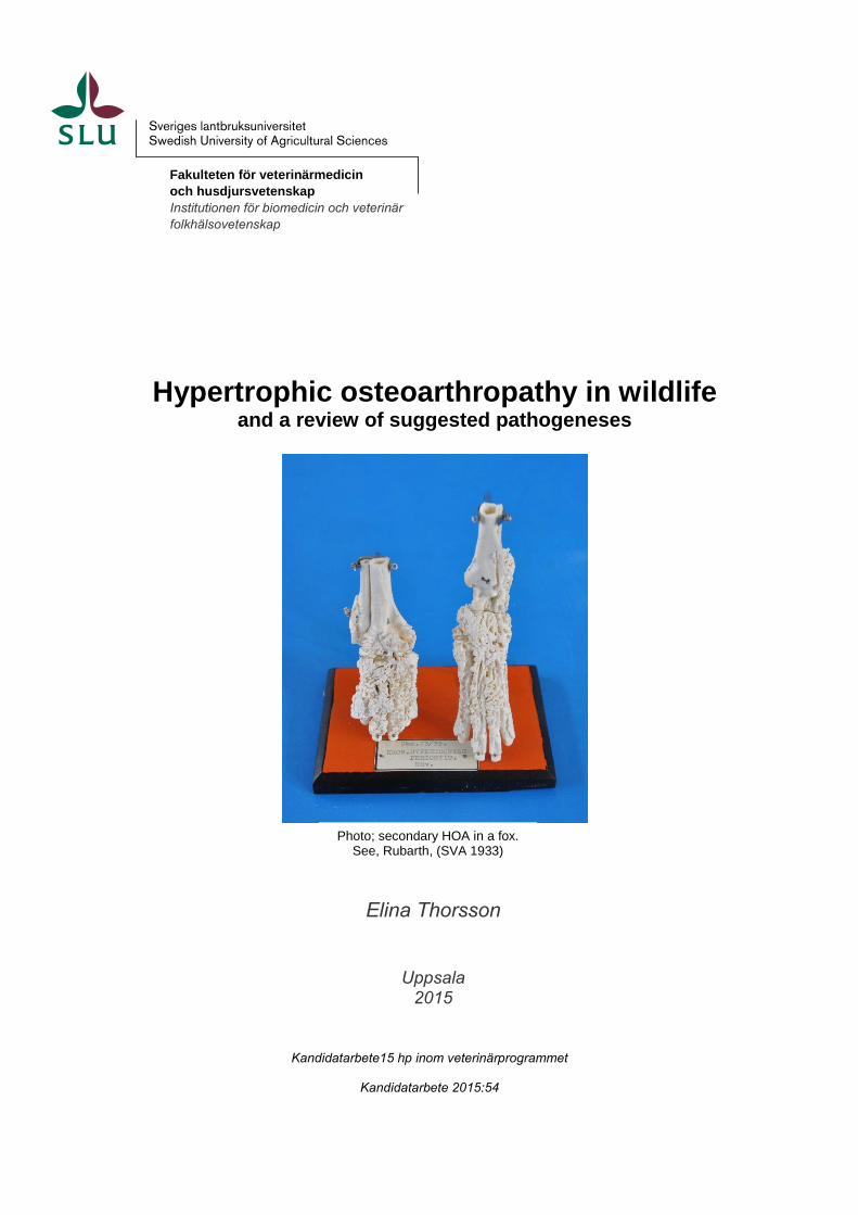
Photo; secondary HOA in a fox. See, Rubarth, (SVA 1933)

Hypertrophic osteoarthropathy in wildlife and a review of suggested pathogeneses
Hypertrofisk osteoartropati hos vilt
och en översikt av föreslagna patogeneser
Elina Thorsson Handledare: Stina Ekman, institutionen för biomedicin och veterinär folkhälsovetenskap, SLU
Biträdande handledare: Erik Ågren, avdelningen för patologi och viltsjukdomar, SVA
Examinator: Eva Tydén, institutionen för biomedicin och veterinär folkhälsovetenskap, SLU
Kandidatarbete i veterinärmedicin Omfattning: 15 hp Nivå och fördjupning: grund nivå, G2E Kurskod: EX0700 Utgivningsort: Uppsala Utgivningsår: 2015
Delnummer i serie: 2015:54
Elektronisk publicering: http://stud.epsilon.slu.se
Key words:
Hypertrophic osteoarthropathy, digital clubbing, osteopathy, pathogenesis, etiology, wild, exotic, zoo
Nyckelord : Hypertrofisk osteoartropati, osteopati, patogenes, etiologi, vilda, exotiska, zoo
Sveriges lantbruksuniversitet
Swedish University of Agricultural Sciences
Fakulteten för veterinärmedicin och husdjursvetenskap
Institutionen för biomedicin och veterinär folkhälsovetenskap


TABLE OF CONTENTS
Summary ................................................................................................................................................. 1
Sammanfattning ...................................................................................................................................... 2
Introduction ............................................................................................................................................. 3
Materials and methods............................................................................................................................. 3
Literature review ..................................................................................................................................... 4
Hypertrophic osteopathy .................................................................................................................... 4
The pathogenesis of secondary hypertrophic osteoarthropathy .................................................... 5
Historical view .............................................................................................................................. 5
Main hypotheses ............................................................................................................................ 5
Review of secondary hypertrophic osteoarthropathy in wild animals ................................................ 7
Case reports, a summary ............................................................................................................... 7
Discussion ............................................................................................................................................. 12
References ............................................................................................................................................. 14

1
SUMMARY
In this essay suggested pathogenesis of secondary hypertrophic osteoarthropathy (HOA) is
reviewed. HOA, characterized by; periostitis, periosteal proliferation of tubular bones and
arthritis can develop due to many different underlying diseases. The syndrome is most
commonly seen with intra-thoracic malignancy or chronic pulmonary infections. HOA has
previously mainly been described in humans and various domesticated species. More
recently, through wildlife disease monitoring, cases have also been found among wild
animals. This essay also aims to summarize the spectra of wild species in which the syndrome
has been reported. Finding a coherent pathogenesis has proven to be difficult. Many theories
overlap and are not yet fully investigated. Factors of importance include hypoxia, VEGF,
PDGF and prostaglandins, but HOA is often suggested to be idiopathic. It is now obvious that
secondary HOA affects a wide range of species. Even though the manifestation of the
syndrome differs slightly between humans and animals it is similar in many aspects.
Considering the similarities it can be presumed that the same pathogenesis applies for
humans, domestic animals and wild animals.
2
SAMMANFATTNING
I denna uppsats beskrivs olika teorier avseende patogenesen till sekundär hypertrofisk
osteoartropati (HOA). Den patologiska bilden av HOA innefattar periostit med typisk
periostal proliferation av rörben samt artrit. Syndromet anses kunna orsakas av flera olika
sjukdomar som underliggande orsak, men ses oftast i samband med intratorakal malignitet
eller kronisk lunginflammation. Äldre literatur har visat att sekundär HOA drabbar människa
och ett flertal domesticerade djurarter. På senare tid har fall också rapporterats hos flera olika
vilda djurarter. Denna uppsats syftar även till att sammanställa det spektrum av vilda djurarter
där sekundär HOA finns rapporterat. Att finna en sammanhängande patogenes har visat sig
vara svårt då teorierna är många, men sparsamt utredda. Faktorer som anses kunna vara
inblandade i processen är hypoxi, tillväxtfaktorer som VEGF, PDGF samt prostaglandiner.
Men ofta anges orsaken vara idiopatisk. Litteraturgenomgången har klart visat att sekundär
HOA drabbar ett stort antal djurarter. Hur syndromet yttrar sig kliniskt skiljer sig något i
beskrivningarna mellan människor och djur, men likheterna överväger vilket gör att man kan
anta att samma patogenes föreligger hos människa och olika djurarter, inklusive vilda djur.
3
INTRODUCTION
Hippocrates (Greece, 460 BC) was first to document findings of digital clubbing, i.e.
periosteal exostoses. This is believed to be the oldest description of clinical signs in human
medicine. Hippocrates described deformed phalanges in association with empyema and
strained respiration (Davidson, 1984). Archeological findings of digital clubbing in skeleton
of humans (7000 years old) gives emphasis to the early presence of the syndrome (Masson et
al., 2013). In more present history, HOA was first described by Bamberger (1889) and Marie
(1890). Digital clubbing (periosteal proliferation of tubular bones) is one part of the triad
constituting HOA, which also includes periostitis and arthritis (Elewaut, 2005). There have
been different perceptions whether all three signs need to be present for accurate diagnosis in
humans. These lesions are often seen together with thickening of the skin of the extremities,
edema, pain at palpation and stiffness (Ginsburg, 1963). Today the general opinion is that
HOA is a progressive syndrome where the symptoms differ over time (Martinez-Lavin et al.,
2007). HOA is in humans classified as primary or secondary (see below for definitions).
Despite the severity and the early historical introduction the pathogenesis of HOA is yet to be
determined. The wide spectra of underlying causes and the rareness of HOA complicate the
research into the pathogenesis of the syndrome. In addition to humans HOA has most
frequently been observed in dogs (Brodey, 1971; Salyusarenko et al., 2013), cats (Huang et
al., 2010), horses (Jp et al., 1992) and cattle (Guyot et al., 2011). The syndrome is however
not limited to domesticated species; as this essay will demonstrate.
The aim of this literature review was to describe the current described hypotheses of the
pathogenesis of secondary HOA. The review also aimed to demonstrate the spectrum of wild
animal species in which occurrence of secondary HOA have been reported. In this essay wild
animals are broadly defined as, and include; undomesticated species, zoo animals and exotic
pets.
MATERIALS AND METHODS
This essay is a literature review divided into two parts, the suggested pathogenesis of
secondary HOA and a summary of case reports describing the syndrome in wild animals. A
web-search was performed in several data-bases; Primo, Scopus, PubMed and Web of
Science, using following key words:
hypertrophic osteoarthropathy, digital clubbing, acropathy, Marie´s disease,
osteopathy
causes, pathogenesis, pathogenetic, source, causation, genesis, pathophysiology,
aetiology
wild, undomesticated, exotic, zoo
4
Regarding the pathogenesis, the search was limited to the years 2005-2015 whereas the case
reports were collected without year limitations. In addition to mentioned data-bases I used
Google, Google scholar and websites of veterinary pathology and wildlife medicine.
LITERATURE REVIEW
Hypertrophic osteoarthropathy
Hypertrophic osteoarthropathy (HOA) is a progressive syndrome characterized by the
presence of bilateral digital clubbing (periosteal proliferation), periostitis and clinical signs of
pain and swollen joints defined as arthritis (Sarkar et al., 2012; Klippel et al., 2008). The
surface of the affected bones is irregular due to spiculated exostoses. The syndrome is
diagnosed by imaging (radiography) and clinical examination (M Martínez-Lavín, 1993).
In animals, the new bone formation usually is concentrated to the diaphyseal region of long
bones. Radius, ulna, tibia and the metatarsal bones are in dogs the first regions to undergo
formation changes. Pelvis, vertebrae, ribs, and bones of the skull may be involved. In 40 % of
human cases, the patients develop articular lesions whereas only 5 % of the dogs show
articular lesions (Brodey, 1971). In humans the proximal phalanges, metatarsal and
metacarpal bones and distal ends of long bones are the most frequent locations with periosteal
proliferation (digital clubbing), consensus reported from WSCO (See The joint pathology
center, 2003).
Primary hypertrophic osteoarthropathy, also known as pachydermoperiostiosis, is a rare
hereditary form of HOA in humans (Sarkar et al., 2012). In addition to periosteal bone
proliferation, periostitis and arthritis thickening of facial skin is seen. The facial skin changes
are the easiest way to separate primary HOA from secondary HOA (Sarkar et al., 2012).
Recently a mutation of the human main enzyme in prostaglandin degradation has shown to be
the cause of primary HOA (Uppal et al., 2008).
Secondary hypertrophic osteoarthropathy, formerly named pulmonary hypertrophic
osteoarthropathy, is most commonly associated with intra-thoracic malignancies or chronic
pulmonary infections (Yao et al., 2009). The changes in the bone tissue is progressive and
develop over months to years and are often detected prior to the suggested underlying disease
(Salyusarenko et al., 2013). The late diagnosis of the underlying cause contributes to a poor
prognosis for individuals diagnosed with secondary HOA.
As an awareness of the syndrome has increased, reports of other primary causes, not
concentrated to the thorax, have been suggested. It is now well established that congenital
heart disease (Ferreira & Camões, 2013), hepatopulmonary syndrome with liver disease,
impaired arterial oxygenation and intrapulmonary shunting (Kaleo Ede, 2008) and chronic
inflammatory bowel disease such as ulcerative colitis and Crohn’s disease (Oppenheimer &
Jones, 1982) also are associated with secondary HOA. Treatment of the underlying disease
results in decreased pain, decreased swelling of the limbs and often a remission of the new-
formed periosteal bone (Ginsburg, 1963).
5
The pathogenesis of secondary hypertrophic osteoarthropathy
The pathogenesis of secondary HOA has remained elusive for centuries. Many of the
hypotheses concerning the pathogenesis were made in the 19th
and 20th
centuries and are in
some cases based on anecdotes.
Neural influence, circulatory changes and growth factors have been discussed as factors of
interest. The theories often overlap which suggests that the pathogenesis of HOA is complex.
Historical view
HOA was in the late 19th century believed to be caused by a toxin derived from the bronchial
tract (Marie, 1890; Bamberger, 1891). However, since the symptoms weren’t reproducible
with sputum, the theory was abandoned (Bamberger, 1891). In the mid-20th century there
were few and unproven theories. The conception was, and in some cases still is, that the
affected extra skeletal organs send an impulse through the vagal nerve inducing periosteal
proliferation with new bone formation (clubbing). The blood flow in the periosteal areas of
the limbs was thought to increase due to an unknown mechanism. A small study of only five
cases showed deceased swelling of the limbs and pain relief following vagotomy (Flayell,
1956). Hyperemia of the periosteal connective tissue with excess of blood and its nutrients is
believed to trigger inflammation (CLUBBING AND HYPERTROPHIC
OSTEOARTHROPATHY. : Medicine). However, increased blood flow has been described in
several conditions with absence of periostitis and periosteal bone proliferation, which clearly
questions that hyperemia is the main cause of clubbing (Ginsburg, 1963).
Main hypotheses
In their review, Yao et al. ( 2009) suggested that the most likely hypothesis of secondary
HOA was presented by Dickinson and Martin (1987). The authors suggest that
megakaryocytes and clusters of platelets in the distal vasculature of the limbs induce release
of platelet derived growth factor (PDGF). Megakaryocytes form in the bone marrow and are
released to the circulation. Normally, clusters of megakaryocytes and platelets become
trapped or fragmented as they pass through the highly dichotomized pulmonary vasculature
(Dickinson & Martin, 1987). Thoracic lesions may cause a pulmonary shunt where this
sequence of events would be lost and the clusters of megakaryocytes and platelets reach the
systemic circulation and subsequently the peripheral vasculature of the distal limbs where
they could activate endothelial cells and release growth factors (Dickinson & Martin, 1987).
In a retrospective study comprising 30 dogs diagnosed with secondary HOA by Salyusarenko
et al. (2013), a significantly higher platelet count and thrombocytosis was found in the dogs
with secondary HOA compared to healthy controls. The dogs with secondary HOA were also
found to have significantly higher proportions of shistocytosis (fragmented red blood cells),
anisocystosis (different sized red blood cells) and pyrexia (Salyusarenko et al., 2013). The
elevated platelet count, thrombocytosis and shistocytosis can be explained by a suspected
pulmonary shunt. The pyrexia refers to an ongoing inflammatory process of the bony lesions.
6
There are contradictory studies on whether hypoxia causes secondary HOA with clubbing or
not. Paton et al. (1991) report a tenfold presence of clubbing in pediatric patients with
hypoxia due to cystic fibrosis, compared to the control group without hypoxia (Paton et al.,
1991). A connection between elevated VEGF (vascular endothelial growth factor) and PDGF
concentrations and hypoxia has been established in human patients with HOA (Silveira et al.,
2000; Atkinson & Fox, 2004). In contrast, a study examining humans diagnosed with
secondary HOA associated with liver disease, showed no difference in oxygen tension
between the patients and the control group was found (O Epstein, 1979). Also, individuals
with an inflamed bowel show increased expression of VEGF-A without evidence of hypoxia
(Scaldaferri et al., 2009).
The above described hypotheses involving megakaryocytes and hypoxia are supported by
presence of an impaired lung function. PDGF and VEGF are formed when platelets aggregate
and the growth factors are regulated by hypoxia (Silveira et al., 2000; Atkinson & Fox, 2004).
PDGF is likely to cause changes such as increased blood flow, edema, collagen deposition
and endothelial hyperplasia, which proceed clubbing (Atkinson & Fox, 2004). However, an
experimental study in rats with intermittent intravenous infusion of PDGF (BB chains) did not
result in development of clubbing. The infusion resulted in an increased bone mass and bone
strength of the skeleton in the rodents (Mitlak et al., 1996).
VEGF is a growth factor produced by malignant tumors to ensure their vascular formation
and nutritional supply. The factor induces edema, vascular hyperplasia, fibroblast
proliferation and new periosteal bone formation, all seen in HOA (Ferrara, 2004). Even
though PDGF may be considered to be the main growth factor involved in the pathogenesis of
HOA (Dickinson & Martin, 1987), it has been noted that elevated levels of VEGF also may
induce pathological changes such as edema, vascular hyperplasia, fibroblast proliferation and
new periosteal bone formation (Atkinson & Fox, 2004). Elevated levels of VEGF have been
seen in patients with both primary and secondary HOA (Silveira et al., 2000).
The most recent hypothesis regarding the pathogenesis of HOA suggests a chronic activation
of macrophages (Toovey & Eisenhauer, 2010). The macrophage activation can be triggered
by a persistent irritant or hypoxia and lead to elevated levels of growth factors and formation
of granulomas (Toovey & Eisenhauer, 2010).
Other circulatory factors such as prostaglandins and hepatocyte growth factor have also been
suggested to be involved in the pathogenesis of digital clubbing (Silveira et al., 2000). In vitro
studies, using rat cell cultures, show that prostaglandins may upregulate expression of VEGF
(PGF2) (Harada et al., 1994) and enhance growth of megakaryocytes (PGF1) (Cooper & Hou,
1988).
Längle (1994) used an isoquinoline derivate in an experimental treatment of bronchial asthma
in rats, and unintentionally induced symptoms corresponding with HOA.
7
Review of secondary hypertrophic osteoarthropathy in wild animals
Case reports, a summary
The database search for secondary HOA in wild animals resulted in a number of case reports,
mainly single affected animals of various species from different genera or families. The
database search for HOA in wild animals resulted in a number of case reports, mainly single
affected animals of various species from different genera or familiesAll animals presented in
the table below are diagnosed with secondary HOA. The definition of HOA in humans is the
triad of lesions, bilateral digital clubbing, periostitis and arthritis (Elewaut, 2005). However,
the most common lesions in animals are the periosteal bone proliferations, hence the case
report authors have diagnosed HOA when such typical lesions were found.
Table 1, Case reports of secondary HOA in wild animals including suggested and/or potential
underlying diseases. The location of the periosteal bone lesions are not always described.
ALP= alkaline phosphatase
Animal species
Periosteal bone lesions Potential underlying disease
Camelidae
Alpaca, female USA, imported from Chile (Curtis et al., 1997)
Diaphysis of the metacarpal bones and the proximal phalanges of the right forelimb
Focal chronic pneumonia Chronic interstitial myocarditis
Canidae
Wolf, Canis lupus Wild, Sweden (SVA, 1997)
Tarsal and metatarsal bones, tibiae, fibulae, femur and the pelvis
-
Fox, Vulpes vulpes female Wild, Sweden (Rubarth SVA, 1933)
Metacarpal bones including the joints, metatarsal bones including the joints, tibiae and the patellae (See front page photo)
Abscending chronic pneumonia and fibrous pleuritis, diplococcus
Fox, Vulpes vulpes male Wild, Sweden (SVA, 1982)
Forelimbs
Pneumonia including pleuritis, isolated bacteria; Clostridium sordellii
8
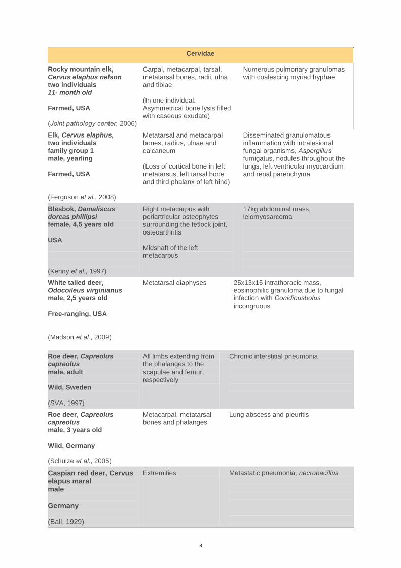
Cervidae
Rocky mountain elk, Cervus elaphus nelson two individuals 11- month old Farmed, USA (Joint pathology center, 2006)
Carpal, metacarpal, tarsal, metatarsal bones, radii, ulna and tibiae (In one individual: Asymmetrical bone lysis filled with caseous exudate)
Numerous pulmonary granulomas with coalescing myriad hyphae
Elk, Cervus elaphus, two individuals family group 1 male, yearling Farmed, USA (Ferguson et al., 2008)
Metatarsal and metacarpal bones, radius, ulnae and calcaneum (Loss of cortical bone in left metatarsus, left tarsal bone and third phalanx of left hind)
Disseminated granulomatous inflammation with intralesional fungal organisms, Aspergillus fumigatus, nodules throughout the lungs, left ventricular myocardium and renal parenchyma
Blesbok, Damaliscus dorcas phillipsi female, 4,5 years old USA (Kenny et al., 1997)
Right metacarpus with periartricular osteophytes surrounding the fetlock joint, osteoarthritis Midshaft of the left metacarpus
17kg abdominal mass, leiomyosarcoma
White tailed deer, Odocoileus virginianus male, 2,5 years old Free-ranging, USA (Madson et al., 2009)
Metatarsal diaphyses 25x13x15 intrathoracic mass, eosinophilic granuloma due to fungal infection with Conidiousbolus incongruous
Roe deer, Capreolus capreolus male, adult Wild, Sweden (SVA, 1997)
All limbs extending from the phalanges to the scapulae and femur, respectively
Chronic interstitial pneumonia
Roe deer, Capreolus capreolus male, 3 years old Wild, Germany (Schulze et al., 2005)
Metacarpal, metatarsal bones and phalanges
Lung abscess and pleuritis
Caspian red deer, Cervus elapus maral male Germany (Ball, 1929)
Extremities Metastatic pneumonia, necrobacillus
9
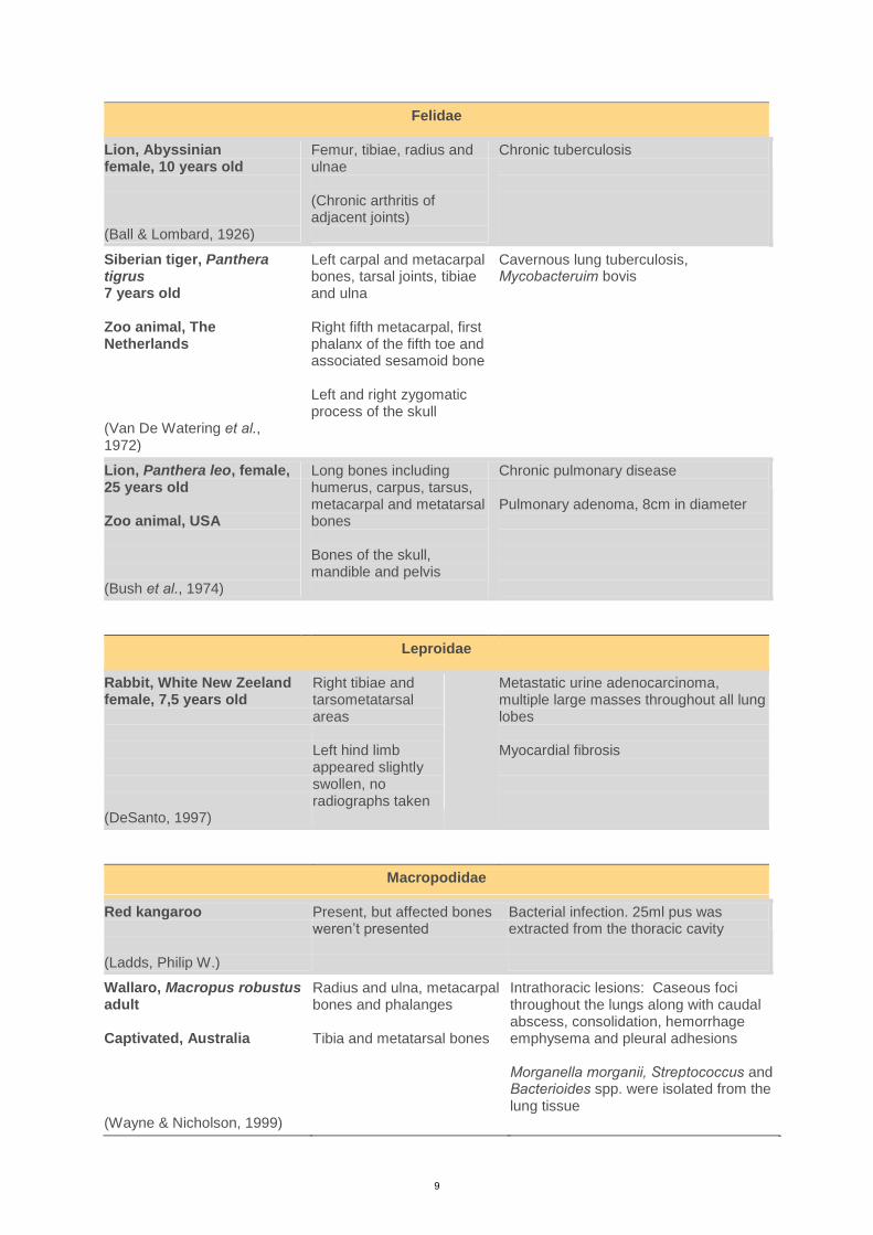
Felidae
Lion, Abyssinian female, 10 years old (Ball & Lombard, 1926)
Femur, tibiae, radius and ulnae (Chronic arthritis of adjacent joints)
Chronic tuberculosis
Siberian tiger, Panthera tigrus 7 years old Zoo animal, The Netherlands (Van De Watering et al., 1972)
Left carpal and metacarpal bones, tarsal joints, tibiae and ulna Right fifth metacarpal, first phalanx of the fifth toe and associated sesamoid bone Left and right zygomatic process of the skull
Cavernous lung tuberculosis, Mycobacteruim bovis
Lion, Panthera leo, female, 25 years old Zoo animal, USA (Bush et al., 1974)
Long bones including humerus, carpus, tarsus, metacarpal and metatarsal bones Bones of the skull, mandible and pelvis
Chronic pulmonary disease Pulmonary adenoma, 8cm in diameter
Leproidae
Rabbit, White New Zeeland female, 7,5 years old (DeSanto, 1997)
Right tibiae and tarsometatarsal areas Left hind limb appeared slightly swollen, no radiographs taken
Metastatic urine adenocarcinoma, multiple large masses throughout all lung lobes Myocardial fibrosis
Macropodidae
Red kangaroo (Ladds, Philip W.)
Present, but affected bones weren’t presented
Bacterial infection. 25ml pus was extracted from the thoracic cavity
Wallaro, Macropus robustus adult Captivated, Australia (Wayne & Nicholson, 1999)
Radius and ulna, metacarpal bones and phalanges Tibia and metatarsal bones
Intrathoracic lesions: Caseous foci throughout the lungs along with caudal abscess, consolidation, hemorrhage emphysema and pleural adhesions Morganella morganii, Streptococcus and Bacterioides spp. were isolated from the lung tissue
10
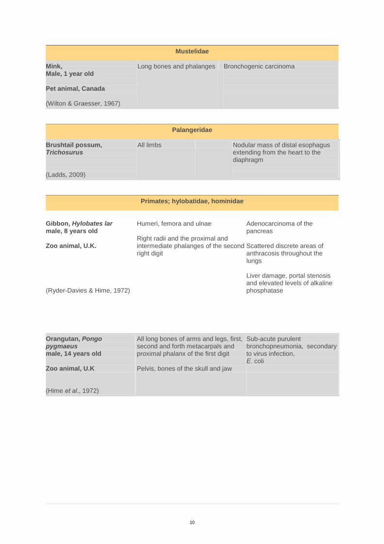
Mustelidae
Mink, Male, 1 year old Pet animal, Canada (Wilton & Graesser, 1967)
Long bones and phalanges Bronchogenic carcinoma
Palangeridae
Brushtail possum, Trichosurus (Ladds, 2009)
All limbs Nodular mass of distal esophagus extending from the heart to the diaphragm
Primates; hylobatidae, hominidae
Gibbon, Hylobates lar male, 8 years old Zoo animal, U.K. (Ryder-Davies & Hime, 1972)
Humeri, femora and ulnae Right radii and the proximal and intermediate phalanges of the second right digit
Adenocarcinoma of the pancreas Scattered discrete areas of anthracosis throughout the lungs Liver damage, portal stenosis and elevated levels of alkaline phosphatase
Orangutan, Pongo pygmaeus male, 14 years old Zoo animal, U.K (Hime et al., 1972)
All long bones of arms and legs, first, second and forth metacarpals and proximal phalanx of the first digit Pelvis, bones of the skull and jaw
Sub-acute purulent bronchopneumonia, secondary to virus infection, E. coli
11
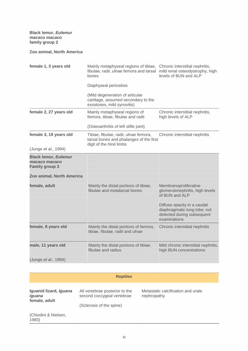
Black lemur, Eulemur macaco macaco family group 2 Zoo animal, North America female 1, 3 years old
Mainly metaphyseal regions of tibiae, fibulae, radii, ulnae femora and tarsal bones Diaphyseal periostisis (Mild degeneration of articular cartilage, assumed secondary to the exostoses, mild synovitis)
Chronic interstitial nephritis, mild renal osteodystrophy, high levels of BUN and ALP
female 2, 27 years old
Mainly metaphyseal regions of femora, tibiae, fibulae and radii (Osteoarthritis of left stifle joint)
Chronic interstitial nephritis, high levels of ALP
female 3, 19 years old (Junge et al., 1994)
Tibiae, fibulae, radii, ulnae femora, tarsal bones and phalanges of the first digit of the hind limbs
Chronic interstitial nephritis
Black lemur, Eulemur macaco macaco Family group 3 Zoo animal, North America female, adult
Mainly the distal portions of tibiae, fibulae and metatarsal bones
Membranoproliferative glomerulonephritis, high levels of BUN and ALP Diffuse opacity in a caudal diaphragmatic lung lobe; not detected during subsequent examinations
female, 8 years old Mainly the distal portions of femora, tibiae, fibulae, radii and ulnae
Chronic interstitial nephritis
male, 11 years old (Junge et al., 1994)
Mainly the distal portions of tibiae, fibulae and radius
Mild chronic interstitial nephritis, high BUN concentrations
Reptiles
Iguanid lizard, Iguana iguana female, adult (Chiodini & Nielsen, 1983)
All vertebrae posterior to the second coccygeal vertebrae (Sclerosis of the spine)
Metastatic calcification and urate nephropathy
12
DISCUSSION
This literature review shows a wide range of wild animal species diagnosed with secondary
HOA. The name and definition of the syndrome has differed through history and
consequently how it has been named in studies, which may make it challenging to retrieve all
documented cases.
Even though digital clubbing was described over 2000 years ago by Hippocrates and has been
reported from even older archeological findings, the exact pathogenesis remains unknown. A
wide range of possible affecting factors have been discussed, including neural influence,
circulatory changes, growth factors and cell activity.
Initially HOA was thought to be associated only with pulmonary diseases, which has
influenced the earlier naming of the syndrome and thoughts on possible pathogenesis. Studies
of human HOA have described hypoxia to be a factor of interest (Paton et al., 1991; Silveira
et al., 2000; Atkinson & Fox, 2004). The presence of hypoxia seems to fit in with other
hypotheses of the underlying pathogenesis of HOA. Induced by hypoxia the growth factors
VEGF and PDGF can cause stromal and vascular changes in the periosteal tissue of the limbs
Black and white tegu lizard, Tupinambus teguixin (Divers & Mader, 2005)
All limbs, mainly forelimbs -
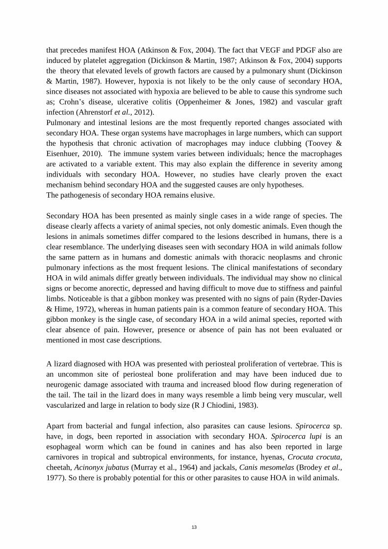
Ursidae
Raccoon dog, Procyon lotor male, adult Conservation and Research Center, USA (Joint pathology center, 2003)
Long bones and the phalanges Pink firm nodule, 2cm in diameter in the caudal proportion of the middle lung lobe. Several smaller nodules present in the pancreas
Raccoon dog, Nyctereutes procyonides female Free-living, Japan (Masegi et al., 1994)
Metacarpal, metatarsal and femorotibial joints, extending to the epiphysis and metaphysis of adjacent bones (Pre-existing metaphyseal bone was thinned and in some areas resorbed Degeneration of tibial articular cartilage and villous proliferation of the synovial membrane of the metacarpal joints)
Chronic bronchitis with pyogranulomatous lesions associated with Nocardia-like bacteria
13
that precedes manifest HOA (Atkinson & Fox, 2004). The fact that VEGF and PDGF also are
induced by platelet aggregation (Dickinson & Martin, 1987; Atkinson & Fox, 2004) supports
the theory that elevated levels of growth factors are caused by a pulmonary shunt (Dickinson
& Martin, 1987). However, hypoxia is not likely to be the only cause of secondary HOA,
since diseases not associated with hypoxia are believed to be able to cause this syndrome such
as; Crohn’s disease, ulcerative colitis (Oppenheimer & Jones, 1982) and vascular graft
infection (Ahrenstorf et al., 2012).
Pulmonary and intestinal lesions are the most frequently reported changes associated with
secondary HOA. These organ systems have macrophages in large numbers, which can support
the hypothesis that chronic activation of macrophages may induce clubbing (Toovey &
Eisenhuer, 2010). The immune system varies between individuals; hence the macrophages
are activated to a variable extent. This may also explain the difference in severity among
individuals with secondary HOA. However, no studies have clearly proven the exact
mechanism behind secondary HOA and the suggested causes are only hypotheses.
The pathogenesis of secondary HOA remains elusive.
Secondary HOA has been presented as mainly single cases in a wide range of species. The
disease clearly affects a variety of animal species, not only domestic animals. Even though the
lesions in animals sometimes differ compared to the lesions described in humans, there is a
clear resemblance. The underlying diseases seen with secondary HOA in wild animals follow
the same pattern as in humans and domestic animals with thoracic neoplasms and chronic
pulmonary infections as the most frequent lesions. The clinical manifestations of secondary
HOA in wild animals differ greatly between individuals. The individual may show no clinical
signs or become anorectic, depressed and having difficult to move due to stiffness and painful
limbs. Noticeable is that a gibbon monkey was presented with no signs of pain (Ryder-Davies
& Hime, 1972), whereas in human patients pain is a common feature of secondary HOA. This
gibbon monkey is the single case, of secondary HOA in a wild animal species, reported with
clear absence of pain. However, presence or absence of pain has not been evaluated or
mentioned in most case descriptions.
A lizard diagnosed with HOA was presented with periosteal proliferation of vertebrae. This is
an uncommon site of periosteal bone proliferation and may have been induced due to
neurogenic damage associated with trauma and increased blood flow during regeneration of
the tail. The tail in the lizard does in many ways resemble a limb being very muscular, well
vascularized and large in relation to body size (R J Chiodini, 1983).
Apart from bacterial and fungal infection, also parasites can cause lesions. Spirocerca sp.
have, in dogs, been reported in association with secondary HOA. Spirocerca lupi is an
esophageal worm which can be found in canines and has also been reported in large
carnivores in tropical and subtropical environments, for instance, hyenas, Crocuta crocuta,
cheetah, Acinonyx jubatus (Murray et al., 1964) and jackals, Canis mesomelas (Brodey et al.,
1977). So there is probably potential for this or other parasites to cause HOA in wild animals.
14
In conclusion, secondary HOA shows similarities across species in regard to clinical
symptoms and pathology of the skeleton. It is seen in combination with other, often thoracic,
lesions, suggesting a common, but still not established pathogenesis in humans and animals,
including wild animals.
REFERENCES
Ahrenstorf, G., Witte, T., Schmidt, R. E., Rihl, M., Pichlmaier, M. A. & Rosenthal, H. (2012).
Unilateral hypertrophic osteoarthropathy in a patient with a vascular graft infection. Journal of
Clinical Rheumatology, 18(6), pp 307–309.
Atkinson, S. & Fox, S. B. (2004). Vascular endothelial growth factor (VEGF)-A and platelet-derived
growth factor (PDGF) play a central role in the pathogenesis of digital clubbing. The Journal
of Pathology [online], 203(2), pp 721–728. Available from:
http://onlinelibrary.wiley.com/doi/10.1002/path.1565/abstract. [Accessed 2015-02-24].
Ball, N. (1929). Fall von multipler Exostosenbildung bei einem Hirsche. Archiv fuer wissenschaftliche
praktische Tierheilkunde, 60, p 235–.
Ball, V. & Lombard, C. (1926). Bull. Acad. Méd., 95(16).
Bamberger, E. (1891). Über Knochenveranderungen bei chronischen Lunger- und Herzkrankheiten.
klin Méd., (93).
Bamberger, E.Wien Klin Wochenschr. 1889(2), pp 226–230.
Brodey, R. S. (1971). Hypertrophic osteoarthropathy in the dog: a clinicopathologic survey of 60
cases. Journal of the American Veterinary Medical Association, 159(10), pp 1242–1256.
Brodey, R. S., Thomson, R. G., Sayer, P. D. & Eugster, B. (1977). Spiroceca lupi infection in dogs in
Kenya. Veterinary Parasitology [online], 3(1), pp 49–59. Available from:
http://www.sciencedirect.com/science/article/pii/0304401777900073. [Accessed 2015-02-18].
Bush, M., James, A. E., Montali, R. J., Sauer, R. M., Heller, R. H. & Gray, C. W. (1974).
Hypertrophic Pulmonary Osteoarthropathy in a Lioness (Panthera Leo). Veterinary Radiology
[online], 15(2), pp 84–90. Available from: http://onlinelibrary.wiley.com/doi/10.1111/j.1740-
8261.1974.tb00688.x/abstract. [Accessed 2015-02-16].
Chiodini, R. J. & Nielsen, S. W. (1983). Vertebral osteophytes in an iguanid lizard. Veterinary
Pathology, 20(3), pp 372–375.
CLUBBING AND HYPERTROPHIC OSTEOARTHROPATHY. : Medicine. [online] (LWW). Available
from: http://journals.lww.com/md-
journal/Fulltext/1942/09010/CLUBBING_AND_HYPERTROPHIC_OSTEOARTHROPATH
Y_.2.aspx. [Accessed 2015-02-24].
Cooper, G. W. & Hou, X. P. (1988). The effects of prostaglandin E1 on megakaryocyte proliferation
in vitro. Advances in Experimental Medicine and Biology, 241, pp 217–224.
Curtis, C., Dart, A., Rawlinson, R. & Hodgson, D. (1997). Hypertrophic osteopathy in an alpaca.
Australian Veterinary Journal [online], 75(1), pp 61–62. Available from:
http://onlinelibrary.wiley.com/doi/10.1111/j.1751-0813.1997.tb13834.x/abstract. [Accessed
2015-02-18].
Davidson, C. S. (1984). Medicine: An Illustrated History. By Albert S. Lyons and R. Joseph
Petrucelli, II. 616 pp. New York: Harry N. Abrams, 1978. Hepatology [online], 4(6), pp 1264–
1264. Available from: http://onlinelibrary.wiley.com/doi/10.1002/hep.1840040633/abstract.
[Accessed 2015-02-23].
DeSanto, J. (1997). Hypertrophic osteopathy associated with an intrathoracic neoplasm in a rabbit.
Journal of the American Veterinary Medical Association, 210(9), pp 1322–3.
Van De Watering, C. C., Zwart, P. & Barker, J. (1972). Cavernous tuberculosis of the lungs and
secondary hypertrophic osteo-arthropathy in a Siberian tiger (Panthera tigrus). Journal of
Small Animal Practice [online], 13(6), pp 321–327. Available from:
http://onlinelibrary.wiley.com/doi/10.1111/j.1748-5827.1972.tb06852.x/abstract. [Accessed
2015-02-18].
15
Dickinson, C. J. & Martin, J. F. (1987). MEGAKARYOCYTES AND PLATELET CLUMPS AS
THE CAUSE OF FINGER CLUBBING. The Lancet [online], 330(8573), pp 1434–1435.
Available from: http://www.sciencedirect.com/science/article/pii/S0140673687911329.
[Accessed 2015-02-24].
Divers, S. J. & Mader, D. R. (2005). Reptile Medicine and Surgery. Elsevier Health Sciences. ISBN
141606477X.
Elewaut, D. (2005). Kelley’s Textbook of Rheumatology, 7th edition (in 2 volumes). Edited by E. D.
Harris Jr, R. C. Budd, G. S. Firestein et al. Elsevier Saunders, 2004. 1916 pp. ISBN
0721601413. Rheumatology [online], 44(4), pp 566–566. Available from:
http://rheumatology.oxfordjournals.org/content/44/4/566. [Accessed 2015-02-23].
Ferguson, N. M., Lévy, M., Ramos-Vara, J. A., Baird, D. K. & Wu, C. C. (2008). Hypertrophic
Osteopathy Associated with Mycotic Pneumonia in Two Juvenile Elk (Cervus Elaphus).
Journal of Veterinary Diagnostic Investigation [online], 20(6), pp 849–853. Available from:
http://vdi.sagepub.com/content/20/6/849. [Accessed 2015-02-18].
Ferrara, N. (2004). Vascular Endothelial Growth Factor: Basic Science and Clinical Progress.
Endocrine Reviews [online], 25(4), pp 581–611. Available from:
http://press.endocrine.org/doi/full/10.1210/er.2003-0027. [Accessed 2015-02-24].
Ferreira, E. & Camões, I. (2013). Hypertrophic osteoarthropathy and congenital heart disease: this is
not casual. BMJ Case Reports [online], 2013, p bcr2013009392. Available from:
http://casereports.bmj.com/content/2013/bcr-2013-009392. [Accessed 2015-02-17].
Flayell, G. (1956). REVERSAL OF PULMONARY HYPERTROPHIC OSTEOARTHROPATHY
BY VAGOTOMY. The Lancet [online], 267(6911), pp 260–262. Available from:
http://www.sciencedirect.com/science/article/pii/S0140673656911862. [Accessed 2015-02-
24].
Ginsburg, J. (1963). Hypertrophic pulmonary osteoarthropathy. Postgraduate medical journal
[online], 39(457), p 639. Available from:
http://www.ncbi.nlm.nih.gov/pmc/articles/PMC2482609/. [Accessed 2015-02-18].
Guyot, H., Sandersen, C. & Rollin, F. (2011). A case of hypertrophic osteoarthropathy in a Belgian
blue cow. The Canadian Veterinary Journal. La Revue Vétérinaire Canadienne, 52(12), pp
1308–1311.
Harada, S. I., Nagy, J. A., Sullivan, K. A., Thomas, K. A., Endo, N., Rodan, G. A. & Rodan, S. B.
(1994). Induction of vascular endothelial growth factor expression by prostaglandin E2 and E1
in osteoblasts. Journal of Clinical Investigation [online], 93(6), p 2490. Available from:
http://www.ncbi.nlm.nih.gov/pmc/articles/PMC294462/. [Accessed 2015-03-01].
Hime, J., Keymer, I. & Appleby, E. (1972). Hypertrophic pulmonary osteoarthropathy in an orang-
utan (Pongo pygmaeus). Veterinary Record., 91(14), pp 334–7.
Hojo, S., Fujita, J., Yamadori, I., Ezaki, T., Watanabe, S., Yamanouchi, H., Miyawaki, H., Yamaji, Y.,
Nishioka, M. & Takahara, J. (1997). Hepatocyte growth factor and digital clubbing. Internal
Medicine (Tokyo, Japan), 36(1), pp 44–46.
Huang, C.-H., Jeng, C.-R., Lin, C.-T. & Yeh, L.-S. (2010). Feline Hypertrophic Osteopathy: A
Collection of Seven Cases in Taiwan. Journal of the American Animal Hospital Association
[online], 46(5), pp 346–352. Available from: http://www.jaaha.org/doi/abs/10.5326/0460346.
[Accessed 2015-02-18].
HYPERTROPHIC OSTEOPATHY ASSOCIATED WITH RENAL GOUT IN A GREEN IGUANA,
Iguana iguana. [online]. Available from: http://arav.conferencespot.org/54999-arav-1999-
1.53493/1999-1.53596/f-019-1.53684/a-171-1.53694?qr=1. [Accessed 2015-03-13].
Joint pathology center (2003). . Available from: http://www.askjpc.org/wsco/wsc/wsc02/02wsc23.pdf.
[Accessed 2015-03-17].
Joint pathology center (2006). . Available from: http://www.askjpc.org/wsco/wsc/wsc06/06wsc03.pdf.
[Accessed 2015-03-17].
Jp, L., Gp, C. & L, G. (1992). Hypertrophic osteopathy in three horses and a pony. Journal of the
American Veterinary Medical Association [online], 201(12), pp 1900–1904. Available from:
http://europepmc.org/abstract/med/1483912. [Accessed 2015-02-23].
Junge, R., Mehren, K., Meehan, T. & Crawshaw, G. (1994). PERIARTICULAR HYPEROSTOSIS
AND RENAL-DISEASE IN 6 BLACK LEMURS OF 2 FAMILY GROUPS. Journal of the
American Veterinary Medical Association, 205(7), p 1024–.
16
Kaleo Ede, D. M. (2008). Hypertrophic osteoarthropathy in the hepatopulmonary syndrome. Journal
of clinical rheumatology : practical reports on rheumatic & musculoskeletal diseases,
14(4), pp 230–3.
Kenny, D. E., Getzy, D. M., Eller, J. L. & Morgan, T. (1997). Hypertrophic osteoarthropathy in a
blesbok (Damaliscus dorcas phillipsi). Journal of Zoo and Wildlife Medicine [online], pp 319–
324. Available from: http://www.jstor.org/stable/20095665. [Accessed 2015-02-18].
Klippel, J. H., Stone, J. H., Crofford, L. eslie J. & White, P. H. (2008). Primer on the Rheumatic
Diseases. Springer Science & Business Media. ISBN 9780387685663.
Ladds, P. (2009). Pathology of Australian Native Wildlife. Csiro Publishing. ISBN 9780643099401.
Ladds, P. W.Pathology of Macropodes. Available from: http://www.arwh.org/sites/default/files/files-
uploads/18%20Pathology%20of%20macropods.pdf. [Accessed 2015-03-17].
Madson, D. M., Loynachan, A. T., Kariyawasam, S. & Opriessnig, T. (2009). Systemic Conidiobolus
incongruus infection and hypertrophic osteopathy in a white-tailed deer (Odocoileus
virginianus). Journal of Veterinary Diagnostic Investigation: Official Publication of the
American Association of Veterinary Laboratory Diagnosticians, Inc, 21(1), pp 167–170.
Marie, P.De l’osteo arthropathie hypertrophiante pneumonique. Rév. Méd. Paris., 1890(10), pp 1–36.
Martinez-Lavin, M., Hochberg, M. & Silman AJ. (2007). Hypertrophic oteoarthropathy.
Rheumatology. 4th Ed. Mosby.
Masegi, T., Yanai, T., Sakai, T., Matsumoto, C., Yamazoe, K., Nukaya, A., Kunimune, Y. & Ueda, K.
(1994). Hypertrophic Pulmonary Osteoarthropathy in a Raccoon Dog (Nyctereutes
procyonoides) with Chronic Pulmonary Inflammatory Lesions. Journal of Wildlife Diseases
[online], 30(4), pp 612–615. Available from: http://www.bioone.org/doi/abs/10.7589/0090-
3558-30.4.612. [Accessed 2015-02-18].
Masson, M., Molnár, E., Donoghue, H. D., Besra, G. S., Minnikin, D. E., Wu, H. H. T., Lee, O. Y.-C.,
Bull, I. D. & Pálfi, G. (2013). Osteological and Biomolecular Evidence of a 7000-Year-Old
Case of Hypertrophic Pulmonary Osteopathy Secondary to Tuberculosis from Neolithic
Hungary. PLoS ONE [online], 8(10), p e78252. Available from:
http://dx.doi.org/10.1371/journal.pone.0078252. [Accessed 2015-02-23].
Mitlak, B. H., Finkelman, R. D., Hill, E. L., Li, J., Martin, B., Smith, T., D’Andrea, M., Antoniades,
H. N. & Lynch, S. E. (1996). The effect of systemically administered PDGF-BB on the rodent
skeleton. Journal of Bone and Mineral Research [online], 11(2), pp 238–247. Available from:
http://onlinelibrary.wiley.com/doi/10.1002/jbmr.5650110213/abstract. [Accessed 2015-02-
24].
M Martínez-Lavín, M. M.-C. (1993). Hypertrophic osteoarthropathy: consensus on its definition,
classification, assessment and diagnostic criteria. The Journal of rheumatology, 20(8), pp
1386–7.
Murray, M., Campbell, M. H. & Jarrett, W. H. F. (1964). Spirocerca lupi in a cheetah. African Journal
of Ecology [online], 2(1), pp 164–164. Available from:
http://onlinelibrary.wiley.com/doi/10.1111/j.1365-2028.1964.tb00209.x/abstract. [Accessed
2015-03-02].
O Epstein, A. B. A. (1979). Hypertrophic hepatic osteoarthropathy. Clinical, roentgenologic,
biochemical, hormonal and cardiorespiratory studies, and review of the literature. The
American journal of medicine, 67(1), pp 88–97.
Oppenheimer, D. A. & Jones, H. H. (1982). Hypertrophic osteoarthropathy of chronic inflammatory
bowel disease. Skeletal Radiology [online], 9(2), pp 109–113. Available from:
http://link.springer.com/article/10.1007/BF00360493. [Accessed 2015-02-23].
Paton, J. Y., Bautista, D. B., Stabile, M. W., Waldman, A. E., Nassar, A. G., Platzker, A. C. & Keens,
T. G. (1991). Digital clubbing and pulmonary function abnormalities in children with lung
disease. Pediatric Pulmonology, 10(1), pp 25–29.
Rj, L., Aj, G., Aa, M., Ww, W., Al, H. & Pd, K. (1978). Relationships among digital clubbing, disease
severity, and serum prostaglandins F2alpha and E concentrations in cystic fibrosis patients.
The American review of respiratory disease [online], 117(4), pp 639–646. Available from:
http://europepmc.org/abstract/med/646216. [Accessed 2015-03-01].
Ryder-Davies, P. & Hime, J. M. (1972). Hypertrophic osteoarthropathy in a gibbon (Hylobates lar).
The Journal of Small Animal Practice, 13(11), pp 655–658.
17
Salyusarenko, M., Peeri, D., Bibring, U., Ranen, E., Bdolah-Abram, T. & Aroch, I. (2013).
Hypertrophic Osteopathy: a Retrospective Case Control Study of 30 Dogs. Front Cover-The
himalayan [online], 68(3), p 3. Available from:
http://isrvma.org/ImageToArticle/Files/DECEMBER%202013%20EN.pdf#page=9. [Accessed
2015-02-18].
Sarkar, M., Mahesh, D. & Madabhavi, I. (2012). Digital clubbing. Lung India [online], 29(4), p 354.
Available from: http://www.lungindia.com/text.asp?2012/29/4/354/102824. [Accessed 2015-
02-17].
Scaldaferri, F., Vetrano, S., Sans, M., Arena, V., Straface, G., Stigliano, E., Repici, A., Sturm, A.,
Malesci, A., Panes, J. & others (2009). VEGF-A links angiogenesis and inflammation in
inflammatory bowel disease pathogenesis. Gastroenterology [online], 136(2), pp 585–595.
Available from: http://www.sciencedirect.com/science/article/pii/S0016508508017319.
[Accessed 2015-03-01].
Schulze, C., Prejawa, T. & Paulick, D. (2005). [Case report: Hypertrophic osteopathy in roe deer
(Capreolus capreolus)]. DTW. Deutsche tierärztliche Wochenschrift, 112(10), pp 393–394.
Silveira, L. H., Martinez-Lavin, M., Pineda, C., Fonseca, M. C., Navarro, C. & Nava, A. (2000).
Vascular endothelial growth factor and hypertrophic osteoarthropathy. Clinical and
experimental rheumatology [online], 18(1), pp 57–62. Available from:
http://www.clinexprheumatol.org/article.asp?a=1557. [Accessed 2015-02-24].
Toovey, O. T. R. & Eisenhauer, H. J. (2010). A new hypothesis on the mechanism of digital clubbing
secondary to pulmonary pathologies. Medical Hypotheses [online], 75(6), pp 511–513.
Available from: http://www.sciencedirect.com/science/article/pii/S0306987710002379.
[Accessed 2015-02-17].
Uppal, S., Diggle, C. P., Carr, I. M., Fishwick, C. W. G., Ahmed, M., Ibrahim, G. H., Helliwell, P. S.,
Latos-Bieleńska, A., Phillips, S. E. V., Markham, A. F., Bennett, C. P. & Bonthron, D. T.
(2008). Mutations in 15-hydroxyprostaglandin dehydrogenase cause primary hypertrophic
osteoarthropathy. Nature Genetics [online], 40(6), pp 789–793. Available from:
http://www.nature.com/ng/journal/v40/n6/full/ng.153.html. [Accessed 2015-02-23].
U W Längle, S. B. (1994). Hypertrophic osteopathy in rats following chronic administration of SDZ
MNS 949, an isoquinoline. Experimental and toxicologic pathology : official journal of the
Gesellschaft für Toxikologische Pathologie, 45(8), pp 473–9.
Wayne, J. & Nicholson, V. (1999). Hypertrophic osteopathy and pneumonia in a macropod.
Australian Veterinary Journal [online], 77(2), pp 98–99. Available from:
http://onlinelibrary.wiley.com/doi/10.1111/j.1751-0813.1999.tb11676.x/abstract. [Accessed
2015-03-15].
Wilton, G. S. & Graesser, F. E. (1967). Hypertrophic pulmonary osteoarthropathy in a mink. The
Canadian Veterinary Journal [online], 8(3), pp 77–78. Available from:
http://www.ncbi.nlm.nih.gov/pmc/articles/PMC1696776/. [Accessed 2015-03-13].
Yao, Q., Altman, R. D. & Brahn, E. (2009). Periostitis and Hypertrophic Pulmonary Osteoarthropathy:
Report of 2 Cases and Review of the Literature. Seminars in Arthritis and Rheumatism
[online], 38(6), pp 458–466. Available from:
http://www.sciencedirect.com/science/article/pii/S0049017208001200. [Accessed 2015-02-
17].