Embed Size (px)
Citation preview

POLICIES AND PROCEDURES INDEX Page 1 of 3
KING KHALID UNIVERSITY HOSPITAL CRITICAL CARE DEPARTMENT INTENSIVE CARE UNIT
POLICIES AND PROCEDURES INDEX
I. ADMINISTRATIVE (ORGANIZATIONAL) POLICIES:
ICU IPP - 001 ADMISSION CRITERIA - ICU
ICU IPP - 002 ADMISSION COMMUNICATION
ICU IPP - 003 DISCHARGE CRITERIA - ICU
ICU IPP - 004 DISCHARGE COMMUNICATION
ICU IPP - 005 TRANSFER OF CRITICALLY ILL PATIENTS FROM ICU FOR DIAGNOSTIC AND
THERAPEUTIC PROCEDURES
ICU IPP - 006 MULTIDISCIPLINARY COLLABORATION IN THE ICU
ICU IPP - 007 WORKING HOURS, ON CALL HOURS
II. COMMON PROCEDURES IN ICU:
ICU IPP - 008 INTERNAL JUGULAR VEIN CANNULATION
ICU IPP - 009 SUBCLAVIAN VEIN CANNULATION
ICU IPP - 010 PULMONARY ARTERY CATHETERIZATION (PAC)
ICU IPP - 011 FEMORAL VEIN CANNULATION
ICU IPP - 012 THORACENTESIS
ICU IPP - 013 CHEST TUBE INSERTION
ICU IPP - 014 CHEST TUBE REMOVAL
ICU IPP - 015 PARACENTESIS (ASCITIC TAP)
ICU IPP - 016 ARTERIAL LINE INTRA-ARTERIAL CATHETERIZATION AND PRESSURE
MONITORING
ICU IPP - 017 ARTERIAL PUNCTURE FOR BLOOD GAS ANALYSIS
ICU IPP - 018 ARTERIAL BLOOD GAS SAMPLE DISPOSAL
ICU IPP - 019 LUMBAR PUNCTURE
ICU IPP - 020 GASTRIC LAVAGE
ICU IPP - 021 INTRACRANIAL PRESSURE MONITORING
ICU IPP - 022 INTRABDOMINAL PRESSURE MONITORING
ICU IPP - 023 PERCUTANEOUS TRACHEOSTOMY
ICU IPP - 024 BRONCHOSCOPY

POLICIES AND PROCEDURES INDEX Page 2 of 3
III. VENTILATION RELATED POLICIES:
ICU IPP - 025 INTUBATION
ICU IPP - 026 VENTILATOR MANAGEMENT AND DOCUMENTATION
ICU IPP - 027 MECHANICAL VENTILATION RECORD -PARAMETER CHANGES
ICU IPP - 028 EXTUBATION
ICU IPP - 029 MECHANICAL VENTILATION WEANING
ICU IPP - 030 OXYGEN ADMINISTRATION
ICU IPP - 031 BIPAP APPLICATION AND MONITORING
ICU IPP - 032 EPIDURAL ANALGESIA
IV. POLICIES RELATED TO MEDICATIONS:
ICU IPP - 033 SEDATION AND ANALGESIA
ICU IPP - 034 MANAGEMENT OF HYPOMAGNESEMIA
ICU IPP - 035 MANAGEMENT OF HYPOPHOSPHATEMIA
ICU IPP - 036 INTRAVENOUS ADMINISTRATION OF POTASSIUM SOLUTIONS IN ADULT
PATIENTS
ICU IPP - 037 HYPERKALEMIA MANAGEMENT IN ADULT PATIENTS
ICU IPP - 038 THERAPEUTIC DRUG MONITORING
ICU IPP - 039 AMIODARONE PROTOCOL
ICU IPP - 040 MANAGEMENT OF HEPARIN INDUCED THROMBOCYTOPENIA
ICU IPP - 041 STAT/URGENT MEDICATION ORDERS
ICU IPP - 042 STRESS ULCERS PROPHYLAXIS
ICU IPP - 043 DEEP VEIN THROMBOSIS PROPHYLAXIS
V. NUTRITION
ICU IPP - 044 ENTERAL FEEDING (FORM INCLUDED)
ICU IPP - 045 PARENTERAL FEEDING
VI. PATIENT/ FAMILY ICU INTERACTION
ICU IPP - 046 CARE OF MUSLIM PATIENTS
ICU IPP - 047 FAMILY-FOCUSED CARE (FORM INCLUDED)
ICU IPP - 048 CONFIDENTIALITY
ICU IPP - 049 WATCHERS
ICU IPP - 050 POLICY FOR LIMITATION AND WITHDRAWAL OF LIFE SUSTAINING
INTERVENTIONS
ICU IPP - 051 VII. CONTINUOUS RENAL REPLACEMENT THERAPY

POLICIES AND PROCEDURES INDEX Page 3 of 3
VIII. GENERAL ICU ENVIRONMENTAL POLICIES
ICU IPP - 052 FIRE SAFETY
ICU IPP - 053 ICU SAFETY
IX. INFECTION CONTROL
ICU IPP - 054 HANDWASHING
ICU IPP - 055 VENTILATOR ASSOCIATED PNEUMONIA PREVENTION
ICU IPP - 056 INFECTION CONTROL PRACTICES IN THE ICU
ICU IPP - 057 TRANSMISSION BASED PRECAUTION
ICU IPP - 058 MANAGEMENT OF OUTBREAKS
ICU IPP - 059 MANAGEMENT OF PATIENTS WITH RESISTANT ORGANISMS
ICU IPP - 060 INFECTION CONTROL GUIDELINES FOR PATIENT’S VISITORS
ICU IPP - 061 TRANSPORT OF PATIENT WITH INFECTIOUS DISEASE

KING KHALID UNIVERSITY HOSPITAL DEPARTMENT OF CRITICAL CARE
INTENSIVE CARE UNIT MANUAL
1.1 VISION Our vision is to develop a nationally and internationally recognized Critical
Care Unit of excellence in patient care, education, research and leadership.
1.2 MISSION
The mission of the Department of Critical Care is to provide quality health care services with a compassionate and caring spirit to all.
To achieve this goal, we will:
Strive for excellence in our care.
Efficiently and effectively use our resources.
Use our innovation to continuously improve our processes.
Consider the total being under our care: physical, mental and spiritual.
Upgrade our skills and education on a continuous basis.
Work as a team to meet the needs of all.
1.3 VALUES
1.3.1 Care is based on continuous healing relationships.
1.3.2 Care is according to patient needs and values.
1.3.3 The patient is the source of control.
1.3.4 Knowledge is shared.
1.3.5 Decision making is evidence-based.
1.3.6 Safety is a system property.
1.3.7 Transparency is necessary.
1.3.8 Needs are anticipated.
1.3.9 Waste is continuously decreased.
1.3.10 Cooperation among clinicians is a priority.

1.4 SCOPE OF SERVICE 1.4.1 CLINICAL SERVICES
The Intensive Care Unit is a 22 bed multidisciplinary care unit located in the 2nd and 3rd floor of the Critical Care building with a future expansion plan to 43 beds.
It specializes in the comprehensive care of all critically ill adult
patients with single and multiple system failure to save their lives and function. These include those experiencing complex pulmonary, renal, neurological, hematological problems. In addition to trauma and post surgical patients who require advanced medical, nursing or respiratory care.
1.4.1.1 The services will be provided through:
1.4.1.1.1 EMERGENCY ROOM CONSULTATIONS will be provided to patients 24 hours a day 7 days a week by the on-call team.
1.4.1.1.2 IN-PATIENT CARE the exceedingly qualified staff in Critical Care Department will provide high quality care to admitted patients.
1.4.1.1.3 ADULT WARDS CONSULTATION WILL be provided to patients 24 hours a day 7 days a week by the on-call team.
1.4.2 ADMINISTRATIVE SERVICES
1.4.2.1 Patient Care
1.4.2.1.1 Provision of emergency medical interventions and resuscitation.
1.4.2.1.2 Assessment, diagnosis, treatment and planning.
1.4.2.1.3 Provision of close observation continuous telemetry and hemodynamic monitoring.
1.4.2.1.4 Provision of mechanical ventilation for patients with respiratory failure.
1.4.2.2 Medical Education
1.4.2.2.1 The Critical Care department is recognized as a training center by the Saudi Council of Medical Specialties therefore, it provides high standard continuous education to both under and post graduates by highly qualified medical staff.
1.4.2.3 Research Activities 1.4.2.3.1 The Intensive Critical Care Unit will be committed
to active participation in research activities. All staff will be encouraged to publish and participate in all educational activities related to research.

1.5 CLIENT AND SUPPLIER
1.5.1 CLIENT 1.5.1.1 Patients 1.5.1.2 Families 1.5.1.3 Other departments and physicians (for consultation)
1.5.2 SUPPLIER
1.5.2.1 Information Technology 1.5.2.2 Medical Supply 1.5.2.3 Other departments ( provide consultation) 1.5.2.4 Clinical Supportive Services( Lab, Radiology, Pharmacy)
1.6 GOALS AND OBJECTIVES 1.6.1 The GOALS of the Intensive Care Unit are to:
1.6.1.1 Provide multidisciplinary patient care on a concentrated and
continuous basis. 1.6.1.2 Provide a multidisciplinary approach/plan to patient care
which includes input from all relevant healthcare professionals.
1.6.1.3 Provide quality nursing care based upon the nursing process
of assessment that includes biophysical, environmental, educational and psychological needs of the patient and family, planning intervention and evaluation.
1.6.1.4 Recruit, orient, assign and maintain a highly qualified,
professional staff, competent to provide individualized, concentrated care and to provide for the continuity of care.
1.6.1.5 Provide an environment conducive to the continuous quality
improvement of the medical, nursing and other healthcare professional staff.
1.6.1.6 Ensure that the standards for professional medical and nursing practice are implemented, evaluated and monitored.
1.6.1.7 Provide an environment conducive to the education needs of the
medical, nursing and other healthcare professional staff, students from healthcare institutions, patients and families.
1.6.1.8 Provide for and participate in relevant research that
investigates problems and provides opportunities to improve patient care.
1.6.1.9 Participate in programs that enhance healthcare education
and research within the community. 1.6.1.10 Affect a system of collaborative, multidisciplinary approach
to unit management that places responsibility and

accountability of interdepartmental functions on the unit team members.
1.6.2 The Intensive Care Unit will maintain the quality of patient care and
achieve their goals by accomplishing the following OBJECTIVES:
1.6.2.1 Written guidelines of nursing care that are reviewed on an annual basis and enforced by the nursing and medical staff. Such standards are kept current by annual review.
1.6.2.2 Written policies and procedures that is standardized and is available to the staff as a reference. They are updated by annual review.
1.6.2.3 A planned, on-going system of monitoring and evaluation of medical, nursing, patient care quality will be performed through the continuous Improving Organizational Performance Program.
1.6.2.4 Job description is kept current. Staff performance is evaluated on an annual basis and mutual goal for continued development will be set to maintain competency.
1.6.2.5 Recertification is kept updated as required and records are kept in the unit. These include:
1.6.2.5.1 Skills checklist 1.6.2.5.2 Emergency Standing Orders 1.6.2.5.3 Basic Life support (BLS) 1.6.2.5.4 Advanced Cardiac Life Support (ACLS)
1.6.2.6 Continuous education is mandatory and will be maintained.
1.6.2.7 Students of other healthcare institutions are directly
supervised by appropriate staff members.
1.6.2.8 Research is encouraged in the unit.
1.6.2.9 Selected staff members participate in community educational and research programs through the hospital and/or community professional organizations.
1.7 ORGANIZATIONAL CHART See attached appendix.
1.8 STAFFING PLAN 1.8.1 Daily ward rounds with Consultant, Registrar and Senior Registrars
along with nursing staff and health care professionals from other disciplines takes place between 7:30AM to 4:30PM. This include patient assessment, discussion, management plan, X-ray meeting, diagnostic and therapeutic interventions.
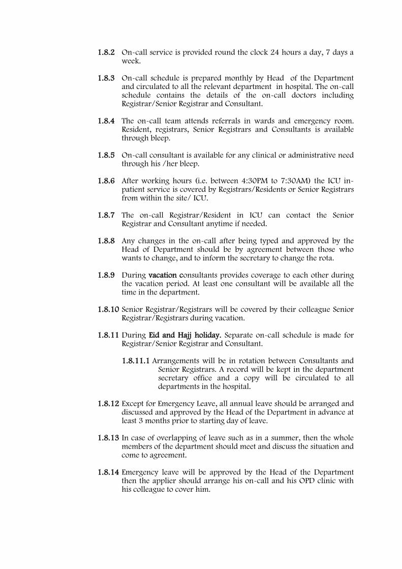
1.8.2 On-call service is provided round the clock 24 hours a day, 7 days a week.
1.8.3 On-call schedule is prepared monthly by Head of the Department and circulated to all the relevant department in hospital. The on-call schedule contains the details of the on-call doctors including Registrar/Senior Registrar and Consultant.
1.8.4 The on-call team attends referrals in wards and emergency room. Resident, registrars, Senior Registrars and Consultants is available through bleep.
1.8.5 On-call consultant is available for any clinical or administrative need through his /her bleep.
1.8.6 After working hours (i.e. between 4:30PM to 7:30AM) the ICU in-patient service is covered by Registrars/Residents or Senior Registrars from within the site/ ICU.
1.8.7 The on-call Registrar/Resident in ICU can contact the Senior Registrar and Consultant anytime if needed.
1.8.8 Any changes in the on-call after being typed and approved by the Head of Department should be by agreement between those who wants to change, and to inform the secretary to change the rota.
1.8.9 During vacation consultants provides coverage to each other during the vacation period. At least one consultant will be available all the time in the department.
1.8.10 Senior Registrar/Registrars will be covered by their colleague Senior Registrar/Registrars during vacation.
1.8.11 During Eid and Hajj holiday. Separate on-call schedule is made for Registrar/Senior Registrar and Consultant. 1.8.11.1 Arrangements will be in rotation between Consultants and
Senior Registrars. A record will be kept in the department secretary office and a copy will be circulated to all departments in the hospital.
1.8.12 Except for Emergency Leave, all annual leave should be arranged and
discussed and approved by the Head of the Department in advance at least 3 months prior to starting day of leave.
1.8.13 In case of overlapping of leave such as in a summer, then the whole members of the department should meet and discuss the situation and come to agreement.
1.8.14 Emergency leave will be approved by the Head of the Department then the applier should arrange his on-call and his OPD clinic with his colleague to cover him.

1.9 COMMUNICATION AND REPORTING
1.9.1 Only Arabic and English languages are to be spoken in the hospital.
1.9.2 Physicians’ are encouraged to learn as much Arabic as possible relative to their clinical area.
1.9.3 Within the department
1.9.3.1 It is the policy of the Critical Care department to improve the care provided to patients thru an effective communication system among all personnel working in the department. Meetings are convened regularly in order to discuss about provided care and all problems encountered aiming to improve service.
1.9.3.2 Regular department academic and administrative meetings are conducted, chaired by Head of the Department or his designee, attended by ICU team members. Others will be invited if needed.
1.9.3.3 The agenda of the meeting will include specific points related to improving the service provided, as well as special concerns of the staff and any problems encountered during course of patient care.
1.9.3.4 In the meeting all points included in the agenda will be discussed. Further points will be suggested by attendants for discussion in the next meeting.
1.9.3.5 Plan for suggested action to sort out problems and improve quality will be approved and duties will be assigned.
1.9.3.6 Summary of the minutes of the meeting, suggested plan and assignment of duties will be finalized and included in the appropriate logbook.
1.9.3.7 All such meeting will be recorded in the appropriate logbook.
1.9.3.8 All unusual incidences will be reported by filling up the incident report form and will be communicated to the appropriate authority.
1.9.4 Communication with patients/family/community 1.9.4.1 Family members are continuously informed by ICU
Registrars/Consultant regarding the condition and prognosis of the patient. This is documented in the Physicians progress note.
1.9.4.2 Where matters of importance need to be communicated it is advisable to obtain an Arabic speaker, eg. Translator to ensure that the patient receives the correct message.

1.9.5 Communication with other departments 1.9.5.1 Referral forms are forwarded from and to the ICU
department from other department. A copy of the referral form is kept in the patient’s file.
1.9.5.2 Meetings are held with the members of other department as dictated by the patient’s condition.

KING KHALID UNIVERSITY HOSPITAL DEPARTMENT OF CRITICAL CARE INTENSIVE CARE UNITS
Head of the Department
MICU Consultants
Interns
&
Students
Clinical Pharmacist
s
Respiratory Therapist
Interns
&
Students
Residents
Registrars
Senior Registrars
SICU Consultants
Residents
Registrars
Senior Registrars
Social Worker
Physiotherapist Dietitian
ORGANIZATIONAL CHART

Page 1 of 7 CCD-ICU IPP – 001 ADMISSION CRITERIA - ICU
King Khalid University Hospital
Department : Unit: Policy Number:
Critical Care Intensive Care CCD-ICU IPP - 001
Title: Issue Date: JUNE 2010 Prepared/Revised by: Date:
Admission Criteria - ICU Revision Date:
Dr. S. H. Ahmad & Dr. F. Azfar ICU IPP’s Committee
Effective Date: JUNE 2010 Due for Revision on: JUNE 2012
Reviewed by: Date: Authorized by: Date: Authorized by: Date:
Dr. Farheen Shaikh Policy and Procedure Review Committee
Dr. Badr Al Jabri KKUH-Medical Director
Prof. Abdul Aziz Alzeer Department Head
Authorized by: Date: Authorized by: Date: Authorized by: Date:
Dr. Ayman Abdo Vice Dean for Quality
Dr. Abdulaziz Al Saif Vice Dean for Hospitals
Prof. Mussaad M.S. Al-Salman Dean of College
1.0 Conditions:
All Physicians and Registered Nurses in the Intensive Care Unit.
2.0 Purpose:
To admit to ICU patients who are likely to benefit from ICU care in order to ensure appropriate utilization of ICU resources.
3.0 Policy:
3.1 The Adult Intensive Care Unit provides care for adult critically ill patients (over 12 years). Every effort is made to facilitate the optimum care and placement of these patients.
3.2 ICU will provide care for patients with actual or potential vital system failure,
which appear reversible with the ICU support. Patients’ will be prioritized based on diagnosis and objective parameters and predicted benefit. The patients’ requiring intensive treatment has priority over terminally ill patients with poor prognosis.
3.3 Establishment of PRIORITY for ICU admission:
3.3.1 Priority 1 : Critically ill, unstable patients in need of life-saving
intensive treatment and nursing care such as ventilatory support, vasoactive drugs, aggressive volume resuscitation, etc. No limits are placed on therapy. Examples include: 3.3.1.1 Hypoxic or hypercapnopeic respiratory failure requiring
mechanical ventilation, aerosol treatment frequency every hour or less and/or supplemental oxygen of 100% by non rebreathing mask. These patients include those with impending failure.
3.3.1.2 Endocrine emergencies such as severe diabetic ketoacidosis requiring insulin infusion or thyrotoxicosis and adrenal insufficiency with hemodynamic instability.
3.3.1.3 Shock states of any kind as defined by inadequate tissue perfusion.

Page 2 of 7 CCD-ICU IPP – 001 ADMISSION CRITERIA - ICU
3.3.1.4 Acute neurologic events requiring frequent neurological or respiratory checks to evaluate progression.
3.3.1.5 Continuous arterial – venous hemofiltration.
3.3.1.6 Massive pulmonary embolism.
3.3.1.7 GI bleeding with risk of exsanguinations, ischemic myocardial event, stroke, etc.
3.3.1.8 Patient postoperative care when underlying background illness can be exacerbated and contribute to a postoperative complication.
3.3.1.9 Patients generally have no limits placed on the extent of therapy they are to receive.
3.3.2 Priority 2: Critically ill patients who have potential immediate risk of
requiring priority 1 care. These patients require intensive monitoring and may potentially need immediate intervention. No therapeutic limits are generally stipulated for these patients. Examples include:
3.3.2.1 Acute GI bleed at risk of rebleed.
3.3.2.2 Uncomplicated vascular surgery.
3.3.2.3 Drug overdose in patients whose hemodynamic,
respiratory and neurologic states are stable.
3.3.2.4 Sub massive P.E.
3.3.2.5 COPD, OSA requiring BIPAP.
3.3.2.6 Acute pancreatitis.
3.3.3 Priority 3: Critically ill patients with chronic illness with or without superimposed acute illness, because of limited physiologic reserve, are less likely to survive or be benefited greatly from intensive care. Care may be limited to supportive and comfort measures. Their need may be for more intensive nursing care delivery rather than acute medical care. 3.3.3.1 These unstable patients are critically ill but have a
reduced likelihood of recovery because of underlying disease or nature of their acute illness.
3.3.3.2 Patients may receive intensive treatment to relieve acute illness but limits on therapeutic efforts may be set such as no intubation or cardiopulmonary resuscitation.
3.3.4 Priority 4: These patients are generally not appropriate for ICU
admission. Admission of these patients should be on an individual basis, and at the discretion of the ICU head of department. These patients can be placed in the following categories:

Page 3 of 7 CCD-ICU IPP – 001 ADMISSION CRITERIA - ICU
3.3.4.1 Little or no anticipated benefit from ICU care based on low risk of active intervention that could not safely be administered in a non-ICU setting.
3.3.4.2 Patients with terminal and irreversible illness facing imminent death (too sick to benefit from ICU care.)
3.4 Establishment of patient DIAGNOSIS :
3.4.1 Patient with severe potential or actual life threatening condition that includes but is not limited to the following disease of organ systems are considered for admission.
3.4.1.1 Pulmonary System
3.4.1.1.1 Acute respiratory failure requiring ventilatory
support.
3.4.1.1.2 Pulmonary emboli with hemodynamic instability.
3.4.1.1.3 Patients in an intermediate care unit who are demonstrating respiratory deterioration.
3.4.1.1.4 Need for nursing/respiratory care not available in lesser care areas such as floor or intermediate care unit.
3.4.1.1.5 Massive hemoptysis
3.4.1.1.6 Respiratory failure with imminent intubation.
3.4.1.2 Neurologic Disorders 3.4.1.2.1 Acute stroke with altered mental status.
3.4.1.2.2 Coma: metabolic, toxic, or anoxic.
3.4.1.2.3 Intracranial hemorrhage with potential for
herniation.
3.4.1.2.4 Acute subarachnoid hemorrhage.
3.4.1.2.5 Meningitis with altered mental status or respiratory compromise.
3.4.1.2.6 Central nervous system or neuromuscular disorders with deteriorating neurologic or pulmonary function.
3.4.1.2.7 Status epilepticus
3.4.1.2.8 Brain dead or potentially brain dead patients who are being aggressively managed while determining organ donation status.

Page 4 of 7 CCD-ICU IPP – 001 ADMISSION CRITERIA - ICU
3.4.1.2.9 Vasospasm
3.4.1.2.10 Severe head injured patients.
3.4.1.3 Drug Ingestion and Drug Overdose 3.4.1.3.1 Hemodynamically unstable drug ingestion.
3.4.1.3.2 Drug ingestion with significantly altered
mental status with inadequate airway protection.
3.4.1.3.3 Seizures following drug ingestion.
3.4.1.4 Gastrointestinal Disorders 3.4.1.4.1 Life threatening gastrointestinal bleeding
including hypotension, angina, continued bleeding or with co morbid conditions.
3.4.1.4.2 Fulminant hepatic failure.
3.4.1.4.3 Severe pancreatitis
3.4.1.4.4 Esophageal perforation with or without mediastinitis.
3.4.1.5 Endocrine
3.4.1.5.1 Diabetic ketoacidosis complicated by
hemodynamic instability, altered mental status, respiratory insufficiency or severe acidosis.
3.4.1.5.2 Thyroid storm or myxedema come with hemodynamic instability
3.4.1.5.3 Hyperosmolar state with come and /or hemodynamic instability.
3.4.1.5.4 Other endocrine problems such as adrenal crises with hemodynamic instability.
3.4.1.5.5 Severe hypercalcemia with altered mental status, requiring hemodynamic monitoring.
3.4.1.5.6 Hypo or hypernatremia with seizures, altered mental status.
3.4.1.5.7 Hypo or hypermagnesemia with hemodynamic compromise or dysrhythmias.
3.4.1.5.8 Hypo or hyperkalemia with dysrhythmias or muscular weakness.

Page 5 of 7 CCD-ICU IPP – 001 ADMISSION CRITERIA - ICU
3.4.1.5.9 Hypophospatemia with muscular weakness.
3.4.1.6 Surgical 3.4.1.6.1 Post-operative patients requiring
hemodynamic monitoring/ventilatory support or extensive nursing care.
3.4.1.7 Miscellaneous
3.4.1.7.1 Septic shock with hemodynamic instability
3.4.1.7.2 Hemodynamic monitoring
3.4.1.7.3 Clinical conditions requiring ICU level nursing
care.
3.4.1.7.4 Environmental injuries (lightning, near drowning, hypo/hyperthermia).
3.4.1.7.5 New/experimental therapies with potential for complications.
3.5 Establish OBJECTIVE Parameters:
3.5.1 Patients with the following parameters are considered for admission:
3.5.1.1 Vital Signs
3.5.1.1.1 Pulse <50 or >150 beats per minute.
3.5.1.1.2 Systolic arterial pressure <80mmHg or
20mmHg below the patient’s usual pressure.
3.5.1.1.3 Mean arterial pressure <60 mmHg.
3.5.1.1.4 Diastolic arterial pressure >120 mmHg
3.5.1.1.5 Respiratory rate >35 or <10 breaths per minute.
3.5.1.2 Laboratory Values (newly discovered)
3.5.1.2.1 Serum Sodium <130 mEq/L or >155 mEq/L
3.5.1.2.2 Serum Potassium <3 mEq/L or > 6 mEq/L
3.5.1.2.3 PaO2 m<60 mmHg
3.5.1.2.4 PaCO2 <25 mmHg or > 50 mmHg
3.5.1.2.5 pH <7.2 or >7.6
3.5.1.2.6 Serum glucose > 800mg/dL

Page 6 of 7 CCD-ICU IPP – 001 ADMISSION CRITERIA - ICU
3.5.1.2.7 Serum Calcium > 15mg/dL
3.5.1.2.8 BUN > 35 mmol/L
3.5.1.2.9 Creatinine > 88 mmol/L
3.5.1.2.10 Urine output <20 cc/hr
3.5.1.2.11 Toxic level of drug or other chemical substance in a hemodynamically or neurologically compromised patient.
3.5.1.3 Radiography/Ultrasonography/Tomography (newly
discovered). 3.5.1.3.1 Cerebral vascular hemorrhage, contusion or
subarachnoid hemorrhage with altered mental status or focal neurological signs.
3.5.1.3.2 Ruptured viscera, bladder, liver, esophageal varices or uterus with hemodynamic instability
3.5.1.3.3 Dissecting aortic aneurysm. 3.5.1.4 Physical Findings (Acute Onset)
3.5.1.4.1 Unequal pupils in an unconscious patient.
3.5.1.4.2 Burns covering > 10% BSA
3.5.1.4.3 Anuria
3.5.1.4.4 Airway obstruction
3.5.1.4.5 Coma
3.5.1.4.6 Continuous seizures
3.5.1.4.7 Cyanosis
3.5.1.4.8 Cardiac tamponade
3.6 Patients will be admitted to ICU based on diagnosis and objective parameters
after prioritizing them for likely benefit.
3.7 Admission to the ICU requires written order after approval of the ICU consultant.
3.8 Patient would be admitted or discharged strictly on their potential to benefit from ICU care.
3.9 Decisions should be made explicitly, and without bias.
3.10 Ethnic origin, race, sex, social status or financial status will not be considered in admission decisions.

Page 7 of 7 CCD-ICU IPP – 001 ADMISSION CRITERIA - ICU
4.0 Forms and Attachment:
4.1 Priority and Ranking System for Admission.
5.0 Reference

Page 1 of 2
CCD-ICU IPP – 002 ADMISSION COMMUNICATION
King Khalid University Hospital
Department : Unit: Policy Number:
Critical Care Intensive Care CCD-ICU IPP - 002
Title: Issue Date: JUNE 2010 Prepared/Revised by: Date:
Admission Communication Revision Date:
Dr. H. Al Otair / ICU IPP’s Committee
Effective Date: JUNE 2010 Due for Revision on: JUNE 2012
Reviewed by: Date: Authorized by: Date: Authorized by: Date:
Dr. Farheen Shaikh Policy and Procedure Review Committee
Dr. Badr Al Jabri KKUH-Medical Director
Prof. Abdul Aziz Alzeer Department Head
Authorized by: Date: Authorized by: Date: Authorized by: Date:
Dr. Ayman Abdo Vice Dean for Quality
Dr. Abdulaziz Al Saif Vice Dean for Hospitals
Prof. Mussaad M.S. Al-Salman Dean of College
1.0 Conditions:
All Physicians, Registered Nurses and Ward Clerks in the Intensive Care Unit.
2.0 Purpose:
To ensure all new admissions are effectively communicated to the relevant departments.
3.0 Policy:
All patients will be admitted according to the department admission policy guidelines and unit protocols.
4.0 Procedure:
4.1 Admission to MICU is arranged by on-cal ICU Senior Registrar/Consultant 24 hours a day.
4.2 Patient admitted directly from the Accident and Emergency Department or
transferred from another hospital must be accepted by the primary Physician on-call before being admitted to MICU.
4.3 Patient is managed by the MICU consultant while in the MICU. After transfer
to the medical ward patient’s care will be back to the primary consultant. 4.4 Admission of a patient from another hospital must be arranged with the ICU
Consultant and admission desk before patient’s transfer. 4.5 Admission dispute must be referred to the on-call ICU consultant and
medical on-call Consultant 4.6 Once admitted the computer will be appropriately checked by the Ward
Clerk / ICU Registered Nurse to ensure the patient’s information has been correctly entered by the Admission Department.

Page 2 of 2
CCD-ICU IPP – 002 ADMISSION COMMUNICATION
4.7 Patient’s profiles will be entered into the computer upon admission to the unit, relevant patient information required by the pharmacy and the dietary department will be entered on HIS by a Registered Nurse or the designee as instructed.
4.8 The ICU Physicians responsible for the patient’s care will be notified of the
admission by ICU Registered Nurse. 4.9 When applicable the Operating Room, Department Nursing staff will be
informed in the admitted is for imminent surgery. 4.10 Notification if the patient admissions to the Nursing Supervisors on duty
undertaken as follows: 4.10.1 DEM will alert supervisors of admission to the unit after 4:30PM.
4.10.2 The ward nurses will then inform the supervisor of the patient’s
admission to the unit, giving up to date information on the patient’s condition.
5.0 Reference:
Nursing Broad Policy Guidelines

Page 1 of 2
CCD-ICU IPP – 003 DISCHARGE CRITERIA - ICU
King Khalid University Hospital
Department : Unit: Policy Number:
Critical Care Intensive Care CCD-ICU IPP - 003
Title: Issue Date: JUNE 2010 Prepared/Revised by: Date:
discharge Criteria - ICU Revision Date:
Dr. H. Al Otair / ICU IPP’s Committee
Effective Date: JUNE 2010 Due for Revision on: JUNE 2012
Reviewed by: Date: Authorized by: Date: Authorized by: Date:
Dr. Farheen Shaikh Policy and Procedure Review Committee
Dr. Badr Al Jabri KKUH-Medical Director
Prof. Abdul Aziz Alzeer Department Head
Authorized by: Date: Authorized by: Date: Authorized by: Date:
Dr. Ayman Abdo Vice Dean for Quality
Dr. Abdulaziz Al Saif Vice Dean Hospitals
Prof. Mussaad M.S. Al-Salman Dean of College
1.0 Conditions:
All Physicians and Registered Nurses in the Intensive Care Unit.
2.0 Purpose:
To discharge from ICU patients who are no longer in need of ICU care in order to ensure appropriate utilization of ICU resources.
3.0 Policy:
3.1 The patients will be discharged from ICU to the general ward once they no
longer need ICU care. 3.1.1 When a patient’s physiologic status has stabilized and the need for
ICU monitoring and care is no longer necessary.
3.1.2 When a patient’s physiological status has deteriorated and active interventions are no longer considered.
3.2 Transfer/discharge will be based on the following criteria: 3.2.1 Stable hemodynamic parameters.
3.2.2 Stable respiratory status (patient extubated with stable arterial
blood gases) and airway patency.
3.2.3 Oxygen requirements not more than 60%
3.2.4 Intravenous inotropic support, vasodilators, vasopressors drugs are no longer required.
3.2.5 Patient on low dose inotropic support may be discharged earlier if ICU bed is needed.
3.2.6 Intracranial pressure monitoring equipment has been removed.
3.2.7 Neurologic stability with control seizures.
3.2.8 Removal of all hemodynamic monitoring catheters.

Page 2 of 2
CCD-ICU IPP – 003 DISCHARGE CRITERIA - ICU
3.2.9 Chronically mechanically ventilated patients whose critical illness
has been reversed or resolved.
3.2.10 Patients with mature artificial airways (tracheostomies) who no longer require excessive suctioning.

CCD-ICU IPP – 004 DISCHARGE COMMUNICATION Page 1 of 1
King Khalid University Hospital
Department : Unit: Policy Number:
Critical Care Intensive Care CCD-ICU IPP - 004
Title: Issue Date: JUNE 2010 Prepared/Revised by: Date:
DISCHARGE COMMUNICATION
Revision Date: Dr. H. Al Otair ICU IPP’s Committee
Effective Date: JUNE 2010
Due for Revision on: JUNE 2012
Reviewed by: Date: Authorized by: Date: Authorized by: Date:
Dr. Farheen Shaikh Policy and Procedure Review Committee
Dr. Badr Al Jabri KKUH-Medical Director
Prof. Abdul Aziz Alzeer Department Head
Authorized by: Date: Authorized by: Date: Authorized by: Date:
Dr. Ayman Abdo Vice Dean for Quality
Dr. Abdulaziz Al Saif Vice Dean Hospitals
Prof. Mussaad M.S. Al-Salman Dean of College
1.0 Conditions:
All Physicians in the Intensive Care Unit.
2.0 Purpose:
To define Physician’s responsibility upon patients discharge. 3.0 Policy:
Patient’s discharge from ICU will be carried out thru proper, communication with patient, family, primary care team.
4.0 Procedure:
4.1 Conscious patient should be notified in advance of pending discharge.
4.2 Where applicable, relatives must be informed of patient’s pending discharge.
4.3 When the patient has left the unit, discharge information is fed into the
computer, effectively informing Medical Records, Pharmacy and Dietary Departments.
4.4 All discharges from the Intensive Care Unit must be approved by the on-call ICU consultant.
4.5 At discharge from ICU the patient will be immediately accepted by the primary team.
4.6 Primary care teams must be informed of all patient discharged and any potential or continuing problems by ICU Registrar
4.7 If appropriate, limitation/non-escalation of treatment must be clearly documented and discussed with the parent team prior to discharge.
4.8 A transfer note must be completed in the patients file before discharge.
5.0 Forms and Attachment:
5.1 Concurrence form – Discharge Communication

Page 1 of 4 CCD-ICU IPP – 005 TRANSPORT OF CRITICALLY ILL PATIENTS FROM ICU FOR DIAGNOSTIC AND THERAPEUTIC PROCEDURES
King Khalid University Hospital
Department : Unit: Policy Number:
Critical Care Intensive Care CCD-ICU IPP - 005
Title: Issue Date: JUNE 2010 Prepared/Revised by: Date:
Transport of Critically ill patients from ICU for Diagnostic and Therapeutic Procedures
Revision Date:
ICU IPP’s Committee Effective Date: JUNE 2010
Due for Revision on: JUNE 2012
Reviewed by: Date: Authorized by: Date: Authorized by: Date:
Dr. Farheen Shaikh Policy and Procedure Review Committee
Dr. Badr Al Jabri KKUH-Medical Director
Prof. Abdul Aziz Alzeer Department Head
Authorized by: Date: Authorized by: Date: Authorized by: Date:
Dr. Ayman Abdo Vice Dean for Quality
Dr. Abdulaziz Al Saif Vice Dean for Hspitals
Prof. Mussaad M.S. Al-Salman Dean of College
1.0 Conditions:
All Physicians and Registered Nurses in the Intensive Care Unit.
2.0 Purpose:
2.1 Transporting critically ill patients is necessary for many diagnostic or therapeutic procedures; however, transporting patient may be associated with risk. Continuous and effective monitoring of a patient’s ventilation, oxygenation and cardiopulmonary and hemodynamic status must be maintained at all times during transport.
2.2 This procedure describes the appropriate patient and equipment preparation
and monitoring required for the safe transport of critically ill patients. 3.0 Policy:
3.1 It is the responsibility of the physician and nurse to maintain patient safety
while transporting critically ill patient within the hospital.
3.2 INDICATION: 3.2.1 When diagnostic testing or therapeutic intervention requires
transport out of the intensive care unit. 3.3 CONTRAINDICATIONS:
3.3.1 Inability to :
3.3.1.1 Maintain patient’s airway during transport. 3.3.1.2 Provide adequate oxygenation and ventilation during transport. 3.3.1.3 Maintain hemodynamic stability during transport. 3.3.1.4 Adequately monitor the patient’s cardiopulmonary status during transport.
3.3.2 All other conditions in which transporting the patient is deemed life- threatening.

Page 2 of 4 CCD-ICU IPP – 005 TRANSPORT OF CRITICALLY ILL PATIENTS FROM ICU FOR DIAGNOSTIC AND THERAPEUTIC PROCEDURES
3.4 PRECAUTIONS: 3.4.1 All procedures for the proper set up, maintenance, and use of all
equipment for transport must be strictly followed. The inappropriate use of any of this equipment may lead to patient compromise.
3.4.2 Some patents may not tolerate movement and/or changes in ventilator support. A trial of body movement, manual ventilation, or application of the Achieve a Oxylog transport ventilator in the intensive care unit is warranted to ensure patient tolerance.
3.5 ADVERSE REACTIONS AND INTERVENTIONS:
3.5.1 Movement may result in accidental extubation and loss of patient
airway. Should accidental extubation occur, immediately institute oxygenation and ventilatory support via a resuscitation mask and manual resuscitator. Assist with re-intubation as necessary.
3.5.2 Movement may result in accidental removal of vascular access devices and/ or unintended discontinuation of pharmacologic support. Hemodynamic instability may result in susceptible patients. Notify the physician and nurse immediately.
3.5.3 Position changes may result in hypotension, hypercarbia, and hypoxemia. Monitor patients throughout all transport maneuvers and during diagnostic/therapeutic procedures. Allow time for patient recovery to baseline vital signs as needed throughout transport procedures.
3.5.4 Hyperventilation or hypoventilation during manual ventilation may cause detrimental changes in acid-base status resulting in cardiac dysrythmias, hypoxemia, and/or hypotension. Susceptible patients should be monitored with cardiopulmonary monitoring and pulse oximetry.
3.5.5 Equipment failures may result in inaccurate data, loss of monitoring capabilities, and patient compromise. Follow all manufacturers' instructions for the maintenance of transport and monitoring equipment, and ascertain proper function of all equipment prior to departure from the intensive care unit.
3.5.6 Loss of PEEP/CPAP may result in hypoxemia. Monitor PEEP/CPAP levels via appropriate pressure monitoring devices in susceptible patients. Ensure rapid, smooth transitions from mechanical ventilation to manual ventilation.
3.5.7 Loss of the patient's oxygen supply may result in hypoxemia. Always ascertain oxygen tank capacity prior to departure and bring extra tanks as needed to ensure an adequate supply.

Page 3 of 4 CCD-ICU IPP – 005 TRANSPORT OF CRITICALLY ILL PATIENTS FROM ICU FOR DIAGNOSTIC AND THERAPEUTIC PROCEDURES
4.0 Equipments:
4.1 Emergency airway management supplies.
4.1.1 Appropriately sized oral airways 4.1.2 Laryngoscope 4.1.3 Endotracheal tubes 4.1.4 Stylet 4.1.5 Portable suction
4.2 Portable oxygen with appropriate oxygen delivery device.
4.3 Manual resuscitator with mask and PEEP valve.
4.4 Pulse Oximeter.
4.5 Cardiopulmonary transport monitor, transducer cables, and modules.
4.6 Emergency pharmacologic agents.
4.7 Stethoscope.
4.8 Universal precautions attire.
5.0 Procedure:
5.1 PERSONNEL
5.1.1 All mechanically ventilated patients in ICU must be accompanied by a
ICU physician and a nurse for diagnostic procedures.
5.1.2 All mechanically ventilated patients in ICU must be accompanied by an anesthesiologist and a nurse for all types of surgical procedures to and from the OR.
5.1.3 The team must be proficient in operation and troubleshooting all of the equipment.
5.2 Gather and assemble all respiratory equipments. Maintain electrical power to portable ventilator prior to departure to ensure the maximum charge of the batteries.
5.3 Set appropriate alarm limits for all parameters.
5.4 Monitor the patient throughout the transport for the adequacy of oxygenation and ventilation, assure hemodynamic stability and tolerance of the procedure, and monitor all mechanical ventilator parameters as indicated to ensure patient safety.
5.5 If ventilatory facilities are available in the procedure room, these can be used to save the oxygen supply and battery of portable ventilator.

Page 4 of 4 CCD-ICU IPP – 005 TRANSPORT OF CRITICALLY ILL PATIENTS FROM ICU FOR DIAGNOSTIC AND THERAPEUTIC PROCEDURES
5.6 POST PROCEDURE:
5.6.1 Upon returning to the unit, place the patient on the appropriate bedside monitoring and respiratory equipment. Check and reset all necessary alarm parameters and ensure patient comfort.
5.6.2 Remove all transport equipment from the patient's room, disinfect as appropriate, and store transport ventilator with connection to AC power for recharging of the batteries.
5.6.3 Document the ventilator or oxygen settings prior to departing and upon returning to the unit.
5.6.4 Document any cardiopulmonary or hemodynamic changes that may have occurred during the transport on the "comments” side of the Ventilator flow sheet. Include the occurrence of adverse reactions and interventions that were made. Report this information to the next shift.
6.0 Forms and Attachment:
Concurrence Form – Transferring of Patients.
7.0 Reference:
AARC Clinical Practice Guideline: Transport of the Mechanically Ventilated Patient.

Page 1 of 2
CCD-ICU IPP – 006 MULTIDISCIPLINARY COLLABORATION IN THE ICU
King Khalid University Hospital
Department : Unit: Policy Number:
Critical Care Intensive Care CCD-ICU IPP - 006
Title: Issue Date: JUNE 2010 Prepared/Revised by: Date:
Multidisciplinary Collaboration in the ICU
Revision Date: Dr. F. Azfar and Dr. H. Al Otair /ICU IPP’s Committee
Effective Date: JUNE 2010
Due for Revision on: JUNE 2012
Reviewed by: Date: Authorized by: Date: Authorized by: Date:
Dr. Farheen Shaikh Policy and Procedure Review Committee
Dr. Badr Al Jabri KKUH-Medical Director
Prof. Abdul Aziz Alzeer Department Head
Authorized by: Date: Authorized by: Date: Authorized by: Date:
Dr. Ayman Abdo Vice Dean for Quality
Dr. Abdulaziz Al Saif Vice Dean Hospitals
Prof. Mussaad M.S. Al-Salman Dean of College
1.0 Conditions:
All ICU Physicians, ICU Registered Nurses, Respiratory Therapist, Clinical Pharmacist, Physical Physiotherapist and Social Worker.
2.0 Purpose:
2.1 The importance of collaboration and communication and its impact on patient outcomes in the ICU is well recognized by many national and international organizations.
2.2 When working together toward common goals, collaboration has been identified as a way of improving care for the critically ill patients as it enables input from the multidisciplinary team members in promoting decision-making based on more useful information.
3.0 Policy:
3.1 Collaboration should be encouraged and promoted in the ICU.
3.2 Multidisciplinary collaboration in the ICU is vital in ensuring appropriate
care and treatment of the critically ill patients as well as an important component for establishing and meeting patient care goals.
4.0 Procedure:
4.1 Daily round in the ICU are carried out by a multidisciplinary team which
consists of the ICU Physicians, ICU Nurses and Clinical Pharmacist.
4.2 Respiratory Therapist and Physical Physiotherapist shall join the round when the case of the patient to whom they are assigned is being discussed.
4.3 A Social Worker attends the daily duty round once a week and will also be invited as needed.
4.4 The Progress Note in the patient’s file is used to promote awareness of patient care goals and improve communication and collaboration in the ICU.
4.5 In daily rounds, the patient care goals for the day are discussed and areas that need addressing are indentified. This promotes collaboration among the

Page 2 of 2
CCD-ICU IPP – 006 MULTIDISCIPLINARY COLLABORATION IN THE ICU
ICU team members as it establishes priority areas of patient care and promotes further discussion throughout the day with updates for team members.
4.6 In working to achieve the patient goals identified in the daily round, ICU team members further collaborate to meet those goals. Each team member documents his/her suggestions and procedures undertaken in the daily progress note.
4.7 Participation of the multidisciplinary team in departmental teaching activities, research and quality improvement initiatives including formulating new protocols and implementing best practices are highly encouraged by ICU department.
5.0 Reference:
5.1 Baggs JG,Schmitt MH, Mushlin Al, et al. Association between nurse-physician collaboration and patient outcomes in three intensive care units, Crit Car Med 1999; 27: 1991-1998.
5.2 National Institute of Health. Consensus conference critical care medicine. JAMA 1983; 250: 789-804.
5.3 Joint Commission on accreditation of Hospitals. Accreditation Manual for Hospitals. 2007; JCAHO Chicago IL.
5.4 American Association of Critical Care Nurses. Collaborative Practice Model: The Organization of Human Resources in Critical Care Units. AACN: Newport Beach CA.
5.5 McCauley K, Irwin RS. Changing the work environment in intensive care units to achieve patient-focused care: the time has come. Am J Crit Care 2006;15:541-548.
5.6 Davidson JE, Powers K, Hedayat KM, et al. Clinical practice guidelines for support of the family in the patient-centered intensive care unit: American College of Critical Car Medicine Taskforce 2004-2005. Crit Care Med 2007;35:605-622.
5.7 Barnato AE, Kahn JM, Rubenfeld G. et al. Prioritizing the organization and management of intensive care services in the United States: The PrOMIS Conference. Crit Care Med 2007;35-1003-1011.
5.8 Baggs JG, Ryan SA, :Phelps CE, Richeson JF, Johnson JE. The association between interdisciplinary collaboration and patient outcomes in a medical intensive care unit. Heart & Lung 1993;21:18-24.
5.9 Baggs JC, Ryan SA, Phelps CE, et al. The association between interdisciplinary collaboration and patient outcomes in a medical intensive care unit. Heart Lung 1993;21:18-24.

Page 1 of 1
CCD-ICU IPP – 007 WORKING HOURS, ON CALL HOURS
King Khalid University Hospital Department : Unit: Policy Number:
Critical Care Intensive Care CCD-ICU IPP - 007
Title: Issue Date: JUNE 2010 Prepared/Revised by: Date:
WORKING HOURS, ON CALL HOURS
Revision Date: Dr. H. Al Otair ICU IPP’s Committee
Effective Date: JUNE 2010
Due for Revision on: JUNE 2012
Reviewed by: Date: Authorized by: Date: Authorized by: Date:
Dr. Farheen Shaikh Policy and Procedure Review Committee
Dr. Badr Al Jabri KKUH- Medical Director
Prof. Abdul Aziz Alzeer Department Head
Authorized by: Date: Authorized by: Date: Authorized by: Date:
Dr. Ayman Abdo Vice Dean for Quality
Dr. Abdulaziz Al Saif Vice Dean for Hospitals
Prof. Mussaad M.S. Al-Salman Dean of College
1.0 Conditions:
All Physicians in the Critical Care Department.
2.0 Purpose:
To define regular working hours and on call hours.
3.0 Policy:
3.1 Staffs are requested to strictly adhere to the scheduled hospital working hours (07:30AM -04:30PM).
3.2 Leaving the unit or hospital early, without permission from the Consultant on duty is a disciplinary offence.
3.3 During a normal nine (9) hour shift staffs are entitled 45 minutes lunch and Prayer break.
3.4 Unauthorized absence from the unit or ward is not allowed.
3.5 All Physicians will work not less than 55 hours/week.
3.6 All staff is expected to be prompt in reporting for duty.
3.7 Early departure from duty may be given at the Head of Department’s discretion.
3.8 On Call duties start 4:30PM till 7:30AM next day.
3.9 On Call Physicians are not allowed to leave the unit till another Physician is present in the unit.
3.10 The On Call Physician will stay in the on call room provided and extension of the room is made known to the nursing staff.

Page 1 of 2
CCD-ICU IPP – 008 INTERNAL JUGULAR VEIN CANNULATION
King Khalid University Hospital
Department : Unit: Policy Number:
Critical Care Intensive Care CCD-ICU IPP - 008
Title: Issue Date: JUNE 2010 Prepared/Revised by: Date:
INTRAJUGULAR VEIN CANNULATION
Revision Date: Dr. S. H. Ahmad /ICU IPP’s Committee
Effective Date: JUNE 2010
Due for Revision on: JUNE 2012
Reviewed by: Date: Authorized by: Date: Authorized by: Date:
Dr. Farheen Shaikh Policy and Procedure Review Committee
Dr. Badr Al Jabri KKUH-Medical Director
Prof. Abdul Aziz Alzeer Department Head
Authorized by: Date: Authorized by: Date: Authorized by: Date:
Dr. Ayman Abdo Vice Dean for Quality
Dr. Abdulaziz Al Saif Vice Dean for Hospitals
Prof. Mussaad M.S. Al-Salman Dean of College
1.0 Conditions:
All Physicians and Registered Nurses in the Intensive Care Unit.
2.0 Purpose:
Provide direct venous access for administration of fluids/medications, blood products, blood sampling, hemodynamic monitoring and total parental nutrition.
3.0 Definitions:
A catheter placed percutaneously in the internal jugular vein with its tip lying near right atrium.
4.0 Policy:
To provide central venous access for monitoring and administration of fluids drugs and medications under aseptic conditions and proper precautions by ICU physicians.
5.0 Procedure:
5.1 Explain procedure to patient/ obtain verbal consent.
5.2 Put patient in trendelenburg or flat with head turned towards opposite side for maximum exposure of sternocleidomastoid triangle.
5.3 Ensure trolley for IJV cannulation has been prepared properly.
5.4 Hand wash, don cap, mask, gloves, gown aseptically.
5.5 Prepare area off around cannulation site with povidone 10% and manorapid.
5.6 Use a large drape to completely cover the body exposing only cannulation site.
5.7 Use 20-22G needle to locate IJV to minimize chances of arterial puncture using landmark technique.

Page 2 of 2
CCD-ICU IPP – 008 INTERNAL JUGULAR VEIN CANNULATION
5.8 Cannulate IJV using seldinger technique.
5.9 Flush all ports of central line catheter.
5.10 Dispose of sharps.
5.11 Order post procedure chest X-ray and check it for position and pneumothorax or other complications.
6.0 Reference:
6.1 Manual of intensive care medicine. Irwin and Rippe; Fourth edition.
6.2 CDC. Guidelines for the prevention of intravascular-catheter related infections.2002:51(RR10).

Page 1 of 2
CCD-ICU IPP – 009 SUBCLAVIAN VEIN CANNULATION
King Khalid University Hospital
Department : Unit: Policy Number:
Critical Care Intensive Care CCD-ICU IPP - 009
Title: Issue Date: JUNE 2010 Prepared/Revised by: Date:
SUBCLAVIAN VEIN CANNULATION
Revision Date: Dr. S.H. Ahmad /ICU IPP’s Committee
Effective Date: JUNE 2010
Due for Revision on: JUNE 2012
Reviewed by: Date: Authorized by: Date: Authorized by: Date:
Dr. Farheen Shaikh Policy and Procedure Review Committee
Dr. Badr Al Jabri KKUH-Medical Director
Prof. Abdul Aziz Alzeer Department Head
Authorized by: Date: Authorized by: Date: Authorized by: Date:
Dr. Ayman Abdo Vice Dean for Quality
Dr. Abdulaziz Al Saif Vice Dean for Hospitals
Prof. Mussaad M.S. Al-Salman Dean of College
1.0 Conditions:
All Physicians and Registered Nurses in the Intensive Care Unit.
2.0 Purpose:
To describe safe insertion of cannula in the subclavian vein.
3.0 Definitions:
Subclavian venous catheter placed percutaneously in the subclavian vein with its tip lying near right atrium.
4.0 Policy:
Subclavian venous catheter is used to provide central venous access for monitoring and administration of fluids/medications, blood products, blood sampling, haemodynamic monitoring and total parental nutrition under aseptic conditions and proper precautions by ICU physicians.
5.0 Procedure:
5.1 Explain procedure to patient/obtain verbal consent.
5.2 Put patient in trendelenburg with towel rolled between shoulder blades to splay anterior chest wall up and out. Arms should be by the patient’s side; head turned away from cannulation site or in a neutral position.
5.3 Hand wash, don cap, mask, gloves, gown aseptically.
5.4 Prepare area of/around cannulation site with povidone10% and manorapid, from mandible and neck down to costal margin, from anterior axillary line to 5cm beyond contra lateral to opposite sternal border.
5.5 Use a large drape to completely cover the body exposing only cannulation site.
Page 2 of 2
CCD-ICU IPP – 009 SUBCLAVIAN VEIN CANNULATION
5.6 Identify landmarks; sternal notch and transition point between middle and medial thirds of clavicle.
5.7 Place the index finger of guiding hand at the sterna notch and thumb at transition point between middle and medial thirds of clavicle. Anthesthetize the skin and subcutaneous tissue just inferior to the clavicle and lateral to thumb.
5.8 Enter the skin with introducer needle, bevel up, lateral to the thumb and inferior to clavicle, aspirating while advancing, aiming at the index finger
5.9 If clavicle is contacted, depress the entire needle with the thumb until it passes under the clavicle, rather than changing angle of approach. The needle is kept as close to the inferior edge of the clavicle as possible to avoid puncturing the dome of pleura.
5.10 Once appropriate venous return is noted, rotate the bevel of the needle inferiorly. Remove the syringe and insert guide wire. Hold the guide wire and remove introducer needle.
5.11 Using a scalpel, make a small nick in the skin at entry site. Pass the dilator over the guide wire, dilate the tract, and remove the dilator.
5.12 Ensure distal port of catheter is open. Pass the catheter over guide wire. When the catheter is near the entry site, feed the guide wire out until it emerges from distal port on catheter. Grasp guide wire distally, and insert the catheter to the desired depth.
5.13 Hold catheter in place and remove guide wire. Flush all ports to ensure that they are functioning properly. Secure the catheter with sutures, cleanse the site and apply sterile dressing.
5.14 Obtain a stat CXR for placement.
6.0 Reference:
6.1 Miller's Anesthesia 7th edition.churchill livingstone
6.2 Current Diagnosis & Treatment: Critical Care.third edition.Lange.

Page 1 of 3
CCD-ICU IPP – 010 PULMONARY ARTERY CATHETERIZATION (PAC)
King Khalid University Hospital
Department : Unit: Policy Number:
Critical Care Intensive Care CCD-ICU IPP - 010
Title: Issue Date: JUNE 2010 Prepared/Revised by: Date:
PULMONARY ARTERY CATHETERIZATION (PAC)
Revision Date: Dr. S. H. Ahmad ICU IPP’s Committee
Effective Date: JUNE 2010
Due for Revision on: JUNE 2012
Reviewed by: Date: Authorized by: Date: Authorized by: Date:
Dr. Farheen Shaikh Policy and Procedure Review Committee
Dr. Badr Al Jabri KKUH-Medical Director
Prof. Abdul Aziz Alzeer Department Head
Authorized by: Date: Authorized by: Date: Authorized by: Date:
Dr. Ayman Abdo Vice Dean for Quality
Dr. Abdulaziz Al Saif Vice Dean for Hospitals
Prof. Mussaad M.S. Al-Salman Dean of College
1.0 Conditions:
All Physicians in the Intensive Care Unit.
2.0 Purpose:
To describe safe insertion of pulmonary artery catheter (PAC)when indicated.
3.0 Definitions:
PAC is a flexible, balloon-tipped, flow-directed catheter that is guided through the right side of the heart and into a branch of the pulmonary artery.
4.0 Policy:
PAC is used for measurement of filling pressures and cardiac output to confirm the diagnosis and optimize use of IV fluids, inotropic agents, and vasopressors by allowing direct, simultaneous measurement of pressures in the right atrium, right ventricle, pulmonary artery, and the filling pressure ("wedge" pressure) of the left atrium.the benefits of PAC should outweigh the risks of procedure. Done by physicians well versed with the procedure.
5.0 Procedure:
5.1 Gain central venous access using appropriate size introducer using sterile
technique. (See policy on central venous cannulation).
5.2 The introducer catheter is inserted using seldinger technique with the difference that dilator is advanced through introducer rather than as a separate piece. Additionally, guide wire and dilator are removed together at the conclusion of the introducer insertion leaving introducer into vessel and secure it.
5.3 After placing the introducer place fresh sterile drape completely covering the patient and leavening the introducer exposed.
5.4 Flush all ports of PAC then check for leaks and integrity of balloon.
5.5 Attach PAC to pressure transducer and flush prior to insertion. Wave the catheter tip prior to insertion with verification of wave form on monitor.

Page 2 of 3
CCD-ICU IPP – 010 PULMONARY ARTERY CATHETERIZATION (PAC)
5.6 Check that the protective sheath has been inserted over the catheter before
insertion; orient the natural curve in catheter to match the projected course in vasculature.
5.7 Advance PAC through introducer and inflate the balloon when in right atrium (approx. 20 cm) and activate lock inflating the syringe. Monitor the wave forms and EKG on monitor continuously. A right atrial wave form on the monitor confirms the proper location of catheter tip.
5.8 Monitoring the waveform, advance the catheter; waveform will increase in amplitude as catheter will enter right ventricle(RV) which occurs at approximately at 30cm (from right IJV approach)
5.9 Keep advancing the catheter till Pulmonary artery (PA) pressure tracing appears identified by an increase in diastolic pressure and development of dicrotic notch in pressure tracing at approximately 40cm.
5.10 If excessive catheter length has been advanced without this transition occurring; most likely catheter has coiled in right ventricle. If this occurs the balloon should be deflated and withdrawn until the right atrial waveform reappears. Re-inflate the balloon and attempt the procedure again.
5.11 Advancement beyond PA position results in a fall on PA pressure tracing from levels of systolic pressure noted in RV and PA. When this is noted, record pulmonary artery occlusion pressure (PAOP) and deflate balloon.
5.12 Phasic PA pressure should reappear on pressure tracing when balloon is deflated. If this does not occur deflate balloon and pull back1-2 cm until PA tracing reappears.
5.13 Carefully record the balloon inflation volume to change pressure from PA to PAOP tracing. If a wedge tracing is obtained and balloon is only partially inflated, signifying the catheter tip is too distal, the catheter should be withdrawn 1-2cm with balloon deflated.
5.14 Perform balloon inflation procedure to obtain a wedge tracing optimally at full balloon inflation. If there is no trace obtained with full inflation of balloon, the catheter should be advanced with balloon inflated till a wedge tracing is obtained.
5.15 The catheter should never be advanced with balloon deflated and should never be withdrawn with balloon inflated.
5.16 Once insertion is completed, the distance of insertion from catheter introducer should be noted and recorded.
5.17 Catheter should be secured and sterile dressing applied.
5.18 Obtain post procedure CXR to verify path of catheter, tip position and exclude complications, like pneumothorax.
6.0 Forms and Attachments:

Page 3 of 3
CCD-ICU IPP – 010 PULMONARY ARTERY CATHETERIZATION (PAC)
6.1 Traces
6.2 Indications
6.3 Contra indications
7.0 Reference:
7.1 Miller's Anesthesia 7th edition. churchill livingstone.
7.2 Practice Guidelines for Pulmonary Artery Catheterization Anesthesiology, V No 4, Oct 2003.

Page 1 of 2
CCD-ICU IPP – 011 FEMORAL VEIN CANNULATION
King Khalid University Hospital
Department : Unit: Policy Number:
Critical Care Intensive Care CCD-ICU IPP - 011
Title: Issue Date: JUNE 2010 Prepared/Revised by: Date:
FEMORAL VEIN CANNULATION
Revision Date: Dr. S. H. Ahmad ICU IPP’s Committee
Effective Date: JUNE 2010
Due for Revision on: JUNE 2012
Reviewed by: Date: Authorized by: Date: Authorized by: Date:
Dr. Farheen Shaikh Policy and Procedure Review Committee
Dr. Badr Al Jabri KKUH-Medical Director
Prof. Abdul Aziz Alzeer Department Head
Authorized by: Date: Authorized by: Date: Authorized by: Date:
Dr. Ayman Abdo Vice Dean for Quality
Dr. Abdulaziz Al Saif Vice Dean for Hospitals
Prof. Mussaad M.S. Al-Salman Dean of College
1.0 Conditions:
All Physicians and Registered Nurses in the Intensive Care Unit.
2.0 Purpose:
Provide direct venous access for administration of fluids/medications, blood products, blood sampling, hemodynamic monitoring and total parental nutrition.
3.0 Definitions:
A catheter placed percutaneously in the femoral vein. 4.0 Policy:
Provide central venous access for monitoring and administration of fluids drugs and medications under aseptic conditions and proper precautions by ICU physicians.
5.0 Procedure:
5.1 Explain procedure to patient/ obtain verbal consent.
5.2 Put patient in trendelenburg, ideally should be flat and supine.
5.3 Position the patients’ leg in slight frog-leg position to open up inguinal fossa.
5.4 Hand wash, don cap, mask, gloves, gown aseptically.
5.5 Prepare area off around cannulation site with povidone10% and manorapid. 10 cm above inguinal ligament, medially to scrotum or labia majora, Inferiorly 15 cm below inguinal ligament, laterally to anterior superior iliac supine.
5.6 Use a large drape to completely cover the body exposing only cannulation site.

Page 2 of 2
CCD-ICU IPP – 011 FEMORAL VEIN CANNULATION
5.7 Palpate femoral artery below inguinal ligament at the junction of its medial and middle third. Make venipuncture 1-1.5 cm medial to femoral artery 2-3 cm below inguinal ligament.
5.8 Insert needle at an angle of 45-60 degrees. Establish venipuncture and confirm free aspiration of blood.
5.9 Pass a guide wire and a dilator, thread catheter over guide wire after removing dilator.
5.10 Flush all ports of central line catheter.
5.11 Dispose of sharps. 6.0 Reference:
6.1 Manual of intensive care medicine. Irwin and Rippe; Fourth edition.
6.2 CDC. Guidelines for the prevention of intravascular-catheter related
infections.2002:51 (RR10).

Page 1 of 2
CCD-ICU IPP – 012 THORACENTESIS
King Khalid University Hospital
Department : Unit: Policy Number:
Critical Care Intensive Care CCD-ICU IPP - 012
Title: Issue Date: JUNE 2010 Prepared/Revised by: Date:
THORACENTESIS
Revision Date: Dr. S. H. Ahmad ICU IPP’s Committee
Effective Date: JUNE 2010
Due for Revision on: JUNE 2012
Reviewed by: Date: Authorized by: Date: Authorized by: Date:
Dr. Farheen Shaikh Policy and Procedure Review Committee
Dr. Badr Al Jabri KKUH-Medical Director
Prof. Abdul Aziz Alzeer Department Head
Authorized by: Date: Authorized by: Date: Authorized by: Date:
Dr. Ayman Abdo Vice Dean for Quality
Dr. Abdulaziz Al Saif Vice Dean for Hospitals
Prof. Mussaad M.S. Al-Salman Dean of College
1.0 Conditions:
All Physicians in the Intensive Care Unit.
2.0 Purpose:
2.1 Patients who have a pleural effusion should undergo diagnostic thoracentesis: 2.1.1 To determine the nature of the effusion (ie, transudate, exudate).
2.1.2 To identify potential causes (eg, malignancy, infection) and tailor the
treatment accordingly.
3.0 Definitions:
Thoracentesis is the introduction of a needle, cannula, or trocar into the pleural space to remove accumulated fluid or air.
4.0 Policy:
4.1 Patients with pleural effusion will undergo diagnostic thoracentesis to
determine the nature of the effusion if not caused by congestive heart failure or does not progress as anticipated.
4.2 Patients with pleural effusion will undergo therapeutic thoracentesis if the effusion results from complicated parapneumonic effusion or leads to respiratory embarassesment or (more than 1cm width).
5.0 Procedure:
5.1 Obtain lateral decubitus CXR to confirm free flowing pleural effusion.
5.2 Obtain informed written consent for procedure.
5.3 Position the patient sitting at the edge of bed, leaning forward over a pillow-
draped bedside table with arms crossed in front. An assistant will support the patient.

Page 2 of 2
CCD-ICU IPP – 012 THORACENTESIS
5.4 Mark the inferior tip of the scapula on the side to be tapped with patients arms by side.
5.5 Percuss the patients’ chest posteriorly for highest point of effusion. Identify the intercostal space below this, mark superior aspect of the rib in posterior axillary line.
5.6 Using sterile technique, cleanse and drape the area surrounding the puncture site.
5.7 Anesthetize the skin and deeper soft tissues with 2% lignocaine aiming for top of the rib.
5.8 Attach a three-way stopcock to a 20G, 1.5 inch needle and to a 50 ml syringe.
5.9 Insert the 20G needle along the anesthetic tract, aspirating as the needle is advanced. When plural fluid is obtained, fill a heparinised blood gas syringe from other port of three way stopcock and send for pH measurement.
5.10 Fill 50ml syringe and transfer its contents into appropriate collection tubes and containers.
5.11 Always maintain a closed system during procedure to prevent air entering the plural space.
5.12 After completing the procedure remove needle from chest, apply pressure for few minutes, and apply sterile dressing.
5.13 Analysis of Pleural Fluid should include: 5.13.1 Grams stain c/s 5.13.2 AGB stains c/s 5.13.3 Cell count differential 5.13.4 pH, Protein, Albumin, LDH, cholesterol, Triglycerides. 5.13.5 Cytology
5.14 Draw venous blood for analysis/comparison with plural fluid analysis.
5.15 Obtain a post procedure CXR.
5.16 For therapeutic thoracentesis in which removal of large volume of pleural
fluid is desired, a catheter is placed in the pleural space and fixed with sutures. Pleural fluid is allowed to drain into a collecting bag, closed system is always maintained.
6.0 Reference:
6.1 Irwin and Rippe. “Manual of intensive care medicine.” Fourth edition.
6.2 Upto Date.com

Page 1 of 6
CCD-ICU IPP – 013 CHEST TUBE INSERTION
King Khalid University Hospital
Department : Unit: Policy Number:
Critical Care Intensive Care Unit CCD-ICU IPP - 013
Title: Issue Date: JUNE 2010 Prepared/Revised by: Date:
Chest Tube Insertion
Revision Date: Dr. A. Al Sharif ICU IPP’s Committee
Effective Date: JUNE 2010
Due for Revision on: JUNE 2012
Reviewed by: Date: Authorized by: Date: Authorized by: Date:
Dr. Farheen Shaikh Policy and Procedure Review Committee
Dr. Badr Al Jabri KKUH-Medical Director
Prof. Abdul Aziz Alzeer Department Head
Authorized by: Date: Authorized by: Date: Authorized by: Date:
Dr. Ayman Abdo Vice Dean for Quality
Dr. Abdulaziz Al Saif Vice Dean for Hospitals
Prof. Mussaad M.S. Al-Salman Dean of College
1.0 Conditions:
All Physicians and Registered Nurses in the Intensive Care Unit.
2.0 Purpose:
To minimize the complications of chest tube insertion in critically ill patient whenever it is indicated.
3.0 Definitions:
CHEST TUBE INSERTION (TUBE THORACOSTOMY) involves placement of a sterile tube into the pleural space to evacuate air or fluid into a closed collection system to restore negative intrathoracic pressure, promote lung expansion, and prevent potentially lethal levels of pressure from developing in the thorax.
4.0 Policy:
4.1 Critically ill patients will undergo chest tube insertion and drainage
whenever it is indicated by trained physicians.
4.2 Before performing the procedure, it is important to review the steps to be taken and to ensure that all necessary equipment is available. Patient comfort and safety are paramount.
5.0 Equipment:
5.1 Povidone 10% and Manorapid
5.2 Sterile towels and drapes
5.3 Sterile sponges
5.4 1% lidocaine without epinephrine (40 mL)
5.5 10-mL syringe
5.6 18-, 21-, and 25-gauge needles
5.7 2 Kelly clamps
5.8 Mayo scissors

Page 2 of 6
CCD-ICU IPP – 013 CHEST TUBE INSERTION
5.9 Standard tissue forceps
5.10 Towel forceps
5.11 Needle holder
5.12 0-Silk suture with cutting needle
5.13 Scalpel handle and no. 10 blade
5.14 Chest tubes (24, 28, 32, and 36 French)
5.15 Chest tube drainage system (filled appropriately)
5.16 Petrolatum gauze
5.17 2 in. adhesive tape
5.18 Sterile gowns and gloves, masks, caps
6.0 Procedure:
6.1 The indications for closed intercostal drainage to palliate a chronic disease
process (e.g., drainage of malignant pleural effusions) or to relieve an acute, life-threatening process (e.g., decompression of a tension pneumothorax). Chest tubes also may provide a vehicle for pharmacologic interventions, as when used with antibiotic therapy for treatment of an emphysema or to instill sclerosing agents to prevent recurrence of malignant effusions.
6.2 Obtain informed written consent for the procedure except in life threatening situations.
6.3 Obtain lateral decubitus CXR to confirm free flowing pleural effusion.
6.4 Choose the Proper size of chest tube as follows:
6.4.1 Adult Male 28-32 Fr
6.4.2 Adult Female 28 Fr
6.4.3 Child 18 Fr
6.4.4 Newborn 12-14 Fr.
6.5 With the patient supine and the head of the bed adjusted for comfort, the involved side is elevated slightly with the ipsilateral arm brought up over the head plus Supplemental oxygen is administered as needed.
6.6 The tube is usually inserted through the fourth or fifth intercostal space in the anterior axillary line. An alternative entry site (for decompression of a pneumothorax) is the second intercostal space in the midclavicular line, but for cosmetic reasons and to avoid the thick pectoral muscles, the former site is preferable in adults.
6.7 Under sterile conditions, the area is prepared with povidone10% and manorapid, and after allowing it to dry, is draped to include the nipple, which serves as a landmark.
6.8 A 2- to 3-cm area is infiltrated with 1% lidocaine to raise a wheal two finger breadths below the intercostal space to be penetrated. (This allows for a subcutaneous tunnel to develop, through which the tube will travel, and discourages air entry into the chest following removal of the tube.)

Page 3 of 6
CCD-ICU IPP – 013 CHEST TUBE INSERTION
6.9 A 2-cm transverse incision is made at the wheal, and additional lidocaine is administered to infiltrate the tissues through which the tube will pass, including a generous area in the intercostal space (especially the periosteum of the ribs above and below the targeted interspace).
6.10 Care should be taken to anesthetize the parietal pleura fully, as it (unlike the visceral pleura) contains pain fibers. Each injection of lidocaine should be preceded by aspiration of the syringe to prevent injection into the intercostal vessels. Up to 30 to 40 mL lidocaine may be needed to achieve adequate local anesthesia.
6.11 To confirm the location of air or fluid, a thoracentesis is then performed at the proposed site of tube insertion. If air or fluid is not aspirated, the anatomy should be reassessed and chest radiographs and CT scans reexamined before proceeding.
6.12 A short tunnel is created to the chosen intercostal space, using Kelly clamps. After the intercostal muscles are bluntly divided, the closed clamp is carefully inserted through the parietal pleura, hugging the superior portion of the lower rib to prevent injury to the intercostal bundle of the rib above. The clamp is placed to a depth of less than 1 cm to prevent injury to the intrathoracic structures and is spread open approximately 2 cm.
6.13 A finger is inserted into the pleural space to explore the anatomy and confirm proper location and lack of pleural symphysis. Only easily disrupted adhesions should be broken. Bluntly dissecting strong adhesions may tear the lung and initiate potentially troublesome bleeding from the systemic circulation.
6.14 The chest tube is inserted into the pleural space and positioned apically for a pneumothorax and dependently for fluid removal. All holes must be confirmed to be within the pleural space. The use of undue pressure or force to insert the tube should be avoided.
6.15 The location of the tube should be confirmed by observing flow of air (seen as condensation within the tube) or fluid from the tube. It is then sutured to the skin securely to prevent slippage. A horizontal mattress suture can be used to allow the hole to be tied closed when the tube is removed. An occlusive gauze dressing is applied.
6.16 The tube is connected to a drainage apparatus and securely taped to the dressing and to the patient. All connections between the patient and the drainage apparatus must also be tight and securely taped.
6.17 Obtain post procedure CXR to confirm proper site of the chest tube.
6.18 While a chest tube is in place, the tube and drainage system must be checked daily for adequate functioning. For pleural fluid we use a three-chambered system that contains a calibrated collection trap for fluid and for pneumothorax an underwater seal unit to allow escape of air while maintaining negative pleural pressure. Suction is routinely established at 15 to 20 cm water, controlled by the height of the column in the suction regulator unit, and maintained as long as an air leak is present.
6.19 The drainage system is examined daily to ensure that appropriate levels are maintained in the underwater seal and suction regulator chambers. If suction is desired, bubbling should be noted in the suction regulator unit.

Page 4 of 6
CCD-ICU IPP – 013 CHEST TUBE INSERTION
Connections between the chest tube and the drainage system should be tightly fitted and securely taped. For continuous drainage, the chest tube and the tubing to the drainage system should remain free of kinks, should not be left in a dependent position, and should never be clamped. The tube can be milked and gently stripped, although with caution, as this may injure adjacent tissues .Irrigation of the tube is discouraged.
6.20 Dressing changes should be performed every 2 or 3 days and as needed. Adequate pain control is mandatory to encourage coughing and ambulation, to facilitate lung expansion.
6.21 Serial chest films are obtained routinely to evaluate the progress of drainage and to ensure that the most proximal drainage hole has not migrated from the pleural space (a situation that may result in pneumothorax or subcutaneous emphysema). If this occurs and the pathologic process is not corrected, replacement of the tube is usually indicated, especially if subcutaneous emphysema is developing.
6.22 The chest tube should never be re-advanced into the pleural space, and if a tube is to be replaced it should always be at a different site rather than through the same hole. If a pneumothorax persists, increasing the suction level may be beneficial, but an additional tube may be required if no improvement results. Proper positioning may also be confirmed by chest CT scanning.
6.23 Contraindication for chest tube insertion is infection at the site of insertion or uncontrolled bleeding diathesis

Page 5 of 6
CCD-ICU IPP – 013 CHEST TUBE INSERTION
7.0 Forms and Attachments:
7.1 Algorithm of Chest Tube insertion
7.2 Indications for chest tube insertion
1-Pneumothorax Spontaneous Traumatic Necrotizing pneumonia Interstitial fibrosis Malignancy Primary Metastatic Bullous emphysema Pulmonary infarction Iatrogenic Central line placement Positive-pressure ventilation Thoracentesis
2-Hemothorax Traumatic Blunt Penetrating Iatrogenic Malignancy Primary Metastatic Infectious Pulmonary arteriovenous malformation Spontaneous pneumothorax Blood dyscrasias Ruptured thoracic aortic aneurysm
3-Empyema Parapneumonic Posttraumatic Postoperative Septic emboli Intraabdominal infection
4-Chylothorax Traumatic
Blunt Penetrating Surgical
Congenital Malignancy Miscellaneous
Filariasis Tuberculosis Subclavian vein obstruction
5-Pleural effusion Transudate Exudate
Malignancy Postoperative Iatrogenic Immunologic Inflammatory
8.0 Reference:
8.1 Interventional Management of Pleural Infections. Heffner, John E.; Klein,
Jeffrey S., Hampson, Christopher. Chest. 136(4):1148-1159, October 2009
8.2 The occult pneumothorax: What have we learned?. Ball, Chad G.; Kirkpatrick, Andrew W; Feliciano, David V. Canadian Journal of Surgery. 52(5):173-179, October 2009.
8.3 New classification and clinical characteristics of reexpansion pulmonary edema after treatment of spontaneous pneumothorax. Kim, Yun Kwon; Kim, Hyun Lee, Christopher C.; Choi, Han Joo; Lee, Kang Hyun; Hwang, Sung Oh; Oh, Joong Hwan; Lee, Young Han; Singer, Adam J. American Journal of Emergency Medicine. 27(8):961-967, October 2009.

Page 6 of 6
CCD-ICU IPP – 013 CHEST TUBE INSERTION
8.4 Simple Aspiration Compared to Chest Tube Insertion in the Management of Primary Spontaneous Pneumothorax. Gaudio, Mark; Hafner, John W., Annals of Emergency Medicine. 54(3):458-460, September 2009.
8.5 Chest drain insertion in pleural effusion. Crossingham, Iain,; Taggart, Simon Clinical Medicine. 9(3):290, June 2009.
8.6 Chest drain insertion: Improving techniques and decreasing complications. Pearce, Andrew P .Emergency Medicine Australasia. 21(2):91-93, April 2009.
8.7 Pleural Interventions: Management of Acute and Chronic Pneumothorax. Rahman, Najib M.; Davies, Robert J.O; Gleeson, Fergus V. Seminars in Respiratory & Critical Care Medicine. Interventional Radiology in Pulmonary and Critical Care Medicine. 29(4):427-440, August 2008.

Page 1 of 4
CCD-ICU IPP – 014 CHEST TUBE REMOVAL.
King Khalid University Hospital
Department : Unit: Policy Number:
Critical Care Intensive Care CCD-ICU IPP – 014
Title: Issue Date: JUNE 2010 Prepared/Revised by: Date:
CHEST TUBE REMOVAL
Revision Date: Dr. A. Al Sharif
/ ICU IPP’s Committee Effective Date: JUNE 2010
Due for Revision on: JUNE 2012
Reviewed by: Date: Authorized by: Date: Authorized by: Date:
Dr. Farheen Shaikh Policy and Procedure Review Committee
Dr. Badr Al Jabri KKUH-Medical Director
Prof. Abdul Aziz Alzeer Department Head
Authorized by: Date: Authorized by: Date: Authorized by: Date:
Dr. Ayman Abdo Vice Dean for Quality
Dr. Abdulaziz Al Saif Vice Dean for Hospitals
Prof. Mussaad M.S. Al-Salman Dean of College
1.0 Conditions:
All Physicians and Registered Nurses in the Intensive Care Unit.
2.0 Purpose:
Minimize the complications and patient discomfort during chest tube removal
3.0 Policy:
3.1 Chest tube removal is indicated whenever there is resolution of the pneumothorax or pleural effusion or whatever the cause it was inserted for.
3.2 Before performing the procedure, it is important to review the steps to be taken and to ensure that all necessary equipment is available. Patient comfort and safety are paramount.
4.0 Equipment:
4.1 Chlorhexidine or povidone-iodine solution. 4.2 Sterile towels and drapes 4.3 Sterile sponges 4.4 1% lidocaine without epinephrine (40 mL) 4.5 10-mL syringe 4.6 18-, 21-, and 25-gauge needles 4.7 Mayo scissors 4.8 Standard tissue forceps 4.9 Towel forceps 4.10 Needle holder 4.11 Scalpel handle and no. 10 blade 4.12 Petrolatum gauze 4.13 2-in. adhesive tape 4.14 Sterile gowns and gloves, masks, caps
5.0 Procedure:
5.1 Chest tube removal is indicated if the amount of drainage has decreased
significantly while on water seal, which resolves the need for chest drainage,

Page 2 of 4
CCD-ICU IPP – 014 CHEST TUBE REMOVAL.
the drainage system is no longer holding suction (as indicated by air leak in drainage system), the chest tube is clogged and unable to be cleared.
5.2 The precautions for chest tube removal are: re-expansion of lung(s) on chest radiograph without other abnormalities, bilateral breath sounds present, improved respiratory status and absence of fluctuations or bubbling in the water seal chamber. Normal coagulation studies (if ordered). Finally the site of the chest tube should be carefully inspected before removal to identify the suture(s) and to look for any signs of infection or inflammation.
5.3 Explain simply to the patient the procedure and obtain lateral decubitus CXR. Put the patient in semi-Fowlers’ position (if tolerable).
5.4 Tube removal is often preceded by oral or parenteral analgesia at an appropriate time interval.
5.5 Under sterile conditions, the area is prepared with povidone10% and manorapid and after allowing it to dry, is draped properly.
5.6 For a pneumothorax, the drainage system is left on suction until the air leak stops. If an air leak persists, brief clamping of the chest tube can be performed to confirm that the leak is from the patient and not the system.
5.7 If, after several days, an air leak persists, placement of an additional tube may be indicated. When the leak has ceased for more than 24 to 48 hours (or if no fluctuation is seen in the underwater seal chamber), the drainage system is placed on water seal by disconnecting the wall suction, followed by a chest film several hours later.
5.8 If no pneumothorax is present and no air leak appears in the system with coughing, deep breathing, and reestablishment of suction, the tube can be removed.
5.9 For pleural effusion, the tube can be removed when drainage is minimal, unless sclerotherapy is planned.
5.10 The suture holding the tube to the skin is cut and removed if knotted or if it is purse-string do not remove but use for closing incision. As the patient takes deep breath and holds it, the tube is gently and firmly removed in single motion. Apply direct pressure to the hole with dressing for at least 2 minutes or until bleeding or drainage have subsided. The hole simultaneously covered with an occlusive petrolatum gauze dressing at peak inspiration (at which point only positive pressure can be generated in the pleural space, minimizing the possibility of drawing air in).
5.11 If the incision is large and purse-string suture was not originally placed, administer local anesthetic and close incision with suture.
5.12 A chest radiograph is performed immediately to check for a pneumothorax and is repeated 24 hours later to rule out re-accumulation of air or fluid.
5.13 Properly dispose of the chest tube catheter and other materials.
5.14 Our recommendations for chest tube removal are:

Page 3 of 4
CCD-ICU IPP – 014 CHEST TUBE REMOVAL.
5.14.1 Level 1 recommendation 5.14.1.1 Chest tube (CT) drainage should be ≤ 2ml/kg or ≤ 200 ml
or <10ml/hr x 6hr pre-removal (whichever is less) before a CT removal is removed.
5.14.2 Level 2 recommendation 5.14.2.1 Chest tube can be removed equally safely at end-
inspiration or end-expiration.
5.14.2.2 Chest tube may be safely removed on suction.
5.14.2.3 A brief trial of water seal prior to CT removal may allow occult air leaks to become clinically apparent and reduce the need for CT reinsertion due to recurrent pneumothorax. Such trials, however, will generally increase hospital length of stay and the number of chest X-rays (CXRs) obtained.
5.14.2.4 After pulmonary resection, small air leaks will resolve significantly more quickly if the CT is placed to water seal.
5.14.3 Level 3 recommendation
5.14.3.1 In non-mechanically ventilated patients, a routine CXR following removal of a CT is generally not indicated. The decision to obtain a CXR should be based on the individual clinical situation and the patient’s signs and symptoms.
5.14.3.2 In mechanically ventilated patients, a CXR obtained between one and three hours after removal of a CT is sufficient to identify a recurrent pneumothorax.
5.14.3.3 A daily CXR is not indicated to monitor CTs in the intensive care unit. Routine monitoring and patient care will identify the need for CXR based on clinical necessity.
6.0 Forms and Attachments:
Chest tube management algorithm 7.0 Reference:
7.1 Varela G, Jimenez MF, Novoa NM, Aranda JL. Postoperative chest
tubemanagement: measuring air leak using an electronic device decreasesvariability in the clinical practice. Eur J Cardiothorac Surg 2009;35:28—31.
7.2 Cerfolio RJ, Bryant AS, Maniscalco LM. A nondivided intercostal muscleflap further reduces pain of thoracotomy: a prospective randomized trial. Ann Thorac Surg 2008;85:1901—7.
7.3 Cerfolio RJ, Bryant AS. Results of a prospective algorithm to remove chesttubes after pulmonary resection with high output. J Thorac Cardiovasc Surg 2008;135:269—73.

Page 4 of 4
CCD-ICU IPP – 014 CHEST TUBE REMOVAL.
7.4 Cerfolio RJ et al. Prospective Randomized Trial Compares Suction vs. Water
Seal for Air Leaks.
7.5 Ann Thoracic Surg 2001; 71:1613-1617.
7.6 Marshall MB et al. Suction vs. Water Seal After Pulmonary Resection: A Randomized Prospective Study. Chest 2002: 121:831-835.
7.7 McCormick JT et al. The Use of Routine Chest X-Ray Films after CT Removal in Postoperative Cardiac Patients. Ann Thorac Surg 2002; 74:2161-2164.
7.8 Pizano LR et al. When Should Chest Radiograph be Obtained after CT Removal in Mechanically
7.9 Ventilated Patients? A Prospective Study. J Trauma, 2002:53:1073-1077.
7.10 Younes RN, et al. When to Remove a CT? A Randomized Study with Subsequent Prospective Consecutive Validation. J Am Coll Surg. 2002; 195:658-662.
7.11 Davis, J et al. Randomized Study of Algorithms for Discontinuing Tube Thoracostomy Drainage. J Am Coll Surg 1994; 179:553-557
7.12 Bell R et al. CT Removal: End-Inspiration or End-Expiration? J Trauma; 2001; 50:674-676.
7.13 Martino K et al. Prospective Randomized Trial of Thoracostomy Removal Algorithms. J Trauma.1999; 46:369-373.
7.14 Pacanowski J et al. Is Routine Roentgenography Needed after Closed Tube Thoracostomy Removal. J Trauma 2000; 48:684-688.
7.15 Palesty JA et al. The Efficacy of X-rays after CT Removal. Am J Surg 2000; 179:13-16.
7.16 Pacharn P et al. Are Chest Radiographs Routinely Necessary Following Thoracostomy Tube Removal. Pediatric Radiology; 202; 32:138-142.
7.17 McCormick JT et al. The Use of Routine Chest X-Ray Films after CT Removal in Postoperative Cardiac Patients. Ann Thorac Surg 2002; 74:2161-2164.
7.18 Pizano LR et al. When Should Chest Radiograph be obtained after CT Removal in Mechanically Ventilated Patients? A Prospective Study. J Trauma, 2002:53:1073-1077.

Page 1 of 2
CCD-ICU IPP – 015 PARACENTESIS (ASCITIC TAP)
King Khalid University Hospital
Department : Unit: Policy Number:
Critical Care Intensive Care CCD-ICU IPP - 015
Title: Issue Date: JUNE 2010 Prepared/Revised by: Date:
PARACENTESIS (ASCITIC TAP)
Revision Date: Dr. S. H. Ahmad ICU IPP’s Committee
Effective Date: JUNE 2010
Due for Revision on: JUNE 2012
Reviewed by: Date: Authorized by: Date: Authorized by: Date:
Dr. Farheen Shaikh Policy and Procedure Review Committee
Dr. Badr Al Jabri KKUH-Medical Director
Prof. Abdul Aziz Alzeer Department Head
Authorized by: Date: Authorized by: Date: Authorized by: Date:
Dr. Ayman Abdo Vice Dean for Quality
Dr. Abdulaziz Al Saif Vice Dean for Hospitals
Prof. Mussaad M.S. Al-Salman Dean of College
1.0 Conditions:
All Physicians in the Intensive Care Unit.
2.0 Purpose:
Paracentesis is undertaken to evaluate for etiology, to alleviate respiratory compromise or abdominal pain due to ascites.
3.0 Definitions:
Paracentesis is a procedure in which a needle or catheter is inserted into the peritoneal cavity to obtain ascitic fluid for diagnostic or therapeutic purposes.
4.0 Policy:
4.1 Paracentesis is performed for diagnostic and therapeutic purposes by trained ICU Physician: 4.1.1 To differentiate transudate from exudates.
4.1.2 To determine SAAG (Serum albumin Ascites Gradient).
4.1.3 To check for presence of malignant cells
4.1.4 To relive pressure effects.
5.0 Procedure:
5.1 Document presence of ascites.
5.2 Explain procedure and obtain consent.
5.3 Using sterile technique, cleanse and drape the area surrounding the puncture site.
5.4 Puncture site approximately 3cm from umbilicus lateral to rectus muscle in lower quadrant.
5.5 Anaesthetize with 2%lignocaine.

Page 2 of 2
CCD-ICU IPP – 015 PARACENTESIS (ASCITIC TAP)
5.6 Retract skin gently (to create a Z-tract) to attenuate the risk of leakage of ascetic fluid at the completion of procedure.
5.7 Under sterile conditions, percutaneously insert paracentesis catheter via seldinger technique into peritoneal cavity.
5.8 If constant drainage is not required, an 18-22G needle may be used.
5.9 Place sterile dressing over puncture site.
5.10 Send the fluid for diagnostic studies.
5.11 Analysis of Pleural Fluid should include: 5.11.1 Grams stain c/s
5.11.2 AGB stains c/s
5.11.3 Cell count differential
5.11.4 pH, Protein, Albumin, LDH, cholesterol, Triglycerides.
5.11.5 Cytology
5.12 Draw blood venous for chemistry analysis for comparison with ascites fluid
analysis.
6.0 Reference:
6.1 Irwin and Rippe. “Manual of intensive care medicine.” Fourth edition.

Page 1 of 4
CCD-ICU IPP – 016 ARTERIAL LINE INTRA-ARTERIAL CATHETERIZATION AND PRESSURE MONITORING
King Khalid University Hospital
Department : Unit: Policy Number:
Critical Care Intensive Care CCD-ICU IPP - 016
Title: Issue Date: JUNE 2010 Prepared/Revised by: Date:
ARTERIAL LINE – INTRA ARTERIAL CATHETERIZATION AND PRESSURE MONITORING
Revision Date:
ICU IPP’s Committee Effective Date: JUNE 2010
Due for Revision on: JUNE 2012
Reviewed by: Date: Authorized by: Date: Authorized by: Date:
Dr. Farheen Shaikh Policy and Procedure Review Committee
Dr. Badr Al Jabri KKUH-Medical Director
Prof. Abdul Aziz Alzeer Department Head
Authorized by: Date: Authorized by: Date: Authorized by: Date:
Dr. Ayman Abdo Vice Dean for Quality
Dr. Abdulaziz Al Saif Vice Dean for Hospitals
Prof. Mussaad M.S. Al-Salman Dean of College
1.0 Conditions:
All Physicians and Registered Nurses in the Intensive Care Unit.
2.0 Purpose:
Proper placement of intra-arterial catheter. 3.0 Policy:
3.1 All intra-arterial catheter should be properly inserted and used for blood
pressure monitoring
3.2 An arterial catheter is to be inserted by a physician qualified in the procedure, following an Allen’s test.
3.3 The patient’s safety and comfort will be maintained by the nurse during the procedure.
3.4 Aseptic technique will be maintained by the nurse and MD throughout the procedure.
3.5 The arterial catheter alarm system will remain activated at all times. Alarm parameters should be set at 20 above and 10 below the patient’s normal arterial pressure.
3.6 The pressure bag will be kept at 200-300 mm Hg to maintain catheter patency.
3.7 Monitoring equipment must be calibrated before the catheter is inserted and then every eight (8) hours.
3.8 Arterial catheter blood pressure reading is to be checked against a cuff pressure after insertion, every shift and PRN.
3.9 The entire monitoring set-up will be changed every 72 hours.
3.10 Gauze dressings will be changed every 72 hours and as required.
3.11 Peripheral arterial catheters should be changed every 96 hours. If arterial pressure monitoring is still required, a new site must be used for insertion.

Page 2 of 4
CCD-ICU IPP – 016 ARTERIAL LINE INTRA-ARTERIAL CATHETERIZATION AND PRESSURE MONITORING
3.12 Arterial line site checks must be documented every two (2) hours on the Nurses progress flow sheet. The physician will be notified of abnormal findings.
4.0 Equipments:
4.1 Arterial catheters, radial or femoral 4.2 Monitor module and pressure cable 4.3 Pressure tubing and bag 4.4 Standard Heparin solution 4.5 Sterile Gloves 4.6 Sterile drapes 4.7 Povidone 10% 4.8 Sterile 4x4 gauze 4.9 Skin dressing 4.10 Transducer holder 4.11 Suture set 4.12 One (1) 10 mL sterile syringe 4.13 BP cuff 4.14 Pre-heparinized syringe with needle samples and appropriate lab tubes for
ABGs or 10 mL syringe with needle for blood. 5.0 Procedure:
5.1 INSERTION:
5.1.1 Explain procedure to patient to alleviate fear and anxiety.
5.1.2 Identify the patient using two (2) patient identifiers.
5.1.3 The Physician will insert the arterial catheter under sterile conditions.
5.1.4 Apply label with date and time to Heparin bag and tubing. Check to
see all fittings are tight on tubing’s.
5.1.5 Flush tubing and transducer to remove all air from system to avoid false readings.
5.1.6 Connect the heparinized saline to pressurized tubing. Keep pressure bag at 300 mmHg to maintain catheter patency.
5.1.7 Set-Up: 5.1.7.1 Place pressure tubing on transducer holder 5.1.7.2 Attach transducer cable to pressure tubing 5.1.7.3 Level transducer air reference port at the same level as the
patient’s right atrium 5.1.7.4 Open stopcock of transducer to air 5.1.7.5 Push “zero” button on monitor module to calibrate 5.1.7.6 Close stopcock off to air and open to patient 5.1.7.7 Place 10 mL sterile syringe on stop cock 5.1.7.8 The distal stopcock must be covered with an injection cap 5.1.7.9 Turn stopcock nearest flush device off to transducer. Pull
back on 10 mL syringe until blood is approximately 12 inches past distal stopcock (at insertion site).

Page 3 of 4
CCD-ICU IPP – 016 ARTERIAL LINE INTRA-ARTERIAL CATHETERIZATION AND PRESSURE MONITORING
5.1.7.10 DO NOT allow blood to enter transducer. If the occurs, transducer must be changed.
5.1.8 To obtain accurate pressures, the transducer must be placed at a
constant reference point. A closed system is required for pressure readings.
5.1.9 Empty syringe in proximal stopcock by flushing the system.
5.1.10 Open proximal stopcock to patient and close to syringe. Flush system (to remove residual blood) until tubing clear.
5.1.11 Check pressure waveform and digital readout to ensure they are essentially the same as before sample was drawn. (If no waveform or readout, flush the system well and ensure stopcocks are aligned in correct position.)
5.1.12 Ensure alarm is on.
5.2 CHANGING MONITOR SYSTEM: 5.2.1 Assemble flush system per procedure.
5.2.2 Remove dressing over insertion site and clean area well with Betadine
solution.
5.2.3 Apply blood pressure cuff to affected limb and inflate pressure 10 mm above systolic blood pressure. Gently remove old pressure line at catheter hub and replace with new setup.
5.2.4 Deflate blood pressure cuff and remove. Redress insertion site using sterile techniques.
5.2.5 Recalibrate transducer per initial setup procedure.
5.2.6 Check waveform and digital blood pressure readout.
5.2.7 Tag monitoring line with time and date changed.
5.3 ARTERIAL LINE REMOVAL: 5.3.1 Arterial line may be removed by ICU Registered Nurse with Physician’s order.
5.3.2 Upon removal, hold adequate pressure for a minimum of five (5)
minutes to seal artery puncture site.
5.3.3 Assess site and the peripheral pulse of the affected limb for adequate circulation every 15 minutes for one (1) hour after removal.
6.0 Reference:
6.1 Clinical Nursing Skills, Basic to Advanced Skills, 5th Edition, 2000, Sandra F.
Smith, RN, MS, ABD; Donna J. Duell, RN, MS, ABN; Barbara C. Martin, RN, MS, CS.

Page 4 of 4
CCD-ICU IPP – 016 ARTERIAL LINE INTRA-ARTERIAL CATHETERIZATION AND PRESSURE MONITORING
6.2 Policies and Procedures for Infusion Nursing, 2nd Edition, Infusion Nurses
Society.

Page 1 of 3
CCD-ICU IPP – 017 ARTERIAL PUNCTURE FOR BLOOD GAS ANALYSIS
King Khalid University Hospital
Department : Unit: Policy Number:
Critical Care Intensive Care CCD-ICU IPP – 017
Title: Issue Date: JUNE 2010 Prepared/Revised by: Date:
ARTERIAL PUNCTURE FOR BLOOD GAS ANALYSIS
Revision Date:
ICU IPP’s Committee Effective Date: JUNE 2010
Due for Revision on: JUNE 2012
Reviewed by: Date: Authorized by: Date: Authorized by: Date:
Dr. Farheen Shaikh Policy and Procedure Review Committee
Dr. Badr Al Jabri KKUH-Medical Director
Prof. Abdul Aziz Alzeer Department Head
Authorized by: Date: Authorized by: Date: Authorized by: Date:
Dr. Ayman Abdo Vice Dean for Quality
Dr. Abdulaziz Al Saif Vice Dean for Hospitals
Prof. Mussaad M.S. Al-Salman Dean of College
1.0 Conditions:
All ICU Physicians and ICU Registered Nurses.
2.0 Purpose:
2.1 To obtain arterial blood sample to measure pH, p02, and pC02 to provide a means of assessing the adequacy of oxygenation and ventilation and the acid base status of a patient.
2.2 To quantitate the response to therapeutic intervention (eg, supplemental oxygen administration, mechanical ventilation) and/or diagnostic evaluation (e.g., exercise desaturation).
2.3 The need to monitor severity and progression of documented disease processes.
3.0 Definitions:
To monitor ventilation, oxygenation and acid base status. 4.0 Policy:
4.1 A verbal or written order must be obtained before obtaining arterial blood
gas samples.
4.2 ICU Physicians can perform radial artery punctures.
5.0 Procedure:
5.1 Gather equipment needed: 5.1.1 Pre-packaged ABG kit containing:
5.1.1.1 1.0 ml heparinized syringe.
5.1.1.2 25-gauge needle.
5.1.1.3 Rubber cube for corking needle.
5.1.1.4 Rubber stopper for syringe.

Page 2 of 3
CCD-ICU IPP – 017 ARTERIAL PUNCTURE FOR BLOOD GAS ANALYSIS
5.1.2 Additionally: 5.1.2.1 Povidone Iodine 10%
5.1.2.2 Alcohol swab.
5.1.2.3 2x2 gauze pad.
5.1.2.4 Transport bag with ice and water (slush).
5.1.2.5 Patient label.
5.2 Place all items on a firm surface close to you. It is helpful to open the betadine, alcohol and gauze pad before you begin.
5.3 Wash your hands.
5.4 Introduce yourself and explain the procedure.
5.5 Apply clean gloves.
5.6 Evaluate pulses for optimal site.
5.7 Examine site for: 5.7.1 Skin rash
5.7.2 Scarring
5.7.3 Trauma or abnormalities.
5.7.4 If present choose alternate site.
5.8 For radial sticks, perform an ALLEN’S TEST by:
5.8.1 Obliterating radial and ulnar pulse simultaneously at the wrist then
have patient clench and unclench fist until blanching occurs.
5.8.2 Release pressure on the ulnar then watch for return of the skin
colour.
5.8.3 If negative, choose alternate site.
5.9 For radial sticks:
5.9.1 Raise patient’s bed to a level that is comfortable.
5.9.2 Position patient’s arm extended flat with the palm up and a rolled
towel underneath the wrist to assist in flexing.
5.10 Cleanse the skin at the puncture site with betadine, let dry, then cleanse
again with alcohol.
5.11 With your non-dominate hand, palpate the artery with your index finger and locate the point of maximal pulsation.
5.12 While palpating the pulse, take the barrel of the sampling syringe in your dominate hand, holding the syringe like a pencil at 45o with the bevel up, slowly penetrate the skin aiming the end of the needle toward the pulsing artery.
5.13 Advance the needle in a straight line until you see a flash of blood in the hub of the syringe.
5.14 If you miss the artery, withdraw the needle to just below the level of the skin.

Page 3 of 3
CCD-ICU IPP – 017 ARTERIAL PUNCTURE FOR BLOOD GAS ANALYSIS
5.15 Re-evaluate the artery’s position and redirect the needle and try again.
5.16 NOTE: Never change the angle of the needle while the needle is deep under the skin as laceration of tissue, veins, muscle, nerves, and even periosteum could occur.
5.17 When a flash of blood appears in the hub of the syringe, stop advancing the needle and hold the position until 0.9-1 mm has filled the syringe.
5.18 Remove the needle from the wrist, foot or groin area and quickly place a gauze pad over the puncture site applying firm pressure for approximately five (5) minutes or until bleeding ceases.
5.19 Expel any air bubbles then cork the needle in the rubber cube, gently rotate the syringe to mix blood with heparin.
5.20 Place a label with the patient ID onto the barrel of the syringe.
5.21 Observe puncture site again to assure bleeding has stopped.
5.22 Information needed on ABG lab slip: 5.22.1 Patient’s name and location.
5.22.2 Time of sampling.
5.22.3 Patient temperature.
5.22.4 FIO2 and device.
5.22.5 Sampling site.
5.23 Document procedures in ABG flow sheet.
6.0 Reference:
6.1 Shapiro BA, Peruzzi WT, Kozelowski-Templin R. Clinical application of blood
gases,5th ed. St Louis: Mosby-Year Book Inc; 1994.
6.2 Fundamental of Respiratory Care EGAN’S. 2003.
6.3 AARC Clinical Practice Guideline.

Page 1 of 1
CCD-ICU IPP – 018 ARTERIAL BLOOD GAS SAMPLE DISPOSAL
DISPOSAL
King Khalid University Hospital
Department : Unit: Policy Number:
Critical Care Intensive Care CCD-ICU IPP - 018
Title: Issue Date: JUNE 2010 Prepared/Revised by: Date:
ARTERIAL BLOOD GAS SAMPLE DISPOSAL
Revision Date: ICU IPP’s Committee
Effective Date: JUNE 2010
Due for Revision on: JUNE 2012
Reviewed by: Date: Authorized by: Date: Authorized by: Date:
Dr. Farheen Shaikh Policy and Procedure Review Committee
Dr. Badr Al Jabri KKUH-Medical Director
Prof. Abdul Aziz Alzeer Department Head
Authorized by: Date: Authorized by: Date: Authorized by: Date:
Dr. Ayman Abdo Vice Dean for Quality
Dr. Abdulaziz Al Saif Vice Dean for Hospitals
Prof. Mussaad M.S. Al-Salman Dean of College
1.0 Conditions:
All Physicians and Registered Nurses in the Intensive Care Unit.
2.0 Purpose:
To assure proper disposal of arterial blood samples and syringes in the Intensive Care Unit.
3.0 Policy:
All ICU personnel are responsible for the proper disposal of blood samples and syringes from the ABG machine.
4.0 Procedure:
4.1 Blood in the disposable samplers will not be expelled before disposal. Dispose
of syringe and needle with unused blood in the waste container.
4.2 When the waste container is two-thirds (2/3) full, secure the lid closed so none of the contents can spill out. Environmental Services personnel will pick up the container.
4.3 The "waste" bottle of the blood gas analyzers will be emptied by the ICU personnel while wearing gloves, into the sink, holding the glass jar next to the drain so that it does not splash.
4.4 Run water for approximately 10-15 seconds behind the water/blood waste.

Page 1 of 2
CCD-ICU IPP – 019 LUMBAR PUNCTURE
King Khalid University Hospital
Department : Unit: Policy Number:
Critical Care Intensive Care CCD-ICU IPP - 019
Title: Issue Date: JUNE 2010 Prepared/Revised by: Date:
LUMBAR PUNCTURE
Revision Date: Dr. S. H. Ahmad ICU IPP’s Committee
Effective Date: JUNE 2010
Due for Revision on: JUNE 2012
Reviewed by: Date: Authorized by: Date: Authorized by: Date:
Dr. Farheen Shaikh Policy and Procedure Review Committee
Dr. Badr Al Jabri KKUH-Medical Director
Prof. Abdul Aziz Alzeer Department Head
Authorized by: Date: Authorized by: Date: Authorized by: Date:
Dr. Ayman Abdo Vice Dean for Quality
Dr. Abdulaziz Al Saif Vice Dean for Hospitals
Prof. Mussaad M.S. Al-Salman Dean of College
1.0 Conditions:
All Physicians in the Intensive Care Unit.
2.0 Purpose:
Ensuring safe and consistent management of patients requiring lumber puncture.
3.0 Definitions:
Lumbar puncture is the insertion of a hollow needle beneath the arachnoid membrane of the spinal cord in the lumbar region to withdraw cerebrospinal fluid for diagnostic purposes or to administer medication.
4.0 Policy:
The procedure must be performed by physicians who have knowledge, skill and demonstrate competence.
5.0 Procedure:
5.1 Explain procedure to patient/ obtain verbal consent.
5.2 Procure LP set.
5.3 Position patient in left lateral knee chest position.
5.4 Identify L3-4, L4-5 space.
5.5 Hand wash, don mask, cap, gown and gloves.
5.6 Scrub the area with povidone1O% and manorapid.
5.7 Place a sterile drape with lumber space exposed.
5.8 Anaesthetize the area with lignocaine 2%.

Page 2 of 2
CCD-ICU IPP – 019 LUMBAR PUNCTURE
5.9 Introduce the spinal needle with stylet in place through the skin between spinous processes at an angle of 15-30 degrees rostally virtually aiming towards navel.
5.10 After confirming lumber puncture by free flow of CSF stylet should be replaced to limit CSF leakage.
5.11 Measure CSF opening pressure, collect CSF for tests.
5.12 Replace stylet and remove spinal needle.
5.13 Apply sterile gauze or band aid to puncture site. 5.14 Return patient to supine position.
5.14 Ensure proper disposal of sharps.
6.0 Reference:
6.1 Manual of intensive care medicine. Irwin and Rippe; Fourth edition.
6.2 Upto Date.com

Page 1 of 2
CCD-ICU IPP – 020 GASTRIC LAVAGE
King Khalid University Hospital
Department : Unit: Policy Number:
Critical Care Intensive Care CCD-ICU IPP - 020
Title: Issue Date: JUNE 2010 Prepared/Revised by: Date:
GASTRIC LAVAGE
Revision Date: Dr. S.M. Khurshid ICU IPP’s Committee
Effective Date: JUNE 2010
Due for Revision on: JUNE 2012
Reviewed by: Date: Authorized by: Date: Authorized by: Date:
Dr. Farheen Shaikh Policy and Procedure Review Committee
Dr. Badr Al Jabri KKUH-Medical Director
Prof. Abdul Aziz Alzeer Department Head
Authorized by: Date: Authorized by: Date: Authorized by: Date:
Dr. Ayman Abdo Vice Dean for Quality
Dr. Abdulaziz Al Saif Vice Dean for University Hospital Affairs
Prof. Mussaad M.S. Al-Salman Dean, College of Medicine
1.0 Conditions:
All Physicians and Registered Nurses in the Intensive Care Unit.
2.0 Purpose:
The purpose of gastric lavage is safe removal of toxins from the body before they get absorbed in the circulation.
3.0 Definitions:
Gastric lavage is a procedure to empty the contents of the stomach, usually for analysis or removal of irritating elements such as poisons.
4.0 Policy:
To do gastric lavage within 60 minutes of ingestion, in patients admitted to critical care unit with suspected poisoning or drug overdose, and for convenient administration of charcoal and antidotes.
5.0 Equipment:
5.1 NG tube (various sizes)
5.2 60cc catheter tip syringe
5.3 2% Lidocaine jelly or KY jelly
5.4 1 inch tape
5.5 Gloves, gown, mask, cap
5.6 Saline
5.7 Charcoal

Page 2 of 2
CCD-ICU IPP – 020 GASTRIC LAVAGE
6.0 Procedure:
6.1 Prepare and assemble equipment.
6.2 Explain procedure to patient.
6.3 Raise head of bed and support patients head with pillow.
6.4 Select appropriate size tube and measure from nose to ear lobe to xyphoid process.
6.5 Curve end of tube and lubricate with Lidocaine jelly.
6.6 Tilt patients head forward and insert tube into nasopharynx. DO NOT FORCE TUBE.
6.7 Verify placement with 20cc of air while listening over the epigastrium.
6.8 Aspirate and save the contents and then lavage repeatedly with 50-100ml aliquots of fluid until the return fluid is clear.
6.9 When stomach content is clear, can introduce medications as needed.
6.10 Secure tube in place and occasionally reassess placement.
6.11 CONTRAINDICATIONS: 6.11.1 Corrosive poisoning
6.11.2 Comatose patients unless they are intubated
7.0 Reference:
Current medical diagnosis and treatment. 48th Ed: 2009.

Page 1 of 2 CCD-ICU IPP – 021 INTRACRANIAL PRESSURE MONITORING
King Khalid University Hospital
Department : Unit: Policy Number:
Critical Care Intensive Care CCD-ICU IPP - 021
Title: Issue Date: JUNE 2010 Prepared/Revised by: Date:
INTRACRANIAL PRESSURE MONITORING
Revision Date: Dr. S.H. Ahmad ICU IPP’s Committee
Effective Date: JUNE 2010
Due for Revision on: JUNE 2012
Reviewed by: Date: Authorized by: Date: Authorized by: Date:
Dr. Farheen Shaikh Policy and Procedure Review Committee
Dr. Badr Al Jabri KKUH-Medical Director
Prof. Abdul Aziz Alzeer Department Head
Authorized by: Date: Authorized by: Date: Authorized by: Date:
Dr. Ayman Abdo Vice Dean for Quality
Dr. Abdulaziz Al Saif Vice Dean for Hospitals
Prof. Mussaad M.S. Al-Salman Dean of College
1.0 Conditions:
All Physician and Registered Nurse in the Intensive Care Unit.
2.0 Purpose:
Maintenance of adequate cerebral perfusion pressure by monitoring intracranial pressure and undertaking therapeutic interventions to keep it within normal range if high.
3.0 Definitions:
INTRACRANIAL PRESSURE (ICP) is defined as the measure of cerebrospinal fluid pressure within the cranium.
4.0 Policy:
ICP is measured in selected cases of severe head injury, intracerebral and subarachnoid hemorrhage, hydrocephalus, or brain edema after large strokes, hypoxic brain injury, central nervous system infections, or fulminant hepatic failure where intraventricular drain is placed by neurosurgeons.
5.0 Procedure:
5.1 Intraventricular drain with three way stopcock for connection to pressure transducer (or water column), drip chamber adjustable over zero for CSF drainage and a reservoir bag for CSF collection are in place.
5.2 Ensure the system is properly connected and sterile.
5.3 Zero the system at the level of external auditory meatus. Keep Zero reference level at the same level for arterial pressure.
5.4 Read the CSF pressure from the monitor/water column depending upon the method used.
5.5 Record CSF pressure and arterial pressure simultaneously to obtain cerebral perfusion pressure. (MAP – ICP = CPP).

Page 2 of 2 CCD-ICU IPP – 021 INTRACRANIAL PRESSURE MONITORING
5.6 Treat increased pressure if ICP raises over 20 mmHg.
5.7 Use inotropes/fluids to increase mean arterial pressure if CPP falls below 60 mmHg.
5.8 Look for signs of infection or hemorrhage at the site of intraventricular catheter site insertion.
6.0 Reference:
6.1 Monitoring the injured brain: ICP and CBF British Journal of Anaesthesia 97
(1): 26–38(2006).
6.2 Miller's Anesthesia. Seventh Edition.2009.

King Khalid University Hospital
Department : Unit: Policy Number:
Critical Care Intensive Care CCD-ICU IPP - 022
Title: Issue Date: JUNE 2010 Prepared/Revised by: Date:
INTRA ABDOMINAL PRESSURE MONITORING
Revision Date:
Dr. S.A. Shah ICU IPP’s Committee
Effective Date: JUNE 2010
Due for Revision on: JUNE 2012
Reviewed by: Date: Authorized by: Date: Authorized by: Date:
Dr. Farheen Shaikh Policy and Procedure Review Committee
Dr. Badr Al Jabri KKUH-Medical Director
Prof. Abdul Aziz Alzeer Department Head
Authorized by: Date: Authorized by: Date: Authorized by: Date:
Dr. Ayman Abdo Vice Dean for Quality
Dr. Abdulaziz Al Saif Vice Dean for Hospitals
Prof. Mussaad M.S. Al-Salman Dean of College
1.0 Conditions:
All Physicians and Registered Nurses in the Intensive Care Unit.
2.0 Purpose:
2.1 To help recognise elevation in intra-abdominal pressure before complication.
2.2 Pressure monitoring can also provide direction in treatment such as the need for decompressive surgery.
3.0 Definitions:
Normal intraabdominal pressure is < 10 cmH2O.Intraabdominal hypertension is sustained elevated pressure > 16 cmH2O.
4.0 Policy:
4.1 The ICU physician will initiate the intraabdominal pressure monitoring in
patients suspected of having raised intraabdominal pressures.
4.2 Pressure is monitored by intermittent connection of a pressure transducer to the Indwelling urinary catheter in order to measure the intra-abdominal pressure via the bladder.
4.3 Indications: 4.3.1 Abdominal distention.
4.3.2 Decrease in pulmonary compliance (eg elevated peak inspiratory
pressure).
4.3.3 Oliguria
4.3.4 Hypotension and decreased cardiac output.
4.3.5 Increased central venous pressures.
4.4 Measurements may be inaccurate when the following is present : 4.4.1 Reduced bladder compliance and bladder spasm.

4.4.2 If there is no bladder, you can’t do it!
4.4.3 Neurogenic bladder
4.4.4 Small or contracted bladder 5.0 Equipment:
5.1 Foley urethral catheter
5.2 Sterile gloves
5.3 Basic dressing pack
5.4 pack sterile square gauze
5.5 Povidone iodine and manorapid solution
5.6 Bladder T piece
5.7 3-way stopcock luerlock
5.8 60ml luer lock tip syringe or 50ml syringe
5.9 CVP pressure monitoring kit
5.10 500ml bag of 0.9% Sodium Chloride
5.11 Clamp or artery forceps
5.12 Pressure transducer holder
5.13 Intravenous stand or pole
5.14 Pressure monitoring cable
5.15 Small blue protective sheet
6.0 Procedure:
6.1 Explain the procedure to the patient
6.2 Place the protective sheet under the connection between the IDC and the
urinary drainage bag
6.3 Adjust the bed height; adjust the patient position, supine < 15 degrees.
6.4 Attend surgical hand wash and don clean gloves
6.5 Spike the bag of saline, place in the pressure bag, inflate and prime the transducer line
6.6 Attach syringe to access tap closest to the end of the transducer line - draw up 25mls sterile saline in luer-lock syringe from transducer line
6.7 attach it to the transducer you have just primed
6.8 Approximate the level of the symphysis pubis (preferably mid-axilla line), tape the transducer at that level, zero it and mark the level for future reference
6.9 Alco-swab the sample port at the side of the urine drainage tubing and firmly insert the end of the transducer line (luer-locked).
6.10 Clamp urinary drainage tube just past the sample port with the artery Forceps x 2 – one each way. Place 1 sheet of gauze under each clamp
6.11 Hold the urinary catheter up with one hand and gently express the sterile saline from the syringe in order to fill the tubing and evacuate all the air from the exposed tubing and back up to the bladder. Do not distend the bladder.
6.12 Turn the tap off to the syringe and open to monitoring. Allow 30-60 seconds before reading measurement.
6.13 Record the pressure from the monitor (which is measured in mmHg) at end expiration.
6.14 Document your findings, including the amount of saline used (this amount will need to be subtracted from the output)
6.15 Cap the tap and the transducer setup with clean swabs and keep for future measurements
6.16 Reconnect the transducer cable to the CVP line and re-zero the CVP transducer
6.17 Discard waste and wash hands. 7.0 Forms and Attachment:
Unit Conversion Table.
8.0 Reference:
8.1 Harrahill,M; (1998).Intra-abdominal pressure monitoring. Journal of Emergency Nursing. vol 24(5) pp465-466.
8.2 Morgan,B;2001. Abdominal Compartment pressure Monitoring. Retrieved
September 18th
2004 from World Wide Web. www.lhsc.on.ca/criticare/icu/education/abccompt.html
8.3 Watson, R.A.; Howdieshell, T.R.;Ga, A.;1998. Abdominal Compartment Syndrome: Southern medical journal.vol91, no4 pp 326-329.l

8.4 Wittmann, D H; Iskander, G A; 2000.The Compartment syndrome of the abdominal cavity: A state of the art review. Journal of Intensive Care Medicine. Vol15 No 4 July/August pp201-220.
8.5 Unkle, D W; 2000. Why Monitor Intra-abdominal Pressure:RN.Vol63(6) pp85-86.

Page 1 of 3
CCD-ICU IPP – 023 PERCUATNEOUS TRACHEOSTOMY
King Khalid University Hospital
Department : Unit: Policy Number:
Critical Care Intensive Care CCD-ICU IPP - 023
Title: Issue Date: JUNE 2010 Prepared/Revised by: Date:
PERCUTANEOUS TRACHEOSTOMY Revision Date:
Dr. S.M. Khurshid ICU IPP’s Committee
Effective Date: JUNE 2010 Due for Revision on: JUNE 2012
Reviewed by: Date: Authorized by: Date: Authorized by: Date:
Dr. Farheen Shaikh Policy and Procedure Review Committee
Dr. Badr Al Jabri KKUH-Medical Director
Prof. Abdul Aziz Alzeer Department Head
Authorized by: Date: Authorized by: Date: Authorized by: Date:
Dr. Ayman Abdo Vice Dean for Quality
Dr. Abdulaziz Al Saif Vice Dean for Hospitals
Prof. Mussaad M.S. Al-Salman Dean of College
1.0 Conditions:
All Physician and Registered Nurses in the Intensive Care Unit. 2.0 Purpose:
The purpose of this document is to describe the policy of our unit in relation to the procedure of subcutaneous tracheostomy.
3.0 Definitions:
TRACHEOSTOMY is the creation of an opening into the trachea through the neck. A tracheostomy tube is then inserted to help facilitate breathing and the removal of secretions.
4.0 Policy:
4.1 Trained ICU Physicians will perform percutaneous bedside tracheostomy in
patients admitted to critical care department using single step cone dilatation procedure, to help in weaning them from the ventilator.
4.2 Timing of tracheotomy is decided by the ICU physicians. (Refer to Forms and Attachments 6.1).
5.0 Procedure:
5.1 Obtain written and informed consent.
5.2 Keep NPO for eight (8) hours before procedure.
5.3 Sedate the patient well.
5.4 Immediately before starting any procedure, the oral cavity, the endotracheal tube
and the trachea are suctioned to clear any secretions and 100% oxygen is applied.
5.5 Make the anesthetized patient supine with the neck extended and the shoulders elevated on a small roll.

Page 2 of 3
CCD-ICU IPP – 023 PERCUATNEOUS TRACHEOSTOMY
5.6 Wear mask and cap, wash hands with soap and water, wear gown and
gloves with asepsis.
5.7 Clean the area with povidone 10% and manorapid and apply drapes.
5.8 Palpate the cricoid cartilage and the suprasternal notch. Identify the space between third and fourth tracheal ring. Puncture the space with the 14G Needle with sheath and enter the trachea.
5.9 The tracheal air column is then identified by aspirating air into the syringe attached to the catheter introducer needle. The syringe is filled with 3-5 ml. of lidocaine with epinephrine and this is injected into the trachea to abolish the cough reflex.
5.10 The outer plastic cannula is advanced into the lumen of the trachea and the inner needle is removed. The J- guide wire is introduced into the trachea and the cannula is removed. Make 1-1.5 cm transverse incision around the guide wire.
5.11 A 14-G dilator is passed over the guide wire to start stoma formation in the anterior tracheal wall.
5.12 Using single step cone dilatation technique progressively dilate the anterior tracheal wall.
5.13 The cone dilator is inserted so that the tracheostomy cannula can be easily introduced into the trachea.
5.14 Remove the cone dilator keeping guide wire in place and introduce the tracheostomy tube over the guide wire using the seldinger's technique.
5.15 Confirm the position of the tracheostomy tube by auscultating the chest and then remove the ET tube.
5.16 Apply the antiseptic dressing after the procedure and obtain chest X-ray.
5.17 Observe for Complications e.g. bleeding, subcutaneous emphysema, esophageal rupture, misplacement of tube.
5.18 Watch for Complications e.g. bleeding.
6.0 Forms and Attachments:
Timing of tracheostomy. 7.0 References:
7.1 Shelden CH, Pudenz RH, Tlchy FY. Percutaneous Tracheostomy. JAMA 1995;
165: 2068-70.
7.2 Joshus S, Darius B, Soly B, Lanny C, Francisco T, Arien M. Percutaneous Tracheostomy. Otolaryngology and Facial Plastic Surgery. www.eMedicine.com. 2006; 6:4-5.

Page 3 of 3
CCD-ICU IPP – 023 PERCUATNEOUS TRACHEOSTOMY
7.3 Marx W, Ciaglia P, Graniero K. Some important details in the technique of
percutaenous dilational tracheostomy via the modified Seldinger technique. Chest 1996; 110: 162-6.
7.4 Amod Karnik. A percutaneous tracheostomy: A review. Austral Anaesth 2003; 186-9.
7.5 W Marx, P Ciaglia. Some important details in the technique of percutaneous dilatational tracheostomy. Am J Chest 1996; 12: 754-62.
7.6 Cobean R, Beah M, Moss C. Percutaneous dilational tracheostomy. A safe, cost effective bedside procedure. Arch Surg 1996; 10: 321-32.
7.7 Hazzard P, Jones C, Benitone J. Comparative trail of standard operative tracheostomy with percutaneous tracheostomy. Crit Care Med 1991; 19: 1018-24.
7.8 Kress lP, O'Connor MF, Pohlman AS. Sedation of critically illpatients during mechanical ventilation. A comparison of propofol and midazolam. Am J Resp Crit Care Med 1996; 153: 1012-22.
7.9 Parker R, Holtmann B, White B. Patient-controlled analgesia: Does a concurrentopioid infusion improve pain management after surgery? JAMA 1991; 266: 1974-52.
7.10 Marik P, Kaufman D, Dast J. The effects of neuromuscular paralysis on systemic and splanchnic oxygen utilization in mechanically ventilated patients. Chest 1996; 109: 1038-42.
7.11 Rodriguez JL, Steinberg SM, Frederich AL, et al. Early tracheostomy for primary airway management in the surgical critical care setting. Surgery. 1990; 108: 655–659.
7.12 Hutchinson RC, Mitchell RD, Webb SR. Life threatenin complications form percutaneous dilational tracheostomy. Crit Care Med 1991; 19: 118-20.
7.13 Byhahn C, Westphal K, Meininger D. Single-dilator percutaneou Tracheostomy: A comparison of PercuTwist and Ciaglia Blue RhinoTechniques. Inten Care Med 2002; 28: 1262-6.

Page 1 of 8
CCD-ICU IPP – 024 BRONCHOSCOPY
King Khalid University Hospital
Department : Unit: Policy Number:
Critical Care Intensive Care CCD-ICU IPP - 024
Title: Issue Date: JUNE 2010 Prepared/Revised by: Date:
BRONCHOSCOPY
Revision Date: Dr. H. Al Otair ICU IPP’s Committee
Effective Date: JUNE 2010
Due for Revision on: JUNE 2012
Reviewed by: Date: Authorized by: Date: Authorized by: Date:
Dr. Farheen Shaikh Policy and Procedure Review Committee
Dr. Badr Al Jabri KKUH-Medical Director
Prof. Abdul Aziz Alzeer Department Head
Authorized by: Date: Authorized by: Date: Authorized by: Date:
Dr. Ayman Abdo Vice Dean for Quality
Dr. Abdulaziz Al Saif Vice Dean for Hospitals
Prof. Mussaad M.S. Al-Salman Dean of College
1.0 Conditions:
ICU Physicians, ICU Registered Nurses and Bronchoscopy Nurses.
2.0 Purpose:
To establish guidelines for the use of bronchoscopy in the Intensive Care Unit.
3.0 Policy:
3.1 In the ICU Flexible Bronchoscopy is performed by trained Physicians for a range of therapeutic and diagnostic indications.
3.2 Patients in ICU should be considered at high risk from complications when undergoing fibreoptic bronchoscopy.
3.3 Continuous multi-modal physiological monitoring must be continued during and after fibreoptic bronchoscopy
3.4 Care must be exercised to ensure adequate ventilation and oxygenation is maintained during fibreoptic bronchoscopy via an endotracheal tube.
3.5 The Bronchoscopy Nurse is responsible of IPP (Internal Policy and Procedure) of bronchoscopy with its application.
4.0 Equipment:
4.1 Required Instruments & Medications:
4.1.1 Sterilized gown and gloves and a surgical face mask.
4.1.2 2% Xylocaine solution for local freezing.
4.1.3 2% Xylocaine viscous
4.1.4 10% Xylocaine spray
4.1.5 Tongue depressor
4.1.6 Gauze, cotton and straight magill's forceps

Page 2 of 8
CCD-ICU IPP – 024 BRONCHOSCOPY
4.1.7 Intravenous Midazolan (5 mg in 5 ml normal saline) for sedation
4.1.8 Normal saline for flushing and broncho-alveolar lavage (BAL)
4.1.9 Oxygen mask/cannula connected to Oxygen source.
4.1.10 Cardiac monitor, BP monitoring device and pulse oximeter.
4.1.11 10 cc syringes for local airways freezing.
4.1.12 20 cc syringe for brancho-alveolar lavage (BAL)
4.1.13 Mouth piece for oral intubation
4.1.14 Bronchial brush
4.1.15 Biopsy forceps
4.1.16 BALtrap
4.1.17 Plastic containers filled with saline for microbiology and cytology
specimens.
4.1.18 Plastic container filled with formalin for histo-pathology specimens.
4.1.19 Cold saline (2-8 CO)
4.1.20 Epinephrine 0.1 mg for local injection.
4.1.21 Swivel connector to ET tube.
5.0 Procedure:
5.1 Indications: 5.1.1 Fibreoptic bronchoscopy may be performed in the ICU in a wide
range of therapeutic and diagnostic indications:
5.1.1.1 To investigate and rectify lobar collapse that has failed to respond to measure such as physiotherapy. Retained bronchial secretions, food material or tooth fragments.
5.1.1.2 Bronchoscopy may identify the source and extent of the hemorrhage and assist in developing a management plan. Massive hemorrhage renders fibreoptic inspection difficult and rigid bronchoscopy is then generally preferred.
5.1.1.3 Bronchoscopically directed lavage or brushing techniques to obtain microbiological samples in patients with pneumonia. Bronchial lavage for microbiological specimens appears to be a relatively safe procedure without lasting or serious sequelae.
5.1.1.4 Transbronchial biopsy may be required for histological diagnosis. In the ventilated patient there is a significant risk

Page 3 of 8
CCD-ICU IPP – 024 BRONCHOSCOPY
of pneumothorax (approximately 10%) and hemorrhage (approximately 5%), and histological diagnosis may only be achieved in about one third of cases.
5.2 Precautions:
5.2.1 It is important that the potential benefits of bronchoscopy outweigh
the risks.
5.2.2 Elevated prothrombin time, increased activated partial thromboplastin time (APTT), reduced fibrinogen titre, or thrombocytopenia indicate clotting dysfunction making biopsy procedures hazardous.
5.2.3 Brushing or lavage for cytological and microbiological examinations may offer a safer alternative. The same reservations apply to patients with renal failure in whom platelets may be dysfunctional.
5.2.4 Critically ill patients may be more susceptible to the toxic effects of local anaesthetics. However, in the ventilated patient intravenous sedation or anesthesia is probably the most appropriate alternative.
5.3 Prior to Bronchoscopy:
5.3.1 Pre-bronchoscopy protocol should be checked out.
5.3.2 In-patient with high suspicion of TB, the need for bronchoscopy
should be carefully weighed against the risks to staff and if the procedure done, N95 mask should be used to all staff in the suit.
5.3.3 Bronchoscopy should be avoided if possible within 6 weeks of a Myocardial Infarction.
5.3.4 Asthmatic should be pre-medicated with bronchodilator before Bronchoscopy.
5.3.5 Patient with chronic obstructive pulmonary disease (COPD) should have spirometric parameters checked before bronchoscopy and if FEV1 < 40% predicted or O2 saturation < 93%, they should have arterial blood gas (ABG).
5.3.6 Oral anticoagulation should be stopped at least 3 days before bronchoscopy and they should have normal coagulation profile prior to the procedure.
5.3.7 On some circumstances, the oral anticoagulation should be stopped 3-5 days and continue on heparin until the day of the procedure if anticoagulation is necessary.
5.3.8 If the patient is intubated check the size of ET Tube (Preferably should be ≥ 7.5 in adults) and inform the pulmonologist.

Page 4 of 8
CCD-ICU IPP – 024 BRONCHOSCOPY
5.4 During Bronchoscopy: 5.4.1 Monitoring.
5.4.1.1 This should include:
5.4.1.1.1 ECG (for heart rate and rhythm)
5.4.1.1.2 Continuous intra-arterial blood pressure or
intermittent cuff blood pressure measurement
5.4.1.1.3 Pulse oximetry (SpO2)
5.4.1.2 Set appropriate alarm limits for heart rate, blood pressure and SpO2 and requesting other attendant staff to monitor physiological variables during the bronchoscopy improves safety.
5.4.1.3 Adverse events require immediate withdrawal of the bronchoscope and resuscitation of the patient. The clinician must then weigh the benefits against the risks of proceeding further.
5.4.1.4 Monitoring intracranial pressure (ICP) in head injured patients is essential if sudden rises in ICP are to be avoided due to CO2 retention or other causes. Monitoring endotracheal CO2 in such patients may also help to detect falls in minute ventilation caused by the presence of the bronchoscope within the endotracheal tube.
5.4.1.5 Profound anesthesia, including effective neuromuscular blockade, is required in patients with head injury while undergoing bronchoscopy.
5.4.2 Sedation and Analgesia.
5.4.2.1 The clinical status of the patient will often determine the
type and level of sedation. Unstable hypoxic patients with ARDS may require deep sedation, analgesia, or even muscle relaxation to maintain oxygenation and prevent the patient “fighting” the ventilator.
5.4.2.2 Synthetic narcotics such as Alfentanil or Fentanyl will suppress cough and provide profound analgesia.
5.4.2.3 Sedation can be induced using incremental doses of a benzodiazepine or Propofol.
5.4.2.4 The total dose of Lidocaine should be limited to 8.2 mg/kg. in adult (approximately 29 ml of a 2% solution for a 70 kg patient) with extra care in the elderly or those with liver or cardiac impairment.
5.4.2.5 Lidocaine gel (2%) is preferred to Lidocaine spray for nasal anesthesia.
Page 5 of 8
CCD-ICU IPP – 024 BRONCHOSCOPY
5.4.2.6 Sedative should be used in incremental doses to achieve
adequate sedation and amnesia.
5.4.2.7 Fluoroscopy is not required routinely during transbronchial biopsy (TBB) in patients with diffuse lung disease but should be considered in patients with localized lung lesions.
5.4.2.8 At least two (2) bronchoscopy assistants should be available at bronchoscopy, and at least one of these should be the assigned ICU Nurse.
5.4.2.9 Resuscitation equipment should be readily available.
5.4.3 Ventilator Settings.
5.4.3.1 Pre-oxygenation should be achieved by increasing the inspired oxygen concentration to 100%. This should be given during bronchoscopy and in the immediate recovery period.
5.4.3.2 The ventilator should be adjusted to a mandatory setting. Triggered modes such as pressure support or assist control will not reliably maintain ventilation during fibreoptic bronchoscopy.
5.4.3.3 The ventilator pressure limit should be increased to ensure that adequate tidal volumes are delivered during each respiratory cycle and the ventilator rate increased if necessary. Most modern microprocessor controlled ventilators will monitor tidal volume and minute ventilation.
5.4.3.4 A special swivel connector (Portex, Hythe, UK) with a perforated diaphragm, through which the bronchoscope can be inserted, allows continued ventilation and maintenance of PEEP/CPAP. This is particularly important when performing a bronchoscopy in hypoxic patients with adult respiratory distress syndrome (ARDS).
5.4.4 Endotracheal Tube Size.
5.4.4.1 The specifications of a bronchoscope suitable for general
ICU work require a degree of compromise. The internal diameter of the endotracheal tube may restrict size of bronchoscope, while efficient suctioning requires a larger bronchoscope with a wide suction channel. The internal diameter of the endotracheal tube, through which the bronchoscope is inserted, must be taken into consideration before bronchoscopy. Adequate lubrication is essential to facilitate passage of the bronchoscope.
5.5 After Bronchoscopy:
5.5.1 Vital signs and oxygen saturation should be monitored every 15
minutes for the 1st hour post bronchoscopy, then every 30 minutes for the 2nd hour, then hourly thereafter for four (4) hours.

Page 6 of 8
CCD-ICU IPP – 024 BRONCHOSCOPY
5.5.2 A chest X-ray (preferably expiratory) should be done at least 1 hour
after TBBx to exclude a pneumothorax.
5.5.3 ICU Nurse needs to observe for any blood coming from the endotracheal tube (Hemoptysis) and inform ICU Physician immediately.
5.6 Patient Preparation:
5.6.1 Administer a single dose of Ventolin nebulizer (5 mg of Ventolin in
3ml saline) over 10 minutes.
5.6.2 Administer a single dose of Xylocaine 2% nebulization (2 ml xylocaine in 3 ml saline).
5.6.3 Check that wall suction is connected and work efficiently.
5.6.4 Start nose preparation as following:
5.6.4.1 Prepare the nose by swabbing the right nostril by 1 % xylocaine solution.
5.6.4.2 Apply xylocaine gel to the right nostril for two minutes and swab it gently.
5.6.5 Spray the mouth and throat by xylocaine 10% spray.
5.6.6 Put the patient flat.
5.6.7 Apply O2 flow (6 L by a face mask or nasal cannula).
5.6.8 Connect the patient to the blood pressure monitor and pulse oximeter to monitor BP, heart rate, cardiac rhythm and O2 saturation continuously during the procedure.
5.6.9 Give Midazolam 1 mg intravenously (up to 5 mg as needed) followed by saline flush.
5.7 BAL Procedure: 5.7.1 Connect the BAL trap to the suction tube.
5.7.2 Inject 20 cc of normal saline in the tested bronchial segment followed by
suction.
5.7.3 Repeat step 7.5.2 until the required volume of bronchial wash is obtained.
5.7.4 Remove the trap and reconnect wall suction.
5.7.5 Send sample for cell count and analysis, microbiology and cytology.

Page 7 of 8
CCD-ICU IPP – 024 BRONCHOSCOPY
5.8 Brushing Procedure: 5.8.1 Intubate the required bronchial segment.
5.8.2 Introduce and advance the brush catheter (brush in) until the
catheter is seen intubating the required bronchial segment.
5.8.3 Advance slowly until a resistance is felt.
5.8.4 Pull back for 2cm and then advance the brush slowly (brush out).
5.8.5 Start brushing slowly and gently, trying to cover as much as you can.
5.8.6 Repeat steps 7.6.2 – 7.6.4 until the required sample is obtained.
5.8.7 Send the sample for cytology (both cytology fluid and slides).
5.9 Trans-Bronchial Procedure: 5.9.1 Intubate the required bronchial segment.
5.9.2 Ask the bronchoscopy nurse to check that the biopsy forceps is functional.
5.9.3 Introduce and advance the forceps until the tip is seen intubating
the required bronchial segment.
5.9.4 Advance further slowly until a resistance is felt.
5.9.5 Pull back for 2 cm, and ask the Bronchoscopy Nurse to open the forceps.
5.9.6 Ask the patient to take deep breath, and advance the forceps further until the resistance is felt again.
5.9.7 Ask the patient to exhale slowly.
5.9.8 Ask the technician to close the forceps.
5.9.9 Pull out the forceps sharply, but not forcefully.
5.9.10 Send the sample in Formalin to histopathology or in Saline for culture.
6.0 Forms and Attachments:
6.1 Pre-Bronchoscopy Protocol
6.2 Post- Bronchoscopy orders
7.0 Reference:
7.1 British Thoracic Society Guidelines on Diagnostic Flexible Bronchoscopy.

Page 8 of 8
CCD-ICU IPP – 024 BRONCHOSCOPY
7.2 British Thoracic Society Bronchoscopy Guidelines Committee. Thorax 2001; 56: (supp I): il-i2l.
7.3 Flexible Bronchoscopy - 2nd Edition by Ko-Pen Wong.
7.4 Clinics in Chest Medicine - June 2001.
Page 1 of 5
CCD-ICU IPP – 025 INTUBATION
King Khalid University Hospital
Department : Unit: Policy Number:
Critical Care Intensive Care CCD-ICU IPP - 025
Title: Issue Date: JUNE 2010 Prepared/Revised by: Date:
INTUBATION Revision Date:
Dr. M. Masood ICU IPP’s Committee
Effective Date: JUNE 2010 Due for Revision on: JUNE 2012
Reviewed by: Date: Authorized by: Date: Authorized by: Date:
Dr. Farheen Shaikh Policy and Procedure Review Committee
Dr. Badr Al Jabri KKUH-Medical Director
Prof. Abdul Aziz Alzeer Department Head
Authorized by: Date: Authorized by: Date: Authorized by: Date:
Dr. Ayman Abdo Vice Dean for Quality
Dr. Abdulaziz Al Saif Vice Dean for Hospitals
Prof. Mussaad M.S. Al-Salman Dean of College
1.0 Conditions: All Physicians and Registered Nurses in the Intensive Care Unit.
2.0 Purpose:
Trained Resident/ Registrar/ Consultant/ Anesthesiologist may perform oral intubation with appropriate didactic training and clinical rotation through the operating room.
3.0 Definitions:
Intubation is the insertion of an endotracheal tube into the trachea.
4.0 Policy:
4.1 It is the responsibility of ICU Nurse to prepare all required equipment to assist in intubation and assure the ETT placement and administer the appropriate post-intubation therapy with close monitoring.
4.2 The ICU Physician may perform oral intubation with appropriate didactic training and clinical rotation through the operating room.
4.3 The Resident on call/ Anesthesiologist will perform endotracheal intubation in a code blue setting in the absence of a qualified physician who can intubate for the indications listed below.
4.4 Indications: 4.4.1 Impending or actual airway compromise
4.4.2 Respiratory failure
4.4.3 Airway protection
4.4.4 Respiratory failure
4.4.5 Impending respiratory failure
4.4.6 Relief of airway obstruction

Page 2 of 5
CCD-ICU IPP – 025 INTUBATION
4.4.7 Airway protection
4.4.8 Selective intubation for procedure, i.e. bronchoscopy.
4.4.9 Specific conditions include, but are not limited to: 4.4.9.1 Full respiratory arrest
4.5 Contraindications:
4.5.1 When the patient’s desire not to be resuscitated has been clearly
expressed and documented in the patient’s medical record or other valid legal document.
4.5.2 Vomiting and aspiration
4.5.3 Hypoxemia with resulting dysrhythmias and/or hypotension.
4.5.4 Esophageal intubation.
4.5.5 Chipped or dislodged teeth.
4.5.6 Trauma to upper airway, tracheal mucosa, or vocal cords.
4.5.7 Vagal nerve stimulation with secondary bradycardia or
hypotension.
4.5.8 Laryngospasm
4.5.9 Failure to intubate.
4.5.10 Relative Contraindications.
4.5.11 Presence of stomach contents.
4.5.12 Inadequate sedation.
4.6 Documentation: 4.6.1 Upon completion of procedure, the Resident should include:
4.6.1.1 Documentation in Respiratory care ICU flow sheet.
4.6.1.2 Complete a department progress notes.
4.6.1.3 Full cardiopulmonary arrest
4.7 Monitoring:
4.7.1 To demonstrate proficiency the Resident shall complete the following:
4.7.1.1 Biweekly manikin intubation demonstration with RC
4.7.1.2 Coordinator or qualified RCP.

Page 3 of 5
CCD-ICU IPP – 025 INTUBATION
4.8 ADVERSE REACTIONS AND INTERVENTIONS: 4.8.1 Vomiting.
4.8.1.1 Stop intubation attempt.
4.8.1.2 Suction oropharynx.
4.8.1.3 Ventilate with 100% oxygen.
4.8.2 Hypoxemia.
4.8.2.1 Stop intubation attempt and ventilate with 100% oxygen.
4.8.2.2 Emergency drugs will be administered as ordered by the physician when needed for control of dysrhythmias.
4.8.3 Esophageal intubation.
4.8.3.1 Remove the endotracheal tube and ventilate with 100%
oxygen.
4.8.3.2 Reattempt tracheal intubation when the patient is well oxygenated.
4.8.4 Chipped or dislodged teeth.
4.8.4.1 Remove these from the airway to prevent their aspiration. 4.8.5 Trauma to the airway mucosa or vocal cords.
4.8.5.1 Take steps to minimize further damage.
4.8.5.2 Suction the airway of blood if necessary to maintain
visualization of anatomical structures.
4.8.6 Vagal stimulation.
4.8.6.1 Stop intubation attempt and ventilate with 100% oxygen.
4.8.6.2 Emergency drugs will be administered as ordered by the physician when necessary.
4.8.7 Laryngospasm.
4.8.7.1 Stop intubation attempt and ventilate with 100% oxygen.
4.8.7.2 Anesthetize the airway as needed prior to another attempt
at intubation; neuromuscular blockade may be necessary. 4.8.8 Failure to intubate.
4.8.8.1 The necessary steps for emergent cricothyrotomy or
tracheostomy must be performed.

Page 4 of 5
CCD-ICU IPP – 025 INTUBATION
4.8.8.2 Either the critical care physician will perform one of these procedures, or he/she will contact personnel who are expert in the performance of these techniques.
4.8.8.3 Assistance should be provided as requested and needed throughout the procedure.
5.0 Equipment:
5.1 Endotracheal tubes of the estimated size needed, one-half size larger, and
one-half size smaller: 5.1.1 The formula for estimating tube size in paediatric patients up to age
12 is (age in years + 16)/4.
5.2 10 cc syringe
5.3 Cardiac monitor
5.4 Endotracheal tube fixation device or tape
5.5 ETCO2 Monitor
5.6 Laryngoscope and blades with functional bulbs.
5.7 Manual resuscitator and appropriate sized mask.
5.8 Oral airways
5.9 Pulse oximeter
5.10 Stethoscope
5.11 Stylet
5.12 Tonsil tip suction
5.13 Yankeur suction
5.14 Xylocaine jelly
5.15 Standby: 5.15.1 Xylocaine spray
5.15.2 Boogie
5.15.3 Laryngeal Mask Airway
6.0 Procedure: Identify patient and determine need for intubation as stated in the indications above.

Page 5 of 5
CCD-ICU IPP – 025 INTUBATION
7.0 Reference:
7.1 AARC 2004
7.2 Egan 8th edition

Page 1 of 7
CCD-ICU IPP – 026 VENTILATOR MANAGEMENT AND DOCUMENTATION
King Khalid University Hospital
Department : Unit: Policy Number:
Critical Care Intensive Care CCD-ICU IPP - 026
Title: Issue Date: JUNE 2010 Prepared/Revised by: Date:
VENTILATOR MANAGEMENT AND DOCUMENTATION
Revision Date:
ICU IPP’s Committee Effective Date: JUNE 2010
Due for Revision on: JUNE 2012
Reviewed by: Date: Authorized by: Date: Authorized by: Date:
Dr. Farheen Shaikh Policy and Procedure Review Committee
Dr. Badr Al Jabri KKUH-Medical Director
Prof. Abdul Aziz Alzeer Department Head
Authorized by: Date: Authorized by: Date: Authorized by: Date:
Dr. Ayman Abdo Vice Dean for Quality
Dr. Abdulaziz Al Saif Vice Dean for Hospitals
Prof. Mussaad M.S. Al-Salman Dean of College
1.0 Conditions:
All Physicians and Registered Nurses in the Intensive Care Unit.
2.0 Purpose:
2.1 Ideally, mechanical ventilatory support should: 2.1.1 Maintain alveolar ventilation to ensure adequate elimination of
carbon dioxide.
2.1.2 Maintain arterial oxygenation to ensure adequate delivery of oxygen to the tissues.
2.1.3 Minimize the risk of adverse pressure and volume effects on the lungs (e.g., baro- /volutrauma) and cardiovascular system.
2.1.4 Aim for patient comfort.
2.1.5 Provide appropriate reconditioning workloads as well as muscle rest during recovery.
2.2 The purpose of this policy is to provide a guideline for proper care of the
patient whose medical management includes the use of any of the devices of mechanical ventilatory support. Specific instruction for the use of any of these devices should be obtained from the Operator’s Manuals for the devices. Additionally, specific indications and procedures exist for some devices (see the corresponding procedures in References), and Section policies still apply.
3.0 Definitions:
3.1 Mechanical Ventilators describe a wide variety of mechanical, pneumatic,
electronic, and microprocessor-driven devices that maybe provided to patients for the purposes of life support during acute respiratory failure, therapeutic support of suboptimal cardiopulmonary function, or therapeutic support of chronic ventilatory failure.
3.2 Positive End Expiratory Pressure (PEEP). A method of mechanical ventilation in which pressure is maintained to increase the volume of gas remaining in

Page 2 of 7
CCD-ICU IPP – 026 VENTILATOR MANAGEMENT AND DOCUMENTATION
the lungs at the end of expiration, thus reducing the shunting of blood through the lungs and improving gas exchange.
3.3 Tidal Volume (Vt) is the amount of air inhaled and exhaled during normal ventilation.
4.0 Policy:
4.1 It is the responsibility of the ICU Physician to setup and administrate all type
of ventilators for critically ill patients.
4.2 It is the responsibility of designated ICU Nurse to document all ventilator data and monitor patient’s cardiopulmonary status and tolerance to the ventilator during duty.
4.3 PRECAUTION: 4.3.1 Mechanical ventilatory devices are highly sophisticated requiring understanding
of the technical components of their design, the pathophysiology of the respiratory system, and the patient-ventilator interaction.
4.3.2 Personnel who are primarily responsible for implementing mechanical ventilation or associated changes to the parameters of mechanical ventilation must demonstrate competence in: 4.3.2.1 The technical setup and operation of the device.
4.3.2.2 Cardiopulmonary physiology and pathophysiology.
4.3.2.3 Interpretation of the results of arterial blood gas analysis.
4.3.2.4 Assessment of the need for mechanical ventilatory support,
therapeutic response, and complications.
4.3.2.5 The ability to respond appropriately to complications as well as to make recommendations to improve the ventilator plan of care.
4.3.2.6 Appropriate application of universal precautions.
4.4 INDICATION: 4.4.1 Hypercapnia respiratory failure.
4.4.2 Hypoxic respiratory failure.
4.4.3 Impending respiratory failure.
4.4.4 Impaired pulmonary function
4.4.5 Apnoea
4.4.6 Airway Protection

Page 3 of 7
CCD-ICU IPP – 026 VENTILATOR MANAGEMENT AND DOCUMENTATION
4.5 CONTRAINDICATION: 4.5.1 Documented refusal to be mechanically ventilated as per an advance
directive signed by the patient or an acceptable surrogate.
4.5.2 Device-specific contraindications may exist.
4.6 COMPLICATION: 4.6.1 Pulmonary barotraumas/ Volutrauma
4.6.2 Ventilator-associated pneumonia
4.6.3 Cardiovascular compromise
4.6.4 Increased intracranial pressure
4.6.5 Mechanical ventilation dependency
5.0 Equipment:
5.1 Cardiopulmonary monitor and supplies.
5.2 Device-specific humidification system.
5.3 Device-specific patient interface and circuit including water trap system
capable of closed disposal of condensation (when necessary).
5.4 Intubation equipment and supplies.
5.5 Manual resuscitator and appropriate size mask.
5.6 Oxygen analyzer
5.7 Pressure monitor
5.8 Pulse oximeter and supplies.
5.9 Stethoscope
5.10 Suction equipment and supplies.
5.11 Test lung
5.12 Universal precautions standard
5.13 Ventilator Flow sheet Record.
5.14 Volume monitor
6.0 Procedure:
6.1 The ICU Nurse will assure device readiness for use through evidence of calibration/performance verification.

Page 4 of 7
CCD-ICU IPP – 026 VENTILATOR MANAGEMENT AND DOCUMENTATION
6.2 The ICU Nurse will ensure proper device function with a test lung.
6.3 The ICU Physician will setup the ventilator parameters which includes: 6.3.1 Flow Rate
6.3.2 Fraction of Inspired Oxygen (FiO2)
6.3.3 Mode
6.3.4 PEEP
6.3.5 Rate
6.3.6 Tidal Volume
6.4 The ICU Nurse will connect the patient to the device. Assess the patient for
tolerance and the patient ventilator system for good coordination and proper function.
6.5 The ICU Nurse will set all applicable alarms including alarms for thermal regulation of the humidification system.
6.6 The ICU Nurse will document ventilator data as well as cardiopulmonary data on the patient’s flow sheet. Perform repeat patient-ventilator checks as per policy.
6.7 The ICU Nurse monitors the patient continuously via cardiopulmonary monitor and pulse oximetry.
6.8 The ICU Physician will perform arterial blood gas analysis and check capnometry after connecting the patient to the ventilator.
6.9 The ICU Physician will order any changes to the ventilator settings as appropriate.
6.10 The ICU Nurse will perform suctioning and other airway care interventions as clinically indicated to ensure optimal pulmonary management of the patient.
6.11 The ICU Nurse will perform routine circuit and related equipment changes as per department policy and whenever required to restore integrity of the circuit or when the circuit is visually soiled.
6.12 POST PROCEDURE: 6.12.1 Refer to the operator’s manual and/or procedure for specific
cleaning instructions.
6.12.2 After appropriate disinfection and reassembly, perform a pre-use functional check according to department policies.

Page 5 of 7
CCD-ICU IPP – 026 VENTILATOR MANAGEMENT AND DOCUMENTATION
6.13 DOCUMENTATION: 6.13.1 A proper record of ventilator care should include documentation of
at least the following every one (1) hour.
6.13.1.1 Ventilator settings
6.13.1.2 The ventilator is functioning properly as evidenced by a check of measured volumes, rates, pressures, and FiO2
6.13.1.3 Alarms are appropriately set.
6.13.1.4 Measured inspired gas temperature.
6.13.1.5 Transcutaneous oxygen saturation (SpO2) or end-tidal carbon dioxide values (when available).
6.13.1.6 The signature or initials of the person performing the patient-ventilator system check are documented at the time of the check.
6.13.2 A proper record of ventilator care should include documentation of the following, at least every twelve (12) hours:
6.13.2.1 The patient’s artificial airway is secure and positioned as previously documented.
6.13.2.2 A manual resuscitator and appropriate size mask are available at the bedside and functional.
6.13.2.3 Physician’s orders for ventilator parameters as written are up-to-date.
6.13.2.4 Physical assessment results are documented.
6.13.3 A proper record of ventilator care should include documentation of the following as needed:
6.13.3.1 Ventilator circuitry and/or manual resuscitation equipment is changed according to policy or as needed when visibly soiled or leaky.
6.13.3.2 Changes to the ventilatory parameters are documented at the time of change, and circled for easy identification.
6.13.3.3 Airway care manoeuvres (including suctioning) are documented when performed.
6.13.3.4 Transport parameters, adverse events, weaning parameters, care plan information, etc. are documented as needed to ensure the most complete information on the patient and a good continuity of care.
6.13.4 This documentation shall be made on the patient’s flow sheet.

Page 6 of 7
CCD-ICU IPP – 026 VENTILATOR MANAGEMENT AND DOCUMENTATION
6.14 COMPLICATIONS AND INTERVENTIONS: 6.14.1 If mechanical ventilation results in life-threatening
cardiopulmonary compromise or the mechanically ventilated patient exhibits life-threatening physical signs, appropriate life support measures must be implemented. Specifically, the ICU Physician/Nurse must:
6.14.1.1 Ensure that:
6.14.1.1.1 The patient has an adequate airway.
6.14.1.1.2 Ventilation is supported via the use of a manual resuscitator (ambu-bag).
6.14.1.1.3 Oxygenation is optimized.
6.14.1.1.4 Steps are taken to preserve cardiac function.
6.14.2 If a malfunction of the device is suspected the ICU Nurse MUST:
6.14.2.1 Remove the patient from the device and ensure appropriate oxygenation and ventilation.
6.14.2.2 Not reinstitute mechanical ventilation with the device until troubleshooting manoeuvres prove proper function.
6.14.2.3 Secure an alternate ventilatory device when necessary.
6.14.2.4 Refer all equipment failures and malfunctions to appropriate service personnel.
7.0 Reference:
7.1 Respiratory care department Policy “Patient Assessment, Care Information”
7.2 Respiratory care department Policy “General Standards for Mechanically Ventilated
Patients”
7.3 AARC Clinical Practice Guideline “Patient-Ventilator System Checks”
7.4 AARC Clinical Practice Guideline “Humidification during Mechanical Ventilation”
7.5 AARC Clinical Practice Guideline “Ventilator Circuit Changes”
7.6 AARC Clinical Practice Guideline “Transport of the Mechanically Ventilated Patient”
7.7 Servo I Ventilator - Operating Manual
7.8 BiPAP Ventilatory Support System Clinical Manual
7.9 “Use of the BiPAP Ventilatory Support System”

Page 7 of 7
CCD-ICU IPP – 026 VENTILATOR MANAGEMENT AND DOCUMENTATION
7.10 Ambu TransCARE Series Ventilators Operator’s Manual
7.11 “TransCARE I Transport Ventilator Procedure”
7.12 “Equipment Change Policy”
7.13 “Ventilator Weaning Procedure”
7.14 “Infant, Paediatric and Adult Ventilator Circuit Size Guidelines Policy”
7.15 “Ventilator Settings Change Policy”

Page 1 of 2
CCD-ICU IPP – 027 MECHANICAL VENTILATION RECORD – PARAMETER CHANGES
King Khalid University Hospital
Department : Unit: Policy Number:
Critical Care Intensive Care CCD-ICU IPP - 027
Title: Issue Date: JUNE 2010 Prepared/Revised by: Date:
MECHANICAL VENTILATION RECORD - PARAMETER CHANGES
Revision Date:
ICU IPP’s Committee
Effective Date: JUNE 2010
Due for Revision on: JUNE 2012
Reviewed by: Date: Authorized by: Date: Authorized by: Date:
Dr. Farheen Shaikh Policy and Procedure Review Committee
Dr. Badr Al Jabri KKUH-Medical Director
Prof. Abdul Aziz Alzeer Department Head
Authorized by: Date: Authorized by: Date: Authorized by: Date:
Dr. Ayman Abdo Vice Dean for Quality
Dr. Abdulaziz Al Saif Vice Dean for Hospitals
Prof. Mussaad M.S. Al-Salman Dean of College
1.0 Conditions:
All ICU Physicians, ICU Registered Nurses and Respiratory Therapist.
2.0 Purpose:
When parameter changes are made, appropriate monitoring of that change should be documented.
3.0 Definitions:
To assure appropriate monitoring of ventilator parameters with or without changes. 4.0 Policy:
It is the responsibility of ICU Physicians and ICU Nurses to monitor and record all ventilator parameters changes.
5.0 Procedure:
5.1 The ICU Physician should inform the attending ICU Nurse whenever changes in the ventilator settings are made.
5.2 The ICU Nurse will write in RED ink all parameter changes and make related adjustments in alarms.
5.3 CHANGE CONSIDERATIONS: 5.3.1 Mode change: Perform a complete check, especially check for
appropriate alarm settings.
5.3.2 Rate change: Total rate and minute volume.
5.3.3 Tidal volume change: Exhaled tidal volume, peak pressure and compliance.

Page 2 of 2
CCD-ICU IPP – 027 MECHANICAL VENTILATION RECORD – PARAMETER CHANGES
5.3.4 Peak Flow: Peak pressure.
5.3.5 Sensitivity: Set and total rate.
5.3.6 FIO2: Pulse Oximeter reading.
5.3.7 PEEP/CPAP: Peak pressure, plateau pressure, compliance.
5.3.8 Pressure Support: Exhaled tidal volume, sensitivity.
5.3.8.1 Remember that the pressure support tidal volume reading is not compensated for compressible volume loss.
5.3.9 Airway temp/RH: Recheck for proper function 15 minutes after change.
5.3.10 Alarms: Reset all alarms to the appropriate level after a change in parameters.
5.3.11 Inspiratory Pressure: tidal volume, minute volume, peak pressure, intrinsic PEEP and oxygen saturation.
5.3.12 Inspiratory Time: Tidal volume, peak pressure, I:E ratio, intrinsic PEEP, check expiratory flow curve for changes.
5.3.13 Apnea Ventilation: Adjust for changes in RR or Vt, FiO2.
5.4 All the changes have to initialed and signed.
6.0 Reference:
6.1 AARC 2004
6.2 EGANS 8th edition

Page 1 of 5
CCD-ICU IPP – 028 EXTUBATION
King Khalid University Hospital
Department : Unit: Policy Number:
Critical Care Intensive Care CCD-ICU IPP - 028
Title: Issue Date: JUNE 2010 Prepared/Revised by: Date:
EXTUBATION
Revision Date: Dr. M. Masood ICU IPP’s Committee
Effective Date: JUNE 2010
Due for Revision on: JUNE 2012
Reviewed by: Date: Authorized by: Date: Authorized by: Date:
Dr. Farheen Shaikh Policy and Procedure Review Committee
Dr. Badr Al Jabri KKUH-Medical Director
Prof. Abdul Aziz Alzeer Department Head
Authorized by: Date: Authorized by: Date: Authorized by: Date:
Dr. Ayman Abdo Vice Dean for Quality
Dr. Abdulaziz Al Saif Vice Dean for Hospitals
Prof. Mussaad M.S. Al-Salman Dean of College
1.0 Conditions:
All Physicians, Registered Nurses and Respiratory Therapists in the Intensive Care Unit.
2.0 Purpose:
2.1 To ensure the responsibilities / procedures are followed correctly for patient safety following extubation.
2.2 To assess ability of patient to breathe without artificial airways and to maintain the patency of the airways.
3.0 Definitions:
3.1 Extubation is the removal of endotracheal tube.
4.0 Policy:
4.1 The ICU Nurse/Respiratory therapist may perform extubation in the
presence of an ICU Physician.
4.2 The ICU Physician will determine extubation readiness by using the following guidelines: 4.2.1 The capacity to maintain adequate arterial partial pressure of oxygen
(arterial oxygen pressure/fraction of inspired oxygen [PaO2/FIO2] ratio > 150-200) on inspired oxygen fractions provided with simple oxygen devices (FIO2 <0.4 to 0.5 and with low levels of positive airway pressure [PEEP] <5 to 8 cm H2O).
4.2.2 The capacity to maintain appropriate pH (pH >7.25) and arterial partial pressure of carbon dioxide during spontaneous ventilation.
4.2.3 Successful completion of 30–120 minute spontaneous breathing trial (SBT) performed with a low level of continuous positive airway pressure (CPAP) (e.g. 5 cm H2O) or low level of pressure support (e.g. 5-7 cm H2O) or T – piece or Trach mask (40% O2) demonstrating adequate respiratory pattern and gas exchange, hemodynamic

Page 2 of 5
CCD-ICU IPP – 028 EXTUBATION
stability, and subjective comfort (Farias et al., 2001; Esteban et al., 1997; Esteban et al., 1999; Vallverdu et al., 1998; Ely et al., 1999).
4.2.4 Respiratory rate < 35 breaths per minute during spontaneous breathing (Adequate respiratory muscle strength (Bellemare & Grassino, 1982; Jubran & Tobin, 1997; Roussos & Macklem, 1982).
4.2.5 Maximum negative inspiratory pressure > -20 cmH2O. Vital capacity > than 10 mL/kg ideal body weight.
4.2.6 Spontaneous exhaled minute ventilation < 10 L/min.
4.2.7 A rapid shallow breathing index (RSBI, respiratory rate-to-tidal-volume ratio) of < 105 breaths/min.
4.2.8 Thoracic compliance > 25 mL/cm H2O.
4.2.9 Work of breathing < 0.8 J/L.
4.2.10 Integrated indices of measured vital capacity (VC, threshold value = 635 mL), respiratory frequency to tidal volume ratio (f/VT, threshold value = 88 breaths/min/L) and maximal expiratory pressure (MEP, threshold value = 28 cm H2O).
4.2.11 Absence of upper airway obstruction or laryngeal edema as detected by adequate gas leak around the endotracheal tube with positive pressure breaths.
4.2.12 Percent cuff leak or the difference between expiratory tidal volume measured with the cuff inflated and then deflated in a volume-controlled mode of ≤15.5%.
4.2.13 Appropriate level of consciousness.
4.2.14 Adequate airway protective reflexes.
4.2.15 Easily managed secretions.
4.2.16 Patient must have no intake of food by mouth for a period of 4 hours prior to airway manipulation.
5.0 Equipment:
5.1 Oxygen source
5.2 Devices to deliver oxygen-enriched gas mixtures.
5.3 High-volume suction source and catheters.
5.4 Pharyngeal and tracheal suction catheters.
5.5 Self-inflating or non-self-inflating manual ventilation system.
5.6 Appropriately sized face masks.

Page 3 of 5
CCD-ICU IPP – 028 EXTUBATION
5.7 Oral and nasopharyngeal airways.
5.8 Endotracheal tubes of various sizes, cuffed and un-cuffed.
5.9 Translaryngeal intubation equipment (laryngoscope blades, handles, extra batteries, stylettes, surgical lubricant, syringes to inflate cuff).
5.10 Airway exchange catheter of various sizes.
5.11 Laryngeal mask airway (LMA) of various sizes .
5.12 Equipment for establishing an emergency surgical airway: 5.12.1 Scalpel 5.12.2 Lidocaine with epinephrine 5.12.3 Appropriately sized endotracheal or tracheostomy tubes
5.13 Nasogastric tubes of various sizes
5.14 Pulse oximeter
5.15 Two-channel cardiac monitor
5.16 Supplies for arterial puncture and blood gas analysis.
5.17 Medication for sedation, analgesia, neuromuscular blockade and prevention
of raised intracranial pressure as indicated by the individual situation.
5.18 Carbon dioxide detection devices (qualitative and/or quantitative devices).
6.0 Procedure:
6.1 Emergency intubation set should be ready before the extubation.
6.2 Place the patient in Fowler’s position.
6.3 Set up the O2 delivery system that will be utilized post-extubation. If no specific order has been written for post extubation O2 the RCP will initiate oxygen utilizing an aerosol mask or cannula system at a FiO2 estimated to be 10% higher than the pre-extubation level while monitoring O2 saturation.
6.4 Explain the procedure to the patient. You should also instruct the patient as to the importance of deep breathing, coughing, and about any systems that will be applied post extubations such as the O2 delivery device or incentive spirometer.
6.5 The ICU Nurse will document the following parameters on the ventilator flow sheet as part of the pre and post extubation assessment: 6.5.1 O2 saturation 6.5.2 Heart rate 6.5.3 f/Vt or respiratory rate 6.5.4 Respiratory pattern 6.5.5 Breath sounds

Page 4 of 5
CCD-ICU IPP – 028 EXTUBATION
6.6 Ensure a functioning bag-mask system with patient label is connected to a flow meter is ready at the bedside for use if needed.
6.7 Hyperoxygenate the patients at 100% for at least 60 seconds.
6.8 Prepare suction equipment as per suction procedure and suction below then thoroughly above cuff.
6.9 Remove tape from patient’s face while holding ETT in place.
6.10 Utilize a new suction catheter and insert the catheter just beyond the distal tip of the ET tube.
6.11 Apply suction while totally deflating the ET cuff. The patient will likely be stimulated to cough at which time the ET tube as a unit with the suction catheter should be withdrawn while applying suction. The clinician that is deflating the cuff should pull the tube as they will have a better perspective of when the air is totally evacuated from the cuff.
6.12 If possible the ET should be pulled out at the point of end inspiration followed by coached coughing to clear secretions.
6.13 The ability of the patient to maintain a patent airway should be immediately assessed and adverse findings reported to the physician. The ICU Nurse will specifically:
6.13.1 Look, listen, and feel for air movement. 6.13.2 Auscultate for the presence of stridor. 6.13.3 Assess vital signs and work of breathing. (i.e. paradoxical, agonal) 6.13.4 Observe for retractions in which the diaphragm moves down and
chest wall and/or intercostals spaces move in. 6.13.5 Be receptive to patient’s complaints. 6.13.6 If the post extubation saturation falls below 92%, or the saturation
limit specified by the physician, notify the physician.
6.14 The ICU Nurse will document the following on the ventilator flow sheet and in Respiratory care treatment sheet:
6.14.1 Procedure 6.14.2 Airway assessment 6.14.3 O2 delivery system
6.15 Notify ICU Physician if the following changes occur DURING or AFTER the
procedure:
6.15.1 Changes in Oz saturation 6.15.2 Heart rate 6.15.3 Respiratory rate of greater than 10% 6.15.4 Presence of a change in respiratory pattern or breath sounds (i.e.
stridor, wheezing, etc.)
6.16 Extubation is best accomplished with ICU Physician, Nurse and respiratory technician at the bedside. The patient's nurse should be present.

Page 5 of 5
CCD-ICU IPP – 028 EXTUBATION
6.17 In emergent situations in which the ICU Nurse is unable to adequately ventilate a patient through the ET (tube obstruction or significant air leak), and the ICU Nurse foresees imminent respiratory/cardiac arrest, the ICU Physician should be notified immediately.
7.0 Reference:
7.1 American Association of Respiratory Care, AARC Clinical Practice
Guideline- Endotracheal Suctioning of Mechanically Ventilated patients with Artificial Airways http://www.rcjournal.com/online_resources/cpgs/cpg_index.asp
7.2 American Association of Respiratory Care, AARC Clinical Practice Guideline-Removal of the Endotracheal Tube. http://www.rcjournal.com/online_resources/cpgs/cpg_index.asp
7.3 Egan’s Fundamentals of Respiratory Care Scanlon, Spearman and Sheldon, 8th edition, Mosby publishing, pgs 612-14.

Page 1 of 3
CCD-ICU IPP – 029 MECHANICAL VENTILATOR WEANING
King Khalid University Hospital
Department : Unit: Policy Number:
Critical Care Intensive Care CCD-ICU IPP - 029
Title: Issue Date: JUNE 2010 Prepared/Revised by: Date:
MECHANICAL VENTILATOR WEANING
Revision Date: Dr. M. Masood /ICU IPP’s Committee
Effective Date: JUNE 2010
Due for Revision on: JUNE 2012
Reviewed by: Date: Authorized by: Date: Authorized by: Date:
Dr. Farheen Shaikh Policy and Procedure Review Committee
Dr. Badr Al Jabri KKUH-Medical Director
Prof. Abdul Aziz Alzeer Department Head
Authorized by: Date: Authorized by: Date: Authorized by: Date:
Dr. Ayman Abdo Vice Dean for Quality
Dr. Abdulaziz Al Saif Vice Dean for Hospitals
Prof. Mussaad M.S. Al-Salman Dean of College
1.0 Conditions:
All Physicians in the Intensive Care Unit.
2.0 Purpose:
To ensure correct procedures are followed for patient safety.
3.0 Definitions:
Weaning is the process of decreasing the amount of support that a patient receives from the mechanical ventilator, assessing the patient's clinical response, and discontinuing mechanical ventilation.
4.0 Policy:
It is the responsibility of the ICU physician to wean the patient from mechanical ventilation.
5.0 Procedure:
5.1 Weaning Patients receiving mechanical ventilation for respiratory failure
should undergo a formal assessment of discontinuation potential if the following criteria are satisfied: 5.1.1 Evidence for some reversal of the underlying cause of respiratory
failure.
5.1.2 Adequate oxygenation (e.g., PaO2/FIO2 > 150-200; requiring positive end-expiratory pressure [PEEP] < or = 5-8 cm H2O; FIO2 < or = 0.4-0.5) and pH (e.g., > or = 7.25)
5.1.3 Hemodynamic stability as defined by the absence of active myocardial ischemia and the absence of clinically important hypotension (i.e., a condition requiring no Vasopressors therapy or therapy with only low-dose Vasopressors such as dopamine or dobutamine < 5 micro g/kg/min).
5.1.4 The capability to initiate an inspiratory effort.
5.1.5 Appropriate level of consciousness.

Page 2 of 3
CCD-ICU IPP – 029 MECHANICAL VENTILATOR WEANING
5.1.6 Adequate airway protective reflexes.
5.1.7 Easily managed secretions.
5.1.8 Electrolyte values within normal range.
Note: The decision to use these criteria must be individualized. Some patients not satisfying all of the above criteria (e.g., patients with chronic hypoxemia below the thresholds cited) may be ready for attempts at discontinuation of mechanical ventilation.
5.2 Ventilator weaning shall be ordered by physicians as for exempt weaning trials the physician shall write daily orders which will include: 5.2.1 Mode (SIMV, PS, CPAP or 'T'-piece or Trach- mask trial )
5.2.2 RR (if SIMV)
5.2.3 Pressure Support Level ( if SIMV,PS or CPAP)
5.2.4 PEEP
5.2.5 Duration and frequency of trial
5.2.6 FiO2
5.3 Physician will be primarily responsible for starting and stopping weaning trials and will assess the patient during weaning trials for : 5.3.1 High RR
5.3.2 20 % change in HR
5.3.3 Desaturation (<92%)
5.3.4 50% reduction in Minute Volume
5.3.5 Hypo or hypertension
5.3.6 Cardiac Arrhythmias
5.3.7 Rise in ICP
5.4 Tracheostomy: Tracheotomy should be considered after an initial period of
stabilization on the ventilator when it becomes apparent that the patient will require prolonged ventilator assistance. Tracheotomy should then be performed when the patient appears likely to gain one or more of the benefits ascribed to the procedure. Patients who may derive particular benefit from early tracheotomy are the following: 5.4.1 Those requiring high levels of sedation to tolerate translaryngeal
tubes.
Page 3 of 3
CCD-ICU IPP – 029 MECHANICAL VENTILATOR WEANING
5.4.2 Those with marginal respiratory mechanics (often manifested as tachypnea) in whom a tracheostomy tube having lower resistance might reduce the risk of muscle overload.
5.4.3 Those who may derive psychological benefit from the ability to eat orally communicate by articulated speech, and experience enhanced mobility.
5.4.4 Those in whom enhanced mobility may assist physical therapy efforts.
5.5 Ventilator alarms may need to be adjusted to accommodate spontaneous
breathing patterns. 6.0 Reference:
6.1 American Association of Respiratory Care, AARC Clinical Practice
Guideline-Weaning and Discontinuing Ventilator Support. http://www.rcjournal.com/online_resources/cpgs/ebgwdvscpg.asp
6.2 Evidence –Based Weaning Guidelines for Weaning and Discontinuing Ventilator Support Respiratory Care, January 2002, Vol. 47, No1. Pg. 69-90.
6.3 Egan’s Fundamentals of Respiratory Care Scanlon, Spearman and Sheldon,
8th edition, Mosby publishing, pg. 974-982.

Page 1 of 5
CCD-ICU IPP – 030 OXYGEN ADMINISTRATION
King Khalid University Hospital
Department : Unit: Policy Number:
Critical Care Intensive Care CCD-ICU IPP - 030
Title: Issue Date: JUNE 2010 Prepared/Revised by: Date:
OXYGEN ASMINISTRATION
Revision Date: ICU IPP’s Committee
Effective Date: JUNE 2010
Due for Revision on: JUNE 2012
Reviewed by: Date: Authorized by: Date: Authorized by: Date:
Dr. Farheen Shaikh Policy and Procedure Review Committee
Dr. Badr Al Jabri KKUH-Medical Director
Prof. Abdul Aziz Alzeer Department Head
Authorized by: Date: Authorized by: Date: Authorized by: Date:
Dr. Ayman Abdo Vice Dean for Quality
Dr. Abdulaziz Al Saif Vice Dean for Hospitals
Prof. Mussaad M.S. Al-Salman Dean of College
1.0 Conditions:
All ICU Physicians, ICU Registered Nurses and Respiratory Therapist.
2.0 Purpose:
2.1 To administrate, initiate and monitor the proper oxygen therapy with its
proper delivery devices.
2.2 To ensure all Registered Nurses (ICU) and Respiratory Therapist will adhere to the policy.
2.3 To provide a proper equipment setup for each ICU bed.
3.0 Policy:
3.1 Each ICU bed will be equipped with two (2) oxygen flow meter and nipple adapter.
3.2 The ICU Nurse and Respiratory Therapist will set up and discontinue oxygen therapy as per ICU physician's order.
3.3 Respiratory therapist may administer oxygen, at their discretion, to any patient demonstrating signs of life threatening hypoxemia.
3.4 The ICU Nurse may initiate oxygen delivery utilizing a nasal cannula if Respiratory Therapist or ICU Physician is not readily available. However, ICU nurse must inform Respiratory therapist or ICU Physician of a new start so that an assessment, appropriate monitoring and documentation of use is performed.
3.5 INDICATION: 3.5.1 Hypoxemia
3.5.2 Increased work of breathing
3.5.3 Increased myocardial work
3.5.4 Pulmonary hypertension

Page 2 of 5
CCD-ICU IPP – 030 OXYGEN ADMINISTRATION
3.5.5 Transport of patients on continuous oxygen therapy who are also being supplied continuous aerosol therapy; use of one of the devices described here eliminates the need for aerosolization during short-term use.
3.6 CONTRAINDICATION: 3.6.1 No absolute contraindications of oxygen therapy exist when
indications are judged to be present. The relative contraindications of oxygen therapy relate to the dangers of hyperoxemia; the goal of oxygen therapy is to achieve adequate tissue oxygenation using the lowest possible FiO2. Although oxygen administration has inherent risks, the dangers of hypoxemia are greater.
3.6.2 In patients with chronic carbon dioxide retention whose stimulus to breathe is a decreased partial pressure of oxygen in arterial blood (PaO2), oxygen administration may depress respiratory drive.
3.6.3 Careful monitoring of these patients for hypoventilation is required during oxygen therapy.
3.7 PRECAUTIONS/HAZARDS/COMPLICATIONS: 3.7.1 Induced hypoventilation.
3.7.2 Oxygen toxicity may result from the long-term exposure to
partially reduced oxygen products which alter the metabolic function and structure of lung cells. Patients who have received certain chemotherapeutic agents (i.e. bleomycin) may be particularly vulnerable to pulmonary toxicity with resulting fibrosis.
3.7.3 Absorption atelectasis (high FiO2s).
3.7.4 Drying of the nasal and pharyngeal mucosa.
3.7.5 Fire hazard.
3.7.6 Potentially inadequate flow resulting in a lower FiO2 delivery than intended due to a high inspiratory demand or an inappropriate oxygen delivery device.
3.7.7 Skin irritation from pressure exerted by the device or reactions to the materials of which the device is made.
3.7.8 Nasal obstruction, especially in infants and children.
3.7.9 Aspiration of vomits may be more likely when a mask is in place.
3.8 ADVERSE REACTIONS AND INTERVENTIONS: 3.8.1 When signs of hypoventilation (decreased level of consciousness in
a suspected or known carbon dioxide retainer) are detected during oxygen administration.

Page 3 of 5
CCD-ICU IPP – 030 OXYGEN ADMINISTRATION
3.8.1.1 Notify the ICU physician, and obtain an arterial blood gas analysis. Confirmation of hypoventilation requires a decrease in the FiO2 and reassessment of ventilatory status after a short period of oxygen delivery at the lower FiO2.
3.8.1.2 The decision to continue oxygen administration must be weighed against the physiological effects of hypoxemia on an individual basis.
3.8.2 Hyperoxemia (i.e. SpO2 greater than 98 percent, or PaO2 greater than 100 torr for an extended period of time) should be attended by an effort to decrease the FiO2.
4.0 Equipment:
4.1 Selection of an appropriate oxygen delivery device must be based on the FiO2
required to correct hypoxemia, comfort to the patient, and practicality of use. All of the devices listed are available in both adult and pediatric sizes. Choosing an appropriately sized device may help avoid skin irritations and nasal obstruction.
4.2 The following chart should be used only as a guideline for titrating the flow of oxygen. As these are low flow oxygen delivery systems (excluding the venture mask), the exact FiO2 will be based on the patient’s anatomic reservoir and minute ventilation. When it is important to ensure delivery of all of a patient’s inspiratory flow demands, a high flow system (venturi mask or aerosol mask) is more appropriate.
4.3 Oxygen Delivery Device LPM FiO2 Comments: Oxygen
Flow
4.3.1 NASAL CANNULA 1-6 lpm FiO2 0.24-0.44 Approx. 4%/liter flow
FiO2 decreases as VE increases.
4.3.2 SIMPLE MASK 5-8 lpm FiO2 0.35-0.55 Approx. 4%/liter flow
minimum flow must be 5 LPM to flush CO2 from mask.
4.3.3 VENTURI MASK Variable* FiO2 0.24-0.50 See pkg. insert for precise flow and corresponding FiO2
4.3.4 PARTIAL
REBREATHER 6-10 lpm FiO2 0.50-0.70 Flow must be sufficient to
keep reservoir bag from deflating upon inspiration.
4.3.5 NONREBREATHER 6-10 lpm FiO2 0.70-1.0 Flow must be sufficient to keep reservoir bag from deflating upon inspiration.
4.4 Humidifier (if appropriate): Oxygen supplied via a nasal cannula at liter
flows less than or equal to 4 LPM need not be humidified.
4.5 Oxygen flow meter
4.6 Nipple adapter
5.0 Procedure:
5.1 Assemble the appropriate supplies.
5.2 Wash your hands thoroughly.

Page 4 of 5
CCD-ICU IPP – 030 OXYGEN ADMINISTRATION
5.3 Introduce yourself and the procedure to the patient.
5.4 Assemble the device and connect it to the flow meter or Blender.
5.5 Adjust the oxygen flow rate appropriately:
5.5.1 When using a Venturi system, adjust the flow to that rate which
corresponds to the Venturi jet device being used. Consult the package insert for further instructions.
5.5.2 When using the partial/non-rebreather system, adjust the flow to that level which maintains an inflated reservoir bag during inspiration.
5.5.3 CAUTION: Flow rates in excess of this may increase the expiratory work of breathing.
5.6 Place the device on the patient’s face.
5.6.1 Masks should fit snugly on the face to ensure an adequate FiO2
delivery.
5.7 Assure patient comfort and tolerance of the device.
5.8 For transport of patients on oxygen therapy: 5.8.1 Obtain a transport cylinder; verify its contents.
5.8.2 “Crack” the cylinder valve to blow out accumulated debris.
5.8.3 Use a regulator with pin rods that match the holes drilled in the
cylinder yoke. NEVER adapt the pin indexing system for use other than that for which it was designed.
5.8.4 Tighten the regulator onto the cylinder; open the valve one turn and verify the pressure.
5.8.5 Calculate the duration of cylinder using:
5.8.6 Attach the delivery device for transport.
5.8.7 Post transport: Return the cylinder to the safe holding area. Mark empty cylinders. Resume oxygen delivery via piped oxygen system.
5.9 Post Procedure: 5.9.1 Monitor the effect of therapy with pulse oximetry and/or blood gas
analysis.

Page 5 of 5
CCD-ICU IPP – 030 OXYGEN ADMINISTRATION
5.9.2 Assess the patient for tolerance and appropriateness of therapy per the Patient Assessment Policy at least once every 6 (six) hours.
5.9.3 All “continuous” and “PRN” oxygen therapy must be verified for proper set up and function.
5.9.4 Change equipment as specified in the Respiratory care Changing of Equipment Policy.
6.0 Reference:
6.1 Fundamental of Respiratory care eighth edition EGAN’S 2003
6.2 Oakes DF. Clinical practitioner’s guide to respiratory care. Old Town, ME: Heath Educator Publications, Inc., 1998:144-146.
6.3 AARC Clinical Practice Guideline: “Oxygen in the Acute Care Hospital.”
6.4 AARC Clinical Practice Guideline: “Neonatal and Pediatric Oxygen NDelivery.”
6.5 Gramlich T. Oxygen therapy. In: Barnhart SL, Czervinske MP, Eds. Perinatal and pediatric respiratory care. Philadelphia: WB Saunders Co., 1995.
6.6 Kreisman H, Wolkive N. Pulmonary toxicity of antineoplastic therapy. In: Perry MC, Ed. The chemotherapy source book. Baltimore: Williams & Wilkins, 1992.

Page 1 of 5
CCD-ICU IPP – 031 BIPAP APPLICATION AND MONITORING
King Khalid University Hospital
Department : Unit: Policy Number:
Critical Care Intensive Care CCD-ICU IPP - 031
Title: Issue Date: JUNE 2010 Prepared/Revised by: Date:
BIPAP APPLICATION AND MONITORING
Revision Date: Dr. S.A. Shah /ICU IPP’s Committee
Effective Date: JUNE 2010
Due for Revision on: JUNE 2012
Reviewed by: Date: Authorized by: Date: Authorized by: Date:
Dr. Farheen Shaikh Policy and Procedure Review Committee
Dr. Badr Al Jabri KKUH-Medical Director
Prof. Abdul Aziz Alzeer Department Head
Authorized by: Date: Authorized by: Date: Authorized by: Date:
Dr. Ayman Abdo Vice Dean for Quality
Dr. Abdulaziz Al Saif Vice Dean for Hospitals
Prof. Mussaad M.S. Al-Salman Dean of College
1.0 Conditions:
All Physicians, Registered Nurses and Respiratory Therapist in the Intensive Care Unit.
2.0 Purpose:
2.1 This policy defines the use of BIPAP within the ICU in which patients are maintained on this mode of ventilation.
2.2 Patients requiring BIPAP for the prevention of intubation during an acute medical crisis shall require monitoring with direct visualization, since life threatening circumstances may arise which may not activate alarms.
3.0 Definitions:
3.1 BiPAP is the application of a user selected level of IPAP (Inspiratory Positive
Airway Pressure) and EPAP (Expiratory Positive Airway Pressure) to the patient’s airway via a nasal mask, face mask or Helmet.
3.2 BiPAP should not be considered a method of continuous ventilator support. It is not a “life support” system and is only intended to augment the patient’s spontaneous ventilation.
4.0 Policy:
4.1 The ICU physician will initiate, adjust, monitor, and evaluate the
effectiveness of BiPAP systems as per policy guidelines.
4.2 All patients requiring BiPAP should be admitted to ICU except those on long term BiPAP or BiPAP for obstructive sleep apnea and DNR patients
4.3 Regardless of indications for the use of BiPAP, full consideration must be given to the limitations in FiO2, rate, and pressure of the BiPAP system and that failure of the patient to immediately respond is an indication for intubation.

Page 2 of 5
CCD-ICU IPP – 031 BIPAP APPLICATION AND MONITORING
4.4 INDICATIONS/ASSESSMENT OF NEED: 4.4.1 Respiratory failure is an indication for BiPAP in a select group of
patients whose acute cause of respiratory distress is of transient origin and who demonstrate a successful response to a trial of BiPAP.
4.4.2 Patients who exhibit clinical evidence of ventilatory muscle fatigue that may be accompanied by CO2 retention, i.e. COPD.
4.4.3 Chronic heart failure patients who may need use of positive pressure while being treated for underlying condition.
4.4.4 Post extubated patients, reintubation may be avoided.
4.4.5 Nocturnal hypoventilation /OSA in which the patient is stable throughout the day but exhibits worsening in alveolar hypoventilation, as reflected by a rising CO2 or decrease in O2 saturation during sleeping hours. The upper airway may be “stented” open by the IPAP and expiratory work minimized by a lower EPAP setting.
4.5 CONTRA INDICATIONS/HAZARDS/LIMITATIONS: 4.5.1 Severe respiratory failure without a spontaneous respiratory drive.
4.5.2 Risk of aspiration of gastric contents, i.e. vomiting within last 48 hours.
4.5.3 Obtunded or comatose patients who are unable to protect their
airway, like patients with Strokes, seizures.
4.5.4 Surgical procedures done on esophagus, surgical anastomosis, stomach.
4.5.5 Patients with respiratory rates greater than 30 breaths per minute.
4.5.6 Patients with acute sinusitis or otitis media.
4.5.7 Patients with preexisting pneumothorax
4.5.8 History of allergy or hypersensitivity to the mask materials where the risk from allergic reaction outweighs the benefit of ventilatory assistance.
4.5.9 In the event the following parameters are ordered, the request must be made by an attending level physician with credentials for mechanical ventilation and will require the presence of the attending or resident level physician at the bedside while the BiPAP device is applied and the patients’ response evaluated.
4.5.9.1 FiO2 > 60 4.5.9.2 Respiratory Rate <4 4.5.9.3 IPAP > 20
Page 3 of 5
CCD-ICU IPP – 031 BIPAP APPLICATION AND MONITORING
5.0 Procedure:
5.1 BiPAP request: 5.1.1 The ICU physician’s order for BiPAP must include:
5.1.1.1 IPAP level 5.1.1.2 EPAP level 5.1.1.3 O2 saturation goal 5.1.1.4 Specified O2 flowrate, or FiO setting. ,rise time 5.1.1.5 Duration/circumstances of use.
5.1.2 Request for transfer of BiPAP patients to the medical ward requires
review by the ICU Physician.
5.2 Patient application: 5.2.1 Patient understanding and acceptance is important for the success
of this modality.
5.2.1.1 The ICU Registered Nurse and Respiratory Therapist should explain:
5.2.1.1.1 The device and its purpose 5.2.1.1.2 How the mask is applied and how it is removed. 5.2.1.1.3 That the patient may or may not be able to speak. 5.2.1.1.4 Duration of use.
5.2.2 Proper mask sizing is a crucial component of success.
5.2.2.1 Mask comfort is often the limiting factor to success.
5.2.2.2 The ICU Registered Nurse and Respiratory Therapist should
select the smallest mask possible for the patient's nasal or facial contour.
5.2.2.3 The nasal mask should fit from the superior bridge of the nose to just below the nares above the upper lip.
5.2.2.4 The facemask should cover the nose and the mouth and extend from the superior bridge of the nose to beneath the lower lip.
5.2.2.5 The head strap should be snug enough to keep the mask in place without significant leaks.
5.3 During initial application of BiPAP the patient's response should be assessed.
5.3.1 Successful trial response to BiPAP is defined as any of the following,
provided the patient is able to tolerate the nasal mask, face mask or Helmet:
5.3.1.1 Improvement in gas exchange.
5.3.1.2 Decrease in respiratory distress

Page 4 of 5
CCD-ICU IPP – 031 BIPAP APPLICATION AND MONITORING
5.4 Monitoring:
5.4.1 The ICU Registered Nurse and Respiratory Therapist will assess the
patient and system no less than every four (4) hours unless the following criteria are met:
5.4.1.1 FiO >.50
5.4.1.2 RR >30
5.4.1.3 Full face mask or IPAP >20 cm H2O in which case the patient will be assessed every 2 hours.
5.4.1.4 Patient assessment documentation should include:
5.4.1.4.1 Neuro status( GCS) 5.4.1.4.2 Tidal Volume 5.4.1.4.3 Heart rate 5.4.1.4.4 Respiratory Assessment (RR, secretions, etc.) 5.4.1.4.5 IPAP in H2O 5.4.1.4.6 EPAP in H2O 5.4.1.4.7 Liter flow or FIO2 5.4.1.4.8 Saturation ABG – after 1 hrs time 5.4.1.4.9 Adverse reactions 5.4.1.4.10 Skin Integrity
5.5 These parameters will be documented in patient’s flow sheet.
5.6 The BiPAP circuit will be changed when soiled.

Page 5 of 5
CCD-ICU IPP – 031 BIPAP APPLICATION AND MONITORING
6.0 Forms and Attachments:
ACTION RATIONALE
1. Explain the rationale of BiPAP to the patient, and gain the patients consent..
The patient needs to understand the therapy and give consent.
2. Record baseline haemodynamic and respiratory parameters.
To ensure progress in therapy.
3. Ensure that the correct size mask has been chosen.
Ill-fitting masks can cause nasal bridge pressure sores, air leaks, dry eyes and conjunctivitis.
4. Suggested initial settings:
CPAP ⇒ PEEP 5-10cm H2O
S/T ⇒ IPAP 12 cm EPAP 5 cm H2O Resp. rate 10 Set FiO2 according to patient’s requirements.
Note that the difference between IPAP and EPAP will be the delivered pressure support.
5. Once the BiPAP has commenced, stay with the patient for 15-30 minutes.
To support the patient through the adjustment period. Psychological preparation is imperative to the overall success.
6. Make adjustments per physiological parameters, doctor’s instructions and the patient’s comfort.
IPAP – increase IPAP in increments of 2 cm H2O to increase pressure support.
EPAP – increase EPAP in increments of 2 cm H2O When increasing EPAP, increase IPAP by the same
amount. N.B IPAP cannot be set lower than EPAP And EPAP cannot be set higher than IPAP.
Increase pressure support if an increase in tidal volume is required. Increased EPAP (PEEP) increases the functional residual capacity. To maintain the pressure support.
7. Set all alarm parameters including, apnoea (20 seconds), high and low pressures, and respiratory rate (if using S/T mode).
To ensure safe practice
8. Monitor clinical and physiological parameters: • Pulse, blood pressure and respiratory rate -1/24, Temp- 2/24 • Oxygen saturations, continuous. • Auscultate chest -2/24 PRN. • Laboratory data – CBC, ABG and CXR, PRN. Assess level of consciousness
To monitor patients progress, and to detect complications. ↓ LOC may indicate worsening respiratory function indicating need for intubation.
9. It is important to carry out 2/24 mouth and eye care.
For patient comfort.
7.0 References:
7.1 AARC 2004 7.2 Egans 8th edition

Page 1 of 3 CCD-ICU-IPP – 032 EPIDURAL ANALGESIA
King Khalid University Hospital
Department : Unit: Policy Number:
Critical Care Intensive Care CCD-ICU-IPP - 032
Title: Issue Date: JUNE 2010 Prepared/Revised by: Date:
EPIDURAL ANALGESIA
Revision Date: Dr. M. Masood /ICU IPP’s Committee
Effective Date: JUNE 2010 Due for Revision on: JUNE 2012
Reviewed by: Date: Authorized by: Date: Authorized by: Date:
Dr. Farheen Shaikh Policy and Procedure Review Committee
Dr. Badr Al Jabri KKUH-Medical Director
Prof. Abdul Aziz Alzeer Department Head
Authorized by: Date: Authorized by: Date: Authorized by: Date:
Dr. Ayman Abdo Vice Dean for Quality
Dr. Abdulaziz Al Saif Vice Dean for Hospitals
Prof. Mussaad M.S. Al-Salman Dean of College
1.0 Conditions:
All Physicians in the Intensive Care Unit. 2.0 Purpose:
To provide standard care for the management of patients receiving epidural controlled substances through a temporary in-dwelling epidural catheter by continuous infusion for the purpose of pain control.
3.0 Definitions:
Epidural anaesthesia is a form of regional anesthesia used to block the nerve roots from the spinal cord with a local anaesthetic or painkilling medication through a catheter placed in the epidural space ( space between duramater and ligamentum flavum).
4.0 Policy:
4.1 The Anesthesiologist is responsible for the Epidural Analgesia Program. 4.2 After the initial dose of medication, the appropriately trained Registered Nurse may perform the continuous infusion of epidural analgesia upon the order of the physician.
5.0 Equipment:
5.1 Catheter Insertion 5.1.1 Masks 5.1.2 Sterile Gloves for physician 5.1.3 Epidural catheter and insertion tray 5.1.4 .1% and 2% lidocaine – without epinephrine 5.1.5 Antiseptic solution (physician’s choice) 5.1.6 Tape 5.1.7 Occlusive dressing
5.2 Continuous Infusion
5.2.1 Epidural pump tubing 5.2.2 Infusion solution (from pharmacy) 5.2.3 Designated pump for epidural infusion

Page 2 of 3 CCD-ICU-IPP – 032 EPIDURAL ANALGESIA
6.0 Procedure:
6.1 Arrange the patient in a sitting position with the spine flexed or a lateral decubitus position (physician’s choice).
6.2 After the physician applies the occlusive dressing, secure the edges of the occlusive dressing with tape and then secure the rest of the catheter with tape. Place gauze square under the epidural filter to prevent pressure on the patient’s skin.
6.3 Spike the epidural tubing into the epidural infusion solution.
6.4 Insert the tubing cassette into the pump and follow the pump guidelines to purge the air.
6.5 Label the infusion tubing (date and time).
6.6 Obtain baseline respiration depth and rate and level of consciousness.
6.7 Set the rate of infusion and start the pump. The pump must be used at all times.
6.8 Monitor: 6.8.1 Sensory block level and motor block level q4h and prn x 24 hours,
then q12h and prn for duration of infusion. 6.8.2 Urine output for a minimum of q6h in patients who are not
catheterized. 6.8.3 After the start of the epidural infusion, and an increase in infusion
rate greater than 4 mL/h. 6.8.4 Vital signs protocol (BP, P, R), level of consciousness and pain score
q½ h x 1 hour, q1h x 2 hours,then q2h x 4 hours, and q4h thereafter for the duration of the infusion.
6.8.5 For an increase in infusion rate less than 4 mL/h:BP and pain score q1h x 2, then q4h.
6.8.6 Stop epidural infusion, restart monitoring for vital signs and call Acute Pain Service or Anesthesia 1st Call if any of the following: 6.8.6.1 SBP less than level noted on epidural infusion orders. 6.8.6.2 Sensory block level above level noted on epidural infusion
orders. 6.8.6.3 Signs of local anesthetic toxicity (light headedness,
numbness of tongue, tinnitus, visual disturbances, muscle twitching, drowsiness, and seizures).
6.8.6.4 Motor block Respiratory rate < 8 or sedation scale > 2. If this occurs, maintain airway, administer oxygen, and administer Naloxone (Narcan) 0.1 mg IV q1 minutes prn until respirations adequate (max of 0.4 mg).
6.8.7 If the infusion is not controlling the pain, notify the Acute Pain Service or Anesthesia 1st Call.
6.8.8 Check the insertion site daily for blood, fluid, swelling or drainage. If unable to inspect the insertion site because of a non-transparent occlusive dressing, do not attempt to remove the dressing.
6.8.9 Document pain score, level of sedation, sensory block level and motor block on the Medication Administration Record – Pain Management form.
Page 3 of 3 CCD-ICU-IPP – 032 EPIDURAL ANALGESIA
6.9 REMOVAL OF EPIDURAL CATHETER:
6.9.1 After the infusion is discontinued continue to monitor the following every 2 hr x 1 Unless otherwise directed by physician: 6.9.1.1 Level of consciousness 6.9.1.2 Vital signs (especially rate and depth and rhythm of
respirations). 6.9.1.3 Sensory level 6.9.1.4 Motor function 6.9.1.5 Level of comfort 6.9.1.6 Emesis score:
6.9.1.6.1 0 = No nausea or vomiting 6.9.1.6.2 1 = Nausea only 6.9.1.6.3 2 = Nausea / vomiting controlled with meds 6.9.1.6.4 3 = Persistent nausea despite meds
6.9.1.7 The Anesthesia Provider only will remove the Epidural Catheter.
6.9.1.8 Apply a dressing to the site. 6.9.1.9 Two (2) licensed nurses in the Inimical will document any
wastage

Page 1 of 6
CCD-ICU IPP – 033 SEDATION AND ANALGESIA
King Khalid University Hospital
Department : Unit: Policy Number:
Critical Care Intensive Care CCD-ICU IPP - 033
Title: Issue Date: JUNE 2010 Prepared/Revised by: Date:
SEDATION AND ANALGESIA
Revision Date: ICU IPP’s Committee
Effective Date: JUNE 2010
Due for Revision on: JUNE 2012
Reviewed by: Date: Authorized by: Date: Authorized by: Date:
Dr. Farheen Shaikh Policy and Procedure Review Committee
Dr. Badr Al Jabri KKUH-Medical Director
Prof. Abdul Aziz Alzeer Department Head
Authorized by: Date: Authorized by: Date: Authorized by: Date:
Dr. Ayman Abdo Vice Dean for Quality
Dr. Abdulaziz Al Saif Vice Dean for Hospitals
Prof. Mussaad M.S. Al-Salman Dean of College
1.0 Conditions:
All Physicians and Registered Nurses in the Intensive Care Unit.
2.0 Purpose:
Maintaining an optimal level of comfort for critically ill patients.
3.0 Definitions:
3.1 Analgesia: blunting or absence of sensation of pain or noxious stimuli.
3.2 Sedation: Absence of anxiety and agitation.
3.3 Delirium: Acutely changing or fluctuating mental status, inattention, disorganized thinking and an altered level of consciousness that may or may not be accompanied by agitation.
4.0 Policy:
4.1 Assessment of pain, sedation, delirium and sleep. Using the appropriate tools
to achieve laid down goals of therapy.
5.0 Procedure:
5.1 Pain assessment tools: 5.1.1 Patient self report. 5.1.2 Modified version of Wong-Baker faces Pain Rating Scale. 5.1.3 Behavior-physiological scale.
5.2 Sedation assessment tools:
5.2.1 Motor activity assessment scale (MAAS). 5.2.2 Bispectral index (BIS).
5.3 Delirium assessment tools:
5.3.1 The confusion assessment method for the diagnosis of Delirium in
ICU (CAM-ICU).

Page 2 of 6
CCD-ICU IPP – 033 SEDATION AND ANALGESIA
5.4 Sleep assessment tools:
5.4.1 Patient self report. 5.4.2 Systematic observation by ICU Nurses. 5.4.3 Bispectral index (bis).
5.5 Goals of Therapy: 5.5.1 Analgesia:
5.5.1.1 Pain free(best) 5.5.1.2 VAS 2-3
5.5.2 Sedation:
5.5.2.1 MAAS 2-3 5.5.2.2 BIS 60-70
5.5.3 Delirium
5.5.3.1 Absence of features of CAM-ICU scale.
5.5.4 Sleep:
5.5.4.1 Allowing patients to obtain adequate amount of sleep.
5.6 Drugs Approved: 5.6.1 Analgesics:
5.6.1.1 Opiods:
5.6.1.1.1 Fentanyl 5.6.1.1.2 Morphine
5.6.1.2 NSAID’S
5.6.1.2.1 Ketorolac (selected cases) Paracetamol
5.6.2 Sedatives:
5.6.2.1 Midazolam 5.6.2.2 Propofol
5.6.3 Neuroleptic:
5.6.3.1 Haloperidol
5.7 Dosages approved (ranges): 5.7.1 FENTANYL: O.7-10mcg/kg/hr.
5.7.2 MORPHINE: 0.07-0.5mg/kg/hr.

Page 3 of 6
CCD-ICU IPP – 033 SEDATION AND ANALGESIA
5.7.3 KETOROLAC: 15-30mg i.v. q 6h ≤ 5 days.
5.7.4 ACETAMINOPHEN: 1g q 4-6 hr maximum ≤ 2-4g/day.
5.7.5 MIDAZOLAM: 0.04-0.2 mg/kg/hr.
5.7.6 PROPOFOL: 5-80 mcg/kg/hr
5.7.7 HALOPERIDOL: 2.5-5 mg q 6 hr
5.8 Define the therapeutic plan and goal of analgesia (VAS 2-3) 9.1 Non pharmacological interventions: 5.8.1 Non Pharmacologic Interventions:
5.8.1.1 Proper positioning of patient.
5.8.1.2 Stabilization of fractures.
5.8.1.3 Proper positioning of ventilator tubing.
5.8.2 Opioid administration:
5.8.2.1 Fentanyl/morphine (Fentanyl preferred).
5.8.2.2 Begin with bolus dose (Fentanyl 0.35-1.5 mcg/kg, repeat till goal achieved and maintain with continuous infusion. Morphine 0.01- 0.15mg/kg i.v., repeat till goal achieved and maintain with continuous infusion.
5.8.2.3 Reevaluate therapy as the clinical condition changes and titrate accordingly.
5.8.2.4 Transdermal Fentanyl patch for chronic pain in hemodynamically stable patients, with i.v. Fentanyl for breakthrough pain.
5.9 Patient Controlled Analgesia for selected patients.
5.9.1 Watch for:
5.9.1.1 Hypotension
5.9.1.2 Respiratory depression (spontaneously breathing patients)
5.9.1.3 Depression of level of consciousness
5.9.1.4 Hallucinations
5.9.1.5 Intestinal hypomotility
5.9.1.6 Muscular rigidity (Fentanyl high doses).
5.9.2 Prolonged effects in renal failure, hepatic insufficiency and elderly.

Page 4 of 6
CCD-ICU IPP – 033 SEDATION AND ANALGESIA
5.9.3 Consider laxative stimulants for constipation, small bowel intubation /prokinetics (for nutrition) for opioid induced gastric and intestinal hypomotility.
5.9.4 Avoid morphine in allergic, asthmatic and hypotensive patients.
5.9.5 Non opioid
5.9.5.1 Acetaminophen: 2-4 gs/24 hr (4gm/24 hr and 2gm/24 hrs for patients with significant alcohol intake or poor nutritional status and impaired liver function).
5.9.5.2 Ketorolac: (selected patients) 15-30 mg i.v. q 6h, dose if:
5.9.5.2.1 Age = 65yrs 5.9.5.2.2 Weight = 50 kg 5.9.5.2.3 Renal impairment, for a maximum 5 days.
5.9.6 Watch for G I bleeding or development of renal failure.
5.10 Sedation Therapy:
5.10.1 First identify and treat any underlying physiological disturbances
and withdrawal from alcohol/drugs. Always begin with analgesia therapy BEFORE Starting Sedation.
5.10.2 Sedative administration:
5.10.2.1 Midazolam: begin with 0.02-0.08 mg/kg to achieve desired effect and maintain with continuous infusion of 0.04-0.2mg/kg/hr.
5.10.2.2 Propofol: begin with 0.02-0.08 mcg/kg/min to achieve desired effect and maintain with continuous infusion of 5-80 mcg/kg/min.
5.10.3 Watch for:
5.10.3.1 Prolonged effects in renal failure, hepatic failure,
elderly, obese, hypo albuminic patients (Midazolam).
5.10.3.2 Hypotension, bradycardia, pain on injection, hypertriglycaedemia, elevation in pancreatic enzymes (Propofol) use dedicated I.V. line, discard tubing and bottles after 12 hrs.
5.10.3.3 Add Propofol infusion to total caloric intake (1.1 Kcal/ml).
5.10.3.4 Monitor for unexplained metabolic acidosis and arrhythmias.

Page 5 of 6
CCD-ICU IPP – 033 SEDATION AND ANALGESIA
5.11 Daily interruption of analgesia therapy for reevaluation and titration unless otherwise specified. 5.11.1 Daily discontinuation of sedation for reassessment and retitration.
5.11.2 Monitor for self extubation, removal of other monitoring devices.
5.12 Delirium therapy:
5.12.1 Ensure appropriate drug regimen for sedation and analgesia.
5.12.2 Haloperidol: begin with 0.03-0.15mg/kg(2-10 mg) to control the
delirium.
5.12.3 Maintain with 2.5-5mg q 4-6 hr.
5.12.4 Watch for:
5.12.4.1 Prolonged QT interval
5.12.4.2 Dysrhythmias
5.12.4.3 Extra pyramidal signs
5.12.4.4 Neuroleptic malignant syndrome
5.13 Sleep: 5.13.1 Promoting sleep by:
5.13.1.1 Titrating environmental stimulation.
5.13.1.2 Noise reduction from equipment,( alarms, telephones,
ventilators, staff conversations, use of ear plugs).
5.13.1.3 Lighting mimicking 24 hour day.
5.13.1.4 Relaxation techniques
5.13.1.5 Massage
5.13.1.6 Adjunctive use of hypnotics
5.14 Sedative and Analgesic withdrawal: 5.14.1 Taper infusion rate of sedatives and analgesics at a rate of 10-
25%/day if:
5.14.1.1 High doses
5.14.1.2 More than 1 week therapy Watch for signs of withdrawal.

Page 6 of 6
CCD-ICU IPP – 033 SEDATION AND ANALGESIA
6.0 Forms and Attachment:
6.1 Modified version of Wong-Baker faces Pain Rating Scale.
6.2 Behavior-physiological scale.
6.3 Motor Activity Assessment Scale.
6.4 The confusion assessment method for the diagnosis of Delirium in ICU (CAM-ICU).
6.5 BIS Index Range: A Continuum of Clinical State and EEG Changes
7.0 Reference:
7.1 Clinical practice guidelines for the sustained use of sedatives and analgesics in the critically ill adult Crit Care Med 2002 Vol. 30, NO.1.
7.2 Society of Critical Care Medicine, AmericanSociety of Health-System Pharmacists: Sedative, analgesia, and neuromuscular blockadeof the critically ill adult: Revised clinical practice guidelines for 2002. Crit Care Med 2002; 30: 117-118.

Page 1 of 2
CCD-ICU- IPP – 034 MANAGEMENT OF HYPOMAGNESIMIA
King Khalid University Hospital
Department : Unit: Policy Number:
Critical Care Intensive Care CCD-ICU- IPP - 034
Title: Issue Date: JUNE 2010 Prepared/Revised by: Date:
MANAGEMENT OF HYPOMAGNESEMIA
Revision Date: Dr. M. Al Sheik ICU IPP’s Committee
Effective Date: JUNE 2010
Due for Revision on: JUNE 2012
Reviewed by: Date: Authorized by: Date: Authorized by: Date:
Dr. Farheen Shaikh Policy and Procedure Review Committee
Dr. Badr Al Jabri KKUH-Medical Director
Prof. Abdul Aziz Alzeer Department Head
Authorized by: Date: Authorized by: Date: Authorized by: Date:
Dr. Ayman Abdo Vice Dean for Quality
Dr. Abdulaziz Al Saif Vice Dean for Hospitals
Prof. Mussaad M.S. Al-Salman Dean of College
1.0 Conditions:
All Physicians and Registered Nurses in the Intensive Care Unit.
2.0 Purpose:
To provide a work frame for the management of hypomagnesaemia in adult ICU patients.
3.0 Definitions:
3.1 Normal Magnesium level 1.5 to 2.4 mg/dL (0.75 -1.2 mmol/L).
3.2 Hypomagnesemia is defined as serum magnesium less than 1.5 mg/dL (0.75 mmol/L).
4.0 Policy:
Management of hypomagnesaemia in adult patients based on the serum magnesium level.
5.0 Procedure:
5.1 Management of Hypomagnesaemia
BODY WEIGHT Symptomatic or Serum Mg < 0.41 mmol/L (seizure, arrhythmia)
Asymptomatic & Serum
Mg 0.41-0.61 mmol/L
Asymptomatic & SerumMg
0.61- 0.75 mmol/L
< 50 kg
4gm in 100 ml NS or D5W over 4 hrs QD for 3 to 5 days
3 gm in 100 ml NS or D5W over 3 hrs QD for 3 to 5 days
Magnesium Oxide 400 mg 1-2 tabs PO QID for 3 to 5 days
> 50 kg 5 gm in 100 ml NS or D5W QD over 5 hrs for 3 to 5 days
4 gm in 100 ml NS or D5W over 4 hrs QD for 3 to 5 days
Magnesium Oxide 400 mg 1-2 tabs PO QID for 3 to 5 days.

Page 2 of 2
CCD-ICU- IPP – 034 MANAGEMENT OF HYPOMAGNESIMIA
5.1.1 These recommendations are applied for patients with normal renal function. In patients with renal disease, dosage must be individualized.
5.1.2 1 ml Magnesium sulfate (50%) provides 500 mg or 4 meq (2 mmol) of elemental magnesium.
5.1.3 Max. Infusion rate is 2 Gm/hr. Rate of administration: 1·2 gm/hr (8-16 meq/hr).
5.1.4 Monitor blood pressure, respiratory rate, magnesium and potassium levels.
6.0 Reference:
6.1 Micromedex Inc. 2009; vol.141
6.2 UpToDate' www.uptodate.com

Page 1 of 3
CCD-ICU -IPP – 035 MANAGEMENT OF HYPOPHOSPHATEMIA
King Khalid University Hospital
Department : Unit: Policy Number:
Critical Care Intensive Care CCD-ICU -IPP - 035
Title: Issue Date: JUNE 2010 Prepared/Revised by: Date:
MANAGEMENT OF HYPOPHOSPHATEMIA
Revision Date: Dr. M. Al Sheik ICU IPP’s Committee
Effective Date: JUNE 2010
Due for Revision on: JUNE 2012
Reviewed by: Date: Authorized by: Date: Authorized by: Date:
Dr. Farheen Shaikh Policy and Procedure Review Committee
Dr. Badr Al Jabri Kkuh-Medical Director
Prof. Abdul Aziz Alzeer Department Head
Authorized by: Date: Authorized by: Date: Authorized by: Date:
Dr. Ayman Abdo Vice Dean for Quality
Dr. Abdulaziz Al Saif Vice Dean for Hospitals
Prof. Mussaad M.S. Al-Salman Dean of College
1.0 Conditions:
All Physicians in the Intensive Care Unit.
2.0 Purpose:
To provide work frame for the management of Hypophosphatemia in ICU patients.
3.0 Definitions:
3.1 Normal Phosphate level 1.5 to 2.4 mg/dL (0.75 to 1.2 mmol/L)
3.2 HYPERPHOSPHATEMIA is defined by a serum phosphorus concentration> 4.5 mg/dL (1.45 mmoIlL).
4.0 Policy:
4.1 The management of Hypophosphatemia in ICU patients according to the
phosphate level. 5.0 Procedure:
5.0 Procedure:
5.1 Management of Hypophosphatemia
MILD MODERATE SEVERE
PHOSPHATE LEVEL 0.7 - 0.81 mmol/L
0.3 - 0.7 mmol/L < 0.3 mmol/L
TREATMENT Phosphate Sandoz* 500 mg PO TID
0.18 - 0.25 mmol/kg
IV(15 mmol/70 kg
patient)
0.25 - 0.5 mmol/kg IV (15 - 30 mmol/70 kg patient)
Dilute the above doses in 100 ml NS or D5W. Infuse the above doses over 4-6 hrs. Re-check phosphate level 6 hrs post infusion. Don't exceed 3 doses of 15 mmol/24 hrs.
Each 500 mg = 16.1 mmol of phosphate.

Page 2 of 3
CCD-ICU -IPP – 035 MANAGEMENT OF HYPOPHOSPHATEMIA
5.1.1 These recommendations are for patients with normal renal function.
5.1.2 Use ideal body weight for dose calculation.
5.1.3 Always order phosphate in mmol.
5.1.4 If K < 3.5 mmol/L use Potassium Phosphate (each ml provides K = 4.4 meq, P04 = 3 mmol).
5.1.5 If K > 3.5 mmol/L use Sodium Phosphate (each ml provides Na = 4 meq, P04 = 3 mmol).
5.1.6 Treatment may be discontinued once the plasma phosphate level is within the normal range (0.8 - 1.44mmol/L) for 48 hours. NOTE: When using Potassium Phosphate follow Potassium Guideline.
5.1.7 Dilution & Infusion Rate:
5.1.7.1 Maximum rate of administration for both potassium and sodium phosphate is 7.5 mmol phosphorus/hour.
5.1.7.2 Maximum concentration of potassium phosphate for central administration is 0.3 mmol phosphorus/mL (i.e. 30 mmol of phosphorus [44 mmol of potassium] /100 mL).
5.1.7.3 Maximum concentration of potassium phosphate for peripheral administration is 0.06 mmol phosphorus/mL (i.e. 15 mmol of phosphorus [22 mmol of potassium] /250 mL).
5.1.7.4 Maximum concentration of sodium phosphate is 0.3 mmol phosphorus/mL (i.e. 30 mmol of phosphorus/l00 mL)
5.1.8 ICU standard Concentrations:
5.1.8.1 15 mmol sodium phosphate in 100 mL 0.9% sodium chloride over 4 hours.
5.1.8.2 20 mmol sodium phosphate in 100 mL 0.9% sodium chloride over 4 hours.
5.1.8.3 30 mmol sodium phosphate in 100 mL 0.9% sodium chloride over 6 hours.
5.1.9 Precautions:
5.1.9.1 Patients with renal failure: dose phosphate judiciously (or even do not give).

Page 3 of 3
CCD-ICU -IPP – 035 MANAGEMENT OF HYPOPHOSPHATEMIA
5.1.9.2 Obese patients: calculate the dose based on ideal body weight.
5.1.9.3 Potassium or sodium overload may occur, depending on the used salt.
5.1.9.4 To avoid soft tissue (metastatic) calcification with symptomatic hypocalcaemia, serum calcium multiplied by serum phosphorus (in SI units) should not exceed 5.
5.1.9.5 Administer dextrose conservatively until phosphorus levels normalize. (Glucose stimulates insulin secretion, increasing phosphorus consumption for ATP-dependent processes.
5.1.9.6 Bolus doses should not be infused in the same lumen as calcium or TPN.
5.1.9.7 For potassium phosphate orders, central or peripheral administration must be specified. If not specified, the peripheral concentration will be dispensed.
6.0 Reference:
6.1 Micromedex Inc. 1974 - 2009; vol.141.
6.2 UpToDate· www.uptodate.com

Page 1 of 3
CCD-ICU IPP – 036 INTRAVENOUS ADMINISTRATION OF POTASSIUM SOLUTIONS IN ADULT PATIENTS
King Khalid University Hospital
Department : Unit: Policy Number:
Critical Care Intensive Care CCD-ICU IPP - 036
Title: Issue Date: JUNE 2010 Prepared/Revised by: Date:
INTRAVENOUS ADMINISTRATION OF POTASSIUM SOLUTIONS IN ADULT PATIENTS
Revision Date:
ICU IPP’s Committee Effective Date: JUNE 2010 Due for Revision on: JUNE 2012
Reviewed by: Date: Authorized by: Date: Authorized by: Date:
Dr. Farheen Shaikh Policy and Procedure Review Committee
Dr. Badr Al Jabri KKUH-Medical Director
Prof. Abdul Aziz Alzeer Department Head
Authorized by: Date: Authorized by: Date: Authorized by: Date:
Dr. Ayman Abdo Vice Dean for Quality
Dr. Abdulaziz Al Saif Vice Dean for Hospitals
Prof. Mussaad M.S. Al-Salman Dean of College
1.0 Conditions:
All Physicians, Registered Nurses & Clinical Pharmacists in the Intensive Care Unit.
2.0 Purpose:
Potassium chloride concentrated injection and other strong potassium solutions can be fatal if administered inappropriately. Research into common medication errors has identified potassium chloride concentrated injection as a potential high risk for patient safety. Therefore, the content of this material is to guide healthcare providers for the appropriate and safe intravenous administration of potassium solutions in adult patients.
3.0 Definitions:
3.1 Normal potassium level …………………..… 3.5 – 5.0 mmol/L. 3.2 Hypokalemia ……………………………..…. < 3.5 mmol/L. 3.3 Mild Hypokalemia ……………………..……. 3.0 – 3.5 mmol/L.
3.4 Moderate Sever Hypokalemia ………..…... < 3.0 mmol/L.
4.0 Policy:
4.1 To reduce the potential for medication error by the following measures: 4.1.1 Premixed potassium intravenous infusion bags are to be used for all
intravenous potassium administration unless it is clinically inappropriate.
4.1.2 Premixed potassium intravenous infusion bags to be available as imprest items on all wards requiring potassium.
4.1.3 No additional potassium to be added to the premixed potassium bags.
4.1.4 Availability of potassium chloride concentrated injection at ward level to be restricted to ICU, ER, OR.

Page 2 of 3
CCD-ICU IPP – 036 INTRAVENOUS ADMINISTRATION OF POTASSIUM SOLUTIONS IN ADULT PATIENTS
4.1.5 Any potassium chloride concentrated injection stored on ICU, and ER must be kept in a locked receptacle and separated from normal saline and water for injection ampoules.
4.1.6 Current potassium prescribing and administration guidelines to be accessible in all wards and departments where potassium administration may be required.
5.0 Procedure:
5.1 Considerations for Intravenous Potassium Administration:
5.1.1 Potassium salts MUST NEVER be given IM or as an IV push.
5.1.1.1 Rapid intravenous administration of potassium is NOT
recommended. (This can cause a high extracellular K+ level and in the presence of a low intracellular K+ level, the large change in the cellular electrochemical gradient can lead to death).
5.1.2 All potassium infusions MUST be administered via a controlled delivery device (e.g., volumetric infusion pump).
5.1.3 The maximum 24-hour dose of potassium:
5.1.3.1 Serum potassium level > 2.5 mmol/L: 200 mmol/24 hrs.
5.1.3.2 Serum potassium level < 2.5 mmol/L: 400 mmol/24 hrs.
5.1.4 WARD SITUATION
5.1.4.1 The rate of administration of intravenous (intermittent or continuous infusion) potassium should never exceed 20 mmol K+/hour without continuous ECG monitoring.
5.1.5 INTENSIVE CARE/HIGH DEPENDENCY SETTING (ICU/HDS)
5.1.5.1 The rate of administration of intravenous (intermittent or
continuous infusion) potassium should not exceed 40 mmol K+/hour in ICU/HDS with continuous ECG monitoring.
5.2 Safe Mixing of Intravenous Potassium Solutions:
5.2.1 If preparing an IV infusion by adding potassium chloride
concentrated injection or any other salt of potassium, the following are essential:
5.2.1.1 The IV bag should be fully inverted at least 10 times to ensure adequate mixing; the resultant solution must be mixed well before connection to the infusion pump.
5.2.1.2 Never add concentrated potassium injection to a hanging bag.

Page 3 of 3
CCD-ICU IPP – 036 INTRAVENOUS ADMINISTRATION OF POTASSIUM SOLUTIONS IN ADULT PATIENTS
5.2.1.3 NO ADDITIVES (including extra potassium) are permitted
to premixed potassium solutions. 5.3 Compatibility/Incompatibility Considerations.
5.3.1 Compatible Fluids: D5W, NS, D5 1/2NS, D5 1/4NS, D10W.
5.3.2 Incompatible Drugs: Adrenaline, Amoxicillin, Amphotericin B,
Atropine Sulphate, Cephalothin, Chloramphenicol, Chlorpromazine, Diazepam, Mannitol, Methylprednisolone, Phenytoin, Promethazine, Suxamethonium.
5.4 Recommended Intravenous Concentration.
5.4.1 Maximum recommended concentration (peripheral line): 80
mmol/L; maximum recommended concentration (central line): 150mmol/L or 15 mmol/100 ml; in severely fluid restricted patients (with central lines): 200 mmol/L or 20 mmol/100 ml (see guidelines).
5.5 Prescribing and Administration Guidelines
5.5.1 The Pharmaceutical Care Department is responsible for
coordinating the periodic review, updating and production of the Guidelines.
6.0 Forms and Attachments:
Adults Intravenous Potassium Administration Guidelines.
7.0 References:
7.1 Micromedex Inc. 1974 - 2009; vol.141.
7.2 Derek Swanson. Implementing an IV potassium policy. Hospital Pharmacist,
2003; 10:348-352.
7.3 Perreault MM, Ostrop NJ, Tierney MG, Efficacy and safety of intravenous phosphate replacement in critically ill patients. Ann Pharmacother 1997 Jun; 31(6):683-688.
7.4 Handbook of Injectable Drugs, 9th ed. Trissel LA ed., Bethesda, MD: American Society of Health – system Pharmacists, 1998.

Page 1 of 3
CCD-ICU IPP – 037 HYPERKALEMIA MANAGEMENT IN ADULT PATIENTS
King Khalid University Hospital
Department : Unit: Policy Number:
Critical Care Intensive Care CCD-ICU IPP - 037
Title: Issue Date: JUNE 2010 Prepared/Revised by: Date:
HYPERKALEMIA MANAGEMENT IN ADULT PATIENTS
Revision Date: Dr. M. Al Sheik /ICU IPP’s Committee
Effective Date: JUNE 2010 Due for Revision on: JUNE 2012
Reviewed by: Date: Authorized by: Date: Authorized by: Date:
Dr. Farheen Shaikh Policy and Procedure Review Committee
Dr. Badr Al Jabri KKUH-Medical Director
Prof. Abdul Aziz Alzeer Department Head
Authorized by: Date: Authorized by: Date: Authorized by: Date:
Dr. Ayman Abdo Vice Dean for Quality
Dr. Abdulaziz Al Saif Vice Dean for Hospitals
Prof. Mussaad M.S. Al-Salman Dean of College
1.0 Conditions:
All Physicians, Registered Nurses & Clinical pharmacists in the Intensive Care Unit.
2.0 Purpose:
To provide a work frame for the management of hyperkalemia in adult patients. 3.0 Definitions:
3.1 Hyperkalemia > 5.0 mmol/L 3.2 Classification (based on severity):
- Mild 5.56.0 mmol/L
- Moderate 6.07.0 mmol/L
- Severe > 7.0 mmol/L
4.0 Policy:
Management of Hyperkalemia in adult patients according to potassium level and the presence of ECG changes.
5.0 Procedure:
5.1 Signs & Symptoms of Hyperkalemia: 5.1.1 The symptoms of Hyperkalemia are related to impaired
neuromuscular transmission (nonspecific symptoms). 5.1.2 The earliest findings are paresthesias and weakness, which can
progress to paralysis affecting respiratory muscles. 5.1.3 These symptoms are similar to those seen with hypokalemia; cranial
nerve function, however, characteristically remains unaffected. 5.2 EKG changes: increase T, S, PR interval, QRS interval; decreased P, R, ST
segment; 2-3 AV block; atrial or vent. Arrhythmia. 5.2.1 Serum K 5.5 - 6.5 mEq/L: peaked T waves; prolonged PR segment. 5.2.2 Serum K 6.5 - 8.0 mEq/L: loss of P wave, prolonged QRS complex,
ST-segment elevation, ectopic beats and escape rhythms. 5.2.3 Serum K > 8.0 mEq/L: progressive widening of QRS complex, sine
wave, ventricular fibrillation, asystole, axis deviations, bundle branch blocks, fascicular blocks.

Page 2 of 3
CCD-ICU IPP – 037 HYPERKALEMIA MANAGEMENT IN ADULT PATIENTS
5.3 Treatment of Hyperkalaemia: 5.3.1 Discontinuance of all medications that adversely affect potassium
balance is mandatory in true hyperkalemia, particularly when it is severe (plasma [K+] > 6.0 mEq/L).
5.3.2 These medications include nonselective beta blockers, ACE inhibitors, potassium-sparing diuretics, NSAIDs, and trimethoprim. Salt substitutes, which contain potassium chloride, should also be avoided. Persons with mild hyperkalemia (plasma [K+] < 6.0 mEq/L) can usually be treated conservatively with reduction of daily intake to less than 2 g and, if indicated, with the addition of a loop diuretic.
6.0 Forms and Attachments:
6.1 Algorithmic management of Hyperkalemia Is life-threatening hyperkalemia present? (ECG changes? Serum K > 6.5 mEq/L? High-risk as renal failure, receiving dialysis, causative medications?) A. If No (Life-threatening hyperkalemia is not present): Resin exchange with laxative, loop diuretic as furosemide, dialysis
Kayexalate (Na Polystyrene Sulfonate) 30 gm in 100 cc 20% sorbitol PO q3-4h. Kayexalate 50 gm in 200 cc 20% sorbitol retention enema 30- 60 min q 4- 6h (decreased 0.5-1 meqK)
Furosemide (Lasix) 40-80-160 mg IV Dialysis B. If Yes (life-threatening hyperkalemia is present) Step 1: Stabilize the myocardium: (IV Calcium infusion) IV Calcium Chloride (27.2 mg/dL calcium) or Calcium gluconate (8.8 mg/dL calcium) 10 mL (1 amp) of 10% solution (500-1000 mg) IV infusion over 2-3 minutes. * Be extra careful when using calcium infusion in patients with concurrent digitalis toxicity, it could worsen brady-arrhythmia and potentially cause cardiac arrest; use EKG monitor. - For slow infusion, may give the calcium solution in 250 mL D5W and given over 30 minutes. Step 2: Shift potassium into cells: (IV glucose +/- insulin +/- Na bicarbonate; Albuterol nebulizer Rx or IV infusion) IV 25 - 50 gm of glucose (25-50 g = 1-2 ampules of 50% dextrose D50W or 250-500 mL of D10W solution) +/- IV Regular insulin 10 units - may add Na HCO3 7.5% 50 cc amp 1 -2 amp in the setting of substandial metabolic acidosis (bicarbonate <22 mEq/L). Albuterol nebulizer Rx can be administered at a dosage of 10 to 20 mg in 4 ml of saline by nasal inhalation over 10 minutes or by a 0.5 mg I.V. infusion. - Beta-agonists decrease plasma potassium levels. Albuterol can be given via a nebulizer (10-20 mg in 4 mL of saline) or via IV infusion (0.5mg). The dosages of B-agonists administered in this setting are relatively high, ranging from 4 to 8 times that recommended for Rx of an acute asthma exacerbation). The major adverse effects are tremor, tachycardia, anxiety, and flushing. Step 3: Enhance elimination of potassium: (Kayexalte, Lasix, Dialysis) Resin exchange with laxative: Kayexalate (Na Polystyrene Sulfonate) 30 gm in 100 cc 20% sorbitol PO q3-4h. Kayexalate 50 gm in 200 cc 20% sorbitol retention enema 30- 60 min q 4- 6h (decreased 0.5-1 meq K)

Page 3 of 3
CCD-ICU IPP – 037 HYPERKALEMIA MANAGEMENT IN ADULT PATIENTS
Loop diuretic as furosemide (Lasix) 40-80-160 mg IV Hemodialysis - It is the Rx of choice for life-threatening hyperkalemia that is refractory to medical management. It may decrease the serum K level by 1.0 - 1.5 mEq/L for each hour of dialysis.
7.0 References:
7.1 Micromedex Inc. 1974 - 2009; vol.141 7.2 UpToDate® www.uptodate.com

Page 1 of 2 CCD-ICU -IPP – 038 THERAPEUTIC DRUG MONITORING
King Khalid University Hospital
Department : Unit: Policy Number:
Critical Care Intensive Care CCD-ICU -IPP – 038
Title: Issue Date: JUNE 2010 Prepared/Revised by: Date:
THERAPEUTIC DRUG MONITORING
Revision Date: Manal Abou Elkheir, PharmD
ICU IPP’s Committee Effective Date: JUNE 2010
Due for Revision on: JUNE 2012
Reviewed by: Date: Authorized by: Date: Authorized by: Date:
Dr. Farheen Shaikh Policy and Procedure Review Committee
Dr. Badr Al Jabri KKUH-Medical Director
Prof. Abdul Aziz Alzeer Department Head
Authorized by: Date: Authorized by: Date: Authorized by: Date:
Dr. Ayman Abdo Vice Dean for Quality
Dr. Abdulaziz Al Saif Vice Dean for Hospitals
Prof. Mussaad M.S. Al-Salman Dean, College of Medicine
1.0 Conditions:
All Physicians, Registered Nurses & Clinical pharmacists in the Intensive Care Unit.
2.0 Purpose:
To ensure safe & effective use of narrow therapeutic index medications.
3.0 Definitions:
3.1 Therapeutic Drug Monitoring: is the measurement of specific drugs levels at intervals in order to maintain a relatively constant concentration of the medication in the bloodstream. Drugs that are monitored tend to have a narrow “therapeutic range”.
3.2 Narrow Therapeutic Index Medications: the medication’s quantity required to be effective is not far away from the quantity that causes significant side effects and/or signs of toxicity.
4.0 Policy:
4.1 KKUH will implement and maintain a process for therapeutic drug
monitoring of narrow therapeutic index medications.
4.2 Therapeutic Drug Monitoring is a multidisciplinary process between ICU Physicians, ICU Nurses and Clinical Pharmacists.
4.3 Therapeutic Drug Monitoring will be performed for the following medications: 4.3.1 Aminoglycosides 4.3.2 Vancomycin 4.3.3 Phenytoin 4.3.4 Phenobarbital 4.3.5 Carbamazepine 4.3.6 Valproic acid 4.3.7 Theophylline 4.3.8 Digoxin 4.3.9 Lidocaine 4.3.10 Procainamide 4.3.11 Quinidine

Page 2 of 2 CCD-ICU -IPP – 038 THERAPEUTIC DRUG MONITORING
4.4 Therapeutic Drug Monitoring form shall be filled by the ICU RN for all the above mentioned medication.
4.5 The exact time and date of the specimen collection in relation to the last dose of the drug should be recorded accurately in the drug monitoring form.
4.6 A clinical pharmacist shall be consulted for interpretation of the drug serum levels, performing the required patient specific pharmacokinetic calculations, and do adequate dose adjustment, and document his recommendation in the patient chart.
4.7 Therapeutic Drug Monitoring form shall be placed in a visible location within the patient’s medical record to assure easy accessibility by physicians.
5.0 Procedure:
Therapeutic drug monitoring (TDM) is of great value for monitoring drugs with Narrow Therapeutic Index. For such measurements to be clinically worthwhile the following criteria should be fulfilled: 5.1 Recording the exact time and date of the specimen collection in relation to
the last dose of the drug.
5.2 A blood specimen should not be taken until "steady-state" has been achieved (approximately five times the drug's half-life). This applies to changes in dosage as well as following the initiation of therapy. Please refer to the table above for guidance on sampling times of specific drugs.
5.3 Samples for drug monitoring should be sent immediately to pharmacokinetic lab.
5.4 Samples should be analyzed maximum within 2 hours from the time of collection (otherwise, should be stored frozen at 0oC) to avoid any artifactual decrease in the measured concentration that might be happened due to drug interactions, inactivation or adsorption to the tube wall. (For example: an aminoglycoside will be deactivated by B-lactam antibiotics if both are present in the same blood sample).
5.5 Note that the "target" or "therapeutic" range is only a guide to proper dosing of the patient. Aim to treat the whole patient rather than the drug concentration.
5.6 In patients with end-stage renal disease, it may take 15-20 days to reach steady-state.
6.0 Forms and Attachments:
6.1 Therapeutic Drug Monitoring Form.

Page 1 of 3
CCD-ICU IPP – 039 AMIODARONE PROTOCOL
King Khalid University Hospital
Department : Unit: Policy Number:
Critical Care Intensive Care CCD-ICU IPP – 039
Title: Issue Date: JUNE 2010 Prepared/Revised by: Date:
AMIODARONE PROTOCOL
Revision Date: ICU IPP’s Committee
Effective Date: JUNE 2010
Due for Revision on: JUNE 2012
Reviewed by: Date: Authorized by: Date: Authorized by: Date:
Dr. Farheen Shaikh Policy and Procedure Review Committee
Dr. Badr Al Jabri KKUH-Medical Director
Prof. Abdul Aziz Alzeer Department Head
Authorized by: Date: Authorized by: Date: Authorized by: Date:
Dr. Ayman Abdo Vice Dean for Quality
Dr. Abdulaziz Al Saif Vice Dean for Hospitals
Prof. Mussaad M.S. Al-Salman Dean of College
1.0 Conditions:
All Physicians and Registered Nurses in the Intensive Care Unit.
2.0 Purpose:
To insure appropriate standards of care in the administration of Amiodarone.
3.0 Definitions:
3.1 Amiodarone is an antiarhythmic indicated for the treatment of severe rhythm disorders: 3.1.1 As an adjunctive short-term treatment prior to DC cardioversion of
atrial flutter/fibrillation.
3.1.2 Tachyarhythmias associated with Wolff-Parkinson-White Syndrome.
3.1.3 Atrial flutter and fibrillation when other drugs cannot be used.
3.1.4 All types of tachyarhythmias of paroxysmal nature including supraventricular, nodal and ventricular tachycardias, ventricular fibrillation, when other drugs cannot be used.
4.0 Policy:
This policy clarifies Amiodarone administration in Critical Care Department under the supervision of cardiologists and/or ICU Physician.
5.0 Procedure:
5.1 ADMINISTRATION GUIDELINES: 5.1.1 Initiating the infusion for Atrial Fibrillation/Atrial Flutter:
5.1.1.1 If a bolus is ordered, administer 150 mg in 100ml D5W
over 10 minutes. 5.1.1.2 Check BP and rhythm frequently during bolus.

Page 2 of 3
CCD-ICU IPP – 039 AMIODARONE PROTOCOL
5.1.1.3 If the systolic BP falls below 100 mm Hg, slow the bolus rate by one-half and notify the covering physician.
5.1.1.4 After bolus dose, begin infusion of 450mg in 250ml D5W (mixed in polyolefin bag or glass container) at 1 mg/min and infuse for 6 hours.
5.1.1.5 After 6 hours, decrease infusion to 0.5mg/min for 18 hours.
5.1.2 Initiating the infusion for Ventricular Tachycardia/ Ventricular Fibrillation.
5.1.2.1 Bolus with 300mg of Amiodarone in 20ml of D5W – may be given IVP over 3-5 minutes.
5.1.2.2 After bolus dose, begin infusion as in atrial fibrillation.
5.2 ONGOING CARE: 5.2.1 Monitor EKG continuously. 5.2.2 Monitor QT intervals on EKG q 4 hours and PRN. Notify physician if
QT interval lengthens to > 500 msec. (QT prolongation may be associated with worsening arrhythmias, including torsades de pointe.)
5.2.3 Monitor VS and BP q 5 minutes during the initial bolus and start of the infusion, then q 4° and PRN.
5.3 DISCONTINUING THE INFUSION:
5.3.1 There is no need to wean the infusion, simply stop. 5.3.2 Patients are normally started on oral Amiodarone after the infusion
has been stopped.
5.4 ORAL REGIMEN: 5.4.1 Initially, 200mg three times a day for 1 week, then reduced to
200mg twice a day for 1 week, then reduced to 200mg daily (or less if appropriate-use the minimum dose required to control the arrhythmia).
5.4.2 In rare cases a maintenance dose of above 200mg daily may be required.
5.5 PRECAUTIONS: 5.5.1 Dosing varies for specific arrhythmias, pay particular attention to
the dosing/concentration for the specific patient age and clinical presentation.
5.5.2 IV Amiodarone will form a precipitate in IV lines if combined with aminophylline, heparin sodium and sodium bicarbonate. If sodium bicarbonate needs to be administered, after Amiodarone has been administered, flush the IV line with 10-20cc of NS.
5.5.3 Amiodarone leeches plasticizers from IV tubing and IV bags; bags should be mixed and run when needed. Do not premix or save unused mixed bags.
5.5.4 Do not use lidocaine if Amiodarone is being used.

Page 3 of 3
CCD-ICU IPP – 039 AMIODARONE PROTOCOL
5.6 CONTRAINDICATIONS: 5.6.1 Known hypersensitivity 5.6.2 Cardiogenic shock 5.6.3 Marked sinus bradycardia 5.6.4 Second or third-degree AV blocks, unless pacemaker is readily
available.
5.7 SIDE EFFECTS AND SPECIAL NOTES: 5.7.1 Hypotension (15.6%) 5.7.2 Bradycardia (4.9%) 5.7.3 Liver function test abnormalities (3.4%) 5.7.4 Cardiac arrest (2.9%), V-Tach (2.4%) 5.7.5 CHF (2.1%) 5.7.6 Cardiogenic shock (1.3%) 5.7.7 AV block (0.5%). 5.7.8 May potentiate the effects of:
5.7.8.1 Oral anticoagulants 5.7.8.2 Digoxin 5.7.8.3 Antiarhythmics 5.7.8.4 Cyclosporine.
5.7.9 Phlebitis may occur at IV site with higher concentrations. 5.7.10 May cause grayish-blue skin discoloration. 5.7.11 Also precipitates with cefazolin sodium.
6.0 Forms and Attachments:
6.1 Amiodarone Protocol.
7.0 Reference:
7.1 Siddoway LA. Amiodarone: guidelines for use and monitoring. Am Fam
Physician. 2003 Dec 1;68(11):2189-96.

Page 1 of 4
CCD-ICU- IPP – 040 MANAGEMENT OF HEPARIN INDUCED THROMBOCYTOPENIA
King Khalid University Hospital
Department : Unit: Policy Number:
Critical Care Intensive Care CCD-ICU- IPP - 040
Title: Issue Date: JUNE 2010 Prepared/Revised by: Date:
MANAGEMENT OF HEPARIN INDUCED THROMBOCYTOPENIA
Revision Date: Dr. Mashael Al Sheik ICU IPP’s Committee
Effective Date: JUNE 2010
Due for Revision on: JUNE 2012
Reviewed by: Date: Authorized by: Date: Authorized by: Date:
Dr. Farheen Shaikh Policy and Procedure Review Committee
Dr. Badr Al Jabri KKUH-Medical Director
Prof. Abdul Aziz Alzeer Department Head
Authorized by: Date: Authorized by: Date: Authorized by: Date:
Dr. Ayman Abdo Vice Dean for Quality
Dr. Abdulaziz Al Saif Vice Dean for Hospitals
Prof. Mussaad M.S. Al-Salman Dean of College
1.0 Conditions:
All Physicians and Registered Nurses in the Intensive Care Unit.
2.0 Purpose:
This policy is intended to provide information on the clinical management and laboratory investigation of immune mediated heparin induced thrombocytopenia for the medical.
3.0 Definitions:
3.1 HEPARIN INDUCED THROMBOCYTOPENIA (HIT) is an immune-mediated disorder characterized by the formation of antibodies against the heparin-platelet factor 4 complex. This disorder has also been called heparin-associated immune thrombocytopenia, heparin-associated thrombocytopenia and thrombosis (HITT), and white clot syndrome, White clot syndrome refers to platelet-rich arterial thrombosis.
4.0 Policy:
In managing patients with suspected heparin induced thrombocytopenia, 2 drugs available in KKUH : Liperudin which is a direct thrombin inhibitor (DTI) that is FDA approved, and Fondararinux which is (factor Xa inhibitor) that used in many case series use in case of HIT
5.0 Procedure:
5.1 Suspect HIT if the platelet count falls by >50% and/or a thrombotic event occurs between days 5-14 following initiation of heparin or a low molecular weight heparin. Use Pre-test probability score to guide initial management. (Attachment 1).

Page 2 of 4
CCD-ICU- IPP – 040 MANAGEMENT OF HEPARIN INDUCED THROMBOCYTOPENIA
5.2 Pre- Test Probability Score Criteria
Suspicion of HIT based on “4Ts” score
Pre-Test Probability Score Criteria
2 1 0
Thrombocytopenia Nadir 20-100 or > 50% platelet fall
Nadir 10-19 or 30- 50% platelet fall
Nadir <10 or < 30% platelet fall
Timing of dose of platelet fall
Day 5-10 or < day 1 with recent heparin
>Day 10 or timing unclear
<day 1 (no recent heparin)
Thrombosis or other sequelae
Proven thrombosis, skin necrosis or acute reaction following administration
Progressive, recurrent, or silent thrombosis; erythematous skin lesions
None
Other cause of platelet fall
None evident Possible Definite
Total Score:
5.3 Action based on scoring system 5.3.1 Score 0-3: Continue heparin or LMWH, consider hematology
consult. 5.3.2 Score 4-5: Consider lepirudin or fondiparinux, order anti-heparin
PF4 assay, hematology consult. 5.3.3 Score 6-8: Discontinue all heparin, begin lepirudin protocol for
HIT, hematology consult.
5.4 Order all the following: 5.4.1 Order CBC without differential daily. 5.4.2 Draw baseline aPTT prior to infusion, then aPTT 4 hours after the
start of the continuous infusion and hours after any rate change. 5.4.3 Stop all heparin or low-molecular weight heparin, including
flushes or locks. 5.4.4 Label all IV sites or catheters as "NO HEPARIN". 5.4.5 Discontinue daily CBC and aPTT when Lepirudin is discontinued. 5.4.6 Adjust rate of infusion based upon Lepirudin (Refludan) Dose
Adjustment Instructions. 5.4.7 If any two sequential aPTTs exceed 80 seconds, page the Clinical
Pharmacy Specialist. 5.4.8 Document the time of initiation, the rate, rate changes, and
discontinuation on the (HIT) Protocol Flow Record. 5.4.9 Document time of drawing and results of each aPTT value on the
Protocol Flow Record. 5.4.10 Order bilateral lower extremity ultrasound for DVT if not done. 5.4.11 Discontinue active orders for any heparin and LMWH and add to
allergy list.

Page 3 of 4
CCD-ICU- IPP – 040 MANAGEMENT OF HEPARIN INDUCED THROMBOCYTOPENIA
5.5 Bolus based on GFR (CrCI)
Optimal Bolus: Appropriate if life-threatening thrombosis and low bleeding risk
CrCL (mL/min) Dose Dose Limit Less than 60 0.2 mg/kg Not to exceed 22mg Greater than 60 0.4 mg/kg Not to exceed 44mg
Administer bolus over 1 minute
5.6 Initial Maintenance Infusion
Initial Maintenance Infusion : (Use standard concentration of 0.4 mg/mL) 50mg in 126ml D5W or NS
CrCL (mL/min) Dose Dose Limit
15-29 0.01 mg/kg/hr 1.1 mg/hr (max rate of 2.8mL/hr)
30-44 0.03 mg/kg/hr 3.3 mg/hr (max rate of 8.3mL/hr)
45-60 0.05 mg/kg/hr 5.5 mg/hr (max rate of 13.7mL/hr)
>60 0.1 mg/kg/hr 11 mg/hr (max rate of 27.5mL/hr)
5.7 Acute renal failure or hemodialysis: infusion is to be avoided or stopped. Following the bolus dose, additional bolus doses of 0.1 mg/kg may be administrated every other day only if a-PTT falls below lower therapeutic limit.
5.8 IN CRRT: contact clinical pharmacist. Note: In patients up to 110 kilograms (kg) body-weight, the dose is calculated on a milligram per kilogram (mg/kg) basis; in patients weighing more than 110 kg, the maximum dose is that for a weight of 110 kg.
5.9 Dosage in Hepatic Insufficiency 5.9.1 No specific dosage adjustments are recommended for patients with
hepatic insufficiency, but more frequent monitoring of activated partial thromboplastin time (aPTT) is strongly recommended.
5.9.2 Due to coagulation defects secondary to reduced production of vitamin K-dependent coagulation factors, serious liver injury such as cirrhosis may enhance the effects of
5.10 Monitoring lepirudin dose adjustment instructions
APTT (seconds) Dose Adjustment/Monitoring
Greater than 80 Stop infusion 2 hours and then restart at 50% slower rate. Draw aPTT 4 hours after rate change.
60-80 Continue at current rate. Draw aPTT in AM.
Less than 60 Increase infusion rate by 20%. Draw aPTT 4 hours after rate change.

Page 4 of 4
CCD-ICU- IPP – 040 MANAGEMENT OF HEPARIN INDUCED THROMBOCYTOPENIA
5.11 Fondaparinux 5.11.1 DVT prophylaxis with history of HIT (unlabeled use): 2.5 mg once daily.
5.11.2 Contraindications to use of Fondaparinux
5.11.2.1 Severe renal impairment (Cler <30 mLiminute); 5.11.2.2 Body weight <50 kg (prophylaxis). 5.11.2.3 Active major bleeding. 5.11.2.4 Bacterial endocarditis. 5.11.2.5 Thrombocytopenia associated with a positive in vitro test
for antiplatelet antibody in the presence of fondaparinux.
5.11.3 Oral Anticoagualtion (Physician’s Order). 5.11.3.1 Do not start warfarin until platelets greater than
150/mm3. 5.11.3.2 Reduce infusion until aPTT 45-50 seconds prior to
starting warfarin. 5.11.3.3 Use doses no greater than 5 mg to initiate warfarin
therapy. 5.11.3.4 Minimum of 5 days overlap with lepirudin and
warfarin. 5.11.3.5 When INR greater than 2 for two days, stop lepirudin.
6.0 Forms and Attachments:
Specific order form must be filled whenever it is planned to use Lepirudin and sent to the pharmacy.
7.0 Reference:
7.1 Warkentin, TE, Greinacher, A, Koster, A, Lincoff, AM. Treatment and
prevention of heparin-induced thrombocytopenia: American College of Chest Physicians Evidence-Based Clinical Practice Guidelines (8th Edition). Chest 2008; 133:3405.
7.2 Keeling, D, Davidson,S, Watson, H. The management of heparin· induced thrombocytopenia. HI' J Haematol 2005; 1.33:259.
7.3 l.o, GK, Juhi, D, Warkentin, TE, et al. Evaluation of pretest clinical score (4 T's) for the diagnosis of heparin-induced thrombocytopenia in two clinical settings. J Thromb Haemost 2006; 4:759.
7.4 Bryant, A, Low, J, Austin, S, Joseph, JE. Timely diagnosis and management of heparin .. induced thrombocytopenia in a frequent request, low incidence single centre using clinical 4T's score and particle gel immunoassay. Br J Haematol 2008; 143:721.
7.5 2009 lexi-Comp.
7.6 2009 Thomson Reuters. All rights reserved. MICROMEDEX(R) Healthcare Series Vol. 14.1

Page 1 of 2 CCD-ICU IPP – 041 STAT/URGENT MEDICATION ORDER
King Khalid University Hospital
Department : Unit: Policy Number:
Critical Care Intensive Care CCD-ICU IPP - 041
Title: Issue Date: JUNE 2010 Prepared/Revised by: Date:
STAT/URGENT MEDICATION ORDER
Revision Date: Manal Abou Elkheir, PharmD
ICU IPP’s Committee Effective Date: JUNE 2010
Due for Revision on: JUNE 2012
Reviewed by: Date: Authorized by: Date: Authorized by: Date:
Dr. Farheen Shaikh Policy and Procedure Review Committee
Dr. Badr Al Jabri KKUH-Medical Director
Prof. Abdul Aziz Alzeer Department Head
Authorized by: Date: Authorized by: Date: Authorized by: Date:
Dr. Ayman Abdo Vice Dean for Quality
Dr. Abdulaziz Al Saif Vice Dean for Hospitals
Prof. Mussaad M.S. Al-Salman Dean of College
1.0 Conditions:
All Physicians, Registered Nurses & Pharmacists in the Intensive Care Unit.
2.0 Purpose:
To identify the processes, including timeframes necessary to ensure safe medication delivery in cases of extreme emergencies (when medication delay would result in serious harm to the patient).
3.0 Definitions:
3.1 STAT/URGENT MEDICATION ORDER: When the medication requested should be received by the patient within 30 minutes in order to prevent or treat a potentially life threatening or emergency situation.
3.2 ROUTINE MEDICATION ORDER: When the medication requested is not of urgent need and will be processed within the time frame established for normal performance (Turnaround Time = 2 hours).
4.0 Policy:
4.1 STAT/URGENT medication orders should be given the priority in handling
by means of physicians, nurses and pharmacists in order to ensure safe medication delivery for patients in an appropriate timely manner.
4.2 Turnaround Time for STAT/URGENT orders should not exceed 30 minutes.
4.3 No STAT/URGENT order should be initiated for medications stocked in the ward.
4.4 Telephone order should be initiated for STAT/URGENT medications when fax machine is not working or not available.
5.0 Procedure:
5.1 Written Orders: 5.1.1 STAT/Urgent medication orders should be written separately from
ROUTINE orders; and must be labeled as "URGENT”, Date and time should be clearly documented in the order.

Page 2 of 2 CCD-ICU IPP – 041 STAT/URGENT MEDICATION ORDER
Example: Meropenem 500 mg IV x1 dose STAT [URGENT] Then, Meropenem 500 mg IV q 8hr. 5.1.2 The physician should communicate STAT/URGENT
order immediately with the assigned nurse.
5.1.3 The nurse/ward clerk should fax the order immediately to the pharmacy; and document the time of order faxing on patient chart.
5.1.4 The nurse/ward clerk should call the pharmacy immediately after faxing the order to insure its safe arrival and ask the pharmacist when the medication will be ready to be picked up. (the nurse should document that in patient chart along with the pharmacist’s name).
5.1.5 STAT/URGENT medication orders will be given priority by the pharmacist for review and preparation. (Unless an order specifically states that a medication is STAT/URGENT, the order will be processed as routine).
5.1.6 After the above mentioned time has been elapsed, the nurse /physician should send a porter directly to the pharmacy to pick up the medication; and he/she will carry the responsibility of following him.
5.2 Telephone Orders: 5.2.1 In cases of extreme emergencies, telephone orders would be
accepted initially from a physician by a pharmacist; the order shall be signed with the name of the physician per the name of the person who accepted the order.
5.2.2 Verbal orders must be repeated from the written transcription to ensure that the listener has properly heard and understood the communication (“read-back” requirement); enunciate what is being read as clearly as possible.
5.2.3 A written order, for the above mentioned telephone order, should be signed by the prescribing physician and sent to the pharmacy within 30 min. (Medications prepared in response to telephone orders will be released only after receiving the written order).
5.3 If the STAT/URGENT medication is not administered to the patient within maximum of 60 min, an incidence report should be documented; and clearly indicates the source of delay.

Page 1 of 6
CCD-ICU -IPP – 042 STRESS ULCER PROPHYLAXSIS
King Khalid University Hospital
Department : Unit: Policy Number:
Critical Care Intensive Care CCD-ICU -IPP - 042
Title: Issue Date: JUNE 2010 Prepared/Revised by: Date:
STRESS ULCER PROPHYLAXSIS Revision Date:
Dr. A. Al Sharif /ICU IPP’s Committee
Effective Date: JUNE 2010
Due for Revision on: JUNE 2012
Reviewed by: Date: Authorized by: Date: Authorized by: Date:
Dr. Farheen Shaikh Policy and Procedure Review Committee
Dr. Badr Al Jabri KKUH-Medical Director
Prof. Abdul Aziz Alzeer Department Head
Authorized by: Date: Authorized by: Date: Authorized by: Date:
Dr. Ayman Abdo Vice Dean for Quality
Dr. Abdulaziz Al Saif Vice Dean for Hospitals
Prof. Mussaad M.S. Al-Salman Dean of College
1.0 Conditions:
All Physicians and Registered Nurses in the Intensive Care Unit.
2.0 Purpose:
To prevent the occurrence of stress ulcer (SU) and its complications in critically ill patients and consequently reduce their morbidity and mortality, hospital length of stay and hospital cost.
3.0 Definitions:
STRESS ULCER: It is stress related mucosal injury encompasses 2 types: first is stress related injury which is diffuse superficial mucosal damage usually cause occult bleeding, and second is discrete focal stress ulcers, which are deep focal lesions that penetrate the submucsoa most often in the body and fundal parts of the stomach .Stress induced lesions that are preceded by mucosal congestion often lead to bleeding in focal areas and eventually multiple areas during the physiologic stress of critical illness.
4.0 Policy:
Prevention of Stress ulcer in patients admitted with critical illness in MICU using different modalities as indicated.
5.0 Procedure:
5.1 Stress ulcer can occur at any time during ICU stay but basically it occurs in the
first 24 hours. Its occurrence is triggered by sever physiological stress leading to break down of various Gastrointestinal protective mechanisms. In the fore mentioned risk factors for stress ulcer their will be increased catecholamine release, decrease cardiac output, vasoconstriction, and proinflammatory cytokines release, resulting in splanchnic hypoperfusion.
5.2 All patients admitted to MICU will be evaluated for their susceptibility to develop SU by doing risk stratification for SU prophylaxis initiation.

Page 2 of 6
CCD-ICU -IPP – 042 STRESS ULCER PROPHYLAXSIS
5.3 Risk factors for SU in intensive care units include mechanical ventilation for >48 hours or any patient with Coagulopathy (platelet count< 50,000 mm -3, INR>1.5 or baseline PT >16 seconds) both are considered as independent high risk factors for occurrence of stress ulcer. Other risk factors include renal failure, hypoperfusion state (shock or sepsis), sever brain or spinal cord injury and past history of upper GI bleeding or peptic ulcer disease.
5.4 Stress ulcer prophylaxis should be initiated once risk factor is identified and it should be continued at least for 7 days but we recommend its continuation throughout the duration of critical illness or MICU stay.
5.5 The strategy and the choice of the pharmacological agent used for SU prophylaxis including dosage and form will be individualized based on each patient physiological condition.
5.6 There are multiple pharmacological options for SU prophylaxis:
5.6.1 HISTAMINE-2 RECEPTOR ANTAGONISTS (E.g.: Ranitidine)
5.6.1.1 Mechanism of action: blocking H2 receptors on the cell membrane of the parietal cell hence inhibiting histamine –driven production of gastric acid.
5.6.1.2 Adverse effects: diarrhea altered liver function, rash, headache, thrombocytopenia and tolerance that require increase the dosage based upon gastric pH measurements.
5.6.2 PROTON PUMP INHIBITORS (E.g.: Omeprazole, pantoprazole)
5.6.2.1 Mechanism of action: they bind irreversibly to the H+/K+ATPase located in the secretory cannaliculae of parietal cells which is the final common pathway for gastric acid secretion. They do not act locally on the stomach wall plus they need acidic environment to be activated; this provides a negative feedback mechanism(i.e. inhibition of acid production reduces the activation of PPI )
5.6.2.2 Adverse effects: Clostridium difficile diarrhea altered liver function, rash, headache, and interstitial nephritis nosocomial pneumonia (due to gram –negative bacterial translocation and colonization).
5.6.3 SUCRALFATE (Aluminum hydroxide sulfate salts with sucrose core).
5.6.3.1 Mechanism of action: It forms protective barrier on the surface of the gastric mucosa, it stimulates mucus and bicarbonate secretion and improves mucosal blood flow and enhances prostaglandin release.
5.6.3.2 Adverse effects: It inhibits absorption of many drugs like phenytion, digoxin, levothyroxine and contraindicated to be administrated through duodenal or jejunostomy feeding tubes because it will bypass its site of action, constipation and aluminum toxicity.

Page 3 of 6
CCD-ICU -IPP – 042 STRESS ULCER PROPHYLAXSIS
5.7 Our recommendations for SU prophylaxis based upon risk factors will be:
5.7.1 Level 1 recommendation. 5.7.1.1 Prophylaxis is recommended for all patients with:
5.7.1.1.1 Mechanical ventilation (>48 hours). 5.7.1.1.2 Coagulopathy (platelet count<50,000 mm-3,INR
>1.5). 5.7.1.1.3 Traumatic brain or spinal cord injury (GCS<8). 5.7.1.1.4 Major burn injury (total BSA >35%) 5.7.1.1.5 Requirement of high-dose steroids (>250 mg
hydrocortisone or equivalent per day)
5.7.2 Level 2 recommendations. 5.7.2.1 Prophylaxis is recommended for all MICU patients with:
5.7.2.1.1 Multi-trauma 5.7.2.1.2 Sepsis 5.7.2.1.3 Acute renal failure 5.7.2.1.4 Concomitant use of anon-steroidal anti-
inflammatory drugs. 5.7.2.1.5 Concomitant or recent corticosteroid use.
5.7.3 Level 3 recommendations.
5.7.3.1 Prophylaxis is recommended for all ICU patients with: 5.7.3.1.1 Injury Severity Score (ISS) >15. 5.7.3.1.2 In patients with no risk factors no SU prophylaxis
is needed. 5.7.4 Medication Choice
5.7.4.1 Level 1 and 2 recommendations. 5.7.4.1.1 There is no difference between H2 antagonists,
cytoprotective agents, and proton pump inhibitors. 5.7.4.1.2 Antacids should not be used as stress ulcer
prophylaxis. 5.7.4.1.3 Aluminum containing compounds should not be
used in patients on dialysis.
5.7.4.2 Level 3 recommendations. 5.7.4.2.1 Enteral feeding alone may be insufficient stress
ulcer prophylaxis.
5.7.5 Duration of prophylaxis 5.7.5.1 Level 1and 2 recommendations.
5.7.5.1.1 During all the duration of critical illness or MICU stay.
5.7.5.2 Level 3 recommendations. 5.7.5.2.1 Until the patient is able to tolerate enteral
nutrition.
5.7.6 Discontinuation of prophylaxis 5.7.6.1 It should be done 24 hour after resolution of the risk
factor or when the patient is tolerating enteral feeding or no longer NPO. Also when the patient general condition improves or the patient is transferred to non-ICU setting.

Page 4 of 6
CCD-ICU -IPP – 042 STRESS ULCER PROPHYLAXSIS
5.7.7 Failure of prophylaxis 5.7.7.1 Consider stress ulcer prophylaxis therapy has failed when
visible bleeding and either decreased HCT with need for transfusion or hypotension. Documented ulcers or gastritis per EGD, blood in gastric aspirate/NG tube. Consistently low gastric pH (<4) despite Optimal therapy.
5.7.8 Summary of our recommendations for SU prophylaxis
5.7.8.1 Current evidence justifies routine prophylaxis for SU in critically ill patients.
5.7.8.2 Patients at high risk of SU related bleeding benefit from SU prophylaxis.
5.7.8.3 Evaluate route of administration: NG/PO access we use PPI or H2RA, if no NG/PO access we use IV PPI or IV H2RA and for H2RA induce thrombocytopenia we use IV PPI.
5.7.8.4 Enteral nutition represents an alternative to medical therapy for SU prophylaxis.
6.0 Forms and Attachments:
6.1 Risk stratification for stress ulceration prophylaxis initiation.
6.1.1 Respiratory failure 6.1.2 Shock/hypotension 6.1.3 Sepsis 6.1.4 Head injury/major neurologic insult (GCS<8) 6.1.5 Coagulopathy 6.1.6 Multiple trauma patients 6.1.7 Prior history of gastric ulceration/bleeding 6.1.8 High-dose corticosteroids (>250mg /day of hydrocortisone
equivalent) 6.1.9 Major surgery, cardiovascular/abdominal 6.1.10 Burn patients, >35% of total body 6.1.11 ICU length of stay, >7 days 6.1.12 Systemic anticoagulation 6.1.13 Transplant patient 6.1.14 Acute hepatic failure 6.1.15 Acute renal insufficiency 6.1.16 Pancreatitis.
6.2 Pharmacologic approaches to acid suppression.
6.2.1 Agents used as first-line therapy for stress ulcer prophylaxis in
the ICU:
CLASS AGENT FORM AND DOSAGE
HISTAMINE-2 RECEPTOR
ANTAGONIST Ranitidine 150mg PO q12h or 50mg IV q 8h Nizatidine 150mg PO q12h
PROTON PUMP INHIBITORS Omeprazole 20mg PO q24h or 20mg NGT q24h Esomeprazole 40mg PO q24h or 40mg NGT q24h Pantoprazole 40mg PO q24h or 40mg NGT q24h Lansoprazole 30mg PO/NG q24h Rabeprazole 20mg PO q24h
CYTOPROTECTIVE AGENTS Sucralfate 1gm PO/NG q6h
PO: orally, NGT: nasogastric tube, OGT :Orogastric tube

Page 5 of 6
CCD-ICU -IPP – 042 STRESS ULCER PROPHYLAXSIS
6.3 Algorithm of stress ulcer prophylaxis initiation.
6.4 Algorithm of pharmaceutical agent choice and dosage for stress ulcer prophylaxis.
6.5 Techniques of administration of PPI capsules and tablets by using NGT/OGT
AGENT TECHNIQUE Omeprazole Open the capsule and suspend the granules in 40ml of apple
juice then deliver and after that flush with 10ml of apple juice.
Esomeprazole Open the capsule and suspend the granules in a syringe with 50ml water then deliver and after that flush with 10ml of water so that all the granules are delivered.
Pantoprazole Crush and dissolve the tablet in 10ml 0f 4.2% sodium bicarbonate add additional 10 ml for total volume of 20 ml then deliver and after that flush with 10 ml of water or sodium bicarbonate.
Lansoprazole Open the capsule and suspend he granule in 10ml in 40ml of apple juice then deliver and after that flush with 10ml of apple juice.
Delayed release orally disintegrating tablets 30 mg are dissolved in 10 ml water then deliver and after flushed with 5ml water.
7.0 References:
7.1 Prevention of stress ulceration: Current trends in critical care. Ryan J. Daley,
Pharm D; Jill A. Rebuck, PharmD, BCPS; Lynda S. Welage, PharmD, FCCP; Frederick B. Rogers, MD, FACS. Crit Care Med Vol. 32, No. 10 (2004).
7.2 When should stress ulcer prophylaxis be used in the ICU? Jean-Pierre Quenot, Nadiejda Thiery and Saber Barbar.Current Opinion in Critical Care, 15:139–143 (2009).
7.3 Update on stress ulcer prophylaxis in critically ill patient. Mitchell J. Spirt, MD Sandra Stanley, RN CRITICAL CARE NURSE Vol: 26, No. 1, FEBRUARY (2006).
7.4 Overuse of stress ulcer prophylaxis in critical care setting and beyond .Christopher P. Farrell DO. Caherine l. Kuntz. Crit Care J. (2009).
7.5 Prevention of Gastrointestinal Complications in the Critically Ill Patient. Beht Martin. Advanced Cri Care Vol 18 No 2 158-166. (2007).
7.6 When should stress ulcer prophylaxis be used in the ICU? .Barbra S. Thiery N. Quenot J. Current Opinion in Critical Care 15:139-143(2009).

Page 6 of 6
CCD-ICU -IPP – 042 STRESS ULCER PROPHYLAXSIS

Page 1 of 4
CCD-ICU -IPP – 043 DEEP VEIN THROMBOSIS PROPHYLAXIS
King Khalid University Hospital
Department : Unit: Policy Number:
Critical Care Intensive Care CCD-ICU -IPP - 043
Title: Issue Date: JUNE 2010 Prepared/Revised by: Date:
DEEP VEIN THROMBOSIS PROPHYLAXIS Revision Date:
Dr. S.M. Khurshid ICU IPP’s Committee
Effective Date: JUNE 2010 Due for Revision on: JUNE 2012
Reviewed by: Date: Authorized by: Date: Authorized by: Date:
Dr. Farheen Shaikh Policy and Procedure Review Committee
Dr. Badr Al Jabri KKUH-Medical Director
Prof. Abdul Aziz Alzeer Department Head
Authorized by: Date: Authorized by: Date: Authorized by: Date:
Dr. Ayman Abdo Vice Dean for Quality
Dr. Abdulaziz Al Saif Vice Dean for Hospitals
Prof. Mussaad M.S. Al-Salman Dean of College
1.0 Conditions:
All Physicians and Registered Nurses in the Intensive Care Unit.
2.0 Purpose:
To prevent VTE and its complications and reducing the morbidity and mortality of the patients and their hospital costs.
3.0 Definitions:
DEEP VENOUS THROMBOSIS (DVT) and PULMONARY EMBOLISM (PE) represent different manifestations of the same clinical entity referred to as a venous thromboembolism (VTE). Venous thrombosis occurs when red blood cells, fibrin and, to a lesser extent, platelets and leukocytes, form a mass within an intact vein. A pulmonary embolism results when a piece of thrombus detaches from a vein wall, travels to the lungs, and lodges within the pulmonary arteries.
4.0 Policy:
Prevention of venous thromboembolism in patients admitted to critical care unit using different modalities according to risk factor scoring.
5.0 Procedure:
5.1 All patients admitted to MICU will be evaluated for the probability of VTE by doing the risk assessment according to the following tables. Those with risk factor 3 will be given low molecular weight heparin plus compressive stockings. Those with risk factor 2 will be given unfractionated heparin plus mechanical thromboprophylaxis, OR low molecular weight heparin. And those with score 0-1, no prophylaxis is recommended.
5.2 For acutely ill medical patients admitted to hospital with congestive heart failure or severe respiratory disease, or who are confined to bed and have one or more additional risk factors, including active cancer, previous VTE, sepsis, acute neurologic disease, or inflammatory bowel disease, will be given thromboprophylaxis with LMWH plus compression devices.

Page 2 of 4
CCD-ICU -IPP – 043 DEEP VEIN THROMBOSIS PROPHYLAXIS
5.3 For medical patients with risk factors for VTE, and for whom there is a high risk of bleeding, mechanical thromboprophylaxis with GCS or IPC will be used, till the high bleeding risk decreases. At that time pharmacological thromboprophylaxis will be added or substituted to mechanical thromboprophylaxis.
5.4 When using ICD or TED stockings special attention should be made to check lower limbs for skin excoriation or reduce perfusion.
6.0 Forms and Attachments:
6.1 RISK FACTOR SCORING CHART FOR DVT:
Total Risk Satisfaction Score:
LOW = 0-1 MODERATE = 2 HIGH = 3
RISK FACTORS WITH VALUE ONE
(1 ) POINT
RISK FACTORS WITH VALUE
TWO(2)POINTS
□ Age 41-60 □ Age 61-70
□ Family history of DVT or PE □ Major surgery
□ Stroke with paralysis □ Malignancy
□ Inflammatory bowel disease □ Multiple trauma
□ Central line □ Spinal cord injury with
paralysis
□ Bed confinement /
immobilization >12h
□ General anesthesia time > 2
hours
□ Pregnancy or post-partum< 1
month
□ Obesity BMI > 30
□ Estrogen therapy

Page 3 of 4
CCD-ICU -IPP – 043 DEEP VEIN THROMBOSIS PROPHYLAXIS
6.2 MEDICAL RISK SATISFACTION:
HIGH Ischemic stroke
History of VTE
Active cancer
Decompensated heart
failure
Acute or chronic
lung disease
Acute inflammatory
disease
Age > 70 years
Inherited or Acquired
thrombophilia
Total Risk Factor
Score of ≥3
LMWH 40
mg/day
OR 30 mg Q 12
hrs
PLUS
Compressive
stockings
Until
resolution
of acute
medical
illness or
hospital
discharge
CAUTIONS
CBC and Platelet
count on 3rd day
and if less than
50000 stop
LMWH and
consult
hematologist.
Use
fondaparinux
2.5 mg SC daily.
Half the dose
for patient with
SCR > 150
mmol/l
Cr Cl <30
ml/min
LMWH be
increased by
25% in the very
obese patient
BMI 35.
If risk of
bleeding is high
use compression
stockings ± SCD
till the risk of
bleeding is
there.
MODERATE Total Risk Factor Score
of 2
UFH 5000 U
every 12 hours
PLUS
Compression
stockings OR
LMWH
40mg/day
LOW Total Risk Factor Score
of 0-1
No prophylaxis
recommended
6.3 ABSOLUTE CONTRAINDICATIONS: 6.3.1 Active hemorrhage. 6.3.2 Heparin or Warfarin use in patient with heparin induced
thrombocytopenia. 6.3.3 Warfarin use in the first trimester of pregnancy. 6.3.4 Severe trauma o head, spinal cord or extremities with hemorrhage
within the last 4 weeks. 6.3.5 Epidural OR indwelling spinal catheter--- placement or removal.
6.4 RELATIVE CONTRAINDICATIONS FOR LMWH/UFH 6.4.1 History of cerebral hemorrhage. 6.4.2 Craniotomy within 2 weeks.

Page 4 of 4
CCD-ICU -IPP – 043 DEEP VEIN THROMBOSIS PROPHYLAXIS
6.4.3 GI, GU hemorrhage within the last 6 months. 6.4.4 Thrombocytopenia 6.4.5 Coagulopathy (PT >18) 6.4.6 Active intracranial lesions/neoplasm/monitoring devices 6.4.7 Proliferative retinopathy 6.4.8 Vascular access/biopsy sites inaccessible to hemostatic control
6.5 SPECIAL INSTRUCTIONS: 6.5.1 If performing epidural or spinal anesthesia, lumbar puncture or
epidural catheter removal/placing, wait for 24 hours from the last dose of enoxaparin. Enoxaparin can be administered 4 hours AFTER the above procedures have been performed.
6.5.2 Wait for 4-6 hours after a prophylactic dose of UFH before placing or removing a catheter.
6.5.3 Initiate unfractionated heparin thromboprophylaxis 1-2 hours after placing or removing a catheter.
6.5.4 In morbidly obese patients (BMI >40), use 60mg of Enoxaparin as subcutaneous injection OD.
6.5.5 In patients with renal insufficiency i.e. Creatinine clearance <30 ml/min, use 30mg of Enoxaparin as subcutaneous injection OD.
7.0 Reference:
7.1 Geerts WH, Bergqvist D, Pineo GF, Heit JA, Samama CM, Lassen MR, Colwell CW. Preventtion of venous thromboembolism: American College of Chest Physicians Evidence-Based Clinical Pratice Guidelines (8th Edition). Chest 2008 Jun;133(6 Suppl):381S-453S.
7.2 Nutescu E A, et al. Low molecular weight heparin in renal impairment and obesity: Available evidence and clinical practice recommendation across medical and surgical settings. The annals of Pharmacotherapy 2009; 43:1064-83.

Page 1 of 2
CCD-ICU -IPP – 044 ENTERAL FEEDING
King Khalid University Hospital
Department : Unit: Policy Number:
Critical Care Intensive Care CCD-ICU -IPP - 044
Title: Issue Date: JUNE 2010 Prepared/Revised by: Date:
ENTERAL FEEDING Revision Date:
Dr. Mashael Al-Shaikh ICU IPP’s Committee
Effective Date: JUNE 2010 Due for Revision on: JUNE 2012
Reviewed by: Date: Authorized by: Date: Authorized by: Date:
Dr. Farheen Shaikh Policy and Procedure Review Committee
Dr. Badr Al Jabri KKUH-Medical Director
Prof. Abdul Aziz Alzeer Department Head
Authorized by: Date: Authorized by: Date: Authorized by: Date:
Dr. Ayman Abdo Vice Dean for Quality
Dr. Abdulaziz Al Saif Vice Dean for Hospitals
Prof. Mussaad M.S. Al-Salman Dean of College
1.0 Conditions:
All Physicians and Registered Nurses in the Intensive Care Unit.
2.0 Purpose:
This policy is intended to provide information on the clinical management of enteral feeding in ICU patients.
3.0 Definitions:
Enteral Feeding is the term used for the delivery of nutrients via tube in patients who cannot receive food and nutrients normally because of health condition.
4.0 Policy:
4.1 Enteral feeding is the preferred method for meeting the nutritional
requirements of the patients’ in ICU.
4.2 In managing patients’ feeding careful assessment of tolerance to feeding is undertaken.
4.3 All efforts should be made to minimize the potential risks for aspiration.
5.0 Procedure:
5.1 Insert an enteric feeding tube within (24-48 hr).
5.2 Confirmation of the tube tip placement by X-ray.
5.3 DO NOT begin feeding until the physician verifies tube position.
5.4 Calculate the calories needed between 25-30kcal/kg.
5.5 Choose the type of enteral feeding based on disease of the patient.
5.6 ROUTINE NURSING EVALUATION: 5.6.1 Keep patient head elevated by 30-45 degree. 5.6.2 Record admission height & daily weights. 5.6.3 Document stool frequency.
Page 2 of 2
CCD-ICU -IPP – 044 ENTERAL FEEDING
5.6.4 Flush feeding tubes every 4-6 hr with 20-30 ml of water as well as whenever administration.
5.6.5 For clogged feeding tube, flush with 20-30 ml warm water.
5.7 ASPIRATION PRECAUTIONS: 5.7.1 For gastric tubes, check residual volumes (RV) every 4 hours and
document in chart. 5.7.2 For RV < 150 ml, return the aspirate & increased feeding 10 ml/hr
every 4 hour. 5.7.3 For RV > l50ml, hold feeding for 2 hr & resume feeding at previous
rate. 5.7.4 Re-check residual after 4 hours. 5.7.5 If> 150 again, hold feeding for 2 hr & resume feeding at 50 % of
the previous rate. 5.7.6 If residual volume remains high call physician (Consider Prokinetic
agents, or post-pyloric type tube). 5.7.7 For patients with post-pyloric tubes, do not routinely check
residuals. However, if the patients develops abdominal distention, ileus or vomiting, attempt aspiration through the feeding tube. If failed, place another nasogastric tube and follow steps 2-5.
5.8 FEEDING INTERRUPTIONS: 5.8.1 Do not stop tube feeds for diagnostic tests, usual nursing care, or
routine bedside procedures unless specially order by the physician. 5.8.2 If feeding had to be stopped, resume feeding at the same rate
preceded by the interruption.
5.9 PRO-KINETICS AGENT: 5.9.1 Metoclopramide 10mg IV q 8 hours. 5.9.2 Erythromycin 200mg IV /PO q 8 hours. 5.9.3 Combination may consider.
6.0 Forms and Attachments:
Specific order form must be filled when starting enteral feeding.

Page 1 of 4
CCD-ICU -IPP – 045 PROCEDURE PARENTERAL NUTRITION SERVICE
King Khalid University Hospital
Department : Unit: Policy Number:
Critical Care Intensive Care CCD-ICU -IPP - 045
Title: Issue Date: JUNE 2010 Prepared/Revised by: Date:
PARENTERAL FEEDING
Revision Date: Dr. R. Al Jaroudi ICU IPP’s Committee
Effective Date: JUNE 2010
Due for Revision on: JUNE 2012
Reviewed by: Date: Authorized by: Date: Authorized by: Date:
Dr. Farheen Shaikh Policy and Procedure Review Committee
Dr. Badr Al Jabri KKUH-Medical Director
Prof. Abdul Aziz Alzeer Department Head
Authorized by: Date: Authorized by: Date: Authorized by: Date:
Dr. Ayman Abdo Vice Dean for Quality
Dr. Abdulaziz Al Saif Vice Dean for Hospitals
Prof. Mussaad M.S. Al-Salman Dean of College
1.0 Conditions:
All Physicians and Clinical Pharmacist Specialist in the Intensive Care Unit. 2.0 Purpose:
Defining the role of the Parenteral Support Service for patient need.
3.0 Definitions:
Clinical Pharmacist Specialist is a consult service that provides nutritional assessment and recommendations for nutritional management of hospitalized patients requiring specialized parenteral nutrition support.
4.0 Policy:
4.1 The Clinical Pharmacist Specialist is responsible for the provision of all
parenteral nutrition to adult and all patients located in the Trauma, Neurosurgery, Medical, Burn, and Surgical Intensive Care Units.
4.2 The primary goals of the Clinical Pharmacist are to maximize patient benefit from parenteral nutrition support, minimize potential complications from these therapies, and to insure the cost-effective application of nutrition therapy within the hospital.
5.0 Procedure:
5.1 The Clinical Pharmacy Specialist will, in collaboration with other members of the Nutrition Support Service, be responsible for: 5.1.1 Assessing 5.1.2 Planning 5.1.3 Implementing 5.1.4 Evaluating clinical approaches to foster the nutrition support of
patients who exhibit potential or actual nutritional deficiencies.
5.2 He/She will promote and continue to monitor for the optimal use of nutrition for patients receiving Total Parenteral Nutrition (TPN).

Page 2 of 4
CCD-ICU -IPP – 045 PROCEDURE PARENTERAL NUTRITION SERVICE
5.3 The clinical pharmacist will also assess the following: 5.3.1 Pharmacologic 5.3.2 Pharmacokinetic 5.3.3 Pharmacodynamic parameters of patients to promote cost effective
drug therapy. 5.3.4 He/She will evaluate for any drug-nutrient interactions.
5.4 Assessment:
5.4.1 Each patient will be assessed for potential nutritional complications
and the provision of adequate micro and macronutrients by The Clinical Pharmacy Specialist.
5.4.2 Patients with a non-functioning gastrointestinal (GI) tract or those in which GI access is not possible for 7-10 days will be assessed and prescribed parenteral nutrition by the ICU consultant.
5.4.3 Each patient ordered parenteral nutrition will receive a nutritional assessment within 24-48 hours that includes but not limited to an evaluation of the patients caloric, protein, fat, vitamin, trace element, fluid and electrolyte.
5.5 Indications for parenteral nutrition include:
5.5.1 Inability to absorb adequate nutrients via the GI tract due to:
5.5.1.1 Massive small bowel resection (<100cm of small bowel or
60cm of small with an IC valve and colon).
5.5.1.2 Short-gut syndrome from previous disease with malabsoprtion.
5.5.2 Enterocutaneous fistulas when bowel rest is indicated for at least 7 - 10 days or enteral nutrition stimulates fistula output.
5.5.3 Early post-operative small bowel obstruction or mechanical bowel obstruction following major surgery when return of GI function is not anticipated for 7-10 days.
5.5.4 Ischemic bowel is suspected or the patient is hemodynamically
unstable for a prolonged period and parenteral nutrition will be necessary for at least 7-10 days.
5.5.5 Obtaining enteral access not possible.
5.6 Contraindications to parenteral nutrition include: 5.6.1 Patients with a functional GI tract capable of absorbing nutrients.
5.6.2 A patient that is anticipated to receive TPN therapy for less than 5
days.

Page 3 of 4
CCD-ICU -IPP – 045 PROCEDURE PARENTERAL NUTRITION SERVICE
5.6.3 Risk of TPN therapy exceeds the potential benefit.
5.6.4 Patient refusal of enteral feeding access.
5.7 TPN Orders:
5.7.1 All TPN orders must be co-signed by clinical pharmacist specialty.
5.7.2 All TPN orders, including changes and discontinuations must be
received in the pharmacy by 12:30 PM. Orders received after 1:00PM will be implemented the following day.
5.7.3 All TPN orders will be evaluated by a clinical pharmacist TPN specialty for appropriateness of indication and provision of adequate nutrition support.
5.8 TPN Administration:
5.8.1 A 24- hour supply of TPN and lipid emulsions are dispensed daily
from the pharmacy and infusion is initiated and 05:00 PM Lipid emulsions are administered over a 10-12 hour infusion for adult and 20 hour infusion for pediatric.
5.8.2 Blood sugars must remain below 11.5 mmol/L prior to initiating TPN or advancing the rate.
5.8.3 If a critical situation occurs that would require an immediate change in the TPN solution. (i.e. Hyperkalemia), discontinue the TPN and infuse 10% dextrose.
5.9 TPN Monitoring:
5.9.1 All patients receiving parenteral nutrition:
5.9.1.1 Will be monitored daily and documentation in the
progressive note of patient file at least twice weekly, for caloric, protein, fat, vitamin, trace element, fluid and electrolytes adjustments.
5.9.1.2 All abnormal values or suboptimal therapies will be addressed to the primary physicians and clinical pharmacist TPN specialty.
5.9.1.3 Will have the following laboratory monitoring measurements to assess the safety and effectiveness of the nutritional therapy:
5.9.1.3.1 A basic metabolic panel (BMP): glucose, BUN, creatinine, sodium, chloride, total calcium, potassium, bicarbonate magnesium, ionized calcium, and phosphorus, levels initiation of TPN for 3 days and then every Saturday and Tuesday. Long-term stable patients may only receive weekly lab monitoring.
Page 4 of 4
CCD-ICU -IPP – 045 PROCEDURE PARENTERAL NUTRITION SERVICE
5.9.1.3.2 Fasting triglyceride will draw upon initiation of
TPN and prior to the first IV fat emulsion administered and then every Saturday. IV fat emulsions are to be held if fasting triglycerides are greater than 2.5 mmol/L.
5.9.1.4 An ionized calcium, SGOT, SGPT, total bilirubin, and albumin levels, to be monitoring every Saturday.
6.0 Forms and Attachments:
6.1 TPN order.
6.2 Attachment A: Policy & Procedure Format/Template
7.0 Reference:
Documents used as reference in the preparation of the policy and procedure.

CCD-ICU -IPP – 046 CARE OF MUSLIM PATIENTS IN ICU
Page 1 of 4
King Khalid University Hospital
Department : Unit: Policy Number:
Critical Care Intensive Care CCD-ICU -IPP - 046
Title: Issue Date: JUNE 2010 Prepared/Revised by: Date:
CARE OF MUSLIM PATIENTS IN ICU
Revision Date: Dr. M. Masood ICU IPP’s Committee
Effective Date: JUNE 2010 Due for Revision on: JUNE 2012
Reviewed by: Date: Authorized by: Date: Authorized by: Date:
Dr. Farheen Shaikh Policy and Procedure Review Committee
Dr. Badr Al Jabri KKUH-Medical Director
Prof. Abdul Aziz Alzeer Department Head
Authorized by: Date: Authorized by: Date: Authorized by: Date:
Dr. Ayman Abdo Vice Dean for Quality
Dr. Abdulaziz Al Saif Vice Dean for Hospitals
Prof. Mussaad M.S. Al-Salman Dean of College
1.0 Conditions:
All Physicians and Registered Nurses in the Intensive Care Unit.
2.0 Purpose:
2.1 To provide health care to the patients in an environment appropriate to their religious and cultural beliefs.
2.2 To make the stay of a patient in hospital more acceptable and comfortable.
2.3 To ensure that the care being provided was of the highest quality possible for Muslim patients.
3.0 Policy:
3.1 VISITING THE SICK:
3.1.1 Strong emphasis is placed on the virtues of visiting the sick. The
sick patient is usually happy to receive visitors. It is a custom to notify of the family members of the sick patient. This is usually done by the close relatives.
3.1.2 It is important for Muslims to recite the Qur'an or prayers (Du’aa)
in front of the patient or in a room close by. The relatives should be invited to pray if they wish.
3.2 HYGIENE:
3.2.1 Patient’s relatives should follow the ICU protocols regarding
Hygiene. 3.2.2 All of them should use the sterilizing gel to sterilize their hands
before entering in to the ICU. 3.2.3 Should follow strictly the isolation precautions (Contact
precautions, Airborne precautions and Droplet precautions) written in front of the patient’s cubicle during the visiting time.

CCD-ICU -IPP – 046 CARE OF MUSLIM PATIENTS IN ICU
Page 2 of 4
3.2.4 In patients who are bed ridden, special care should be taken with cleanliness, especially with discharges, urine and stools and bleeding from any orifice, bearing in mind that the patient may wish to pray in bed.
3.3 RELIGIOUS OBSERVANCE:
3.3.1 The hospital or health institution should know the direction of
Mecca, ie. Roughly west in Riyadh. This could then be easily pointed out to the patient as all Muslims face Mecca for prayer.
3.3.2 Wudhu: Washing the hands, mouth, nostrils, face, forearms,
wiping the head, ears, and the neck and washing the feet with clean water, in accordance with the Sunnah of the Prophet Muhammad (PBUH), so as to be pure for prayer/ Salaat.
3.3.3 Tayammum:
3.3.3.1 A form of purification for prayer using dust, earth or
stone, when water for ghusl or wudhu is either
unavailable or would be detrimental to health.
3.3.3.2 Tayammum is performed by striking the earth with the
palms of the hands and lightly wiping the face and
forearms.
3.3.3.3 Thurab (clean sand in a flat box) is commonly used in our
ICU.
3.4 CLINICAL OR NURSING CARE:
3.4.1 It is preferable for a female Muslim to be cared for by females and a
male Muslim by males if adequate staff is available. This is most important during confinement when strict privacy is very essential. Female health workers should be present when a female patient is examined by a male health care provider. Exposure should be kept to a minimum.
3.4.2 When a female and a male patient are kept in either opposite
cubicles or side cubicles, strict precautions to be taken to cover the female patient’s cubicle.
3.4.3 Where a choice exists, medicines containing alcohol should not he
used. In emergency situations, this rule does not apply if an alternate drug is not available, but this should be explained to the patient. If the medication is absolutely necessary, then Islam permits its use.
3.4.4 Present medical examination techniques should be modified where
possible so that as little of the patient is exposed as possible, whilst not inhibiting the medical procedures.

CCD-ICU -IPP – 046 CARE OF MUSLIM PATIENTS IN ICU
Page 3 of 4
3.4.5 Strict adherence to fasting may lead to problems with medication and compliance. Exempted from fasting are pregnant, lactating or menstruating women, the ill and travelers. So it is permissible to give medications and nutrition to those who are on ventilators and sedated.
3.5 PATIENT’S RIGHTS:
3.5.1 Patients should be allowed to be dressed according to their
requirements. Suitable clothing should be made available so that the patient can be covered appropriately.
3.5.2 Patient or their relatives must be informed about any procedure or
examination. This information should be given to them in their own language if they do not understand the English language.
3.5.3 In these circumstances informed consent must not only be in
English. It must be verbal as well as written in their own language so that the issue is fully understood. In circumstances where the patient has difficulty communicating in English, a professional interpreter should be arranged.
3.6 BILINGUAL STAFF:
Hospitals should employ bilingual (e.g. English/Arabic and English/Urdu) where possible and if the demand exists. This would be an invaluable resource.
3.7 MATERNITY SERVICES
Intra-Uterine Death. A fetus after the age of 120 days is regarded as a viable baby. A miscarriage or an intra-uterine death occurring after 120 days after conception would require burial. Therefore, fetuses from such events must be given to the parents for proper burial. The fetus is given a name before burial.
3.8 FOR A TERMINALLY ILL PATIENT:
3.8.1 If a patient is in coma, it is preferred that the face of the patient be
turned to face Mecca, i.e. roughly west in Riyadh. 3.8.2 It is important for Muslims to recite the Qur'an or prayers in front
of the patient or in a room close by. The relatives should be invited to pray if they wish.
3.8.3 Access to a religious leader should be made available if not already
arranged by the relatives. 3.8.4 A member of the family may wish to remain with the patient at all
times, in line with medical arrangements as necessary.
3.9 TRANSPLANTS: Transplants of various human organs are acceptable in Islam; this would include blood transfusions. Certain conditions have to be fulfilled, namely:

CCD-ICU -IPP – 046 CARE OF MUSLIM PATIENTS IN ICU
Page 4 of 4
3.9.1 The donor must not be at risk while alive (e.g. blood transfusion, kidney transplants).
3.9.2 The donor's and/or family's permission has to be obtained. 3.9.3 Organ donations should not be the outcome of compulsion, family
embarrassment, social or other pressures, exploitation for financial or other reasons.
3.9.4 No vital organ is to be removed while the person is alive. In heart
transplantation, the donor has to be clinically dead before the heart is removed.
3.10 DNR: If the disease is irremediable and his death is almost certain, as
witnessed by three competent physicians, there is no need to use resuscitative measures. (Refer to Hospital DNR Policy)
3.11 FOR A PATIENT WHO JUST DIED: 3.11.1 The face of the deceased should preferably be turned towards
Mecca, (i.e. west) and eyes closed. 3.11.2 The face and indeed the whole body of the deceased must be
covered by a sheet. The body must be handled as little as possible. Muslims believe that the body 'feels' pressure and pain numerous times more than that applied. Muslims also believe that the soul remains close to the body until burial.
3.11.3 The body must be handled with utmost respect only by a person of
the same sex if available. 3.11.4 Relatives may wish to pray close to the body or in a room close by. 3.11.5 Islam prohibits post-mortems. However, the statutory laws of the
country must be followed with respect to post-mortems.
3.11.6 The body should not be washed. Islamic washing of the body is done before burial (Ghusl before burial). If no relatives are available then the Islamic Council should be contacted. Muslim burials are performed as soon as possible after death, sometimes on the same day.
3.12 EUTHANASIA:
3.12.1 Euthanasia is forbidden in Islam. It is regarded as murder by the
person who is performing it and suicide for the person ending his/her life.
4.0 Reference:
Islamic Council of Queensland, Health care provider’s Hand book on Muslim Patients. 1996.

Page 1 of 2
CCD-ICU IPP – 047 FAMILY FOCUSED CARE
King Khalid University Hospital
Department : Unit: Policy Number:
Critical Care Intensive Care CCD-ICU IPP - 047
Title: Issue Date: JUNE 2010 Prepared/Revised by: Date:
FAMILY FOCUSED CARE Revision Date:
Dr. H. Al Otair and Dr. F. Azfar ICU IPP’s Committee
Effective Date: JUNE 2010 Due for Revision on: JUNE 2012
Reviewed by: Date: Authorized by: Date: Authorized by: Date:
Dr. Farheen Shaikh Policy and Procedure Review Committee
Dr. Badr Al Jabri KKUH-Medical Director
Prof. Abdul Aziz Alzeer Department Head
Authorized by: Date: Authorized by: Date: Authorized by: Date:
Dr. Ayman Abdo Vice Dean for Quality
Dr. Abdulaziz Al Saif Vice Dean for Hospitals
Prof. Mussaad M.S. Al-Salman Dean of College
1.0 Conditions:
All Physicians in the Intensive Care Unit.
2.0 Purpose:
2.1 The high morbidity and mortality associated with ICU setting causes intense
feelings of anxiety, uncertainty, compounded by the fact that most families lack experience with catastrophic events.
2.2 Illness in one family member can disrupt the synergy of the entire family. Families suddenly face multiple stressors such as role changes, disruption of everyday routines, dependency on others, financial concerns, and emotional distress.
2.3 Regular communication with family members during the critically ill patient’s hospitalization helps them better cope with the overwhelming stressor and provide them with reasonable expectations for their patient’s outcome while at the same time observing the patients confidentially.
3.0 Policy:
It is our policy in the ICU to communicate regularly with the authorized family spokesperson preferably next of kin or immediate blood relations.
4.0 Procedure:
4.1 Prepare the setting:
4.1.1 Assure team consensus on facts. 4.1.2 Decide who comes to the meeting and leads the discussion.
4.2 Introduce the participants.
4.3 Identify the spokesperson of the family and two (2) authorized family
members.
4.4 Assess family members understanding and what they want to know.

Page 2 of 2
CCD-ICU IPP – 047 FAMILY FOCUSED CARE
4.5 Summarize the patient’s medical condition and key clinical decisions.
4.6 Explore and address family fears and concerns.
4.7 Frame recommendations.
4.8 Explain to the spokesperson that he/she will be responsible to convey
information about patient’s condition to others.
4.9 Plan for follow-up.
4.10 Document meeting and communicate content to team.
4.11 Inform family regularly about goals of care and how we know if goals are met.
4.12 Avoid false certainty. Describe treatment as a therapeutic time trial aimed at specific short-term goals.
4.13 Explain care always continues but treatment may be withdrawn or withheld.
4.14 Do not ask the family about each diagnostic or treatment options.
4.15 Do not discuss patient related issues with unauthorized family members or other relatives or friends.
5.0 Forms and Attachments:
Authorization Form
6.0 Reference:
6.1 Gonzalez, C.E., Carroll, D. L., Elliot, J.S. Fitzgerald, P.A., & Vallents, H.J.
(2004). “Visiting preferences of patients in the intensive care unit and in a complex care medical unit.” American Journal of Critical Care, 13(3), 194-197.
6.2 Institute for Healthcare improvement (IHI). (2004). A Challenge accepted: Open visiting in the ICU at Geisinger. Retrieved November 18, 2004, from http://www.ihi.org/IHI/Topics/Improv....tGeisinger.htm
6.3 Berwick, D.M. & Kotagal, M. (2004). Restricted visiting hours in ICUs: Time to change: Journal of the American Medical Association, 292, 736-737.

Page 1 of 2
CCD-ICU IPP – 048 CONFIDENTIALITY
King Khalid University Hospital
Department : Unit: Policy Number:
Critical Care Intensive Care CCD-ICU IPP - 048
Title: Issue Date: JUNE 2010 Prepared/Revised by: Date:
CONFIDENTIALITY
Revision Date: Dr. H. Al Otair ICU IPP’s Committee
Effective Date: JUNE 2010
Due for Revision on: JUNE 2012
Reviewed by: Date: Authorized by: Date: Authorized by: Date:
Dr. Farheen Shaikh Policy and Procedure Review Committee
Dr. Badr Al Jabri KKUH-Medical Director
Prof. Abdul Aziz Alzeer Department Head
Authorized by: Date: Authorized by: Date: Authorized by: Date:
Dr. Ayman Abdo Vice Dean for Quality
Dr. Abdulaziz Al Saif Vice Dean for Hospitals
Prof. Mussaad M.S. Al-Salman Dean of College
1.0 Conditions:
All Physicians in the Intensive Care Unit.
2.0 Purpose:
To ensure that all patients’ and Physicians’ confidentiality is protected.
3.0 Policy: It is the policy of ICU that all information released by the patients should be treated in confidential manner
4.0 Procedure:
4.1 All Physicians’ are accountable for his or her practice and in the exercise of professional accountability shall always respect confidential information obtained in the course of professional practice and refrain from disclosing such information without the consent of the patient or a person entitled to act on the patient’s behalf.
4.2 The only exception to the above is disclosure of the information is required by law or by the order of a court or is necessary in the public interest.
4.3 When information is given to Physician by the patient he or she has the right to believe that his information is given in confidence and will only be used for the purpose for which it was given and will not be released to others without his or her consent.
4.4 In the course of medical duties the Physician may have access to confidential information material about patients or members of staff. On no account must information related to identifiable patients be divulged to anyone other than authorized persons who are concerned with the direct care diagnosis or treatment of the patient.
4.5 Similarly no information of a personal or confidential nature concerning members of staff should be divulged to anyone without the proper authority having first been given.

Page 2 of 2
CCD-ICU IPP – 048 CONFIDENTIALITY
4.6 If you are in any doubt whatsoever regarding the authority of a person
asking for information, advice should be sought from your immediate superior.

Page 1 of 2
CCD-ICU IPP – 049 WATCHERS
King Khalid University Hospital
Department : Unit: Policy Number:
Critical Care Intensive Care CCD-ICU IPP - 049
Title: Issue Date: JUNE 2010 Prepared/Revised by: Date:
WATCHERS
Revision Date: Dr. H. Al Otair ICU IPP’s Committee
Effective Date: JUNE 2010
Due for Revision on: JUNE 2012
Reviewed by: Date: Authorized by: Date: Authorized by: Date:
Dr. Farheen Shaikh Policy and Procedure Review Committee
Dr. Badr Al Jabri KKUH-Medical Director
Prof. Abdul Aziz Alzeer Department Head
Authorized by: Date: Authorized by: Date: Authorized by: Date:
Dr. Ayman Abdo Vice Dean for Quality
Dr. Abdulaziz Al Saif Vice Dean for Hospitals
Prof. Mussaad M.S. Al-Salman Dean of College
1.0 Conditions:
All Physicians and Registered Nurses in the Intensive Care Unit.
2.0 Purpose:
To ensure watchers fulfill their purpose. 3.0 Policy:
3.1 No Watchers are allowed in the ICU. In extreme circumstances and in rare
occasions, where the patient is very sick. The watcher is allowed but requires written permission from the Consultant in charge and Chairman of the Department.
3.2 Assigned nurse should record in patient's nursing notes: 3.2.1 The name and relationship of watcher on arrival and if there is a
change of watcher.
3.2.2 Length of stay allowed.
3.2.3 Shift the Watcher is in attendance.
3.3 On admission : 3.3.1 The nursing staff should ensure that no watcher should undertake
specific nursing responsibilities such as wound dressing and administration of medications.
3.3.2 In cases of patients with diagnosis of communicable diseases such as hepatitis, HI V, the physician and the nursing staff should educate the watcher/relatives in appropriate and specific precautions required when assisting in the nursing care of the patient.
3.4 Watchers are responsible for their own personal hygiene and must bathe and change their clothes daily. They are not allowed to leave the patient bedside or ward and wander around the Hospital.

Page 2 of 2
CCD-ICU IPP – 049 WATCHERS
3.5 When a Watcher is deemed unfit either physically or emotionally to fulfill the role, the Consultant should be notified and the family asked to change the Watcher. A new companion request form to be filled.
3.6 When a Watcher becomes ill on a Ward, no intervention or care will be given at Ward level, but the Watcher referred /taken to DEM.
3.7 The Watcher should be notified in advance if he/she is no longer required and encouraged to arrange for the family to come to collect and the companion request form should be collected back and filed in patient file.
3.8 When a person remains as a Watcher without permission the Nursing staff should complete an incident Report and inform the Nurse Supervisor on duty.
4.0 Reference:
Nursing Administrative Manual.

Page 1 of 5
CCD-ICU IPP – 050 POLICY FOR LIMITATION AND WITHDRAWAL OF LIFE SUSTAINING INTERVENTIONS
King Khalid University Hospital
Department : Unit: Policy Number:
Critical Care Intensive Care CCD-ICU IPP - 050
Title: Issue Date: JUNE 2010 Prepared/Revised by: Date:
POLICY FOR LIMITATION AND WITHDRAWAL OF LIFE SUSTAINING INTERVENTIONS
Revision Date: ICU IPP’s Committee
Effective Date: JUNE 2010
Due for Revision on: JUNE 2012
Reviewed by: Date: Authorized by: Date: Authorized by: Date:
Dr. Farheen Shaikh Policy and Procedure Review Committee
Dr. Badr Al Jabri KKUH-Medical Director
Prof. Abdul Aziz Alzeer Department Head
Authorized by: Date: Authorized by: Date: Authorized by: Date:
Dr. Ayman Abdo Vice Dean for Quality
Dr. Abdulaziz Al Saif Vice Dean for Hospitals
Prof. Mussaad M.S. Al-Salman Dean of College
1.0 Conditions:
All Physicians and Registered Nurses in the Intensive Care Unit.
2.0 Purpose:
2.1 To understand and articulate the desirable outcomes of care from the patient’s perspective so that health care teams can provide diagnostic, therapeutic and palliative interventions consistent with each patient’s goals of care, and to limit interventions including cardiopulmonary resuscitation that are not consistent with the patient’s goals of care. Pursuit of curative and palliative goals of care can occur simultaneously. The processes and outcomes involved in meeting the goals of care should be: 2.1.1 Medically appropriate. 2.1.2 Ethically permissible. 2.1.3 Respectful of patients’ values and cultures. 2.1.4 Delivered in a way that will promote comfort, meet family’s needs
for privacy, grieving, and spirituality. 2.1.5 Respectful of the complementary roles and perspectives of all health
care professionals involved in the patient’s care. 2.1.6 Consistent with religious jurisprudence.
3.0 Definitions:
3.1 LIFE SUSTAINING INTERVENTIONS may include but are not limited to:
3.1.1 Ventilatory support 3.1.2 Cardiopulmonary resuscitation 3.1.3 Vasopressors 3.1.4 Blood product 3.1.5 Hemodialysis 3.1.6 Antibiotics 3.1.7 Intravenous Fluids 3.1.8 Chemotherapy 3.1.9 Intra-Aortic balloon pump (IABP) 3.1.10 Surgical Procedures 3.1.11 EXCLUDING medical or surgical procedures that are intended for
palliative purpose.

Page 2 of 5
CCD-ICU IPP – 050 POLICY FOR LIMITATION AND WITHDRAWAL OF LIFE SUSTAINING INTERVENTIONS
4.0 Policy:
4.1 The ICU Physician is responsible for the patient’s care and for assuring that
discussions regarding goals of care are accomplished in a timely, prospective manner. These discussions should occur on a regular basis to discuss changes in condition, prognosis and response to treatments. Goals of care may be adjusted as clinical circumstances change. All discussions shall be clearly documented in the patient’s chart.
4.2 The ICU Physician is responsible for involving and coordinating the efforts of all members of the patient’s health care team to assess condition, determine prognosis, and define treatment options. The attending or designee is responsible for obtaining perspectives and concerns from members of the health care team to facilitate planning and to identify potential conflicts. Conflicts between members of the health care team should be identified and resolved.
4.3 The ICU Physician or designee is responsible for apprising all members of the health care team about pertinent decisions and treatments, including the rationale.
4.4 When decisions are made not to resuscitate the patient and to limit life sustaining interventions. The primary consultant and ICU Consultant (or a member of the DNR committee) must sign the DNR (Do Not Resuscitate) form.
4.5 Life-sustaining interventions by ICU Nurse must check the progress notes for documentation of conversation with the primary Physician. Documentation must be present prior to initiating this policy.
4.6 The ICU Nurse will: 4.6.1 Evaluate patient responses to interventions for symptom
management. 4.6.2 Determine if any additional resources need to be present for patient
support before and /or during implementation of these orders.
4.7 PATIENT CARE OBJECTIVE: 4.7.1 The ICU Physician is responsible for the patient’s care, and for
assuring that discussions regarding goals of care are accomplished in a timely, prospective manner. These discussions should occur on a regular basis. Goals of care may be adjusted as clinical circumstances change. All discussions shall be clearly documented in the patient’s medical record.
4.7.2 Discussions should include an assessment of the patient’s current condition and prognosis.
4.7.3 The following stepwise approach is helpful for structuring communication regarding care during critical illness. These seven (7) steps can be utilized in situations such as breaking bad news, setting treatment goals, planning advance care, limiting or withdrawing therapy, and guiding patients and families through critical illness:

Page 3 of 5
CCD-ICU IPP – 050 POLICY FOR LIMITATION AND WITHDRAWAL OF LIFE SUSTAINING INTERVENTIONS
4.7.3.1 Confirm medical facts and establish appropriate environment. 4.7.3.2 Determine how information is to be handled at the
beginning of the family-Physician relationship. 4.7.3.3 Deliver the information in a sensitive but straightforward manner. 4.7.3.4 Respond to the emotions of the patients, parents and families. 4.7.3.5 Establish goals for care and treatment priorities when
possible. 4.7.3.6 Establish an overall plan.
4.7.4 The attending will discuss plans for limiting or withdrawing life-sustaining interventions with the interdisciplinary team.
4.7.5 The attending Physician shall explore and reaffirm goals of a care on a regular basis. The goals of care shall guide the patient’s treatment plan and treatments or interventions that are consistent with the DNR or limiting life sustaining interventions.
4.7.6 Once the decision is made, treatments that are more burdensome than beneficial can be limited or withdrawn based on the patient’s goal of therapy.
4.7.7 Some patients and families may want to consider organ donation as one of the options related to withdrawal of life-sustaining therapies. Consideration of organ donation shall occur only after the patient or family and Physician have made the decision to withdraw care should follow the recommended procedure.
4.7.8 Determine if any additional staff needs to be present for patient and family support before and/or during limitation or withdrawal of life support.
4.7.9 Although an often-asked question, it is not wise to declare a particular time-frame for the dying process to occur after life- sustaining treatment is withdrawn (when questioned but offer a range of possibilities, e.g. days to weeks, weeks to months , to allow the family needed time to prepare).
4.8 OBTAINING ORDERS FOR SEDATION AND ANALGESIA: 4.8.1 Determine patient’s desire or need for analgesia, sedation or other
symptoms in need of management, both prior to the limitation or withdrawal of life-sustaining interventions.
4.8.2 Obtain order for analgesia and sedation. Indicate level of sedation desired prior to the withdrawal of life-sustaining interventions in the doctor’s order sheets. Utilize appropriate sedation and/ or pain scale for patient population.
4.8.3 The ICU nurse will confirm that the ICU Physician has signed orders for sedation and DNR and has discussed these orders with the primary consultant. This is accomplished by checking the progress notes for documentation of conversation with the ICU Physician. Documentation of DNR must be present prior to initiating this policy, if applicable.

Page 4 of 5
CCD-ICU IPP – 050 POLICY FOR LIMITATION AND WITHDRAWAL OF LIFE SUSTAINING INTERVENTIONS
4.8.4 Order set for Withdrawal of Life Support or the doctor’s order sheet
can be utilized to indicate treatments and interventions to be discontinued.
4.9 DURING THE DYING PROCESS:
4.9.1 Monitor the patient regularly for sings of discomfort and distress and
administer additional sedative/pain medication per orders as needed for comfort. Medications intended for the patient’s comfort need not be withheld if the patient becomes hypotensive, bradycardic, or there is a decrease level in the consciousness. Signs that indicate discomfort and /or distress in the adult patient may include:
4.9.1.1 Moderate to significant use of accessory muscles. 4.9.1.2 Respiratory rate exceeding 35 / minute. 4.9.1.3 Gasping, noisy and/or increased respiratory effort,
coughing or choking. 4.9.1.4 Sings of agitation 4.9.1.5 Increased heart rate or mean arterial pressure more than
20%.
4.9.2 The ICU Nurse will:
4.9.2.1 Document the time the limitation of Life-sustaining measures is initiated and the sedation level prior to withdrawal in the vital sign section of the nursing flow sheet.
4.9.2.2 Document any sedation/analgesia given in the medication administration record, or per unit policy.
4.9.2.3 Document the rationale for administering additional sedation or analgesics in the nurse’s notes section.
4.9.2.4 This should occur with all doses or drip changes given for the comfort of the patient after withdrawal of Life-Support.
4.10 PROLONGED DYING/ANTICIPATION OF THE UNEXPECTED: 4.10.1 The charge ICU nurse and ICU Physician should take steps to assure
a respectful environment, including reminding staff members to limit noise or laughter coming from employee work areas.
4.10.2 The cardiac monitoring display in the patient’s room is to be kept on based on the discretion of the health care team. Explain to the family that you are monitoring the patient you and that you will notify them of any changes in the patient’s vital sign.
4.11 AFTER DEATH HAS OCCURRED: 4.11.1 Once the patient has died, allow the family as much unhurried time
as they need to be with the patient. Remove all equipment and tubes, unless otherwise indicated i.e., autopsy). Make provisions for cultural, religious or personal rituals (i.e, preparation of the body) see “Care of Muslim Patients Policy.”
4.11.2 Discuss final details with the family. Determine if there are autopsy wishes. Allow the family to participate in preparation of the body, if requested. Answer all questions completely and encourage them to call back if they have questions later).
Page 5 of 5
CCD-ICU IPP – 050 POLICY FOR LIMITATION AND WITHDRAWAL OF LIFE SUSTAINING INTERVENTIONS
4.11.3 Attend to the feelings of the health care team. Sometimes a debriefing afterwards can be helpful.
5.0 Reference:
5.1 Cambell, M.L. (1998). Forgoing life-sustaining therapy: How to care for the patient who is near death. American Association of Critical Care Nurses.
5.2 End-of-Life Physician Education Resource Center (EPERC). www.eperc.mcw.edu
5.3 Gunten, C.F., Feris, F.D., and Emanuel, L.L. (2000). Ensuring competency in en-of-life care: Communication and relational skills. Journal of American Medical Association, 284, (23) 3051-3057.
5.4 McCue, K. & Bonn, R. (1994). How to help children through a parent’s serious illness. New York: St. Martin’s Press.
Page 1 of 5
CCD-ICU -IPP – 051 CONTINUOUS RENAL REPLACEMENT THERAPY IN ICU
King Khalid University Hospital
Department : Unit: Policy Number:
Critical Care Intensive Care CCD-ICU -IPP - 051
Title: Issue Date: JUNE 2010 Prepared/Revised by: Date:
CONTINUOUS RENAL REPLACEMENT THERAPY IN ICU
Revision Date: Dr. S.A. Shah ICU IPP’s Committee
Effective Date: JUNE 2010 Due for Revision on: JUNE 2012
Reviewed by: Date: Authorized by: Date: Authorized by: Date:
Dr. Farheen Shaikh Policy and Procedure Review Committee
Dr. Badr Al Jabri KKUH-Medical Director
Prof. Abdul Aziz Alzeer Department Head
Authorized by: Date: Authorized by: Date: Authorized by: Date:
Dr. Ayman Abdo Vice Dean for Quality
Dr. Abdulaziz Al Saif Vice Dean for Hospitals
Prof. Mussaad M.S. Al-Salman Dean of College
1.0 Conditions:
All Physicians and Registered Nurses in the Intensive Care Unit.
2.0 Purpose:
To describe the assessment and interventions undertaken for a patient who is on Continuous Renal Replacement Therapy.
3.0 Definitions:
CONTINUOUS RENAL REPLACEMENT THERAPY (CRRT) is a continuous dialysis therapy provided to the patients as a continuous 24hr/day therapy and includes dialysis modalities like Slow Continuous Venovenous Ultrafiltration (SCUF), Continuous Venovenous Hemofiltration (CVVH), Continuous Venovenous Hemodialysis (CVVHD), or Continuous Venovenous Hemodiafiltration (CVVHDF).
4.0 Policy:
4.1 Patients receiving CRRT in a Intensive Care Unit must be on a continuous
cardiac monitor and pulse oximeter.
4.2 Nephrologists /ICU physician must write “daily” orders, but modifications throughout the day may be made by the ICU team.
4.3 It is the responsibility of the ICU nurse to perform CRRT, monitor and document all events taking place during the procedure.
5.0 Procedure:
5.1 The nephrology physician, intensivist in collaboration will plan renal
replacement therapy and place catheter for access.
5.2 The nephrology physician, intensivist in collaboration will: 5.2.1 Write the initial and daily CVVT orders that include:
5.2.1.1 Fluid removal rate (usually 0-300 mL/hr). 5.2.1.2 Blood flow rate. 5.2.1.3 Dialysate fluid and infusion rate.

Page 2 of 5
CCD-ICU -IPP – 051 CONTINUOUS RENAL REPLACEMENT THERAPY IN ICU
5.2.1.4 Pre & post -filter replacement fluid and infusion rate. 5.2.1.5 Anticoagulation agents (heparin) and rate of infusion
(delivered) pre filter. 5.2.1.6 NSS circuit flushing.
5.3 ICU staff nurse will:
5.3.1 Complete the order and pre-assessment section on the CRRT order set. 5.3.2 Prime the CRRT circuit on the blood pump device as ordered
(circuit is usually primed with normal saline). 5.3.3 Prepare rinse-back setup. 5.3.4 Transport the pump and circuit, as well as completed order set to
the patient’s bedside. 5.3.4.1 Validate that orders meet the patient’s current physiologic needs. 5.3.4.2 FAX orders to pharmacy and place completed orders in
patient’s chart. 5.3.5 Initiate CRRT therapy. 5.3.6 Monitor the CRRT blood pump device and the patient’s tolerance of therapy. 5.3.7 Document pump settings, pressures and intake and output. 5.3.8 Change dialysate, pre-filter, replacement or effluent bags. 5.3.9 Obtain revisions in the CRRT orders based upon the patient’s
clinical condition and tolerance of planned therapy. 5.3.10 Will assess the CRRT pump and circuit at least twice daily. 5.3.11 Routine circuit change every 48 hours unless order to change every 72
hours. 5.3.12 Catheter site dressing changed per the venous access device policy.
5.4 The ICU Physician will provide follow-up orders (after initial daily orders)
for CRRT based upon changes in the patient’s clinical condition or advertisement of the nephrologists.
5.5 The ICU Physician may order temporary discontinuation of CRRT based upon changes in the patient’s clinical condition.
5.6 PATIENT CARE MANAGEMENT: 5.6.1 ASSESSMENT (BY ICU NURSE )
5.6.1.1 Hourly machine and line assessments. 5.6.1.1.1 Circuit assessment
5.6.1.1.1.1 Blood circuit for patency, integrity, warmth, color, and absence of serum separation.
5.6.1.1.1.2 Blood filter for absence of dark mottled areas or completely white areas.
5.6.1.1.1.3 Effluent (collection bag) for straw color and absence of blood tingeing.
5.6.1.1.1.4 Absence of alarm conditions.
5.6.1.1.2 CRRT pump settings and readings. 5.6.1.1.2.1 Blood flow rate matches ordered flow
rate. 5.6.1.1.2.2 Net loss matches ordered patient removal
rate. 5.6.1.1.2.3 Access (arterial) pressure (usually between
negative 50 and negative 150 mm Hg).

Page 3 of 5
CCD-ICU -IPP – 051 CONTINUOUS RENAL REPLACEMENT THERAPY IN ICU
5.6.1.1.2.4 Return (venous) pressure (usually between positive 50 and positive 150 mm Hg).
5.6.1.1.2.5 Filter pressure (usually between positive 150 and positive 250 mm Hg.
5.6.1.1.3 Treatment history from previous hour
5.6.1.1.3.1 Pre-filter & post – filter intake 5.6.1.1.3.2 Dialysate intake 5.6.1.1.3.3 Effluent output 5.6.1.1.3.4 Actual patient fluid removed.
5.6.1.1.4 Every hour
5.6.1.1.4.1 Heart rate, blood pressure, and respiratory rate.
5.6.1.1.4.2 Dialysis-related intake and output measurement.
5.6.1.1.5 Every 2-4 hours (individualized) 5.6.1.1.5.1 Body temperature, Central venous
pressure (if access available and fluid is being removed), Peripheral pulse in extremity distal to dialysis catheter (if indicated).
5.6.1.1.5.2 Total intake and output measurement. 5.6.1.1.5.3 Assess catheter site per venous access
device protocol. 5.6.1.1.5.4 Daily weight.
5.6.1.1.6 Laboratory test assessment
5.6.1.1.6.1 Labs as ordered based on patient’s condition, indication for therapy, and clinical response: Serum chemistry with calcium, magnesium, phosphate, glucose, creatinine and blood urea nitrogen at least every 24 hours & PRN.
5.6.1.1.7 Coagulation studies PT/PTT every 6 hours while titrating the dose of anticoagulation, then every 6 hours, if receiving anticoagulation.
5.6.2 INTERVENTIONS: 5.6.2.1 Positioning to facilitate best catheter flow (e.g. minimize
head of bed elevation in patients with a femoral catheter). 5.6.2.2 Implement patient or circuit warming therapy as needed
to prevent or treat hypothermia. 5.6.2.3 Evaluate blood product requirements and medications that
may need to be adjusted during continuous dialysis therapy.
5.6.3 REPORTABLE CONDITIONS: 5.6.3.1 Hypotension 5.6.3.2 Net loss more or less than ordered 5.6.3.3 Hypothermia

Page 4 of 5
CCD-ICU -IPP – 051 CONTINUOUS RENAL REPLACEMENT THERAPY IN ICU
5.6.3.4 Acidosis- new onset or lack of change in response to interventions. 5.6.3.5 Electrolyte abnormalities unresponsive to ordered interventions. 5.6.3.6 Depleted electrolytes requiring replacement (more
common with large amounts of fluid removal). 5.6.3.7 Hyperglycemia 5.6.3.8 Signs of catheter site infection. 5.6.3.9 Dysrhythmias 5.6.3.10 Bleeding from catheter exit site.
5.6.4 STRATEGIES TO DECREASE THE RISK OF CATHETER CLOTTING: 5.6.4.1 Avoid routine use of return line for fluids, blood products,
or blood drawing. 5.6.4.2 Rinse back as needed for discontinuation of therapy, when
being transported to a test or procedure, or when clinically unstable.
5.6.4.3 Rinse-back is NOT indicated when: 5.6.4.3.1 The filter or lines are clotted. 5.6.4.3.2 When there is blood in the effluent solution. 5.6.4.3.3 If the circuit has become disconnected. 5.6.4.3.4 Observe for hypertension and bradycardia which
indicates the patient is not tolerating the rinse-back.
5.6.5 DISCONTINUE TREATMENT: 5.6.5.1 Flush both catheter ports with normal saline. 5.6.5.2 Instill heparin flush per the venous access device policy. 5.6.5.3 Obtain final intake and output values. 5.6.5.4 Turn pump off. 5.6.5.5 ICU nurse can resume therapy if notified by the ICU
physician or nephrologists.
6.0 Forms and Attachments:
6.1 Normal access (arterial) pressures: -50 to -150
6.2 Normal return (venous) pressures: +50 to +150
6.3 Normal filter pressures: +150 to +250
7.0 Reference:
7.1 Giuliano, K.K. (2001) Continuous Renal Replacement Therapies. In D.J.
Lynn-McHale & K.K.Carlson (Eds). AACN Procedure manual for Critical Care,Philadelphia, PA: W.B. Saunders, Co. pp.717-732.
7.2 Morimatsu, H., Uchino, S., Bellomo, R., Ronco, C. (2002). Continuous veno-venous hemodiafiltration or hemofiltration: impact on calcium, phosphate, and magnesium concentrations, IntJ Artif Organs, 25(6): 512-519.
7.3 Price, C.A., Compton, A.D., Martin, R.K.. (1999). Standards and Guidelines of Clinical Practice for Continuous Renal Replacement Therapy. Endorsed by the American Nephrology Nurses’ Association.
7.4 Ronco, C., Bellomo, R., Kellum, J.A. (2002). Continuous renal replacement therapy: opinions and evidence, Adv Ren Replace Ther, 9(4): 229-244

Page 5 of 5
CCD-ICU -IPP – 051 CONTINUOUS RENAL REPLACEMENT THERAPY IN ICU
7.5 Ronco, C., Kellum, J.A., Mehta, R. (2002). The Acute Dialysis Quality Initiative: the New York conference, Adv Ren Replace Ther, 9(4): 248-251
7.6 Schetz, M., LeBlanc, M., Murray, P.T. (2002). The Acute Dialysis Quality Initiative-part VII: fluid composition and management in CRRT. Adv Ren Replace Ther, 9(4): 282-289.

Page 1 of 2
CCD-ICU -IPP - 052 FIRE SAFETY
King Khalid University Hospital
Department : Unit: Policy Number:
Critical Care Intensive Care CCD-ICU -IPP - 052
Title: Issue Date: JUNE 2010 Prepared/Revised by: Date:
FIRE SAFETY
Revision Date: ICU IPP’s Committee
Effective Date: JUNE 2010
Due for Revision on: JUNE 2012
Reviewed by: Date: Authorized by: Date: Authorized by: Date:
Dr. Farheen Shaikh Policy and Procedure Review Committee
Dr. Badr Al Jabri KKUH-Medical Director
Prof. Abdul Aziz Alzeer Department Head
Authorized by: Date: Authorized by: Date: Authorized by: Date:
Dr. Ayman Abdo Vice Dean for Quality
Dr. Abdulaziz Al Saif Vice Dean for Hospitals
Prof. Mussaad M.S. Al-Salman Dean of College
1.0 Conditions:
All Health Care Workers in the Intensive Care Unit. 2.0 Purpose:
Minimizing the fire hazards and ensuring patients’ and staff safety. 3.0 Policy:
All staff should be familiar with the proper response to fire or smoke. 4.0 Procedure:
4.1 The proper response to fire or smoke is R.A.C.E.
R = RESCUE patients immediately from fire or smoke area. A = Pull fire ALARM station and call emergency number to give exact location. C = CONTAIN the smoke or fire by closing all doors to rooms and corridors. E = EXTINGUISH the fire (when it is safe to do so).
4.2 Rescue individuals from the immediate fire or smoke area. Always rescue people before pulling the fire alarm. 4.3 Pull the fire alarm and call the emergency number to report the fire. Be sure to take this step immediately after rescuing so that the appropriate emergency response personnel are notified and can start to the scene of the fire. 4.4 Contain the fire and smoke by closing all doors in the area. 4.5 After all doors are closed in the fire area, attempt to extinguish the fire if it is safe to do so. All employees shall be familiar with the location and operation of fire extinguishers through the Fire Safety Education Program. 4.6 Prior to a fire, ensure that staff members have been delegated for each of the following duties:
4.6.1 Turn on all corridor lights. 4.6.2 Monitor the telephone, emergency calls and relay messages. 4.6.3 Close all room doors.

Page 2 of 2
CCD-ICU -IPP - 052 FIRE SAFETY
4.6.4 Make a current list of all patients so that all are accounted for in the event of fire.
4.7 If fire or water threatens your area, initiate the following procedures:
4.7.1 Remove all patients from the fire area. 4.7.2 Turn off all equipment, but leave the lights on. 4.7.3 Calm and reassure any patients who are in your area, but not
immediately threatened by the fire. 4.7.4 Close oxygen shutoff valves as necessary. 4.7.5 Keep telephone lines clear. 4.7.6 Close all doors and windows. 4.7.7 Use the fire extinguisher to suppress the fire only if you are trained
and it is safe to do so. 4.7.8 Notify the Operations Center when you are in readiness for
evacuation. 4.7.9 Stand by for orders. 4.7.10 Post someone at exit door to maintain order. 4.7.11 Supply a blanket to each patient and a wet towel to cover his/her face
if necessary. 4.7.12 Carry bed patients out (use blanket carry or fireman's carry).
4.8 If the fire is not in your area, be alert; be guided by the instructions of your area Fire Marshal, or department manager.
4.8.1 Area Fire Marshals will direct activities of staff members within their units.
4.8.1.1 Assign personnel to take wet blankets, fire extinguishers, etc., and report to scene of the fire.
4.8.1.2 Calm and reassure any patients who may be in your department.
4.8.1.3 Assign personnel to coordinate traffic flow at all fire doors and corridors.
4.8.1.4 Turn off all equipment. 4.8.1.5 Close all doors and windows. 4.8.1.6 Stand by for further orders.
5.0 Reference:
Nursing Department Service

Page 1 of 4
CCD-ICU -IPP – 053 ICU SAFETY
King Khalid University Hospital
Department : Unit: Policy Number:
Critical Care Intensive Care CCD-ICU -IPP - 053
Title: Issue Date: JUNE 2010 Prepared/Revised by: Date:
ICU SAFETY Revision Date:
ICU IPP’s Committee
Effective Date: JUNE 2010 Due for Revision on: JUNE 2012
Reviewed by: Date: Authorized by: Date: Authorized by: Date:
Dr. Farheen Shaikh Policy and Procedure Review Committee
Dr. Badr Al Jabri KKUH-Medical Director
Prof. Abdul Aziz Alzeer Department Head
Authorized by: Date: Authorized by: Date: Authorized by: Date:
Dr. Ayman Abdo Vice Dean for Quality
Dr. Abdulaziz Al Saif Vice Dean for Hospitals
Prof. Mussaad M.S. Al-Salman Dean of College
1.0 Conditions:
All Health Care Professionals in Intensive Care Unit. 2.0 Purpose:
The maintenance of safety standards in the Intensive Care Unit is in order to ensure the safety of patients and staff.
3.0 Policy:
The Department of Critical Care Medicine will develop safety rules and monitor their implementation.
4.0 Procedure:
4.1 The ICU Head Nurse is responsible for:
4.1.1 Maintaining safety standards, developing safety rules, supervising and training personnel in departmental standards.
4.1.2 Notifying the Safety Officer in case of any safety hazard.
4.2 All ICU employees shall report defective equipment, unsafe conditions, acts or safety hazards to supervisor.
4.3 Keep electrical cords clear of passageways. Do not use electrical extension cords without written approval of the Engineering Services Department.
4.4 All equipment and supplies must be properly stored. All personal electric appliances shall be inspected by the Engineering Services Department for safe use.
4.5 Scissors, knives, pins, razor blades and other sharp instruments must be safely stored and used. Use of sharp spindles is prohibited.
4.6 All electric machines with heat producing elements must be turned off when not in use.

Page 2 of 4
CCD-ICU -IPP – 053 ICU SAFETY
4.7 Smoking is prohibited as per hospital wide smoking policy. 4.8 Do not permit rubbish to accumulate.
4.9 Notify the Engineering Services Department immediately of improper
illumination and ventilation.
4.10 Furniture and equipment must be arranged to allow passage and access to exits at all times.
4.11 Minor spills (i.e., water) shall be cleaned by the employee who discovers the spill. This shall be done immediately.
4.12 Major spills will be cleaned by Environmental Services.
4.13 Report faulty equipment to the Engineering Services Department or vender as per policy.
4.14 Obey warning signs.
4.15 File drawers and cabinet doors shall be closed when not in use.
4.16 Wear suitable clothing (avoid high heels or jewelry that may catch in machinery).
4.17 When breaking ampules, protect your fingers by using a file and covering the tips with gauze.
4.18 Protruding “gatch handles” on beds shall be turned in.
4.19 All instruments, pins, needles and other articles shall be removed from soiled linen and clothing. (Use proper disposal for used needles and disposable instruments.)
4.20 Be careful that stretchers, examining tables, etc., are properly secure, when assisting patients onto them, by setting brakes on wheels.
4.21 Transporting patients on wheelchairs, stretchers or wheeled tables requires the following: 4.21.1 Every precaution shall be taken to ensure the patient’s safety. 4.21.2 Safety belts will be used and side rails placed in the UP position. 4.21.3 Stand at the patient’s head and push slowly. 4.21.4 Guide the vehicle from in front when going down in incline. 4.21.5 Always use added care when approaching corridor intersections. 4.21.6 Use restraining straps on all wheeled stretchers. Check straps before
using. Look for fraying and loose fasteners.
4.22 In extreme circumstances where it is necessary that instructions be taken over the telephone for patient treatment, the instructions shall be read back before proceeding. Make certain that physician signs the order at his or her first opportunity thereafter.
4.23 Never use contents of an unlabeled bottle. Unlabeled containers shall be discarded.

Page 3 of 4
CCD-ICU -IPP – 053 ICU SAFETY
4.24 Medicine cabinet is to be locked at all times when not being used.
4.24.1 Key shall be kept by narcotic nurse and will not be released by the
narcotic nurse within his/her shift.
4.24.2 Cabinet is locked at any time that nurse must leave the medication cabinet.
4.25 Prevention of microshock hazards is a safety factor for the compromised critical care patient:
4.25.1 Cardiac catheter and conductor handling:
4.25.1.1 Avoid touching the exposed end of the electrical conductive portion of the catheter.
4.25.1.2 Prevent it from coming in touch with any instrument or conductive surface other than the point to which it is intended to be connected. Any other contact might allow a hazardous current to find its way to the heart.
4.25.1.3 Always wear plastic or rubber gloves when handling the external end of a cardiac catheter or conductor.
4.25.2 Instruments and Facilities: (i.e., suction machines, monitors)
4.25.2.1 Instruments within the vicinity of the patient must be properly grounded and regularly inspected.
4.25.2.2 Equipment leads that are connected to the patient must be insulated from any possible contact with electrically “hot” wiring or surfaces including those within the instrument.
4.25.2.3 Equipment shall be periodically checked to ensure that no hazardous current is available from the leads. This is most important with older equipment.
4.25.3 Equipotential Grounding System:
To maintain effectiveness, be sure that all exposed conductive surfaces within six (6) feet of the patient, including such items as a metal bed frame, are electrically connected to one “reference point.” This is vital for the safety of patients with externalized catheters of conductors.
4.25.4 Defibrillators:
4.25.4.1 Do test the defibrillator performance once a day by
discharging it into a testing device. 4.25.4.2 Do have it serviced at least twice a year. 4.25.4.3 When testing defibrillators, use tester provided “ONLY.” 4.25.4.4 When using the defibrillator, be sure to stand on a dry
floor. Do not let any part of your body make contact with the bed or the patient receiving the cardioversion.
4.25.4.5 When touching any electrical appliances, be sure that your hands are dry to reduce the possibility of conduction of any static electricity.

Page 4 of 4
CCD-ICU -IPP – 053 ICU SAFETY
4.25.5 Understand and practice good body mechanics.
4.25.6 Keep to right when going down corridors.
4.25.7 Approach intersections carefully.
4.25.8 Be sure traffic on other side is clear when opening swinging doors.
4.25.9 Do not push doors open with equipment. Use push panel or door knob.
4.25.10 Do not leave equipment standing in traffic lanes. Return equipment to its proper location when not in use.
4.25.11 Do not obstruct fire equipment. 4.25.11.1 Know location of firefighting equipment and how to use it. 4.25.11.2 Know evacuation routes and what to do in case of fire.

Page 1 of 2
CCD-ICU-IPP – 054 HANDWASHING
King Khalid University Hospital
Department : Unit: Policy Number:
Critical Care Intensive Care CCD-ICU-IPP - 054
Title: Issue Date: JUNE 2010 Prepared/Revised by: Date:
HANDWASHING Revision Date:
ICU IPP’s Committee
Effective Date: JUNE 2010 Due for Revision on: JUNE 2012
Reviewed by: Date: Authorized by: Date: Authorized by: Date:
Dr. Farheen Shaikh Policy and Procedure Review Committee
Dr. Badr Al Jabri KKUH-Medical Director
Prof. Abdul Aziz Alzeer Department Head
Authorized by: Date: Authorized by: Date: Authorized by: Date:
Dr. Ayman Abdo Vice Dean for Quality
Dr. Abdulaziz Al Saif Vice Dean for Hospitals
Prof. Mussaad M.S. Al-Salman Dean of College
1.0 Conditions:
All Health Personnel in the Intensive Care Department.
2.0 Purpose:
To remove contaminants and transient micro-organisms from the hands.
3.0 Policy:
All Physicians should practice handwashing when indicated in the proper way.
4.0 Procedure:
4.1 Handwashing is classified according to the purpose.
4.2 Routing handwashing is performed:
4.2.1 Before and after patient contact. 4.2.2 Before invasive procedures 4.2.3 After touching sources or inanimate objects that are likely to be contaminated. 4.2.4 At the beginning and end of the duty. 4.2.5 Before and after performing bodily functions. 4.2.6 Before entering or leaving patients areas 4.2.7 Before serving food to patients 4.2.8 Before and after wearing gloves. 4.2.9 Before and after collecting specimens
4.3 Routine handwashing should be performed for a minimum of 15 (fifteen)
seconds.
4.4 Handwashing is done using liquid soap or a hospital approved antiseptic solution.
4.5 For assisting/performing high risk procedures, e.g. (changing a central line dressing) hands must be washed for a minimum of 2 (two) minutes using a hospital approved antiseptic.
4.6 When assisting/performing surgical procedure, hands must be washed for a minimum of 5 (five) minutes using a hospital approved antiseptic solution.

Page 2 of 2
CCD-ICU-IPP – 054 HANDWASHING
4.7 Where staffs develop sensitivity to any handwashing solution, this must be reported to the head nurse, and then an alternative solution can be sought.
5.0 Reference:
Nursing Broad Policy Guidelines.

Page 1 of 3
CCD-ICU IPP – 055 VENTILATOR ASSOCIATED PNEUMONIA PREVENTION
King Khalid University Hospital
Department : Unit: Policy Number:
Critical Care Intensive Care CCD-ICU IPP - 055
Title: Issue Date: JUNE 2010 Prepared/Revised by: Date:
VENTILATOR ASSOCIATED PNEUMONIA PREVENTION
Revision Date: Dr. M. Masood ICU IPP’s Committee
Effective Date: JUNE 2010 Due for Revision on: JUNE 2012
Reviewed by: Date: Authorized by: Date: Authorized by: Date:
Dr. Farheen Shaikh Policy and Procedure Review Committee
Dr. Badr Al Jabri KKUH-Medical Director
Prof. Abdul Aziz Alzeer Department Head
Authorized by: Date: Authorized by: Date: Authorized by: Date:
Dr. Ayman Abdo Vice Dean for Quality
Dr. Abdulaziz Al Saif Vice Dean for Hospitals
Prof. Mussaad M.S. Al-Salman Dean of College
1.0 Conditions:
All Physicians, Registered Nurses and Respiratory Therapist in the Intensive Care Unit.
2.0 Purpose:
To reduce incidences of ventilated pneumonia. (VAP)
3.0 Definitions:
3.1 VAP is a pneumonia that occurs > 48 hours after initiation of mechanical ventilation.
3.2 Bundle: Bundles are a method used to implement evidence-based clinical practice guidelines and are a grouping of best practices that, when used individually, are found to be effective.
3.3 VAP Bundle: 3.3.1 Elevation of head of bed (HOB) to between 30 and 45 degrees. 3.3.2 Deep vein thrombosis prophylaxis. 3.3.3 Peptic ulcer disease prophylaxis. 3.3.4 Daily sedation vacation and daily assessment of readiness to
extubated.
4.0 Policy:
It is the responsibility of the ICU Physician, Registered Nurse and Respiratory therapist to follow VAP bundle in addition to other measures described below.
5.0 Procedure:
5.1 Elevation of head of bed to between 30 and 45 degrees:
5.1.1 Patients with an artificial airway will have their HOB maintained > 30 degrees at all times.
5.1.2 Reverse trendelenberg positioning may be used if HOB elevation is contraindicated. (i.e. Spinal precautions).
5.1.3 If clinical conditions preclude HOB > 30 degrees in which case elevation to the level tolerated should be achieved.Hypotension (MAP less than 70 mmHg). 5.1.3.1 Posterior strokes 5.1.3.2 Tachycardia greater than 150 bpm
Page 2 of 3
CCD-ICU IPP – 055 VENTILATOR ASSOCIATED PNEUMONIA PREVENTION
5.1.3.3 Cardiac Index less than 2.0 5.1.3.4 Central line procedures 5.1.3.5 Proning
5.2 Daily sedation vacation and daily assessment of readiness to extubated. 5.2.1 Titrate the sedation down by one-half of dose each hour until a Motor
Activity Agitation score (MAAS) of two to three is achieved. (For example, if your initial rate is 10 cc/hourly, you would decrease to 5 ccs/hourly for the first hour, then decrease to 2.5 ccs over the second hour, ongoing until appropriate score is met).
5.2.2 Assess patient’s underlying mental status and ability to follow commands, document findings in electronic medical record.
5.2.3 Maintain sedation at lowest rate which allows for (MAAS) of two to three following assessment.
5.2.4 If patient becomes agitated during sedation vacation, give loading dose and restart sedation at ½ the previous rate. Titrate to score of two to three on the MAAS scale.
5.3 Deep vein thrombosis prophylaxis:
All mechanically ventilated patients require DVT prophylaxis as outlined in the Deep vein thrombosis prophylaxis policy and procedures.
5.4 Stress ulcer disease prophylaxis:
All mechanically ventilated patients require stress bleeding prophylaxis as outlined in the Stress Bleeding Prophylaxis policy and procedures.
5.5 Use orotracheal rather than nasotracheal intubation when possible.
5.6 Use a cuffed endotracheal tube with suction tube with an endotracheal cuff
pressure of at least 20cmH2O and in-line or subglottic suctioning. 5.6.1 ASPIRATION OF SUBGLOTTIC SECRETIONS: Removing potentially contaminated
secretions from above the tracheal cuff may reduce VAP. 5.6.2 Deep orophyayngeal (subglottic ) suctioning will be completed. 5.6.3 Every (Q4) hours and as need. 5.6.4 Prior to manipulation of the tracheal cuff air volume. 5.6.5 Prior to retaping/repositioning of the endotracheal tube. 5.6.6 Prior to extubation.
5.7 Perform regular oral care with an antiseptic solution.
5.8 Ventilator equipment sterilization as outlined in the Ventilator equipment
policy and procedures.
6.0 Reference:
6.1 U. S. Department of Health and Human Sciences. HHS action plan to prevent healthcare-associated infections: prevention – prioritized recommendations.
6.2 http://www.hhs.gov/ophs/initiatives/hai/prevention.html. [Accessed 22 March2009].
Page 3 of 3
CCD-ICU IPP – 055 VENTILATOR ASSOCIATED PNEUMONIA PREVENTION
6.3 American Association of Respiratory Care, AARC Clinical Practice Guideline-
Weaning and Discontinuing Ventilator Support.
6.4 Evidence –Based Weaning Guidelines for Weaning and Discontinuing Ventilator Support Respiratory Care, January 2002, Vol 47, No1. Pgs 69-90.
6.5 Egan’s Fundamentals of Respiratory Care Scanlon, Spearman and Sheldon, 8th edition, Mosby publishing, pgs 974-982.

Page 1 of 5
CCD-ICU IPP – 056 INFECTION CONTROL PRACTICES IN ICU
King Khalid University Hospital
Department : Unit: Policy Number:
Critical Care Intensive Care CCD-ICU IPP - 056
Title: Issue Date: JUNE 2010 Prepared/Revised by: Date:
INFECTION CONTROL PRACTICES IN ICU
Revision Date: ICU IPP’s Committee
Effective Date: JUNE 2010
Due for Revision on: JUNE 2012
Reviewed by: Date: Authorized by: Date: Authorized by: Date:
Dr. Farheen Shaikh Policy and Procedure Review Committee
Dr. Badr Al Jabri KKUH-Medical Director
Prof. Abdul Aziz Alzeer Department Head
Authorized by: Date: Authorized by: Date: Authorized by: Date:
Dr. Ayman Abdo Vice Dean for Quality
Dr. Abdulaziz Al Saif Vice Dean for Hospitals
Prof. Mussaad M.S. Al-Salman Dean of College
1.0 Conditions:
All Physicians, Registered Nurses and Health Care Workers in the Intensive Care Unit.
2.0 Purpose:
To reduce Hospital Acquired Infection (HAI) morbidity and mortality of patients and post-exposure management of personnel by preventing and/or reducing exposure to infectious agents, through strict compliance of Infection Control Protocols.
3.0 Policy:
3.1 Infection Control Surveillance and Reporting
3.1.1 The Hospital Infection Control Specialist, as a representative of the Infection Control Committee conducts rounds and surveillance of the Intensive Care Unit.
3.1.2 Any significant findings and trends are reported to the Infection Control Supervisor, ICU Nurse Manager and Medical Director of the ICU by the Infection Control Practitioner.
3.2 Measures for Prevention and Control of Infections
3.2.1 Health Standards for Personnel:
3.2.1.1 Personnel who have significant contact with patients who are at risk due to immunosuppression should be free of transmittable infectious diseases.
3.2.1.2 Health Care Workers with respiratory, Cutaneous, mucocutaneous, herpetic, gastrointestinal or other communicable infections should not have direct contact with these patients.
3.2.1.3 Employees who work in an intensive care environment are considered at higher risk of developing TB, and exposure to blood borne diseases.
Page 2 of 5
CCD-ICU IPP – 056 INFECTION CONTROL PRACTICES IN ICU
3.2.1.4 Nursing staff who are unable to utilize the Standard Precautions outlined in the Infection Control Manual because of current health conditions outlined in (i.e., rash on hands and cannot use gloves) will be evaluated by the Employee Health Clinic staff. They will not be allowed to handle ICU patients until the problem is resolved.
3.2.1.5 The ICU Head Nurse will be responsible for advising personnel of exposure to pathogens as well as the infection control recommendations from Infection Control Representatives.
3.2.1.6 Each employee is responsible for notifying supervisory personnel when exposure occurs.
3.2.1.7 Exposure incidents will be reported to Infection Control Department thru the Infection Control Nurse assigned in the area and include the following information: 3.2.1.7.1 Name and medical file number of involved
employee.
3.2.1.7.2 Area of work
3.2.1.7.3 Diagnosis of patient involved.
3.2.1.7.4 Steps taken as post exposure management by Employee Health Clinic /Department of Emergency Medicine.
4.0 Procedure:
4.1 Body Substance Precautions:
4.1.1 Wash hands before initiating contact with patients; and when body substances have soiled the hands. Hands are to be washed with soap or hospital approved antiseptic agent, running water and friction for 15 minutes paying particular attention to around and under fingernails and between the fingers. Hands should be washed thoroughly and immediately when contaminated.
4.1.2 Gloves on both hands are to be worn as protection for anticipated contact with mucous membranes, nonintact skin and body substances from all patients. Gloves protect the hands from being soiled by body substances, keep body substances from beneath fingernails and protect the caregiver from localized infections. Hand wash after gloves are removed.
4.1.3 Gloves are to be changed between each patient and/or each task involving blood and/or body substances. Gloves are changed after touching contaminated areas, before going back to care on clean areas.

Page 3 of 5
CCD-ICU IPP – 056 INFECTION CONTROL PRACTICES IN ICU
4.1.4 Protective gown is worn whenever contamination of clothing or arms with blood or body substance is anticipated.
4.1.5 Masks, Face Shields and/or eye protection are to be worn during tasks where splashing, splattering or spraying with body substance is anticipated, i.e., line placement, suctioning, etc. Masks are worn above the nose and below the chin and immediately discarded when not in use. They are not allowed to be left hanging on or under the chin.
4.2 Standard Precautions:
4.2.1 Standard Precautions combine the features of universal precautions and body substance isolation. Standard precautions apply to all patients regardless of their diagnosis or suspected infection status. Standard precautions apply to the following: 4.2.1.1 Blood 4.2.1.2 All body fluids, secretions and excretions except sweat
whether or not they contain visible blood. 4.2.1.3 Non-intact skin. 4.2.1.4 Mucous membranes.
4.2.2 Standard Precautions include the following:
4.2.2.1 Hand Hygiene - hands are to be washed after touching blood, body fluids, secretions, excretions or other contaminated items, whether or not gloves have been worn. Hands must be washed immediately after removal of gloves, between any patient contact and when otherwise indicated. This will help prevent transmission of microorganisms. To prevent cross contamination of different body sites on the same patient, it may be necessary to wash hands between tasks an procedures.
4.2.2.2 Gloves - gloves are to be worn when touching blood, body fluids, secretions excretions and other contaminated items. Clean, non-sterile gloves will be adequate. Gloves shall be changed between tasks and procedures on the same patient after contact with material that may contain a high concentration of microorganisms.
4.2.2.3 Mask, Eye Protection, Face Shields - when performing procedures that may be likely to generate splashes or sprays of blood, body fluids, secretions or excretions, wear a mask and eye protection or a face shield. This will protect the mucous membranes of the eyes, nose and mouth. Masks must be worn properly covering the nose and the mouth and immediately discarded when not in use.
4.2.2.4 Gowns - when performing procedures that may likely to generate splashes or sprays of blood, body fluids, secretions or excretions, wear a gown to protect the skin and to prevent soiling of clothing. Always remove the soiled gown as soon as possible and wash the hands.

Page 4 of 5
CCD-ICU IPP – 056 INFECTION CONTROL PRACTICES IN ICU
4.2.2.5 Uncontaminated PPE is discarded in black bags; contaminated PPE in orange bag.
4.2.2.6 Patient Care Equipment - all patient care equipment that is soiled with blood, body fluids, secretions or excretions shall be handled in a manner that will prevent skin and mucous membrane exposures. Single use, disposable items must be disposed properly. Make sure that the reusable equipment has been cleaned and reprocessed appropriately prior to use on another patient.
4.2.2.7 Environmental Controls - make sure that the facility has adequate implemented procedures for the routine cleaning of all surfaces including beds, bedrails, bedside equipment and other frequently touched surfaces.
4.2.2.8 Linen - used linen soiled with blood, body fluids, secretions and excretions will be handled, transported and processed in a way that prevents skin and mucous membrane exposure, contamination of clothing and the transfer of microorganisms to other patients and the environment. Non-infected linens are placed on blue laundry bags and infected linens on the water soluble laundry bags.
4.2.2.9 Occupational Health and Blood borne Pathogens - avoid injuries if at all possible when using needles, scalpels and other sharp instruments. Never recap needles, place all contaminated needles, syringes, scalpel blades and other sharp items in designated puncture resistant containers. These containers should be located as close as possible to the area where the items are used. They should be replaced when ¾ full.
4.2.2.10 Instead of doing mouth-to-mouth resuscitation, use mouthpieces, resuscitation bags or other ventilation devices when the need for resuscitation is anticipated.
4.2.2.11 Patient Placement - ensure that patients who may be a source of contamination to other patients or the environment be placed in a private room. If single rooming is not possible, Cohorting of patients and staff assigned is observed and consult with your infection control professional.
4.2.2.12 Respiratory Hygiene / Cough Etiquette. 4.2.2.12.1 Applied to all persons who enter the health care
setting including health care personnel, patients and visitors with signs and symptoms of respiratory tract infections to cover their mouths / noses when coughing or sneezing using disposable tissue and dispose the contaminated tissue properly.
Page 5 of 5
CCD-ICU IPP – 056 INFECTION CONTROL PRACTICES IN ICU
4.2.2.12.2 Perform hand hygiene after hands have been in contact with respiratory secretions.
5.0 Reference:
Infection Control Manual (2009)
Page 1 of 4
CCD-ICU IPP – 057 TRANSMISSION BASED PRECAUTIONS
King Khalid University Hospital
Department : Unit: Policy Number:
Critical Care Intensive Care CCD-ICU IPP - 057
Title: Issue Date: JUNE 2010 Prepared/Revised by: Date:
TRANSMISSION BASED PRECAUTIONS
Revision Date: ICU IPP’s Committee
Effective Date: JUNE 2010
Due for Revision on: JUNE 2012
Reviewed by: Date: Authorized by: Date: Authorized by: Date:
Dr. Farheen Shaikh Policy and Procedure Review Committee
Dr. Badr Al Jabri KKUH-Medical Director
Prof. Abdul Aziz Alzeer Department Head
Authorized by: Date: Authorized by: Date: Authorized by: Date:
Dr. Ayman Abdo Vice Dean for Quality
Dr. Abdulaziz Al Saif Vice Dean for Hospitals
Prof. Mussaad M.S. Al-Salman Dean of College
1.0 Conditions:
All Physicians and Registered Nurses in the Intensive Care Unit.
2.0 Purpose:
2.1 To decrease the number of Hospital Acquired Infection.
2.2 To ensure the safety of the patients and visitors.
3.0 Policy:
3.1 Beside Standard Precaution, Transmission Based Precaution is implemented to patients with certain infectious disease that needs extended precaution.
3.2 Types of Transmission Based Precautions:
3.2.1 AIRBORNE PRECAUTION:
3.2.1.1 Indications: Used for patients with known or suspected to have
serious illness transmitted by airborne droplet nuclei such as:
3.2.1.1.1 Measles 3.2.1.1.2 Varicella (including disseminated zoster) 3.2.1.1.3 Tuberculosis
3.2.1.2 Patient Placement
3.2.1.2.1 Private Room 3.2.1.2.2 Monitored negative air pressure 3.2.1.2.3 6-12 air changes per hour 3.2.1.2.4 Keep the room door always closed.
3.2.1.3 Respiratory Protection
3.2.1.3.1 Wear respiratory protection (N95 respirator) when
entering the room. 3.2.1.3.2 For persons immune to measles, there is no need to
wear respiratory protection when dealing with a measles patient.

Page 2 of 4
CCD-ICU IPP – 057 TRANSMISSION BASED PRECAUTIONS
3.2.1.3.3 For persons immune to varicella, there is no need to wear respiratory protection when dealing with a varicella patient.
3.2.1.4 For Visitors
3.2.1.4.1 Visitors must report to Nurses’ station before entering the room and adhere to the following:
3.2.1.4.1.1 Special Masks - indicated for all persons entering the room.
3.2.1.4.1.2 Hands – must be washed before leaving the room.
3.2.1.5 Patient Transport
3.2.1.5.1 Limit patients’ movement and transport form the room to essential purposes only.
3.2.1.5.2 Place a surgical mask on the patient if possible.
3.2.2 DROPLET PRECAUTION: 3.2.2.1 Indications: Used for patients with known or suspected to have
serious illness transmitted by large particle droplets such as:
3.2.2.1.1 Invasive Hemophilus influenza type B disease including:
3.2.2.1.1.1 Meningitis 3.2.2.1.1.2 Pneumonia 3.2.2.1.1.3 Epiglottis 3.2.2.1.1.4 Sepsis
3.2.2.1.2 Invasive Neisseria Meningitis disease including:
3.2.2.1.2.1 Meningitis 3.2.2.1.2.2 Pneumonia 3.2.2.1.2.3 Sepsis
3.2.2.1.3 Other serious bacterial respiratory infections spread
by droplet transmission, including: 3.2.2.1.3.1 Diptheria (pharyngeal) 3.2.2.1.3.2 Mycoplasma pneumonia 3.2.2.1.3.3 Pertusis 3.2.2.1.3.4 Pneumonic plague 3.2.2.1.3.5 Streptococcal (group A) Pharyngitis
3.2.2.1.4 Serious Viral infection spread by droplet
transmission include: 3.2.2.1.4.1 Adenovirus infection 3.2.2.1.4.2 Influenza 3.2.2.1.4.3 Mumps 3.2.2.1.4.4 Parvovirus B19 infection 3.2.2.1.4.5 Rubella
3.2.2.2 Patient Placement
3.2.2.2.1 Private Room, if not available place the patient in a room with other patients who have similar infections.

Page 3 of 4
CCD-ICU IPP – 057 TRANSMISSION BASED PRECAUTIONS
3.2.2.2.2 If a private room is not available and cohorting is not possible, maintain special separation of at least 1 meter between the infected patient and other patients. Visitors should also be 1 meter away from the patient.
3.2.2.3 Respiratory Protection Masks - wear mask when working within 1 meter of the patient.
3.2.2.4 For Visitors
3.2.2.4.1 Visitors must report to Nurses’ station before entering the room and adhere to the following:
3.2.2.4.1.1 Special Masks - indicated for all persons entering the room.
3.2.2.4.1.2 Hands – must be washed before leaving the room.
3.2.2.5 Patient Transport
3.2.2.5.1 Limit patients’ movement and transport form the room to essential purposes only.
3.2.2.5.2 Place a surgical mask on the patient if possible.
3.2.3 CONTACT PRECAUTION: 3.2.3.1 Indications: Used for patients with known or suspected to
have serious illness transmitted by direct patient contact or by contact with items in the patient’s environment such as:
3.2.3.1.1.1 Gastrointestinal, respiratory, or skin and wound infections:
Adenovirus infection Clostridium difficile enterocolitis Diptehria Escherichia coli Furunculosis Group A Streptococcal magjor skin,
burn or wound infection Hemorrhagic fevers Hepatitis A Impetigo Major abscess, cellulites or decubitus
ulcers Shigellosis
3.2.3.2 Patient Placement
3.2.3.2.1 Private Room, if not available place the patient in a
room with other patients who have similar infections.
3.2.3.2.2 If a private room is not available and cohorting is not possible, consult with infection control professionals.

Page 4 of 4
CCD-ICU IPP – 057 TRANSMISSION BASED PRECAUTIONS
3.2.3.3 For Visitors
3.2.3.3.1 Visitors must report to Nurses’ station before
entering the room and adhere to the following:
3.2.3.3.1.1 Hands – must be washed before leaving the room.
4.0 Forms and Attachments:
Table 4.1 "Isolation precaution for each specific disease "
5.0 Reference:
Infection Control Manual 2009.

Page 1 of 2
CCD-ICU-IPP – 058 MANAGEMENT OF OUTBREAKS
King Khalid University Hospital
Department : Unit: Policy Number:
Critical Care Intensive Care CCD-ICU-IPP - 058
Title: Issue Date: JUNE 2010 Prepared/Revised by: Date:
MANAGEMENT OF OUTBREAKS
Revision Date: ICU IPP’s Committee
Effective Date: JUNE 2010
Due for Revision on: JUNE 2012
Reviewed by: Date: Authorized by: Date: Authorized by: Date:
Dr. Farheen Shaikh Policy and Procedure Review Committee
Dr. Badr Al Jabri KKUH-Medical Director
Prof. Abdul Aziz Alzeer Department Head
Authorized by: Date: Authorized by: Date: Authorized by: Date:
Dr. Ayman Abdo Vice Dean for Quality
Dr. Abdulaziz Al Saif Vice Dean for Hospitals
Prof. Mussaad M.S. Al-Salman Dean of College
1.0 Conditions:
All Physicians and Registered Nurses in the Intensive Care Unit. 2.0 Purpose:
Investigate outbreaks and implement proper control measures 3.0 Policy:
Cluster epidemiology will become the immediate top priority at any time an unexpected occurrence or frequency of infection becomes evident.
4.0 Procedure:
4.1 The nurse has to report infection clusters or unusual patterns (especially viral or parasitic infection).
4.2 Indicators for such increased incidence may include reports of a particular organisms, service, site or unit.
4.3 All infections which fit the previously mentioned criteria will be reported to the Infection Control Team.
4.4 The outbreak investigation is to be directed by Infection Control Team with the cooperation of nursing staff. Please refer to Infection Control Manual.
4.5 When a resistant organism is isolated from any site in two or more patients housed on the same unit, Infection Control will be advised and an epidemiological investigation will be initiated, if indicated.
4.6 The type of control measure and their duration (e.g., closure of unit or closure to new admissions) will be determined by the Supervisor of the Infection Control Department and Head of the Unit.
4.7 If diversion of new admits is required, they will be located in an intensive care environment which can best meet their treatment needs

Page 2 of 2
CCD-ICU-IPP – 058 MANAGEMENT OF OUTBREAKS
4.8 ASSIGNMENT OF NURSING PERSONNEL Nursing personnel will be given patient care assignments which minimize the risk of transmission of infectious organisms, if at all possible. In the event such assignments are not possible, patients will be grouped based on an infectious agents/ colonizing organism to minimize the spread of accidental contamination.
5.0 Reference:
Infection Control Manual (2009).

Page 1 of 2
CCD-ICU- IPP – 059 MANAGEMENT OF PATIENTS WITH RESISTANT ORGANISMS
King Khalid University Hospital
Department : Unit: Policy Number:
Critical Care Intensive Care CCD-ICU- IPP - 059
Title: Issue Date: JUNE 2010 Prepared/Revised by: Date:
MANAGEMENT OF PATIENTS WITH RESISTANT ORGANISMS
Revision Date:
ICU IPP’s Committee Effective Date: JUNE 2010 Due for Revision on: JUNE 2012
Reviewed by: Date: Authorized by: Date: Authorized by: Date:
Dr. Farheen Shaikh Policy and Procedure Review Committee
Dr. Badr Al Jabri KKUH-Medical Director
Prof. Abdul Aziz Alzeer Department Head
Authorized by: Date: Authorized by: Date: Authorized by: Date:
Dr. Ayman Abdo Vice Dean for Quality
Dr. Abdulaziz Al Saif Vice Dean for Hospitals
Prof. Mussaad M.S. Al-Salman Dean of College
1.0 Conditions:
All Physicians and Registered Nurses in the Intensive Care Unit. 2.0 Purpose:
To prevent transmission of resistant organisms in the Intensive Care Unit. 3.0 Policy:
3.1 In the Intensive Care Units, when multiple drug resistant bacteria are cultured from any site, contact precaution should be implemented. The need for isolation /transfer will be evaluated in consultation with the Infection Control Team. The following organisms are examples: 3.1.1 Gram negative bacilli with ESBL (Extended spectrum Betalactamase). 3.1.2 Staphylococcus aureus resistant to methicillin (oxacillin) 3.1.3 Vancomycin resistant enterococcus 3.1.4 Pan-resistant organisms.
4.0 Procedure:
4.1 Precautions: 4.1.1 Body Substance Precautions (including the use of gloves) for all
patient contact. 4.1.2 Patient equipment will not be shared (e.g., blood pressure cuffs). 4.1.3 Every effort shall be made to place patients in private areas. 4.1.4 Isolation signs will be placed in a conspicuous place at the entrance
of the room.
4.2 Contact Precautions will be maintained until: 4.2.1 Three negative cultures are obtained from the original site at least 72
hours apart and following completion of effective therapy (negative culture is defined as a report of ‘no growth’, ‘normal flora’ an organism which does not conform to any of the preceding definitions).
4.2.2 If unable to obtain cultures: - consult with Infection Control Team.

Page 2 of 2
CCD-ICU- IPP – 059 MANAGEMENT OF PATIENTS WITH RESISTANT ORGANISMS
4.3 Nursing personnel shall document on the patients plan of care, standard and transmission based precautions in use for patients with resistant organisms (contact). Documentation will list the patient’s response to these interventions.
4.4 When a resistant organism is isolated from any site, in two or more patients on the same unit, the Infection Control Services will be advised and an epidemiological investigation will be initiated, if indicated
4.5 For additional information regarding the management of patients with multiple resistant organisms, please refer to Infection Control Manual.
5.0 Reference:
Infection Control Manual (2009).

Page 1 of 2
CCD-ICU IPP – 060 INFECTION COTNROL GUIDELINES FOR PATIENT’S VISITORS
King Khalid University Hospital
Department : Unit: Policy Number:
Critical Care Intensive Care CCD-ICU IPP - 060
Title: Issue Date: JUNE 2010 Prepared/Revised by: Date:
INFECTION COTNROL GUIDELINES FOR PATIENT’S VISITORS
Revision Date: ICU IPP’s Committee Effective Date: JUNE 2010
Due for Revision on: JUNE 2012
Reviewed by: Date: Authorized by: Date: Authorized by: Date:
Dr. Farheen Shaikh Policy and Procedure Review Committee
Dr. Badr Al Jabri KKUH-Medical Director
Prof. Abdul Aziz Alzeer Department Head
Authorized by: Date: Authorized by: Date: Authorized by: Date:
Dr. Ayman Abdo Vice Dean for Quality
Dr. Abdulaziz Al Saif Vice Dean for Hospitals
Prof. Mussaad M.S. Al-Salman Dean of College
1.0 Conditions:
All Physicians and Registered Nurses in the Intensive Care Unit. 2.0 Purpose:
To ensure both patients and their visitors are protected from exposure to infectious contaminates.
3.0 Policy:
3.1 Nursing personnel are responsible for ensuring adequate precautions are taken based on each patient’s diagnosis / condition.
3.2 Staff will instruct visitors in standard precautions, handwashing, gowning, gloving, etc., as indicated.
3.3 Nursing personnel observe for visitors in the patient care areas with a noticeable illness (cold, flu, etc.).
3.4 Staff should evaluate appropriateness of the visit to prevent patients from exposure to communicable diseases.
3.5 Staff will inform the visitor when exposure would potentially affect the patient’s condition adversely and request they leave the area.
3.6 Traffic Control:
3.6.1 Not more than 2 visitors are allowed at the same time for each patient.
3.6.2 All visitors should enter/exit a unit from the main entrance following the hospital/unit visitor protocol. They also are advised to observe hand hygiene and Personal Protection Equipment use according to the need.
3.6.3 In the unit, their contact will be limited to whomever they have come to see.
3.6.4 No visitors will be allowed access to any other patient area or where medications, intravenous or wound care supplies are prepared or stored.
3.6.5 Visitors are only allowed during visiting hours.

Page 2 of 2
CCD-ICU IPP – 060 INFECTION COTNROL GUIDELINES FOR PATIENT’S VISITORS
4.0 Reference: Infection Control Manual (2009).

Page 1 of 2
CCD-ICU-IPP- 061 TRANSPORT OF PATIENTS WITH INFECTIOUS DISEASE
King Khalid University Hospital
Department : Unit: Policy Number:
Critical Care Intensive Care CCD-ICU-IPP- 061
Title: Issue Date: JUNE 2010 Prepared/Revised by: Date:
TRANSPORT OF PATIENTS WITH INFECTIOUS DISEASES
Revision Date: ICU IPP’s Committee
Effective Date: JUNE 2010 Due for Revision on: JUNE 2012
Reviewed by: Date: Authorized by: Date: Authorized by: Date:
Dr. Farheen Shaikh Policy and Procedure Review Committee
Dr. Badr Al Jabri KKUH-Medical Director
Prof. Abdul Aziz Alzeer Department Head
Authorized by: Date: Authorized by: Date: Authorized by: Date:
Dr. Ayman Abdo Vice Dean for Quality
Dr. Abdulaziz Al Saif Vice Dean for Hospitals
Prof. Mussaad M.S. Al-Salman Dean of College
1.0 Conditions:
All Physicians and Registered Nurses in the Intensive Care Unit. 2.0 Purpose:
To ensure the safe transport of patient with infectious disease. 3.0 Policy:
When a patient with an infectious process requires transport from an intensive care
setting, the goal is to protect the patient and those who come in contact with them. 4.0 Procedure:
4.1 During transport all non essential personnel should not have contact with the
patient.
4.2 When preparing the patient for transport the staff should create a closed system
as much as possible.
4.3 Personnel will wear protective barriers if required by the patient’s condition,
gowns, masks, etc.
4.4 Receiving department must be notified of the patient coming to prepare for
precautionary actions needed.
4.5 Consideration should be given for discontinuation of any equipment not
necessary for the patient’s short term care needs.
4.6 Invasive lines should be secured to minimize the potential for body substance
contamination.
4.7 Intubated patients requiring oxygen should be transported in a way which
minimizes the potential for respiratory contamination.
4.8 Non-intubated patients requiring respiratory hygiene will wear a mask at all
times when off their unit or during transport.

Page 2 of 2
CCD-ICU-IPP- 061 TRANSPORT OF PATIENTS WITH INFECTIOUS DISEASE
5.0 Reference:
Infection Control Manual (2009)