Embed Size (px)
Citation preview
7129
Abstract. – OBJECTIVE: To determine the regulatory role of interleukin 1 beta (IL-1β) in the Nuclear Factor kappa B (NF-κB) -mediated cata-bolic effects of the nucleus pulposus cells in hu-man intervertebral disc degeneration under hy-poxic conditions.
PATIENTS AND METHODS: Human nucleus pulposus cells were cultured and exposed to IL-1β under hypoxic or normoxic environments, with or without NF-κB inhibition. The cell growth was determined using cell counting kit-8; gene and protein expressions were analyzed by Re-al-time polymerase chain reaction and Western blotting, respectively.
RESULTS: Co-treatment with IL-1β and hypox-ia decreased cell viability in human nucleus pul-posus cells. There was a positive effect of IL-1β on human nucleus pulposus cells under hypox-ia, which was through the up-regulation of ma-trix metalloproteinase-3 (MMP-3), MMP-13, a disin-tegrin and metalloproteinase with thrombospon-din motifs (ADAMTS)-4, and ADAMTS-5. IL-1β-in-duced expressions of MMP-3, MMP-9, ADAMTS-4, and ADAMTS-5 under hypoxia were accompanied by increased activation of NF-κB. Inhibition of NF-κBp65 by small interfering RNA or specific inhib-itor BAY11-7082 blocked IL-1β–dependent gene upregulation of MMP-3, MMP-13, ADAMTS-4, and ADAMTS-5 in a hypoxic environment. The gene expression of aggrecan was decreased by IL-1β under hypoxic conditions, which was reversed by either BAY11-7082 or NF-κBp65 knockdown.
CONCLUSIONS: IL-1β and hypoxia synergeti-cally contributed to the catabolic effects of the nucleus pulposus cells by upregulating the ex-pression of MMP-3, MMP-13, ADAMTS-4 and ADAMTS-5 through the activation of NF-κB sig-naling pathway, indicating that the NF-κB signal-ing pathway is a key mediator of intervertebral disc degeneration.
Key Words:Hypoxia, NF-κB, Disc degeneration, Signaling
pathway, Nucleus pulposus cells, ADAMTS, Aggre-can, IL-1β, MMP-3, MMP-13, ADAMTS-4, ADAMTS-5.
Introduction
Intervertebral disc degeneration (IDD) is a ma-jor health issue worldwide, with comprehensive socio-economical consequences. IDD is gener-ally associated with low back ache in the clinic, and a lifetime incidence of IDD can reach up to 80% in general populations1. Although the exact etiology and pathophysiological mechanisms of IDD are largely unknown, it has been suggest-ed that the degenerative process happens by the slow inhibition of the physiological turnover of the extracellular matrix (ECM), which is usually balanced by anabolic factors, such as growth fac-tors, catabolic factors, such as matrix metallopro-teinases (MMPs), aggrecanases/a disintegrin and metalloproteinase with thrombospondin motifs (ADAMTSs), and other proteolytic enzymes, re-sulting the loss of aggrecan (Agg) and type II col-lagen (Col II) from the ECM in IDD2-5. Recently, there is an increasing interest in investigating the effectiveness of inhibition of ECM degradation for experimental therapy of IDD6. Intervertebral disc (IVD) is the largest avascular structure in the body7. The lack of blood supply in the nucleus pulposus (NP) cells and the inner annulus fibro-sus (AF) cells is an important feature of IVD, which imposes a hypoxic state on the cells8,9.
European Review for Medical and Pharmacological Sciences 2018; 22: 7129-7139
Z.-Y. SUN1, Y.-T. LIU2, H. LIANG1, Y. LI1, D.-G. WANG1, J.-W. TIAN3
1Shanghai Songjiang District Central Hospital, Shanghai, China2Shandong Rizhao City Chinese Medicine Hospital, Rizhao, Shandong, China3Department of Orthopedics, School of Medicine, Shanghai General Hospital Affiliated to Shanghai Jiao Tong University, Shanghai, China
D.-G. Wang and J.-W. Tian contributed equally to this paper
Corresponding Author: Jiwei Tian, Ph.D; e-mail: [email protected] D.G. Wang, MD; e-mail: [email protected]
Interleukin-1β exacerbates the catabolic effects of human nucleus pulposus cells through activation of the Nuclear Factor kappa B signaling pathway under hypoxic conditions
Z.-Y. Sun, Y.-T. Liu, H. Liang, Y. Li, D.-G. Wang, J.-W. Tian
7130
Evidence10-12 supports the notion that hypoxia is an important cellular stress in many diseases, such as cerebral ischemia, cancer, and chronic degenerative disorders. It has also been reported that a low-oxygen-tension environment plays an important role in maintaining the physiological functions of IVD cells, including energy metabo-lism, matrix synthesis and cell viability13-15. Low oxygen tension (5%) enhances the ECM synthesis in the porcine NP cells in a pellet culture sys-tem16. Although the NP cells can survive under hypoxia conditions in vitro, without a significant loss of cell viability17, the mechanism underlying IDD in a hypoxic environment is not fully under-stood. In addition to hypoxia, pro-inflammatory cytokines may affect ECM degradation in IDD. Degenerative disc disease is characterized by elevated levels of proinflammatory cytokines, including tumor necrosis factor-beta (TNF-β), interleukin-1 beta (IL-1β), interleukin-6 (IL-6), and interleukin 6 (IL-8), and decreased levels of matrix molecules, such as aggrecan (Agg) and collagen II (Col-II) 18-21. Many studies have shown that IL-1β promotes IDD; it is elevated in the de-generated human IVDs, up-regulates the expres-sion of matrix degrading enzymes and down-reg-ulates the expression of matrix genes in IVD cells22-25. The results from in vitro experiments with human cells have demonstrated that IL-1β can significantly increase the matrix-degrading enzyme activity in the NP and AF cells of nor-mal and degenerated IVDs26-28. Although the role of IL-1β has been extensively studied in IDD, its effect on ECM degradation of NP cell under hypoxia conditions is still not well understood. In addition, most studies29,30 have focused on the effect of IL-1β or hypoxia alone; the combined effect of IL-1β and hypoxia on IDD is largely unknown. Therefore, clarifying the combined effects of IL-1β and hypoxia on IDD is pivotal. Also, it is very important to identify the regula-tory pathways that could be targeted to eliminate the negative effects of IL-1β and hypoxia on IDD. The present work was designed to test our hypothesis that IL-1β and hypoxia would have a synergetic effect in promoting ECM degradation. To better understand the underlying mechanisms, we also attempted to examine the effects of IL-1β on the expressions of MMPs, ADAMTS, Agg and Col II in human NP cells under hypoxia condi-tions, determining whether the IL-1β effects were modulated by activating the nuclear factor kappa B (NF-κB) signaling pathway. To the best of our knowledge, this work was the first to evaluate the
physiological and pathophysiological roles of IL-1β, hypoxia, and NF-κB pathway in human IVD homeostasis. Our results may provide novel in-formation on NF-κB-mediated IVD metabolism in humans.
Patients and Methods
Human NP Cell Isolation and CultureEthical approval for human patients was ob-
tained from the Medical Ethics Committee of Shanghai General People’s Hospital (2015KY017). All patients and their next of kin signed an in-formed consent form allowing the researchers to use IVD tissues obtained during the patients’ spinal surgery. All the donor patients underwent spinal fusion surgery that required their inter-vertebral discs to be resected. The patients aged between 19 and 45 years old with the mean age of 30.52 years. Based on the Pfirrmann grading scale31, all the disc tissues selected for the present study were non-degenerative IVD (Pfirrmann < grade III). Of note, all the IVD tissues used in the present study would have been discarded as med-ical waste and would not be used in any purposes in the clinical treatment. The NP tissues obtained were washed thrice with phosphate-buffered saline (PBS; Gibco, Grand Island, NY, USA), minced into small fragments, and digested in a solution of 0.25% trypsin (Gibco, Grand Island, NY, USA) and 0.2% type II collagenase (Gibco, Grand Is-land, NY, USA). They were then placed in PBS in a gyratory shaker at 37°C for 3 h. The cells were filtered through a 70-μm mesh filter (BD Biosci-ences, Franklin Lakes, NY, USA), and cultured in 100-mm culture dish with the growth medium (Dulbecco’s Modified Eagle’s Medium and Ham’s F-12 Nutrient Mixture [DMEM-F12; Gibco, Grand Island, NY, USA], 20% fetal bovine serum [FBS; Gibco, Grand Island, NY, USA], 50 U/mL penicil-lin, 50 μg/mL streptomycin (Gibco, Grand Island, NY, USA) in an incubator with 5% CO2, 21% O2, and 75% N2. The cells were passaged by trypsin treatment at approximately 80% confluence, and sub-cultured in 60-mm culture dish at a cell den-sity of 2.5×10⁵ cells/dish. All the experiments in the present study used first or second generation of cells. For the hypoxic experiments, the NP cells were serum starved and placed in a hypoxic incu-bator with an atmosphere of 1% O2, 5% CO2 and 94% N2. For each experiment, the parallel cultures were grown in serum-free medium under normox-ic conditions in a 5% CO2 incubator.
IL-1β exacerbates the catabolic effects of nucleus pulposus
7131
Cell Viability Assay The cell viability was determined using the
Cell Counting Kit-8 assay (CCK-8, Dojindo Lab-oratories, Kumamoto, Japan), according to the manufacturer’s instructions. Briefly, the NP cells were grown in 96-well plates (5000 cells/well) without treatment. Next, the cells were incubated with a vehicle control (DMSO; Sigma-Aldrich, St. Louis, MO, USA) or 5 different concentra-tions of recombinant human IL-1β (0~100 ng/mL, Peprotech, London, UK). After a 48-h in-cubation, 10 ul of the WST-8 reagent [2-(2-me-thoxy-4-nitrophenyl)-3-(4-nitrophenyl)-5-(2,4-di-sulfonyl)-2H-tetrazolium] was added to each well, and the cells were incubated at 37°C for 2 h. The absorbance of each well was measured at 450 nm using a Bio-Rad 680 microplate reader (Bio-Rad, Hercules, CA, USA). Each experiment was performed in triplicate. Cell viability was calculated based on the optical density of experi-mental sample/optical density of control (×100%), and the cell viability in the hypoxic or (hypoxic and IL-1β) treated cells was calculated relative to the control. At least five independent repeats of this experiment were performed.
Cell Treatment The NP cells obtained were grown to conflu-
ence in 60-mm culture dishes, serum starvation overnight to synchronize the cell cycles and then stimulated with 10 ng/mL of recombinant human IL-1β under normoxic or hypoxic conditions for 24 h, the NF-κB activation and catabolic response was examined. Our preliminary CCK-8 experi-ments showed that BAY11-7082 (Sigma-Aldrich, St. Louis, MO, USA) at concentrations of less than 6.25 uM was not toxic to the NP cells29. In the next experiment, the cells were pretreated
with BAY11-7082 (2.5 and 5.0 uM) under a hy-poxic environment for 1 h and then with IL-1β (10 ng/mL) and BAY11-7082 for 48 h. The cells were harvested for subsequent gene and protein expression analyses.
Gene Expression Analysis Total RNA was extracted from the NP cells
using TRIzol reagent (Invitrogen, Carlsbad, CA, USA), according to the manufacturer’s instruc-tions. The mRNA level was analyzed with Re-al-time polymerase chain reaction (PCR) using the ABI 7500 Fast Real-time PCR system (Ap-plied Biosystems, Foster City, CA, USA). cDNA was then reverse transcribed (Thermo Fisher, Waltham, MA, USA), according to the manu-facturer’s instructions. Real-time PCR reactions were done in triplicate in 96-well plates using a SYBR Premix Ex Taq Kit (TaKaRa, Dalian, Chi-na) in a final volume of 20 uL. All primers (Ta-ble I) were designed based on published coding sequences and obtained from Sangon (Shanghai, China). The cycle threshold values were obtained, and data were normalized to β-actin expression using the 2-ΔΔCt method32.
Western Blotting Analysis The cultured NP cells were lysed in RIPA buf-
fer and the nuclear lysates were generated with Thermo Nuclear Extraction Kit (Thermo Fisher, Waltham, MA, USA), supplemented with phos-phatase inhibitors and protease inhibitor cocktail (Biotech. Well, Shanghai, China), according to the manufacturers’ instructions. The lysates were centrifuged at 12,000 g for 20 min. The protein concentrations were determined using the BCA protein assay (Beyotime, Shanghai, China), and equivalent amounts of total protein (40 ug) were
Table I. Sequences primers used in the present study.
Gene Primer sequence
Aggrencan Forward: 5’-TGAAACCACCTCTGCATTCCA-3’ Reverse: 5’- GACGCCTCGCCTTCTTGAA-3’MMP-3 Forward: 5’- GCTGTTTTTGAAGAATTTGGGTTC-3’ Reverse: 5’- GCACAGGCAGGAGAAAACGA-3’MMP-13 Forward: 5’- CCAGGCATCACCATTCAAG-3’ Reverse: 5’- ATCATCTTCATCACCACCACTG-3’ADAMTS-4 Forward: 5’- ACTGGTGGTGGCAGATGACA-3’ Reverse: 5’- TCACTGTTAGCAGGTAGCGCTTT-3’ADAMTS-5 Forward: 5’- GCTTCTATCGGGGCACAGT-3’ Reverse: 5’- CAGCAGTGGCTTTAGGGTGTAG-3’ACTIN Forward: 5’- AGCGAGCATCCCCCAAAGTT-3’ Reverse: 5’- GGGCACGAAGGCTCATCATT-3’
Z.-Y. Sun, Y.-T. Liu, H. Liang, Y. Li, D.-G. Wang, J.-W. Tian
7132
separated by electrophoresis on sodium dodecyl sulfate polyacrylamide gel electrophoresis (SDS-PAGE) and transferred onto polyvinylidene diflu-oride (PVDF) membranes. After being blocked in Tris-buffered saline and Tween-20 (TBST) with 5% milk-powder for 2 h, the membranes were incubated with specific antibody for MMP-3, MMP-13, and p65 (Epitomics, Burlingame, CA, USA) and antibodies for ADAMTS-4, ADAMTS-5 and Agg (Abcam, Cambridge, MA, USA) at 4°C with gentle shaking overnight. After washing, the membranes were incubated with the respective secondary antibody (Jackson, MS, USA) at the appropriate concentration for 2 h at room temperature. Immuno-blotting was finally done with enhanced chemiluminescence (ECL) reagents (Amersham Biosciences, Roosendaal, The Netherlands).
Lentivirus InfectionLentivirus (8×10⁸ TU/mL) packaging of green
fluorescent protein (GFP) LV-shp65 and nega-tive control (NC) was constructed by Genechem (Shanghai, China). The cells (1×10³) were plated into 96-well plates 48 h before the LV-shp65 at various volumes and negative control were added to determine the best MOI value at which con-centrations have no virus toxicity effect on the cells. The GFP gene expression was observed at 96 h after infection, under a fluorescence micro-scope, and the infected cells were collected for subsequent silencing treatments. The cells were harvested at 5 days after viral transduction for protein extraction. The experiment was repeated three times.
Statistical AnalysisThe SPSS 17.0 software (SPSS, Inc., Chicago,
IL, USA) was used for statistical calculations. The data were represented as means ± standard deviation. Student’s t-test was used for com-parisons between 2 different groups. One-way analysis of variance and a post-hoc Tukey HSD test (S-N-K method) were used for comparisons among 3 or more groups. p < 0.05 was considered statistically significant.
Results
IL-1β Decreases NP Cellular Viability Under Hypoxic Conditions
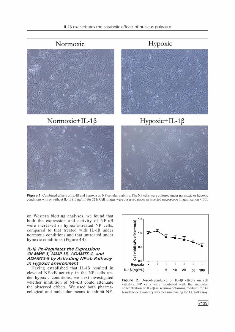
To evaluate the functional effects of IL-1β on NP cellular viability in normoxia or hypox-
ia, we first examined the effects of IL-1β and hypoxia on NP cellular viability. As shown in Figure 1, the NP cells gradually became spindle shape with the cytoplasm being less abundant, refractivity was reduced and the rate of growth was also decreased. The NP cells were also treated with various concentrations of IL-1β in serum-containing medium for 24 h under normoxic or hypoxic conditions, and the cell viability was assessed using the CCK-8 viability assay. As shown in Figure 2, hypoxia increased the cell viability by approximately 1.075-fold, compared to the normal group. The treatment with 5-100 ng/mL of IL-1β signifi-cantly decreased the cell viability in a IL-1β concentration-dependent manner in a hypoxic environment, compared to the cells without IL-1β treatment.
IL-1β Enhances NF-κB Activity in NP Cells Under Hypoxic Conditions and Regulates Catabolic Effects
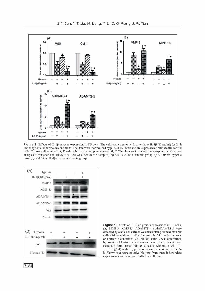
Since overexpressed MMPs and ADAMTS play important roles in ECM degradation of the IVD, we investigated the effects of IL-1β on the expression MMPs and ADAMTS in the NP cells under hypoxia. The expres-sion of MMP-3, MMP-13, ADAMTS-4, and ADAMTS-5 were analyzed by qRT-PCR and compared among the IL-1β-treated and un-treated cells under hypoxic or normoxic con-ditions, respectively. The results showed that, compared to the untreated cells, the expres-sions of MMP-3, MMP-13, ADAMTS-4, and ADAMTS-5 were upregulated in IL-1β-treat-ed NP cells under both normoxic and hypox-ic conditions, but the effect under hypoxic conditions was greater (Figure 3). Western blotting analyses showed that the protein lev-els of MMP-3, MMP-13, ADAMTS-4, and ADAMTS-5 were upregulated in IL-1β-treat-ed NP cells under both normoxic and hypoxic conditions, compared to the untreated cells (Figure 4A). Considering that some prelim-inary data showed that IL-1β increased the expression of MMPs and ADAMTS by acti-vating the NF-κB pathway in other types of cells, we next determined whether the upreg-ulation of MMP-3, MMP-13, ADAMTS-4, and ADAMTS-5 in NP cells under hypoxic condi-tions by IL-1β was attributable to increased NF-κB activity. The NP cells were cultured under normoxic or hypoxic conditions with or without 10 ng/mL of IL-1β for 24 h. Based
IL-1β exacerbates the catabolic effects of nucleus pulposus
7133
on Western blotting analyses, we found that both the expression and activity of NF-κB were increased in hypoxia-treated NP cells, compared to that treated with IL-1β under normoxic conditions and that untreated under hypoxic conditions (Figure 4B).
IL-1β Pp-Regulates the Expressions Of MMP-3, MMP-13, ADAMTS-4, and ADAMTS-5 by Activating NF-κb Pathway in Hypoxic Environment
Having established that IL-1β resulted in elevated NF-κB activity in the NP cells un-der hypoxic conditions, we next investigated whether inhibition of NF-κB could attenuate the observed effects. We used both pharma-cological and molecular means to inhibit NF-
Figure 1. Combined effects of IL-1β and hypoxia on NP cellular viability. The NP cells were cultured under normoxic or hypoxic conditions with or without IL-1β (10 ng/ml) for 72 h. Cell images were observed under an inverted microscope (magnification ×100).
Figure 2. Dose-dependence of IL-1β effects on cell viability. NP cells were incubated with the indicated concentration of IL-1β in serum-containing medium for 48 h and the cell viability was measured using the CCK-8 assay.
Z.-Y. Sun, Y.-T. Liu, H. Liang, Y. Li, D.-G. Wang, J.-W. Tian
7134
Figure 3. Effects of IL-1β on gene expression in NP cells. The cells were treated with or without IL-1β (10 ng/ml) for 24 h under hypoxic or normoxic conditions. The data were normalized by β -ACTIN levels and are expressed as ratios to the control cells. Control cell value = 1. A, The data for matrix component genes. B, C, The change of catabolic gene expression; One-way analysis of variance and Tukey HSD test was used (n = 6 samples). *p < 0.05 vs. he normoxia group. †p < 0.05 vs. hypoxia group, ♦p < 0.05 vs. IL-1β-treated normoxia group.
Figure 4. Effects of IL-1β on proiein expressions in NP cells. (A) MMP-3, MMP-13, ADAMTS-4 andADAMTS-5 were detected by whole cell extract Western blotting from human NP cells with or without IL-1β (10 ng/ml) for 24 h under hypoxic or normoxic conditions. (B) NF-κB activity was determined by Western blotting on nuclear extracts. Nucleoprotein was extracted from human NP cells treated without or with IL-1β (10 ng/ml) under hypoxic or normoxic conditions for 24 h. Shown is a representative blotting from three independent experiments with similar results from all three.
IL-1β exacerbates the catabolic effects of nucleus pulposus
7135
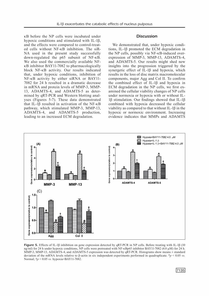
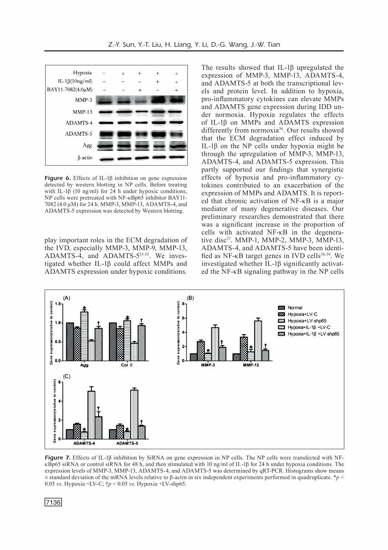
κB before the NP cells were incubated under hypoxic conditions and stimulated with IL-1β, and the effects were compared to control-treat-ed cells without NF-κB inhibition. The siR-NA used in the present study successfully down-regulated the p65 subunit of NF-κB. We also used the commercially available NF-κB inhibitor BAY11-7082 to pharmacologically block NF-κB activity. Our results indicated that, under hypoxic conditions, inhibition of NF-κB activity by either siRNA or BAY11-7082 for 24 h resulted in a dramatic decrease in mRNA and protein levels of MMP-3, MMP-13, ADAMTS-4, and ADAMTS-5 as deter-mined by qRT-PCR and Western blotting anal-yses (Figures 5-7). These data demonstrated that IL-1β resulted in activation of the NF-κB pathway, which stimulated MMP-3, MMP-13, ADAMTS-4, and ADAMTS-5 production, leading to an increased ECM degradation.
Discussion
We demonstrated that, under hypoxic condi-tions, IL-1β promoted the ECM degradation in the NP cells, possibly via NF-κB-induced over-expression of MMP-3, MMP-13, ADAMTS-4, and ADAMTS-5. Our results might shed new insights into the progression triggered by the synergetic effect of IL-1β and hypoxia, which results in the loss of disc matrix macromolecular components, major Agg and Col II. To confirm the combined effect of IL-1β and hypoxia in ECM degradation in the NP cells, we first ex-amined the cellular viability changes of NP cells under normoxia or hypoxia with or without IL-1β stimulation. Our findings showed that IL-1β combined with hypoxia decreased the cellular viability as compared to that without IL-1β in the hypoxic or normoxic environment. Increasing evidence indicates that MMPs and ADAMTS
Figure 5. Effects of IL-1β inhibition on gene expression detected by qRT-PCR in NP cells. Before treating with IL-1β (10 ng/ml) for 24 h under hypoxic conditions, NP cells were pretreated with NF-κBp65 inhibitor BAY11-7082 (4.0 µM) for 24 h. MMP-3, MMP-13, ADAMTS-4, and ADAMTS-5 expression was detected by qRT-PCR. Histograms show means ± standard deviation of the mRNA levels relative to β-actin in six independent experiments performed in quadruplicate. *p < 0.05 vs. Normal; †p < 0.05 vs. hypoxia+BAY11-7082.
Z.-Y. Sun, Y.-T. Liu, H. Liang, Y. Li, D.-G. Wang, J.-W. Tian
7136
play important roles in the ECM degradation of the IVD, especially MMP-3, MMP-9, MMP-13, ADAMTS-4, and ADAMTS-533-35. We inves-tigated whether IL-1β could affect MMPs and ADAMTS expression under hypoxic conditions.
The results showed that IL-1β upregulated the expression of MMP-3, MMP-13, ADAMTS-4, and ADAMTS-5 at both the transcriptional lev-els and protein level. In addition to hypoxia, pro-inflammatory cytokines can elevate MMPs and ADAMTS gene expression during IDD un-der normoxia. Hypoxia regulates the effects of IL-1β on MMPs and ADAMTS expression differently from normoxia36. Our results showed that the ECM degradation effect induced by IL-1β on the NP cells under hypoxia might be through the upregulation of MMP-3, MMP-13, ADAMTS-4, and ADAMTS-5 expression. This partly supported our findings that synergistic effects of hypoxia and pro-inflammatory cy-tokines contributed to an exacerbation of the expression of MMPs and ADAMTS. It is report-ed that chronic activation of NF-κB is a major mediator of many degenerative diseases. Our preliminary researches demonstrated that there was a significant increase in the proportion of cells with activated NF-κB in the degenera-tive disc37. MMP-1, MMP-2, MMP-3, MMP-13, ADAMTS-4, and ADAMTS-5 have been identi-fied as NF-κB target genes in IVD cells38,39. We investigated whether IL-1β significantly activat-ed the NF-κB signaling pathway in the NP cells
Figure 6. Effects of IL-1β inhibition on gene expression detected by western blotting in NP cells. Before treating with IL-1β (10 ng/ml) for 24 h under hypoxic conditions, NP cells were pretreated with NF-κBp65 inhibitor BAY11-7082 (4.0 µM) for 24 h. MMP-3, MMP-13, ADAMTS-4, and ADAMTS-5 expression was detected by Western blotting.
Figure 7. Effects of IL-1β inhibition by SiRNA on gene expression in NP cells. The NP cells were transfected with NF-κBp65 siRNA or control siRNA for 48 h, and then stimulated with 10 ng/ml of IL-1β for 24 h under hypoxia conditions. The expression levels of MMP-3, MMP-13, ADAMTS-4, and ADAMTS-5 was determined by qRT-PCR. Histograms show means ± standard deviation of the mRNA levels relative to β-actin in six independent experiments performed in quadruplicate. *p < 0.05 vs. Hypoxia +LV-C; †p < 0.05 vs. Hypoxia +LV-shp65.
IL-1β exacerbates the catabolic effects of nucleus pulposus
7137
under hypoxic conditions. Our results showed that IL-1β increased activity of NF-κB under hypoxia, suggesting that pro-inflammatory cy-tokines such as IL-1β may exacerbate catabolic effects of the NP cells through the NF-κB sig-naling pathway under hypoxia. In recent years, clear evidence showed that when treatment with IL-1β under normoxia, NF-κB is activated in NP cells, leading to Agg and Col II loss and produc-tion of multiple matrix-degrading enzymes40,41. Therefore, we examined whether IL-1β exacer-bated the ECM degradation in the NP cells via the NF-κB signaling pathway under hypoxia. In the present study, we used both molecular and pharmacological means to inhibit NF-κB, which resulted in remarkable decreases in the expres-sions of MMP-3, MMP-13, ADAMTS-4 and ADAMTS-5, compared with the control cells without NF-κB inhibition. The effects of NF-κB inhibition were also confirmed at the protein level. This effect of IL-1β on the expression of MMPs and ADAMTS of NP cells under hypoxic was effectively reduced by NF-κB inhibitors, suggesting that the upregulation of MMPs and ADAMTS expression by IL-1β might be through the activation of an NF-κB signal pathway in hy-poxia. This is important because pro-inflamma-tory cytokines, even at low concentrations, can have a synergistic effect with hypoxia, enhanc-ing or maintaining a high level of gene expres-sion induced by pro-inflammatory cytokines. These results demonstrated that NF-κB pathway might play a critical role in the regulation of MMPs and ADAMTS expression in human NP cells under hypoxia and inflammatory environ-ment. We speculated that the loss of Agg could be due to the action of NF-κB pathway, which could play a key role in IVD degradation. For patients with IDD, this could be a new treatment to protect against, decrease or even reverse IDD. Such a new approach would help to decrease IDD patients’ suffering and the economic bur-den associated with IDD-induced low backache. Further studies will be required to develop this strategy for clinical application.
Conclusions
We demonstrated that the overexpression of MMPs and ADAMTS mediated by the NF-κB pathway promoted ECM degradation in the NP cells, in the presence of both IL-1β and hypox-ia. The synergetic effect of IL-1β and hypoxia
might contribute to promoting the activation of NF-κB pathway, thus inducing expression of MMPs and ADAMTS. NF-κB inhibition could mitigate IL-1β-induced upregulation of MMPs and ADAMTS, and consequently reverse the degradation of disc matrix macromolecules Agg and collagen II. Our study suggested a critical mechanism underlying the effect of combining IL-1β with hypoxia on promoting ECM deg-radation in the NP cells, providing important information that may be helpful in future devel-opment of potential therapy for IDD.
Conflict of InterestThe Authors declare that they have no conflict of interests.
References
1) Waddell G. Low back pain: a twentieth centu-ry health care enigma. Spine 1996; 21: 2820-2825.
2) HanGai M, KaneoKa K, Kuno S, HinotSu S, SaKaneM, MaMizuKa n. Factors associated with lumbar inter-vertebral disc degeneration in the elderly. Spine J 2008; 8: 732-740.
3) StruGlicS a, HanSSon M. MMP proteolysis of the human extracellular matrix protein aggrecan is mainly a process of normal turnover. Biochem J 2012; 446: 213-223.
4) Gruber He, inGraM Ja, HoelScHer Gl, zincHenKo n, norton HJ. Constitutive expression of cathepsin K in the human intervertebral disc: new insight in-to disc extracellular matrix remodeling via cathep-sin K and receptor activator of nuclear factor-κB ligand. Arthritis Res Ther 2011; 13: 1-7.
5) tiaden an, KlaWitter M, lux V. Detrimental role for human high temperature requirement serine pro-tease A1 (HTRA1) in the pathogenesis of interver-tebral disc (IVD) degeneration. J Biol Chem 2012; 287: 21335-21345.
6) naSto la, Seo HY, robinSon ar. ISSLS prize winner: inhibition of NF-κB activity ameliorates age-associated disc degeneration in a mouse model of accelerated aging. Spine 2012; 37: 1819-1825.
7) Fernández-torreS J, zaMudio-cueVaS Y, Martínez-na-Va Ga, lópez-reYeS aG. Hypoxia-Inducible Factors (HIFs) in the articular cartilage: a systematic re-view. Eur Rev Med Pharmacol Sci 2017; 21: 2800-2810.
8) StairMand JW, HolM S, urban Jp. Factors influenc-ing oxygen concentration gradients in the inter-vertebral disc. A theoretical analysis. Spine 1991; 16: 444-449.
Z.-Y. Sun, Y.-T. Liu, H. Liang, Y. Li, D.-G. Wang, J.-W. Tian
7138
9) bartelS eM, FairbanK Jc, WinloVe cp, urban Jp. Ox-ygen and lactate concentrations measured in vi-vo in the intervertebral discs of patients with sco-liosis and back pain. Spine 1998; 23: 1-7.
10) zHao Y, liu x, lu Yx. MicroRNA-143 regulates the proliferation and apoptosis of cervical cancer cells by targeting HIF-1α. Eur Rev Med Pharma-col Sci 2017; 21: 5580-5586.
11) Wen zW, lianG dS, cai xH, cHen J. The role of AMPK/mTOR signal pathway in brain injury fol-lowing chronic intermittent hypoxia in growing rats. Eur Rev Med Pharmacol Sci 2018; 22: 1071-1077.
12) HitcHon ca, el-GabalaWY HS, bezabeH t. Char-acterization of synovial tissue from arthritis pa-tients: a proton magnetic resonance spectro-scopic investigation. Rheumatol Int 2009; 29: 1205-1211.
13) FuJita n, MarKoVa d, anderSon dG. Expression of prolyl hydroxylases (PHDs) is selectively con-trolled by HIF-1 and HIF-2 proteins in nucleus pulposus cells of the intervertebral disc: distinct roles of PHD2 and PHD3 proteins in controlling HIF-1alpha activity in hypoxia. J Biol Chem 2012; 287: 16975-16986.
14) riSbud V, Guttapalli a, StoKeS G. Nucleus pulposus cells express HIF-1 alpha undernormoxic culture conditions: a metabolic adaptation to the inter-vertebral disc microenvironment. J Cell Biochem 2006; 98: 152-159.
15) riSbud MV, ScHipani e, SHapiro iM. Hypoxic regula-tion of nucleus pulposus cell survival: from niche to notch. Am J Pathol 2010; 176: 1577-1583.
16) pei M, SHouKrY M, li J. Modulation of in vitro micro-environment facilitates synovium-derived stem cell-based nucleus pulposus tissue regeneration. Spine 2012; 37: 1538-1547.
17) Yuan Fl, WanG H r, cao l. New evidence of the role of the hypoxia regulated pathway in nucleus pulposus cell survival: comment on the article by Hiyama et al. Arthritis Rheum 2012; 64: 940-941.
18) SMitH lJ, cHiaro J a, nerurKar nl. Nucleus pulpo-sus cells synthesize a functional extracellular ma-trix and respond to inflammatory cytokine chal-lenge following long-term agarose culture. Eur Cell Mater 2011; 22: 291-301.
19) HolM S, MacKieWicz z, HolM aK. Pro-inflammato-ry, pleiotropic, and anti-inflammatory TNF-a, IL-6, and IL-10 in experimental porcine intervertebral disk degeneration. Vet Pathol 2009; l46: 1292-1300.
20) lee JM, SonG J Y, baeK M, Interleukin-1beta induc-es angiogenesis and innervation in human inter-vertebral disc degeneration. J Orthop Res 2011; 29: 265-269.
21) pientKa FK, Hu J, ScHindler SG, brix b, tHiel a, JöHren o, FandreY J, bercHner-pFannScHMidt u, dep-pinG r. Oxygen sensing by the prolyl-4-hydroxy-lase PHD2 within the nuclear compartment and the influence of compartmentalisation on HIF-1 signalling. Cell Sci 2012; 125: 5168-5176.
22) WanG x, WanG H, YanG H, li J, cai Q, SHapiro iM, riSbud MV. Tumor necrosis factor- and Interleu-kin-1-dependent matrix metalloproteinase-3 ex-pression in nucleus pulposus cells requires coop-erative signaling via syndecan 4 and mitogen-ac-tivated protein kinase-NF-κB axis: implications in inflammatory disc disease. Am J Pathol 2014; 184: 2560-2572.
23) HoYland Ja, le Maitre c, FreeMont aJ. Investigation of the role of IL-1 and TNF in matrix degradation in the intervertebral disc. Rheumatology (Oxford) 2008; 47: 809-814.
24) ellMan Mb, KiM JS, an HS, Kroin JS, li x, cHen d, Yan dY, buecHter dd, naKaYaMa K, liu b, Mor-Gan S, iM HJ. The pathophysiologic role of the protein kinase C δ pathway in the intervertebral discs of rabbits and mice: in vitro, ex vivo, and in vivo studies. Arthritis Rheum 2012; 64: 1950-1959.
25) SMitH lJ, cHiaro Ja, nerurKar nl. Nucleus pulpo-sus cells synthesize a functional extracellular ma-trix and respond to inflammatory cytokine chal-lenge following long-term agarose culture. Eur Cell Mater 2011; 22: 291-301.
26) ponnappan rK, MarKoVa dz, antonio pJ. An or-gan culture system to model early degenerative changes of the intervertebral disc. Arthritis Res Ther 2011; 13: R171.
27) HoYland Ja, le Maitre c, FreeMont aJ. Investigation of the role of IL-1 and TNF in matrix degradation in the intervertebral disc. Rheumatology 2008; 47: 809-814.
28) Sun zY, Yin z, liu c, tian JW. IL-1β promotes ADAMTS enzyme-mediated aggrecan degrada-tion through NF-κB in human intervertebral disc. J Orthop Surg Res 2015; 10: 159.
29) Sun z Y, Sai z, liu c, tian J W. Effects of NF-κB sig-naling pathway in human intervertebral disc de-generation. Spine 2015; 40: 224-232.
30) FenG G, li l, liu H, SonG Y, HuanG F, tu c, SHen b, GonG Q, li t, liu l. Hypoxia differentially reg-ulates human nucleus pulposus and annulus fi-brosus cell extracellular matrix production in 3D scaffolds. Osteoarthritis Cartilage 2013; 21: 582-588.
31) pFirrMann c W, MetzdorF a, zanetti M , Hodler J, booS n. Magnetic resonance classification of lum-bar intervertebral disc degeneration. Spine 2001; 26: 1873-1878.
32) liVaK KJ, ScHMittGen td. Analysis of relative gene expression data using real-time quantitative PCR and the 2(-Delta Delta C(T)) Method. Methods 2001; 25: 402-408.
33) robertS S, caterSon b, MenaGe J, eVanS eH, JaFFraY dc, eiSenStein SM. Matrix metalloproteinases and aggrecanase: their role in disorders of the human intervertebral disc. Spine (Phila Pa 1976) 2000; 25: 3005-3013.
34) boeuF S, GraF F, FiScHer J, Moradi b, little cb, ricHter W. Regulation of aggrecanases from the ADAMTS family and aggrecanneoepitope for-
IL-1β exacerbates the catabolic effects of nucleus pulposus
7139
mation during in vitro chondrogenesis of human mesenchymal stem cells. Eur Cell Mater 2012; 4: 320-332.
35) FoSanG aJ, little cb. Drug insight: aggrecanases as therapeutic targets for osteoarthritis. Nat Clin Pract Rheumatol 2008; 4: 420-427.
36) YanG SH, Hu MH, Sun YH, lin FH. Differential phe-notypic behaviors of human degenerative nucle-us pulposus cells under normoxic and hypox-ic conditions: influence of oxygen concentration during isolation, expansion, and cultivation. Spine J 2013; 13: 1590-596.
37) Sun z, Yin z, liu c, tian J. The changes in the ex-pression of NF-κB in a degenerative human inter-vertebral disc model. Cell Biochem Biophys 2015; 72: 115-122.
38) SéGuin ca, boJarSKi M, pilliar rM, rouGHleY pJ, Kan-del ra. Differential regulation of matrix degrading
enzymes in a TNF alpha-induced model of nu-cleus pulposus tissue degeneration. Matrix Biol 2006; 25: 409-418.
39) Xia M, zHu Y. Fibronectin fragment activation of ERK increasing integrin α5 and β1 subunit ex-pression to degenerate nucleus pulposus cells. J Orthop Res 2011; 29: 556-561.
40) aKeda K, an H, GeMba t. A new gene therapy ap-proach: In vivo transfection of naked NF-κB de-coy oligonucleotide restored disc degeneration in the rabbit annular needle puncture model. Trans Orthop Res Soc 2005; 30: 45.
41) nerlicH a G, bacHMeier b e, ScHleicHer e, roHr-bacH H, paeSold G, booS n. Immunomorphological analysis of RAGE receptor expression and NF-κB activation in tissue samples from normal and degenerated intervertebral discs of various ages. Ann N Y Acad Sci 2007; 1096: 239-248.