Embed Size (px)
Citation preview

7/31/2019 Immuno . Lec 4
http://slidepdf.com/reader/full/immuno-lec-4 1/14
Wednesda 6/ /2011
Ziad Al-Nasser
Emad Al-Gazo
Antigen antibody reactions -
Immunoglobulin
2
7/31/2019 Immuno . Lec 4
http://slidepdf.com/reader/full/immuno-lec-4 2/14
Immunoglobulins
*Represents 20 % of our plasma proteins that prevent us against infectious agents
(pathogens).
*They come from B cells when they change to plasma cells.
*They are detected by analytic techniques such as electrophoresis in the (gamma
globulin) fraction of serum ((from book)) see figure 4.3.
*The specificity of Igs depends on B-cell receptor structure and the protein
composition of the Ig itself ((the gamma wave of the protein electrophoresis and
within this gamma wave we see sub-waves of different Igs)).
IgG:
*It is the most common Ig in our body that is produced in most of infections related to
secondary immune response.
*Has 4 subtypes IgG1 IgG2 IgG3 IgG4.
*Has the longest half life and the only antibody that crosses the placenta.
*It is the main neutralizing antibody and antitoxin.
IgM:
*It is first antibody to be produced; it is the one that presents on B-cell surface as a
monomer. Now when it comes out it comes as a pentamer.
*Has a violencey of 10 in order to bind 10 antigenic determinants, we need that in the
area of infections and this is so important in newborns to protect them against
pathogens .
7/31/2019 Immuno . Lec 4
http://slidepdf.com/reader/full/immuno-lec-4 3/14
*IgM can’t pass placenta so the baby develop them by himself.
IgA:
*it is the Ig of the mucous membranes so we see it in the respiratory tract and GIT and
urogenital tract so it acts as a 1st
line of defense.
*it is a dimer 2 monomers adherent to each other and we have 2 types (IgA1) (IgA2).
IgD:
*it is essential component of B-cell receptor so it is important to IgM function on the
surface and it is an indicator of maturity of B-cells and has a role in the stability of B-
cells and rarely we see it free ,usually on the surface .
IgE:
*it is the antibody of inflammation it is the one that responsible for type 1
hypersensitivity reactions and what we call it anaphylactic shock.
*we see it in response of parasitic type of infections.
*IgE and eosinophils are highly related to each other.
*hapten: simple chemical compounds can’t -by itself- induce antibody response so it has
be to coupled to a macromolecule (eg: protein carrier) and this protein carrier has a
role in activating the T-helper cells and the cytokines that are needed to stimulate the
B-cells then antibodies can be generated .
*immunogen: that a substance that can (always) produce antibodies by itself and cause
immune response so it has to be: 1.forgien body to host.
2. Fairly large, MW must be more than 6000.

7/31/2019 Immuno . Lec 4
http://slidepdf.com/reader/full/immuno-lec-4 4/14
3. Chemically complex.
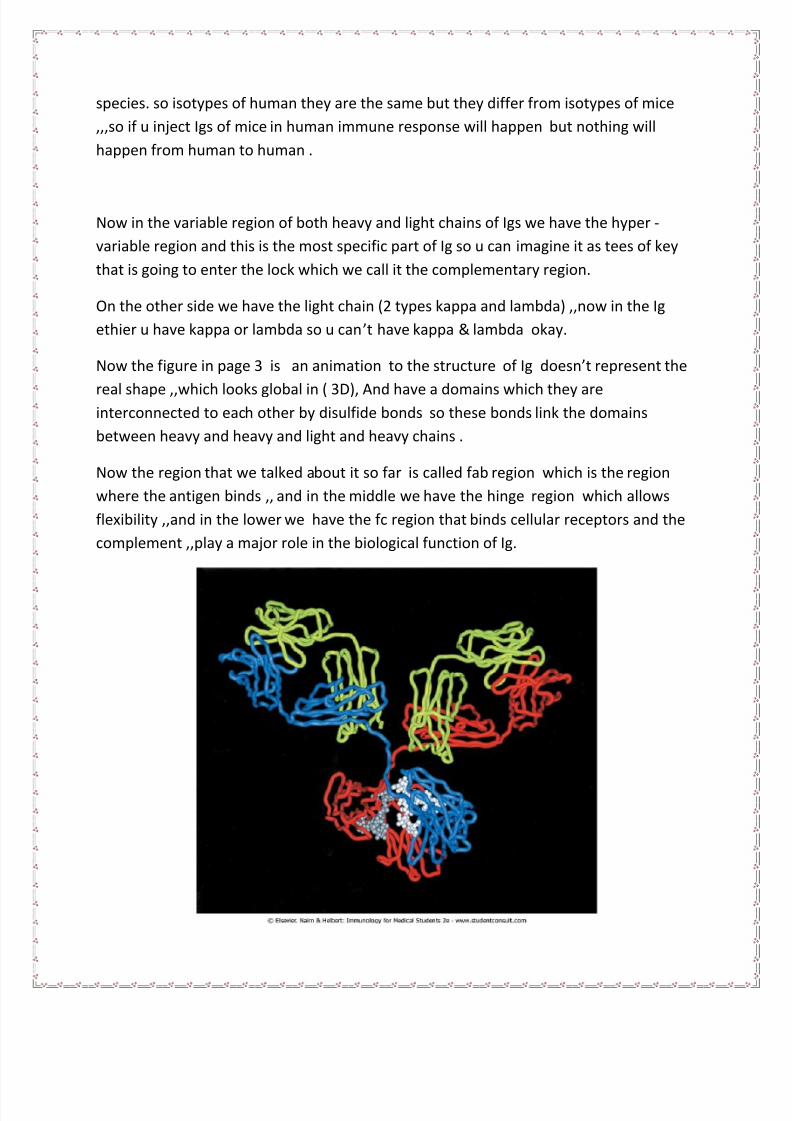
Now in Ig model in the pic above we have 4 chains 2 heavy and 2 light chains ,,the 2
heavy in which the substance of it are the determinant of the type of Ig so when I say (
IgG ANTIBODY) then the heavy chain here we call it of the gamma type ,,we have 4
types of IgG (1,2,3,4) and we have 4 types of heavy chains( gamma 1)(2)(3)(4),,,, and the
substance of heavy chains we call them the isotypes that form the classes and
subclasses of Igs. and the same for IgA we have IgA1 and IgA2 SO the heavy chains
are either alpha1 or alpha2,,,, and IgM the heavy chain is MUO ,,, IgE the heavy chain
is epsilon and for IgD it is delta .and all these heavy chains are coded by chromosome
num 14 .
Now if we look at the heavy chain we see what we call it the variable region and the
constant region,, here the variable means the amino acids variation so they differ from
Ig to the other of the same type while the constant region all are the same in same
7/31/2019 Immuno . Lec 4
http://slidepdf.com/reader/full/immuno-lec-4 5/14
species. so isotypes of human they are the same but they differ from isotypes of mice
,,,so if u inject Igs of mice in human immune response will happen but nothing will
happen from human to human .
Now in the variable region of both heavy and light chains of Igs we have the hyper -
variable region and this is the most specific part of Ig so u can imagine it as tees of key
that is going to enter the lock which we call it the complementary region.
On the other side we have the light chain (2 types kappa and lambda) ,,now in the Ig
ethier u have kappa or lambda so u can’t have kappa & lambda okay.
Now the f igure in page 3 is an animation to the structure of Ig doesn’t represent the
real shape ,,which looks global in ( 3D), And have a domains which they areinterconnected to each other by disulfide bonds so these bonds link the domains
between heavy and heavy and light and heavy chains .
Now the region that we talked about it so far is called fab region which is the region
where the antigen binds ,, and in the middle we have the hinge region which allows
flexibility ,,and in the lower we have the fc region that binds cellular receptors and the
complement ,,play a major role in the biological function of Ig.

7/31/2019 Immuno . Lec 4
http://slidepdf.com/reader/full/immuno-lec-4 6/14
(The pic above shows (3D) shape of Ig )
In the heavy chain of Igs the constant part is divided to 3 parts in most Igs and 4 in IgM
and IgE constant 1 ,2,3 from upper to lower in the figure in page 4 of the lecture. Now
the CH2 (CONSTANT HEAVY 2) Is the area where complement is going to bind and will
be fixed so it plays a role in complement fixation in the heavy chain and also u can see in
the CH2 the carbohydrates are there ,,,so Igs they are glycoproteins =carbohydrates
+proteins,,, so if u inject those into animals, they are going to develop antibodies against
them,,, and if u inject them into human then antibodies could developed against the
variable part of heavy chain protein ,,, Now u have to know we call the heavy chain
proteins isotypes and the variable region proteins allotypes and the hyper-variable
region proteins adiotypes .
Now our body will develop antibodies against the adiotypes we call them anti-adiotypes(Igs against Igs), those anti-adiotypic molecules, our body use them as messenger for
interaction between cells, exactly our body use them as cytokines for activation or
suppression of cells.
Back to fc portion,, u have to know that on the lymphocytes we have receptors for fc
portion of Ig so fc portion will bind on these receptors of the B-cells and T-cells or even
the NK cells ,,, this important to NK cells function ,,so tumor cells or infected cells bind
to fab region and the NK cells will come and bind to fc region by fc receptors,, those fc
receptors are not specific ,,so NK cells also not specific they are servalent cells ,,so they
will bind and become activated and produce the cytolysins and interferons that will kill
the virally infected cells and the tumor cells we call that APCC (ANTIBODY DEPENDANT
CELLULAR CYTOTOXISTY ).

7/31/2019 Immuno . Lec 4
http://slidepdf.com/reader/full/immuno-lec-4 7/14
Now the Dr. showed this slide and started to talk:
You can see here the Igs (M,D,G,E,A) the heavy chain symbol for IgM is ( mu) this is the
isotype the mean serum concentration = 0.4-2.5,,, we can compare it with the highestconcentration of IgG which is from 7 to 18,,, but the IgE concentration is less than
0.0005 this is the lowest concentration (important) ,,and u have to know the heaviest
one is IgM and it is the most efficient in activating the complement by opsenization then
phagocytosis take place ,,,now the IgE: the mast cells and basophils have a receptors for
IgE and this is important for its function and anaphylactic. About IgA: that is important
in mucosal immunity, and about IgG is the only one that can pass the placenta and
longest half life so it is the Ig that allow maternal protection of newborns.
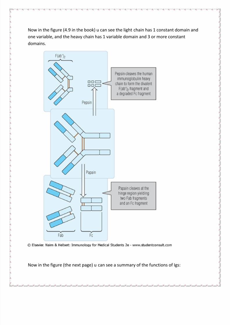
Then the doc showed the pic that in the next page which is about the effect of enzymes
on Igs for example: IgG the most stable one.
If we add pepsin enzyme then the fab region will appear as f(ab’)2 because still there is
a disulfide bond but the pepsin enzyme will degrade the fc region(the biological region )
into fragments . Now if we add the papain enzyme then it will degrade the Ig into 3
parts; 2 parts of fab and one fc ,,the fab one can bind antigen (fragment antigen –binding
), the fc doesn’t bind the antigen but activate the complement and do the biologicaleffector function .
7/31/2019 Immuno . Lec 4
http://slidepdf.com/reader/full/immuno-lec-4 8/14
Now in the figure (4.9 in the book) u can see the light chain has 1 constant domain and
one variable, and the heavy chain has 1 variable domain and 3 or more constant
domains.
Now in the figure (the next page) u can see a summary of the functions of Igs:
7/31/2019 Immuno . Lec 4
http://slidepdf.com/reader/full/immuno-lec-4 9/14
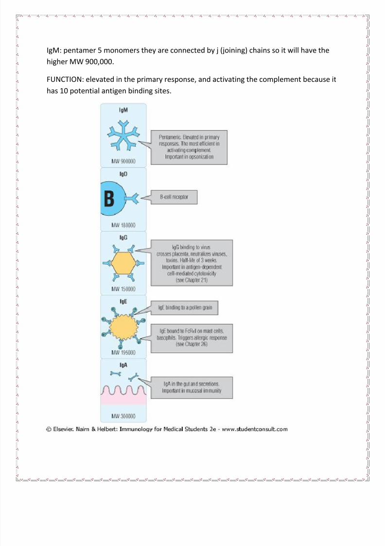
IgM: pentamer 5 monomers they are connected by j (joining) chains so it will have the
higher MW 900,000.
FUNCTION: elevated in the primary response, and activating the complement because it
has 10 potential antigen binding sites.

7/31/2019 Immuno . Lec 4
http://slidepdf.com/reader/full/immuno-lec-4 10/14
Now IgD: it is chiefly found on the surface of B-cell receptor molecule and involved in
cell activation and has a role in maturity of B-cells.
Now IgG: is the main neutralizing antibody, look here for example at IgG in the 3rd
box u
can see the virus each angle of this virus act as ligand (protein binding ) which binds to
the receptor on cell surface so our body will produce IgG that block the function of
ligand (neutralize the virus ) ,,so u have to remember IgG it has the longest half life (3
weeks) so it is most prevalent in the serum ,,, and remember that it is the one that cross
the placenta ,, we need 2 IgG to fix the complement while 1 of IgM is enough because it
is a pentamer.
Now IgE: it is responsible for inflammation and anaphylactic shock ,,it binds to the mast
cells and basophils by fc region not fab,, so when fab bind to the allergen then the mast
cell will be granulated and produce mediators like histamine ,,, so u have to know thatIgE is responsible for hypersensitivity reaction ,,,it is important in clinic to avoid these
hypersensitivity reactions by taking a good history from the patient ,,, but sometimes
we can’t predict the allergy example: a patient sensitive to epinephrine ,,,or
hydrocortisone and these are rare cases u cant predicts it.
Now IgA : 2 subtypes A1 AND A2 ,,, it is a dimer 2 monomers that are connected by
(J CHAINS) ,, and this Ig normally in the serum ,,now when infection happen in the
mucous membranes like in the gut ,,then the Ig transferred to the gut by a process
resembles the endocytosis by binding to the S protein receptors, then it will pass to the
site of infection, now the S protein will protect the IgA from microbial enzymes.
Now the Dr. showed slide 23 and said :
U have to know what( fab ,fc ,f(ab’)2 ,VL,VH ,CL,CH ) MEAN ,,,,,
The domains: they are segments of Igs.
The Ig supergene family: similar folded polypeptide domain.
Now slide 24:
The opsonization : is the process that facilitate the phagocytosis by binding Ig with
microbe ,,we call this microbe opsonic ,,and the complement activation also help in
phagocytosis ,,and remember the complement is fixed on (CH2) .
7/31/2019 Immuno . Lec 4
http://slidepdf.com/reader/full/immuno-lec-4 11/14
,,,,,SO FAR WE END CHAPTER 4 ,,,,
((NOW LET’S GO TO GHAPTER 5: ANTIBODY ANTIGEN INTERACTION))
Antibody antigen interactions they are similar to those between enzyme and its
substrate, u have to know there are 4 forces contribute in this interaction:
1. Electrostatic forces.
2. Hydrogen bonds.
3. Van der walls forces.
4. Hydrophobic interaction.
All of these forces are non covalent bonds (weak bonds) but as a sum it will act strongly.
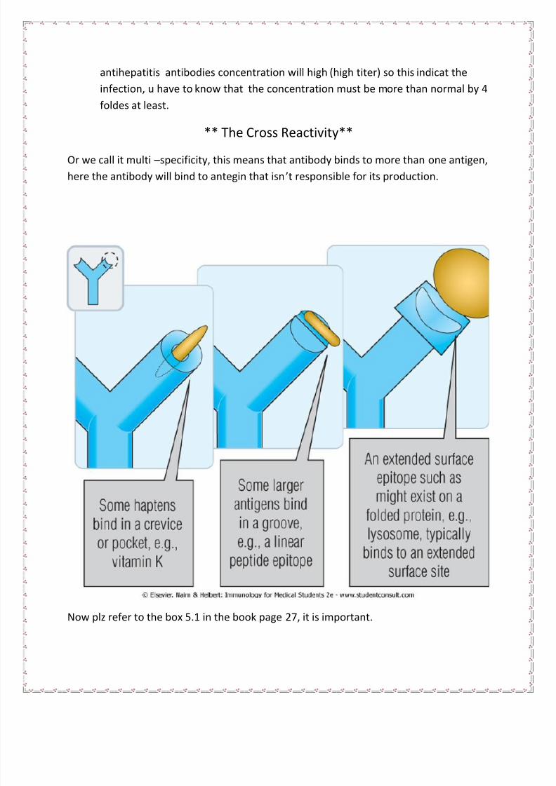
Now we have different types of antigens, the variety is infinite and we have different
binding sites at the Ig. Now the strength of anti-body antigen interaction we call it the
affinity, and the accumulative antigen antibody interactions we call it additive, these are
important two terms as the Dr. said.
Look at figure in the next page:
You have to know that we have different structures and shapes of antigens:
1. Simple antigen.
2. Larger epitope.
3. Complex antigen.
And we have different binding sites of Igs to bind these antigens. So the simple antigen
(like: Vitamin k) will bind to binding site which is crevice (pocket), and the larger epitope
(like: linear peptide epitope) this will bind with a groove binding site, and complex
antigen like: folded protein that present in lysosome this will bind to extended surface.
Antibodies importance:
1. protection our cells from the antigen by neutrolizing them and preventing them
from binding to their receptors.
2. for diagnostic tests: meuserment of the antibodies concentration indicate the
infection by the pathogen for exampl: if u infected with hepatitis B virus then the
7/31/2019 Immuno . Lec 4
http://slidepdf.com/reader/full/immuno-lec-4 12/14
antihepatitis antibodies concentration will high (high titer) so this indicat the
infection, u have to know that the concentration must be more than normal by 4
foldes at least.
** The Cross Reactivity**Or we call it multi –specificity, this means that antibody binds to more than one antigen,
here the antibody will bind to antegin that isn’t responsible for its production.
Now plz refer to the box 5.1 in the book page 27, it is important.
7/31/2019 Immuno . Lec 4
http://slidepdf.com/reader/full/immuno-lec-4 13/14
Now if u remember (group A Beta Hemolytic Strep) that cause tonsillitis then it will
cause rheumatic fever or rheumatic heart disease and that happened by cross reactivity
of protein M that present in the bacteria and our tissue and this will cause autoimmune
disease. quantity
Now refer to the diagnostic tests, in the medical microbiology lab there is a section we
call it immunology or serology department, in this department simply they do antibody
antigen interactions, so if the antigen is known then I will know the antigen and vice
versa because they are matching, the problem sometimes is the cross reactivity then the
test will be false positive :/.
In these tests u have to know some terms like specificity and sensitivity, 100% specificity
then no false positive results, sensitivity: this means that any small amounts of Igs
should be detected .
Now in the serology tests u can determine quantity (how much positive results there)
(the titer) and quality (positive or negative) of the tests.
The 1st
tests done in the past was the agglutination and precipitation tests, u are familiar
with agglutination in experiments of blood grouping, now the precipitation: this happen
when the antigen and the antibody are part of a solution so they will bind together and
form what we call it latex (network-like ) and if u draw this as curve it will looks like a
bell shape ,,this means that the maximum amount of immune complexes in the middle
and these immune complexes will precipitate and this happen better in the optimal
concentration, this optimal means that the proportion of antibody to antigen nearly 1:1
so no excess antibodies or antigens .
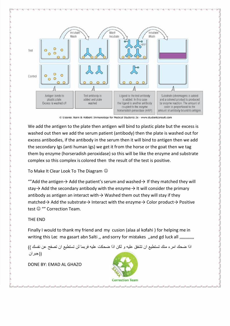
***ELISA test:
Enzyme linked Immunosorbent Assay:
The most common test in the lab through the measure of antibody titer. Here in the
experiment we know the antigen but the antibody is unknown.
7/31/2019 Immuno . Lec 4
http://slidepdf.com/reader/full/immuno-lec-4 14/14
We add the antigen to the plate then antigen will bind to plastic plate but the excess is
washed out then we add the serum patient (antibody) then the plate is washed out for
excess antibodies, if the antibody in the serum then it will bind to antigen then we add
the secondary Igs (anti human Igs) we get it from the horse or the goat then we tag
them by enzyme (horseradish peroxidase) so this will be like the enzyme and substrate
complex so this complex is colored then the result of the test is positive.
To Make It Clear Look To The Diagram
“”Add the antigen→ Add the patient’s serum and washed→ If they matched they will
stay→ Add the secondary antibody with the enzyme→ It will consider the primary
antibody as antigen an interact with→ Washed them out they will stay if they
matched→ Add the substrate→ Interact with the enzyme→ Color product→ Positive
test “” Correction Team.
THE END
Finally I would to thank my friend and my cusion (alaa al kofahi ) for helping me in
writing this Lec ma gasart abn 5alti ,, and sorry for mistakes ,,and gd luck all ,,,,,,,,,,,,
))
))
DONE BY: EMAD AL GHAZO