Embed Size (px)
Citation preview
1
Immunohistochemical profile of odontogenic epithelium in developing dog
teeth (Canis familiaris)
S. NELa,*, M. B. VAN HEERDENa, G. STEENKAMPb W. F. P. VAN HEERDENa, S. C.
BOYa
a Department of Oral Pathology and Oral Biology, School of Dentistry, Faculty of Health
Sciences, University of Pretoria, Oral and Dental Hospital, Bophelo Road, Pretoria,
South Africa
b Department of Companion Animal Clinical Studies, Faculty of Veterinary Science,
University of Pretoria, Old Soutpan Road, Onderstepoort , South Africa
Running Title: Immunohistochemical profile of odontogenic epithelium
* Corresponding author: Sulette Nel PO Box 1266, Pretoria, 0001, South Africa Tel.: +27 12 319 2664 Fax: +27 12 321 2225 E-mail address: [email protected]
2
ABSTRACT
Tumours of the jaw bones and oral soft tissue are relatively common lesions in dogs.
The aim of this study was to find cell markers to differentiate odontogenic epithelium
from non-odontogenic epithelium for future research on the pathogenesis and pathology
of odontogenic neoplasms in dogs. Keratin 14 and 19 staining was observed in
odontogenic and non-odontogenic epithelium, while amelogenin and p75 neurotrophin
receptor immunoreactivity was observed in certain odontogenic epithelial cells at various
stages of development, but not in other epithelial cells. Calretinin staining was observed
in the alveolar epithelial cells directly overlying the developing tooth germ in 28/39
sections (71,8%), as well as the dental laminae in 30/35 sections (85,7%) and Serres
rests in 24/28 sections (85,7%). Focal positivity was detected in the respiratory mucosa,
some hair follicles and fusion epithelium of the palate but no calretinin staining was
observed in other oral epithelial cells, and therefore calretinin has potential to be utilized
as a marker to differentiate odontogenic form non-odontogenic epithelium.
Key words: amelogenin, calretinin, canine, dogs, keratin-14, keratin-19, odontogenesis,
odontogenic epithelium, p75 neurotrophin receptor
3
Dogs frequently present with oral epulides defined as tumours of the gingiva. Many of
these can easily be identified as one of the commonly recognised odontogenic
neoplasms.13,38,39 Problematic entities however do exist when the tumours do not have
the classic histological features of any specific odontogenic tumour or cyst. In an
attempt to elucidate the origin and character of these epulides of uncertain histogenesis,
it is imperative to find molecular markers which could discriminate between odontogenic
epithelium and other types of epithelium in the mouths of dogs. Such markers could
then be utilized in future molecular research on the pathogenesis and pathology of
odontogenic versus non-odontogenic neoplasms in dogs. To the best of our knowledge
no in situ markers for odontogenic epithelium have been described in developing dog
teeth. We therefore evaluated keratin 14 (K14) and 19 (K19), amelogenin, p75
neurotrophin receptor (p75NTR) and calretinin, which have been previously described as
markers of odontogenic epithelium in developing human and rat odontogenic tissues,7-
9,19,26 on developing dog teeth.
Materials and methods
Twenty four foetuses of large breed dogs were obtained under ethical clearance of the
Animal Use and Care Committee of the Faculty of Veterinary Sciences, University of
Pretoria, South Africa, from female dogs scheduled for elective pregnancy termination.
The foetuses were fixed in 10% buffered formalin and the heads carefully cut into
coronal sections, dehydrated and embedded in paraffin wax blocks. Those samples that
contained calcified bone or dental hard tissues were decalcified in routine decalcifying
solution (70ml HNO3, 50ml HCL, 880ml distilled water) for 60 minutes and then rinsed in
running tap water for 60 minutes. Tissue specimens were sectioned at 3µm, stained
with haematoxylin and eosin (H&E), and microscopically examined to select slides with
4
well-formed enamel organs in which the respective odontogenic epithelial cells were
morphologically clearly identifiable.
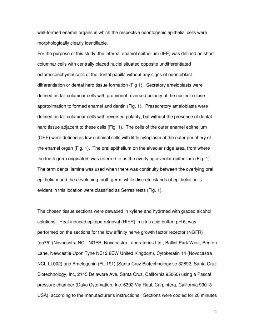
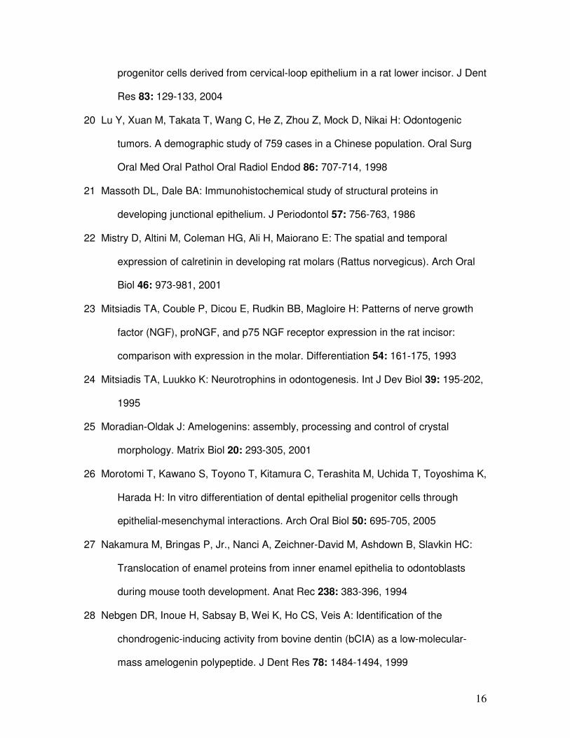
For the purpose of this study, the internal enamel epithelium (IEE) was defined as short
columnar cells with centrally placed nuclei situated opposite undifferentiated
ectomesenchymal cells of the dental papilla without any signs of odontoblast
differentiation or dental hard tissue formation (Fig 1). Secretory ameloblasts were
defined as tall columnar cells with prominent reversed polarity of the nuclei in close
approximation to formed enamel and dentin (Fig. 1). Presecretory ameloblasts were
defined as tall columnar cells with reversed polarity, but without the presence of dental
hard tissue adjacent to these cells (Fig. 1). The cells of the outer enamel epithelium
(OEE) were defined as low cuboidal cells with little cytoplasm at the outer periphery of
the enamel organ (Fig. 1). The oral epithelium on the alveolar ridge area, from where
the tooth germ originated, was referred to as the overlying alveolar epithelium (Fig. 1).
The term dental lamina was used when there was continuity between the overlying oral
epithelium and the developing tooth germ, while discrete islands of epithelial cells
evident in this location were classified as Serres rests (Fig. 1).
The chosen tissue sections were dewaxed in xylene and hydrated with graded alcohol
solutions. Heat induced epitope retrieval (HIER) in citric acid buffer, pH 6, was
performed on the sections for the low affinity nerve growth factor receptor (NGFR)
(gp75) (Novocastra NCL-NGFR, Novocastra Laboratories Ltd., Balliol Park West, Benton
Lane, Newcastle Upon Tyne NE12 8EW United Kingdom), Cytokeratin 14 (Novocastra
NCL-LL002) and Amelogenin (FL-191) (Santa Cruz Biotechnology sc-32892, Santa Cruz
Biotechnology, Inc, 2145 Delaware Ave, Santa Cruz, California 95060) using a Pascal
pressure chamber (Dako Cytomation, Inc. 6392 Via Real, Carpintera, California 93013
USA), according to the manufacturer’s instructions. Sections were cooled for 20 minutes
5
at room temperature and treated with hydrogen peroxide for 5 minutes at 37ºC, to
quench endogenous peroxidase activity. Sections stained for Calretinin (Novocastra
NCL-Calretinin), and Cytokeratin 19 (Novocastra NCL-CK19) were first treated with
hydrogen peroxide and then HIER was done in ethylenediaminetetraacetic acid (EDTA)
buffer pH 8 using the Pascal.
Sections were then incubated in their various anti serum: Cytokeratin 14, (1:60),
Cytokeratin 19 (1:100), NGFR-gp75 (1:50), Calretinin (1:100) and Amelogenin (1:50) for
60 minutes at room temperature. All the sections, except the Amelogenin, were then
incubated in EnvisionTM+ System-HRP Labelled Polymer Mouse (Dako K4000, Dako
Cytomation Inc) for 35 minutes at room temperature. The Amelogenin sections were
incubated in EnvisionTM+ System-HRP Labelled Polymer Rabbit (Dako K4009) for 35
minutes at room temperature. All the sections were then stained with AEC+ chromogen
(Dako Cytomation Inc) for 4 minutes at 37ºC. Sections were counterstained with
Haematoxylin, mounted with Faramount Aqueous Mounting Media (Dako S3025) and
then examined microscopically.
Results
The exact time of gestation of the developing dog foetuses were unknown and the
foetuses were classified in 2 groups based on the stage of odontogenic development.
Foetuses 1-8 (group 1) only had tooth germs in the bud and cap stages of development
with no ameloblast differentiation or dental hard tissue formation. The tooth germs of
foetuses 9-24, (group 2), were in the bell stage of development with visible cell
differentiation and hard tissue formation. In 15 cases, a rostral (anterior) and a caudal
(posterior) section of the same foetus were used for comparison. In the remaining 9
cases, only one section complied with the inclusion criteria. A total of 39 sections were
therefore harvested from the 24 foetuses. Although the number of tooth germs on a
6
single section varied from 1 to 4, only one, defined as the best representative of the bell
stage in the given section, was chosen for analysis.
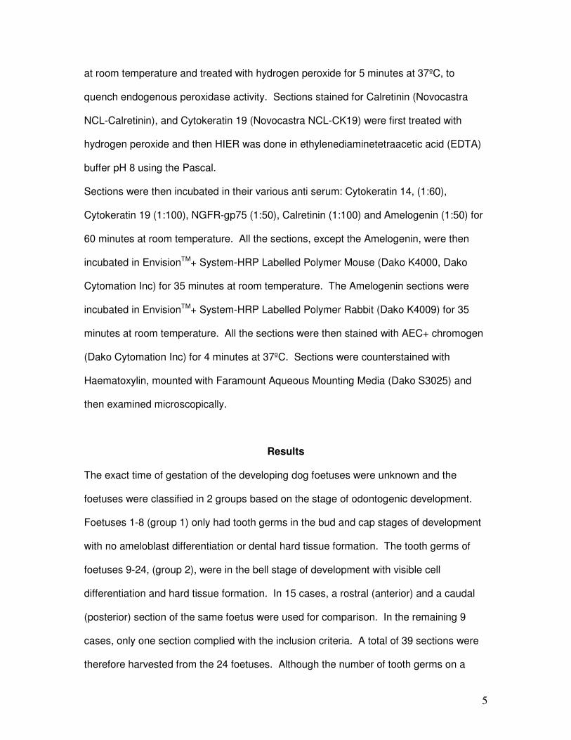
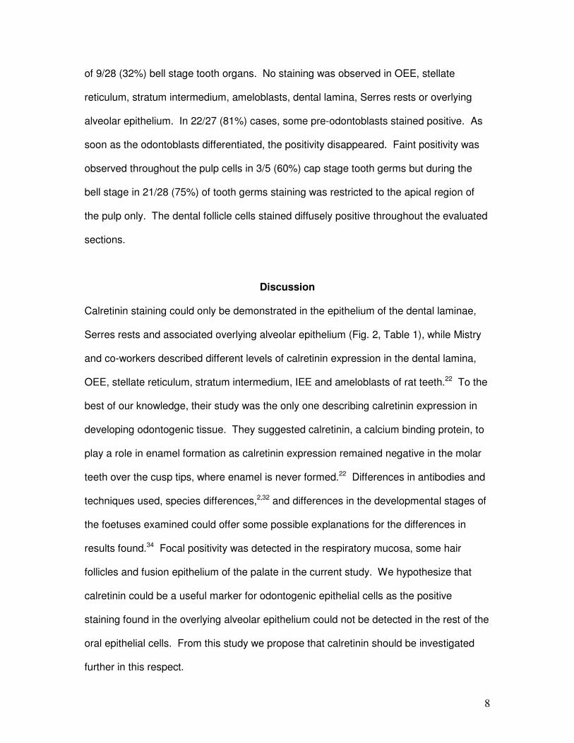
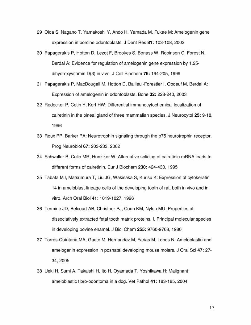
Calretinin
Group 1 foetuses showed calretinin staining of the OEE but no calretinin staining was
observed in the OEE of group 2, IEE, ameloblasts, odontoblasts or epithelial cells of the
stellate reticulum or stratum intermedium at any stage of development. Thirty of the 35
dental laminae evaluated stained positive for calretinin (85.7%). Twenty four of 28 cases
with Serres rests showed positive staining in the epithelial rests (85.7%). The overlying
alveolar epithelium, in close approximation to the lamina and Serres rests, stained
positive in 28 of the 39 cases (71.8%) evaluated (Fig. 2).
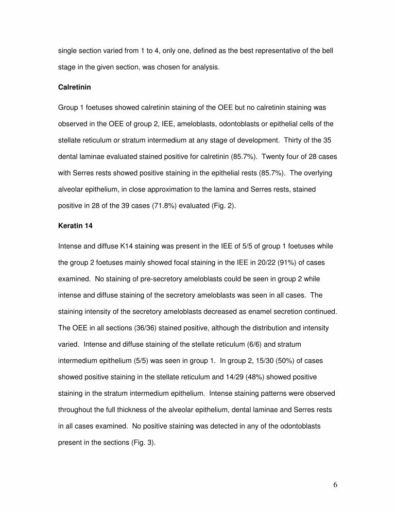
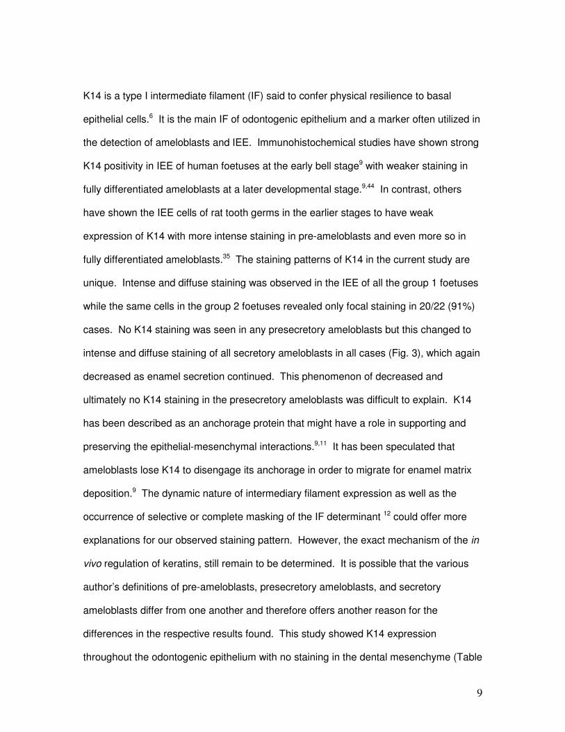
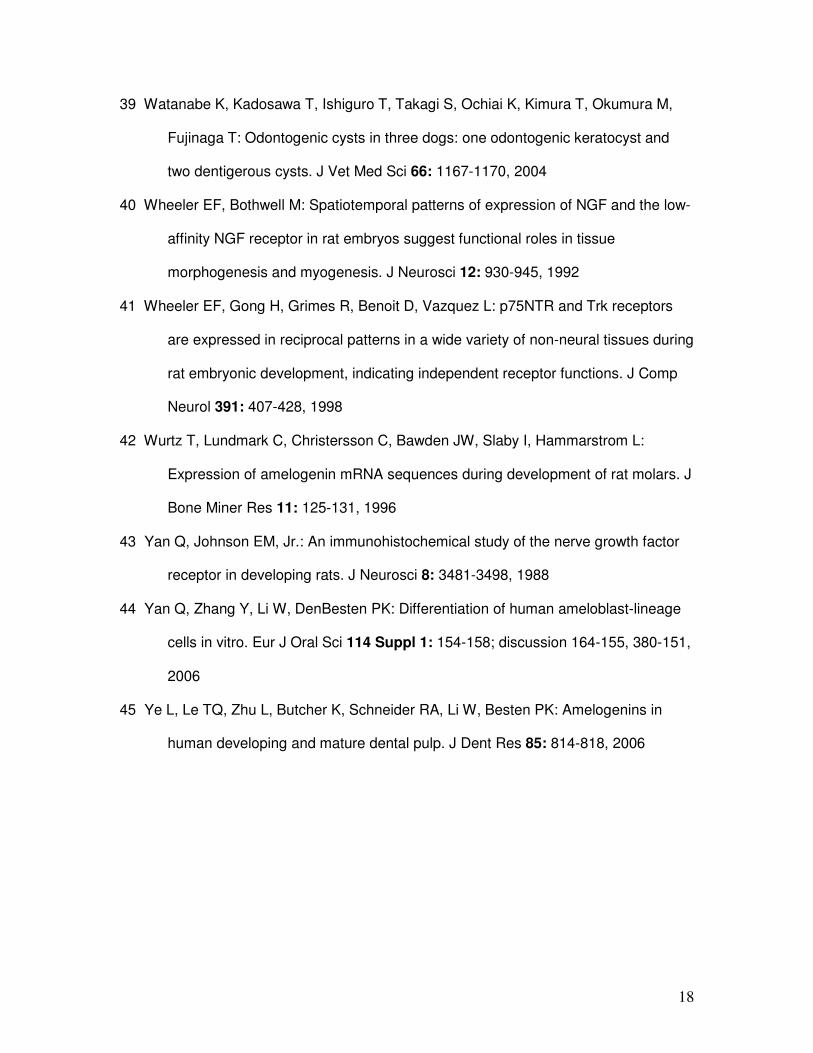
Keratin 14
Intense and diffuse K14 staining was present in the IEE of 5/5 of group 1 foetuses while
the group 2 foetuses mainly showed focal staining in the IEE in 20/22 (91%) of cases
examined. No staining of pre-secretory ameloblasts could be seen in group 2 while
intense and diffuse staining of the secretory ameloblasts was seen in all cases. The
staining intensity of the secretory ameloblasts decreased as enamel secretion continued.
The OEE in all sections (36/36) stained positive, although the distribution and intensity
varied. Intense and diffuse staining of the stellate reticulum (6/6) and stratum
intermedium epithelium (5/5) was seen in group 1. In group 2, 15/30 (50%) of cases
showed positive staining in the stellate reticulum and 14/29 (48%) showed positive
staining in the stratum intermedium epithelium. Intense staining patterns were observed
throughout the full thickness of the alveolar epithelium, dental laminae and Serres rests
in all cases examined. No positive staining was detected in any of the odontoblasts
present in the sections (Fig. 3).
7
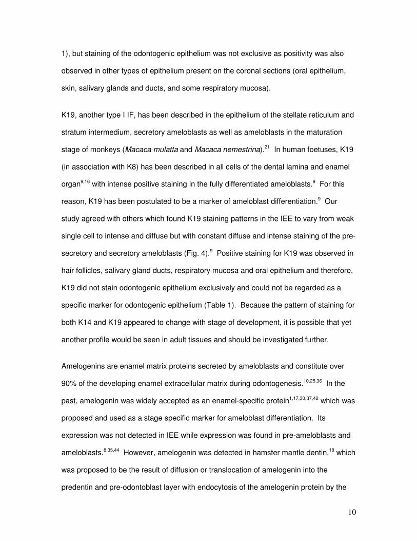
Keratin 19
In both group 1 and 2 the epithelial cells of the enamel organs of all the sections stained
positive with variable intensities and distribution patterns. The IEE cells of group 1
exhibited a heterogeneous pattern of staining as staining intensities varied. In group 2,
diffuse expression of K19 was observed in the IEE cells, except in the cervical loop
region. The pre-secretory and secretory ameloblasts stained diffusely positive in all
cases although with varied intensities. Positive staining was observed in the full
thickness of the overlying alveolar epithelial cells in all of the group 1 foetuses, but in
group 2 it was mainly restricted to the superficial epithelial cell layers. Diffuse staining
was observed in all the dental laminae and Serres rests in all cases examined. No
positive staining was detected in any of the odontoblasts present on the sections (Fig.
4).
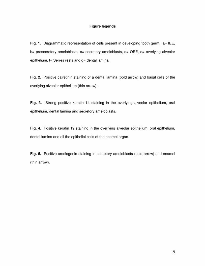
Amelogenin
No amelogenin staining was present in group 1. In group 2, small amounts of
amelogenin were observed in presecretory ameloblasts seen in close approximation to
the cell nucleus. Diffuse granular staining for the amelogenin protein was observed in all
secretory ameloblasts present in group 2 sections. No staining was detected in the IEE,
OEE, stellate reticulum, stratum intermedium, dental laminae, Serres rests or alveolar
epithelium of both groups. Of the 30 evaluated group 2 sections, 6 revealed weak
staining in some odontoblasts (20%). Staining seemed to be localized to the intercellular
spaces between the odontoblasts and other pulp cells. Staining of these sections was
repeated but the same results were obtained. No amelogenin staining was observed in
or around odontoblasts at advanced stages of odontogenesis (Fig. 5).
p75NTR
No epithelial staining was observed in bud stage dental organs. Focal positivity was
observed in the IEE of 5/5 cap stage germs as well as in the region of the cervical loops
8
of 9/28 (32%) bell stage tooth organs. No staining was observed in OEE, stellate
reticulum, stratum intermedium, ameloblasts, dental lamina, Serres rests or overlying
alveolar epithelium. In 22/27 (81%) cases, some pre-odontoblasts stained positive. As
soon as the odontoblasts differentiated, the positivity disappeared. Faint positivity was
observed throughout the pulp cells in 3/5 (60%) cap stage tooth germs but during the
bell stage in 21/28 (75%) of tooth germs staining was restricted to the apical region of
the pulp only. The dental follicle cells stained diffusely positive throughout the evaluated
sections.
Discussion
Calretinin staining could only be demonstrated in the epithelium of the dental laminae,
Serres rests and associated overlying alveolar epithelium (Fig. 2, Table 1), while Mistry
and co-workers described different levels of calretinin expression in the dental lamina,
OEE, stellate reticulum, stratum intermedium, IEE and ameloblasts of rat teeth.22 To the
best of our knowledge, their study was the only one describing calretinin expression in
developing odontogenic tissue. They suggested calretinin, a calcium binding protein, to
play a role in enamel formation as calretinin expression remained negative in the molar
teeth over the cusp tips, where enamel is never formed.22 Differences in antibodies and
techniques used, species differences,2,32 and differences in the developmental stages of
the foetuses examined could offer some possible explanations for the differences in
results found.34 Focal positivity was detected in the respiratory mucosa, some hair
follicles and fusion epithelium of the palate in the current study. We hypothesize that
calretinin could be a useful marker for odontogenic epithelial cells as the positive
staining found in the overlying alveolar epithelium could not be detected in the rest of the
oral epithelial cells. From this study we propose that calretinin should be investigated
further in this respect.
9
K14 is a type I intermediate filament (IF) said to confer physical resilience to basal
epithelial cells.6 It is the main IF of odontogenic epithelium and a marker often utilized in
the detection of ameloblasts and IEE. Immunohistochemical studies have shown strong
K14 positivity in IEE of human foetuses at the early bell stage9 with weaker staining in
fully differentiated ameloblasts at a later developmental stage.9,44 In contrast, others
have shown the IEE cells of rat tooth germs in the earlier stages to have weak
expression of K14 with more intense staining in pre-ameloblasts and even more so in
fully differentiated ameloblasts.35 The staining patterns of K14 in the current study are
unique. Intense and diffuse staining was observed in the IEE of all the group 1 foetuses
while the same cells in the group 2 foetuses revealed only focal staining in 20/22 (91%)
cases. No K14 staining was seen in any presecretory ameloblasts but this changed to
intense and diffuse staining of all secretory ameloblasts in all cases (Fig. 3), which again
decreased as enamel secretion continued. This phenomenon of decreased and
ultimately no K14 staining in the presecretory ameloblasts was difficult to explain. K14
has been described as an anchorage protein that might have a role in supporting and
preserving the epithelial-mesenchymal interactions.9,11 It has been speculated that
ameloblasts lose K14 to disengage its anchorage in order to migrate for enamel matrix
deposition.9 The dynamic nature of intermediary filament expression as well as the
occurrence of selective or complete masking of the IF determinant 12 could offer more
explanations for our observed staining pattern. However, the exact mechanism of the in
vivo regulation of keratins, still remain to be determined. It is possible that the various
author’s definitions of pre-ameloblasts, presecretory ameloblasts, and secretory
ameloblasts differ from one another and therefore offers another reason for the
differences in the respective results found. This study showed K14 expression
throughout the odontogenic epithelium with no staining in the dental mesenchyme (Table
10
1), but staining of the odontogenic epithelium was not exclusive as positivity was also
observed in other types of epithelium present on the coronal sections (oral epithelium,
skin, salivary glands and ducts, and some respiratory mucosa).
K19, another type I IF, has been described in the epithelium of the stellate reticulum and
stratum intermedium, secretory ameloblasts as well as ameloblasts in the maturation
stage of monkeys (Macaca mulatta and Macaca nemestrina).21 In human foetuses, K19
(in association with K8) has been described in all cells of the dental lamina and enamel
organ9,16 with intense positive staining in the fully differentiated ameloblasts.9 For this
reason, K19 has been postulated to be a marker of ameloblast differentiation.9 Our
study agreed with others which found K19 staining patterns in the IEE to vary from weak
single cell to intense and diffuse but with constant diffuse and intense staining of the pre-
secretory and secretory ameloblasts (Fig. 4).9 Positive staining for K19 was observed in
hair follicles, salivary gland ducts, respiratory mucosa and oral epithelium and therefore,
K19 did not stain odontogenic epithelium exclusively and could not be regarded as a
specific marker for odontogenic epithelium (Table 1). Because the pattern of staining for
both K14 and K19 appeared to change with stage of development, it is possible that yet
another profile would be seen in adult tissues and should be investigated further.
Amelogenins are enamel matrix proteins secreted by ameloblasts and constitute over
90% of the developing enamel extracellular matrix during odontogenesis.10,25,36 In the
past, amelogenin was widely accepted as an enamel-specific protein1,17,30,37,42 which was
proposed and used as a stage specific marker for ameloblast differentiation. Its
expression was not detected in IEE while expression was found in pre-ameloblasts and
ameloblasts.8,35,44 However, amelogenin was detected in hamster mantle dentin,18 which
was proposed to be the result of diffusion or translocation of amelogenin into the
predentin and pre-odontoblast layer with endocytosis of the amelogenin protein by the
11
odontoblasts.18,27 It has also been suggested that the presence of trace amounts of
amelogenin splice products in dentin could be the result of contamination.28 Since then,
amelogenin mRNA was detected in odontoblasts of porcine and rat teeth29,31 and in 2006
it was suggested that odontoblasts actually synthesize and secrete amelogenins after in
situ hybridization and immunohistochemistry studies on human odontogenic tissue.45
The mRNA of splice forms of amelogenin have been detected in periodontal ligament
cells (cementoblasts) of wild-type mice14 and recently amelogenin mRNA and protein
was described in dog and rat osteoblasts, osteoclasts, in some osteocytes and also in
articular cartilage chondrocytes.15 It was therefore proposed that amelogenin is also
expressed in mesenchymal cells and is not only an ameloblast-specific protein as
previously believed.14,15
Expression of the amelogenin protein in this study was similar to previous studies which
found it to vary from only small amounts in presecretory ameloblasts,17,30 to diffuse
positivity throughout differentiated ameloblasts in the secretory stage (Fig. 5).17,30,37
Single cell positivity of odontoblasts was observed in 6/30 (20%) sections with
odontoblasts present. On close examination, the staining appeared to be localized to
the intercellular spaces. It is therefore possible that the single cell positivity observed in
the odontoblasts in our study could in fact be artifactual. The amelogenin protein
actually located outside the cell could have been misinterpreted as intracellular staining
due to superimposition on the slide. No staining was observed in odontoblasts at
advanced stages of odontogenesis and the single cells that did seem to stain were
associated with the mantle dentin only. Although staining has been described in other
tissues before,15 amelogenin was only detected in odontogenic tissues in this study and
seems to be a promising marker to distinguish between odontogenic and non-
odontogenic tissues in dogs (Table 1).
12
In 1985 it was observed that two classes of NGFRs exists, originally described as high
and low affinity receptors.4 Today it is better known as the tropomyosin-related kinase
(Trk) tyrosine kinase receptors (high affinity receptor) and the p75NTR (low affinity
receptor).33 p75NTR has been described in epithelial cells of the growing tooth germ as
well as the associated ectomesenchymal cells of rat teeth.5,23,24 In the bell stage,
staining has been observed in proliferating cells of the IEE,5,23,43 some cells of the
stratum intermedium and in pre-odontoblasts/polarizing odontoblasts.5,23,24 More recent
studies revealed immunoreactivity against p75NTR to be restricted to the IEE, the dental
papilla and the dental follicle of rat incisors and therefore p75NTR has been used as a
marker for IEE cells.19,26 In the current study p75NTR expression was restricted to focal
regions of the IEE in 14/33 sections (42%) and no staining was observed in other
epithelial cells (Table 1). It could therefore be utilized as a marker to differentiate
between odontogenic epithelium in the early stages of development versus non-
odontogenic epithelium. The widespread staining observed in the mesenchymal tissue
does not permit p75NTR to be a specific marker for odontogenic epithelium although the
morphological differences between epithelium and mesenchymal tissue would aid in the
histological investigation. The precise function of p75NTR expression in the IEE cells
remains a speculative topic.
Odontogenic tumours may originate from the epithelial or ectomesenchymal cells of the
developing tooth germ or its remnants.20 Most of the odontogenic tumours are found in
adult dogs where all that is left of the odontogenic epithelium is the Serres and Malassez
odontogenic epithelium rests and in certain cases the reduced enamel epithelium
associated with an unerupted (or impacted) tooth. One can therefore assume that the
odontogenic tumours in adult dogs would most probably originate from these epithelial
remnants in the gingiva and the periodontal ligament space.3 As it was shown in this
13
study, the expression profile of certain markers was altered in group 2 compared to
group 1 foetuses in many respects. This expression profile may change even more in
post-natal and adult tissue as previously indicated.34,40,41,43 Therefore, it would be
possible that a marker could retain its expression in Serres- and Malassez rests, but
could loose its expression in oral epithelial cells that lost their odontogenic potential. We
propose that the oral epithelial cells would gain more mature differentiated
characteristics in adult tissue as opposed to the Serres- and Malassez rests that may
retain their original foetal characteristics and expression profiles, as they are quiescent
remnants of foetal developmental cells. Finding a suitable marker that will stain the
odontogenic rests without staining the remainder of the oral epithelial cells in adult tissue
could aid in finding a marker to distinguish between tumours originating from
odontogenic tissue compared to those taking origin from the adjacent oral epithelium.
The expression of the same markers that were used in this study should therefore be
tested in adult dog tissue, specifically to evaluate the immunoreactivity of the Serres and
Malassez odontogenic epithelium rests and associated gingiva.
Acknowledgements
We would like to thank the Veterinary Academic Hospital at Onderstepoort and Dr. MF
Visser (Lakeside Veterinary Clinic) for providing us with the canine foetuses used in this
study.
14
References
1 Abiko Y, Murata M, Ito Y, Taira T, Nishimura M, Arisue M, Inoue T, Shimono M,
Kuboki Y, Kaku T: Immunohistochemical localization of amelogenin in human
odontogenic tumors, using a polyclonal antibody against bovine amelogenin.
Med Electron Microsc 34: 185-189, 2001
2 Andressen C, Blumcke I, Celio MR: Calcium-binding proteins: selective markers of
nerve cells. Cell Tissue Res 271: 181-208, 1993
3 Buchner A, Sciubba JJ: Peripheral epithelial odontogenic tumors: a review. Oral Surg
Oral Med Oral Pathol 63: 688-697, 1987
4 Buxser S, Puma P, Johnson GL: Properties of the nerve growth factor receptor.
Relationship between receptor structure and affinity. J Biol Chem 260: 1917-
1926, 1985
5 Byers MR, Schatteman GC, Bothwell M: Multiple functions for NGF receptor in
developing, aging and injured rat teeth are suggested by epithelial, mesenchymal
and neural immunoreactivity. Development 109: 461-471, 1990
6 Chan Y, Anton-Lamprecht I, Yu QC, Jackel A, Zabel B, Ernst JP, Fuchs E: A human
keratin 14 "knockout": the absence of K14 leads to severe epidermolysis bullosa
simplex and a function for an intermediate filament protein. Genes Dev 8: 2574-
2587, 1994
7 Crivelini MM, de Araujo VC, de Sousa SO, de Araujo NS: Cytokeratins in epithelia of
odontogenic neoplasms. Oral Dis 9: 1-6, 2003
8 DenBesten PK, Machule D, Zhang Y, Yan Q, Li W: Characterization of human primary
enamel organ epithelial cells in vitro. Arch Oral Biol 50: 689-694, 2005
9 Domingues MG, Jaeger MM, Araujo VC, Araujo NS: Expression of cytokeratins in
human enamel organ. Eur J Oral Sci 108: 43-47, 2000
15
10 Fincham AG, Moradian-Oldak J, Simmer JP: The structural biology of the developing
dental enamel matrix. J Struct Biol 126: 270-299, 1999
11 Fuchs E, Cleveland DW: A structural scaffolding of intermediate filaments in health
and disease. Science 279: 514-519, 1998
12 Fuchs E, Weber K: Intermediate filaments: structure, dynamics, function, and
disease. Annu Rev Biochem 63: 345-382, 1994
13 Gardner DG: Epulides in the dog: a review. J Oral Pathol Med 25: 32-37, 1996
14 Hatakeyama J, Sreenath T, Hatakeyama Y, Thyagarajan T, Shum L, Gibson CW,
Wright JT, Kulkarni AB: The receptor activator of nuclear factor-kappa B ligand-
mediated osteoclastogenic pathway is elevated in amelogenin-null mice. J Biol
Chem 278: 35743-35748, 2003
15 Haze A, Taylor AL, Blumenfeld A, Rosenfeld E, Leiser Y, Dafni L, Shay B,
Gruenbaum-Cohen Y, Fermon E, Haegewald S, Bernimoulin JP, Deutsch D:
Amelogenin expression in long bone and cartilage cells and in bone marrow
progenitor cells. Anat Rec (Hoboken) 290: 455-460, 2007
16 Heikinheimo K, Hormia M, Stenman G, Virtanen I, Happonen RP: Patterns of
expression of intermediate filaments in ameloblastoma and human fetal tooth
germ. J Oral Pathol Med 18: 264-273, 1989
17 Hu JC, Sun X, Zhang C, Simmer JP: A comparison of enamelin and amelogenin
expression in developing mouse molars. Eur J Oral Sci 109: 125-132, 2001
18 Karg HA, Burger EH, Lyaruu DM, Woltgens JH, Bronckers AL: Gene expression and
immunolocalisation of amelogenins in developing embryonic and neonatal
hamster teeth. Cell Tissue Res 288: 545-555, 1997
19 Kawano S, Saito M, Handa K, Morotomi T, Toyono T, Seta Y, Nakamura N, Uchida
T, Toyoshima K, Ohishi M, Harada H: Characterization of dental epithelial
16
progenitor cells derived from cervical-loop epithelium in a rat lower incisor. J Dent
Res 83: 129-133, 2004
20 Lu Y, Xuan M, Takata T, Wang C, He Z, Zhou Z, Mock D, Nikai H: Odontogenic
tumors. A demographic study of 759 cases in a Chinese population. Oral Surg
Oral Med Oral Pathol Oral Radiol Endod 86: 707-714, 1998
21 Massoth DL, Dale BA: Immunohistochemical study of structural proteins in
developing junctional epithelium. J Periodontol 57: 756-763, 1986
22 Mistry D, Altini M, Coleman HG, Ali H, Maiorano E: The spatial and temporal
expression of calretinin in developing rat molars (Rattus norvegicus). Arch Oral
Biol 46: 973-981, 2001
23 Mitsiadis TA, Couble P, Dicou E, Rudkin BB, Magloire H: Patterns of nerve growth
factor (NGF), proNGF, and p75 NGF receptor expression in the rat incisor:
comparison with expression in the molar. Differentiation 54: 161-175, 1993
24 Mitsiadis TA, Luukko K: Neurotrophins in odontogenesis. Int J Dev Biol 39: 195-202,
1995
25 Moradian-Oldak J: Amelogenins: assembly, processing and control of crystal
morphology. Matrix Biol 20: 293-305, 2001
26 Morotomi T, Kawano S, Toyono T, Kitamura C, Terashita M, Uchida T, Toyoshima K,
Harada H: In vitro differentiation of dental epithelial progenitor cells through
epithelial-mesenchymal interactions. Arch Oral Biol 50: 695-705, 2005
27 Nakamura M, Bringas P, Jr., Nanci A, Zeichner-David M, Ashdown B, Slavkin HC:
Translocation of enamel proteins from inner enamel epithelia to odontoblasts
during mouse tooth development. Anat Rec 238: 383-396, 1994
28 Nebgen DR, Inoue H, Sabsay B, Wei K, Ho CS, Veis A: Identification of the
chondrogenic-inducing activity from bovine dentin (bCIA) as a low-molecular-
mass amelogenin polypeptide. J Dent Res 78: 1484-1494, 1999
17
29 Oida S, Nagano T, Yamakoshi Y, Ando H, Yamada M, Fukae M: Amelogenin gene
expression in porcine odontoblasts. J Dent Res 81: 103-108, 2002
30 Papagerakis P, Hotton D, Lezot F, Brookes S, Bonass W, Robinson C, Forest N,
Berdal A: Evidence for regulation of amelogenin gene expression by 1,25-
dihydroxyvitamin D(3) in vivo. J Cell Biochem 76: 194-205, 1999
31 Papagerakis P, MacDougall M, Hotton D, Bailleul-Forestier I, Oboeuf M, Berdal A:
Expression of amelogenin in odontoblasts. Bone 32: 228-240, 2003
32 Redecker P, Cetin Y, Korf HW: Differential immunocytochemical localization of
calretinin in the pineal gland of three mammalian species. J Neurocytol 25: 9-18,
1996
33 Roux PP, Barker PA: Neurotrophin signaling through the p75 neurotrophin receptor.
Prog Neurobiol 67: 203-233, 2002
34 Schwaller B, Celio MR, Hunziker W: Alternative splicing of calretinin mRNA leads to
different forms of calretinin. Eur J Biochem 230: 424-430, 1995
35 Tabata MJ, Matsumura T, Liu JG, Wakisaka S, Kurisu K: Expression of cytokeratin
14 in ameloblast-lineage cells of the developing tooth of rat, both in vivo and in
vitro. Arch Oral Biol 41: 1019-1027, 1996
36 Termine JD, Belcourt AB, Christner PJ, Conn KM, Nylen MU: Properties of
dissociatively extracted fetal tooth matrix proteins. I. Principal molecular species
in developing bovine enamel. J Biol Chem 255: 9760-9768, 1980
37 Torres-Quintana MA, Gaete M, Hernandez M, Farias M, Lobos N: Ameloblastin and
amelogenin expression in posnatal developing mouse molars. J Oral Sci 47: 27-
34, 2005
38 Ueki H, Sumi A, Takaishi H, Ito H, Oyamada T, Yoshikawa H: Malignant
ameloblastic fibro-odontoma in a dog. Vet Pathol 41: 183-185, 2004
18
39 Watanabe K, Kadosawa T, Ishiguro T, Takagi S, Ochiai K, Kimura T, Okumura M,
Fujinaga T: Odontogenic cysts in three dogs: one odontogenic keratocyst and
two dentigerous cysts. J Vet Med Sci 66: 1167-1170, 2004
40 Wheeler EF, Bothwell M: Spatiotemporal patterns of expression of NGF and the low-
affinity NGF receptor in rat embryos suggest functional roles in tissue
morphogenesis and myogenesis. J Neurosci 12: 930-945, 1992
41 Wheeler EF, Gong H, Grimes R, Benoit D, Vazquez L: p75NTR and Trk receptors
are expressed in reciprocal patterns in a wide variety of non-neural tissues during
rat embryonic development, indicating independent receptor functions. J Comp
Neurol 391: 407-428, 1998
42 Wurtz T, Lundmark C, Christersson C, Bawden JW, Slaby I, Hammarstrom L:
Expression of amelogenin mRNA sequences during development of rat molars. J
Bone Miner Res 11: 125-131, 1996
43 Yan Q, Johnson EM, Jr.: An immunohistochemical study of the nerve growth factor
receptor in developing rats. J Neurosci 8: 3481-3498, 1988
44 Yan Q, Zhang Y, Li W, DenBesten PK: Differentiation of human ameloblast-lineage
cells in vitro. Eur J Oral Sci 114 Suppl 1: 154-158; discussion 164-155, 380-151,
2006
45 Ye L, Le TQ, Zhu L, Butcher K, Schneider RA, Li W, Besten PK: Amelogenins in
human developing and mature dental pulp. J Dent Res 85: 814-818, 2006
19
Figure legends
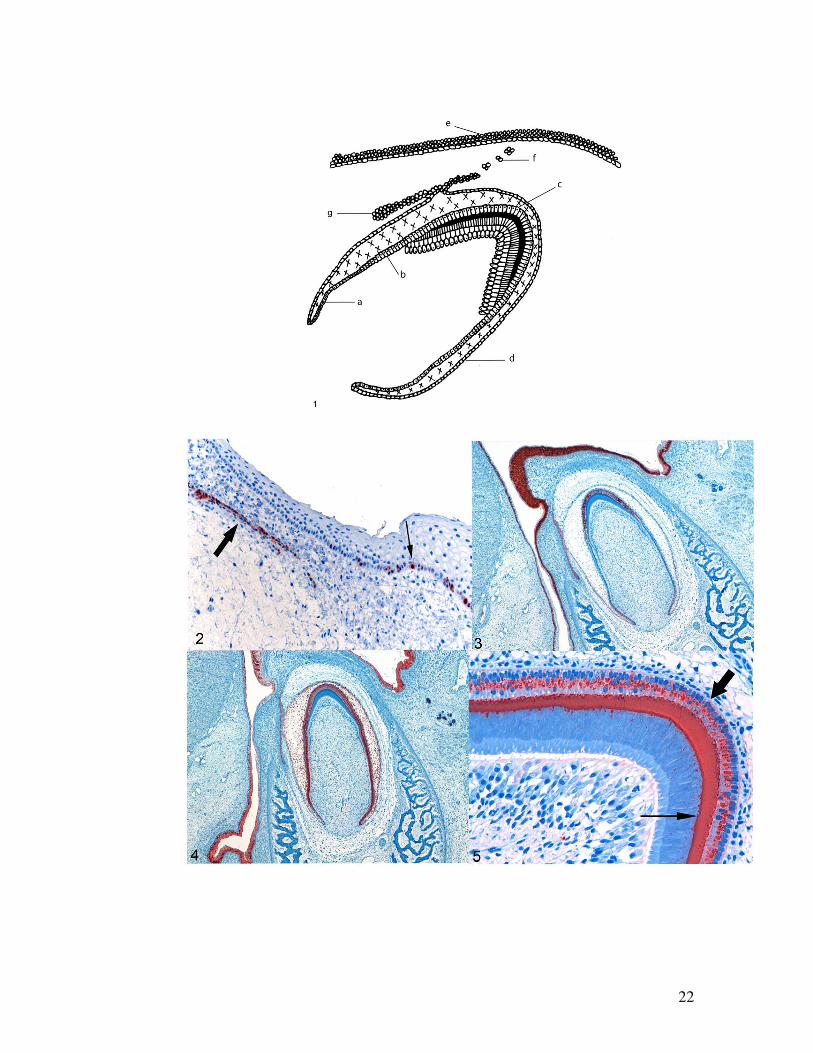
Fig. 1. Diagrammatic representation of cells present in developing tooth germ. a= IEE,
b= presecretory ameloblasts, c= secretory ameloblasts, d= OEE, e= overlying alveolar
epithelium, f= Serres rests and g= dental lamina.
Fig. 2. Positive calretinin staining of a dental lamina (bold arrow) and basal cells of the
overlying alveolar epithelium (thin arrow).
Fig. 3. Strong positive keratin 14 staining in the overlying alveolar epithelium, oral
epithelium, dental lamina and secretory ameloblasts.
Fig. 4. Positive keratin 19 staining in the overlying alveolar epithelium, oral epithelium,
dental lamina and all the epithelial cells of the enamel organ.
Fig. 5. Positive amelogenin staining in secretory ameloblasts (bold arrow) and enamel
(thin arrow).
20
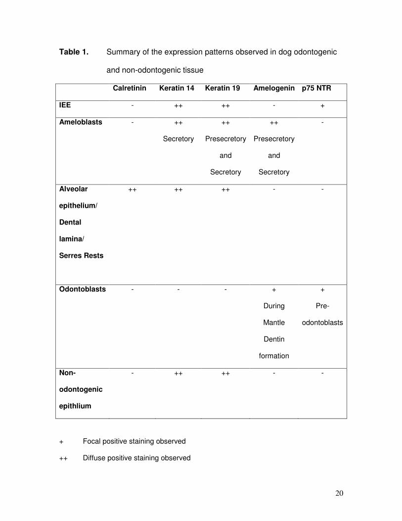
Table 1. Summary of the expression patterns observed in dog odontogenic
and non-odontogenic tissue
Calretinin Keratin 14 Keratin 19 Amelogenin p75 NTR
IEE - ++ ++ - +
Ameloblasts - ++
Secretory
++
Presecretory
and
Secretory
++
Presecretory
and
Secretory
-
Alveolar
epithelium/
Dental
lamina/
Serres Rests
++ ++ ++ - -
Odontoblasts - - - +
During
Mantle
Dentin
formation
+
Pre-
odontoblasts
Non-
odontogenic
epithlium
- ++ ++ - -
+ Focal positive staining observed
++ Diffuse positive staining observed

21
- No positive staining observed
22
, I
.t· ..J: ' <
H ",
.~ 'ii ,